Embed Size (px)
Citation preview


PREEMPTIVE ULTRASOUND FOR A-V ACCESS
Sitthichai Vachirasrisirikul , MD.
Diploma of The Thai Board of Vascular Surgery
Vascular surgeon , Buddhachinaraj Hospital , Phitsanulok

ULTRASOUND VASCULAR
MAPPING FOR
PREOPERATIVE PLANNING OF DIALYSIS ACCESS
Sitthichai Vachirasrisirikul , MD.
Diploma of The Thai Board of Vascular Surgery
Vascular surgeon , Buddhachinaraj Hospital , Phitsanulok
K/DOQI GuidelinesVascular access in hemodialysis patients
1) The nondominant arm AVF
: Dominant forearm (surgical preference)
2) Cephalic vein AVF : Forearm > Upper arm
3) Basilic vein transposition AVF, or other AVF configuration
4) AVG : forearm loop>upper arm straight>upper arm loop
5) Thigh AVG
6) Catheter based hemodialysis
Clinical practice guidelines for vascular access. Am J Kidney Dis. 2006;48 Suppl 1:S176-247.
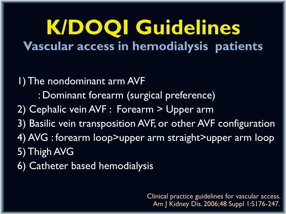
Mandate vascular mapping in all patients approaching chronic dialysis
1) Physical examination
2) Duplex ultrasonography (DUS)
3) Venography
1. Clinical practice guidelines for vascular access. Am J Kidney Dis. 2006;48 Suppl 1:S176-2472. Fistula First : the National Vascular Access Improvement Initiative. WMJ 2006 May; 105(3): 71-3.

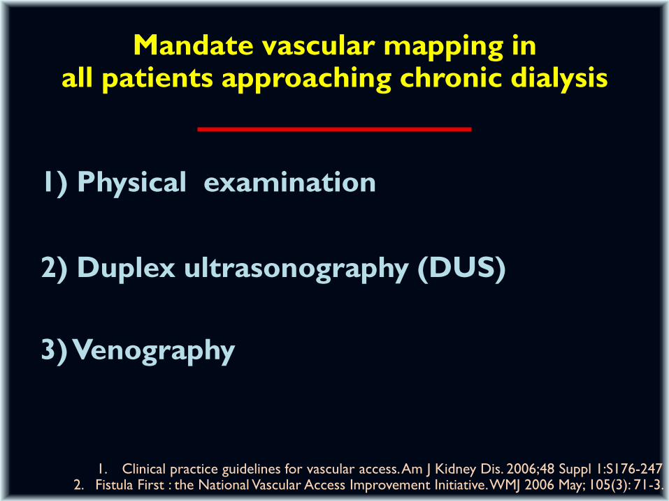
IDEAL
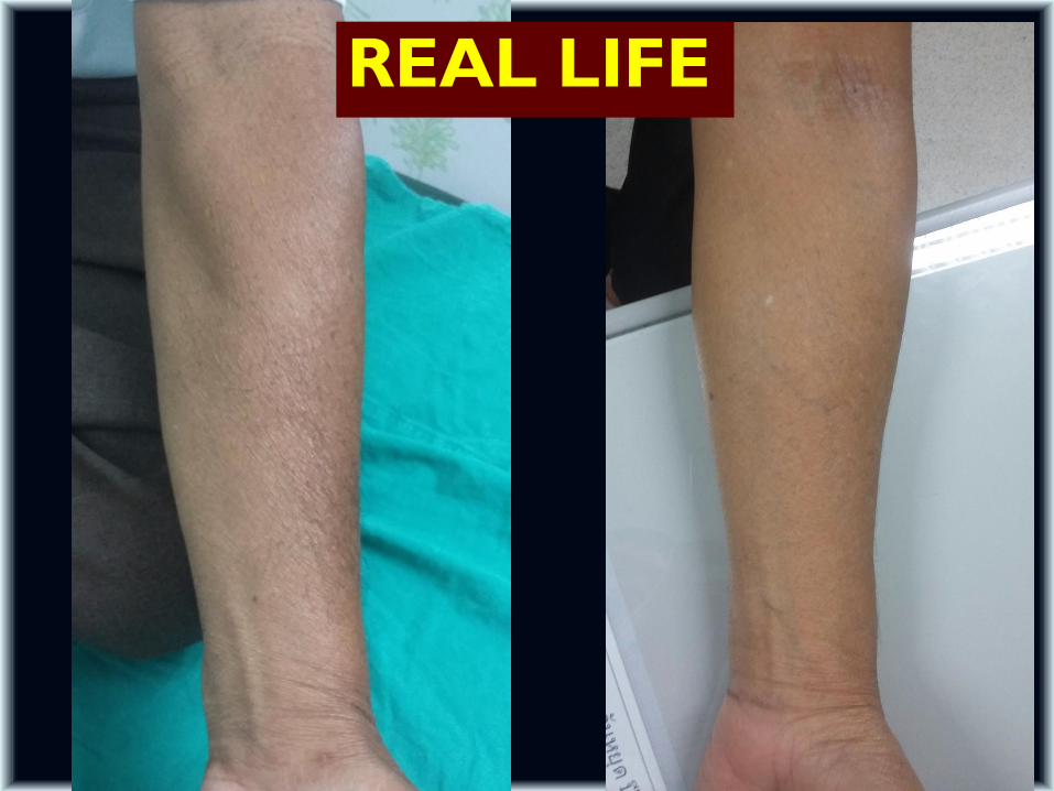
REAL LIFE

VASCULAR MAPPING
1) Physical examination (PE.)
- An adequate vein : only 47% CKD patients
- Poor clinically visible or clinically absent veins : 54%
2) Duplex ultrasonography (DUS)
- Poor clinically visible or clinically absent veins
>> 75% : showed adequate veins
3) Venography
- Complex vascular access , multiple prior access
procedures
Malovrh M. Am J Kidney Dis 39:1218–1225, 2002.

PE. CAN BE LIMITED
• Obese patients
• Multiple previous access surgeries
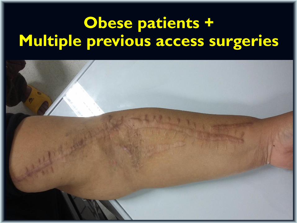
Obese patients +Multiple previous access surgeries

PREOPERATIVE DUS MAPPING
• Mapping of arm vessels before surgical creation of
dialysis access
- Higher percentage of AVF placements
as well as an increased fistula success rate
- Reduction in use of tunneled HD catheters
1. Allon M, et al. Kidney Int 2001; 60:2013–2020.
2. Allon M, Robbin ML. Kidney Int 2002; 62:1109–1124.
3. Robbin ML, et al. Radiology 2000; 217:83–88.
4. Silva MB Jr, et al. J Vasc Surg 1998; 27:302–308.
5. Wong CS, et al. J Vasc Surg. 2013;57(4):1129-1133.
6. Ferring M, et al. Clin J Am Soc Nephrol. 2010;5(12):2236-2244.
7. Asif A, et al. Kidney Int 67:2399–2406, 2005.

ROUTINE
PREOPERATIVE DUS
VS
PE. or Selective DUS ?

Routine Pre-operative Ultrasound Mapping Before AVF Creation : A Meta-analysis
(Based mainly on moderate quality RCTs)
Odds ratio (OR) 0.32
95% CI 0.17-0.60p < .01
THE IMMEDIATE FAILURE RATEDUS VS Clinical exam or Selective US
G.S. Georgiadis et al.Eur J Vasc Endovasc Surg (2015) 49, 600-605

THE EARLY/MIDTERM ADEQUACY
FOR HEMODIALYSIS (usability for HD, at 1 or 6 mo. post-op)
Odds ratio (OR) 0.66
95% CI 0.42-1.03p =0.06
Routine DUS VS Clinical exam or Selective US
G.S. Georgiadis et al.Eur J Vasc Endovasc Surg (2015) 49, 600-605

THE EARLY/MIDTERM ADEQUACY
FOR HEMODIALYSIS (usability for HD, at 1 or 6 mo. post-op)
Routine DUS VS Selective DUS
Odds ratio (OR) 0.56
95% CI 0.33-0.95p =0.03
G.S. Georgiadis et al.Eur J Vasc Endovasc Surg (2015) 49, 600-605
CONCLUSION :
The clinical examination should always be supplemented with routine DUS mapping before AVF creation

Routine preoperative DUS
improves patency and use of AVFs : A Randomized Trial
Clinical
assessment group
Ultrasound group
P - value
Rate of immediate failure 11% 4% 0.028Failed AVFs, thrombosis 67% 38% 0.029Primary AVF survival 1 year 56% 65% 0.081Assisted primary AVF survival 1 year
65% 80% 0.012
Ferring M, et al. Clin J Am Soc Nephrol. 2010 Dec; 5(12): 2236–2244.

Duplex Ultrasound(DUS)
• B-mode imaging :
Gray scale
• Color-flow Doppler
• Spectral Doppler waveforms
GE HEALTH CARE ; VIVID7

Duplex Ultrasonography (DUS)Linear array transducer
• Higher –frequency
• 12-18 MHz
• Better sensitivity to low
flow
• More superficial vein
Curved linear or Phased array transducer
• Lower -frequency
• 2.5 -3.5 MHz
• Better penetration
• Central veins : innominate vein or SVC

SUCCESSFUL VASCULAR ACCESS CREATION
1) OPTIMAL INFLOW
: Arterial Examination
2) OPTIMAL OUTFLOW
: Venous Examination
3) OPTIMAL CONDUIT & GOOD
ANASTOMOSIS
DUS
OPTIMAL INFLOW ARTERIAL EXAMINATION
• Brachial artery pressures both arms for
comparison
• Pulse examination
- Axillary ,Brachial ,Radial ,Ulnar arteries
• Modified Allen’s test
DUSARTERIAL EXAMINATION
• Sufficient size ( variable & series dependent)
: diameter ≥ 0.20 cmSilva MB Jr, et al. J Vasc Surg 1998; 27:302–308.
Parmar J, et al. Eur J Vasc Endovasc Surg 33:113–115, 2007.
Sidawy AN, et al. J Vasc Surg. 2008;48(5 Suppl):2S-25S.Nakata et al. SpringerPlus 2016;5:462
• The internal luminal diameter of the artery

ARTERIAL DIAMETER
DUSARTERIAL EXAMINATION
0.23cm

DUSARTERIAL EXAMINATION
• Sufficient size ( variable & series dependent)
: diameter ≥ 0.20 cmSilva MB Jr, et al. J Vasc Surg 1998; 27:302–308.
Parmar J, et al. Eur J Vasc Endovasc Surg 33:113–115, 2007.
Sidawy AN, et al. J Vasc Surg. 2008;48(5 Suppl):2S-25S.Nakata et al. SpringerPlus 2016;5:462
• The internal luminal diameter of the artery
• Presence of significant concentric calcification ?

Concentric calcification : Radial artery

DUSARTERIAL EXAMINATION
• Sufficient size ( variable & series dependent)
: diameter ≥ 0.20 cmSilva MB Jr, et al. J Vasc Surg 1998; 27:302–308.
Parmar J, et al. Eur J Vasc Endovasc Surg 33:113–115, 2007.
Sidawy AN, et al. J Vasc Surg. 2008;48(5 Suppl):2S-25S.Nakata et al. SpringerPlus 2016;5:462
• The internal luminal diameter of the artery
• Presence of significant concentric calcification ?
• Arterial spectral waveforms: screen for Inflow or Outflow disease ?

Triphasic waveform &
No significant focal velocity increases
Detecting
stenoses :
sensitivity
70 - 90.9 %
specificity
98.7 - 100%
Wittenberg G, et al. Ultraschall Med 19:22–27, 1998.

For a forearm AVF
- Radial artery at the wrist
- Ulnar artery may be assessed
- A modified duplex Allen test
DUSARTERIAL EXAMINATION

A MODIFIED DUPLEX ALLEN TEST
• Patency of the deep palmar arch
• Radial artery at the wrist and/or at the
dorsum of the hand
• Reversal of blood flow distal to the proximal occlusion
Zimmerman P, et al. Radiology 2001; 220:299–302.


For a forearm AVF
- Radial artery at the wrist
- Ulnar artery may be assessed
- A modified duplex Allen test
Kian K, et al. Semin Dial. 2012;25(2):244-247.
For either AVF or graft creation
- Brachial artery at the antecubital fossa
- Brachial artery upper arm & Axillary artery
- High brachial artery bifurcation ? ( 10% )
DUSARTERIAL EXAMINATION
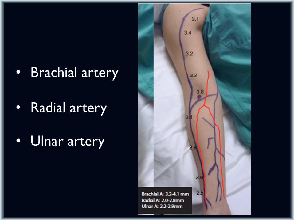
• Brachial artery
• Radial artery
• Ulnar artery

OPTIMAL OUTFLOWVENOUS EXAMINATION
• Adequate size , length , depth
• No outflow stenosis
: Forearm veins >> SVC
DUSVENOUS EXAMINATION
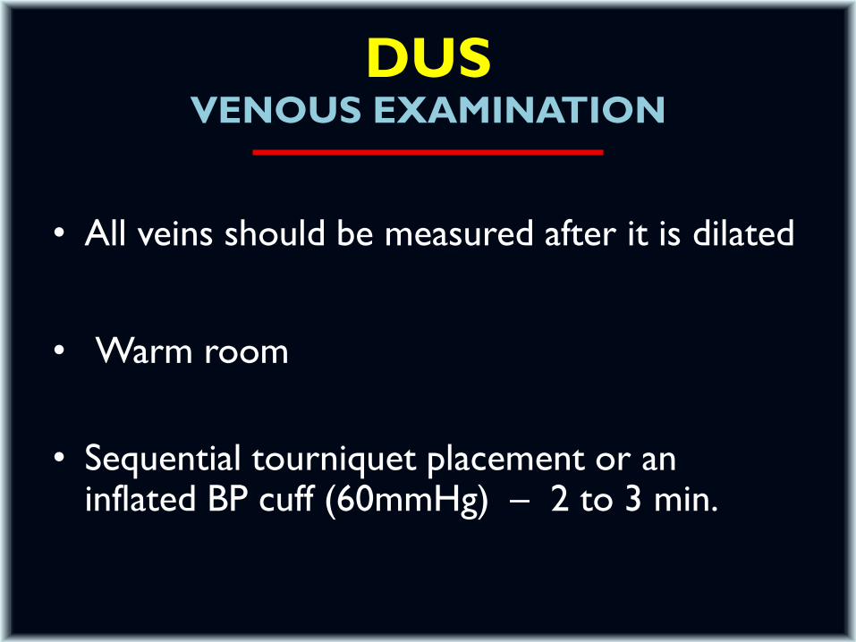
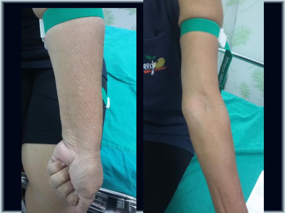
• All veins should be measured after it is dilated
• Warm room
• Sequential tourniquet placement or an inflated BP cuff (60mmHg) – 2 to 3 min.

• Diameter (ID) : variable & series dependent
: ≥ 0.25 cm for an AVF
: ≥ 0.4 cm for an AVG or Basilic upper arm
transposition
Silva MB Jr, et al. J Vasc Surg 1998; 27:302–308.
Malovrh M, Am J Kidney Dis 39:1218–1225, 2002.
Sidawy AN, et al. J Vasc Surg. 2008;48(5 Suppl):2S-25S.
Zierler RE: Strandness’s duplex scanning in vascular disorders, ed 4, 2010.
Arroyo MR, et al. J Vasc Surg 2008; 47:1279-1283.
DUSVENOUS EXAMINATION

0.33cm
DIAMETER (ID)

Lockhart ME, et al. J Ultrasound Med 2006; 25:1541–1545.van Bemmelen PS, et al. J Vasc Surg 42:957–962,2005.
• More focused percussion or after application
of a warm-water immersion (43-44oC ) : Borderline in size (0.05cm)
• Daily variation : alteration in vascular tone
- Hydration related to dialysis cycle
Planken RN, et al: Nephrol Dial Transplant 21:802–806, 2006.
DUSVENOUS EXAMINATION
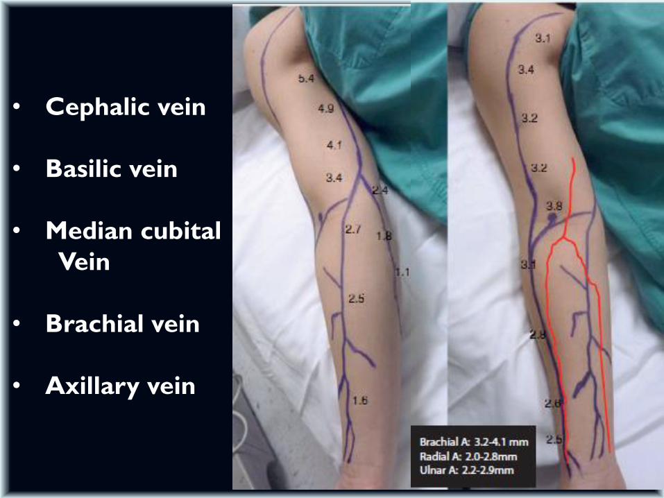
• Cephalic vein
• Basilic vein
• Median cubital
Vein
• Brachial vein
• Axillary vein
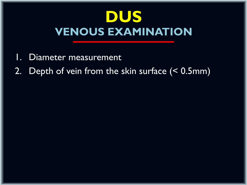
1. Diameter measurement
2. Depth of vein from the skin surface (< 0.5mm)
DUSVENOUS EXAMINATION

DEPTH OF VEIN

1. Diameter measurement
2. Depth of vein from the skin surface (< 0.5mm)
3. Contiguous length of nondiseased vein (8-10 cm)
4. Compressibility : thrombus
DUSVENOUS EXAMINATION

Chronic thrombosis of the cephalic vein

1. Diameter measurement
2. Depth of vein from the skin surface (< 0.5mm)
3. Contiguous length of nondiseased vein (8-10 cm)
4. Compressibility : thrombus
5. Sclerotic or thick-walled vein
DUSVENOUS EXAMINATION

Vein wall thickeningfrom needle injury

1. Diameter measurement
2. Depth of vein from the skin surface (< 0.5mm)
3. Contiguous length of nondiseased vein (8-10 cm)
4. Compressibility : thrombus
5. Sclerotic or thick-walled vein
6. Adequate venous drainage
7. Large branches of veins near the site of a fistula
Beathard GA, et al. Kidney Int 2003; 64:1487–1494.
Singh P, et al. Radiology 2008; 246:299–305.Wiese P, et al. Nephrol Dial Transplant 19:1956–1963, 2004.
DUSVENOUS EXAMINATION

CENTRAL VENOUS STENOSIS
Should be suspected if
1) Any prominent venous collaterals or edema
2) A differential in extremity diameter
3) Any history of previous central venous catheter placement
4) Multiple previous access sites
• Should be examined with deep venous duplex ultrasound imaging, followed by venography if necessary
Clinical practice guidelines for vascular access.Am J Kidney Dis. 2006;48 Suppl 1:S176-247.
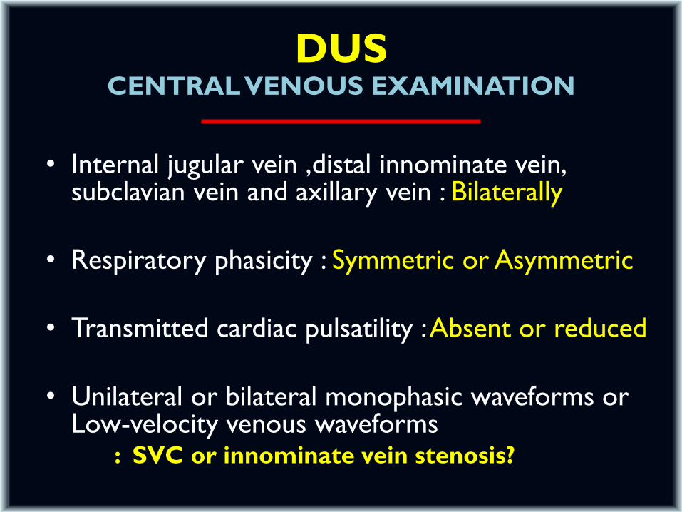
DUSCENTRAL VENOUS EXAMINATION
• Internal jugular vein ,distal innominate vein, subclavian vein and axillary vein : Bilaterally
• Respiratory phasicity : Symmetric or Asymmetric
• Transmitted cardiac pulsatility : Absent or reduced
• Unilateral or bilateral monophasic waveforms or Low-velocity venous waveforms
: SVC or innominate vein stenosis?
DUS ACCURACY
• Detection of venous stenosis, thrombosis, and
occlusion
: Sensitivity 81%, Specificity 90%
• Sensitivities decrease for more proximal veins
Nack TL, et al. J Vasc Technol 16:69–73, 1992.

TAKE HOME MESSAGE
• Routine preoperative DUS mapping :
AVF failure rate, Early/midterm adequacy
Assisted primary patency(1yr)
• Optimal inflow & outflow DUS
• Sufficient diameter :
Artery 0.2cm & Vein 0.25cm / 0.4cm