Embed Size (px)
Citation preview

Biotechnology and Bioprocess Engineering 19: 1-7 (2014)
DOI 10.1007/
Predictive Evaluation for the Preparation of a Synthetic Y-shaped
DNA Nanostructure
Kyung Soo Park, Seung-Won Shin, Jin-Ha Choi, Byung-Keun Oh, Jeong-Woo Choi, and Soong Ho Um
Received: / Revised: / Accepted:
© The Korean Society for Biotechnology and Bioengineering and Springer 2014
Abstract With the advent of deoxyribonucleic acid
(DNA) nanotechnology, the Y-shaped DNA nanostructure
(Y-DNA) as a basic block was first created. Due to their
characteristic selectivity and specificity, Y-DNA-based
materials have been utilized in a variety of scientific fields
including multiplexed nanobarcoding. Basically, the tripod
DNA nanostructure was prepared by simple hybridization
of three different single stranded DNA (ssDNA). Before
the synthetic process, the optical densities (OD) of the
three ssDNAs were measured to accurately estimate the
concentration. Through repeated temperature fluctuations,
three ssDNAs were hybridized into a Y-shaped block with
both a central junction and three blunt ended arms. After
the reaction, the ODs of the synthesized DNA products
were measured and compared with the theoretical OD
values calculated by a MATLAB program (‘matrix laboratory’)
with different molar concentrations and volumes to predict
the presence of Y-DNA. Simultaneously, the product was
analyzed by agarose gel electrophoresis to confirm the Y-
DNA structure. The measured ODs of the solutions with
confirmed Y-DNA structures were close to the theoretical
maximum OD values. This article provides means to help
understand and prepare Y-DNA by performing OD
measurements. It is highly expected that this guide will be
an excellent starting point for structural DNA nanotechnology.
Keywords: DNA nanotechnology, optical density, MATLAB
program, gel electrophoresis
1. Introduction
Along with the fast development of nanobiotechnology,
DNA has been used as an infra-structural block for a
variety of supramolecular nanostructures with conformational
flexibility at the nano-scale level and molecular specific
recognitions in predesigned manners [1-4]. For instance,
DNA origami [5] has been further applied to DNA tubes
[6,7], triangles [8], dimensional structures [9-11], and DNA
junctions [12-15]. In particular, Y-shaped DNA (Y-DNA)
junctions as a basic unit have been highlighted. Moreover,
by modifying the overhang sequences of each arm of Y-
DNA, the Y-DNAs ligate together in a controlled manner
resulting in a tree-shaped DNA supramolecular nanostructure
referred to as dendrimer-like DNA [16]. Furthermore, each
end in the DNA nanostructure may be functionalized with
various color codes in order to realize anisotropic built-in
and multiplexed barcodes [17-20].
To prepare such DNA blocks, each ssDNA is first
quantitated by measuring its optical density (OD) at a
wavelength of 260 nm. The initial OD measurement of
ssDNA is directly associated with the final product efficiency
and cost-effectiveness. Y-DNA is made by simply annealing
three single stranded oligonucleotides whose sequences are
complimentary to each other so that under the given
optimal conditions, the three oligo-pieces form Y-DNA as
designed. To prevent the loss of oligonucleotides, it may be
beneficial to accurately assess the quantitation of each
Seung-Won Shin, Jin-Ha Choi, Soong Ho UmSchool of Chemical Engineering, Sungkyunkwan University, Suwon 440-746, Korea
Soong Ho Um*
SKKU Advanced Institute of Nanotechnology (SAINT), SungkyunkwanUniversity, Suwon 440-746, KoreaTel: +82-31-290-7348; Fax: +82-31-290-7272E-mail: [email protected]
Kyung Soo Park, Byung-Keun Oh, Jeong-Woo ChoiDepartment of Chemical and Biomolecular Engineering, Sogang University,Seoul 121-742, Korea
Jeong-Woo ChoiGraduate School of Management Technology, Sogang University, Seoul121-742, Korea
RESEARCH PAPER

2 Biotechnology and Bioprocess Engineering 19: 000-000 (2014)
DNA molecule and predict the state of the expected Y-
DNA before completely forming the Y-DNA. In the same
sense, quantitative evaluations of the created Y-DNA are
required to analyze its state before further applications. As
a result, a variety of experimental parameters must be
cautiously investigated such as the total reaction volume,
concentration of the synthesis solution, and dilution steps
taken before the OD measurements. To assess these
parameters, the three main variables were varied as
follows: (1) reaction volumes of 30 and 100 µL (2) molar
concentrations of 0.00600 and 0.00900 mM, and (3) one
dilution and two consecutive dilutions and for further
investigation, 2 or 10 µL of the stock solution was mixed
with DNase-free water for dilution.
Empirically, the measured OD was close to the theoretical
maximum OD. For example, the theoretical minimum and
maximum OD values of the solution with a concentration
of 0.00600 mM were 0.290 and 0.439, respectively, where
the average measured OD after nine trials was 0.445. It is
speculated that better products were generated in this study
for advanced DNA technological applications.
In this article, we provide a guide to a proficient
experimental and theoretical approach for the preparation
of Y-DNA by introducing detailed experimental information
which results in more precise OD measurements by
evaluating influential experimental parameters. Thus, it will
enable researchers to easily reproduce Y-DNA and predict
the final as-designed products, which will help confirm if
the Y-DNA was successfully synthesized. In addition, this
guide will be quite helpful for DNA nanotechnology novices.
2. Materials and Methods
Three complementary oligonucleotides, TE buffer (10 mMTris
(pH 8.0), 1 mM ethylenediaminetetraacetic acid (EDTA),
50 mM NaCl, DNase-free water (DFW), micro-pipettes,
graduated pipette tips, microtubes, a thermal cycler
(Eppendorf), and latex gloves were used in the experiments.
For the UV absorbance measurements of the DNA molecules
at a specific wavelength, which is typically 260 nm for
nucleic acids, a UV-vis spectrophotometer (BioPhotometer
Plus 6132, Eppendorf) and cuvettes (UVette®, Eppendorf)
were used.
2.1. OD measurement of ssDNAs and synthesized Y-DNA
To measure the OD of nucleic acids, three different
oligonucleotides, Y1, Y2, and Y3, were first separately
dissolved thoroughly in TE buffer with volumes of 500 µL
(Fig. 1A). 10 µL of each sample was then taken and
serially diluted into DFW to a final volume of 100 µL.
Then, the concentration of each oligonucleotide solution
was calculated by measuring OD of the solution at a
wavelength of 260 nm using a UV-vis spectrophotometer
(Fig. 1B). In order to measure the OD of a solution, the
solvent used to dissolve the oligonucleotides has to be
measured first to obtain the background signal. This
background is then subtracted from the total signal of the
oligonucleotide solution leaving only the signal from the
solute (oligonucleotide). If the OD value is larger than 1,
the sample solution must be additionally diluted until the
OD becomes less than 1 and within the machine’s
readability. Once the ODs of the ssDNA are measured, the
actual molar concentration of each oligonucleotide can be
calculated using the following equation.
33 µg/ml × Absorbance260 nm × Dilution factor =
DNA concentration (µg/ml)
Then, to synthesize Y-DNA, three oligonucleotides were
mixed at an equal molar ratio in a 0.1 mL microcentrifuge
tube and DFW was added to the final volume, which was
determined depending on the experiment design. The
Fig. 1. Schematic drawing of the overall experiment: (A) preparation of three oligonucleotides (ssDNA), where Y1, Y2, and Y3 (Table 4)are represented by red, yellow, and blue, respectively, (B) quantitation of each oligonucleotide through spectrophotometry, (C) annealingof the three oligonucleotides into Y-DNA using a thermal cycler, and (D) Y-DNA represented as three ssDNAs twisted into Y-shapedblocks after thermal hybridization.
Predictive Evaluation for the Preparation of a Synthetic Y-shaped DNA Nanostructure 3
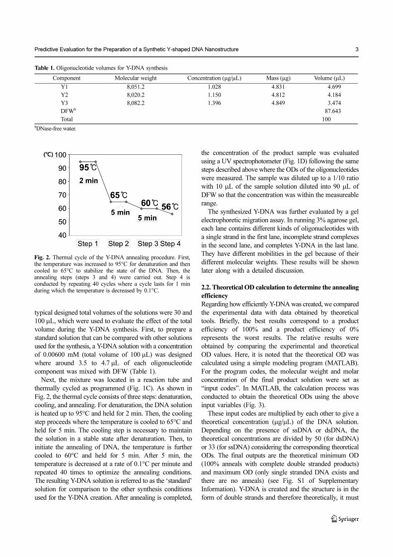
typical designed total volumes of the solutions were 30 and
100 µL, which were used to evaluate the effect of the total
volume during the Y-DNA synthesis. First, to prepare a
standard solution that can be compared with other solutions
used for the synthesis, a Y-DNA solution with a concentration
of 0.00600 mM (total volume of 100 µL) was designed
where around 3.5 to 4.7 µL of each oligonucleotide
component was mixed with DFW (Table 1).
Next, the mixture was located in a reaction tube and
thermally cycled as programmed (Fig. 1C). As shown in
Fig. 2, the thermal cycle consists of three steps: denaturation,
cooling, and annealing. For denaturation, the DNA solution
is heated up to 95°C and held for 2 min. Then, the cooling
step proceeds where the temperature is cooled to 65°C and
held for 5 min. The cooling step is necessary to maintain
the solution in a stable state after denaturation. Then, to
initiate the annealing of DNA, the temperature is further
cooled to 60°C and held for 5 min. After 5 min, the
temperature is decreased at a rate of 0.1°C per minute and
repeated 40 times to optimize the annealing conditions.
The resulting Y-DNA solution is referred to as the ‘standard’
solution for comparison to the other synthesis conditions
used for the Y-DNA creation. After annealing is completed,
the concentration of the product sample was evaluated
using a UV spectrophotometer (Fig. 1D) following the same
steps described above where the ODs of the oligonucleotides
were measured. The sample was diluted up to a 1/10 ratio
with 10 µL of the sample solution diluted into 90 µL of
DFW so that the concentration was within the measureable
range.
The synthesized Y-DNA was further evaluated by a gel
electrophoretic migration assay. In running 3% agarose gel,
each lane contains different kinds of oligonucleotides with
a single strand in the first lane, incomplete strand complexes
in the second lane, and completes Y-DNA in the last lane.
They have different mobilities in the gel because of their
different molecular weights. These results will be shown
later along with a detailed discussion.
2.2. Theoretical OD calculation to determine the annealing
efficiency
Regarding how efficiently Y-DNA was created, we compared
the experimental data with data obtained by theoretical
tools. Briefly, the best results correspond to a product
efficiency of 100% and a product efficiency of 0%
represents the worst results. The relative results were
obtained by comparing the experimental and theoretical
OD values. Here, it is noted that the theoretical OD was
calculated using a simple modeling program (MATLAB).
For the program codes, the molecular weight and molar
concentration of the final product solution were set as
“input codes”. In MATLAB, the calculation process was
conducted to obtain the theoretical ODs using the above
input variables (Fig. 3).
These input codes are multiplied by each other to give a
theoretical concentration (µg/µL) of the DNA solution.
Depending on the presence of ssDNA or dsDNA, the
theoretical concentrations are divided by 50 (for dsDNA)
or 33 (for ssDNA) considering the corresponding theoretical
ODs. The final outputs are the theoretical minimum OD
(100% anneals with complete double stranded products)
and maximum OD (only single stranded DNA exists and
there are no anneals) (see Fig. S1 of Supplementary
Information). Y-DNA is created and the structure is in the
form of double strands and therefore theoretically, it must
Table 1. Oligonucleotide volumes for Y-DNA synthesis
Component Molecular weight Concentration (µg/µL) Mass (µg) Volume (µL)
Y1 8,051.2 1.028 4.831 4.699
Y2 8,020.2 1.150 4.812 4.184
Y3 8,082.2 1.396 4.849 3.474
DFWb 87.643
Total 100bDNase-free water.
Fig. 2. Thermal cycle of the Y-DNA annealing procedure. First,the temperature was increased to 95°C for denaturation and thencooled to 65°C to stabilize the state of the DNA. Then, theannealing steps (steps 3 and 4) were carried out. Step 4 isconducted by repeating 40 cycles where a cycle lasts for 1 minduring which the temperature is decreased by 0.1°C.

4 Biotechnology and Bioprocess Engineering 19: 000-000 (2014)
exhibit theoretical minimum OD values. If there are no
reactions for Y-DNA formation, only three single stranded
oligonucleotides may exist in the solution and it must
demonstrate theoretical maximum OD values. However,
experimentally, the ODs values of the synthesized Y-DNA
were around the theoretical maximum values.
2.3. Evaluation of synthesis conditions
To evaluate the influence of the synthesis conditions on
the Y-DNA creation, several solutions synthesized under
different parameters including the reaction volume, solution
concentration, and dilution factors were investigated by
measuring the OD at specific wavelengths of 230, 260,
280, and 340 nm. In addition, for more accurate results, the
entire steps from the solution synthesis to the OD
measurement were carried out nine times and then averaged.
First, to evaluate the effect of the solution volume, a solution
with a molar concentration and volume of 0.00600 mM
and 30 µL, respectively, was made (Table 2A) and compared
with the aforementioned ‘standard’ solution. Secondly, the
influence of how the dilution steps are performed on the
resulting OD was investigated. A solution with a
concentration and total volume of 0.00600 mM and 100 µL,
respectively, was prepared. Its parameters were kept the
same as those of the ‘standard’ and only differed in terms
of the dilution steps taken before measuring the OD. The
sample was diluted by a 1/100 ratio in two dilution steps,
each at a 1/10 ratio (Table 2B). Next, the evaluation of the
effect of the solution concentration to the Y-DNA creation
was conducted by making a solution with a molar
concentration and volume of 0.00900 mM and 100 µL,
respectively (Table 2C). As with the previous solutions,
its OD was measured and compared with that of the
‘standard’. Lastly, the dilution factors were evaluated. The
OD of a solution with a concentration and total volume
of 0.00667 mM and 30 µL, respectively, was measured
(Table 2D). During the OD measurement, the dilution was
carried out in two consecutive steps where the first step
diluted 2 µL of solution into 18 µL of DFW, resulting in
the intermediate solution at a 1/10 dilution ratio. Then,
10 µL of the intermediate solution was diluted into 90 µL
of DFW, resulting in a 1/100 diluted sample.
3. Results and Discussion
The full sequence information of the Y-DNA is given in
Table 3. The ‘standard’ solution synthesized at a molar
concentration of 0.00600 mM with a reaction volume of
100 µL was first evaluated. Before taking the OD measure-
ment, the Y-DNA formed was confirmed by a 3% agarose
gel electrophoretic assay (Fig. 4). Interestingly, the band
indicating the Y-DNA components composed of three
single strands was most slowly retarded because of the
Fig. 3. Algorithm of the MATLAB process used for the theoreticalOD calculations. The molar concentration (mM) and molecularweight are input codes which can be converted to the theoreticalconcentration (µg/µL). To hypothesize the condition for thetheoretical concentration calculation, the type of the DNA isdetermined whether to be single or double stranded and dependingon its type, the theoretical concentration is divided by 5 for singleor 3.3 for double stranded DNA yielding the theoretical minimumand maximum ODs, respectively.
Table 2. Optical densities of solutions obtained under different synthesis conditions
Standard (a) (b) (c) (d)
Concentration (mM) 0.00600 0.00600 0.00600 0.00900 0.00667
Total volume (µL) 100 30 100 100 30
OD (230 nm) 0.389 0.388 0.0343 0.557 0.06044
OD (260 nm) 0.445 0.433 0.0459 0.663 0.0569
OD (280 nm) 0.291 0.279 0.0342 0.279 0.0371
OD (340 nm) 0.000667 0.00122 0.00167 0.00122 0
STDV (260 nm) 0.00425 0.00372 0.00668 0.0119 0.00836
Dilution ratio 1/10 1/10 1/100 1/10 1/100
Dilution step (µL/µL) 10/90 10/90 (10/90)x2 10/90 2/18, 10/90

Predictive Evaluation for the Preparation of a Synthetic Y-shaped DNA Nanostructure 5
higher molecules weights (24,000 Da) relative to other
single strands (8,000 Da) and incomplete Y-DNAs consisting
of two single strands (16,000 Da). Compared with the
ladder DNA, it is evident that there are stepwise increments
of approximately 30 bp in the DNA nanostructures which
directly indicates the creation of Y-shaped DNA nano-
structures with three arms. Next, the OD of Y-DNA was
measured using a UV spectrophotometer (Table 2). The
OD260 nm obtained was 0.445 with a standard deviation of
0.004. It can be considered that the value is fairly reliable
when it is within the tenth digit of the OD and the standard
deviation is in the thousandth digit.
This experimental result was compared with the
theoretical OD calculated via MATLAB, in which we used
a molar concentration of 0.00600 mM and a molecular
weight of 24,153.6 as input codes. The calculated theoretical
minimum and maximum ODs were 0.290 and 0.439,
respectively, in which case the theoretical maximum OD is
similar to the experimentally obtained value of 0.445
(Table 4A). This phenomenon may be ascribed to the
characteristic topological features of Y-DNA [21]. It is
known that the topology or morphology of a molecule
affects its molecular optical properties.
Secondly, solution (a) in Table 2 whose reaction volume
was modified to 30 µL compared to the ‘standard’ was
investigated. For the MATLAB operation, because the
molar concentration and molecular weight are fixed to
0.00600 mM and 24,153.6, respectively, the theoretical OD
did not change from the previous case with minimum and
maximum OD values of 0.290 and 0.439, respectively. After
the nine trials of solution synthesis and OD measurements,
an average OD260 nm of 0.433 (Table 2A) was obtained,
which is again around the theoretical maximum OD
(0.439), which is slightly within the theoretical range. This
supports the above notion that light absorption of Y-DNA
with its unique morphology is somewhat different than that
of the common double stranded DNA. With only a subtle
decrease of the standard deviation compared to that of the
‘standard’ solution, a significant difference was not observed
when the total reaction volume was changed.
Next, the influence of how the dilution steps are performed
on the resulting OD was investigated. Solution (b) in Table 2
was prepared and its OD was measured. After carrying out
nine steps of solution synthesis and OD measurements, the
average was 0.0459 (Table 2B) which is similar to that of
the ‘standard’ solution (0.445) considering the one extra
dilution step, which requires multiplication by 10 for
Table 3. Oligonucleotide sequence
Strand Sequencesa
Y1 5'-Phos-TGGATCCGCATGACATTCGCCGTAAG-3'
Y2 5'-Phos-CTTACGGCGAATGACCGAATCAGCCT-3'
Y3 5'-Phos-AGGCTGATTCGGTTCATGCGGATCCA-3'aThe sequences were obtained from Li, Y.; Tseng, Y. D.; Kwon, S. Y.;D’espaux, L.; Bunch, J. S.; Mceuen, P. L.; Luo, D. Nat. Mater. 2004, 3:38-42.
Fig. 4. Stepwise confirmation of Y-DNA creation by agarose gelelectrophoresis: (1) 25 bp ladder DNA, (2) 26 bp singleoligonucleotide, (3) two 26 bp oligonucleotides partially annealedforming double oligonucleotides, and (4) triple oligonucleotidesannealed into Y-DNA forming a 78 bp DNA structure.
Table 4. Calculated theoretical optical density range
Theoretical OD Range
Molar concentration (mM) MW (g/mol) Theoretical concentration (µg/µL) Theoretical OD
(a) Minimum (ds) 0.00600 24,153.6 0.145 0.290
Maximum (ss) 0.0180 8,051.2 0.145 0.439
(b) Minimum (ds) 0.00900 24,153.6 0.217 0.435
Maximum (ss) 0.0270 8,051.2 0.217 0.659
(c) Minimum (ds) 0.00667 24,153.6 0.161 0.322
Maximum (ss) 0.0200 8,051.2 0.161 0.488

6 Biotechnology and Bioprocess Engineering 19: 000-000 (2014)
comparison. However, the standard deviation increased to
0.007. Considering the average OD is in the hundredth
digit, the standard deviation increased tenfold compared to
that of the ‘standard’ solution. It can be concluded that the
effect of taking extra dilution steps on the overall OD value
is not of significance but each trial may not be as precise
as that of when less dilution steps are taken considering the
increased standard deviation. Once again, similar to other
solutions, the measured OD was near the theoretical
maximum OD.
To evaluate the influence of the solution concentration
on Y-DNA creation, solution (c) in Table 2 was prepared.
It is usually expected that when the solution concentration
is higher, the solutes in the solution absorb more
transmitting light. The measured OD of 0.663 (Table 2C)
is higher compared to that of the previously mentioned
solutions, which are at lower concentrations. According to
the MATLAB program, the theoretical OD range was
calculated by applying a molar concentration of 0.00900 mM
and a solution volume of 100 µL as input codes resulting
in theoretical minimum and maximum OD values of 0.435
and 0.659, respectively (Table 4B). It was observed again
that the measured ODs were close to the theoretical
maximum ODs. However, the standard deviation almost
doubled compared to those of the ‘standard’ and previously
mentioned solutions. This may be due to the higher
concentration of the solution where only a small amount of
difference in every handling of the solution leads to a
higher deviation in the results.
Lastly, to test how the dilution factors influence the
solution OD, solution (d) was synthesized. It was preliminarily
concluded that if a lower amount of the original solution
was used compared to the above trials and diluted into
DFW, it induced less accurate results. Through the MATLAB
operation, the theoretical minimum and maximum OD values
of the 1/100 diluted solution with a molar concentration of
0.00667 mM and solute molecular weight of 24,153.6 were
calculated to be 0.0322 and 0.0488, respectively (Table 4C).
The measured OD result was 0.0569 (Table 2D), which is
not as close to the theoretical maximum OD as those of the
other solutions which underwent different dilution steps
where a larger volume (10 µL) of original stock solution
was diluted into DFW. This phenomenon may be ascribed
to the capillary phenomena occurring while handling a
much smaller volume using a micropipette.
It is concluded that the solution concentrations, volumes,
and dilutions during sample preparation do not have
significant effects on the OD values. Meanwhile, the
concentration is proportional to the OD and it was observed
that once the solution concentration was higher, the OD
deviated more from the overall OD obtained by averaging
many trials. While the samples were diluted, too small a
volume of the stock solution may cause substantial error in
the resulting OD. Empirically, we highly recommend that
at least 10 µL of a stock solution be used for dilution to
obtain reliable data based on the comparative and theoretical
ODs.
Interestingly, the DNA purity of the solutions also
influenced the preciseness of the measured OD. From the
ODs obtained at different wavelengths, the DNA purity of
the solutions can be deduced. The closer the OD340 nm is to
0 and OD260 nm/OD280 nm is to 1.8, the higher the DNA
purity. If OD260 nm/OD280 nm is lower than 1.8, some other
molecules that absorb light at a wavelength of 280 nm,
such as proteins or chemicals with phenols inside the
structure, exist within the solution. Lastly, OD260 nm/OD230
nm should be higher than OD260 nm/OD280 nm for a higher
DNA purity. Under different conditions, there were some
trials where the OD340 nm values were not zero (see Table S1
of Supplementary Information). Excluding those trials, the
average OD260 nm was recalculated which leads to a reduced
standard deviation. This means that if contamination is
reduced, more accurate results can be achieved.
4. Conclusion
The conditions which may influence the optical density of
a solution were assessed through comparisons to a standard
solution, in which Y-DNA was created and confirmed by
agarose gel electrophoresis. Other solutions that differed in
terms of any one of three synthesis parameters of
concentration, volume, and dilution steps were evaluated.
The volume was determined to not be a significant factor
when controlling a small amount of volume of less than
100 µL, whereas how the dilution steps were carried out
influenced the standard deviation of the obtained OD and
therefore, the preciseness of the results. The concentration
is proportional to the measured OD where an increased
concentration results in an increased OD. Also, changing
the concentration greatly affects the standard deviation
even when the reaction volume and dilution steps are fixed.
Additionally, if not contaminated by chemicals or biomaterials
other than DNA, more reliable data can be achieved which
can be reflected in a decrease of the standard deviation of
the average OD260 nm when trials with high values of OD340
nm are excluded from the calculation of the average. Also,
by comparing the measured OD260 nm with the theoretical
OD260 nm range, it could be concluded that solutions with Y-
DNA blocks successfully created had ODs around the
theoretical maximum OD. With the above result in mind,
knowing how the synthesis conditions affect the Y-DNA
creation, effective Y-DNA synthesis if possible even when
certain conditions need to be varied in different situations

Predictive Evaluation for the Preparation of a Synthetic Y-shaped DNA Nanostructure 7
simply by comparison with theoretical OD values obtained
using MATLAB rather than taking images after gel
electrophoresis, resulting in time and cost savings.
Acknowledgements
We thank Kyung-ho Nam and Hyun-ju Kim, both at the
School of Chemical Engineering of Sungkyunkwan
University, for their valuable advice and experimental
assistance. This work was supported by a grant from the
Korea Health Technology R&D Project of the Ministry of
Health & Welfare of the Republic of Korea (Grant No.
A110552) and by Basic Science Research Program through
the National Research Foundation of Korea (NRF) funded
by the Ministry of Education, Science and Technology
(2013R1A1A2016781).
References
1. Pinheiro, V., D. Han, W. Shih, and H. Yan (2011) Challenges andopportunities for structural DNA nanotechnology. Nat. Nano-
technol. 6: 763-772.2. Seeman, N. C. (2010) Nanomaterials based on DNA. Annu. Rev.
Biochem. 79: 65-87.3. Lee, C. C., J. A. MacKay, J. M. Frechet, and F. C. Szoka (2005)
Designing dendrimers for biological applications. Nat. Biotech-
nol. 23: 1517-1526.4. Nilsen, T. W., J. Grayzel, and W. J. Prensky (1997) Dendritic
nucleic acid structures. J. Theor. Biol. 187: 273-284.5. Rothemund, P. W. K. (2006) Folding DNA to create nanoscale
shapes and patterns. Nature 440: 297-302.6. Rothemund, P. W. K., A. Ekani-Nkodo, N. Papadakis, A. Kumar,
D. K. Fygenson, and E. Winfree (2004) Design and characteriza-tion of programmable DNA nanotubes. J. Am. Chem. Soc. 126:16344-16352.
7. Lin, C., R. Jungmann, A. M. Leifer, C. Li, D. Levner, G. M.Church, W. M. Shih, and P. Yin (2012) Submicrometre geomet-rically encoded fluorescent barcodes self-assembled from DNA.
Nat. Chem. 4: 832-839.8. Kershner, R. J., L. D. Bozano, C. M. Micheel, A. M. Hung, A. R.
Fornof, J. N. Cha, C. T. Rettner, M. Bersani, J. Frommer, P. W. K.Rothemund, and G. M. Wallraff (2009) Placement and orienta-tion of individual DNA shapes on lithographically patterned sur-faces. Nat. Nanotechnol. 4: 557-561.
9. Han, D., S. Pal, Y. Liu, and H. Yan (2010) Folding and cuttingDNA into reconfigurable topological nanostructures. Nat. Nano-
technol. 5: 712-717.10. Ke, Y., S. M. Douglas, M. Liu, J. Sharma, A. Cheng, A. Leung,
Y. Liu, W. M. Shih, and H. Yan (2009) Multilayer DNA origamipacked on hexagonal and hybrid lattices. J. Am. Chem. Soc. 131:15903-15908.
11. Douglas, S. M., H. Dietz, T. Liedl, B. Högberg, F. Graf, and W.M. Shih (2009) Self-assembly by DNA into nanoscale three-dimensional shapes. Nature 459: 414-418.
12. Seeman, N. C. and N. R. Kallenbach (1982) Design of immobilenucleic acid junctions. J. Theor. Biol. 99: 237-247.
13. Um, S. H., J. B. Lee, N. Park, S. Y. Kwon, C. C. Umbach, and D.Luo (2006) Enzyme-catalysed assembly of DNA hydrogel. Nat.
Mater. 5: 797-801.14. Park, N., S. H. Um, H. F. Funabashi, J. Xu, and D. Luo (2009) A
cell-free protein-producing gel. Nat. Mater. 8: 432-437.15. Park, N., J. S. Kahn, E. J. Rice, M. R. Hartman, H. Funabashi, J.
Xu, S. H. Um, and D. Luo (2009) High-yield cell-free proteinproduction from P-gel. Nat. Protoc. 4: 1759-1770.
16. Li, Y., Y. D. Tseng, S. Y. Kwon, L. D’espaux, J. S. Bunch, P. L.Mceuen, and D. Luo (2004) Controlled assembly of dendrimer-like DNA. Nat. Mater. 3: 38-42.
17. Li, Y., Y. T. H. Cu, and D. Luo (2005) Multiplexed detection ofpathogen DNA with DNA-based fluorescence nanobarcodes.Nat. Biotechnol. 23: 885-889.
18. Lee, J. B., Y. H. Roh, S. H. Um, H. Funabashi, W. Cheng, J. J.Cha, P. Kiatwuthinon, D. A. Muller, and D. Luo (2009) Multi-functional nanoarchitectures from DNA-based ABC monomers.Nat. Nanotechnol. 4: 430-436.
19. Um, S. H., J. B. Lee, S. Y. Kwon, Y. Li, and D. Luo (2006) Den-drimer-like DNA-based fluorescence nanobarcodes. Nat. Protoc.
1: 995-1000.20. Roh, Y. H., J. B. Lee, P. Kiatwuthinon, M. R. Hartman, J. J. Cha,
S. H. Um, D. A. Muller, and D. Luo (2011) DNAsomes: Multi-functional DNA-based nanocarriers. Small 7: 74-78.
21. Chatterjee, S., J. Lee, N. V. Valappil, D. Luo, and V. M. Menon(2012) Probing Y-shaped DNA structure with time-resolvedFRET. Nanoscale 4: 1568-1571.