Embed Size (px)
Citation preview

RESEARCH Open Access
Prediction of Class II improvement afterrapid maxillary expansion in early mixeddentitionAlberto Caprioglio1, Chiara Bergamini2, Lorenzo Franchi3, Nicolò Vercellini4, Piero Antonio Zecca4,Riccardo Nucera5 and Rosamaria Fastuca6,7*
Abstract
Background: The aim of this study is to identify cephalometric pretreatment parameters for prediction of Class IIimprovement induced by rapid maxillary expansion.
Methods: Lateral cephalograms of 30 patients (mean age 8.3 ± 1.6 years old) showing Class II molar relationship andundergone to rapid maxillary expansion on the upper deciduous molars were traced before treatment, and molarrelation changes were evaluated on dental casts before and after treatment. Overall treatment time lasted 10.2 ± 2 months. Good responders (18 subjects, 10 females and 8 males) showed improvement of at least 2.50 mm, andbad responders (12 subjects, 7 females and 5 males) showed no improvement, improvement less than 2.50 mm, orworsening of molar relationship after treatment. Student’s t test was used to assess significance of differencesbetween groups, and discriminant analysis allowed identification of predictive pretreatment variables.
Results: Articular angle, superior gonial angle, and mandibular dimensions (Co-Gn, S-Ar, Ar-Go, Go-Me) showedsignificant differences in the comparison between groups. Mandibular length Co-Gn and superior gonial angle wereselected as significant predictive variable for discrimination.
Conclusions: Patients with smaller mandibular length and more acute superior gonial angle are expected to havemore chances to improve molar Class II after rapid maxillary expansion.
Keywords: Class II malocclusion, Maxillary expansion, Mixed dentition
BackgroundDistal relationship of the mandible to maxilla is usually de-scribed as Class II malocclusion, and it represents the mostcommon disharmony in white race populations [1]. Eithersagittal or vertical components were showed in Class IImalocclusion patients; however, another relevant compo-nent is transverse dimension. Several authors [2, 3] evalu-ated transverse component of Class II malocclusion andfound narrower maxillary arch in Class II division 1 mal-occlusion. Transverse maxillary deficiency, in fact, mightnot be evident in Class II patients due to occlusion of max-illary posterior teeth on narrower portions of the mandible
[2, 3]. Indeed maxillary constriction might often be clinic-ally observed by forcing lower jaw of Class II patients for-ward in dental Class I relationship. Tollaro et al. [4] found3- to 5-mm narrower maxillary transverse dimension inClass II patients compared to ideal maxillary width relativeto mandible without presenting posterior crossbite in cen-tric occlusion. Franchi et al. [5] and Buschang et al. [6]showed that maxillary dental arch was narrower in Class IIdivision 1 malocclusion compared to maxillary arch widthsin normal occlusion in adult patients.Based on previously reported findings, rapid maxillary
expansion (RME) treatment was frequently suggested be-fore Class II therapy [3, 4, 7–9]. Maxillary transverse defi-ciency might cause functional interferences, and removingmaxillary constriction might lead to Class II spontaneousimprovement. Even though improvement of dental ClassII was showed after RME, disagreement was reported
* Correspondence: [email protected] of Medical, Surgical and Health Sciences, University of Messina,Messina, Italy7C/O Dental School, Via G. Piatti, 10, Velate, 21100 Varese, ItalyFull list of author information is available at the end of the article
© The Author(s). 2017 Open Access This article is distributed under the terms of the Creative Commons Attribution 4.0International License (http://creativecommons.org/licenses/by/4.0/), which permits unrestricted use, distribution, andreproduction in any medium, provided you give appropriate credit to the original author(s) and the source, provide a link tothe Creative Commons license, and indicate if changes were made.
Caprioglio et al. Progress in Orthodontics (2017) 18:9 DOI 10.1186/s40510-017-0163-3
suggesting that RME might be detrimental for correctionof Class II malocclusion, since the maxilla might be dis-placed downward and forward causing post-rotation ofthe mandible and then worsening Class II [10–12].It was suggested that after RME treatment of the
upper jaw, a “spontaneous” correction of Class II mighttake place due to forward posturing of the mandible to amore comfortable position [7, 8, 13]. McNamara [7]showed spontaneous improvement of dental Class IIduring retention phase of RME treatment in early mixeddentition patients. Disruption of occlusion and tendencyto posture their jaw slightly forward improving sagittalocclusal relationships were reported [7]. In addition tovariability in treatment response among different studies,similarly wide variability can be assessed within individ-ual studies, i.e., a significant variability in response of in-dividual patients to the same treatment protocol. Thepossibility to find any predictive variables might help theclinicians to distinguish favorable and unfavorable situa-tions in order to provide when further correction ofClass II malocclusion after RME would be needed.The aim of this cephalometric investigation was there-
fore to identify possible pretreatment parameters for theprediction of individual Class II improvement inducedby RME in early mixed dentition patients.
MethodsThe initial sample of the present retrospective studyconsisted of 122 Class II patients treated with RME se-lected from private practice (private practice Dr. A.Caprioglio, Pavia, Italy) and treated by the same trainedoperator (AC). Signed informed consent for releasingdiagnostic records for scientific purposes was availablefrom parents of patients. Among all patients only whosatisfied inclusion and exclusion criteria were selectedfor the final group. Inclusion criteria were as follows: (i)Class II molar relationship described as end-to-end orfull-cusp measured at the first permanent molars onboth sides on dental casts; (ii) patients without discrep-ancy between centric relation (CR) and centric occlusion(CO); (iii) early mixed dentition (all first permanent mo-lars erupted, as well as upper and lower permanent inci-sors and presence of all healthy deciduous molars) withstages 1 in cervical vertebral maturation (CVM); (iv) noother orthodontic or pediatric dentistry treatment; and(v) good general health (absence of craniofacial syn-dromes [14, 15] or other craniofacial anomalies [16]).Exclusion criteria were as follows: (i) unilateral/bilateralcrossbite, asymmetrical Class II molar relationship, and/or open bite; (ii) loss of deciduous teeth during treat-ment; and (iii) use of other appliances before or duringRME treatment.From the initial sample of 122 patients, 30 patients
(mean age 8.3 ± 1.6 years old; 13 males, 17 females)
treated between January 2013 and December 2015 whosatisfied inclusion and exclusion criteria were selected.Maxillary expander used for all subjects was Haas-type
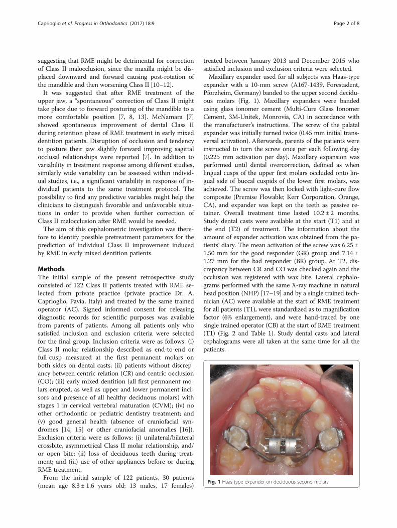
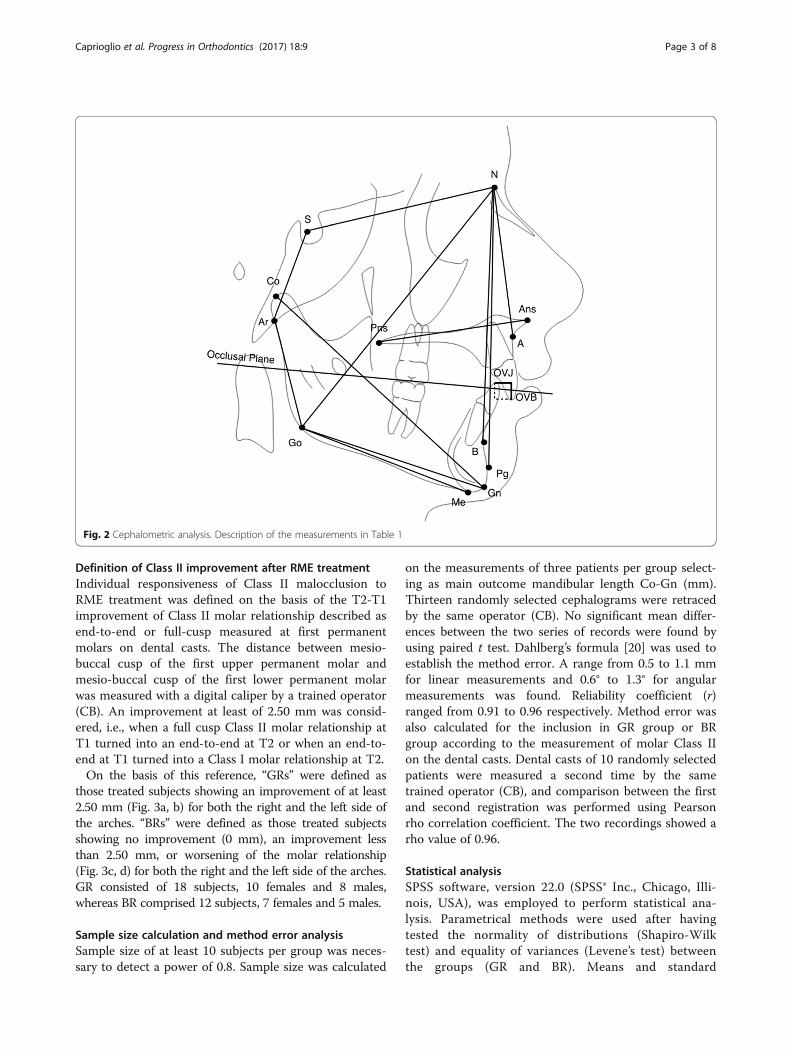
expander with a 10-mm screw (A167-1439, Forestadent,Pforzheim, Germany) banded to the upper second decidu-ous molars (Fig. 1). Maxillary expanders were bandedusing glass ionomer cement (Multi-Cure Glass IonomerCement, 3M-Unitek, Monrovia, CA) in accordance withthe manufacturer’s instructions. The screw of the palatalexpander was initially turned twice (0.45 mm initial trans-versal activation). Afterwards, parents of the patients wereinstructed to turn the screw once per each following day(0.225 mm activation per day). Maxillary expansion wasperformed until dental overcorrection, defined as whenlingual cusps of the upper first molars occluded onto lin-gual side of buccal cuspids of the lower first molars, wasachieved. The screw was then locked with light-cure flowcomposite (Premise Flowable; Kerr Corporation, Orange,CA), and expander was kept on the teeth as passive re-tainer. Overall treatment time lasted 10.2 ± 2 months.Study dental casts were available at the start (T1) and atthe end (T2) of treatment. The information about theamount of expander activation was obtained from the pa-tients’ diary. The mean activation of the screw was 6.25 ±1.50 mm for the good responder (GR) group and 7.14 ±1.27 mm for the bad responder (BR) group. At T2, dis-crepancy between CR and CO was checked again and theocclusion was registered with wax bite. Lateral cephalo-grams performed with the same X-ray machine in naturalhead position (NHP) [17–19] and by a single trained tech-nician (AC) were available at the start of RME treatmentfor all patients (T1), were standardized as to magnificationfactor (6% enlargement), and were hand-traced by onesingle trained operator (CB) at the start of RME treatment(T1) (Fig. 2 and Table 1). Study dental casts and lateralcephalograms were all taken at the same time for all thepatients.
Fig. 1 Haas-type expander on deciduous second molars
Caprioglio et al. Progress in Orthodontics (2017) 18:9 Page 2 of 8
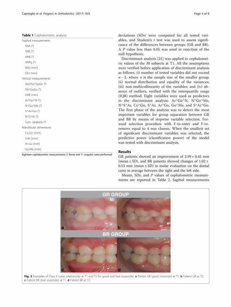
Definition of Class II improvement after RME treatmentIndividual responsiveness of Class II malocclusion toRME treatment was defined on the basis of the T2-T1improvement of Class II molar relationship described asend-to-end or full-cusp measured at first permanentmolars on dental casts. The distance between mesio-buccal cusp of the first upper permanent molar andmesio-buccal cusp of the first lower permanent molarwas measured with a digital caliper by a trained operator(CB). An improvement at least of 2.50 mm was consid-ered, i.e., when a full cusp Class II molar relationship atT1 turned into an end-to-end at T2 or when an end-to-end at T1 turned into a Class I molar relationship at T2.On the basis of this reference, “GRs” were defined as
those treated subjects showing an improvement of at least2.50 mm (Fig. 3a, b) for both the right and the left side ofthe arches. “BRs” were defined as those treated subjectsshowing no improvement (0 mm), an improvement lessthan 2.50 mm, or worsening of the molar relationship(Fig. 3c, d) for both the right and the left side of the arches.GR consisted of 18 subjects, 10 females and 8 males,whereas BR comprised 12 subjects, 7 females and 5 males.
Sample size calculation and method error analysisSample size of at least 10 subjects per group was neces-sary to detect a power of 0.8. Sample size was calculated
on the measurements of three patients per group select-ing as main outcome mandibular length Co-Gn (mm).Thirteen randomly selected cephalograms were retracedby the same operator (CB). No significant mean differ-ences between the two series of records were found byusing paired t test. Dahlberg’s formula [20] was used toestablish the method error. A range from 0.5 to 1.1 mmfor linear measurements and 0.6° to 1.3° for angularmeasurements was found. Reliability coefficient (r)ranged from 0.91 to 0.96 respectively. Method error wasalso calculated for the inclusion in GR group or BRgroup according to the measurement of molar Class IIon the dental casts. Dental casts of 10 randomly selectedpatients were measured a second time by the sametrained operator (CB), and comparison between the firstand second registration was performed using Pearsonrho correlation coefficient. The two recordings showed arho value of 0.96.
Statistical analysisSPSS software, version 22.0 (SPSS® Inc., Chicago, Illi-nois, USA), was employed to perform statistical ana-lysis. Parametrical methods were used after havingtested the normality of distributions (Shapiro-Wilktest) and equality of variances (Levene’s test) betweenthe groups (GR and BR). Means and standard
Fig. 2 Cephalometric analysis. Description of the measurements in Table 1
Caprioglio et al. Progress in Orthodontics (2017) 18:9 Page 3 of 8
deviations (SDs) were computed for all tested vari-ables, and Student’s t test was used to assess signifi-cance of the differences between groups (GR and BR).A P value less than 0.05 was used in rejection of thenull hypothesis.Discriminant analysis [21] was applied to cephalomet-
ric values of the 30 subjects at T1. All the assumptionswere verified before application of discriminant analysisas follows: (i) number of tested variables did not exceedn − 2, where n is the sample size of the smaller group;(ii) normal distribution and equality of the variances;(iii) non-multicollinearity of the variables; and (iv) ab-sence of outliers, verified with the interquartile range(IQR) method. Eight variables were used as predictorsin the discriminant analysis: Ar^Go^N, N^Go^Me,N^S^Ar, Co^Gn, S^Ar, Ar^Go, Go^Me, and S^Ar^Go.The first phase of the analysis was to detect the mostimportant variables for group separation between GRand BR by means of stepwise variable selection. For-ward selection procedure with F-to-enter and F-to-remove equal to 4 was chosen. When the smallest setof significant discriminant variables was selected, thepredictive power (classification power) of the modelwas tested with discriminant analysis.
ResultsGR patients showed an improvement of 2.99 ± 0.45 mm(mean ± SD), and BR patients showed changes of 1.02 ±0.53 mm (mean ± SD) in molar evaluation on the dentalcasts in average between the right and the left side.Means, SDs, and P values of cephalometric measure-
ments are reported in Table 2. Sagittal measurements
Fig. 3 Examples of Class II molar relationship at T1 and T2 for good and bad responder. a Patient GR (good responder) at T1. b Patient GR at T2.c Patient BR (bad responder) at T1. d Patient BR at T2
Table 1 Cephalometric analysis
Sagittal measurements
SNA (°)
SNB (°)
ANB (°)
ANPg (°)
Wits (mm)
OVJ (mm)
Vertical measurements
AnsPns^GoGn (°)
SN^GoGn (°)
OVB (mm)
Ar^Go^N (°)
N^Go^Me (°)
S^Ar^Go (°)
N^S^Ar (°)
Sum. Jaraback (°)
Mandibular dimensions
Co-Gn (mm)
S-Ar (mm)
Ar-Go (mm)
Go-Me (mm)
Eighteen cephalometric measurements (7 linear and 11 angular) were performed
Caprioglio et al. Progress in Orthodontics (2017) 18:9 Page 4 of 8
showed no significant differences between the twogroups. Among the vertical measurements, articularangle (S^Ar^Go) showed significant reduced values inBR group (140.85 ± 4.22°) when compared to GR group(144.66° ± 4.81°). On the contrary, superior gonial angle(Ar^Go^N) showed significant greater values in BRgroup (55.26° ± 3.48°) than in GR group (53.01° ± 2.46°).All mandibular dimensions (Co-Gn, S-Ar, Ar-Go, Go-Me) showed significant reduced values in GR groupwhen compared to BR group.Stepwise variable selection generated a two-variable
model that produced the most efficient separation be-tween the two groups (GR vs BR). The variables se-lected were the mandibular length (Co-Gn) and thesuperior gonial angle (Ar^Go^N) (Fig. 4 and Table 3).The classification power of the selected two-variablemodel was 83.3% (Table 4). Only one out of five casesin each group was not classified correctly. Unstan-dardized discriminant function coefficients of the se-lected variable together with a calculated constant(Table 5) lead to the following equation that providesindividual scores for the assignment of a new case toGR or to BR:
Individual Score ¼ 0:147 Co‐Gnð Þþ 0:221 Ar∧Go∧Nð Þ−26:399
The critical score (i.e., the value dividing GR from BR)is 0.667, i.e., the mean value of the group centroids ofthe two groups (−0.690 and 1.034 for GR and BR, re-spectively) (Table 5). Each new patient with dental ClassII malocclusion at CS 1–2 who will show an individualscore smaller than the critical score is expected to re-spond favorably to RME treatment in terms of at least2.50 mm of improvement in molar relationship. On thecontrary, each new patient with dental Class II mal-occlusion at CS 1–2 who will show an individual scoregreater than the critical score is expected to have a poorresponse to RME treatment in terms of at least 2.50 mmof improvement in molar relationship.
DiscussionSeveral studies investigated possible “spontaneous” cor-rection of Class II malocclusion after maxillary expan-sion; nevertheless, these studies present differentmethodology and controversial results [22, 23]. To thebest of our knowledge, this is the first study that differ-entiates bad and good responders in improving occlusalrelationship after palatal expansion. Previous studies in-vestigated Class II patients measuring changes aftertreatment without making any differences between pa-tients who improved and who did not improve mal-occlusion [7, 8, 13]. This study design might not allowto evidence possible variables influencing improvement,since mean changes might not reach clinical significance;in fact, they are the result of average of patients who im-proved and who showed no improvement pooled to-gether. Present study design, separating GR from BR,allowed detecting significant differences between the twogroups. Indeed, according to the results of present inves-tigation, the amount of dental and skeletal Class II doesnot seem a discriminant variable in influencing improve-ment. In fact, patients with similar sagittal measure-ments of skeletal Class II (ANB, ANPg in Table 2) mightshow improvement of the malocclusion after RME ornot. On the contrary, mandibular lengths and mandibu-lar sagittal position showed significant differences incomparisons between groups. GRs showed statisticallysignificant greater articular angle, more acute superiorgonial angle, and reduced mandibular dimensions (Co-Gn, S-Ar, Ar-Go, Go-Me) when compared to BRs.Discriminant analysis confirmed mandibular dimen-
sion (Co-Gn) and superior gonial angle (Ar^Go^N) ascephalometric variables with significant predictive valuein assigning patients to one group or the other. In par-ticular, patients with smaller mandibular length andmore acute superior gonial angle showed significant im-provement of Class II malocclusion after RME. Smaller
Table 2 Comparison between BR and GR groups
BR group GR group
Mean SD Mean SD P
Age 8.32 1.10 8.29 1.06 0.945
SNA (°) 81.65 2.57 80.60 4.33 0.457
SNB (°) 76.79 2.01 75.01 4.62 0.223
ANB (°) 4.87 2.32 5.38 1.96 0.520
ANPg (°) 4.18 2.43 4.78 2.28 0.497
Wits (mm) 1.97 3.88 1.03 2.76 0.447
OVJ (mm) 6.82 3.53 5.04 1.94 0.091
AnsPns^GoGn (°) 22.83 3.31 24.29 4.67 0.356
SN^GoGn (°) 30.18 3.16 33.23 5.44 0.092
OVB (mm) 3.22 1.94 2.74 1.74 0.496
Ar^Go^N (°) 55.26 3.48 53.01 2.36 0.048*
N^Go^Me (°) 70.28 3.69 71.84 4.78 0.354
N^S^Ar (°) 125.38 3.95 124.94 4.97 0.804
S^Ar^Go (°) 140.85 4.22 144.66 4.81 0.036*
Sum. Jaraback (°) 391.77 3.58 394.46 6.79 0.222
Co-Gn (mm) 103.68 5.83 95.36 6.31 0.001**
S-Ar (mm) 32.59 3.75 28.93 3.18 0.008**
Ar-Go (mm) 41.17 3.99 37.49 2.75 0.006**
Go-Me (mm) 63.36 5.45 57.63 5.05 0.006**
Data are shown as mean and SDs. Student’s t test was used to assess significanceof the differences between groups, and significance levels are shown in P columnBR bad responder, GR good responder*P < 0.05, **P < 0.01
Caprioglio et al. Progress in Orthodontics (2017) 18:9 Page 5 of 8
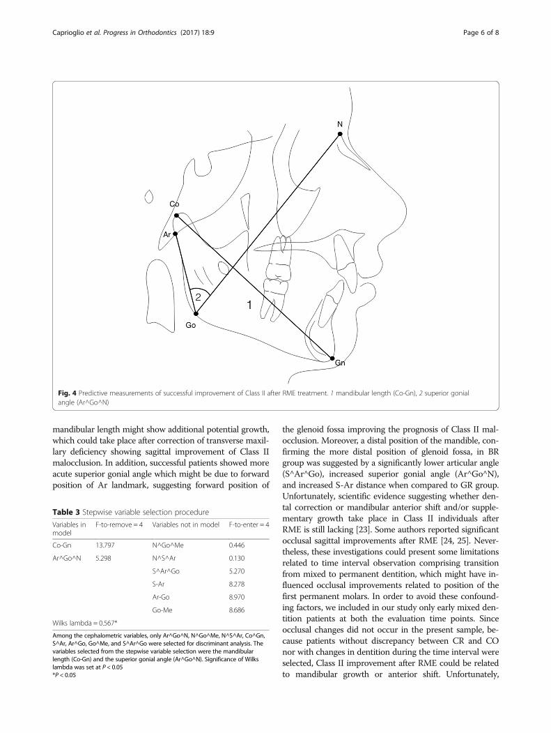
mandibular length might show additional potential growth,which could take place after correction of transverse maxil-lary deficiency showing sagittal improvement of Class IImalocclusion. In addition, successful patients showed moreacute superior gonial angle which might be due to forwardposition of Ar landmark, suggesting forward position of
the glenoid fossa improving the prognosis of Class II mal-occlusion. Moreover, a distal position of the mandible, con-firming the more distal position of glenoid fossa, in BRgroup was suggested by a significantly lower articular angle(S^Ar^Go), increased superior gonial angle (Ar^Go^N),and increased S-Ar distance when compared to GR group.Unfortunately, scientific evidence suggesting whether den-tal correction or mandibular anterior shift and/or supple-mentary growth take place in Class II individuals afterRME is still lacking [23]. Some authors reported significantocclusal sagittal improvements after RME [24, 25]. Never-theless, these investigations could present some limitationsrelated to time interval observation comprising transitionfrom mixed to permanent dentition, which might have in-fluenced occlusal improvements related to position of thefirst permanent molars. In order to avoid these confound-ing factors, we included in our study only early mixed den-tition patients at both the evaluation time points. Sinceocclusal changes did not occur in the present sample, be-cause patients without discrepancy between CR and COnor with changes in dentition during the time interval wereselected, Class II improvement after RME could be relatedto mandibular growth or anterior shift. Unfortunately,
Fig. 4 Predictive measurements of successful improvement of Class II after RME treatment. 1 mandibular length (Co-Gn), 2 superior gonialangle (Ar^Go^N)
Table 3 Stepwise variable selection procedure
Variables inmodel
F-to-remove = 4 Variables not in model F-to-enter = 4
Co-Gn 13.797 N^Go^Me 0.446
Ar^Go^N 5.298 N^S^Ar 0.130
S^Ar^Go 5.270
S-Ar 8.278
Ar-Go 8.970
Go-Me 8.686
Wilks lambda = 0.567*
Among the cephalometric variables, only Ar^Go^N, N^Go^Me, N^S^Ar, Co^Gn,S^Ar, Ar^Go, Go^Me, and S^Ar^Go were selected for discriminant analysis. Thevariables selected from the stepwise variable selection were the mandibularlength (Co-Gn) and the superior gonial angle (Ar^Go^N). Significance of Wilkslambda was set at P < 0.05*P < 0.05
Caprioglio et al. Progress in Orthodontics (2017) 18:9 Page 6 of 8

cephalograms at T2 were not available for the tested sam-ple due to ethical reasons, and this might be considered asa limit of the present study since the Class II improvementwas measured on dental casts only. Nevertheless, the com-prehension of reasons for improvement was not the aim ofthe present study.The rigidity of applied inclusion and exclusion criteria
led to a small final sample compared to the initial sam-ple of the present study. This rigid selection might havecaused selection bias, which might be consider as a limitof the present investigation, but on the other hand, thismethodology assured a great homogeneity among the se-lected patients which is challenging in a retrospectivestudy. This homogeneity in patients’ selection was con-sidered of great importance since RME treatment mighthave caused high variation in individual response.Some authors have reported that maxillary expansion
might be detrimental for correction of Class II malocclu-sion, due to downward and backward displacement ofmandible that frequently occurs after RME [10–12]caused by extrusion due to buccal tipping of the firstupper molars involved in expansion appliance [26]. Differ-ent treatment outcomes might be related to collateralfunction such as changes in the breathing pattern [27–29],different mandibular displacement [30, 31], and/or spon-taneous dental changes in the lower arch [32], but thesevariables were not evaluated in the present study. Thepresent study employed maxillary expander banded onthe upper second primary molars [33, 34], and resultsshould be limited to this appliance. Unfortunately, none ofthe previous cited studies evaluated mandibular responsein Class II patients after RME on the upper second pri-mary molars, but Rosa et al. [31] suggested spontaneous
changes in mandibular position in Class III patients withthe use of this appliance. Since the upper permanent mo-lars are not anchored in the appliance and are free tomove within the occlusal forces, spontaneous movementand distal rotation [26] might have occurred allowing for-ward placement of mandible after treatment.Considering clinical importance of outcomes although
the limitation of present retrospective design, furtherstudies conducted with prospective design are necessaryto confirm present results.
Conclusions
� The assessment of spontaneous improvement ofClass II malocclusion after RME therapy wasperformed by means of discriminant analysis, toidentify a significant model of predictive variables.Two predictive measurements were selected: (1)length of mandible (Co-Gn) and (2) superior gonialangle (Ar^Go^N).
� The classification power of the model for predictingsuccess or failure is 83.3% for each new patient.Spontaneous correction of Class II malocclusionafter RME in early mixed dentition might befavorable when patient’s cephalometric records showdecreased mandibular length and more acutesuperior gonial angle at the start of treatment.
� The important role of mandibular dimensions andmandibular sagittal position in diagnostic andprognostic evaluation of Class II patients deserves tobe emphasized, suggesting poor response whenincreased mandibular dimensions and more distalposition of the mandible are identified inpretreatment cephalograms. Class II skeletal angularmeasurements before treatment are not able toimprove this prediction based upon mandibulardimensions and superior gonial angle.
AbbreviationsBR: Bad responder; CO: Centric occlusion; CR: Centric relation; CVM: Cervicalvertebral maturation; GR: Good responder; NHP: Natural head position; RME: Rapidmaxillary expansion; SD: Standard deviation
Authors’ contributionsAC treated the patients, coordinated the research project, and revised themanuscript critically for important intellectual content. CB acquired the data,performed the tracings, and drafted the manuscript. LF revised the manuscriptcritically for important intellectual content and revised the statistical analysis. NVparticipated in the data acquisition and manuscript drafting. PZ designed thestudy protocol and helped in the interpretation of the statistical analysis andthe results and drafting the manuscript. RN revised the manuscript critically forimportant intellectual content and English language. RF drafted the manuscriptand performed the statistical analysis. All authors read and approved the finalmanuscript.
Competing interestsThe authors declare that they have no competing interests.
Table 4 Discriminant analysis
Predicted group membership
GR BR
Group No. of cases n % n %
Group GR (success) 12 10 83.3 2 16.7
Group BR (failure) 18 3 16.7 15 83.3
Percentage of cases correctly classified 83.3%. Classification results ofdiscriminant analysis
Table 5 Discriminant function
Predictive variables Unstandardized canonical discriminantfunction coefficients
Co-Gn 0.147
Ar^Go^N 0.221
Constant −26.399
Individual Score = 0.147(Co-Gn) + 0.221(Ar^Go^N) − 26.399. Discriminant scores forgroup means (group centroids): successful group = −0.690; unsuccessful group =1.034; and critical score = 0.667. Unstandardized discriminant function coefficientsof the selected variable together with a calculated constant (−26.399) lead to theequation that provides individual scores for the assignment of a new case to GRor to BR
Caprioglio et al. Progress in Orthodontics (2017) 18:9 Page 7 of 8

Ethics approval and consent to participateSigned informed consent for releasing diagnostic records for scientific purposeswas available from parents of patients. Protocol was reviewed and approved bythe Ethical Committee of the University of Insubria (Varese, Italy) (approval no.826), and procedures followed adhered to the World Medical OrganizationDeclaration of Helsinki.
Publisher’s NoteSpringer Nature remains neutral with regard to jurisdictional claims in publishedmaps and institutional affiliations.
Author details1Division of Orthodontics, Department of Surgical and MorphologicalSciences, School of Medicine, University of Insubria, Varese, Italy. 2Division ofOrthodontics, Department of Surgical and Morphological Sciences,Orthodontic Programme, School of Medicine, University of Insubria, Varese,Italy. 3Division of Dentistry, Department of Surgery and TranslationalMedicine, University of Florence, Florence, Italy. 4Department of Surgical andMorphological Sciences, School of Medicine, University of Insubria, Varese,Italy. 5Division of Orthodontics, Department of Medical, Surgical and HealthSciences, University of Messina, Messina, Italy. 6Department of Medical,Surgical and Health Sciences, University of Messina, Messina, Italy. 7C/ODental School, Via G. Piatti, 10, Velate, 21100 Varese, Italy.
Received: 9 January 2017 Accepted: 20 March 2017
References1. Fontana M, Cozzani M, Caprioglio A. Non-compliance maxillary molar
distalizing appliances: an overview of the last decade. Prog Orthod. 2012;13(2):173–84.
2. Will LA. Transverse maxillary deformities: diagnosis and treatment. OralMaxillofac Surg. 1996;5:1–28.
3. Baccetti T, Franchi L, McNamara Jr JA, Tollaro I. Early dentofacial features ofClass II malocclusion: a longitudinal study from the deciduous through themixed dentition. Am J Orthod Dentofacial Orthop. 1997;11:502–9.
4. Tollaro I, Baccetti T, Franchi L, Tanasescu CD. Role of posterior transverseinterarch discrepancy in Class II, division 1 malocclusion during the mixeddentition phase. Am J Orthod Dentofacial Orthop. 1996;110:417–22.
5. Franchi L, Baccetti T. Transverse maxillary deficiency in Class II and Class IIImalocclusions: a cephalometric and morphometric study on postero-anteriorfilms. Orthod Craniofac Res. 2005;8(1):21–8.
6. Buschang PH, Stroud J, Alexander RG. Differences in dental arch morphologyamong adult females with untreated Class I and Class II malocclusion. Eur JOrthod. 1994;16:47–52.
7. McNamara Jr JA. Early intervention in the transverse dimension: is it worththe effort? Am J Orthod Dentofacial Orthop. 2002;121:572–4.
8. McNamara Jr JA. Long-term adaptations to changes in the transverse dimensionin children and adolescents: an overview. Am J Orthod Dentofacial Orthop.2006;129(4 suppl):S71–4.
9. Bishara SE, Bayati P, Jakobsen JR. Longitudinal comparisons of dental archchanges in normal and untreated Class II, division 1 subjects and theirclinical implications. Am J Orthod Dentofacial Orthop. 1996;110:483–9.
10. Haas AJ. Palatal expansion: just the beginning of dentofacial orthopedics.Am J Orthod. 1970;57:219–55.
11. Sarver D, Johnston M. Skeletal changes in vertical and anterior displacementof the maxilla with bonded rapid palatal expansion appliances. Am J OrthodDentofacial Orthop. 1989;95:462–6.
12. Wertz RA. Skeletal and dental changes accompanying rapid mid-palatal sutureopening. Am J Orthod. 1970;58:41–65.
13. Lima Filho R, Lima A, Ruellas A. Spontaneous correction of Class II malocclusionafter rapid palatal expansion. Angle Orthod. 2003;73:745–52.
14. Bianchi B, Ferri A, Brevi B, Di Blasio A, Copelli C, Di Blasio C, Barbot A, Ferri T,Sesenna E. Orthognathic surgery for the complete rehabilitation of Moebiuspatients: principles, timing and our experience. J Craniomaxillofac Surg.2013;41(1):e1–4.
15. Di Blasio A, Cassi D, Di Blasio C, Gandolfini M. Temporomandibular jointdysfunction in Moebius syndrome. Eur J Paediatr Dent. 2013;14(4):295–8.
16. Anghinoni ML, Magri AS, Di Blasio A, Toma L, Sesenna E. Midlinemandibular osteotomy in an asymmetric patient. Angle Orthod. 2009;79(5):1008–14.
17. Di Blasio A, Mandelli G, Generali I, Gandolfini M. Facial aesthetics andchildhood. Eur J Paediatr Dent. 2009;10(3):131–4.
18. Cassi D, De Biase C, Tonni I, Gandolfini M, Di Blasio A, Piancino MG. Naturalposition of the head: review of two-dimensional and three-dimensionalmethods of recording. Br J Oral Maxillofac Surg. 2016;54(3):233–40.
19. Zecca PA, Fastuca R, Beretta M, Caprioglio A, Macchi A. Correlation assessmentbetween three-dimensional facial soft tissue scan and lateral cephalometricradiography in orthodontic diagnosis. Int J Dent. 2016;2016:1473918.
20. Dahlberg G. Statistical methods for medical and biological students.London, United Kingdom: Allen and Unwin; 1940. p. 122–32.
21. Franchi L, Baccetti T. Prediction of individual mandibular changes inducedby functional jaw orthopedics followed by fixed appliances in Class II patients.Angle Orthod. 2006;76(6):950–4.
22. Volk T, Sadowsky C, Begole EA, Boice P. Rapid palatal expansion for spontaneousClass II correction. Am J Orthod Dentofacial Orthop. 2010;137:310–5.
23. Feres MF, Raza H, Alhadlaq A, El-Bialy T. Rapid maxillary expansion effects inClass II malocclusion: a systematic review. Angle Orthod. 2015;85(6):1070–9.
24. McNamara Jr JA, Sigler LM, Franchi L, Guest SS, Baccetti T. Changes in occlusalrelationships in mixed dentition patients treated with rapid maxillaryexpansion. A prospective clinical study. Angle Orthod. 2010;80:230–8.
25. Guest SS, McNamara Jr JA, Baccetti T, Franchi L. Improving Class IImalocclusion as a side-effect of rapid maxillary expansion: a prospectiveclinical study. Am J Orthod Dentofacial Orthop. 2010;138:582–91.
26. Rosa M, Lucchi P, Manti G, Caprioglio A. Rapid palatal expansion in theabsence of posterior cross-bite to intercept maxillary incisor crowding in themixed dentition: a CBCT evaluation of spontaneous changes of untouchedpermanent molars. Eur J Paediatr Dent. 2016;17(4):286–94.
27. Fastuca R, Perinetti G, Zecca P, Nucera R, Caprioglio A. Airway compartmentsvolume and oxygen saturation changes after rapid maxillary expansion: alongitudinal correlation study. Angle Orthod. 2015;85(6):955–61.
28. Caprioglio A, Meneghel M, Fastuca R, Zecca PA, Nucera R, Nosetti L. Rapidmaxillary expansion in growing patients: correspondence between 3-dimensional airway changes and polysomnography. Int J PediatrOtorhinolaryngol. 2014;78(1):23–7.
29. Fastuca R, Meneghel M, Zecca PA, Mangano F, Antonello M, Nucera R,Caprioglio A. Multimodal airway evaluation in growing patients after rapidmaxillary expansion. Eur J Paediatr Dent. 2015;16(2):129–34.
30. Fastuca R, Zecca P, Caprioglio A. Role of mandibular displacement andairway size in improving breathing after rapid maxillary expansion. ProgOrthod. 2014;15:40.
31. Rosa M, Lucchi P, Mariani L, Caprioglio A. Spontaneous correction ofanterior crossbite by RPE anchored on deciduous teeth in the early mixeddentition. Eur J Paediatr Dent. 2012;13(3):176–80.
32. Ugolini A, Cerruto C, Di Vece L, Ghislanzoni LH, Sforza C, Doldo T, Silvestrini-Biavati A, Caprioglio A. Dental arch response to Haas-type rapid maxillaryexpansion anchored to deciduous vs permanent molars: a multicentricrandomized controlled trial. Angle Orthod. 2015;85(4):570–6.
33. Giuliano Maino B, Pagin P, Di Blasio A. Success of miniscrews used asanchorage for orthodontic treatment: analysis of different factors. ProgOrthod. 2012;13(3):202–9.
34. Mutinelli S, Manfredi M, Guiducci A, Denotti G, Cozzani M. Anchorage ontodeciduous teeth: effectiveness of early rapid maxillary expansion in increasingdental arch dimension and improving anterior crowding. Prog Orthod.2015;16:22.
Submit your manuscript to a journal and benefi t from:
7 Convenient online submission
7 Rigorous peer review
7 Immediate publication on acceptance
7 Open access: articles freely available online
7 High visibility within the fi eld
7 Retaining the copyright to your article
Submit your next manuscript at 7 springeropen.com
Caprioglio et al. Progress in Orthodontics (2017) 18:9 Page 8 of 8


![Comparison of dentopalatal change after maxillary expansion … · 2020. 8. 21. · maxillary expansion in growing children (bonded rapid maxillary expander [RME], plate with median](https://img.dokumen.tips/doc/110x75/60bb6f74a371f41140243d51/comparison-of-dentopalatal-change-after-maxillary-expansion-2020-8-21-maxillary.jpg)