Embed Size (px)
Citation preview

Pre-and perinatal pathology Children's Infection
Sepsis
as.-prof. V.Voloshyn
(By Ya.Ya. Bodnar, Yu. Orel)

Essence and structure of prenatal pathology
Prenatal (antenatal) pathology includes pathological processes of human embryo from fertilization and ending with the birth of a child. Prenatal period lasts 280 days or 40 weeks .

Kimatogenesic Period
The whole development from zygote forming to birth is called kimatogenesis. Before it – progenesis – the period of maturing of male and female sex cells (gametes) to fertilization.Kimatogenesis period is divided into three periods:- Blastogenesis lasts from fertilization to 15-th day of pregnancy;- Embryogenesis - from 16-th to 75-th day of pregnancy;- Fetogenesis - lasts from 76-th to 280-th day of pregnancy .

GametopatiesThere are genetic, chromosomal and genomic mutations that cause congenital defects (malformations).
Defects incompatible with life ended
spontaneous miscarriage (abortion).

Blastopaties violation of blastocyst implantation (ectopic pregnancy)
twins malformations
single malformations
malformations (defect) of the placenta and umbilical cord.

Embriopaties Pathology of the embryonic period from 16-th to 75-th day of pregnancy, during which followed the main organ.
Embriopaties include mainly congenital defects - malformations.

Classification of congenital malformations
1) the lack (absence) of any organs or body part - agenesis, aplasia;
2) underdeveloped organ - hypoplasia;
3) excessive development - hyperplasia;
4) modified form: the merging of organs, atresia or stenosis of holes or channels, uninosculation of embryonic holes or fissures- dysraphia or persistence, inside out - extrophia.
5) change in the organs location - ectopia;
6) persistence of embryonic organs, often branchial arches or their residues .

Fetopatia pathology of fetal period (period from 76-th to 280-th day of pregnancy. The main tissue differentiation of organs is formed. There are typical two types of displays: the violation of tissue morphogenesis and reactive changes in the form of blood disorder, dystrophy and necrosis, perverse immune reactions and compensatory-adaptive processes .

Perinatal periodThe term "perinatal period" includes late
fetal period (29 weeks of intrawomb development to the delivery beginning), intranatal period (parturition/during delivery) and early neonatal (from birth till six days inclusive).

Classification of perinatal period
Antenatal (pre-natal) (predelivery);
Intrapartum (during labor);
Postpartum (postnatal or neonatal)

Newborn baby - baby which began to breathe independently.Stillborn fetus - Fetus which has absent breathing in the moment of birth, and he does not succeed to be caused artificial a way. Palpitation can proceed some time in such fetus.Immaturity - Anatomical unripe and undifferentiate up some structures and organs.Overmaturity - Fetus of pregnancy over 42 weeks
Classification of perinatal pathology (GLOSSARY)

Reasons of prematurity
illness of fetus, especially intrawomb infection, combined with the defeat of placenta of same etiology;disease of sexual organs of pregnant;placenta insufficiency;heavy toxicosis of pregnancy (severe gestosis);extragenital pathology of mother;criminal intervention.

Signs of prematurity :gestation term is less than 37 weeks;a little mass and length of child (less than 2,5 kg and 47 centimeters);absence or weak expressed of ossification nuclei;nose and ears cartilages mildness (auricles densely adjoin to the cranium);nails are soft, don't reach to the tip of fingers;superfluous fluff is saved, especially on a shoulder girdle and superior portion of the back;boys testicles not in scrotum omitted;in girls major pudenda lips don’t cover a clitoris and small pudenda lips ;

Signs of immaturity :
myocardium is poor by sarcoplasm;the cardiomyocytes transversal striped is weak;the follicles of spleen are shallow;the kidney glomeruluses are a like goblet capsule .

Signs of overmaturity dryness, shelling, partial skin maceration;general hypotrophy;presence of ossify nucleuses of proximal epiphysis of tibia and humeral bones;an umbilical cord and placenta membranes are painted by meconium .


Infectious DiseasesEtiology :
viruses: herpes, cytomegalic, hepatitis, rubella, AIDS and others;bacteria: pale treponema, listeria, tuberculosis;mycoplasma;chlamydia;fungus;protozoa (simplest) (toxoplasm).
Ways of infection:antenatal;intranatal .
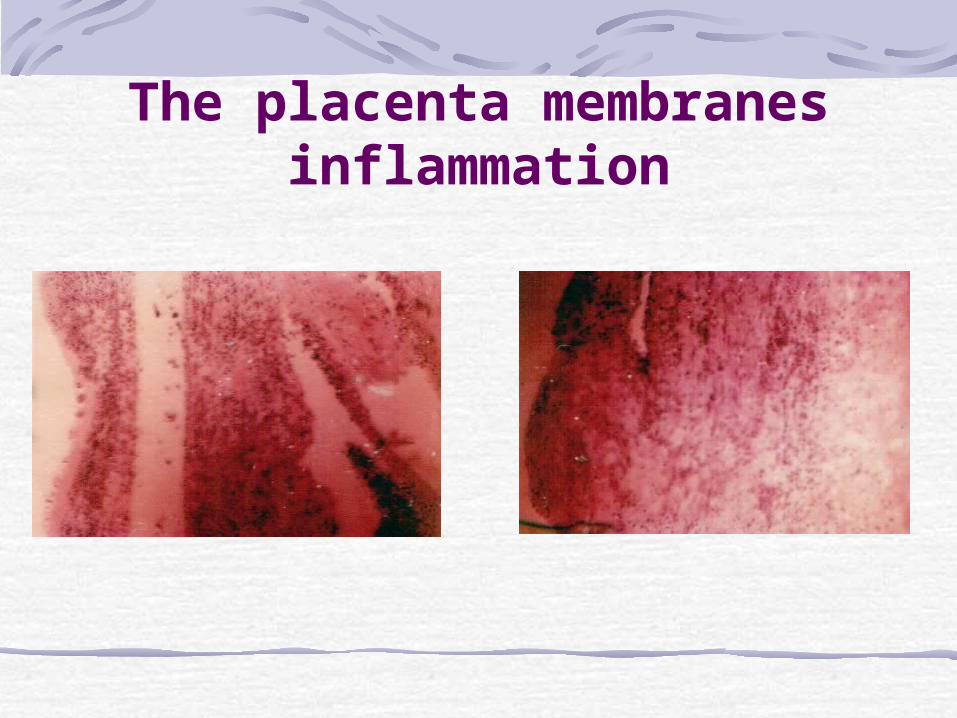
The placenta membranes inflammation

Intrauterine herpesPathogen - Herpes simplex II (rarer I type).
The virus multiplies in cells of the epithelium.

Morphological signs of intrauterine herpes:
increasing of the epithelium size, especially the nuclei;acidophilic and basophilic inclusions in the nuclei;fragmentation of chromatin with marginal location of lumps;an inflammatory reaction is poorly expressed or absents round the alteration areas;gigantic cells metamorphoses of hepatocytes and Kupffer cells;gigantic cells pneumonia;gigantic cells brain defeat;macroscopically: the area of the injury organ have rather yellow or grey color.

Cytomegalia Etiology - Cytomegalovirus hominis.
Pathogen multiplies in the epithelium.

Morphological signs of cytomegalia
the oxyphilic and basophilic inclusion in the epiteliocells nuclei with the brighten area around them;the light basophilic inclusion in a cytoplasm;limphohystiocells infiltration with the admixtures of erythromyeloblastes around the injury areas;sialadenitis develops most often;macroscopic displays are expressed poorly.

Listeriosis Etiology - Listeria monocytogenes.

Morphological signs of listeriosis
listerios granulema: in a center is an accumulation of leucocytes around Listerias;on peripheries is fibrinoid necrosis, in the perifocal areas is granulation tissue which consists of hystiocytes;in the inner organs – granulemas (listeriomas)

Complications of listeriosis
phlegmon of the new-born;omphalitis;umbilical sepsis

Classification of non-infectious perinatal
pathology:asphyxia;birth injury (maternity trauma);hemolytic disease;hemorrhagic disease of newborn;pneumopathy;pneumonia.

Asphyxia
Fetus or new-born hypoxygen condition, which is combined with a hypercapnia. It can develop before births, during births and after births

Asphyxia reasons
Reasons of asphyxia in antenatal period:anoxic state of mother;acute violations of the utero-placenta or placento-fetus blood circulation;B) Reasons of asphyxia in intranatal period:anoxic state of mother;
Reasons of asphyxia in intranatal period:premature placenta exfoliation (abruptio placenta);violation of the utero-placenta blood circulation;abnormal position of placenta (placenta previa (передлежання));blood stream violation on an umbilical cord as a result of: pressure by fetus head, umbilical cord falling out of mother's maternity ways, umbilical cord overstrain, umbilical cord tight arounds the fetus neck, veritable knots of umbilical cord.


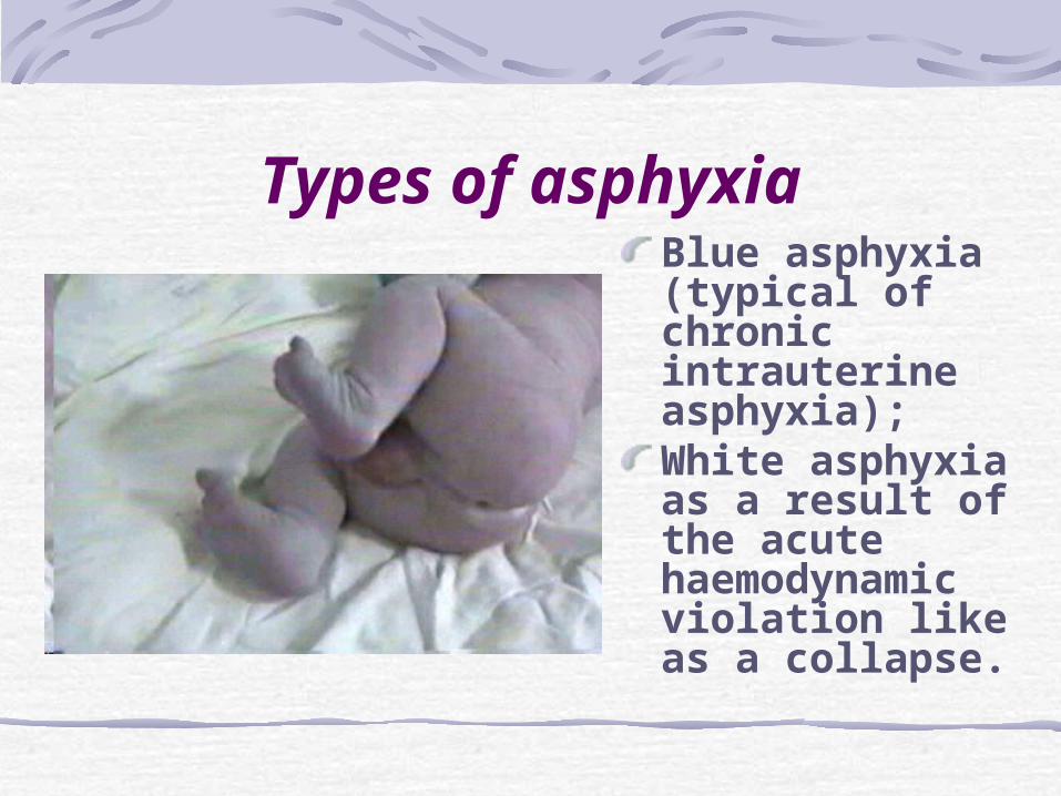
Types of asphyxiaBlue asphyxia (typical of chronic intrauterine asphyxia);White asphyxia as a result of the acute haemodynamic violation like as a collapse.

Morphological signs of asphyxia
dark liquid blood in the cavities of heart and large blood vessels;acrocyanosis and cyanosis;swelling of the feet, scrotum and sexual labia;hemorrhage in serous membranes;lungs have a meaty consistency (fleshy consistency), do not fill a thoracic cavity, airless pieces sink in water;aspiration of amniotic fluid elements;consumption coagulopathy

Maternity traumalocal damage of fetus tissues
during fetal descent act, which arose up as a result of operating of mechanical force directly on a fetus, but not on a placenta or umbilical cord, and shows up breaks, fractures, dislocations, laying out (smashed?) of the tissues.

The damage degree of fetus depends from:
degree of prematurity or overmaturity of fetus;degree of forming and size of the skull;degree of forming of the cerebral falx and cerebella tentorium;rigidity of maternity channel tissues;form and sizes of pelvis;violation of moving apart of maternity ways tissues at the premature break of fetus bubble;dynamics of maternity act (rapid delivery);standing duration of the fetus head in the uterus neck channel.

Morphological signs of birth injury
labour tumor;hemorrhages;cephalic haematoma;hemorrhages in the cranial cavity;hemorrhages in the cerebrum ventricles;damage of cranial bones.

Haemolytic illnessarises up at incompatibility of mother blood and fetus blood mainly on the rhesus-factor (mother has Rh"–", fetus has Rh“+”), causing hemolysis of fetus erythrocytes by the mother's antibodies.

Haemolytic disease neonate is translated in the neonate intensive therapy department; perforation and peritonitis
developed through intestinal impassability ІV degrees;After 22 days - the child died.
ІІІ pregnancy, ІІ deliveryІ pregnancy (1999) – healthy baby,ІІ pregnancy (2002) – died down.Mother has ІІІ Rh (-), titre аntibodies 1:64;Caesarean section; 37-38 weeks,valuation by Apgar scale 7/8 balls, Mass 2550;Child АВ (ІV) Rh (+);Bilirubin from umbilical cord – 62,1;through 7 hours - 101,3 mkmoll/l; through 13 hours - 133,6 mkmoll/l

The forms of hemolytic jaundice
anaemic;icteric;oedematous.

Pathoanatomy of the haemolytic illness
1) in intrauterine fetal death of 7.5 months:autolysis;maceration;swelling of face;moderate enlargement of the liver, spleen;Pathoanatomy of the haemolytic illness (continuation)
2) at an anaemic form :icterus is absent;anaemia of inner organs;erytroblastosis is expressed moderately;skin pallor & mucus covers pallor.

Pathological anatomy of hemolytic disease(continuation)
3) at a heavy post-natal jaundice:bilirubin encephalopathy;nuclear icterus (kernicterus);erythroblastosis;haemosyderosis;bile stasis;hyperplasia of spleen;
at an oedematous form:• a skin is pale,
semilucent, brilliant. partly is macerationed;
• a hypoderm and tissues of brain oedematic;
• transudation in the body cavity;
• liver multiplying is in 4-6 times;
• lungs mass is diminished.

Bilirubin encephalopathy

Hemorrhagic disease of newborn
Hemorrhagic disease of newborn - is a clinic & anatomic syndrome, which is characterized by internal and external hemorrhages which arises up in new-born in the first days after births.
Etiology – 1) related to heredity or; 2) influence of exogenous factors (acceptance of
medications by the pregnancy woman), and also; 3) infectious diseases of new-born.

Hemorrhagic disease of newborn
Mechanisms of development:coagulopathia;thrombocytopathia;angiopathia;
Pathological anatomy :partial or segmental pulmonary hemorrhage;linear or spotty hemorrhages on pleura;massive adrenal hematomas;spotty hemorrhages in cortical and medullar layers of kidneys;melena (false);


PneumopathiesPneumopathies are a
group of uninfectious defeats of breathings organs which include disease of hyaline membranes, oedematous-haemorrhagic syndrome, atelectasis of lungs

PneumoniaPneumonias – exciters are mostly
cocci which get to the respiratory tracts of foetus in an intranata period together with aspirated perifoetus maintenance or after births at application of instrument room artificial ventilation of liungs.
Morphological signs:- aspirated perifoetus maintenance
and water are presence in bronchial tubes, alveolus ducts and alveolus.

Sepsis features, which distinguish it from other infectious diseases:1. Septicemia - polyethiological disease. It can be caused by different microorganisms. But most of all - staphylococcus. Meningococcus, Klebsiella, aeruginosa, and mixed infections.2. Epidemiological feature – uncontagious disease. Sepsis is not reproduced in the experiment.3. Clinical feature: the absence of recurrence is not possible to allocate periods for specific infectious diseases.4. Immunological - no immunity.

Stages of pathogenesis Systemic inflammatory response syndrome (destruction of the endothelium, mediated cascade).Septic arterial hypotension (NO, TNF).Septic shock (cardiac weakness, disturbance of microcirculation).Syndrome of multiple organ failure ("shock kidney" distress syndrome, etc.).
According to clinical morphological features distinguish septicemia, septic-piyemiyu, septic (bacterial) endocarditis and chroniosepsis.

Apostematic nephritis

SEPTIC EMBOLISM IN LUNGS

CHANGES IN THE LUNGS AND KIDNEYS IN SEPTIC
SHOCK

Thank you for attention!