Embed Size (px)
Citation preview

Practical Hematology Manual

Wh
ole
blo
od
8%
of b
od
y
weig
ht
Plasma55%
Cells45%
Water 91.5%
Plasma protein 7%
Other solutes 1.5%
Albumin 54%Globulin 38%
Fibrinogen 7%Other 1%
Enzymes & HormonesNutrient
Electrolytes &Gases Wastes
RBCs 4.5-6 million/mm3
Platelets150,000-400,000/mm3
WBCs 4000- 11000/mm3
Neutrophils 60-70%
Lymphocytes 20-25%
Monocytes 4-8%
Eosinophils 2-4%
Basophils 0-1%

Erythrocyte Count
• Normal ranges:– Male 5 - 6 million/mm3 or μl– Female 4.5 - 5 million/mm3 or μl– New born 6 - 7 million/mm3 or μl
• Erythrocyte count increased in case of polycythemia and decreased in anemia

How to collect blood sample to do Complete blood count (CBC)When blood come in contact with wettable surface e.g.
glass (tube), it will normally clot (by intrinsic pathway) as follow
We need to prevent this blood clotting to be able to proceed through the intended CBC,To do this we have to add anti-coagulants.


Hemocytometer
• The hemocytometer counting chamber is used for cell counting.
• It is constructed so that the distance between the bottom of the coverslip and the surface of the counting area of the chamber is 0.1 mm.
• The surface of the chamber contains two square ruled areas separated by an H-shaped moat.
Hemo / cyto / Meter Blood / cell / counter
Thus, it is an instrument used to count the blood cells.

It includes :
-Neubauer’s slide -Cover slip
-RBC pipette -WBC pipette

Erythrocyte Count
• Principle:
• In order to facilitate RBCs count a specified volume of blood is diluted with a specified volume of isotonic fluid
• Diluting fluid:• One of the following solutions may be used:
1. Isotonic saline: 0.9 % NaCl in distilled water.2. Hayam’s solution
• Sample:
– Whole blood using EDTA or heparin as anticoagulant.
• Equipments:
1. RBCs pipettes.
2. Neubauer's chamber.
3. Cover slips.
4. Conventional light microscope.
5. Clean gauze.

haemocytometer chamber
Pipette
Rubber sucking tube
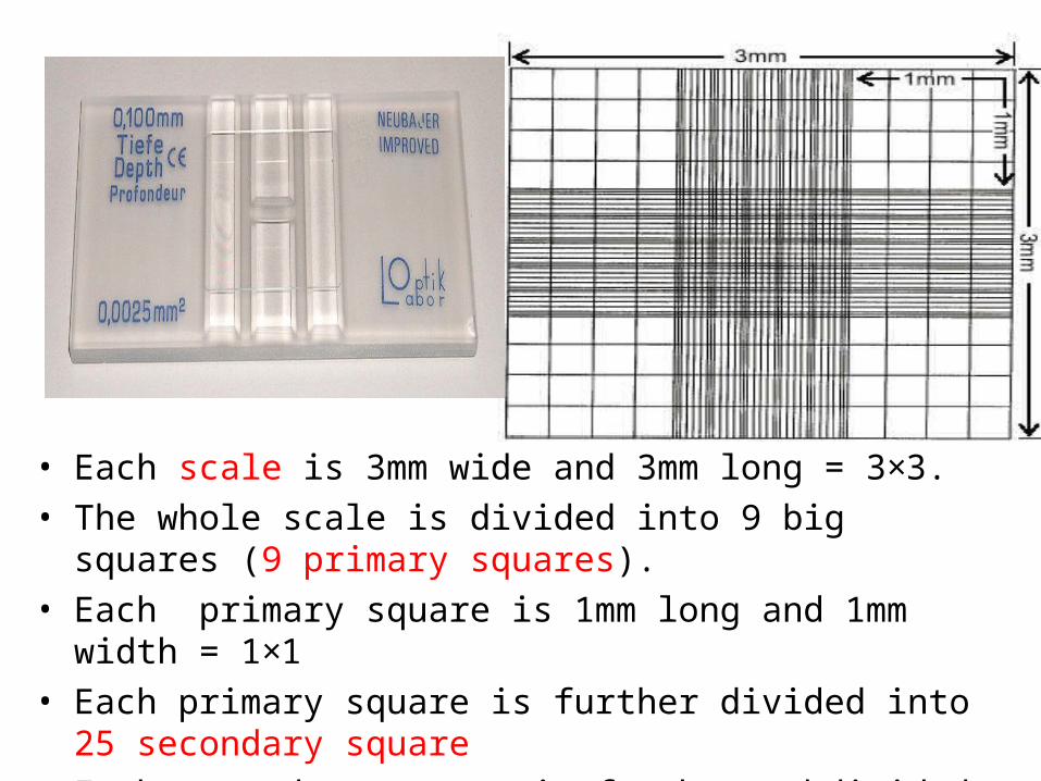
• Each scale is 3mm wide and 3mm long = 3×3.
• The whole scale is divided into 9 big squares (9 primary squares).
• Each primary square is 1mm long and 1mm width = 1×1
• Each primary square is further divided into 25 secondary square
• Each secondary square is further subdivided into 16 tertiary square
• So the primary square is divided into 400 tertiary square


DILUTION FACTORSFor RBC counting
Blood is filled till mark 0.5 and Hayam's fluid is then filled till mark 101. Both are thoroughly mixed and then few drops are discarded which contain just the diluting fluid in the stem. Thus, 1 portion out of 101 is discarded. So, 0.5 part of blood is in 100 parts of fluid or, 1 part of blood is mixed in 200 parts of fluid Thus, dilution factor for RBC counting is 200.


ProcedureCarefully charge hemocytometer with diluted blood by gently squeezing sides of reservoir to expel contents until chamber is properly filled.

4X to see the general formation of slide.
10X for WBC counting
40X for RBC counting
FOCUSING

Adjust the microscopic objective lens 40X for RBC counting

RBC COUNTING
• Total no. of RBCs in 5 secondary squares = X
• No. of RBCs in 1 secondary squares = X/ 5
• X/ 5 × 25 = No. of RBCs in the primary sq (1mm2) of diluted blood
• X/ 5 × 25 × 200 = No. of RBCs in the primary sq (1mm2) of pateint blood
• Because the depth between the cover and slide was only 0.1 mm
• X/ 5 × 25 × 200 × 10 = No. of RBCs in the primary sq (1mm3) (= 1 μl of blood)
X = number of RBCs in 5 secondary squares
RBC COUNT = X × 10,000/mm³ or µL

Principle of WBCs count
Principle:
The diluting fluid lyses the RBCs but preserves leukocytes and platelets. The diluted blood is added to the hemocytometer chamber.
Sample:– EDTA- anticoagulated blood or capillary blood is preferred.
WBCs count diluting fluid :
• Acetic acid 1% (v/v) in distilled water.• HCl 1% (v/v) in distilled water.• Turk's solution.

Equipment
1. White blood cells count diluting fluid
2. WBCs pipette
3. Hemocytometer and coverslip
4. Microscope
5. Lint-free wipe
6. Alcohol pads
For WBC counting
0.5 part of blood is mixed in 10 parts of fluid
So, 1 part of blood is in 20 parts of fluid
Thus, dilution factor for WBC counting is 20.


WBC COUNTING
Total no. of WBCs in 4 Primary squares = X
No. of WBCs in one Primary square (1 mm2) = X/4
(diluted)
X/4 × 20 = No. of WBCs in 1 mm2 of undiluted blood
X/4 × 20 x 10= No. of WBCs in 1 mm3 of undiluted
blood.
While X is the No of WBCs counted in the 4 primary
square
• Normal Value: 5000 - 11000/mm3
or µL
No. of WBCs = X × 50/mm³ or µl

Coagulation timeDefinition :-
It is the time needed for formation of a hard clot.
Material:-•Clean dry glass slide, stop watch , water bath 37ºC, syringe, alcohol , cotton .Procedure:-•Put a drop of the blood on the slide.•The slide is placed on a warm plate at 37ºC. •Every 30 seconds put a needle in the middle of the blood drop and try to elevate it •blood coagulation start when fibrin threads appear attached to the needles.Normal : 5-10 minutes.
This test assess the factors involved in
intrinsic pathway

Bleeding timeThe time needed for
bleeding to stop.• Materials:
• Filter paper, sterile lancets, stop watch, sphygmomanometer , ethyl alcohol.
• Method:
• A sphygmomanometer cuff is placed on the upper arm and inflated to 40mm Hg .
• A sterilized area of forearm without visible superficial veins is selected and is pierced by sterilized lancet in 3 points .
• Wipe the blood every 30sec from the 3 points alternatively until no blood comes to filter paper .
• Normal : 3-5 min.3-5 min.
• To assess the platelet functions.

Hematocrite value (H.V.) packed cell volume (PCV)
Definition:-
H.V. is the volume of RBCs in 100 ml blood or it is the percentage of the volume of RBCs to the volume of whole blood.
Volume of RBCs
H.V. = × 100
Volume of blood
Materials:-• Microhematocrite tube (75 mm long, 1 mm pore, heparinized )• Microhematocrite centrifuge .• Microhematocrite tube reader.• Sterile lancet. • 70 % ethyl alcohol.

Procedure-: Obtain blood drop by pricking the thumb.
• Micro hematocrite tube is filled up to it's 2/3 with blood sample by touch the drop of blood by one end of the tube.
• Close the empty end of the tube by plasticine .
• Centrifuge the tube at 13000 per minute for 5 minutes .
• Remove the tube and read H.V. by putting the tube on special micro hematocrite scale.
Normal values-:• Adult male 45% - 47 %
• Adult female 42 % - 44%
• Higher in new born 45 % - 49%

Hematocrite (PCV) determinationCapillary methods


Female:36% - 48%
Male:45% - 47%

Hematocrite (PCV) determinationWintrobe method


Significance of the haematocrite
The PCV layer normally 45% - 47%
Increased in Polycythemia and dehydration
Decreased in anemia and overhydration
The Buffy coat normally 1% Increased in leukocytosis and
leukemia
Plasma layer
Colour : Normally faint yellow Deep yellow color indicate
Jaundice Red color indicate
Hemolysis

Determination of Hb values• Haemoglobin can be measured by manual or automatic.• Two method are common use:
1. Oxyhaemoglobin (HbO2method).
2. Haemiglobincyanid. (HicN;Cyanmethaemoglobin).
• A major a advantage of the HicN method is availability of stable and reliable reference preparation.
• Other methods: Sahli’s acid haematin and alkalin-haematin method.

Cyanmethaemoglobin method• Use Drabkins solution which contain
potassium cyanide and potassium ferricynide at pH 7 - 7.4
• Hb oxidation to methaemoglobin by potassium ferricynide then combine to potassium cyanide to form cyanmethaemoglobin which measure at 540 nm by spectrophotometer.

Normal range of Hb in g/dl
• Hb g/dl=
• Men 15 ± 2 g/dl
• Women 13.5 ± 1.5 g/dl
• At birth 18 ± 4 g/dl
Principle of Hb cyanide method• Blood is diluted in a solution:• Contains potassium cyanide + potassium ferricyanide (Drabkin
solution)• As a result this reaction will occur: • Hgb, HbCO methemoglobin HiCN (cyanmethemoglobin)• HbS will not be converted using this method

Methods
• Set spectrophotometer to 0.0 reading at 540 wave length, using
diluted drubkin’s solution as blank
• Test the reading of the cyanohemoglobin standard solution
• Dilute blood sample in the working solution as 1:250 dilution
(blood: diluents)
• 10µL of blood in 2.5 mL of working solution in the cuvet, Cover
and invert the tube several times
• Let it stand at R.T for 5-10 minutes to insure complete conversion
• Put the cuvet in the hemoglobinometer
• Continue reading patient samples and determin Hb value of sample

RBCs indices
Red Cell Indices includes
1. Mean Corpuscular Volume (MCV)
2. Mean Corpuscular Hemoglobin (MCH)
3. Mean Corpuscular Hemoglobin
Concentration (MCHC)
These calculation requires the presence of
1. RBCs count
2. Hb value
3. PCV

Mean Corpuscular Volume (MCV)
• The MCV indicates the average volume of the red blood cells.
• MCV =
= (fl)
• Normal value for the MCV : 86±8 fl
• If the MCV is less than 78 fl, the RBCs are Microcytic.
• If the MCV is greater than 94 fl, the RBCs are Macrocytic.
• If the MCV is within the normal range, the RBCs are Normocytic.
Volume of RBC in femtoliters (fl) / μl of blood
RBC / μl of blood
Hematocrit x 10
RBC count in millions
1 μl = 109 fl

Mean Corpuscular Hemoglobin (MCH)
• The MCH indicates the average weight of hemoglobin in the red blood cells.
• MCH =
• Normal value for the MCH : 27~31 pg
• If MCH is lower than 27 pg the condition is called Hypochromic
• If MCH is higher than 31 pg the condition is called Hyperchromic
• If MCH is within the rang of 27~31 pg condition is called Normochromic
Weight of hemoglobin in 1 μl of blood
Number of red blood cells in 1 μl of blood
Hemoglobin * 10
Red blood cell count in millions(pg)
1 g = 1012 pg
1 ml = 103 μl
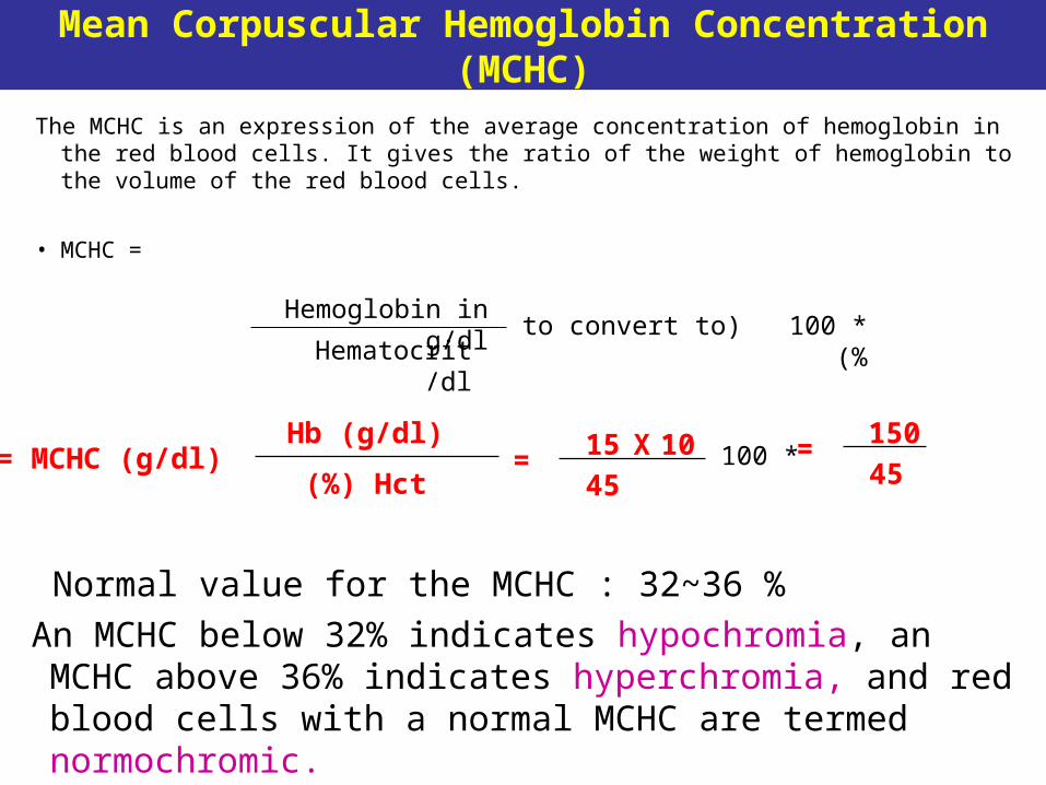
Mean Corpuscular Hemoglobin Concentration (MCHC)
The MCHC is an expression of the average concentration of hemoglobin in the red blood cells. It gives the ratio of the weight of hemoglobin to the volume of the red blood cells.
• MCHC =
Hemoglobin in g/dl
Hematocrit /dl *100( to convert to)%
Normal value for the MCHC : 32~36 %
An MCHC below 32% indicates hypochromia, an MCHC above 36% indicates hyperchromia, and red blood cells with a normal MCHC are termed normochromic.
Hb (g/dl)
Hct(%) MCHC (g/dl)= 15
45= X 10 150
45= *100

What are blood types? Blood Types
AA or AO = Type ABB or BO = Type B
OO = Type OAB = Type AB
There are 3 alleles or genes for blood type: A, B, & O. Since we have 2 genes, there are 6 possible
combinations.

Average Percents…
•Type O—46%
•Type A—40%
•Type B—10%
•Type AB—4%

Rh Factors• Scientists sometimes study Rhesus monkeys
to learn more about the human anatomy because there are certain similarities between the two species. While studying Rhesus monkeys, a certain blood protein was discovered. This protein is also present in the blood of some people. Other people, however, do not have the protein.
• The presence of the protein, or lack of it, is referred to as the Rh (for Rhesus) factor.
• If your blood does contain the protein, your blood is said to be Rh positive (Rh+). If your blood does not contain the protein, your blood is said to be Rh negative (Rh-).
A+ A-B+ B-
AB+ AB-O+ O-
http://www.fi.edu/biosci/blood/rh.html

How common is your blood type?
46.1%
38.8%
11.1%
3.9%

Blood Typing Test
•Determines blood type and compatibility
Figure 19–7

Red blood cell compatibility table
AB+
AB-
B+
B-
A+
A-
O+
O-
AB+AB-B+B-A+A-O+O-DonorRecipient

• Specimen Specimen : EDTA blood within 2 to 3 hours & collected to the : EDTA blood within 2 to 3 hours & collected to the mark on tube.mark on tube.
PROCEDUREPROCEDURE
placing a drop of blood placing a drop of blood from mixed sample from mixed sample on a clean glass slide.on a clean glass slide.
Spreader slide using another clean glass slide at 30-40 degree Spreader slide using another clean glass slide at 30-40 degree angle.angle.
Control thickness of the smear by changing the angle of spreader Control thickness of the smear by changing the angle of spreader slideslide
Allow the blood film to air-dry completely before staining. (Do Allow the blood film to air-dry completely before staining. (Do not blow to dry. The moisture from your breath will cause RBC not blow to dry. The moisture from your breath will cause RBC artifact.)artifact.)
Differential leukocytic Countblood smear examination

STEPS FOR BLOOD FILM


Characteristics of a Good Smear1.1. Thick at one end, thinning out to a smooth rounded feather edge.Thick at one end, thinning out to a smooth rounded feather edge.
2.2. Should occupy 2/3 of the total slide area.Should occupy 2/3 of the total slide area.
3.3. Should not touch any edge of the slide.Should not touch any edge of the slide.
4.4. Should be margin free, except for point of application.Should be margin free, except for point of application.
Normal peripheral blood smear


![Thymoglobulin (anti-thymocyte globulin [rabbit]) - Sanofiproducts.sanofi.ca/en/thymoglobulin.pdf · Thymoglobulin® (Anti-thymocyte Globulin [Rabbit]) ... (ATG) products, as protein](https://img.dokumen.tips/doc/110x75/5aa587a17f8b9ab4788d4753/thymoglobulin-anti-thymocyte-globulin-rabbit-anti-thymocyte-globulin-rabbit.jpg)
![Thymoglobulin (anti-thymocyte globulin [rabbit]) · 2020. 12. 14. · DESCRIPTION . Thymoglobulin® (Anti-thymocyte globulin [rabbit]) is a purified, pasteurized, gamma immune globulin](https://img.dokumen.tips/doc/110x75/60c2dece3812e518472963b9/thymoglobulin-anti-thymocyte-globulin-rabbit-2020-12-14-description-thymoglobulin.jpg)