Embed Size (px)
Citation preview

Collège de France Pr Nicholas Ayache Symposium
24th June 2014
Computational Physiology: Connecting molecular systems biology with
clinical medicine
Peter Hunter FRS Auckland University & Oxford University

Examples (from the ABI):
1. Circulatory system: Heart
2. Respiratory system: Lungs
3. Musculo-skeletal system
4. Digestive system: Stomach
5. Brain & facial muscles
Part 1

1. Circulatory system 2. Respiratory system 3. Musculo-skeletal system 4. Digestive system 5. Brain & facial muscles
Cardiac team Peter Hunter Ian LeGrice Denis Loiselle Martyn Nash Greg Sands Bruce Smaill Nic Smith Andrew Taberner Alistair Young Jichao Zhao Jesse Ashton
Bruce Smaill Martyn Nash Alistair Young

cell-cell connections
proteins genomic
sequence
amino acid
sequence
torso
Heart physiome: Multi-physics and multi-scale
3D cell
tissue
heart
cellular processes
nm
m
=109nm
Hunter PJ, Pullan AJ, Smaill, BH. Modeling total heart function. Annual Review of Biomedical Engineering, 5:147-177, 2003 LeGrice IJ, Hunter PJ, Smaill BH. Am.J.Physiol. 272:H2466-H2476, 1997
Myocardial activation Ventricular wall mechanics Ventricular blood flow Heart valve mechanics Coronary blood flow Neural control
Torso model
Composite lumped parameter
cell model
Hodgkin-Huxley type ion channel model
Markov ion channel model
3D protein model
Coarse grained MD model
Quantum mechanics model
Molecular dynamics model
Continuum tissue model
Organ model
Discrete tissue structure model
Calcium transport models Myofilament mechanics Signal pathway models Metabolic pathway models Gene regulation models
3D cell model

MRI (100m)
MicroCT (5m340nm)
Confocal light microscopy
(0.5m)
Electron tomography
(5nm)
X-ray diffraction
(5A)
Imaging Modelling
Fluid flow
Reaction-diffusion
Electro-magnetic
Finite elasticity
Partial differential equations (PDEs)
Bayesian network description
Molecular dynamics/coarse graining
Poisson-Boltzmann …
Differential algebraic equations
10-9
10-6
10-3
1m
Tissue
Organ system
Organ
Organism
Cell
Protein Gene Atom
Network
Scale Multi-scale

Tissue level function: passive properties
Hunter PJ, Smaill BH, Nielsen PMF. Biophysical J, 49(2):90a, 1986 Malcolm DTK, Nielsen PMF, Hunter PJ, Charette G. BMMB, 1(3):197-210, 2002 Schmid, H., Nash, M.P., Young, A.A., Röhrle, O., Hunter, P.J. J Biomech Eng, 129(2):279-283, 2007
Axial
tension
Axial strain
sheet axis
fibre axis
sheet normal
fibre
sheet
normal
Epi
Transmural confocal image of rat myocardium
Endo

4 mm
Thermopile arrays
Trabecula
RV inner wall
1 mm
Hunter PJ, McCulloch AD & ter Keurs HEDJ. Prog Biophys Molec Biol 69:289-331, 1998 Niederer, S.A., Hunter, P.J., Smith, N.P. Biophysical Journal, 90(5):1697–1722, 2006
- electrophysiology - myofilament mechanics - metabolism - signalling
Model:
Tissue level function: active properties

Radiological data
Structural data
Molecular data
Model provides framework for aligning data
Mathematical model
Kim, Cannell & Hunter. Changes in calcium current among different transmural regions contributes to action potential heterogeneity in rat heart. PBMB 103(1):28-34, 2010
-60 -40 -20 0 20 40 60
-20
-16
-12
-8
-4
0
4
mV
I Ca d
en
sity (
pA
/pF
)
-20pA/pF
-60mV 40mV
LV epi
-60 -40 -20 0 20 40 60
-20
-16
-12
-8
-4
0
4
mV
I Ca d
ensity (
pA
/pF
)
RV
-60 -40 -20 0 20 40 60
-20
-16
-12
-8
-4
0
4
mV
I Ca d
ensity (
pA
/pF
)
Septum
-60 -40 -20 0 20 40 60
-20
-16
-12
-8
-4
0
4
mV
I Ca d
en
sity
(p
A/p
F)
LV endo
Physiological data

Lung team Merryn Tawhai Kelly Burrowes Alys Clark Hari Kumar Barbara Breen Kerry Hedges Kelly Murphy Josh Lee Mabelle Lin Karthik Subramaniam
1. Circulatory system 2. Respiratory system 3. Musculo-skeletal system 4. Digestive system 5. Brain & facial muscles

A multi-scale model of the lung
Respiratory system
Tawhai MH, Clark AR, Donovan GM, Burrowes KS. Computational modeling of airway & pulmonary vascular structure & function: development of a `Lung Physiome'. Critical Reviews in BME, 2011.

1. Circulatory system 2. Respiratory system 3. Musculo-skeletal system 4. Digestive system 5. Brain & facial muscles
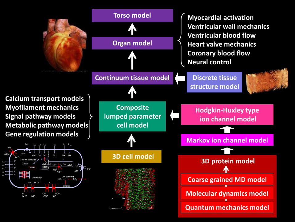
Musculo-skeletal team Thor Besier Vickie Shim Justin Fernandez Peter Hunter Poul Nielsen Martyn Nash Alice Hung Jessica Jor Duane Malcolm Kumar Mithraratne Mark Finch Tim Wu Yu Zhang
Musculo-skeletal system
Load generic models into the anatomical component under study:
Web-accessible database of generic models (+ tissue structure):

Generic models of the joints
Shim VB, Hunter PJ, Pivonka P, Fernandez JW. A multiscale framework based on the physiome markup languages for exploring the initiation of osteoarthritis at the bone-cartilage interface. IEEE Trans Biomed Eng. 58(12):3532-6, 2011


GI team Andrew Pullan (1962-2012) Leo Cheng Peng Du Greg O’Grady Shawn Means Tim Angeli Jerry Gao Rachel Lees-Green Niranchan Paskaranandavadivel Shameer Sathar Binny Paul Vinodh Vedachalam
1. Circulatory system 2. Respiratory system 3. Musculo-skeletal system 4. Digestive system 5. Brain & facial muscles

Digestive system: stomach
Faville et al. BiophysJ. 96, 4834-4852, 2009. Biophysically based mathematical modeling of interstitial cells of Cajal slow wave activity generated from a discrete unitary potential basis.

1. Circulatory system 2. Respiratory system 3. Musculo-skeletal system 4. Digestive system 5. Brain & facial muscles
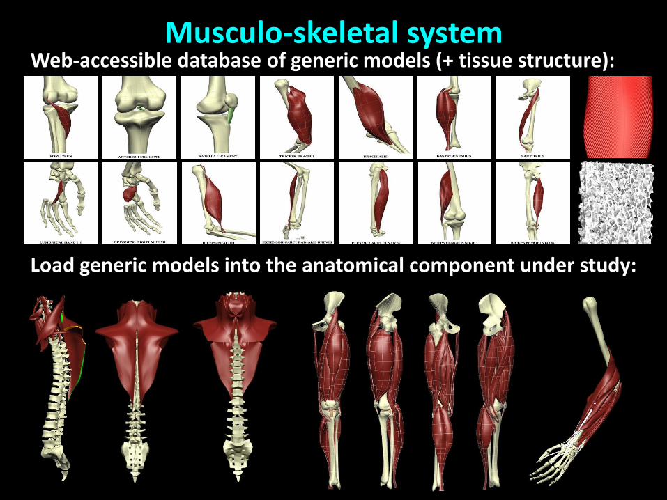
Lab for Animate Technologies Mark Sagar David Bullivant
Skin / Dermis
Hypodermis
Facial Space / Fat Compartment
Muscle Fibres
Ligament
Deep Fascia
SMAS
Modelling the facial muscles


Muscles need a control system
Facial Nerve Circuits
Models include • Neurobehavioral Models • Emotion and Motivation • Learning • Neuronal Dynamics
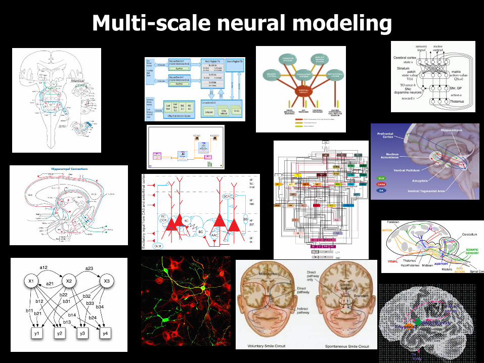
Multi-scale neural modeling
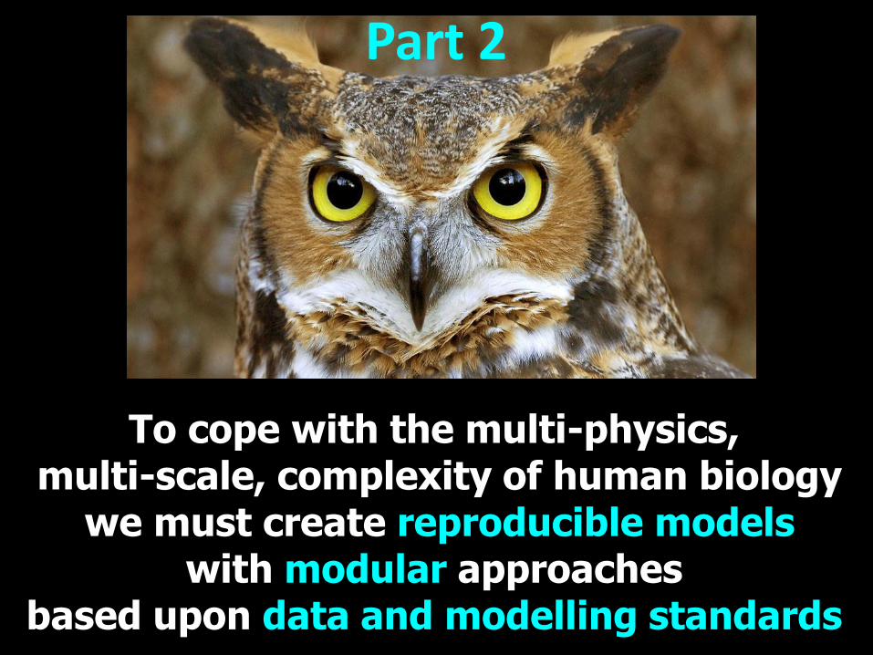
To cope with the multi-physics, multi-scale, complexity of human biology
we must create reproducible models with modular approaches
based upon data and modelling standards
Part 2

History of Physiome Project
1999 Systems Biology Markup Language
2006 STEP: Strategy for European Physiome 2008 VPH Network of Excellence
2011 VPH Institute
2009 Drug Disease Model Resources (DDMoRe)
1997 IUPS Physiome Committee
1998 CellML, FieldML
2003 IMAG (NIH, NSF, FDA, NASA, DOE, DOD, ..)
2010 German Virtual Liver Network

Verification, Benchmarks
Experimental measurements
Model standards: SBML, CellML, FieldML
Model repositories: Biomodels, PMR2
Data standards: DICOM, BioSignalML, ..
Minimum information standards: MIAME, MICEE, ..
Data repositories: PhysioNet, CAPdb, CVRG, ..
Standards for models, data & software
Validation, Limitations
(Journal review)
APIs, webservices
Software: OpenCOR, OpenCMISS, FEniCS, Chaste, ..
Simulation standards: SED-ML Functional curation
APIs, webservices
Curation Annotation
Reference description
Metadata: Ontologies
Need modules!
Metadata: Ontologies GO, FMA, ..

Biophysical Journal
“To assure public access to computational models, authors are strongly encouraged to deposit their models in the CellML Model Repository models.cellml.org/cellml or Biomodels Database www.ebi.ac.uk/biomodels-main/”
Note on model publishing
Similarly for many other journals.

Tissue
Osteon Nephron Acinus Liver lobule Lymph node Cardiac sheets
Organ
Heart Lungs Diaphragm Colon Eye Knee Liver
Environment
Organ system
Organism
Cell
Protein Gene Atom
Network
x 1million 20 generations
The challenge: organs to proteins

• Biophysically based models at every level – as much as possible (there’s always a black box!)
• Adoption of model and data standards – SBML, CellML, FieldML for models
• Automated assembly of multi-scale models – molecule to organ(ism)
• Automated model reduction – otherwise too expensive
• New instrumentation – new instruments → new expts → new knowledge
A multi-scale bioengineering approach needs:

Cardiovascular system Respiratory system Musculo-skeletal system Digestive system Skin (integument) Urinary system Lymphoid system Female reproductive system Special sense organs Central nervous system Endocrine system Male reproductive system
Organ system Physiome Projects

CellML – standards, databases and tools
(www.cellml.org)
Cuellar AA, Lloyd CM, Nielsen PF, Halstead MDB, Bullivant DP, Nickerson DP, Hunter PJ. An overview of CellML 1.1, a biological model description language.SIMULATION: Transactions of the Society for Modeling and Simulation, 79(12):740-747, 2003

Cell cycle (25 models)
Calcium dynamics (63 models) Cell migration (2 models)
Circadian rhythms (9 models) Endocrine system (29)
PKPD models (7 models)

Myofilament mechanics (15) Metabolism (35 models)
Electrophysiology (117 models) Excitation-contaction (15 models)
Gene regulation DNA repair (3) Synthetic biology (5 models)
Material constitutive
laws

CellML enables modular construction

Cardiac myocyte a-adrenergic
Muscarincic, ACh
PLCb
Ga Gbg PIP2
Gq
a1
DAG + IP3
Ca2+
CaN CaMK PKCa PKCd PKCb PKCe
b-adrenergic NE, Iso
PKA
ATP cAMP
Ga Ga Gbg
AC
Gi
b2
Gs
b1
MEF2C
GATA4
NFAT p
p p
TFs
t
V
Ca2+
EC-coupl.g & mechs
PLB Serca2
TnC
Ca2+
RyR2
TnI
MHC
p p
p
p
T
p
p
p
p p
ms seconds hours days Time scale
Nucleus
Membrane
Cytosol
DNA
Inputs
Outputs c-myc, c-fos, c-jun, ras, hsp-70
TFs
p
Eccentric hypertrophy Concentric hypertrophy
Physiological hypertrophy
CellML signalling modules for the cardiac myocyte
Ion channels, transporters
INa Na+
INa,b Na+
ICl Cl- ICa,L
Ca2+
ICa,T Ca2+
ICa,b Ca2+
IKr K+
IKs K+
IKto K+
IKp K+
IK1 K+
NCX
Ca2+
3Na+
3K+
2Na+ NKA
CHE OH-
NHE H+
NBC
HCO3-
Na+
AE
HCO3-
Cl- *
NO
sGC sGC
eNOS nNOS
Ca2+
NO
cGK I
p
cGK II
p
cGMP
pGC
ANP BNP
iNOS
NO
Peptide GFs
RAS
MAPK
ERK JNK p38K
TFs
RTK
p p p
p p p
p
Apoptosis
Cytokines
cytokine receptor
gp130
STAT
Jac IkB
NFkB
Inactive Class II HDACs
Ca2+
CaMK PKD
PKC
histone
Insulin, IGF, GH
TFs
mTOR
p
GSK3
p
PKB
PI3K
PIP3
RTK
p
Hypertrophic cardiac myopathy
FOXO

• Glucose transporter (GLUT2) • Glucokinase (GK) • Glucose-6 phosphatase (G6Pase) • Glucose-6-phosphate isomerase (GPI) • Glucose-1-phosphate 1,6-phosphomutase (G16PI) • UTP: Glucose-1-phosphate uridylyltransferase (UGT) • Pyrophosphate phosphohydrolase (PPase) • Glycogen synthase (GS) • Glycogen phosphorylase (GP) • Nucleosid diphosphate kinase (NDK) • Adenylate kinase (AK) • Phosphofructo kinase 2 (PFK2) • Fructo-2,6-bisphosphatase (FBP2) • Phosphofructo kinase (PFK1) • Fructose-1,6-bisphosphatase (FBP1) • Aldolase (ALD) • Triosephosphate isomerase (TPI) • D-Glyceraldehyde-3-phosphate: NAD+
oxidoreductase (GAPDH) • Phosphoglycerate kinase (PGK) • 3-Phosphoglycerate mutase (PGM) • Enolase (EN) • Pyruvate kinase (PK) • Phosphoenolpyruvate carboxykinase (PEPCK) • Pyruvate carboxylase (PC) • Lactate dehydrogenase (LDH) • Lactate transporter (LACT) • Pyruvate transporter (PYRT) • PEP transporter (PEPT) • Pyruvate dehydrogenase (PDH) • Citrate synthase (CS) • Nucleosid diphosphate kinase (NDK) • Oxalacetate flux (OAAflx) • Acetyl-CoA flux (ACOAflx) • Citrate flux (CITflx)

Linking models to medical informatics
Bernard de Bono

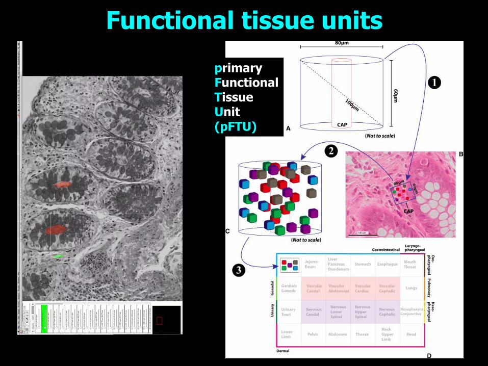
Functional tissue units
primary Functional Tissue Unit (pFTU)

Organ Tissue Cells Cell function

Acknowledgements

ABI colleagues Bruce Smaill
Martyn Nash
Poul Nielsen
Merryn Tawhai
Thor Besier
Chris Bradley
Leo Cheng
Mark Sagar
Alistair Young
Denis Loiselle
Bernard de Bono

Our instrumentation engineers

The CellML/FieldML team
Andrew Miller Randall Britten
Richard Christie
Alan Garny
David Nickerson
Tommy Yu
Mike Cooling
Hugh Sorby
Poul Nielsen
Alan Wu

ABI graduate students & postdocs

NZ Health Research Council NZ Ministry of Science & Innovation NZ Maurice Wilkins Centre CoRE UK Wellcome Trust (Heart Physiome) FP7 (euHEART, NoE, VPH-Share) NIH (Cardiac Atlas Project - CAP)
Funding Acknowledgements
www.vph-institute.org


![Toward Patient-Speci c Myocardial · Reza Razavi, MDb, Nicholas Ayache, PhDa ... The complete 3D reconstruction of ber ori-entations from histologic sections[28], and more ... from](https://img.dokumen.tips/doc/110x75/5c4d047c93f3c34c550ae146/toward-patient-speci-c-myocardial-reza-razavi-mdb-nicholas-ayache-phda-.jpg)