Embed Size (px)
Citation preview

[Frontiers in Bioscience 9, 3453-3478, September 1, 2004]
INSIGHTS INTO THE CATALYTIC MECHANISM OF PEPTIDYL PROLYL CIS/TRANSISOMERASES
Jörg Fanghänel and Gunter Fischer
Max-Planck-Forschungsstelle für Enzymologie der Proteinfaltung Weinbergweg 22, D-06120 Halle, Saale, Germany
TABLE OF CONTENTS
1. Abstract2. Introduction3. Mechanism of catalyzed prolyl cis/trans isomerization
3.1. Non enzymatic acceleration of prolyl cis/trans isomerization3.1.1. Environmental effects3.1.2. Intramolecular catalysis3.1.3. Intermolecular catalysis3.2. PPIase catalyzed prolyl cis/trans isomerization3.2.1. Cyclophilins3.3.2. FKBP3.3.3. Parvulins
4. Perspective5. Acknowledgment6. References
1. ABSTRACT
A large body of physiological, cell biological, kinetic and structural data about peptidyl prolylcis/trans isomerases (PPIases) has been accumulated during the past 20 years, but despite the simplicity of the catalyzed reaction the question of how the enzyme action is performed is still not fully answered. In this review the center of attention is the molecular background of the catalytic mechanism of PPIases and the spontaneously occurring peptidyl prolyl cis/trans isomerization. We summarize and compare the available kinetic, structural and amino acid sequence data of all three PPIase families, the cyclophilins, FKBP and parvulins. Different catalytic mechanisms that have been suggested in the literature are discussed. A comprehensive comparison of enzyme active site structures reveals a hitherto unnoticed similarity between the three PPIase families and might suggest that PPIases utilize mechanisms that are more similar than previously suspected.
2. INTRODUCTION
The pioneering work of L. Pauling over half a century ago prepared the ground for understanding the atomic structure of proteins and the molecular basis of enzyme catalysis which should be especially valuable in studying catalysis of chemically simple reactions (1).

Twenty years later, J. F. Brandts proposed that the slow phases observed in the unfolding kinetics of proteins might be due to the cis/trans isomerization of peptidyl prolyl bonds (the terms prolyl bond and prolyl isomerization are used throughout this review for the peptide bond preceding a proline residue and the peptidyl prolyl cis/trans isomerization, respectively) (2). An enzyme class discovered 9 years later with its first member isolated from pig kidney represented a powerful catalyst of prolyl isomerization operating on a level of one of the simplest chemical reactions: the rotation about a single bond (3). According to their substrate specificity these cis/trans isomerases were named peptidyl prolyl cis/trans isomerases (PPIases). These enzymes have opened up a broad new area into the investigations of assisted protein folding. Recently the secondary amid peptide bondcis/trans isomerases (APIases) have been found which are actively engaged in lowering the rotational barrier of a secondary amide peptide bond C(O)-NH- in oligopeptide- and protein substrates(4). Another 20 years have passed since the first PPIase was characterized and what have we learned about these enzymes in that time? A great deal of data has been published about the physiological significance and the enzyme mechanism of PPIases. The best characterized enzymes among the PPIases are human cyclophilin 18 (human Cyp18) and human FKBP12 (human FKBP12). Both are prototypic members of their corresponding PPIase families known as cyclophilins and FK506 binding proteins (FKBP). The family names derive from the ability of their respective members to bind to cyclosporine A or FK506, both highly active immunosuppressive compounds. A third PPIase family, the parvulins, was discovered in 1994 (5). The most prominent and best investigated member of this family is human Pin1 (human Pin1). The finding that cyclophilins and FKBP participate in immunosuppressive processes initiated a rush of scientific interest. Since then PPIases have been found to take part in a great number of physiological processes such as in vivo protein folding, heat shock response, transcription and translation, channel gating, virus assembly, signal transduction, tumor metastasis, pathogen virulence, cell cycle control and others (6-10). These topics have been reviewed exhaustively and shall not concern us here. In this review we want to focus on publications related to the catalytic mechanism and try to combine all available data to give a complete overview of this subject.
Despite the amount of data, the molecular basis of the PPIase mechanism is still only poorly understood. For biochemical investigation, the simplicity of the reaction is a blessing and a curse at the same time. Since the reaction is so undemanding, evolution did not "bother" to equip prototypic PPIases with cofactors; which can often be exploited to unveil the enzyme mechanisms. Furthermore the difference between the substrate and the product state of a polypeptide chain is very subtle and therefore sometimes difficult to investigate. On the other hand the acceleration of the peptide bond rotation is already achieved by globular low-molecular-mass proteins, making these enzymes a perfect subject of structural investigations. Since the reaction is, at least for oligopeptide substrates, completely reversible, PPIases can be studied under equilibrium conditions, an advantage if one wants to employ NMR spectroscopic tools which often require long measuring times under biological conditions.
There exists no sequence homology between the three PPIase families, but they all catalyze the same chemical reaction. A fundamental question arises from this fact:
Do PPIases facilitate their enzymatic action by utilizing a common catalytic pathway?
3. MECHANISM OF CATALYZED PROLYL CIS/TRANS ISOMERIZATION

3.1. Non enzymatic acceleration of prolyl cis/trans isomerization
To fully understand the enzyme mechanism of PPIases, a profound knowledge of the chemical properties of prolyl bond isomers is unavoidable. Therefore a short excursion into the nonenzymatic acceleration of prolyl bond isomerization is necessary here. Although a full description requires a more complex explanation, the classical concept of �resonance stabilization", where the lone electron pair of the peptide bond nitrogen is delocalized over the entire peptide bond, is now widely accepted. This model explains many of the peptide bond features such as the limited number of stable ground state conformations, the high barrier to rotation about the C-N bond, a shortened C-N bond length, the kinetic stability of peptide bonds towards a nucleophilic attack and the isomer-sensitive carbonyl stretching frequencies in the IR-spectrum, and it defines the chemical means to lower the rotational barrier catalytically (Figure 1). Reviews on this topic are available (11, 12). Here we will focus on the data directly linked to the acceleration of the prolyl bond isomerization, supplemented in some cases with results obtained from secondary amides.
3.1.1. Environmental effects
The amide group shows a greater charge separation when planar than in its proposed orthogonal transition state, therefore the isomerization rate should be faster in less polar solvents. Accordingly the isomerization rate of N,N-dimethylacetamide (DMA) increases by a factor of 60 as the solvent is changed from water to cyclohexane (13). A similar increase in isomerization rates were observed for peptides containing 2-C substituted proline-like oxazolidine and thiazolidine derivatives, when the solvent was shifted from DMSO to CDCl3 (14). Contrarily, it was found that the isomerization rate of Ac-GP-OMe did not correlate with the solvent dielectric constant or the solvent polarity but depended on the ability of the solvent to donate hydrogen bonds. Further analysis showed that the barrier to rotation is proportional to the strength of hydrogen bonds formed to the peptide bond oxygen. From the experimental results it was calculated that desolvation of the prolyl bond can reduce the free energy of activation by 1.3 kcal/mol, which is only little when compared with the decrease achieved by PPIases (15). A pH shift from 7.0 to 1.8 increases the rate of isomerization of DMA 130-fold (16). Although the preferred protonation site of a peptide bond is the carbonyl oxygen, the rotational barrier of amides can be lowered by a small but kinetically significant amount of an amide nitrogen protonated species. The direct participation of hydroxide ions to the acceleration of the cis/trans isomerization of DMA at pH 11.8 has been observed. The rate enhancement was attributed to the formation of a tetrahedral hemiorthoamide, in which the amide resonance is disrupted (16). Such extreme pH conditions are usually not found in biological systems under native conditions but reactive hydroxide species are frequently observed in enzymes utilizing Zn2+ ions like carbonic anhydrase (17). The findings that the isomerization rate of short proline containing peptides in buffered solutions is independent of the pH within the range of 5 to 9 apparently indicates the lack of general intermolecular acid/base catalysis (18). In this case the observed deuterium kinetic solvent isotope effect (KSIE) is nearly in unity and therefore it was postulated that no proton movement during transition state formation occurs (19).
Not mentioned in either of the categories above are effects on the isomerization rate of prolyl bonds due to modifications of the prolyl bond itself or of the two neighboring amino acid residues. In general, one finds that electron donating substituents near the carbonyl moiety accelerate the isomerization rate whereas a deceleration is observed when the electron density increases at the peptide nitrogen. Only a few modifications are of physiological

relevance, such as hydroxylation of proline in position 4 or O-glycosylation and phosphorylation of serine or threonine side chains preceding proline. Proline homologues with differing ring size and heteroatom substitution in the proline ring influence the imidic rotational barrier of oligopeptides containing these analogues. For Aze, Pip, 4-Oxa and 2-Thz (Figure 2) a substantial acceleration of the cis/trans isomerization was detected (20). A similar effect was observed for fluoroproline derivatives (Ac-(4R)-FPro-OMe, Ac-(4S)-FPro-OMe and Ac-4,4-F2Pro-OMe), where the inductive effect of the fluorine substitutions leads to a weakening of the Ac-Pro amide bond (21). Higher isomerization rates of the imide bond have also been reported for short peptides containing different 2-C substituted proline-like oxazolidine and thiazolidine derivatives (Figure 2) (14). On the other hand, the presence of 3-C alkylated proline derivatives in the chain leads to a marked decreasein the imidic isomerization rates in water. These effects were mainly attributed to sterical restrictions arising from these bulky side chains (22). O-glycosylation of serine residues preceding proline and hydroxyl substitution of proline were shown to have no effect on the isomerization rate, whereas serine or threonine phosphorylation decreased the isomerization rate up to 7 fold (22-25). The substitution of the prolyl bond carbonyl oxygen by sulfur in short peptides reduces the isomerization rate by a factor of about 100. This is in accordance with the resonance theory since the thioxo group increases the electron density along the C-N bond (26).
3.1.2. Intramolecular catalysis
Intramolecular catalysis of prolyl bond isomerization was first observed during the refolding reaction of denatured dihydrofolate reductase (27). Proline 66 undergoes a trans to cisisomerization catalyzed by the guanidinium group of arginine 44, which interacts with the imide nitrogen of the Gln65-Pro66 peptide bond. Analyzing protein structures available from the RCSB data bank revealed that in almost 6% of all presently available structures at least one arginine guanidinium group is within 4 Å of a proline imide nitrogen (Wille, G. et al., in preparation) suggesting a general concept for accelerating proline-limited protein folding. It was also found that a hydrogen bond between the imide nitrogen and the adjacent amidic NH within a five-membered ring, a so called 5-NH- -Na hydrogen bond, can accelerate cis/trans isomerization up to 260-fold, depending on the used solvent (28). A tenfold increase of the isomerization rate was reported for Cys-Pro bonds in disulfide bonded cyclic peptides (Ac-CPPC-NH2 and Ac-TCPPCR-NH2) as compared to the corresponding acyclic compounds. With the help of Monte Carlo molecular mechanics simulations it was concluded that the NH proton of the residue following Pro can establish a hydrogen bond to the sp3 lone electron pair of the imide nitrogen in the transition state (29). An about tenfold increased rate of prolyl isomerization was found in peptides containing a His-Pro moiety after a pH jump from basic to acidic conditions. It was proposed that the protonation of the imidazol ring promotes the isomerization by either providing an intramolecular hydrogen bond or by localizing a positive charge close to the prolyl bond. Since the KSIE under acidic conditions was determined to be 2.0, it was concluded that a direct proton transfer from the protonated histidine moiety to the imide nitrogen during transition state formation occurs (30). The sequence specific catalytic effect to the neighbouring prolyl bond rotation suggests that His-Pro may play a general role in preventing proline-limited slow folding phases in proteins..
3.1.3. Intermolecular catalysis
Monoclonal antibodies raised against α-keto dicarbonyl containing haptens (Figure 3) were described to accelerate the cis/trans isomerization of short proline containing peptides and

the refolding rate of RNase T1, which is limited by prolyl bond cis/trans isomerization. The authors suggested that the used α-keto dicarbonyl function might mimic a twisted amide bond of a PPIase bound substrate. It was further suggested that this hapten might result in antibodies capable of forming a tetrahedral adduct to the electrophilic carbonyl group of the α-keto functionality. However, such a specific catalytic mechanism was ruled out because no pH dependence or KSIE was observed and the small rate enhancement was comparable to that achieved by hydrophobic solvents alone (31, 32). Similar acceleration factors were observed for vesicle- and micelle-entrapped oligopeptides. Thermodynamic results as well as the effects of side chain variations of the investigated peptide substrates will be discussed in the FKBP section of this review (33).
3.2. PPIase catalyzed prolyl cis/trans isomerization
Among PPIases are single and multidomain enzymes, a review summarizing the domain structure of human PPIase is available (34), here we focus on the enzymatic activity and therefore only the function of PPIase domains will be discussed. Figure 1 shows five possible catalytic mechanisms to accelerate the cis/trans isomerization of prolyl bonds. Many of them have been proposed to be the driving force behind the catalytic power of PPIases but only few have been shown to be at least involved in the catalytic cycle of these enzymes.
3.2.1. Cyclophilins
After the development of an in vitro PPIase assay based on the isomer-specific proteolytic cleavage of peptide bonds it became feasible to investigate the catalytic mechanism of PPIases (3). Since then a number of different activity assays have been developed, including a protease free UV/Vis assay, dynamic NMR based methods, fluorescence based assays and protein folding/unfolding assays. A complete summary and descriptions of all available PPIase assays has been given in a recent review published by Fischer and Aumuller (34). In the last two decades a large number of cyclophilins from different organisms have been purified and kinetically characterized. Almost all cyclophilins investigated so far show a second order rate constant kcat/KM between 105 and 107M-1s-1 when measured using the oligopeptide Suc-AAPF-pNA as substrate. Under optimal conditions the prototypic human Cyp18 is a perfectly evolved catalyst that approach the limit of diffusion control for enzyme catalyzed reactions; for the most suitable oligopeptide substrates it has a high turnover number (kcat > 600 s-1) combined with a low ground state affinity (KM > 80 �M). Table 1 gives an overview of the catalytic constants (kcat/KM) for thecis to trans isomerization of cyclophilins, mostly estimated with the oligopeptide substrate Suc-AAPF-pNA.
To date the three dimensional structures of 11 cyclophilins from 9 different organisms have been determined (Table 2), most of these in complex with a tight-binding inhibitor such as CsA. All these cyclophilin domains consist of an eight-stranded antiparallel β-barrel capped at either end by two α-helices (Figure 4A). Despite the large quantity of structural data only three cyclophilins from different organisms, namely human Cyp18, E.coli CypA and C.elegance Cyp3 have been co-crystallized with a substrate (35-42). The first view of the active site of human Cyp18 in complex with a substrate (Ac-AAPA-amc) was provided by Kallen et al. 1992. Amino acid residues of human Cyp18 in close contact (< 3.8 Å) with the substrate are R55, I57, F60, Q63, A101, N102, Q111, F113, L122, H126, and R148 (38). These residues form a channel on top of two antiparallel β-strands (Figure 4B). The binding pocket for the proline pyrrolidine ring is formed by residues F60, M61, F113 and L122 (Figure

4D).
The same hydrophobic pocket is occupied by the MeVal-11 moiety of the tight binding inhibitor cyclosporin A (Figure 5A). The shape of the cavity is optimized to interact with a five-membered ring. The catalytic efficiency of human Cyp18 is greatly reduced if the prolyl moiety of peptides is replaced by four- or six-membered ring derivatives of proline, and completely abolished in cases of a secondary amide peptide bond (18, 43). This finding makes it clear that the correct spatial orientation of the proline residue in the transition state plays a crucial role in catalysis.
A pairwise amino acid sequence alignment of cyclophilin domains originating from cyclophilins that have been reported as active, shows that all residues comprising the binding pocket are conserved, with the exception of R148 (Table 3). The highest degree of variability is found at the three positions 57, 102 and 126. The exchange in position 57 of isoleucine with valine in six sequences is conservative, but nevertheless four out of these six cyclophilins posses a strongly reduced catalytic activity. The side chain of N102 is not involved in substrate interactions but the backbone atoms of this residue are in close contact with the substrate. The observed substitutions are tolerated accordingly; only OvCyp16, in which this residue is exchanged with a serine residue, possesses a low PPIase activity. The exchange of H126 with tyrosine found in four cyclophilin sequences appear not necessarily to impair the catalytic activity since both PPIases E.coli CypA and E.coli CypB are described as highly active. The same substitution seems to be responsible for the reduced activity of Cyp-10 from C. elegans. Mutational analysis of seven active site residues of human Cyp18 showed that only the variants R55A, F60A, and H126Q posses less than 1% residual activity (Table 4) (44). The phenylalanine residue in position 60 and arginine in position 55 are completely conserved among the cyclophilins. The side chain of F60 forms part of the prolyl binding pocket and is therefore essential for proper positioning of substrates. The importance of R55 for the catalytic power of human Cyp18 will be discussed later. On the basis of the first structure of a human Cyp18/substrate complex, it was suggested that H126 might attack the prolyl bond, but further refinement showed that H126 is not involved in any direct or indirect hydrogen bonds with the substrate (38, 45). It was pointed out that the hydrophobic interaction of this residue is essential for substrate binding (39). It was further concluded that the hydrophobic properties of H126 are more important than its ability to participate in hydrogen bonds, which was supported by the strongly reduced but still significant activity of the H126Q variant (Table 4).
The structural data in combination with kinetic experiments and site directed mutagenesis resulted in a variety of theories describing the molecular mechanism of cyclophilin catalysis. From the observed inactivation of human Cyp18 by sulfhydryl group modifying agents, an involvement of an active site thiol was proposed. The suggested mechanism assumed a nucleophilic attack of an activated sulfhydryl group to the prolyl bond carbonyl carbon resulting in hemithioorthoamides as a covalently bound tetrahedral intermediate (Figure 1, path C) (46). For this intermediate the resonance stabilization of the former peptide bond is destroyed, resulting in a greatly reduced rotational barrier about the C-N bond. This mechanism was supported by the observation that the kinetic secondary deuterium isotope effect of the cis to trans isomerization of Suc-AG(d2)PF-pNA changed from kH/kD >1 to 0.91 � 0.01 when cyclophilin was added to the reaction (47). A number of experiments disagree with this isotope effect derived mechanism. Site directed mutagenesis showed that all four cysteine residues (C52, C62, C115, and C161) of human Cyp18 are dispensable for activity (48). In accordance with these results is the observation that no cysteine is in the proximity of the substrate, the closest cysteine (C115) is, with 8 Å, too far away to build the proposed

hemithioorthoamide intermediate (38).
Harrison and Stein suggested a mechanism which they called "catalysis by distortion", where the enzyme induces strain or distortion in the substrate by utilizing the binding energy provided by the enzyme/substrate complex formation (Figure 1, path A) (18, 19). Cyclophilin was proposed to tightly bind a transition state in which the prolyl bond is characterized by a partially rotated C-N imide bond. In
agreement with this mechanism is the observed small enthalpy of activation ( H* =4.3 kcal/mol) and the relatively large negative entropy of activation ( S* = -47 eu). It should be pointed out that the Eyring plots used to calculate the activation parameters were nonlinear and that two interchangeable enzyme forms were proposed to account for it (49). This mechanism was also supported by a KSIE on kcat/KM and kcat close to unity for the cyclophilin catalyzed isomerization of Suc-AAPF-pNA (19, 50) and a normal temperature independent secondary deuterium isotope effect of 1.12 � 0.02 using Suc-AG(d2)PF-pNA as substrate. The authors suggested that the transfer of the substrate into the hydrophobic active site results in a reduced hyperconjugation of the β-CH electrons generating the normal -deuterium isotope effect
They also attribute a part of the catalytic power to the reduced stabilization of the polar resonance structure of the prolyl bond in the apolar active site. Structural analyses of human Cyp18 substrate complexes did not provide further proof for this theory. Neither in crystal structures nor in solution do short peptides such as Ala-Pro, Ac-AAPA-amc or Suc-AAPF-pNA adopt a twisted conformation (36, 37, 39, 40, 51). Further investigation using dynamic NMR spectroscopic techniques revealed that human Cyp18 binds to both ground state conformers with similar affinities (52). Studies using modified bicyclic lactam inhibitors and phosphinic Ala-Pro surrogates, which were thought to resemble the above mentioned twisted transition state, failed to display significant binding affinity towards the active site of human Cyp18 (Figure 6) (53, 54). Peptides harboring a thioxo prolyl bond or peptides with a reversed chirality at the C carbon of peptides in P1 and P1� position (nomenclature according to Schechter and Berger (55)) can not activate the catalytic pathway of human Cyp18 (26, 56). This suggests that the pathway from ground state to transition state is sterically and electronically demanding, thereby ruling out the above mentioned enzyme mechanisms like ground state distortion and desolvation by binding to a hydrophobic cavity.
New grounds for speculation were given by a water molecule observed in a human Cyp18/Ala-Pro complex. This water molecule is hydrogen bonded to the Q63 active site residue of human Cyp18. Since the water is placed in the right orientation to stabilize a twisted transition state via a hydrogen bond to the carbonyl oxygen of the prolyl bond, the authors concluded that the human Cyp18 catalysis is "solvent-assisted" (Figure 1, path B). The necessary energy to distort the peptide bond should arise from thermodynamic fluctuations (39). Later it was found that the Ala-Pro dipeptide is not a substrate but rather a competitive inhibitor of human Cyp18 (37). The authors discussed that the additional charge of the free proline carboxyl group disturbs the active site of human Cyp18. It forces the guanidinium group of R55 away from the proline nitrogen and into an additional hydrogen bond with the unprotected C-terminus of the peptide. It could therefore be concluded that an additional amino acid residue C-terminally located to the proline might be a prerequisite for catalysis. The authors also suggested that the guanidinium group of R55 might be the hydrogen donor proposed earlier by Kofron et al., which lowers the rotational barrier of the prolyl bond by forming a hydrogen bond to the proline imide nitrogen (Figure 1, path D) (50).

Consistent with this mechanism is the greatly reduced activity caused by the R55A mutation and the finding that intramolecular general acid catalysis by NH groups will lead to an increased isomerization rate of prolyl bonds (30, 44). The distance between the guanidinium NH2 group and the nitrogen of the prolyl bond ranges from 3.3 to 3.8 Å depending on the structure used and is therefore too far away to form a hydrogen bond. A shortening of this distance while the prolyl bond proceeds from ground to transition state was suggested (36, 41) and confirmed by a computational investigation (57, 58). Further support for this mechanism was provided by the observation that the R44 side chain of dihydrofolate reductase intramolecularly catalyzes the rate limiting step of folding by accelerating the cis to trans isomerization of the Q65-P66 peptide bond (Figure 7) (27). The spatial arrangement of the R44 guanidinium group and the imide nitrogen resembles the situation found in cyclophilin/substrate complexes. On the other hand, the observation that the peptide benzyl-Phe-Ala-Pro, which has the same free carboxy terminus as the dipeptide, acts as a substrate for human Cyp18 as shown by NMR spectroscopic line shape experiments casts doubt on this mechanism (59). Human Cyp18 activity (kcat/KM) has been shown to be pH dependent; a pKa value of 5.9 was calculated (26). The enzyme is almost two orders of magnitude more active at neutral or basic conditions than at pH 4.5. Since the observed decrease of activity originates mainly from a lowered kcat value it is feasible that ionizable active site side chains are involved in catalysis. The guanidinium group of R55 is expected to be protonated within the pH range investigated thereby making this residue unlikely to be the source of the pH dependency.
All experiments mentioned so far utilized short peptides to gain information on the catalytic mechanism, whereas cyclophilins will act on proteins in biological systems. As it is obvious from the contradicting enzyme mechanisms derived from crystal structures harboring dipeptides compared to structures in complex with oligopeptides, it is necessary to ask whether peptides utilize the complete enzymatic machinery of cyclophilins. A new tool to help answer this question was provided by the interesting finding that human Cyp18 is incorporated into the human immunodeficiency type 1 virus (HIV) by interacting with the HIV Gag protein (60). Several structures showed that human Cyp18 binds specifically to a G89-P90 motive, which is situated within a solvent exposed loop of the capsid protein (35, 41, 61). The G89-P90 bond in this complex was shown to be in the trans conformation, unlike all other human Cyp18 peptide substrate complexes, where the prolyl bond was found exclusively in the cis conformation. An exchange of G89 to alanine resulted in a reduced binding affinity (62). The binding affinity depended not only on the amino acid moiety preceding the proline residue (P1 position) but also on the chain length of the substrate used, suggesting an extended substrate binding site of cyclophilin (63). NMR exchange spectroscopy experiments of the native N-terminal domain of the capsid protein in the presence of catalytic amounts of human Cyp18 showed clearly that the isomerization of the G89-P90 bond is catalyzed by human Cyp18 in the native state of the capsid protein (64). Structures of variants of the HIV capsid protein co-crystallized with human Cyp18 resulted in complexes in which both cis and trans conformations of the X-P90 bond occurred in crystallographically equivalent positions (41). The authors proposed a mechanism where the transition state deviates only minimally from the cis and trans ground states. During reaction progress the C-terminal part of the substrates including the proline remains fixed relative to the enzyme, while the carbonyl oxygen of the amino acid preceding proline performs a flip-over. This model is consistent with a computational analysis (57). However, it is inconsistent with NMR relaxation rate changes and an NMR line shape analysis of human Cyp18 main chain amides during catalysis, where a substantial movement of substrate and enzyme residues, especially in the C-terminal region of the tetrapeptide-4-nitroanilide substrate, was

observed (52, 65).
3.3.2. FKBP
Even though FKBP are a conserved family of PPIases, their PPIase domains show a much higher degree of sequence variability than the cyclophilins. Some FKBP, like the E.coli trigger factor andE.coli SlyD can not be inhibited by FK506 or rapamycin. As described for higher molecular mass cyclophilins, the catalytic domain of FKBP was found to be complemented with other domains in several cases. That these additional domains can influence the PPIase activity by binding to their designated interacting proteins has been shown for human FKBP38 (Edlich F et.al, submitted). Some of the multidomain FKBP harbor up to four nonidentical FKBP PPIase domains within the same polypeptide chain. This domain copying is a feature not found within the cyclophilin family of PPIases. The functional significance of these multiple FKBP PPIase domains is still unclear, especially since it has been reported that like in the case of human FKBP51, human FKBP52, and rabbit FKBP59 the PPIase activity is mainly due to one single N-terminally located FKBP PPIase domain when tested with oligopeptide substrates (66-68). It remains to be discussed whether the inactivity of the other FKBP PPIase domains is due to the experimental conditions used, due to autoinhibitory segments within these proteins or if the inactivity is indeed an intrinsic property of these domains.
The majority of FKBP accelerate the cis to trans isomerization of the prolyl bonds with a second order rate constant between 104 and 107 M-1 s-1 (Table 5). Several FKBP exhibit kcat/Km values in the range of enzyme variants point mutated in the active site of human FKBP12 (Table 6). A few exceptions are found where only little or no activity could be detected (69-72). FKBP achieve the observed rate enhancement by lowering the rotational barrier by app. 6.6 kcal/mol (50, 73). In contrast to the uncatalyzed reaction, where the Eyring activation parameter is characterized by a large enthalpic contribution of H*= 18.9 kcal/mol and a small entropic part of S*= -1.16 eu , the FKBP catalyzed reaction is characterized by a large entropic ( S*= -43.95 eu) and small enthalpic ( H*=5.85 kcal/mol) contribution to G* (74). The marked entropic contribution to the free energy of activation led the authors to the conclusion that the rate limiting step is a physical rather than a chemical one. They proposed that the enzyme selectively stabilizes a twisted amide by using the binding energy to compensate for the loss of amide resonance. In contrast to cyclophilins, the catalytic efficiency of FKBP depends strongly on the side chain in position P1 of the substrate. Peptides containing large hydrophobic residues like leucine or phenylalanine in this position show an up to 100- to 1000-fold increased second order rate constant compared to substrates with charged or small side chains in this position (73, 74). The difference is not due to an impaired substrate binding since the KM value is not affected within the experimental error. It is rather an effect on kcat which decreases more than 1000-fold when the leucine residue in the P1 position of Suc-ALPF-pNA is changed to glycine (73). These data in combination with the observed pH independence of the FKBP from pH 5 to 10, the solvent isotope effect close to unity and the reported, mainly entropic contribution to the activation parameter led to an enzyme mechanism called "catalysis by distortion" which had also been proposed for the cyclophilin catalyzed reaction. The energy necessary to destabilize the substrate was thought to arise from the binding energy. Within the active site the substrate is then distorted by electrostatic and/or geometric means towards the transition state. In addition to the induced strain, the transition state is further stabilized by the hydrophobic environment of the active site (18). The normal secondary kinetic deuterium isotope effect observed for the specificity constant of the substrate Suc-AG(d2)PF-pNA with
FKBP12 has been used to argue in favor of a transition state characterized by a partial rotation about the C-N bond. However, since this -G-P- substrate might not be able to activate the complete machinery of the biocatalytic pathway, the mechanistic significance of the isotope effect remains questionable (73). A so called "unassisted conformational twist mechanism" was proposed on the basis of this isotope effect in combination with a slight inverse solvent isotope effect ((kcat/KM(H2O))/(kcat/KM(D2O)) = 0.92) (73). This mechanism is a modified version of the "assisted conformational twist mechanism" also suggested for cyclophilins, but it assumes that catalysis by FKBP does not require favorable hydrogen bonding to the lone electron pair of nitrogen from an active site residue.
The central question that needed to be addressed was what kinds of enzyme/substrate interactions were used by FKBP to lower the rotational barrier in a FKBP-like manner. Some answers could be found by investigating the FKBP structures reported. As many as 51 structures of FKBP from 12 different organisms have been determined (Table 7), none of them with a standard peptide substrate bound. They all show a very similar general fold which consist of a four to six stranded -sheet wrapped around a short -helix (Figure 8A). Inhibitor/enzyme complexes have been used to localize the active site of FKBP. The high resolution structure of the FK506/human FKBP12 complex revealed that the pipecolinyl moiety of FK506 (Figure 5B) is deeply buried in a hydrophobic cleft of human FKBP12 (Figure 8B). It was concluded that the pipecolinyl moiety might substitute for prolyl moieties of putative peptide or protein substrates (75). In accordance with this proposal is the finding that in the crystal structure of the GPI-1046/human FKBP12 complex, the five-membered pyrrolidyl moiety of GPI-1046 is located similar to the pipecolinyl function of FK506 (76) (Figure 8C & D). One of the most striking properties of the proposed active site of FKBP is its hydrophobic character. In case of human FKBP12 thirteen residues (Y26, F36, D37, R42, F46, F48, Q53, E54, I56, W59, Y82, H87, and F99) make direct contacts with FK506 (74, 75, 77). Four residues (E54, I56, Y82 and D37) stabilize the drug/enzyme interaction via hydrogen bonds. The residues in direct contact with the pipecolinyl ring have an exclusively aromatic character (Y26, F46, W59, and F99). In contrast to earlier proposals (78) the pipecolinyl-C8 carbonyl bond exhibits a trans conformation in its enzyme bound state and does not mimic a twisted amide bond (77). Later it was proposed that the orthogonal conformation of the -dicarbonyl moiety of FK506 might act as a twisted amide surrogate (74), but the very low activity of catalytic antibodies derived from - dicarbonyl containing haptens could not provide further evidence (31, 32). To determine how conserved the thirteen drug binding residues are among the FKBP tested for PPIase activity, a pairwise sequence alignment of these enzymes with human FKBP12 was performed and analyzed (Table 5). The degree of conservation is quite low for all positions. It appears that most of the side chains within the active site are dispensable for FKBP activity. Even P.horikoshii PhFKBP29, which has only one active site residue in common with human FKBP12, was reported to be enzymatically active. For some FKBP the low PPIase activity coincides with a low sequence identity in comparison to human FKBP12, namely for M.jannaschiiMjFKBP26, M.thermoautotrophicum FKBP28.3, M.genitalium trigger factor, P.horikoshiiPhFKBP29, A.thaliana AtFKBP42, and Thermococcus sp. TcFK. On the other hand - the E.colitrigger factor - that has only very few active site residues in common with human FKBP12 posses a high catalytic activity. It appears as though the enzymatic activity does not correlate directly with sequence conservation. In contrast, FK506 binding constants provide a better measure of sequence similarity. This might indicate that the residues involved in FK506 binding are not necessarily part of the active site. The active site residues conserved best among 51 FKBP domains aligned with human FKBP12 are Y26, F36, D37, I56, Y82, and F99 (Table 5). Two of these (Y26 and F99) form the hydrophobic cavity thought to accommodate the substrate proline residue. The importance of D37 for the

enzymatic activity will be discussed later. From the 15 mutants of human FKBP12 depicted in Table 6 only the D37L, the W59A, the F99Y and the Y26F/Y82F double variant posses less than 10% catalytic activity as compared to the wild type enzyme. The tyrosine mutations do not influence the formation of the Michaelis-Menten complex since the KM value remained unchanged while kcat is greatly reduced (73). Since a single point mutation Y82L diminished the slight pH dependence observed between pH 8 and 10.5 of human FKBP12, it was concluded that the tyrosine hydroxyl group of Y82 might be acting as a hydrogen bond donor to the prolyl nitrogen and thereby assist the prolyl cis/trans isomerization (18, 79). One has to bear in mind that the single Y82L mutation of human FKBP12 still leaves a residual activity of app. 10%, indicating that human FKBP12 stabilizes the transition state by hydrogen bonding only to a limited amount. Further mutations of potential nucleophiles and hydrogen bond donors (S8A, S38A, S67A, S77A, C22A, T75A and T96A) led only to insignificant reduction of the catalytic function (73). Quantitative modification of the cysteine thiols did not impair the catalytic activity (80). Since all nucleophilic residues close to the active site of human FKBP12 are dispensable for activity a side chain assisted covalent mechanism can be been ruled out (73).
Similar to FKBP, a preference for large hydrophobic side chains in the P1 position of oligopeptides was found for the prolyl bond isomerization catalyzed by micelles. Various surfactants have been shown to lower the free activation energy of isomerization by D G*= 1.8 kcal/mol. In contrast to PPIases, micelles also accelerate the isomerization rate of thioxoproline containing peptides (33). These results proved that only a small part of the catalytic power of FKBP arises from the desolvation of the peptide bond and that the rate enhancement of FKBP depends on a finely tuned spatial and electronic arrangement within the active site. The same conclusions were drawn from the observation that human FKBP12 cannot catalyze the cis/trans isomerization of oligopeptides which harbor D-amino acids in position P1, P1� or P2� while ground state binding was found to occur (56).
Molecular dynamics and free energy perturbation calculations where the peptides Ac-AAPF-Ame and Ac-LPF-Ame were fitted into the active site of human FKBP12 provided evidence that these peptides adopt a VIa -turn type conformation. This allows intrachain hydrogen bonding between the backbone nitrogen proton of the phenylalanine residue and the proline imide nitrogen. This interaction is believed to lower the rotational barrier via an "enzyme-induced autocatalysis" mechanism (81, 82). Against such a mechanism stands the observation that an acidification of the respective NH proton in thioxopeptide derivatives does not lead to a substantial lowering of the rotational barrier neither for the free thioxopeptide nor for the FKBP12/thioxopeptide complex. For example for the human FKBP12 catalyzed cis to trans isomerization of AGP-psi[CS-N]-F-pNA a kcat/KM value of 67 M-
1 s-1 was estimated which compares to the value of 920 M-1 s-1 of the oxo congener. In addition to the above mentioned transition state stabilization, examples for ground state destabilization were calculated as well. In agreement with the observed reduced activity of the D37L variant an unfavorable ground state interaction between the carbonyl moiety of the prolyl bond and the D37 side chain was calculated. This interaction changes to a more favorable charge-dipole interaction when the substrate proceeds from the planar ground state to the twisted transition state. The simulation also predicted a catalytically relevant water molecule, which is bound between the carbonyl oxygen of the prolyl bond and the hydroxyl oxygen of Y82 during transition state formation. The computational analysis could not pinpoint the catalytic efficiency to certain side chains or favor one of the proposed enzyme mechanisms (73). It was concluded that a combination of factors like desolvation of the imide bond by the hydrophobic active site, ground state destabilization, preferential transition state binding and enzyme induced autocatalysis contribute to the enzyme activity.

The theoretical methods mentioned here all suffer from the same limitations, in particular the lack of FKBP/substrate complex structures to provide valid starting points for the calculations. The methods used rely on the assumption that the conformation of complexed FK506 or rapamycin resembles the substrate conformation in a FKBP bound state. Some doubt is cast on the validity of this assumption by the observation that proline containing peptides quench the fluorescence of W59 located at the bottom of the drug binding site of human FKBP12 differently from FK506 (73).
3.3.3. Parvulins
The first member of the parvulin family of PPIases was identified in E.coli in 1994 (5). The substrate specificity of the 92 amino acid long E.coli Par10 is similar to the FKBP family of PPIases but the second order rate constant is at least one order of magnitude higher and the activity could not be inhibited by either FK506 or cyclosporin. The differences in the substrate specificity for human Pin1 and E.coli Par10 point out that within the parvulin family two subfamilies exist. Whereas the enzymatic activity of all phosphate specific parvulins reported so far is exceptionally high, the observed kcat/KM values among the parvulins that show no such preference varies within a range from 103 to 107 M-1 s-1 (Table 8). The sequence alignment shows that all phosphate specific parvulins have an identical set of active site residues. On the other hand, E.coli Par10 type parvulins show greater sequence variability and both residues which have been proposed to attack the prolyl carbon (C113 and S154 in human Pin1) are not conserved, questioning the described enzymatic mechanism.
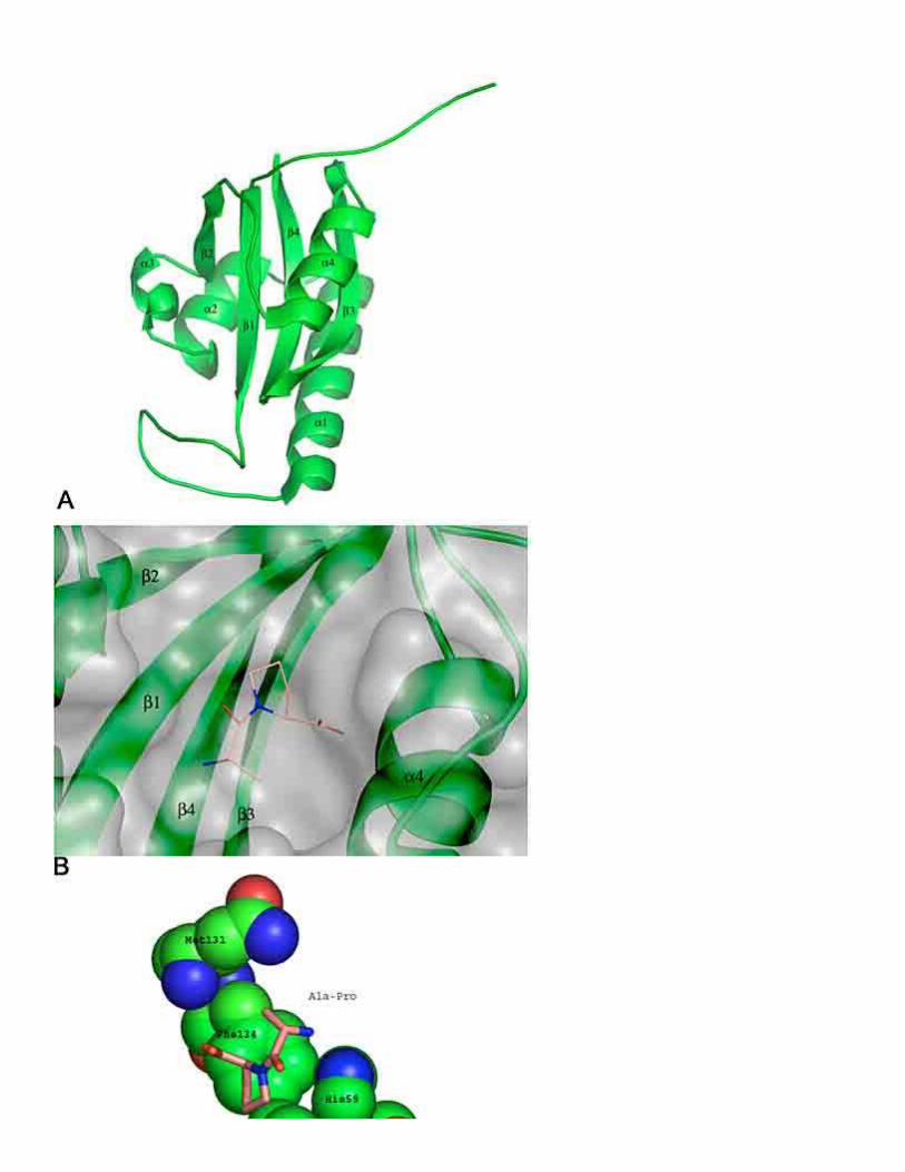
A first glimpse of the catalytic mechanism of parvulins was provided by the crystal structure of the human parvulin-like human Pin1 in complex with an Ala-Pro dipeptide (83). Since then, 10 structures of parvulins from human, A.thaliana and E.coli have been solved (Table 9). The human Pin1 consists of a C-terminally located PPIase domain and an N-terminally located WW-domain, which was found to interact with phosphoserine (pS) or phosphothreonine (pT) containing peptides and proteins. The presence of the WW-domain has no influence on the PPIase activity of the whole enzyme when the standard in vitro assay is applied (84). Whereas no sequence homology between FKBP and parvulin exists, a similar general fold can be observed (Figure 9). The PPIase domain of human Pin1 consists of a four stranded anti-parallel -sheet surrounded by four -helices (Figure 10A). The active site of human Pin1 is formed by 10 residues (H59, K63, R68, R69, C113, L122, M130, F134, S154 and H157). A basic cluster of side chains (K63, R68 and R69) in the 1/1 loop region binds a sulfate ion in the crystal structure. The sequestered sulfate is in close proximity to the bound dipeptide and has led to the assumption that human Pin1 possesses a strong preference for negatively charged side chains in position P1 of proline containing substrates. Accordingly it was found that the second order rate constant of Suc-AEPF-pNA is at least two orders of magnitude higher than that of an uncharged peptide like Suc-AQPF-pNA. Later it was observed that substrates with phosphorylated serine or threonine residues in this position were catalyzed most effectively and that the glutamate moiety preceding proline can not activate the full catalytic power of Pin1. Especially the remarkable pH dependence of Pin1 catalyzing thecis to trans isomerization of Ac-AAS(PO3H2)PR-pNA was not found to the same extend using the substrate Suc-AEPF-pNA, pointing out that glutamate can not mimic the phosphorylated serine residue completely (25, 85). The rate enhancement for substrates with phosphorylated side chains vs. their unphosphorylated counterparts is about 1300-fold. Most effective catalysis was observed at pH values where the phosphorylated side chain is in its dianionic form (25). Simultaneous substitution of R68 and R69 with alanine reduced the catalytic efficiency to the level observed with unphosphorylated peptides. The main contribution arises from the positive charge of the R69 side chain, since the R69L variant

lowers the enzymatic activity about 50-fold more efficiently than the R68L variant (84). The finding that the 1/1 sulfate/phosphate binding loop is extended into the solvent when no ligand is bound to the PPIase domain led to the hypothesis that this loop might act as a lid which opens and closes during the catalytic cycle (86). Structural analysis of human Pin1 in solution could not strengthen this induced fit mechanism and the authors concluded that the observed open conformation might be due to the crystallization conditions or crystal contacts (87).
The prolyl moiety of the dipeptide in the crystal structure of human Pin1 is located within a hydrophobic pocket similar to that which is occupied by the pipecolinyl moiety of FK506 in FKBP12/FK506 complexes (Figure 10A). It is formed by residues L122, M130 and F134 (Figure 10C). A minimum of three peptide bonds is essential to enable activation of the catalytic machinery of Pin1. Dipeptides as used in the crystal structure act as weak competitive inhibitors. These results are in agreement with those found for the other two PPIase families. It seems that the extended substrate/enzyme interaction is necessary to stabilize the transition state. Similar to human FKBP12 D-amino acid residues in position P1 are not tolerated in substrates for human Pin1 (88). Close to the dipeptide prolyl bond are the residues H59, C113, S154 and H157. A covalent mechanism was suggested, where the deprotonated side chain of His59 abstracts a proton from the C113 thiol, the resulting thiolate than attacks the carbonyl carbon of the prolyl bond. According to path C (Figure 1) the isomerization barrier of the formed intermediate is greatly reduced and therefore the rate of cis/trans isomerization is increased (83). Active site variants C113A and H59A showed a 123-fold and a 17-fold reduced activity respectively towards phosphorylated and unphosphorylated substrates (85). In accordance with this mechanism is the bell-shaped pH dependence of kcat/KM with apparent pKa values of 5.6 and 7.5, both pKa values are consistent with the titration of the active site residues H59 and His157. Further investigations confirmed that the side chain of His59 is responsible for the single dissociation step found below pH 6. No pH dependence was found with substrates containing an Ala-Pro bond and it was concluded that peptides that interact only unspecifically with the active site of human Pin1 are catalyzed via a desolvation mechanism (25).
Although the model of covalent catalysis via a cysteine residue is consistent with a great deal of work, some results argue against it. Juglone (Figure 11A) has been found to covalently modify the active site cysteine of E.coli Par10, the reported rate of modification was about 5 times faster than the observed enzyme inactivation. In accordance with the reported CD-spectroscopic data, it was suggested that the juglon-modified E.coli Par10 is destabilized and that the misplacement of catalytic residues leads to partial unfolding and thereby inactivation of the enzyme (89). Interestingly, it was also observed that juglone derived inhibitors (Figure 11B & C) could inhibit human Pin1 as well as human Par14 in a competitive and noncovalent manner (90).
The solution structure of a human Pin1 homolog from A.thaliana (Pin1At) also questioned the participation of an activated cysteine in the catalytic mechanism (91). The protein is highly homologous to human Pin1, but lacks the WW domain, like all plant human Pin1 homologues reported so far. All above mentioned active site residues are present in Pin1At and the general fold reveals a high level of similarity. The substrate binding pocket was mapped using NMR chemical shift perturbation experiments initiated by addition of a phospho-threonine peptide to Pin1At. Whereas it was found that the residues R21, R22, M87 and F91 (corresponding to R68, R69, M130 and F134 in human Pin1) were clearly involved in substrate binding, no or only slight changes in chemical shifts were observed for L79, C70, H12 and H114 (L122, C113, H59 and H157 in human Pin1) upon addition of saturating

concentrations of substrate. Structural comparison of the active sites showed that the C70 side chain of Pin1At pointed away from the hydrophobic prolyl binding pocket, making it unlikely to interact with the prolyl bond. The authors suggested that the serine residue S71 (corresponds to S154 in human Pin1), which showed large chemical shift perturbation upon ligand binding, might instead be involved in catalysis.
Human parvulin 14 displays only a weak PPIase activity (kcat/KM = 103 M-1 s-1) with a preference towards positively charged residues in P1 position (92). This preference is due to a shortened loop region where the positively charged side chains K63, R68 and R69 of the human Pin1 are replaced by a negatively charged area made of two negatively charged side chains D74 and E73 of human Par14 (93). The active site C113 of human Pin1 is exchanged to D74 in human Par14 and S154 is exchanged to F120. Only the two histidine residues in the active center of human Pin1 are conserved in human Par14. It is thought that the negative charges provided by the E73-D74 patch might be necessary to support catalytic assistance of other active site residues or enzyme bound water molecules.
4. PERSPECTIVE
The question asked in the introduction - "Do PPIases facilitate their enzymatic action by utilizing a common catalytic pathway?" is still unanswered, not even the nature of the catalytic pathway of a single PPIase family is yet fully understood. As it seems none of the mechanisms depicted in Figure 1 can explain all of the published data, but some can be ruled out. A direct involvement of a tetrahedral intermediate formed by a nucleophilic amino acid sidechain originating from the PPIase active site seems not to be supported by the data. Furthermore, it has been shown for different members of the three PPIase families that a number of atomic features in the substrate chain are necessary to activate the full catalytic power of these enzymes, therefore mechanisms which do not require specific enzyme/substrate interactions can also be ruled out. On grounds of the existing data and new results obtained by investigating the influence of cosolvents and heavy water on PPIase catalysis (Fanghänel et al., in preparation) we favor a concerted mechanism in which the electron pair is stabilized on the amide nitrogen bond by an H-bond donor of the protein. A simultaneous electrostatic transition state stabilization by an enzyme bound general base-polarized water molecule of the perpendicularly oriented carbonyl group is thought to occur (Figure 1, path D and E). One major handicap to elucidate a common catalytic pathway of PPIases is obviously the lack of structures of FKBP and parvulins in complex with substrates. In addition, the danger posed by cyclophilin/substrate complexes is the potential problem of analyzing dead complexes which do not map to the catalytic pathway. On a molecular level we have only limited knowledge of how these two enzyme families bind to their natural protein ligands. That the three enzyme families might catalyze the prolyl isomerization in a similar way became evident in structural comparisons.
The active site residues of human Cyp18 have been superimposed with some of the known drug binding residues of human FKBP12 (Figure 12). Four of the depicted active site residues of human Cyp18 have identical counterparts in human FKBP12, the other three residues are conservatively exchanged (L122 to I56, H126 to Y26 and Q63 to D37). Interestingly, all residues crucial for the activity of human FKBP12 (Y26, D37, W59 and F99) coincide with active site residues found to be essential (H126, Q63, F60) or at least important (W121) for human Cyp18 catalysis. The pipecolinyl residue of FK506 does not superimpose with the proline residue of the human Cyp18 bound substrate, underlining the doubt that it occupies the active site of human FKBP12. It should also be noted that in the crystal

structure of the human FKBP12 in complex with the cytoplasmic domain of the type I TGF-beta receptor the proposed proline binding pocket of human FKBP12 is occupied by a leucine residue of the receptor (94).
When the same human Cyp18 residues are superimposed with the active site of human Pin1 a similar picture is observed (Figure 13). Four residues of human Cyp18 have identical counterparts in the active site of human Pin1 (R55 to R68, F113 to F134, Q63 to Q131 and L122 to L122). Again, among them are three residues necessary for human Cyp18 activity (R55, Q63, F113), and F60 and H126 show a mirror symmetry to the human Pin1 residues H157 and F125. The superimposed active site residues of human Pin1 also include R68 and His59, which have been shown to be important for efficient human Pin1 catalysis. In contrast to the superimposed human Cyp18 and human FKBP12 structures, the proline residues of the bound ligands of human Cyp18 (Suc-AAPF-pNA) and human Pin1 (Ala-Pro) share a common space.
Despite the lack of a common general fold cyclophilins, FKBP and parvulins have a very similar active site structure. This observation leads us to the assumption that the catalytic pathway utilized by the different PPIase families is closely related.
INSIGHTS INTO THE CATALYTIC MECHANISM OF PEPTIDYL PROLYL CIS/TRANSISOMERASES
Jörg Fanghänel and Gunter Fischer
Max-Planck-Forschungsstelle für Enzymologie der Proteinfaltung Weinbergweg 22, D-06120 Halle, Saale, Germany
FIGURES

Figure 1. Canonical structures of the prolyl bond and possible catalytic mechanism for thecis/trans isomerization. Catalysis by the hydrophobic enzyme environment which stabilizes a more apolar twisted prolyl bond (A). Catalysis by a solvent assisted mechanism in which a enzyme bond water stabilizes the intermediate by a hydrogen bond to the carbonyl oxygen (B). Nucleophilic catalysis (C). Catalysis by an intra- or intermolecular protonation of the imide nitrogen (D). An active site structural water molecule assists bond rotation by electrostatic transition state stabilization (E).

Figure 2. Haptens (R = CO(CH2)3CO2H or R = CHO) used to raise catalytic antibodies.
Figure 3. Proline analogues used to investigate the uncatalyzed prolyl cis/trans isomerization. A) (S)-azetidine-2-carboxylic acid, Aze; B) (S)-oxazolidine-4-carboxylic acid, 4-Oxa; C) (R)-thiazolidine-2-carboxylic acid, 2-Thz; D) R)-thiazolidine-4-carboxylic acid, 4-Thz; E) 2-C substituted thiazolidine derivatives, F) 2-C substituted oxazolidine derivatives.

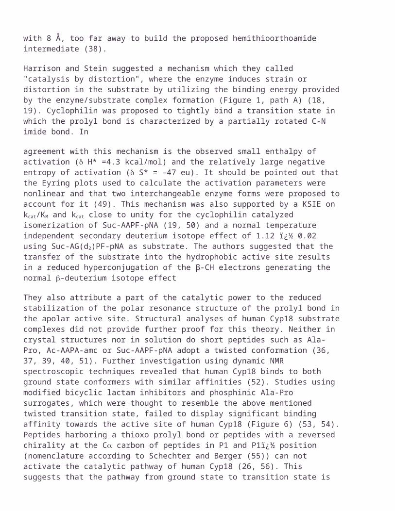
Figure 4. Ribbon representationof the global fold of cyclophilin domains, here shown for human Cyp18. The protein consists of a -barrel formed by eight antiparallel -sheets comprising residues 5 to 12 (1), 15 to 24 (2), 53 to 57 (3), 61 to 64 (4), 91 to 100 (5), 112 to 115 (6), 127 to 134 (7) and 156 to 163 (8) and two amphipathic -helices (1 residues 30 to 41 and 3 residues 136 to 145) and a short 3-helix (residues 120 to 122) (A). Two of the eight -strands (4, 6) form the cavity that accommodates the proline residue (B). This binding pocket is formed by four hydrophobic residues (Phe60, Met61, Phe113, and Leu122). The cavity accommodates the MeVal-11 moiety of the inhibitor CsA (C) as well as the prolyl moiety of a substrate (D).

Figure 5. CsA (A) and FK506 (B); residues that penetrate the prolyl binding pocket (MeVal-11 of CsA and the pipecolinyl moiety of FK506) are indicated.
Figure 6. Compounds used as surrogates of the prolyl bond in their ground state (A) or a twisted transition state (B, C and D).

Figure 7. Distances from the NH2 nitrogen of arginine to proline in DHFR and human Cyp18/substrate complexes. Panel A depicts the interactions in DHFR where R44 is involved in intramolecular catalysis of the Gln65-Pro66 bond cis/trans isomerization. Panel B shows the active site R55 residue of human Cyp18 which interacts with a ground state bound peptide substrate.

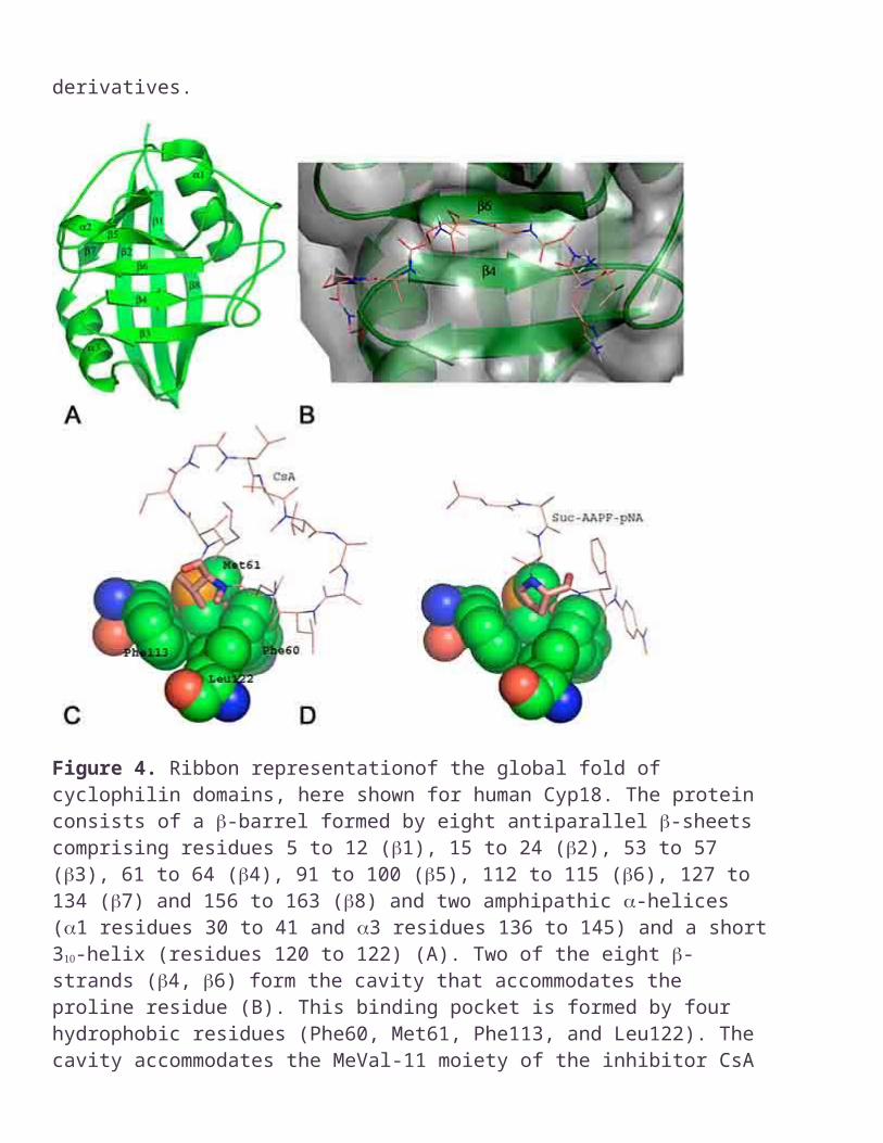
Figure 8. General fold of a FKBP domain, which is exemplarily shown for human FKBP12 (A). The six antiparallel -sheets exhibit +3, +1, -3, +1 side chain topology; the -sheets are capped with an amphiphilic -helix. The pipecolinyl ring protrudes into the drug binding site of human FKBP12 (B). The four hydrophobic residues F99, Y26, F46 and W59 are located in 2, 4, 6 strands and the1 helix respectively (C). The shape of the cavity can accommodate five- and six-membered rings as found in the structures of FK506 (C) and GPI-1046 (D). The four residues are shown in spacefilling mode and labeled; the inhibitors are depicted as stick models.

Figure 9. Arrangement of secondary structure elements of human FKBP12 (left), human Pin1 (middle) and human Par14 (right). Conserved secondary structures are in black; grey structures are found to be variable within the individual structures. Bars represent -strands; wires correspond to -helices and lines to loops and turns.


INSIGHTS INTO THE CATALYTIC MECHANISM OF PEPTIDYL PROLYL CIS/TRANS ISOMERASES
Jörg Fanghänel and Gunter Fischer
Max-Planck-Forschungsstelle für Enzymologie der Proteinfaltung Weinbergweg 22, D-06120 Halle, Saale, Germany
TABLES
Table 1. Cyclophilins described as active using Suc-AAPF-pNA as substrate in the standard in vitro PPIase assays
Organism Name as in publication or data bank/generic name
Swissprot (kcat/KM) M-1s-1
References
A. nidulans CypB / Cyp23 O94190 active 95
A. niger CypA / Cyp19 O94184 active 96
A. niger CypB / Cyp23 Q8X166 active 97
A. thaliana AtCyp22 P34790 5.7 106 98
A. thaliana AtCyp28 P34791 active 99
B. malayi Cyp-1 / Cyp98 Q27450 7.5 106 100
B. malayi Cyp-2 / Cyp19 Q17246 1.23 107 101
B. subtilis PPiB / Cyp15 P35137 1.1 106 102
B. taurus CypA / Cyp18 P04374 1.3 107 50
B. taurus CypB / Cyp23 P80311 3.0 106 103
C. albicans Cyp1 / Cyp18 P22011 active 104
C. elegans cyp-1 / Cyp21 P52009 7.0 104 105
C. elegans cyp-2 / Cyp18.5 P52010 6.1 105 105
C. elegans cyp-3 / Cyp18.6 P52011 3.6 105 105
C. elegans cyp-4 / Cyp59 P52012 1.8 104 105
C. elegans cyp-5 / Cyp22 P52013 7.4 104 105
C. elegans cyp-6 / Cyp21.9 P52014 8.4 106 105
C. elegans cyp-8 / Cyp54 P52016 1.95 104 105
C. elegans cyp-9 / Cyp36 Q09637 1.5 104 105
C. elegans Cyp-10 / Cyp18 P52017 1.9 104 105
C. elegans Cyp-11 / Cyp20 P52018 1.5 104 105
C. elegans CeCyp-16 / Cyp25 Q9XXI7 2 103 a) 106
D. discoideum CypE / Cyp17 Q9NI62 Active 107
D. melanogaster Moca-CypA / Cyp113 Q8ISE5 5.6 104 108
D. immitis Dicyp-3 / Cyp60 O61300 3.95 105 109
E. coli CypA / Cyp20 P20752 5.71 107 110
E. coli CypB / Cyp18 P23869 6.74 107 110
E. histolytica EhCyp / Cyp18 O15729 active 111
H. cutirubrum Cyp19 O50586 active 112
H. sapiens NKCR_HUMAN / Cyp166 P30414 7.5 105 113
H. sapiens Cyp 40 Q08752 1.9 106 114
H. sapiens SnuCyp-20 O43447 active 115
H. sapiens Cyp18 P05092 1.3 107 b) 116
H. sapiens CypF / Cyp22 P30405 2.3 107 117
H. sapiens CypB / Cyp23 P23284 1.1 107 117
L. esculentum CypA / Cyp18 P21568 active
L. major LmCyp19 O02614 1.6 106 119
L. pneumophila lpCyp18 Q48822 4.6 106 120
N. crassa CPH / Cyp24 P10255 2.8 106 121
N. crassa NcCyP41 Q9P3X9 6.5 105 122
O. volvulus OvCyp16 Q8IA80 5.2 102 c) 100
Orpinomyces sp. CypB / Cyp22 Q01490 9.3 106 123
P. falciparum PFCyP / Cyp25 Q8I6S4 active 124
P. falciparum PFCyP / Cyp22 Q8IIK8 2.3 106 125
R. norvegicus Matrin CYP / Cyp88 O55035 1.0 106 126
R. norvegicus CypF / Cyp22 P29117 0.9 106 127
S. cerevisiae Cpr1 / Cyp17 P14832 active 128
S. cerevisiae Cpr3 / Cyp20 P25719 5.8 106 129
S. cerevisiae Cpr6 / Cyp42 P53691 5 105 130

S. cerevisiae Cpr7 / Cyp45 P47103 7 104 131
S. mansoni SmCypB / Cyp23 Q26551 8.2 105 132
S. mansoni SmCypA / Cyp31 Q26548 3.65 105 132
S. mansoni Smp17.7 Q26565 active d) 133
S. pombe SpCyp3 / Cyp19 O74729 1.5 106 134
S. chrysomallus ScCYPA / Cyp18 Q06118 3.73 106 135
S. chrysomallus ScCYPB / Cyp19 P77949 7.5 106 136
T. cruzi TcCyP19 AI021872e) active 137
T. gondii Cyp18.5 Q26994 active 138
T. gondii Cyp20 Q26995 active 138
T.mentagrophytes Cyp13 B019518 active 139
T. tridentatus CypG / Cyp24 O44073 1.8 105 f) 140
T. inflatum no name/ Cyp25 Q99009 active 141
V. faba CypB / Cyp27 Q41651 active 142
X. laevis XlCyp / Cyp17 AJ496795g) 1.1 107 143
Z. mays zmCyp18 P21569 1.1 107 144
Z. mays CypB Q10724 h) 2.5 107 144
The reported enzymatic constants were obtained as described in the respective publication. The generic name consists of the abbreviation of the respective PPIase family followed by the rounded molecular weight of the full length protein. a) Suc-AVPF-pNA was used as substrate, b) active site titration for this enzyme has been performed (145), c) Suc-ALPF-pNA was used as substrate, d) no data about the substrate used, e) no swissprot entry, SRS EMBL nucleotide accession number, f) Suc-AAPF-mca was used as substrate, g) no swissprot entry, SRS EMBL protein accession number, h) protein fragment, no sequence data available.
Table 2. Cyclophilin structures available in RCSB Data Bank
Organism Name as used in
publication
Number of
structures
PDB identifier
H. sapiens human Cyp18
43 1ak4, 1awq, 1awr, 1aws, 1awt, 1awu, 1bck, 1cwa, 1cwb, 1cwc, 1cwf, 1cwh, 1cwi, 1cwj, 1cwk, 1cwm, 1cwl, 1cwo, 1cya, 1cyb, 1fgl, 1m63, 1m9c, 1m9d, 1m9e, 1m9f, 1m9x, 1m9y, 1mf8, 1mik, 1nmk, 1oca,

1rmh, 1vbs, 1vbt, 2cpl, 2cyh, 2rma, 2rmb, 3cyh, 3cys, 4cyh, 5cyh
H. sapiens hCypB 1 1cyn
H. sapiens SnuCyp-20 2 1mzw, 1qoi
B. malayi CYP-1 3 1a58, 1a33, 1c5f
M. musculus
CypC 1 2rmc
C. elegans Cyp-3 2 1dyw 1e3b
C. elegans Cyp-5 1 1hop
P. falciparum
Cyp18 2 1qng, 1qnh
B. taurus Cyp40 2 1ihg, 1iip
E. coli CypB 2 1clh, 1csa
E. coli CypA 2 1lop, 2nul
S. cerevisiae
Cpr-1 1 1ist
Table 3. Conserved active site residues of cyclophilins; activity has been measured using Suc-AAPF-pNA if not stated otherwise
Residue (human Cyp18 nomenclature)
Number of
exchanges
Organism/Name as used in publication
Found exchange
(kcat/KM) M-1s-1
Swissprot entry
I57 6 C.elegans / Cyp-9
C.elegans / Cyp-16
R.norvegicus / Matrin CYP
H.sapiens / NKCR_HUMAN
S.cerevisiae / Cpr7
D.melanogaster / Moca-CypA
V
V
V
V
V
V
1.5 104
2 103 a
1.0 106
7.5 105
7 104
5.6 104
Q09637
Q9XXI7
O55035
P30414
P47103
Q8ISE5
N102 5 E.coli / CypA T 5.71 107 P20752
E.coli / CypB
B.subtilis / PPiB
L.pneumophila / Cyp18
O.volvulus / Cyp16
T
H
R
S
6.74 107
1.1 106
4.6 106
5.2 102 a
P23869
P35137
Q48822
Q8IA80
L122 1 C.elegans / Cyp-9 C 1.5 104 Q09637
H126 4 E.coli / CypA
E.coli / CypB
C.elegans / Cyp-10
P.falciparum / CyP
Y
Y
Y
N
5.71 107
6.74 107
1.9 104
no number
P20752
P23869
P52017
Q8I6S4
a) Enzyme was inactive with Suc-AAPF-pNA, the value was obtained by using Suc-ALPF-pNA. Residues R55, F60, Q63, A101, Q111, and F113 were conserved throughout all sequences
Table 4. Relative activities of recombinantly expressed variants of human Cyp18
Variant % rel. activity
WT 100
H54Q 15.0
R55A 0.1
F60A 0.32
Q111A 15.0
F113A 3.0
W121A 8.7
H126Q 0.53
Table 5. FKBP examined for their in vitro PPIase activity
Organism Name as in publication/generic name
Swissprot
(kcat/KM)
M-1s-1
Active site residues as found in a pair wise alignment with human FKBP12, +) identical residue, -)
residue not present
Xa)
FK506
binding
Ref.

H. sapiens FKBP12 b) P20071
1.6 106
Y 26
F
36
D
37
R
42
F
46
F
48
Q
53
E
54
I
56
W
59
Y
82
H
87
F
99
L Ki=0.5 nM
146
H. sapiens FKBP12.6 Q16645
6.2 105
+ + + + + + + + + F + + + L Kd=0.55 nM
147
148
H. sapiens FKBP13 P26885
3.1 106
+ + + Q + + G Q + + + A + L Ki=74 nM
149
H. sapiens FKBP25 Q00688
active + + + K L + G K + + + Q + A IC50=400 nM
150
A. thaliana
AtFKBP42 Q9LDC0
inactive
+ + E E I L E K L L + N Y L no binding
69
B. malayi FKBP13 O96335
6.3 104
+ + + + + + G Q + + + S + L IC50 = 1.7 µM
151
B. taurus FKBP12 P18203
6.6 105
+ + + + + + + + + + + + + L Ki = 0.25 nM
50
B. taurus FKBP25 P26884
8 105 + + + K L + G K + + + Q + A Ki = 160 nM
152
C. burnetii
CbMip / FKBP26
P51752
active + + + + A + K S + + + A + F no number
153
C. tracho-matis
chl-mip / FKBP27
P26623
active + + + N I L - - + F + Q + F no number
154
E. coli trigger factor/FKBP48
P22257
6.5 105
V + T K + L G R + F F G S L no inhibition
155
E. coli FKBP22 P39311
1.3 106
+ + + + A + - - + + + A + L Ki = 25 nM
156
E. coli SlyD/FKBP21 P30856
1.4 104
G + + A Y Q + R P V A + + L no inhibition
157
H. cutiru-brum
HcFKBP33 Q9P9H4
9.7 105
+ L + E R I E H F V + + + A no inhibition
158
M. genita-lium
trigger factor / FKBP51
P47480
7.2 103
F L A Q Y L N S + F + L + A no inhibit
159
ion
M. janna-schii
MjFKBP18 Q57726
9.2 105
I Y Y Y I + G + + F + I + L IC50 = 170 nM
71
M. janna-schii
MjFKBP26 Q58235
6.4 102
L I N F V + E + V I T L I L 70% at 20 µM
71
M. thermo-autotrophicum
FKBP28.3 O27197
3.6 102
E E E A I V G H + L F + M L IC50 >20 µM
72
M. thermo-litho-trophicus
MtFK / FKBP17
O52980
3.5 105
D A G + L + G Q + F + I + L IC50= 250 nM
160
N. crassa NcFKBP22 O60046
6.9 105
+ + + + + + G Q + + + - + F Ki = 4.5 nM
161
N. crassa NcFKBP / FKBP13
P20080
active + + + + L + G Q + + + V + A no numbers
162
N. menin-gitidis
NmFKBP / FKBP12
P25138
active + + + + L I G Q + + + A + A no numbers
163
P. hori-koshii
PhFKBP29 no entry
1.6 103
D I Y I V I G H + L E Q Y L 75% at 20 µM
164
S. cere-visiae
yFKBP12 P20081
8.2 107
+ + + + + C G Q + + + F + L Kd = 0.9 nM
165
S. cere-visiae
yFKBP13 P32472
5.4 107
+ + + + I + G R + + + V + L Kd = 18 nM
165
S. cere-visiae
yFKBP70 P38911
active + + + - + + G + + + + L + L no numbers
166
S. frugi-perda
FKBP46 / Q26486
7.8 106
+ M F K + + K + + + + S + L IC50 = 5 µM
167
Thermo-coccus sp. KS-1
TcFK / FKBP18
O93778
3.5 102
+ L V Y M V G + + L + K + L IC50 = 7 µM
70
V. faba FKBP15 Q41649
active + + + + I + G Q + + + S + L Ki = 30 nM
168
H. sapiens hFKBP51
domain I
Q13451
1.24 106
+ + + + + + G Q + + + S + L no numbers
169
H. sapiens FKBP51 Q1345 L + + + + V H D P I F K Y

domain II
1
H. sapiens FKBP52
domain I
Q02790
3.8 105
+ + + + + + G + + + + S + L Ki = 10 nM
170
H. sapiens FKBP52
domain II
Q02790
inactive
L + + + + I N D P L F K Y no numbers
171
M. musculus
FKBP51
domain I
Q64378
4.8 105
+ + + + + + G Q + + + + + L Ki= 10-15 nM
148
M. musculus
FKBP51
domain II
Q64378
L + + + + V H D P I F K Y
O. cuni-culus
rFKBP59
domain I
P27124
1.2 106
+ + + + + + G + + + + S + L no numbers
66
O. cuni-culus
rFKBP59
domain II
P27124
0.02 106
L + + + + V L D P L F K Y L no numbers
66
A. thaliana
FKBP72
domain I
Q9M326
1.5 104
C V E + I D S K + L + A + no number
172
A. thaliana
FKBP72
domain II
Q9M326
I I F - Y + S + P L L L +
A. thaliana
FKBP72
domain III
Q9M326
+ Y + N L + G L P F + R W
M. musculus
FKBP60
domain I
Q9Z247
active + + + + + V G Q + M + V + L no numbers
173
M. musculus
FKBP60
domain II
Q9Z247
+ + + + Y T G W + M + D +
M. musculus
FKBP60
domain III
Q9Z247
+ + + + + T G Y + M + R +
M. musculus
FKBP60
domain IV
Q9Z247
+ L + L Y I G Q V M + V +
M. musculus
FKBP65
domain I
Q61576
6.5 105
+ + + + V I G R + M + V + A IC50 = 45 nM
174

M. musculus
FKBP65
domain II
Q61576
+ + + + Y T G W + M + Y +
M. musculus
FKBP65
domain III
Q61576
+ + + + Y T G Y + M + T +
M. musculus
FKBP65
domain IV
Q61576
+ L F Y Q I N K + L H A +
Activity was measured with the substrate Suc-AXPF-pNA. The reported enzymatic constants were obtained as described in the respective publication. All FKBP domains of multi domain FKBP have been aligned individually; the activity shown was determined for the full length protein, if not stated otherwise. The alignment of MjFKBP18, MjFKBP26 and FKBP 28.3 showed only very weak homologies, the given result should therefore be interpreted with caution. The statement "active" means that in the publication PPIase activity assays were performed but no (kcat/KM) values were provided. The generic name consists of the abbreviation of the respective PPIase family followed by the rounded molecular weight of the full length protein, a) X Stands for the amino acid proceeding proline in the used substrate (Suc-AXPF-pNA), b) active site titration for this protein has been performed (175).
Table 6. Catalytic activity of recombinantly expressed human FKBP12 active site variants determined using Suc-ALPF-pNA as substrate
Variant kcat/KM M-1s-1 KM kcat References
Wt 1.2 106 b)
3.5 106 a)
1.2 ± 0.4 106 c)
2.2 ± 0.2 106
3.6 106 c
5 10-4 M b)
0.9-1.3 10-3 M c)
600 s-1 b)
1000-1300 s- 1 c)
176
176
155
177
79
Y26F ~ 1 105 a) 176
F36L ~ 4 106 a) 176
D37V 3 105 a) 176
D37L 6.6 ± 0.4 104 c) 155
R42A 1.1 ± 0.2 106 177
F46L ~ 5 106 a) 176
F48L ~ 0.9 106 a) 176

Q53A 1.8 ± 0.4 106 177
W59A ~ 1 105 a) 176
Y82L 3.6 105 c) 0.7-1.0 10-3 M c) 20-24 s-1 c) 79
Y82F 2.6 105 b) 2.9 10-4 M b) 75 s-1 b) 176
Y26F/Y82F 6.4 104 b) 7.5 10-4 M b) 48 s-1 b) 176
H87A 1.9 ± 0.2 106 177
F99Y 6.4 104 c) 155
a) numbers estimated from diagram, assay performed at 15 °C, b) assay performed at 5 °C, c) assay performed at 10 °C
Table 7. FKBP structures available in the RCSB Data Bank
Organism Name as used in publication
Number of structures
PDB identifier
B. taurus FKBP12 2 1fkk, 1fkl
C. elegans FKB-6 1 1r9h
E. coli FKPA 3 1q6h, 1q6i, 1q6u
H. sapiens FKBP12 33 1a7x, 1aui, 1b6c, 1bkf, 1bl4, 1d6o, 1d7h, 1d7i, 1d7j, 1eym, 1f40, 1fab, 1fkb, 1fkd, 1fkf, 1fkg, 1fkh, 1fki, 1fkj, 1fkr, 1fks, 1fkt, 1j4h, 1j4i, 1j4r, 1nsg, 1qpf, 1qpl, 1tco, 1fap, 2fke, 3fap, 4fap
H. sapiens FKBP12.6 1 1c9h
H. sapiens FKBP25 1 1pbk
H. sapiens FKBP52 1 1n1a
H. sapiens FKBP51 1 1kto
L. pneumophila LpMip 1 1fd9
M. genitalium Trigger factor 1 1hxv
M. thermolithotrophicus
MtFK 1 1ix5
O. cuniculus FKBP59 2 1rot, 1rou
S. boliviensis FKBP51 1 1kt1

S. cerevisiae FKBP12 1 1yat
T. cruzi TcMip 1 1jvw
Table 8. Parvulins examined for their in vitro PPIase activity
Organism
Name as in
publication or data
bank/ generic name
swissprot
(kcat/KM)
M-1s-1
Active site residues as found in a pair wise alignment with human Pin1, +) identical residue, -) residue not present
substrate Ref.
H. sapiens
Pin1 / Par18
Q13526 1.9 107 H 59
K 63
R 68
R 69
C 113
M 130
F 134
S 154
H 157
AApSPR-pNA a)
85
H. sapiens
Par14 Q9Y237 3.9 103 + + - - D + + F + Suc-ARPF-pNA
92
B. subtilis PrsA / Par33
P24327 6 103 + A - - D Q + Y + Suc-AKPF-pNA
178
E. coli Par10 P39159 1.35 107 + + - - + + + F + Suc-ALPF-pNA
92
E. coli SurA / Par47
Domain I
P21202 1.9 104 + P P T D L + V + Suc-ALPF-pNA
92
E. coli SurA / Par47
Domain II
P21202 + + - - D F + F +
E. coli PpiD / Par68
P77241 3.4 106 - Q - - I I L V L Suc-AEPF-pNA
179
A. thaliana
AtPar13 Q9SL42 2.7 106 + + + + + + + + + Ac-AApSPF-pNA a)
180
D. lanata DlPar13 Q9LEK8 1.5 107 + + + + + + + + + Ac-ApSPY-pNA a)
181
M. domes-tica
MdPar13 Q94G00 3.1 106 + + + + + + + + + Ac-AApSPF-pNA a)
180
N. crassa Ssp1 / O60045 6.5 106 + + + + + + + + + Ac- 182

Par21 AApSPF-pNA a)
S. cervicea
Ptf1 / Par22
P22696 1.7 107 + + + + + + + + + Ac-ApSPY-pNA a)
181
X. laevis Pin1 / Par18
Q9I9K6 active + + + + + + + + + no information
183
The activity was measured with the indicated substrate. The reported enzymatic constants were obtained as described in the respective publications. The two parvulin domains of E.coli SurA have been aligned individually; the activity shown was determined for the full length protein. The statement "active" means that in the publication PPIase activity assays were performed but no (kcat/KM) values were provided. The generic name consists of the abbreviation of the respective PPIase family followed by the rounded molecular weight of the full length protein, a) the substrate contains a phosphorylated serine (pS) preceding proline
Table 9. Parvulin structures available in the RCSB Data Bank
Organism Name as used in publication
Number of structures PDB identifier
H. sapiens Pin1 4 1nmv, 1nmw, 1pin, 1f8a
H. sapiens Par14 2 1eq3, 1fjd
A. thaliana Pin1At 1 1j6y
E. coli Par10 2 1jns, 1jnt
E. coli SurA 1 1m5y