Embed Size (px)
Citation preview

Copyright © 2004 Pearson Education, Inc., publishing as Benjamin Cummings
Fundamentals of Anatomy & Physiology
SIXTH EDITION
Frederic H. M
artini
PowerPoint® Lecture Slide Presentation prepared by Dr. Kathleen A. Ireland, Biology Instructor, Seabury Hall, Maui, Hawaii
Chapter 12, Neural tissue

Copyright © 2004 Pearson Education, Inc., publishing as Benjamin Cummings
Learning Objectives
• Describe the two major divisions of the nervous system and their characteristics.
• Identify the structures/functions of a typical neuron.
• Describe the location and function of neuroglia.
• Explain how resting potential is created and maintained.
• Describe the events in the generation and propagation of an action potential.

Copyright © 2004 Pearson Education, Inc., publishing as Benjamin Cummings
Learning Objectives
• Define the structure/function of a synapse.• List the major types of neurotransmitters
and neuromodulators.• Explain the processing of information in
neural tissue.

Copyright © 2004 Pearson Education, Inc., publishing as Benjamin Cummings
SECTION 12-1 An Overview of the Nervous System

Copyright © 2004 Pearson Education, Inc., publishing as Benjamin Cummings
nervous system overview
• Nervous system• Provides swift, brief responses to stimuli
• Endocrine system• Adjusts metabolic operations and directs
long-term changes• Nervous system includes
• All the neural tissue of the body• Basic unit = neuron

Copyright © 2004 Pearson Education, Inc., publishing as Benjamin Cummings
Divisions of the Nervous system
• CNS (Central Nervous system)• Brain and spinal cord
• PNS (Peripheral Nervous system)• Neural tissue outside CNS• Afferent division brings sensory
information from receptors• Efferent division carries motor
commands to effectors• Efferent division includes somatic nervous system and autonomic nervous system

Copyright © 2004 Pearson Education, Inc., publishing as Benjamin Cummings Figure 12.1
Figure 12.1 Functional Overview of the Nervous System

Copyright © 2004 Pearson Education, Inc., publishing as Benjamin Cummings
SECTION 12-2 Neurons

Copyright © 2004 Pearson Education, Inc., publishing as Benjamin Cummings
• Perikaryon• Neurofilaments, neurotubules,
neurofibrils• Axon hillock• Soma• Axon• Collaterals with telodendria
Neuron structure
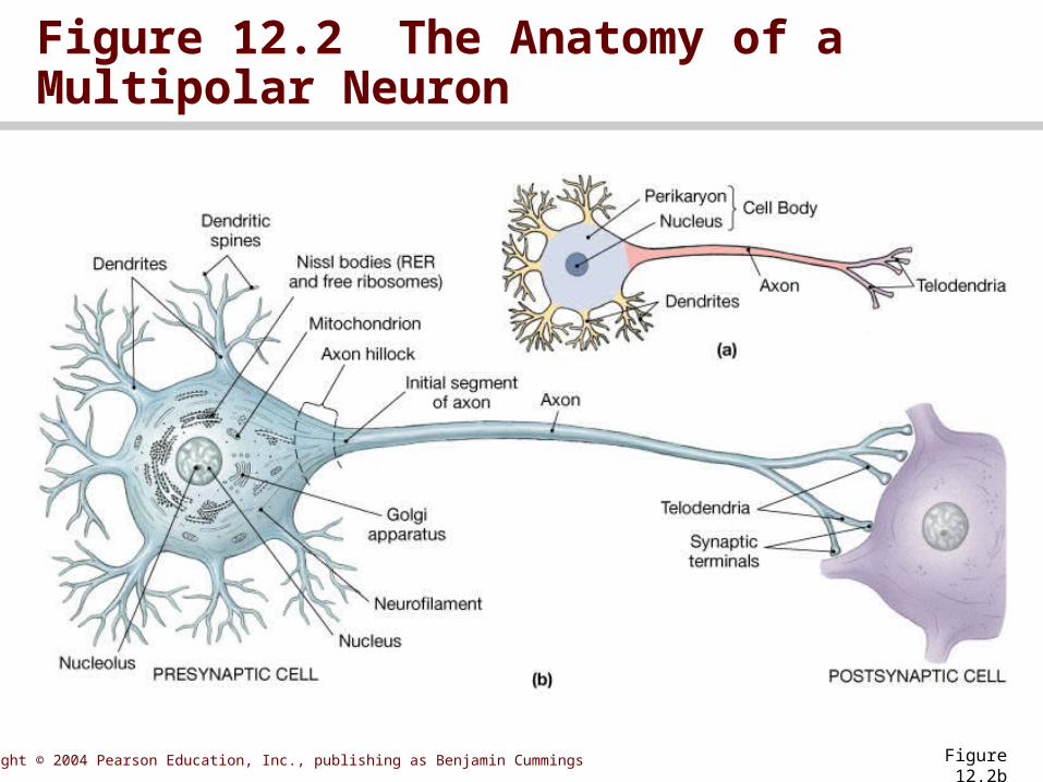
Copyright © 2004 Pearson Education, Inc., publishing as Benjamin Cummings Figure 12.2b
Figure 12.2 The Anatomy of a Multipolar Neuron

Copyright © 2004 Pearson Education, Inc., publishing as Benjamin Cummings
• Site of intercellular communication• Neurotransmitters released from synaptic
knob of presynaptic neuron
Synapse

Copyright © 2004 Pearson Education, Inc., publishing as Benjamin Cummings
Figure 12.3 The Structure of a Typical Synpase
Figure 12.3

Copyright © 2004 Pearson Education, Inc., publishing as Benjamin Cummings
• Anatomical• Anaxonic • Unipolar• Bipolar• Multipolar
Neuron classification

Copyright © 2004 Pearson Education, Inc., publishing as Benjamin Cummings Figure 12.4
Figure 12.4 A Structural Classification of Neurons

Copyright © 2004 Pearson Education, Inc., publishing as Benjamin Cummings
• Sensory neurons • deliver information from exteroceptors, interoceptors, or proprioceptors
• Motor neurons• Form the efferent division of the PNS
• Interneurons (association neurons)• Located entirely within the CNS• Distribute sensory input and coordinate motor output
Functional

Copyright © 2004 Pearson Education, Inc., publishing as Benjamin Cummings Figure 12.5
Figure 12.5 A Functional Classification of Neurons

Copyright © 2004 Pearson Education, Inc., publishing as Benjamin Cummings
SECTION 12-3 Neuroglia

Copyright © 2004 Pearson Education, Inc., publishing as Benjamin Cummings
• Four types of neuroglia in the CNS• Ependymal cells
• Related to cerebrospinal fluid• Astrocytes
• Largest and most numerous• Oligodendrocytes
• Myelination of CNS axons• Microglia
• Phagocytic cells
Neuroglia of the Central Nervous System

Copyright © 2004 Pearson Education, Inc., publishing as Benjamin Cummings
Figure 12.6 An Introduction to Neuroglia
Figure 12.6

Copyright © 2004 Pearson Education, Inc., publishing as Benjamin Cummings
Figure 12.7 Neuroglia in the CNS
Figure 12.7a

Copyright © 2004 Pearson Education, Inc., publishing as Benjamin Cummings
Figure 12.7 Neuroglia in the CNS
Figure 12.7b

Copyright © 2004 Pearson Education, Inc., publishing as Benjamin Cummings
• Two types of neuroglia in the PNS• Satellite cells
• Surround neuron cell bodies within ganglia
• Schwann cells• Ensheath axons in the PNS
Neuroglia of the Peripheral Nervous System
Animation: Nervous system anatomy reviewPLAY

Copyright © 2004 Pearson Education, Inc., publishing as Benjamin Cummings
• Electrochemical gradient• Sum of all chemical and electrical forces
acting across the cell membrane• Sodium-potassium exchange pump
stabilizes resting potential at ~70 mV
The transmembrane potential

Copyright © 2004 Pearson Education, Inc., publishing as Benjamin Cummings Figure 12.11
Figure 12.11 An Introduction to the Resting Potential

Copyright © 2004 Pearson Education, Inc., publishing as Benjamin Cummings
Figure 12.12 Electrochemical Gradients
Figure 12.12

Copyright © 2004 Pearson Education, Inc., publishing as Benjamin Cummings
• Membrane contains • Passive (leak) channels that are always
open• Active (gated) channels that open and
close in response to stimuli
Changes in the transmembrane potential

Copyright © 2004 Pearson Education, Inc., publishing as Benjamin Cummings
Figure 12.13 Gated Channels
Figure 12.13

Copyright © 2004 Pearson Education, Inc., publishing as Benjamin Cummings
• Chemically regulated channels• Voltage-regulated channels• Mechanically regulated channels
Three types of active channels

Copyright © 2004 Pearson Education, Inc., publishing as Benjamin Cummings
• A change in potential that decreases with distance • Localized depolarization or
hyperpolarization
Graded potential

Copyright © 2004 Pearson Education, Inc., publishing as Benjamin Cummings
Figure 12.14 Graded Potentials
Figure 12.14.1

Copyright © 2004 Pearson Education, Inc., publishing as Benjamin Cummings
Figure 12.14 Graded Potentials
Figure 12.14.2

Copyright © 2004 Pearson Education, Inc., publishing as Benjamin Cummings Figure 12.15
Figure 12.15 Depolarization and Hyperpolarization

Copyright © 2004 Pearson Education, Inc., publishing as Benjamin Cummings
• Appears when region of excitable membrane depolarizes to threshold
• Steps involved• Membrane depolarization and
sodium channel activation• Sodium channel inactivation• Potassium channel activation• Return to normal permeability
Action Potential

Copyright © 2004 Pearson Education, Inc., publishing as Benjamin Cummings Figure 12.16.1
Figure 12.16 The Generation of an Action Potential

Copyright © 2004 Pearson Education, Inc., publishing as Benjamin Cummings Figure 12.16.2
Figure 12.16 The Generation of an Action Potential

Copyright © 2004 Pearson Education, Inc., publishing as Benjamin Cummings
• Generation of action potential follows all-or-none principle
• Refractory period lasts from time action potential begins until normal resting potential returns
• Continuous propagation • spread of action potential across
entire membrane in series of small steps
• salutatory propagation• action potential spreads from node
to node, skipping internodal membrane
Characteristics of action potentials

Copyright © 2004 Pearson Education, Inc., publishing as Benjamin Cummings Figure 12.17
Figure 12.17 Propagation of an Action Potential along an Unmyelinated Axon

Copyright © 2004 Pearson Education, Inc., publishing as Benjamin Cummings Figure 12.18.1
Figure 12.18 Saltatory Propagation along a Myelinated Axon

Copyright © 2004 Pearson Education, Inc., publishing as Benjamin Cummings Figure 12.18.2
Figure 12.18 Saltatory Propagation along a Myelinated Axon

Copyright © 2004 Pearson Education, Inc., publishing as Benjamin Cummings
• Type A fibers• Type B fibers• Type C fibers
• Based on diameter, myelination and propagation speed
Axon classification

Copyright © 2004 Pearson Education, Inc., publishing as Benjamin Cummings
• Muscle tissue has higher resting potential• Muscle tissue action potentials are longer
lasting• Muscle tissue has slower propagation of
action potentials
Animation: The action potentialPLAY
Muscle action potential versus neural action potential

Copyright © 2004 Pearson Education, Inc., publishing as Benjamin Cummings
• Action potential travels along an axon• Information passes from presynaptic
neuron to postsynaptic cell
Nerve impulse

Copyright © 2004 Pearson Education, Inc., publishing as Benjamin Cummings
• Electrical • Rare• Pre- and postsynaptic cells are bound by
interlocking membrane proteins
General properties of synapses

Copyright © 2004 Pearson Education, Inc., publishing as Benjamin Cummings
• Chemical synapses• More common• Excitatory neurotransmitters cause
depolarization and promote action potential generation
• Inhibitory neurotransmitters cause hyperpolarization and suppress action potentials
General properties of synapses

Copyright © 2004 Pearson Education, Inc., publishing as Benjamin Cummings
• Release acetylcholine (ACh) • Information flows across synaptic cleft• Synaptic delay occurs as calcium influx and
neurotransmitter release take appreciable amounts of time
• ACh broken down• Choline reabsorbed by presynaptic
neurons and recycled• Synaptic fatigue occurs when stores of ACh
are exhausted
Cholinergic synapses

Copyright © 2004 Pearson Education, Inc., publishing as Benjamin Cummings
Animation: Overview of a cholinergic synapsePLAY
Figure 12.19 The Function of a Cholinergic Synapse
Figure 12.19.1

Copyright © 2004 Pearson Education, Inc., publishing as Benjamin Cummings
Figure 12.19 The Function of a Cholinergic Synapse
Figure 12.19.2

Copyright © 2004 Pearson Education, Inc., publishing as Benjamin Cummings
• Adrenergic synapses release norepinephrine (NE)
• Other important neurotransmitters include• Dopamine• Serotonin• GABA (gamma aminobutyric acid)
Other neurotransmitters

Copyright © 2004 Pearson Education, Inc., publishing as Benjamin Cummings
• Influence post-synaptic cells response to neurotransmitter
• Neurotransmitters can have direct or indirect effect on membrane potential• Can exert influence via lipid-soluble
gases
Neuromodulators
Animation: Synaptic potentials, cellular integration, and synaptic transmissionPLAY

Copyright © 2004 Pearson Education, Inc., publishing as Benjamin Cummings
Figure 12.21 Neurotransmitter Functions
Figure 12.21a

Copyright © 2004 Pearson Education, Inc., publishing as Benjamin Cummings
Figure 12.21 Neurotransmitter Functions
Figure 12.21b

Copyright © 2004 Pearson Education, Inc., publishing as Benjamin Cummings
Figure 12.21 Neurotransmitter Functions
Figure 12.21c

Copyright © 2004 Pearson Education, Inc., publishing as Benjamin Cummings
SECTION 12-6 Information Processing

Copyright © 2004 Pearson Education, Inc., publishing as Benjamin Cummings
• Simplest level of information processing occurs at the cellular level• Excitatory and inhibitory
potentials are integrated through interactions between postsynaptic potentials
Information processing

Copyright © 2004 Pearson Education, Inc., publishing as Benjamin Cummings
• EPSP (excitatory postsynaptic potential) = depolarization • EPSP can combine through summation
• Temporal summation• Spatial summation
• IPSP (inhibitory postsynaptic potential) = hyperpolarization
• Most important determinants of neural activity are EPSP / IPSP interactions
Postsynaptic potentials

Copyright © 2004 Pearson Education, Inc., publishing as Benjamin Cummings
Figure 12.22 Temporal and Spatial Summation
Figure 12.22a

Copyright © 2004 Pearson Education, Inc., publishing as Benjamin Cummings
Figure 12.22 Temporal and Spatial Summation
Figure 12.22b

Copyright © 2004 Pearson Education, Inc., publishing as Benjamin Cummings
Figure 12.23 EPSP – IPSP Interactions
Figure 12.23

Copyright © 2004 Pearson Education, Inc., publishing as Benjamin Cummings
• GABA release at axoaxonal synapse inhibits opening calcium channels in synaptic knob• Reduces amount of neurotransmitter
released when action potential arrives
Presynaptic inhibition

Copyright © 2004 Pearson Education, Inc., publishing as Benjamin Cummings
Figure 12.24 Presynaptic Inhibition and Facilitation
Figure 12.24

Copyright © 2004 Pearson Education, Inc., publishing as Benjamin Cummings
• Activity at axoaxonal synapse increases amount of neurotransmitter released when action potential arrives• Enhances and prolongs the effect of the
neurotransmitter
Presynaptic facilitation

Copyright © 2004 Pearson Education, Inc., publishing as Benjamin Cummings
Figure 12.24 Presynaptic Inhibition and Facilitation
Figure 12.24

Copyright © 2004 Pearson Education, Inc., publishing as Benjamin Cummings
• Neurotransmitters are either excitatory or inhibitory• Effect on initial membrane segment
reflects an integration of all activity at that time
• Neuromodulators alter the rate of release of neurotransmitters
Rate of generation of action potentials

Copyright © 2004 Pearson Education, Inc., publishing as Benjamin Cummings
• Can be facilitated or inhibited by other extracellular chemicals
• Effect of presynaptic neuron may be altered by other neurons
• Degree of depolarization determines frequency of action potential generation
Rate of generation of action potentials

Copyright © 2004 Pearson Education, Inc., publishing as Benjamin Cummings
You should now be familiar with:
• The two major divisions of the nervous system and their characteristics.
• The structures/ functions of a typical neuron.
• The location and function of neuroglia.• How resting potential is created and
maintained.

Copyright © 2004 Pearson Education, Inc., publishing as Benjamin Cummings
You should now be familiar with:
• The events in the generation and propagation of an action potential.
• The structure / function of a synapse.• The major types of neurotransmitters and
neuromodulators.• The processing of information in neural
tissue.