Embed Size (px)
Citation preview


Cardiotocography
recording (-graphy) the fetal heartbeat (cardio-) and the uterine contractions (-toco-)
e.g. Electrocardiography (ECG or EKG from German: Elektrokardiogramm) electric (electro) recording (graphy) of the heartbeat (cardio/kardio)

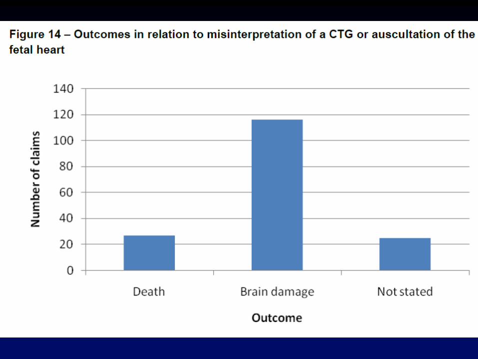
In the ten years (2000-2010) covered by the study, 300 claims involving alleged CTG misinterpretation were reported to the NHSLA. The total value of these claims is estimated to be in the region of £466million.
2,330,000,000,000 Toman, 2.3 trillion T or 2300 Billion T ( assuming 1£=5000 T)
Source: NHSLA

Source: NHSLA


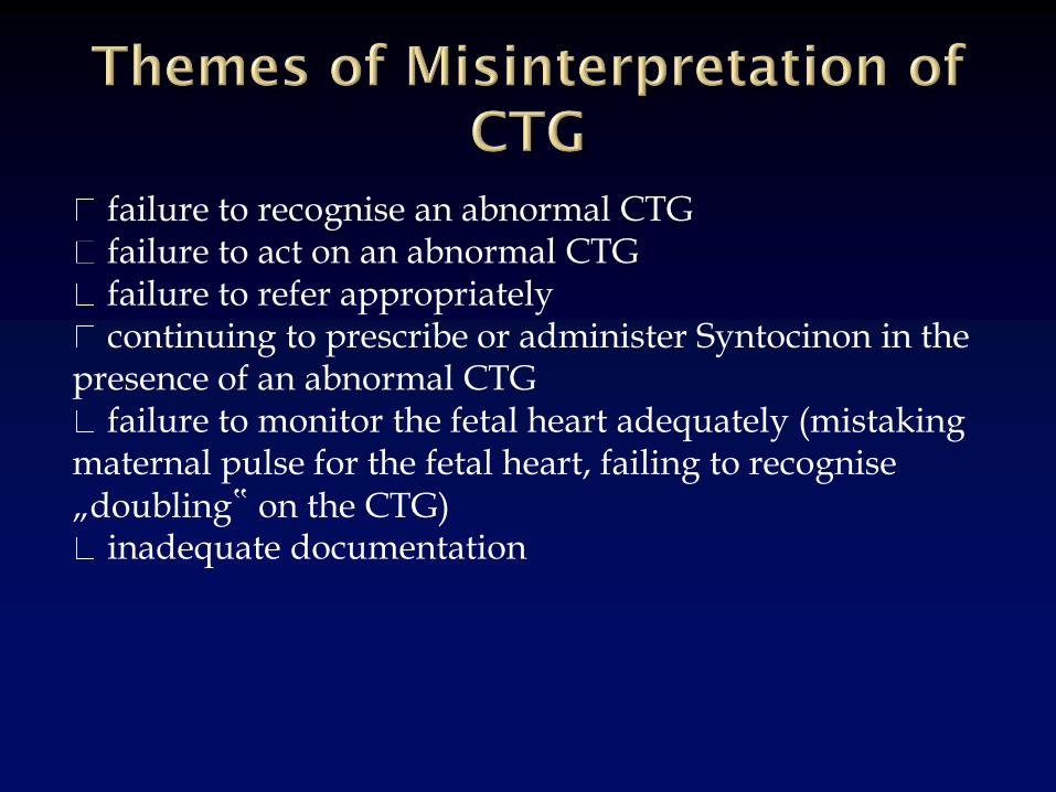
failure to recognise an abnormal CTG failure to act on an abnormal CTG failure to refer appropriately continuing to prescribe or administer Syntocinon in the
presence of an abnormal CTG failure to monitor the fetal heart adequately (mistaking
maternal pulse for the fetal heart, failing to recognise „doubling‟ on the CTG)
inadequate documentation


Intrapartum (IP) hypoxia is a significant cause of fetal death and disability.
In theory, the worst consequences and the majority of the adverse outcomes of IP hypoxia can be avoided.


Cerebral palsy is a group of permanent disorders of the development of movement and posture, causing activity limitation, that are attributed to non-progressive disturbances that occurred in the developing fetal or infant brain.
Source: cerebralpalsy.org

Severity Mild
Moderate
Severe
Motor Function Spastic (Pyramidal)80% can affect any limb(s) Monoplegia, paraplegia, hemiplegia, tetraplegia
Non-Spastic (Extrapyramidal) 20% Ataxia & Dyskinesia
Mixed

In addition to the motor impairment, individuals with cerebral palsy may have sensory impairments, cognitive impairment, and epilepsy.
Ambulation status, intelligence quotient, quality of speech, and hand function together are predictive of employment status.

Current strategies to decrease the risk of cerebral palsy due to IP hypoxia include induced hypothermia for neonates with hypoxic-ischemic encephalopathy.

chronically or
acutely


Some episodes of hypoxaemia are common in labour and are tolerated well by the healthy fetus, unless the episodes are profound (e.g. abruption, shoulder dystocia) or protracted or repetitive for a long time (prolonged labour or hyperstimulation).

Maternal
Fetal

Pre-eclampsia : materno-fetal oxygen transfer (uteroplacental vascular disease).
Maternal Diabetes: uteroplacental vascular disease esp Type I, relative intrauterine growth restriction in diabetic women ( size of baby in relation to placenta)
Antepartum Haemorrhage: placenta seperating
Maternal cardiac disease may cause reduced uterine perfusion
Intrauterine growth restriction and therefore fetal hypoxia is more common in autoimmune disorders such as: Systemic lupus erythematosus (SLE)
Antiphospholipid syndrome

Small fetus especially IUGR
Even in the absence of utero-placental vascular disease causing intrauterine growth restriction, the constitutionally small fetus will be predisposed to hypoxia due to reduced fetal nutritional reserves
The severity of placental dysfunction can be assessed by features including:
Degree and gestation at onset of growth restriction
Degree of oligohydramnios
Doppler ultrasonography of the umbilical artery

Associated pathology that may have triggered preterm labour, in particular
intrauterine growth restriction
Intrauterine infection (which may not be apparent clinically)
The reduced nutritional reserves (especially cardiac glycogen) of a preterm fetus to cope with the normal intermittent hypoxic insult with each contraction in labour

Intrauterine growth restriction and PPROM
Increased risk of cord compression resulting in fetal hypoxia in labour

Fetuses with alloimmunisation are at risk of severe anaemia and therefore hypoxia

Preterm labour
Pre-eclampsia
Intrauterine growth restriction
acute intrapartum hypoxia in twin 2 after delivery of twin 1 due to :
Decompression of the uterus (can cause abruption)
Sustained contraction of the uterus
Possible cord compression

The mechanism is not known, but breech presentation may be more common amongst fetuses with growth restriction, and may predispose to cord compression or cord prolapse.

Intrauterine infection leads to a rise in perinatal morbidity and mortality
Making the CTG less sensitive at detecting hypoxia
Sensitising the fetus so that perinatal injury occurs at a lesser degree of hypoxia

If VBAC , ruptures in labour (1:250)
Absolute risk : same as having the first baby vaginally

Infection, which can alter the fetal response to hypoxia by making the CTG less sensitive at detecting hypoxia
sensitising the fetus so that perinatal injury occurs at a lesser degree of hypoxia
Cord compression, which can cause hypoxia directly
Abruption, which can cause hypoxia directly

No time to breathe
Can cause abruption

Old meconium, especially in mature fetuses, is less significant than fresh meconium. The incidence of meconium passage rises from less than 4% before 34 weeks to over 30% at 42 weeks.
Meconium aspiration can occur with normal cord PH
Is there a place for c/section for grade III Meconium?

Cord prolapse
Uterine rupture (which can occur in an unscarred uterus)
Watch very carefully Parous ladies on Syntocinon
Placental abruption
Maternal hypotension
Usually transient and after Epidural
Hyperstimulation
Uncommon in non-induction group

Each uterine contraction is associated with a temporary reduction of placental blood flow and placental oxygen exchange. The healthy fetus has enough metabolic reserves to be able to cope with these periods of hypoxia for hours during labour. Between contractions, uterine perfusion normalises and placental oxygen exchange resumes.
Resembling : swimming

Intrinsic Spontaneous dominant pacemaker activity of the sinoatrial
node in the atrium
Extrinsic The cardio-regulatory/vasomotor centre in the brain stem
Baroreceptors
Chemoreceptors
Autonomic nervous system: Sympathetic causes increase in FHR and lowers baseline variability
Parasympathetic slows FHR and increases baseline variability
Circulating catecholamines
Aldosterone production and kidneys contribute to control of blood volume
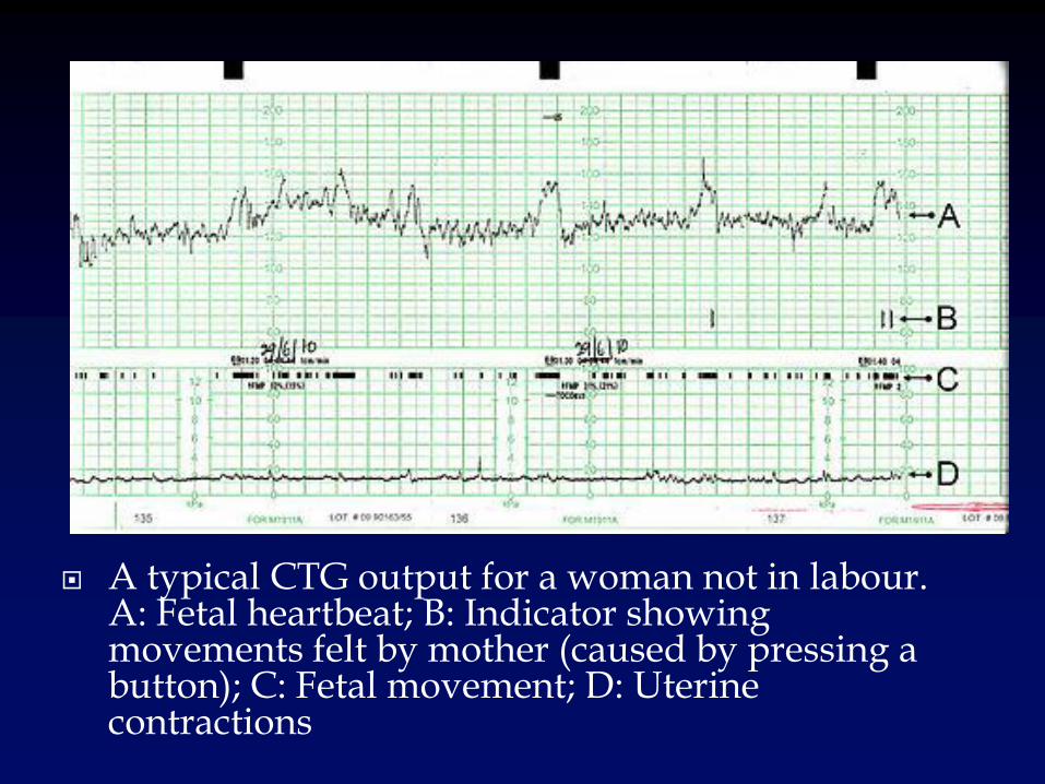
A typical CTG output for a woman not in labour. A: Fetal heartbeat; B: Indicator showing movements felt by mother (caused by pressing a button); C: Fetal movement; D: Uterine contractions

Recognizing a pattern is a daily job
How you recognise a car , face etc. etc.?


Check the name
Check the speed
Check the time and the date
Check the maternal pulse
Define risk and the indication of CTG monitoring

1cm/minute
3cm/minute
In UK it is 1cm/minute
What is yours?
Does it need to be standardised in Iran?

From 14 weeks to term there is a progressive fall in the mean baseline FHR which is unaffected by whether the fetus is Active or Quiescent

The baseline variability of the FHR in early pregnancy is low and increases with gestation, but that is refined by the behavioural state of the fetus . Thus over the second half of pregnancy the baseline variability increases progressively during fetal activity; this increase is less marked during fetal quiescence and declines from 30 weeks onwards.
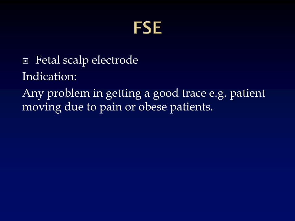
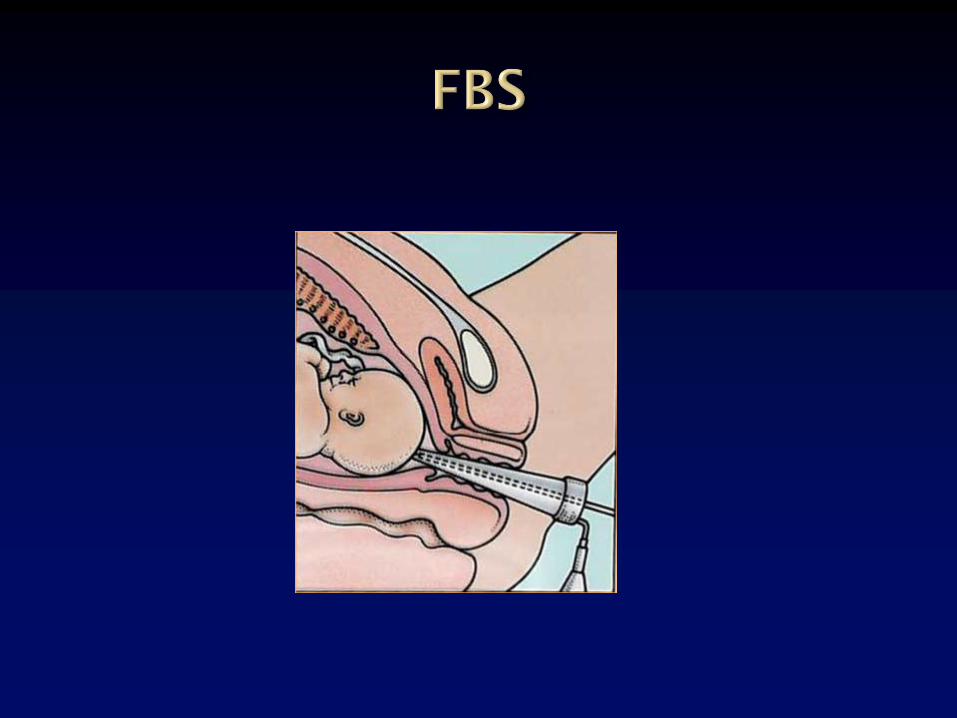
Fetal scalp electrode
Indication:
Any problem in getting a good trace e.g. patient moving due to pain or obese patients.

Pethidine/opioids
Methyldopa

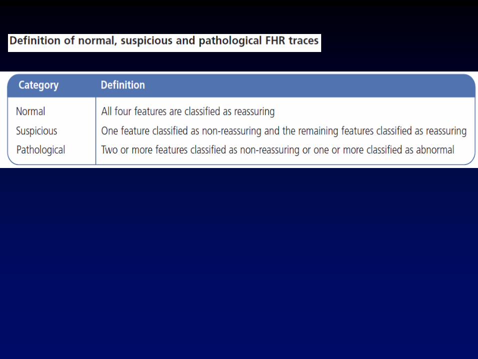
Normal
Suspicious
Pathological

Normal CTG
Suspicious CTG
Pathological CTG

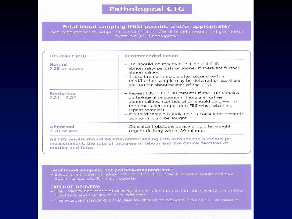

Pathological CTG
Sample or Deliver

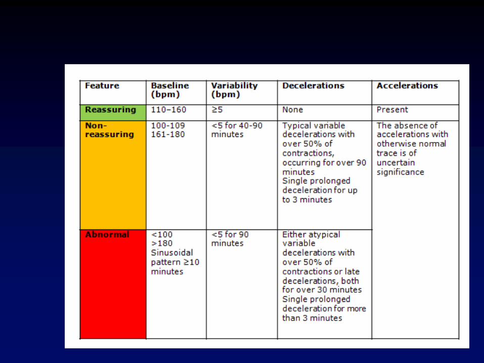
Baseline
Reassuring, Non Reassuring , Abnormal
Variability
Reassuring, Non Reassuring , Abnormal
Deceleration
Reassuring, Non Reassuring , Abnormal
Accelerations
Lack : unknown significance in Labour

The FHR in between periodic changes is called the baseline FHR
The normal baseline FHR varies between 110-160 bpm (slightly quicker for preterm).
A baseline FHR of 100-109 bpm or 161-180 bpm is non-reassuring.
A baseline FHR of <100 or >180 bpm is classified as an abnormal feature.
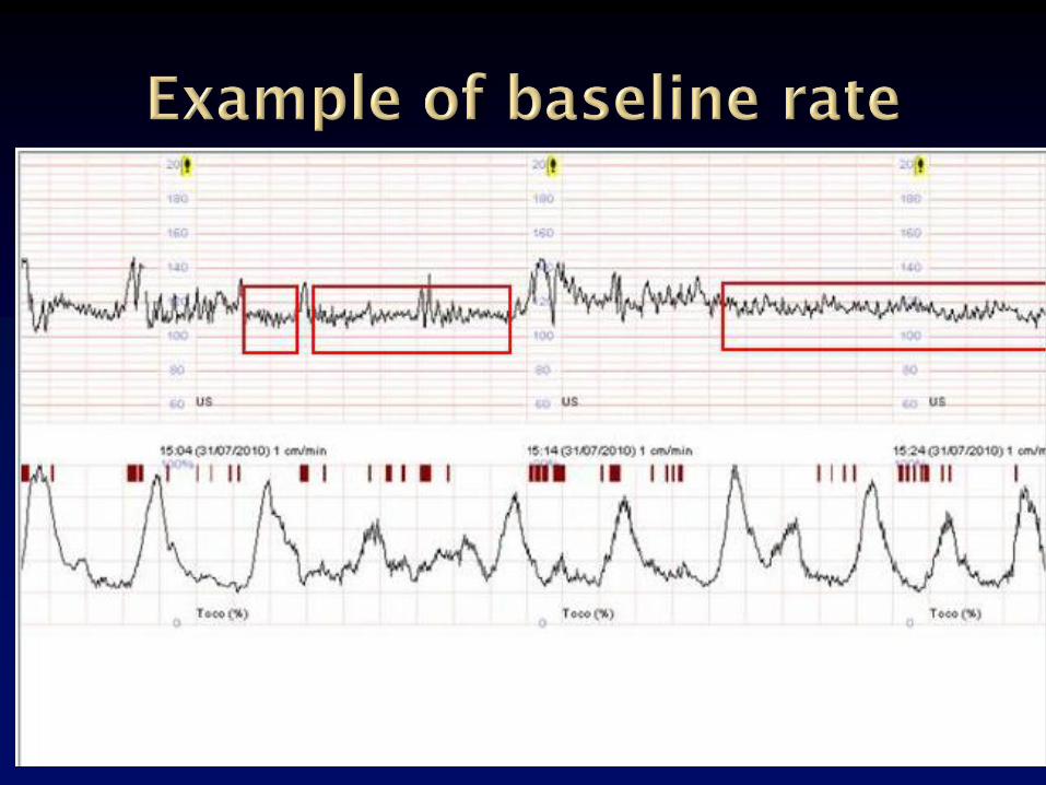

Look at the paper from the side
Watch for creeping baseline (Look at the side of folded papers)

This is the difference between the upper and lower limits of the baseline heart rate over a short period of time, for example, over one minute.
Variability between 5 and 25 bpm is considered ‘reassuring’. Reduced variability of <5 bpm can be physiological during periods of fetal sleep.
Variability of <5 bpm is considered a non-reassuring feature if it lasts between 40 to 90 minutes.
A variability of <5 bpm for more than 90 minutes is an abnormal feature.

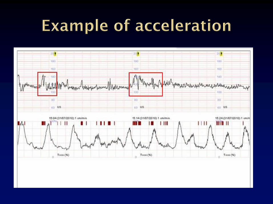
An acceleration is a transient increase in FHR of at least 15 bpm above the baseline which lasts for at least 15 seconds .
Accelerations are an essential feature of a non-labouring CTG, however, in labour the fetus may preserve energy by reducing its movements. Therefore, the absence of accelerations in labour is common and is of no known clinical significance.

A deceleration is a transient decrease in FHR of at least 15 bpm below the baseline which lasts for at least 15 seconds.
The presence of decelerations is a non-reassuring or abnormal feature in a CTG and is classified according to the type, duration and frequency of the decelerations.

Early Decel
Late Decel

Caused by head ompression


Must be uniform in both length and depth; for this reason true late decelerations are uncommon (more commonly they are atypical variable decelerations that have been wrongly classified)
Late decelerations are one feature of uteroplacental insufficiency.

vary in length and amplitude and are not uniform (unlike early and late decelerations).
Typical
'Shoulders' (primary and secondary acceleratory phases)
Atypical Overshoots
Loss of primary shoulder/acceleratory phase
Smooth deceleration
Slow return to baseline (late component)
Baseline returns to a lower level (after deceleration)
Biphasic (W shape)

'Shoulders' (primary and secondary acceleratory phases)


Loss of variability
Complicated tachycaardia


How reliable is the clock in the labour ward and the hospital?

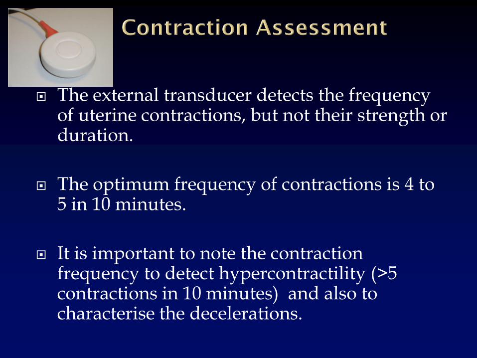
The external transducer detects the frequency of uterine contractions, but not their strength or duration.
The optimum frequency of contractions is 4 to 5 in 10 minutes.
It is important to note the contraction frequency to detect hypercontractility (>5 contractions in 10 minutes) and also to characterise the decelerations.

DR C BRAVADO
Determine Risk
Contractions
Baseline RAte
Variability
Accelerations
Decelerations
Overall Assessment

NICE Guideline


Left Lateral
Lithotomy


Insert sepculum
Clean the scalp
Spray the scalp with ethyl chloride to encourage a reactive hyperaemia in the skin
Use petroleum jelly to lightly cover the scalp and smooth the hair in a single direction
Use the blade in line with the hair and use a single firm stab
Wait for a minute and blood will come into a bubble
Use a glass tube to take up blood by capillary action
Put the sample into machine - use clean end to avoid contamination

Acute compromise : Bradycardia , average time to get an FBS is 18 minutes
Vertical transmission of virus or bacteria possible, e.g. HIV, hepatitis C, suspected chorioamnionitis
Fetal bleeding disorders suspected, e.g. haemophilia, low platelets
Be careful in preterm infants - avoid below 34 weeks

Parous patient whom needs one or two FBS’s before delivery

Maternal Oxyge:n NICE recommends that using maternal facial
oxygen therapy for more than 10 minutes may be harmful to the baby and should be avoided.
Maternal position: A significant degree of compression of the inferior
vena cava is demonstrable with up to 15° of lateral tilt . The use of a lateral tilt (>15°) at delivery, both by caesarean section and during labour, reduces the cardiovascular compromise considerably by reducing this compression
Drugs


Reducing or discontinuing an oxytocin infusion
Removing vaginal prostaglandin agents
Administration of a fluid bolus
Administering tocolytics

Terbutaline 0.25 mg , Subcutaneous
rapid onset of action with a reduction in uterine activity of up to 87.3% in 15 minutes


If decelerating , perform c/section/vaginal delivery immediately
Don’t delay delivery for abrution
Continue with labour if CTG Normal

Meconium ( fresh or thick meconium)
Infection ( Pyrexia, prolonged ROM, Maternal and fetal tachycardia, Offensive discharge)
(Don’t get falsely re-assured by normal FBS )
CTG training courses
Annual mandatory CTG training preferably online
CTG weekly meeting to discuss interesting cases
Make the definition of the CTG findings as standard
Make the speed of the machine standard
When in doubt ask for a second opinion
Practise , Practise and Practise
Good documentation
Legal cases to be judged by CTG experts in interpretation and labour management