Embed Size (px)
Citation preview

Potential Methods of Eliminating Swine
Pathogens in Livestock Transport Trailers
Literature Review
For University of Saskatchewan Saskatoon, Saskatchewan
Dr. Tadele Kiros Gebreyohannes
March 31, 2015

i
Acknowledgement
The financial support provided to the University of Saskatchewan by Swine Innovation Porc and
Agriculture and Agri-Food Canada is gratefully acknowledged.
ii
Executive Summary
Increased animal transportation has introduced additional challenges to biosecurity. A great deal
of attention has been given to transportation biosecurity associated with the outbreaks of
Porcine Respiratory and Reproductive Syndrome (PRRS), Transmissible Gasteroenteritis (TGE),
and, more recently, the widespread outbreak of the Porcine Epidemic Diarrhea virus (PEDv) that
has occurred primarily in the United States. Even though these viruses can be transmitted via
several different vectors, the major concern is transportation of the virus from one farm to
another or even across international boundaries via livestock transport trailers. Thus, there are
two primary goals of this literature review. The first goal is to identify bacterial, viral, and parasitic
swine diseases that can easily be transported through animal transport trailers. Along with this,
all possible modes of transmission are noted. The second goal is to investigate the methods that
have been documented in the scientific literature as being effective in deactivating each of the
viruses, bacteria, and parasites of interest. This report identifies ten viral diseases, nine bacterial
diseases, and two parasitic diseases that represent economically significant biosecurity
challenges for the Canadian swine industry.
The viral diseases of interest include African Swine Fever (ASF), Classical Swine Fever (CSV),
Porcine Epidemic Diarrhea (PED), Transmissible Gastroenteritis (TGE), Porcine Reproductive and
Respiratory Syndrome (PRRS), Swine Influenza, Rotavirus, Porcine Circoviruses, Aujesky’s Disease
(Pseudorabies), and Foot and Mouth Disease (FMD). The bacterial diseases of interest include
Salmonellosis; Colibacillosis; Brachyspiral colitis, B. hyodysenteriae, and B. pilosicoli; Swine

iii
Erysepelas; Leptospira; Actinobacillosis; Streptococcus; and Haemophilus parasuis (Glasser’s
Disease). The two parasitic diseases of interest include Sarcoptic Mange (Scabies) and Ascariasis.
It was found that the majority of viruses, bacteria, and parasites are much more resistant to
disinfection when there is organic matter such as feces and bedding present. This indicates that
an effective biosecurity protocol requires cleaning of the trailer prior to disinfection. In addition,
drying of the trailer following disinfection was found to be an important process step. Taken
together, the results of this extensive study have shown that low temperature, short contact
time, and high organic matter decreased the efficacy of most of the disinfectants tested. It is
important to note that no disinfectant is universally effective against all pathogens. However, it
appears, based on the literature, that application of a 10% sodium hypochlorite (household
bleach) solution in combination with heating to a temperature of 70 degrees for ten minutes has
the potential to be effective in deactivating the majority of viruses, bacteria, and parasites.

iv
Table of Contents
1. Introduction ............................................................................................................................. 1
2. Viral diseases ........................................................................................................................... 3
2.1. African swine fever ........................................................................................................... 3
2.1.1. Etiology and mode of transmission .......................................................................... 3
2.1.2. Environmental survival and susceptibility to disinfectants ...................................... 4
2.2. Classical Swine Fever ........................................................................................................ 5
2.2.1. Etiology and Transmission ........................................................................................ 5
2.2.2. Environmental survival and susceptibility to disinfectants ...................................... 6
2.3. Porcine Epidemic Diarrhea ............................................................................................... 7
2.3.1. Etiology and Transmission ........................................................................................ 7
2.3.2. Environmental Stability and Susceptibility to Disinfectants ..................................... 8
2.4. Transmissible Gastroenteritis ........................................................................................ 11
2.4.1. Etiology and Transmission ...................................................................................... 11
2.4.2. Environmental survival and susceptibility to disinfectants .................................... 12
2.5. Porcine Reproductive and Respiratory Syndrome (PRRS) ............................................. 13
2.5.1. Etiology and Transmission ...................................................................................... 13
2.5.2. Survival in the environment and susceptibility to disinfectants ............................ 14
2.6. Swine Influenza .............................................................................................................. 16
2.6.1. Etiology and Transmission ...................................................................................... 17
2.6.2. Environmental resistance and susceptibility to disinfectants ................................ 18
2.7. Rotaviral enteritis ........................................................................................................... 18
2.7.1. Etiology and Transmission ...................................................................................... 19
2.7.2. Environmental resistance and susceptibility to disinfectants ................................ 19
2.8. Porcine Circovirus Associated Disease ........................................................................... 20
2.8.1. Etiology and Transmission ...................................................................................... 20
2.8.2. Environmental stability and susceptibility to disinfectants .................................... 21
2.9. Pseudorabies (Aujesky’s Diseases) ................................................................................. 23
2.9.1. Etiology and Transmission ...................................................................................... 23

v
2.9.2. Environmental resistance and susceptibility to disinfectants ................................ 24
2.10. Foot and Mouth Disease ............................................................................................ 24
2.10.1. Etiology and transmission ....................................................................................... 26
2.10.2. Environmental survival and susceptibility to disinfectants .................................... 27
3. Bacterial diseases .................................................................................................................. 29
3.1. Salmonellosis .................................................................................................................. 29
3.1.1. Etiology and Transmission ...................................................................................... 29
3.1.2. Environmental survival and susceptibility to disinfectants .................................... 30
3.2. Colibacillosis ................................................................................................................... 32
3.2.1. Etiology and Transmission ...................................................................................... 32
3.2.2. Environmental survival and susceptibility to disinfectants .................................... 33
3.3. Swine Brachyspiral Colitis............................................................................................... 34
3.3.1. Etiology and Transmission ...................................................................................... 34
3.3.2. Survival in the environment and susceptibility to disinfectants ............................ 36
3.4. Swine Erysipelas ............................................................................................................. 37
3.4.1. Etiology and Transmission ...................................................................................... 38
3.4.2. Environmental survival and susceptibility to disinfectants .................................... 38
3.5. Mycoplasmal Pneumonia ............................................................................................... 39
3.5.1. Etiology and Transmission ...................................................................................... 40
3.5.2. Environmental survival and Susceptibility to disinfectants .................................... 41
3.6. Leptospirosis .................................................................................................................. 42
3.6.1. Etiology and Transmission ...................................................................................... 42
3.6.2. Environmental survival and susceptibility to disinfectants .................................... 43
3.7. Actinobacillosis ............................................................................................................... 44
3.7.1. Actinobacillus pleuropneumoniae (APP) ................................................................ 44
3.7.2. Actinobacillus suis ................................................................................................... 47
3.8. Streptococcosis .............................................................................................................. 47
3.8.1. Etiology and Transmission ...................................................................................... 48
3.8.2. Environmental resistance and susceptibility to disinfectants ................................ 50

vi
3.9. Glӓsser’s Disease ............................................................................................................ 50
3.9.1. Etiology and Transmission ...................................................................................... 51
3.9.2. Environmental resistance and susceptibility to disinfectants ................................ 52
4. Parasitic diseases of swine .................................................................................................... 54
4.1. Mange............................................................................................................................. 54
4.1.1. Etiology and Transmission ...................................................................................... 55
4.1.2. Environmental survival and susceptibility to disinfectants .................................... 55
4.2. Ascariasis ........................................................................................................................ 56
4.2.1. Etiology and transmission ....................................................................................... 57
4.2.2. Environmental resistance and susceptibility to disinfectants ................................ 57
5. Disinfectants and Disinfection ............................................................................................... 60
5.1. Definitions ...................................................................................................................... 61
5.2. Factors to consider during disinfection .......................................................................... 62
5.2.1. Nature of the surface/material ............................................................................... 63
5.2.2. Environmental factors............................................................................................. 63
5.2.3. Organic matter ........................................................................................................ 64
5.2.4. Type of pathogens .................................................................................................. 66
5.2.5. Nature of the disinfectant....................................................................................... 67
6. Animal Transport Biosecurity ................................................................................................ 70
7. Conclusions ............................................................................................................................ 74
8. Appendices ............................................................................................................................ 75
8.1. Summary of characteristics of disinfectants .................................................................. 75
8.2. Effect of different disinfectants against various pig pathogens .................................... 76
9. References ............................................................................................................................. 80

1
1. Introduction
The Food and Agricultural Organization (FAO) has defined biosecurity as “a strategic and
integrated approach to analyze and manage relevant biological risks to human, animal and plant
life or/and health and the associated risks to environment” (FAO, 2007). The concept of
biosecurity started to make an appearance in the late 1990’s with an extensive literature review
and series of studies conducted by Amass and colleagues from Purdue University on the
importance of biosecurity as a tool to control the transmission of diseases, and the effectiveness
of commonly used disinfectants to inactivate or eliminate pathogens (Amass, 2004; Amass and
Clark, 1999; Amass et al., 2000a; Amass et al., 2000b). Since then, farm biosecurity has become
an essential part of farm management in the pork industry, with an overall objective of
preventing the introduction of new diseases to the farm and controlling the spread of endemic
diseases within and between farms. As extensively discussed by (Levis and Baker, 2011),
biosecurity measures may vary from farm to farm. Despite this, every effective biosecurity plan
should include the following measures: bio-exclusion, bio-management, and bio-containment
(Levis and Baker, 2011). Levis defined bio-exclusion as measures taken by a farm to prevent the
introduction of a new disease to the swine herd in the farm, while bio-management refers to the
efforts made by the farm management to minimize the economic impact of endemic diseases in
a given farm using different management practices. The focus of most hog producers is on the
first two biosecurity measures. However, bio-containment is equally important, not only to
protect a given farm from new diseases but also to prevent the spread of diseases into

2
neighboring farms. In addition to protecting a given farm or neighboring farms, bio-containment
is the most effective method of preventing the introduction of trans-boundary animal diseases
(TAD) into Canada. It is also important to notice the importance of bio-containment in preventing
consumers from contracting zoonotic animal diseases. All of the three biosecurity measures are
very important, and it is difficult to effectively apply one without considering the other. The major
focus of this review, however, will be on bio-containment, with the aim of preventing the
introduction of diseases into a country, a region, or a farm, primarily through the trailers used
for live animal transportation.
Animal transportation is becoming a major concern of biosecurity, and a great deal of attention
has been given to transportation biosecurity in association with the outbreaks of porcine
respiratory and reproductive syndrome (PRRS), transmissible gastroenteritis (TGE) and, more
recently, the widespread outbreak of the porcine epidemic diarrhea virus (PEDv) that has
occurred primarily in the United States. Even though these viruses can be transmitted between
animals or farms through several different vectors, the major concern is transportation of the
virus from one farm to another or even across boundaries through livestock transporting trailers.
Thus, the main goal of this literature review is to identify bacterial, viral and parasitic swine
diseases that can easily be transported through animal transporting trailers for the purpose of
determining physical or chemical conditions that can be applied on transportation vehicles to
inactivate or eliminate the pathogens. The ultimate goal of this project is to develop an
automated system for effectively cleaning and disinfecting livestock transport vehicles to allow
for improved biosecurity in livestock transportation.

3
2. Viral diseases
2.1. African swine fever African Swine Fever (ASF) is a highly contagious hemorrhagic viral infection of pigs that is endemic
to the continent of Africa. The disease is still causing serious problems in some Sub-Saharan
African countries, but outbreaks have also been reported in Europe, Central Asia, South America
and the Caribbean that consumed a huge amount of financial resources for eradication by
stamping out all pigs in the outbreak area (Penrith and Vosloo, 2009). The disease has not yet
been reported in North America and most Western countries, but, with the existing globalization
and increasing trade relations among countries, there is serious concern that ASF could soon be
introduced into the North American hog industry.
2.1.1. Etiology and mode of transmission
The etiology of African swine fever is a double stranded DNA virus, which belongs to the genus
Asfivirus in the Asfarviridae family. The virus affects mainly the domesticated pig, but can also
affect the European wild boar and the American wild pig. In Africa, warthogs and bush pigs are
reservoir hosts with a persistent subclinical infection, and they are important sources of infection
to domestic pigs (Spickler, 2010). Clinical signs range from hyper acute/acute infection with close
to 100% mortality in domesticated pigs to subclinical infection that cannot be diagnosed based
on clinical signs in the reservoir hosts. Soft ticks of the genus Ornithodoros are believed to be the
biological vector of the virus. However, the virus can easily spread among pigs through direct pig-
to-pig contact and may be spread indirectly through contaminated fomites and livestock
transport vehicles (Oura, 2013).

4
2.1.2. Environmental survival and susceptibility to disinfectants
The virus is relatively resistant to environmental conditions and can remain viable for several
days in pig products and the environment, particularly if the virus is covered with organic matter.
It is highly resistant to cold temperatures and can survive for more than a year in blood and other
animal products stored at 4oC. It can also survive for several years in a frozen carcass. The virus
can also survive for more than a month in contaminated pig pens and more than 11 days in feces
at room temperature (EAZWV, 2011). The virus is also resistant to a wide range of pH (3.9 -11.5),
particularly if it is covered with organic matter like blood and feces (Spickler, 2010). On the other
hand, the virus is highly sensitive to high temperature and can be inactivated by heating at 56oC
for 70 minutes and within 20 minutes at 70oC (OIE, 2012). Like any other enveloped virus, ASFv
is reported to be susceptible to a wide variety of lipid solvent detergents and disinfectants
including ether, chloroform, iodine, formalin, phenols, and quaternary ammonium compounds
(EAZWV, 2011). In agreement with this, several in vitro studies have shown that the virus can be
inactivated easily in less than 30 minutes with common disinfectants such as sodium hypochlorite
and citric acid (Krug et al., 2011), as well as sodium hydroxide and calcium hydroxide (Turner and
Williams, 1999). However, all these results are from in vitro studies and need to be interpreted
with caution. In line with this, an extensive study from the old literature that used more than 10
disinfectants has concluded that, despite the ability of several disinfectants to inactivate ASF virus
in vitro, only One-Stroke Environ, a phenolic compound, applied as a spray at 1% concentration
was able to completely inactivate ASFv when applied in a contaminated pig room (Stone and
Hess, 1973) This suggests that only disinfectants specifically approved for African Swine Fever
should be used for effective decontamination during disease outbreaks.

5
2.2. Classical Swine Fever Classical Swine Fever (CSF), also known as hog cholera, is a highly contagious disease of swine
that causes systemic infection characterized by high body temperature, viraemia, vomiting,
diarrhea, and purple skin discoloration around the ear and the lower abdomen. The virus affects
both wild and domesticated pigs and is notifiable to the World Organisation for Animal Health
(OIE) due to its high economic importance. Despite its worldwide distribution, most developed
countries, including Canada, have eradicated the disease from swine herds and are declared free
of CSF. However, due to the endemic nature of the disease in most of the countries of South and
Central America, the risk for introduction of CSF to North America is still high.
2.2.1. Etiology and Transmission
Classical Swine Fever is caused by an enveloped RNA virus that belongs to the genus Pestivirus of
the Flaviviridae family. Infected animals shed the virus in all body secretions and excretions like
blood, semen, urine, feces, nasal and ocular discharges, as well as saliva. The virus is highly
contagious and reproduction ratios (R0) of 100 and 15.5 were reported within pens for weaned
pigs and slaughter pigs, respectively (Klinkenberg et al., 2002). The most efficient method of
transmission is by the oronasal route, either through direct contact between healthy and infected
pigs or by indirect transmission through contaminated feed, such as swill (Moennig et al., 2003).
Vertical transmission via contaminated semen from infected boars is also possible (Floegel et al.,
2000). Furthermore, indirect transmission due exposure to contaminated fomites like pens,
buckets, farm equipment, boots, clothing, veterinary and insemination equipment, as well as
transport vehicles, is well documented (Edwards, 2000). For instance, contaminated vehicles are
believed to be responsible for spreading the CSFv to uninfected herds during the 1997 CSF

6
outbreak in the Netherlands (Stegeman et al., 2000). Mechanical transmission via vectors such
as flies, rodents, and birds is also a concern (Edwards, 2000). The virus has been isolated from air
samples originating from infected pigs (Weesendorp et al., 2008) and aerosol transmission in the
close confinement of crowded farms is a major means of direct transmission. There is no good
evidence describing how far the virus can travel through the air; however, there are several
reports that show airborne transmission might have reached farms within a 0.25 to 6 Km radius
of an infected herd (Roberts, 1995). The general consensus is that farms within a radius of 1 km
from the infected herd are considered to be at a very high risk (Ribbens et al., 2004b).
2.2.2. Environmental survival and susceptibility to disinfectants
Like other enveloped viruses, CSF virus is moderately fragile and does not persist in the
environment for a prolonged period of time. It is very difficult to determine the exact length of
time the virus can survive in the environment as this is influenced by the level of moisture, pH
and temperature; however, studies show that it can survive in contaminated pens for about 4
weeks under winter conditions with no exposure to direct sunlight. It can also survive for months
and even years in refrigerated or frozen meat, respectively (Ribbens et al., 2004a). An extensive
review of the old literature by Steven Edwards on the survival and inactivation of CSF virus has
shown that the virus is highly sensitive to drying and ultra-violet light. It is also susceptible to heat
and can be easily inactivated within 30 minutes at a temperature greater than 65OC. The virus is
also susceptible to extreme pH conditions and can be rapidly inactivated at pH <3 and pH >11,
but it is generally stable at neutral to slightly alkaline conditions in the pH 5 to 10 range.
Furthermore, like all enveloped viruses, CSF virus is easily inactivated by detergents and organic
solvents like ether and chloroform. A wide range of chemicals, including sodium hypochlorite,

7
phenolic compounds, quaternary ammonium compounds, and aldehydes are used to disinfect
CSF virus in the field (Edwards, 2000). For example, 1000 ppm sodium hypochlorite was found to
completely inactivate CSF virus both from metallic and plastic surfaces in field conditions. In
contrast, 2% citric acid, which was effective against ASF virus, was not effective against CSF virus
(Krug et al., 2011).
2.3. Porcine Epidemic Diarrhea Porcine epidemic diarrhea (PED) is a highly contagious viral disease of swine characterized by
rapid onset of diarrhea, vomiting and dehydration. PED is associated with high morbidity and
mortality. Mortality can reach 100% in young piglets of less than one week age. The disease is
clinically indistinguishable from Transmissible Gastroenteritis (TGE), but there is no antigenic
relation or cross protection between the two viruses (Pospischil et al., 2002). The disease was
first reported in England in 1971 and had resulted in several devastating disease outbreaks
throughout Europe in the 70s and 80s (Chasey and Cartwright, 1978). Porcine epidemic diarrhea
was first detected in the United States in May of 2013 on Iowa pig farms (Stevenson et al., 2013).
Since then, the disease has been spreading rapidly throughout the USA and Canada. The first
confirmed case of PED in Canada was reported in January, 2014 on an Ontario farm (Ojkic et al.,
2015).
2.3.1. Etiology and Transmission
The etiology is an enveloped single stranded RNA virus that belongs to the family Coronaviridea,
genus Alphacoronavirus. The virus was first identified in 1978 from the 1970’s outbreaks of PEDv
in Europe (Pensaert and de Bouck, 1978). However, the recent outbreak in the United States and
Canada is believed to have its sources in China (Huang et al., 2013). The strain circulating in the

8
US since April 2013 is similar to the strain that resulted in a PED outbreak in China between 2010
and 2012 (Chen et al., 2014; Stevenson et al., 2013).The virus is excreted in the feces and the
fecal-oral route of transmission is the major route of direct transmission between pigs.
Like any other enveloped viruses, PEDv is moderately fragile, but can survive outside the host for
some time under suitable environmental conditions like in organic matter and in cool
temperatures. Therefore, contaminated fomites, including transport vehicles, can serve as a
mode of indirect transmission of the disease between farms (Lowe et al., 2014). Ingestion of
contaminated feed material is suspected as the source of the first PEDv outbreak in Canada (Dee
et al., 2014; Pasick et al., 2014). Furthermore, virus genetic material was detected as far as 10
miles from an infected herd, suggesting that airborne transmission is a potential means for
disseminating porcine epidemic diarrhea virus among farms (Alonso et al., 2014).
2.3.2. Environmental Stability and Susceptibility to Disinfectants
The survival of the virus in the environment is dependent on several factors including humidity,
temperature, pH, and the presence of organic matter. The virus was reported to be stable
between a pH of 5 and 9, in a temperature range of 4oC to 37oC in an environment with a suitable
organic matter like feces, but was inactivated by heating at 60oC for 30 minutes (Pospischil et al.,
2002). A group of researchers, from Iowa State University have shown that, in the absence of
proper washing and disinfection, the virus can be completely inactivated in livestock trailers
(cleaned by scraping and sweeping only) by heating at 71oC for 10 minutes. However, it took 7
days to completely inactivate the virus when the trailers were left at room temperature (Thomas
et al., 2014).

9
Table showing summary of pig bioassay and PED virus result by treatment
F, Fahrenheit; M, minute; H, hour; D, days
The above table, taken from (Thomas et al., 2014), shows that treatment of PEDv containing feces
at 160oF (71oC) for 10 minutes or at room temperature (60oF) for 7 days completely inactivated
the virus and none of the 4 pigs exposed to the heat treated feces were infected. On the other
hand, treatment of the virus at a temperature less than 160oF (71oC) for 10 minutes or at room
temperature (68oF) for 24 hours did not completely inactivate the virus and 25-50% of the pigs
exposed to the heat treated feces were infected.
Considering the conditions discussed above concerning time and temperature combinations
required to inactivate PED virus, it is possible that the virus can easily be transmitted by the
consumption of contaminated pelleted pig feed (Dee et al., 2014). The typical retention time in
the conditioner machine during the pelleting process is 30 to 60 seconds at a temperature range
of 71 to 99oC (personal communication). This may suggest a risk that the virus may not be

10
completely inactivated during the short retention time when the lower temperature range (71oC)
is used to pellet pig diets containing porcine products. Therefore, it is recommended either to
use the higher temperature range (100 to 121oC) or increase the conditioning time. Further
processing of the conditioned feed material with an expander may also help to completely
inactivate the virus. However, the Canadian Food Inspection Agency (CFIA) could not confirm a
link between feed containing porcine plasma and PED outbreaks in Canada, despite the presence
of infectious virus particles in porcine plasma originating from the United States ,which was used
for the preparation of pig feed in Canada (CFIA, 2015).
The virus is highly susceptible to direct sun light when it is not protected with organic material
like feces. Therefore, UVC irradiation and treatment with ionizing radiations like gamma rays can
be used to inactivate the virus in contaminated feed materials or surfaces. There is no published
evidence on PEDv to support this hypothesis; however, studies performed on a group of viruses
similar to the TGEv suggest that corona viruses, including the PEDv, can be inactivated using a
UVC dose of 500 J per m2 (Terpstra et al., 2008) or by gamma irradiation at a dose of 25 to 35 kGy
(Nims et al., 2011). Similarly, like other enveloped viruses, PEDv can be easily inactivated using
ionic and non-ionic detergents, as well as organic solvents such as ether and chloroform. The
virus is also susceptible to most virucidal disinfectants including cresol, sodium hydroxide (2%),
formalin (1%), sodium carbonate (4% anhydrous or 10% crystalline, with 0.1% detergent), strong
iodophors (1%) in phosphoric acid, oxidizing agents like potassium peroxymono-sulfate and
sodium hypochlorite, and phenolic compounds (Pospischil et al., 2002). These results are from
an in vitro study and results may be different under natural conditions in the environment where
the virus is usually covered with organic matter like feces. For example, a report from Iowa State

11
University researchers has shown that the use of a common virucidal disinfectant, Stalosan F
powder, was not an effective means of inactivating PEDv in scraped, but unwashed livestock
trailers (PorkCheckoff, 2013). These results reiterate the importance of the gold standard
inactivation method that includes washing, disinfection, and drying of trailers. However, a recent
study conducted at Iowa State University and published in 2015 by the PorkCheckoff showed that
under circumstances when washing, disinfecting, and drying the trailer was not possible, full
inactivation of the virus in trailers was accomplished within 40 minutes of contact time by using
accelerated hydrogen peroxide® (AHP®) disinfectant, which is sold under the brand name Accel®,
at a minimum concentration of 1:32 in a 10 percent propylene glycol solution (PorkCheckoff,
2015).
2.4. Transmissible Gastroenteritis Transmissible gastroenteritis (TGE) is a highly contagious, acute, rapidly spreading viral infection
of the intestinal tract of pigs. It multiplies in and damages the enterocytes covering the small
intestine, producing villus atrophy and enteritis. The disease is characterized by diarrhea,
vomiting, dehydration and high mortality in piglets less than 2 weeks of age. TGE is clinically
indistinguishable from Porcine Epidemic Diarrhea virus (PEDv), but the two corona viruses are
not related to each other (Neumann, 2014).
2.4.1. Etiology and Transmission
TGE is caused by a virus that belongs to the genus Coronavirus of the Coronaviridae family. The
natural host is the domestic pig, but wild and domesticated carnivores can also be infected and
act as reservoir hosts. The virus is shed in feces and the main route of transmission is direct pig-
to-pig contact by the fecal-oral route. Indirect transmission through contaminated fomites,

12
including transportation vehicles, is also possible, particularly during winter because of the
improved survival of the virus in the environment in cold temperatures. The virus was also
isolated from flies (Gough and Jorgenson, 1983) and dogs (McClurkin et al., 1970), and, therefore,
mechanical transmission by flies and other animals, including birds and rodents, is a major
concern.
2.4.2. Environmental survival and susceptibility to disinfectants
The virus is highly susceptible to direct sun light; and thus cannot survive in the environment for
a prolonged period of time. The virus is generally susceptible to high temperatures and can be
easily inactivated by temperatures above 30oC (Laude, 1981). There are also unpublished reports
which show that the virus can be fully inactivated by heating at 56oC for 30 minutes or at 65oC
for 10 minutes. However, the virus is resistant to freezing cold weather, and this results in
seasonal outbreaks during the winter (Harris, 2013). The virus is highly susceptible to almost all
virucidal disinfectants, including iodides, peroxygen, quaternary ammonium compounds, phenol
and sodium hypochlorite (Brown, 1981). Thus, a good biosecurity policy of washing, disinfection
and drying of equipment, including animal transport vehicles, can effectively control the spread
of the disease between herds.
There are two other swine diseases worth mentioning here, caused by the same group of corona
viruses but with a different degree of severity and economic impact. The first one is the Porcine
Respiratory Corona Virus (PRCV) infection caused by a virus identical to the TGE virus that lost its
tropism and virulence to the intestines; it replicates only in the lung interstitial cells. The virus
causes only mild respiratory infection in young piglets of 2-3 weeks age, but its economic
importance lays on its ability to quickly spread by air between farms and its cross reaction with

13
TGE virus, which makes it difficult to differentially diagnose TGE outbreaks (Neumann, 2014). A
second disease caused by the same corona virus group that affects both the intestinal and
respiratory tract of young pigs was reported in Ohio and Indiana in February of 2014. The disease
rapidly spread to other states in the United States and Canada and caused a significant economic
loss to the swine industry in 2014. The etiology is a novel corona virus known as porcine
deltacorona virus (PdCV), which is closely related to the TGE and PED viruses and is believed to
have originated in Hong Kong and South Korea (Ma et al., 2015).
2.5. Porcine Reproductive and Respiratory Syndrome (PRRS) PRRS is a highly infectious and the most economically significant disease of pigs in North America,
which is costing hundreds of millions of dollars every year to the swine industry. The total cost of
productivity losses due to PRRS to the US pork industry was estimated to be around $560 million
in 2005 (Neumann et al., 2005), and this increased to $664 million in 2011 (Holtkamp et al., 2013).
The disease was first reported in North America in the 1980s, and spread rapidly throughout the
world in a very short period of time. The disease has now been reported in almost all countries
with the exception of Australia, New Zealand and the Scandinavian countries (OIE, 2008).
Clinically, it is characterized by overlapping symptoms of the reproductive system (failure or
impaired breeding in sows, gilts, and even boars) and respiratory illness in pigs of all age
(Neumann, 2014).
2.5.1. Etiology and Transmission
The etiology of PRRS is an enveloped, single stranded and positive sense RNA virus classified
under the family Arteriviridae and the genus Arterivirus viruses. The virus affects only pigs, and
no other mammalian or arthropod host acts as a biological vector for the transmission of the

14
disease. The virus is shed in all body secretions and excretions of the infected pig including milk,
blood, semen, saliva, nasal discharge, urine and feces. The main route of transmission is through
direct contact between pigs through the oronasal route or through artificial insemination (OIE,
2008). Indirect transmission through contaminated fomites is also a major problem in the control
of PRRS. A group from the University of Minnesota conducted extensive research to determine
the mechanical vectors for the transmission of PRRSv and compiled evidence that the virus can
be carried from pen to pen within a farm or between farms with contaminated fomites such as
farm equipment, clothing, boots, veterinary equipment (Otake et al., 2002b), house flies and
mosquitoes (Otake et al., 2004; Otake et al., 2002c; Pitkin et al., 2009b), as well as transport
vehicles (Dee et al., 2004b; Dee et al., 2007). The same group also identified the airborne
transmission potential of the PRRS virus to be as long as 3.3 km (Otake et al., 2002a; Pitkin et al.,
2009a) and developed an air filtration system to control aerosol transmission of the virus (Dee et
al., 2005a; Dee et al., 2009b).
2.5.2. Survival in the environment and susceptibility to disinfectants
Comparable to other enveloped viruses, PRRSv is moderately fragile in the environment and can
only survive for a few hours if not covered by organic materials like feces or bedding. The stability
of the virus in the environment depends on several factors including moisture, temperature and
pH. (Bloemraad et al., 1994). According to this report, the virus was stable for an extended period
of time at pH 6.5-7.5, but infectivity was rapidly lost at pH values below 6 and above 7.5. Similarly,
the virus was stable at lower temperatures and remained viable for up to 4 months in
temperatures between -70 to -20oC. Viability decreased with increasing temperature and almost
90% of the viruses were inactivated within a week when stored at 4oC; however, few infectious

15
virus were able to survive for about 30 days at 4oC (Bloemraad et al., 1994). In another report,
PRRS virus was reported to survive in a solution for weeks at 4oC, for about 1-6 days at room
temperature, and for 24 hours at 37oC. Its viability decreased with increasing temperature and
was completely inactivated within 20 minutes at 56oC (Zimmerman et al., 2012). The virus is also
highly susceptible to direct sunlight (Benfield et al., 1992).The virus is also sensitive to almost all
lipid solvent detergents and virucidal disinfectants; however, a combination of Quaternary
ammonium compound and glutaraldehyde (synergize 0.8%) or a modified potassium
monopersulfate (Virkon 1%) are the recommended disinfectants for use under natural farm
conditions (Pitkin et al., 2009c).
A good biosecurity program at the national and farm level can effectively control the spread of
PRRS and over the years the focus has been on the role of livestock transporting trailers as a
major means of spreading the virus between farms and biosecurity protocols that can be applied
effectively on trailers and drivers. A group led by Scott Dee from the Swine Disease Eradication
Center at the University of Minnesota College of Veterinary Medicine has developed a model to
study the role of trailers in the spread of the virus and demonstrated healthy pigs could become
infected with PRRSV through contact with the contaminated interior of the transport vehicle (Dee
et al., 2004b). Using this model, they developed a number of sanitation protocols to disinfect
PRRSV contaminated full size trailers applicable at room temperature (Dee et al., 2004a) and cold
temperatures (4oC and -20oC) representing winter conditions (Dee et al., 2005b). The conclusion
from all these studies is that livestock transporting vehicles could be effectively cleared from
PPRS virus by washing with cold water and drying for 8 hours at room temperature instead of the
usual overnight drying time. Furthermore, washing plus fumigation with a combination of

16
glutaraldehyde and quaternary ammonium chloride (Synergize) was able to completely
inactivate PRRS virus within 90 minutes on the interiors of livestock transport trailers. The effect
of Synergize was compromised under freezing weather (-20oC) conditions unless it was used with
antifreeze reagents such as a 10% propylene glycol or a 40% methanol solution (windshield
washer fluid).
2.6. Swine Influenza Influenza is a viral infection of birds and mammals, including humans. Swine influenza is a highly
contagious and economically important disease that can infect almost all pigs in a herd within 1-
3 days. In the absence of secondary complicating bacterial infections, the mortality rate is
generally very low, ranging between 1% and 4%, (Dee, 2014 ), but the high morbidity of infected
pigs results in high production losses to the hog industry. As shown on The Pig Site, Holtkamp
and colleagues estimated the economic losses due to swine influenza in 2007 to be only second
to PRRS (Anonymous, 2013). Moreover, swine influenza is important because of its zoonotic
significance (Khan et al., 2013); the virus has been isolated from lung tissues of man (Smith et al.,
1976). In addition to this, pigs possess pulmonary epithelial cells with receptors that can bind
both to avian and human influenza viruses (Ito et al., 1998). Therefore, it is possible that different
types of influenza viruses infecting the same host cell in the pig mix and produce novel strain of
virus by genetic re-assortment (Castrucci et al., 1993). Due to this, pigs are considered as “mixing
vessels” for human and avian influenza viruses (Thacker and Janke, 2008). The new viruses that
arise from genetic re-assortment as a result of antigenic shift or antigenic drift are highly virulent
type of influenza viruses that can cause severe and fatal disease outbreaks even in the natural
hosts. A typical example is the H3N2 influenza virus reported in the United States swine

17
population in the late 90’s and was composed of genes from swine, avian, and human influenza
viruses (Webby et al., 2000) .
2.6.1. Etiology and Transmission
Influenza in birds and mammals is caused by enveloped and single stranded negative-sense RNA
viruses that belong to the Orthomyxoviridae family. There are three types of influenza virus
affecting mammals and birds, designated as influenza type A, B, and C. While type B and C are
limited to humans and result in mild respiratory infection, type A is wide spread in birds and
mammals, including man. The natural hosts for influenza A virus are wild aquatic birds, but it can
also affect domestic birds, swine and humans (Webby and Webster, 2001). There are many
subtypes of influenza A viruses, which differ based on the combination of two glycoproteins on
the surface of the influenza virus designated as protein ‘H’ (Hemagglutinin) and protein ‘N’
(Neuraminidase). There are at least 16 H (H1-H16) and 9 N (N1-N9) proteins, and many
combinations of H and N proteins are possible (Fouchier et al., 2005). The three subtypes of
influenza A virus that cause infection in swine are H1N1, H1N2 and H3N2 (Ma and Richt, 2010).
The first influenza virus isolated from pigs was the H1N1 subtype known as the classical swine
influenza virus (Myers et al., 2007). Even though all three subtypes of the swine influenza can
affect humans, most of the recent outbreaks particularly the 2009 pandemic in humans, are
widely believed to be related to the lineage of the swine H1N1 influenza virus (Dawood et al.,
2009). The virus is spread among pigs by aerosols, through direct contact between pigs, and
through indirect contact with contaminated fomites. Transmission may also occur between
humans and pigs as well as other animals and pigs (Tellier, 2006).

18
2.6.2. Environmental resistance and susceptibility to disinfectants
Mammalian influenza viruses in general, including swine influenza viruses, are labile in the
environment and can only survive for 8 to 24 hours on the surface of materials depending on the
nature of the surface (Greatorex et al., 2011); however, an earlier report has shown that human
influenza A virus could survive for more than 3 days on the surface of bank notes (Thomas et al.,
2008). Survival of influenza viruses in the environment is influenced by several factors such as
presence of organic matter, temperature, humidity and pH. For example, swine influenza viruses
survived for 9 weeks in a fecal slurry stored at 4oC, 2 weeks at 20oC, but only for 1 to 2.5 hours at
50 to 55oC (Haas et al., 1995). In contrast, influenza A viruses were inactivated by exposure to
direct sunlight for 30 minutes and by heating at 56oC for 30 minutes, 60oC for 10 minutes and
70oC or 1 minute (Zou et al., 2013). Influenza viruses are also susceptible to a wide variety of
disinfectants including sodium hypochlorite (1:10 dilution of household bleach), aldehydes
(formalin, glutaraldehyde, formaldehyde), quaternary ammonium compounds (Lysol, No-Rinse
sanitizer), phenolic compounds (Tek-Trol and One-Stroke Environ), peroxygen compounds
(Virkon-S), 70% ethanol, oxidizing agents and lipid solvents (Suarez et al., 2003). Even though
trans-species transmission of the virus may complicate control of swine influenza, good
biosecurity and sanitation measures at a farm level can be effective in eradicating influenza from
a given farm.
2.7. Rotaviral enteritis Rotaviral enteritis is a disease that affects the small intestine of animals and humans. It was first
identified in calves in 1969 and subsequently reported from humans and pigs (Neumann, 2014).
Porcine rotavirus enteritis is reported in all age groups of pigs; however, it is a major cause of

19
diarrhea in nursing or post-weaning piglets. Passive immunity through colostrum is believed to
protect piglets in the first 7 to 10 days of life, and hence rotavirus diarrhea outbreaks usually
occur in post weaning or later stage nursery piglets (Wieler et al., 2001).
2.7.1. Etiology and Transmission
The etiological agent, rotavirus, is a non-enveloped double stranded RNA virus that belongs to
the Reoviridae family of viruses. There are 7 serotypes of rotavirus designated A to G based on
the main viral protein (VP6) of the intermediary capsid layer (Yuan et al., 2006). Even though
serotypes A, B and C have been reported from pigs (Medici et al., 2011), the most important
cause of diarrhea in young piglets is rotavirus group A (Will et al., 1994). The viruses are excreted
in the feces of infected piglets and transmission is mainly through direct contact by the fecal-oral
route. The viruses are resistant to the external environment and were isolated from dust and
fomites of a nursery room that had been free of pigs for three months (Fu et al., 1989), suggesting
that the spread of the virus to long distance farms either by aerosol or via contaminated fomites,
including vehicles, is a big concern.
2.7.2. Environmental resistance and susceptibility to disinfectants
The virus is shed in feces and can stay viable for an extended period in manure or manure
contaminated environment. For example, the virus was reported to stay viable for 7 to 9 months
at room temperature and even for years when the feces were stored at a lower temperature in
a pH range of 3 to 9 (Ramos et al., 2000). Based on data from different published articles, it was
concluded that the viral particles are able to remain infectious for an extended period of time
(enough time to infect other hosts) in the air, soil, water, and fomites (CAST, 2008). However, the
virus can be easily inactivated by heating at 50oC for 5 minutes (Ramos et al., 2000). Rotaviruses

20
are also highly susceptible to UV and gamma irradiation (Ojeh et al., 1995). Rotaviruses are non-
enveloped viruses and hence relatively resistant to inactivation by lipid solvent such as ether and
chloroform, as well as, nonionic disinfectants such as sodium hypochlorite and formaldehydes.
However, 95% ethanol, phenols, formalin and chlorine compounds have been reported to be
very effective in killing the virus (Ojeh et al., 1995; Yuan et al., 2006). Chlorine at a concentration
of 0.2 to 0.3 mg/L was found to be the best disinfectant to inactivate rotaviruses in water (Vaughn
et al., 1986).
2.8. Porcine Circovirus Associated Disease In the mid 90’s, pig farmers in North America and Europe reported a disease similar to the
previously described postweaning multisystemic wasting syndrome (PMWS), but causing more
severe illness characterized by rapid weight loss and higher rates of mortality in finisher pigs
(Harding, 1996; Segales et al., 1997). The disease was manifested as a syndrome affecting the
respiratory, intestinal and reproductive tracts and the full expression of the disease sometimes
required a concurrent viral infection with PRRSv, Porcine Parvovirus, and TGEv (Allan and Ellis,
2000). An ad hock committee established by the American Association of Swine Veterinarians
(AASV) identified the new Porcine Cirocvirus in 2006 and subsequently designated the virus and
the disease as PCV2 and Porcine Circovirus Associated Disease (PCVAD), respectively (Halbur,
2006).
2.8.1. Etiology and Transmission
Porcine Circoviruses (PCV) are extremely small, non-enveloped, and single-stranded circular DNA
viruses found in pigs throughout the world. This original PCV, now termed PCV1, is
nonpathogenic to pigs and was usually reported as a common laboratory contaminant

21
(Neumann, 2014). A new genotype of circovirus associated with a severe and fatal PMWS was
reported in the late 1990’s and subsequently designated as PCV2 by the AASV in 2006 (Halbur,
2006). The virus is excreted in almost all body secretions of the pig including nasal, ocular, saliva,
urine, feces (Patterson et al., 2011b; Yang et al., 2003) and even semen from infected boars
(Larochelle et al., 2000). Therefore, transmission is mainly by direct nose-to-nose contact
between pigs through the nasal-oral route or indirectly through contaminated fomites including
artificial insemination with semen containing a high viral load (Rose et al., 2012). There is no clear
evidence yet that show porcine circoviruses can spread to distant farms or between pens in a
given farm by aerosol transmission; however, detection of the virus from air samples taken from
swine confinement buildings in Canadian pig farms (Verreault et al., 2010) strongly suggests that
airborne transmission of PCV2 is a possibility.
2.8.2. Environmental stability and susceptibility to disinfectants
Porcine circovirus 2 (PCV2) is a hardy virus that can survive in the environment under a wide
range of pH and temperature (Welch et al., 2006). It can resist heating at 56oC for 1 hour and it
required heating at 70oC for 6 hours to completely inactivate the virus (Kim et al., 2009). In
addition to this, Kim and colleagues showed that PCV2 is also resistant to many lipid solvent
disinfectants and irradiation. As shown in the figure 1 below, out of the 8 disinfectants tested,
only Virkon S (1:100), Clorox Bleach (1:23), and 3% Sodium Hydroxide were the most effective
disinfectants that completely inactivated the virus within 10 minutes of contact time. Roccal D
Plus from Pfizer animal health (1:256) and Synergize (1:256) were also able to kill the virus in 30
minutes and 12 hours respectively (Kim et al., 2009). A similar experiment conducted in vitro
reported only partial reduction in virus titers following a 10 minutes exposure of PCV 2 virus to
22
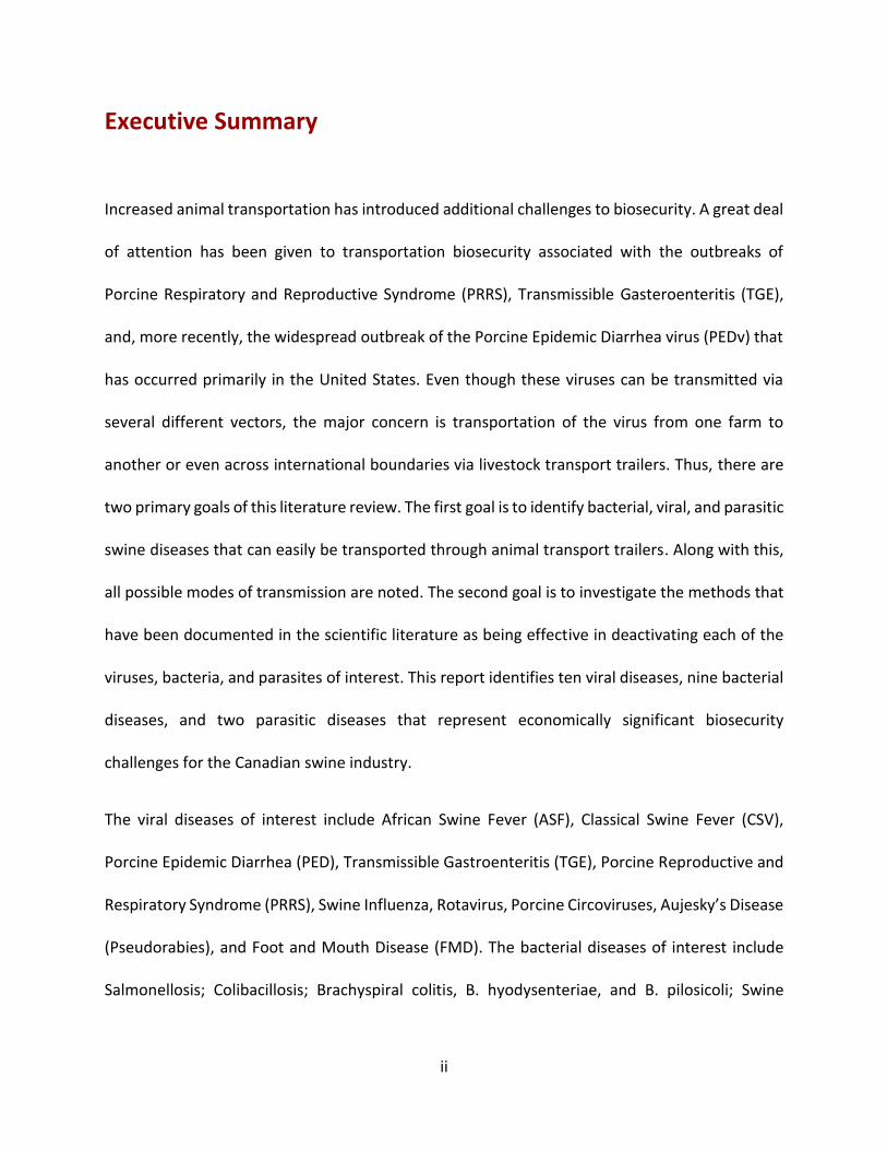
the same disinfectants (Royer et al., 2001). However, the highest effect was also observed with
Virkon S, Sodium hydroxide, Roccal D Plus, and Clorox Bleach, but only resulted in 74%, 61%, 50%
and 46% virus titer reduction, respectively (Royer et al., 2001).
FIG 1: Reduction of porcine circovirus type 2 (PCV-2) infectivity following treatment with eight
different disinfectants, at a final dilution in line with manufacturers’ recommendations, or 3 per
cent sodium hydroxide. Arrows indicate the time of complete inactivation of PCV-2 by certain
disinfectants (source Kim, et al, 2009)
These results coming from in vitro studies need to be interpreted with caution, as the effect of
the disinfectant in the field could be affected by several factors. For example, Patterson and
colleagues (Patterson et al., 2011a) tried a combination of four different livestock transporting
trailer washing and disinfection protocols using disinfectants reported to be effective in the in
vitro studies. Their results showed that none of the disinfectants used alone was better than
washing only to inactivate the PCV 2 viruses in the trailer, but a combination of oxidizing agents

23
followed by sodium hypochlorite was enough to reduce the number of viruses to a level where
they were not able to induce infection in naïve pigs kept for some time in the trailer. The ability
of PCV2 to resist common disinfectants and its ubiquitous distribution makes it difficult to
effectively control PCVAD through strict biosecurity measures; however, studies in France by
Madec and colleagues have shown that a list of management measures, now known as Madec’s
20-point plan, were effective in managing PCVAD and minimizing its economic impact in farms
(Madec et al., 2000). Furthermore, there are now commercially available effective vaccines that
can help in the fight against PCVAD in pig farms (Chae, 2012).
2.9. Pseudorabies (Aujesky’s Diseases) Aujesky’s disease is a highly contagious and economically important disease of pigs that cause a
fatal central nervous system (CNS) infection and high mortality in young pigs. It also causes
respiratory and reproductive diseases in adult pigs. The natural host is the pig, but nearly all
mammals except horses can be infected, resulting in fatal CNS infection (Neumann, 2014). The
disease does not pose a significant risk to human health (Spickler, 2006).
2.9.1. Etiology and Transmission
The etiology of Pseudorabies, the Aujesky’s diseases virus (ADV), is an enveloped, double
stranded DNA virus, which belongs to the genus Varicellovirus of the Herpesviridae family. The
virus is distributed globally, but has been eradicated from most of the developed countries
including Canada and USA; however, the presence of the virus in feral pigs in North America is a
major concern for hog farmers in the US and Canada (Spickler, 2006). The virus is shed in most
body secretions including saliva, nasal discharges, urine, and semen. Hence, transmission is
mainly by direct nose-to-nose contact between pigs or through indirect contact with
24
contaminated fomites, including feed (Spickler, 2006). Long distance airborne transmission
(Christensen et al., 1990; Scheidt et al., 1991) as well as venereal transmission in feral swine
(Romero et al., 2001) were also reported.
2.9.2. Environmental resistance and susceptibility to disinfectants
The virus is fairly resistant outside the host and can survive for several days in a moist and cool
environment. An extensive review by Whittmann showed that the virus is stable over a wide pH
range (5.0 to 12) and can survive for about 6 to 9 weeks at room temperature, about 9 weeks at
15°C, 20 weeks at 4°C and for years at -40°C. However, the virus is inactivated within 30 to 60
minutes at 60oC, within 10 minutes at 70oC, within 3 minutes at 80°C and within a minute at
100°C (Wittmann, 1985). The virus is rapidly inactivated by direct sunlight and drying.
Furthermore, the virus is highly sensitive to most commonly used disinfectants like phenols,
bleach, iodine based disinfectants, formaldehyde and quaternary ammonium compounds
(Spickler, 2006). However, chlorine based disinfectants were reported to be the best
disinfectants; a 3% chloramine solution inactivated the virus within 10 min and a 1% solution
within 30 min (Brown, 1981; Lee and Wilson, 1979). In contrast, the use of caustic soda solution
is not advised, since the virus was not completely inactivated by 1% NaOH even after 6 hours of
contact time (Wittmann, 1985). Therefore, good biosecurity measures supplemented by control
of wild animals and birds can be effective in preventing a herd from contracting the disease.
2.10. Foot and Mouth Disease Foot and mouth disease (FMD) is a highly contagious vesicular disease of cloven-hooved
mammals including domestic and wild pigs. In pigs, the disease is characterized by vesicular
lesions on the feet, snout and around the mouth. All age groups can be affected, but mortality is

25
high only in young animals. Although infected animals usually recover, the morbidity is very high
and results in huge economic losses to the livestock industry (Neumann, 2014). Economic losses
associated with FMD can be direct, as a result of decrease in meat and milk production, or indirect
economic losses associated with diseases control measures. In addition, there are losses due to
the trade embargoes imposed on FMD endemic countries that make these countries un-eligible
to export animals and animal products to disease free countries. A recent review (Knight-Jones
and Rushton, 2013) on the economic impacts of FMD has estimated losses in FMD endemic
regions to be between USD 6.5 and 21 billion per annum, while sporadic outbreaks in disease-
free countries could cost them around USD 1.6 billion a year. Because of the potential that FMD
viruses have for rapid spread within and between countries, and the associated economic
impacts that include trade barriers between endemic and disease-free countries, FMD is one of
the transboundary animal diseases (TAD) that should be reported to the world organization on
animal health, OIE (Leforban and Gerbier, 2002).
FMD has been eradicated from North America in the early 1950s by a combination of vaccination
and stamping out measures (Sutmoller et al., 2003); however, the disease is still devastating the
livestock industry in developing countries, particularly in Sub-Saharan Africa (Sinkala et al., 2014),
South East Asia (Perry et al., 1999) and South America(Clavijo et al., 2015). Despite the FMD free
status of Europe, an outbreak in 2001 in the UK has resulted in the death of close to 10 million
livestock that cost the UK economy around 8 billion pounds (Kao, 2003). More recently, there
was a devastating outbreak in East Asian countries including Japan, South Korea and Taiwan as a
result of the spread of the virus from endemic countries in mainland South East Asia (Knowles et
al., 2012; Valdazo-Gonzalez et al., 2013). Given the endemic nature of the disease in South

26
America, there is always a risk that FMD can spread into North America including Canada.
Therefore, it is important to know about the disease (epidemiology, routes of transmission and
control measures) so that appropriate biosecurity measures will be set at all levels to prevent
introduction of the disease into the Canadian livestock industry.
2.10.1. Etiology and transmission
The etiology of FMD is a non-enveloped, positive sense single stranded RNA virus that belongs to
genus Alphthavirus of the picornaviridae family. There are seven different serotypes of FMD virus
designated as O, A, C, South African Type 1 (SAT 1), SAT 2, SAT 3 and Asian 1. Serotypes A and O
are the most common serotype responsible for most of the outbreaks in Africa, South East Asia
and South America. In contrast, serotype C is very rare and has not been reported from any
country since 2004 (Spickler, 2014). Cross protection between serotypes is very rare and specific
vaccines should be prepared during outbreaks. There are more than 60 strains within these
serotypes and cross protection between strains is variable. The FMD virus common in pigs is the
Cathay strain within the serotype O group (Spickler, 2014).
The virus is shed in all secretions and excretions from an acutely infected animal, including saliva,
milk, urine, feces, expired air and semen, as well fluids from the vesicular lesions in the mouth
and feet area. The FMD virus is the most contagious virus known (Grubman and Baxt, 2004).
Transmission of FMD virus can occur as a result of direct contact between healthy and infected
pigs through skin abrasions or aerosol. Indirect transmission through contaminated fomites,
including ingestion of FMD virus contaminated swills is also possible. However, the primary route
of transmission is through the respiratory tract, as a result of inhalation of virus-containing
aerosols arising from secretions and excretions of infected animals. Pigs respire more infectious

27
FMD virus than other animals, and they are an important source of airborne transmission for
cattle, which are highly susceptible to aerosol infection than pigs are (Alexandersen et al., 2012).
The virus can easily spread via air under suitable environmental conditions, such as high relative
humidity, steady wind and level topography. Airborne transmission up to a distance of 50km
overland (Gloster et al., 2005) and 200km over water (Gloster et al., 1982) has been previously
reported. Furthermore, movement of people and vehicles are claimed to be the reason for the
spread of the virus between farms in the southern part of Japan during the 2010 FMD outbreak
in East Asia (Muroga et al., 2013).
2.10.2. Environmental survival and susceptibility to disinfectants
The virus is hardy and can remain infective for a prolonged period of time in the environment.
Data compiled by (Alexandersen et al., 2012) has shown that the virus can remain viable for up
to 6 months in beddings and fecal slurry, but the typical survival time is for a couple of weeks in
dry feces, up to 4 weeks on hair, up to 39 days in urine, and between 3 to 28 days in soil depending
on the temperature. Survival in the environment is temperature dependent, and the virus
remained infective for about 50 to 70 days on a wool stored at 4oC, for about 12 days at 18oC to
20oC and 2 to 3 days at 37oC (McColl et al., 1995). On the other hand, FMD virus is rapidly
inactivated by high or low pH outside the pH range of 7 to 8 and temperature above 40oC,
resulting in complete inactivation of the virus within one hour at 49oC and in a matter of seconds
at 55oC (Bachrach et al., 1957). The virus is not as such sensitive to sunlight or UV irradiation, but
exposure to direct sunlight kills it within minutes due to the combined effect of drying and
temperature (Alexandersen et al., 2012).

28
Like any other nonenveloped viruses, FMD virus is not sensitive to lipid solvents such as ether
and chloroform, but can be easily inactivated by acidic or alkaline disinfectants, such as acetic
acid, sodium hydroxide and sodium carbonate. Furthermore, oxidizing disinfectants, including
sodium hypochlorite, Virkon S, are very effective against FMD viruses (Neumann, 2014). A recent
study by Krug and colleagues has shown that drying alone can decrease FMD virus by about 3
logs, and disinfectants such as sodium hypochlorite (1000ppm), 1 to 2% citric acid and 4% sodium
carbonate completely inactivated FMD virus on metallic, plastic, and wooden surfaces (Krug et
al., 2012; Krug et al., 2011).

29
3. Bacterial diseases
3.1. Salmonellosis Salmonellosis is a disease that affects a wide variety of host species, including man, and is caused
by any one of the more than 2000 serotypes of salmonella (Carlson et al., 2012). In swine, the
disease is characterized by septicemia and/or enterocolitis. Diarrhea may appear in both types
of salmonella infection, but it is more common in the enterocolitis form of salmonellosis, which
is characterized by severe inflammation and necrosis of the small and large intestine (ISU, 2015b;
Reed et al., 1986). Infection does not always result in disease; most infected pigs remain
asymptomatic carriers, shedding the bacteria for some time and thus infecting other pigs (Carlson
et al., 2012). Pigs are also important sources of infection to humans via contaminated pork
products or as a result of direct contact with infected pigs (Alsop, 2005). Infection can occur in all
age groups of pigs, but the disease is primarily observed in weaned piglets or grower/finisher pigs
between weaning and 180 days of age (Carlson et al., 2012). The disease is precipitated by
stressful conditions like weaning, change of diet and transportation (Isaacson et al., 1999).
Clinical cases of salmonellosis is higher in younger pigs, but the rate of bacterial shedding is very
high in breeding sows (Wilkins et al., 2010). Special attention should be given to farrowing sows,
as they are the primary source of infection to newborn piglets.
3.1.1. Etiology and Transmission
Salmonellosis in swine is caused by small, rod shaped, gram negative, facultative intracellular and
facultative anaerobic bacteria that belong to family enterobacteriaceae, genus salmonella.
Salmonella species are classified into several serotypes based on the lipopolysaccharide somatic
(O) and flagella protein (H) antigens. There are more than 2000 serotypes and more than a dozen

30
of them have been isolated from pigs, but the two major causes of salmonellosis in swine are
Salmonella cholerasuis and Salmonella typhimurium, which are responsible for the septicemic
and enterocolic forms of salmonellosis, respectively (Reed et al., 1986). Salmonella species are
shed in the feces of infected animals; sub clinically infected animals (carriers) are a special
concern as they can shed the bacteria for a long period of time without showing any clinical signs.
Carrier pigs can be important sources of infection to healthy pigs and even humans. Feeding
probiotics or prebiotics to pigs was reported to decrease shedding of salmonella in the feces of
subclinically infected carrier pigs (Letellier et al., 2000).
The major route of salmonella transmission is direct contact by the fecal-oral route through
ingestion of contaminated feed and water or indirectly through contact with contaminated
fomites. Salmonella species were isolated from pig feed and vehicles used to transport feed to
pig farms (Fedorka-Cray et al., 1997), suggesting the possibility that contaminated fomites
including vehicles can act as a reservoir host for salmonella infection. Some reports also showed
that aerosol transmission, either through direct nose-to-nose contact or through long distant
airborne dissemination, was a possible means of transmission (Proux et al., 2001). A role for other
animals like cats, rodents, flies and birds in the transmission of salmonella to pigs has also been
documented (Barber et al., 2002).
3.1.2. Environmental survival and susceptibility to disinfectants
Salmonella are hardy bacteria that can resist environmental conditions and stay viable for
months and even years in suitable organic substances such as manure and wet bedding.
Salmonella species were recovered after 180 days from hog manure treated soil and up to 21
weeks in contaminated water (Cote and Quessy, 2005; Holley et al., 2006). The host adapted

31
salmonella (S. cholerasuis) was believed to be sensitive to the environment and unable to survive
outside the host; however, a group from the United States Department of Agriculture-
Agricultural Research Service were able to recover the host adapted S. cholerasuis after 3 months
from fecal samples stored under wet condition and after 13 months from a desiccated fecal
sample (Gray and Fedorka-Cray, 2001).
On the other hand, salmonella species can be easily killed in a short period of time by moist heat.
Moist heat at 71oC or higher can kill salmonella species in less than a minute if not covered with
any organic matter (Spickler, 2005a). However, inactivation of salmonella species covered with
feces or chicken litter required more than 80 to 100 minutes, depending on the moisture level,
for complete inactivation by heating at 70oC (Kim et al., 2012a). Salmonella are also susceptible
to many disinfectants and can be easily inactivated with chlorine compounds (1% Sodium
Hypochlorite), formaldehyde, iodine-based disinfectants, 2% glutaraldehyde, 70% Ethanol, and
phenols (Spickler, 2005a). In addition to this, ozone has been used in the food industry to
inactivate a broad range of microbes including S.typhimurium (Restaino et al., 1995). The
difficulty with effective disinfection of salmonella species occurs when they form biofilms on the
surface of biotic and abiotic materials. In this regard, the emerging S. typhimurium strain DT104
is of particular concern due to its resistance to major antibiotics and its ability to form biofilms
on abiotic surfaces (Ngwai et al., 2006). Biofilm forming salmonella are tough to kill with common
disinfectants; however, a combination of ozone and organic acids was reported to have a
synergistic effect to inactivate biofilm forming S. typhimurium on the surface of abiotic materials
(Singla et al., 2014).

32
3.2. Colibacillosis Colibacillosis is a disease caused by E.coli and characterized mainly by diarrhea and occasionally
septicemia and bowel/gut edema. Diarrhea in the newborn (neonatal diarrhea) occurs between
0 to 4 days after birth, while post-weaning diarrhea (PWD) and bowel edema, also known as ED,
occurs later in the nursery or 1 to 2 weeks post weaning. PWD and ED are economically important
diseases for hog producers due to the high morbidity, mortality and weight loss in piglets, as well
as the high cost of drugs and vaccines incurred to control the disease (Fairbrother and Gyles,
2012). Some serotypes of E.coli, such as the O157:H7 are also zoonotic and can infect humans
through contaminated pork products; however, pig does not seem to be the natural host for this
strain of zoonotic E.coli (Chapman et al., 1997).
3.2.1. Etiology and Transmission
Escherichia coli are gram negative, facultative anaerobic, flagellated and rod shaped bacteria
classified under the family Enterobacteriaceae. Species of E.coli are classified into different
serotypes or strains based on the Somatic (O), Capsular (K), Flagellar (H) and Fimbrial (F) antigens.
Currently, at least 175 O, 80 K, 56 H, and 20 F antigens have been identified (Fairbrother and
Gyles, 2012). In swine, five antigenically distinct types are reported. F4 (K88) and F18 are common
in postweaning pigs, while F4 (K88), F5 (K99), F41 and F6 (987P) are commonly isolated from
neonatal piglets (Francis, 1999). These strains of E.coli are further classified into pathotypes
based on their virulence mechanisms as Enterotoxigenic E.coli (ETEC) or attaching and effacing
E.coli (AEEC) also known as EPEC, Enteropathogenic E.coli (Fairbrother and Gyles, 2012). The
primary sources of infection are infected animals shedding the bacteria in their feces, urine, nasal
and oral secretions. Sows are assumed to be the silent carriers that infect newborn piglets

33
immediately after birth (Fairbrother and Gyles, 2012) and transmission is either directly through
the fecal-oral route by ingestion of contaminated feed and/or nose-to-nose contact or indirectly
through by the aerosol route (Cornick and Helgerson, 2004; Cornick and Vukhac, 2008).
3.2.2. Environmental survival and susceptibility to disinfectants
Generally E.coli are resistant to adverse environmental conditions and can remain viable and
infectious outside the host for prolonged period of time. Reviews that compiled data from several
published papers on the environmental survival of E.coli showed the ability of the bacteria to
resist high temperature fluctuations, acidic pH, and drying/desiccation conditions and survive for
more than a year in soils, manure and water (Cote and Quessy, 2005; van Elsas et al., 2011). E.
coli was also reported to survive for more than 14 months on the surface of inanimate objects or
fomites that can then act as a source of infection to pigs through indirect contact (Kramer et al.,
2006).
On the other hand, E.coli bacteria are susceptible to heating and can be killed within seconds by
boiling at 70oC (Oie et al., 1999). The bacteria are also highly susceptible to most of the commonly
used disinfectants; however, some strains of E.coli, such as, the O157:H7 are able to make
biofilms when they are outside the host and become resistant to most oxidizing disinfectants
(Vogeleer et al., 2014). Treatment of biofilms with aqueous chlorine dioxide followed by drying
at 22oC and 43% relative humidity completely inactivated biofilm forming E.coli within 6hrs (Bang
et al., 2014), suggesting that washing, disinfection, and drying of livestock transporting vehicles
can completely inactivate all kinds of E.coli species.

34
3.3. Swine Brachyspiral Colitis Swine Brachyspiral colitis is a severe bacterial infection of pigs characterized by
mucohemorrhagic diarrhea known as swine dysentery (SD) and mild spirochaetal colitis (SC)
associated with inflammation of the large intestine (cecum and/or colon) of pigs (Neumann,
2014). Swine dysentery is seldom reported in young pigs, but it is wide spread in the
growing/finishing period of the pig cycle and can result in a huge economic losses to the hog
industry due to mortality and suboptimal performance of pigs with reduced feed conversion
efficiency and retarded growth during diseases outbreaks (Wills, 2000). The second form of
Brachyspiral infection, spirochaetal colitis (SC) usually occurs as mild non-hemorrhagic colitis in
young pigs between the age of 2 to 3 weeks (Neumann, 2014).
3.3.1. Etiology and Transmission
Swine dysentery is caused by gram negative, motile, spiral shaped, anaerobic bacteria that belong
to the genus Brachyspira in the Spirochaetaceae family (Paster and Dewhirst, 2000). Brachyspira
inhabit the large intestine of birds and mammals and six species of Brachyspira have been
isolated from the large intestine of swine (Hampson, 2012). Most Brachyspira species in swine
are nonpathogenic commensals; however, B. hyodysenteriae and B. pilosicoli are pathogenic and
induce disease in pigs characterized by severe hemorrhagic dysentery (SD) and mild spirochaetal
colitis (SC), respectively (Hampson, 2012). Previously, B. hyodysenteriae and B. pilosicoli were
classified under genus Treponema and then genus Serpulina until they were finally classified
under genus Brachyspira (Ochiai et al., 1997).
The natural host for B. hyodysenteriae and B. pilosicoli is the pig, including feral pigs; however,
the bacteria have also been isolated from birds and other mammals like mice, rats and dogs that

35
act as an important source of infection to pigs (Alvarez-Ordonez et al., 2013). The bacteria are
shed in feces of both sick and asymptomatic carrier animals and transmission is through ingestion
of feces contaminated feed and water. Transmission can also occur through indirect contact with
contaminated fomites like boots, clothing and livestock transport vehicles, as well as by
mechanical vectors like rodents, flies and birds (Hampson, 2012; Neumann, 2014). In addition to
this, a comprehensive review on Brachyspira, recently published in the International Journal of
Environmental Research and Public Health (Alvarez-Ordonez et al., 2013) has extensively
described the determinants that influence survival of the bacteria in the environment, all the
sources and routes of transmission, and factors contributing to the establishment of swine
dysentery in pigs. The figure shown below taken from the above review is a summary of the
factors involved in the transmission of Brachyspira species.
Factors influencing the establishment and transmission of Swine Dysentery (SD), taken from Alvarez-
Ordonez et al., 2013.

36
3.3.2. Survival in the environment and susceptibility to disinfectants
Brachyspira species are anaerobic, but can stay viable for a few weeks to several months
depending on the presence of organic matter, moisture level, and temperature of the
environment. Compared to other Brachyspira species, B. hyodysentery is sensitive to
environmental factors and was reported to survive only for 10 days in pure soil held at 10oC. The
survival time in soil was increased to 78 days in the presence of 10% pig feces and was even
longer (112 days) in pure wet pig feces. On the other hand, the less pathogenic B. pilosicoli was
reported to survive for 119 days in pure soil and for more than 210 days in soil in the presence of
10% pig feces (Boye et al., 2001). Brachyspira species, however, are highly susceptible to high
environmental temperatures and drying, which can be killed by desiccation and temperatures
above room temperature (Chia and Taylor, 1978). The optimal environmental temperature for
long term survival of Brachyspira is 0-10oC and survival decreases with decreasing moisture and
increasing temperature and pH. Temperature above 37oC and alkaline pH >8 is lethal to
Brachyspira and they can be effectively inactivated by heating at 56oC for few minutes (Alvarez-
Ordonez et al., 2013; Chia and Taylor, 1978), suggesting that control of the diseases will be more
effective during the hot summer season using alkaline solutions.
Brachyspira species are also susceptible to common disinfectants like phenols and sodium
hypochlorite (Hampson, 2012). A study conducted to examine the effect of seven different
disinfectants (quateranery ammonium compounds and organic acids) showed that B. pilosicoli
can be easily inactivated by most disinfectants in the presence or absence of organic matter, even
though the inactivation process was reduced or/and slowed down in the presence of organic
matter like feces (Corona-Barrera et al., 2004). For example, the efficacy of Virkon S was highly

37
reduced in the presence of feces and required a more concentrated solution (1:10) than the
recommended dose (1:200) to attain 100% inactivation. Comparable results were also reported
from another experiment, which used Virkon S and Chirox to inactivate B. hyodysenteriae in the
presence of 10% fecal slurry from SPF pigs as source of organic matter, (Lobova and Cizek, 2004).
The implications of all these results is that good biosecurity measures applied in the farm and on
fomites as well as livestock transport vehicles can effectively control Brachyspira infection and
spread. These results also suggest the importance of cleaning and removal of organic matter by
washing before disinfection.
3.4. Swine Erysipelas Erysipelas is an infectious disease of pigs that can occur in all stages of pork production cycle. It
manifests itself clinically either as an acute septicemia associated with endocarditis and sudden
death in grow-finisher pigs or as chronic arthritis and lameness in adult pigs (Opriessnig and
Wood, 2012). The septicemic form of the disease is also characterized by a typical diamond-
shaped skin discoloration around the ear, snout and the abdomen (Neumann, 2014). If
uncontrolled, both the septicemic and the chronic form can result in significant economic losses
to the pork industry due to mortality, reduction in pig growth, and a decrease in meat quality
(Opriessnig and Wood, 2012). Swine Erysipelas is also a zoonotic disease and infection in man is
mostly occupationally related, affecting primarily farm and abattoir workers as a result of contact
with animals, their products, or their wastes. In humans, Erysipelas can cause mild cutaneous
infection known as erysipeloid, or a serious systemic complication with septicemia and
endocarditis (Brooke and Riley, 1999).

38
3.4.1. Etiology and Transmission
The causative agent of Erysipelas in pigs is Erysipelothrix rhusiopathiae, which is a gram positive,
slightly curved, rod shaped bacteria that belongs to the genus Erysipelothrix. The bacteria can
grow under a wide temperature range (5oC to 44oC), but the optimal temperature is 30oC to 37oC
(Opriessnig and Wood, 2012; Reboli and Farrar, 1989). The domestic pig is the natural host, but
the bacteria can also infect a wide range of domestic and wild mammals including chicken,
turkeys, ducks and lambs that act as reservoir hosts and hence as sources of infection to the
domestic pig (Wang et al., 2010). Not all infected pigs show symptoms of infection, and 30 to
50% of infected pigs remain as carriers harboring the bacteria in the oropharynx and tonsils
(Okolo, 1986). However, both sick and carrier animals shed the bacteria in their feces, as well as
oral and nasal secretions, representing a significant source of infection to healthy animals and an
important source of environmental contamination (Okolo, 1986; Takahashi et al., 1987).
Transmission is mainly by the fecal-oral route through ingestion of contaminated feed and water,
but direct inoculation through skin wounds can also occur. Indirect transmission through
contaminated fomites and carrier vectors including wild animals, birds, rodents, reptiles, and flies
is also an important source of infection to domestic pigs (Neumann, 2014).
3.4.2. Environmental survival and susceptibility to disinfectants
Erysepalothrix are stable in the environment and can survive for a prolonged period of time.
Therefore, the environment is an important source of Erysepalothrix infection to susceptible
animals. A comprehensive review compiled by (Brooke and Riley, 1999) showed that the
organism prefers cooler temperatures, alkaline pH (6.7 to 9.2) and the presence of organic matter
like feces for long time survival. Erysepalothrix can survive for up to 5 days in drinking water,

39
about 14 days in sewage, at least 35 days in the soil, and 1 to 5 months in feces. However, the
bacteria are sensitive to heat and can be killed in moist heating at 55oC in 15 minutes (Reboli and
Farrar, 1989). They are also highly susceptible to most household disinfectants (Fidalgo et al.,
2002). As an important consideration for food safety, Erysepalothrix are resistant to salting,
curing and many other food preserving techniques, which make it difficult to control the
dissemination of the bacteria to humans (Opriessnig and Wood, 2012). Despite the susceptibility
of the bacteria to many antimicrobial agents, including common disinfectants, and the availability
of an effective vaccine, controlling the diseases and eradication of Erysepalothrix from swine
herds is still difficult and complicated primarily. This could be, as discussed above, due to the
involvement of a wide variety of animals able to act as reservoir hosts and the ability of the
bacteria to survive in the environment for a prolonged period of time.
3.5. Mycoplasmal Pneumonia Mycoplasmal pneumonia, also known as Enzootic pneumonia, is an infection of the respiratory
tract of pigs resulting in chronic bronchopneumonia associated with suppression of immunity in
the lung, thereby creating a suitable condition for opportunistic bacterial and viral pathogens to
flourish and infect the lung (ISU, 2015a). Even though Enzootic pneumonia can occur as a single
entity by itself, it is usually reported as part of the porcine respiratory diseases complex (PRDC)
together with swine influenza, PRRS, porcine circovirus infections, and other bacterial infections
(ISU, 2015a; Thacker et al., 1999). It is worldwide in distribution, reported from almost all pork
producing countries, and is commonly seen in its chronic form characterized by dry cough, high
morbidity and low mortality rates. Acute fatal cases, however, can also occur in naïve and
immunocompromised animals (Dee, 2014). It usually affects grower-finishers and incurs

40
significant economic losses to swine farmers due to reduced feed conversion and retarded
growth rate of infected animals, and also due to the central role it plays in the pathogenesis of
PRDC (Pointon et al., 1985; Thacker et al., 1999). A review compiled by Thacker and colleagues
on the economic importance of enzootic pneumonia to pig farmers found a decrease of 17% and
14% for daily weight gain and feed efficiency, respectively. The cost of the disease was estimated
to be around $4.08 per pig, and when the cost of control measures is included, the disease may
cost the US hog industry close to $500 million per year (Thacker and Minion, 2012).
3.5.1. Etiology and Transmission
The causative agents of enzootic pneumonia, Mycoplasma pneumonia, also known as
Mycoplasma hypopneumenia, are the smallest bacteria classified under the Mollicutes class of
bacteria that lack a cell wall. They are very sensitive to environmental factors outside the host.
However, the absence of the cell wall structure allows these organisms to be resistant to
antibiotics such as penicillin that target the cell wall. The absence of a cell wall also prevents the
bacteria from staining using gram’s stain (Razin et al., 1998). Despite difficulties in staining, they
are believed to be descendants, by mutation, of gram positive bacteria, and thus they are
grouped under gram positive bacteria (Pieper et al., 1995). Mycoplasma are the smallest known
bacteria that can grow in a cell-free media, but the bacteria need special requirements for
successful in vitro cultivation. Hence, Mycoplasma are termed fastidious bacteria (Thacker,
2004).
M. hypopneumonia (MHP) is excreted in nasal secretions of infected or carrier pigs and the main
route of transmission is by direct nose-to-nose contact between infected and healthy pigs (Meyns
et al., 2004). Even though the role of indirect transmission through contaminated fomites is not

41
well investigated, there is still a possibility of transmission through this medium as the organism
can live for a few days in a cool and wet environment (Goodwin, 1972). In one study, however,
where personnel were using routine standard hygienic measures before entering a susceptible
herd, enzootic pneumonia was not transmitted from infected to healthy pigs during the 20 weeks
observation period when the workers had a frequent contact with both the infected and healthy
pigs (Batista et al., 2004). Therefore, even though a possibility for indirect transmission of
mycoplasma by contaminated fomites (Fano et al., 2005), including animal transport vehicles
(Hege et al., 2002) was previously reported, it is limited to poorly managed farms. Thus,
introduction of enzootic pneumonia into a farm or population of pigs is most probably caused by
infected or/and carrier pigs (ISU, 2015a). Despite its sensitivity to the external environment, there
is convincing evidence in the literature that show airborne dissemination of the bacteria is
possible. Airborne transmission up to 150 m was reported by (Cardona et al., 2005), while others
reported a spread of the bacteria to distant farms up to 3.2 to 9.2 Km is possible (Dee et al.,
2009a; Goodwin, 1985; Otake et al., 2010).
3.5.2. Environmental survival and Susceptibility to disinfectants
Due to lack of the cell wall, Mycoplasma bacteria are highly sensitive to the external environment
and cannot survive outside the host body for an extended period of time. However, they can
stay alive for some time in a cool and moist condition. For example, (Cardona et al., 2005)
reported survival of the bacteria for more than 30 days in water at 2 to 7oC. Similarly, the bacteria
was reported to survive up to 7 days if covered in organic matter such as nasal discharges or
mucous (Hurnik, 2005). Nonetheless, the bacteria are killed within a few hours under dry and hot
weather conditions in the environment as well as by heating to temperatures above 45OC. Typical

42
mycoplasma inactivation protocols to avoid laboratory contamination are conducted by heating
for 30 minutes at 45OC or for 10 minutes at 60OC, or by irradiation at 25 to 35 kGy (personal
communication), suggesting that heating and irradiation could be used to inactivate M.
hypopneumonia from fomites, including transport vehicles. Mycoplasma are also highly sensitive
to almost all commonly used disinfectants (ISU, 2015a) making it easy to control the spread of
mycoplasma in pigs farms using good biosecurity procedures.
3.6. Leptospirosis Leptospirosis is a highly contagious disease of both domestic and wild animals. The disease is
distributed globally, with a major economic impact on the livestock industries (Ellis, 2015).
Leptospirosis is also an important zoonotic disease in humans (Bharti et al., 2003). Swine
leptospirosis is wide spread in all pig farming countries, but its economic impact is largely
confined to intensive pig farming systems in western countries, including Australia, New Zealand
and Brazil (Ellis, 2012). The causative agent of leptospirosis persists in the kidney and genital tract
of pigs, resulting in urinary and reproductive tract infection (Ellis et al., 1986b). Reproductive tract
infection is associated with huge economic losses in breeding sows due to abortion, still birth,
reduced litter size, and reduced number of weaned piglets (Kemenes and Suveges, 1976; Ramos
et al., 2006).
3.6.1. Etiology and Transmission
The etiological agents of leptospirosis are small, motile, aerobic, gram negative spirochetes that
belong to the genus Leptospira. The genus includes several species of pathogenic and saprophytic
bacteria, which are further classified into more than 250 serovars. Pigs can be infected by several
serovars of Leptospira, but the most prominent one is L. interogans serovar Pomona (Spickler,

43
2013). The bacteria persists in the urinary and genital tract of pigs and is excreted in urine and
genital discharge of infected or carrier animals (Ellis et al., 1986b). Transmission is either by direct
contact with infected pigs, particularly aborted sows, or by indirect contact via contaminated
feed and water. Live animal vectors, including rats, mice, skunks, raccoons, foxes, and possums
are also an important source of infection (Neumann, 2014). Water contaminated with urine from
carrier animals including boars (Ellis et al., 1986a), foxes (Kingscote, 1986a), and skunks
(Kingscote, 1986b) is reported to be important source of infection for healthy pigs, and infection
is primarily through skin wounds or contact with the mucus membrane.
3.6.2. Environmental survival and susceptibility to disinfectants
Leptospira can survive outside the host for a considerable period of time, making the
environment an important reservoir of infection. The length of time Leptospira survives outside
the host depends on moisture, temperature, and pH of the environment, and the bacteria prefers
warm, moist, and close to neutral pH conditions (Levett, 2001). It is difficult to determine the
exact length of time Leptospira survives under different conditions, as the requirement for
various serovars could be different, but a review of several published data compiled by (Levett,
2001) has shown that Leptospira species can remain viable for several months in water at room
temperature in a pH range of 6.7 to 7.3. Survival was prolonged in fecal slurry kept at cooler
temperature under shade. On the other hand, Leptospira were reported to be sensitive to salty
water, exposure to direct sun light, and to desiccation (Khairani-Bejo et al., 2004). The bacteria
are also sensitive to heating, and were killed in a short period of time by heating at temperatures
> 45oC (Parker and Walker, 2011). Leptospira are highly sensitive to most commonly used
detergents and disinfectants including 1% sodium hydroxide, 70% ethanol, iodine compounds,

44
quaternary ammonium compounds, hydrogen peroxide, glutaraldehyde, and formaldehyde
(Spickler, 2013).
3.7. Actinobacillosis Actinobacillosis is a highly contagious respiratory infection of pigs caused by several species of
bacteria that belongs to the family Pasteurellaceae and genus Actinobacillus. There are more
than 22 species of bacteria under this genus, but only four species are associated with diseases
in animals and two of these, Actinobacillus pleuropneumoniae and Actinobacillus suis, are
important pathogens in pigs (Smith, 2013).
3.7.1. Actinobacillus pleuropneumoniae (APP)
APP is gram negative coccobacillus that affects only pigs and causes a highly contagious and
severe pleuropneumonia, also known as contagious porcine pleuropneumonia (CPP). The disease
is one of the most economically important respiratory diseases of pigs and is characterized by
sudden onset and death in susceptible populations (Neumann, 2014). It is worldwide in
distribution and economically important to the hog industry because of high morbidity (retarded
growth) and also mortality. Mortality is high when the disease occurs in conjunction with other
respiratory infections as a component of the porcine respiratory diseases complex, PRDC
(Gottschalk, 2012). It is still a problem in many European countries and is also widely distributed
in North America. In one study, 78% and 70% of the herds in Ontario were positive for APP based
on PCR or ELISA tests, respectively (MacInnes et al., 2008). It can affect all age groups of pigs, but
it is primarily reported from pigs between 6 to 20 weeks of age (Neumann, 2014).

45
3.7.1.1. Etiology and Transmission
The etiological agent of APP is Actinobacillus pleuropneumoniae. There are two biotypes
depending on their requirement for nicotinamide adenine dinucleotide (NAD) for in vitro growth
and cultivation (Pohl et al., 1983). There are more than 15 serotypes within these two biotypes,
which differ in their virulence and epidemiological distribution. The most virulent strains include
serotype 2 in Europe, serotype 15 in Australia, and serotypes 1 and 5 in North America
(Gottschalk, 2012). However, in Canada, there seems to be a shift in prevalence from the most
virulent serotypes 1 and 5 into the less virulent serotypes 3 and 7, which explains the high
prevalence of the diseases in swine herds but very low clinical disease reported from those farms
(MacInnes et al., 2008).
APP is primarily a disease of swine and resides in the tonsils of asymptomatic carriers and
recovered pigs. Healthy carriers and survivor pigs can shed the virus for months in nasal
discharges, and transmission is mainly by direct contact through nose-to-nose. Therefore,
dissemination between farms or pens within a farm is believed to be mainly by the introduction
of infected live animals (Gottschalk, 2012). However, there are also studies that show indirect
transmission through contaminated fomites or aerosol transmission through droplets are also
possible ways to disseminate APP within or between swine herds (Desrosiers and Moore, 1998;
Tobias et al., 2014). Regardless, the direct transmission is 10 times more efficient than the
indirect transmission in spreading the bacteria between pigs on a farm (Tobias et al., 2014).
Airborne transmission between pens over a short distance, up to 2.5 meters, has also been
reported (Jobert et al., 2000). Even though the role of vectors like rodents and birds is limited,
long-distance dissemination of the bacteria to other farms via air and livestock transport vehicles

46
should be considered in any control plan to prevent the spread of APP between farms.
Epidemiological investigation, using molecular typing methods, on the route of transmission of
the disease during an outbreak in specific pathogen-free herds in Denmark implicated airborne
transmission in 5 out of 12 cases. Transmission by trailers was implicated in 6 out of 9 cases
investigated (Fussing et al., 1998).
3.7.1.2. Environmental survival and susceptibility to disinfectants
APP are delicate bacteria that are highly fragile in the environment, particularly under hot and
dry conditions; therefore, it is unlikely that APP would survive for a prolonged period of time
outside the host on inanimate objects. Nevertheless, there are several reports which show APP
can survive outside the host for enough time to be a concern for indirect transmission through
contaminated fomites and even livestock transport vehicles. In one study, APP was reported to
survive for 3 to 4 days in a dried nasal secretion and even for weeks in physiologically buffered
saline (PBS) stored at 4oC. Furthermore, APP was reported to survive storage at -20oC or -70oC
for about 4 months, suggesting the possibility of long term survival of the bacteria in frozen pig
carcasses (Assavacheep and Rycroft, 2013). The survival time of APP decreases with increasing
environmental temperatures. APP was reported to survive for less than 8 hours at 37oC and less
than 4 hours at 42oC (Morozumi and Hiramune, 1982).
Actinobacillus are also highly sensitive to most disinfectants; however, the efficacy of various
disinfectants against APP may vary depending on the presence or absence of organic matter such
as nasal discharges. In one study which tested the efficacy of 23 different disinfectants against
APP, Chloramine-T, hydrogen peroxide, glutaraldehyde, quaternary ammonium compound, 2.5%
glutaraldehyde, 6.8% glyoxal, and 6% formaldehyde were found to be effective in killing APP even

47
in the presence of organic matter (Gutierrez et al., 1995). In the same study, the efficacy of most
disinfectants was reported to decrease in the presence of serum as an organic matter, suggesting
the importance of cleaning and washing organic matter from the surface before applying
disinfectants for effective inactivation of AAP.
3.7.2. Actinobacillus suis
Actinobacillus suis is an infectious disease of pigs characterized by hemorrhagic septicemia that
mainly affects the lungs but is also seen in other organs. The bacteria reside in the upper
respiratory tract of pigs, and infection is asymptomatic in most cases. For example, in one study,
more than 94% of the herds tested in Ontario, Canada were estimated to be infected with A. suis
without showing clinical signs (MacInnes et al., 2008). In a naïve herd, the bacteria may affect
primarily suckling piglets, but weaned, fattening and adult pigs can also be affected (Yaeger,
1996). In a naïve and high health status herds, the disease may manifest itself in various clinical
forms including sudden death and acute septicemia in suckling and recently weaned piglets,
respiratory disease characterized by hemorrhagic and necrotizing pleuropneumonia in grow-
finish pigs, and septicemia in adult pigs (Yaeger, 1995). Actinobacillosis due to A. suis infection
may not be economically as important as infection with A. pleuropneumonia; however, all the
transmission and epidemiological features are more or less the same as what has been discussed
for APP.
3.8. Streptococcosis Streptococcosis refers to a variety of infectious disease that affect both man and animals.
Streptococcosis is caused by a group of gram positive cocci that belong to the phylum Firmicutes,
order Lactobacillales, family Streptococcaceae, and genus Streptococcus. The bacteria reside in

48
the body of animals and humans as part of the normal microflora, and disease occurs either as a
result of decreased immunity in the host, appearance of more virulent strains by mutation, or
transfer of the bacteria to an unusual site within the host body (Spickler, 2005b). Some species
of streptococcus are zoonotic, in that they use animals as reservoir hosts and cause severe illness
in humans. The most important zoonotic streptococcus is S. suis, reported to affect around 1600
people in 2012 alone, primarily in south east Asia but also in Europe and North America (Huong
et al., 2014). Streptococci are classified into three groups, based on their ability to lyse red blood
cells on blood agar, as Alpha hemolytic, Beta hemolytic and non-hemolytic groups. They are
further classified into different groups (Lancefield grouping) and serotypes based on serological
reaction to cell wall and capsular antigens of the bacteria (Spickler, 2005b). Even though several
species of streptococcus are able to cause sporadic disease in pigs, emphasis will be given here
to S. suis due to its economic significance in the swine industry worldwide (Staats et al., 1997)
and its zoonotic significance in humans (Huong et al., 2014). The bacteria primarily affect young
pigs immediately after weaning or in the nursery; however, the disease can also affect adult pigs.
The disease in young pigs is characterized by acute onset of illness, septicemia, meningitis,
polyarthritis, polyserositis, and bronchopneumonia usually followed by sudden death, and
sometimes endocarditis in adult pigs. The infection may also occur in other domestic animals,
including cattle, sheep, goats, horses and dogs, as well as in humans (Neumann, 2014) .
3.8.1. Etiology and Transmission
Streptococcus suis is a gram positive, non-hemolytic streptococcus with cell wall antigenic
determinant related to the Lancefield group D (previously, R, S, RS, and T groups). There are 35
serotypes identified based on serological reaction to the capsular antigens. Several serotypes of

49
S. suis may infect pigs, but serotype 2 is the most commonly isolated serotype from sick pigs and
humans worldwide (Gottschalk et al., 2007). The bacteria normally reside in the upper respiratory
tract (tonsils and nasal cavity), gastrointestinal tract, and genital tract of healthy carriers. It is
estimated that 60 to 100% of healthy pigs in a herd carry S. suis without showing clinical signs of
the disease, but only 2 to 15% develop the clinical disease (Spickler, 2005b).
Streptococcus suis are excreted in the nasal discharge of healthy carriers or sick pigs, and
transmission is primarily by direct nose-to-nose contact between pigs or aerosol over a short
distance. Thus, introduction into a farm is mainly through live animals (Staats et al., 1997).
However, the organism has been isolated from manure-coated farm and veterinary equipment,
suggesting the possibility of indirect transmission through contaminated fomites (Dee and Corey,
1993). The bacteria was also isolated from house flies (Enright et al., 1987) implicating the role
of mechanical vectors like flies, birds, and rodents in the transmission of S. suis. Furthermore, the
organism is commonly isolated from the reproductive tract of aborting sows, and piglets can be
infected in the birth canal during delivery (Robertson et al., 1991). The role of transport vehicles
in the dissemination of S. suis has not been fully investigated, but, as the organism can survive
on fomites for a considerable period of time, it is possible that trailers can spread the bacteria in
the absence of good biosecurity measures. In agreement with this, manure containing S. suis
spread on truck tires was isolated from the tire treads after the truck was driven for 4.82 km at
an average speed of 64.3 km/h, but not after an additional 12.87 km at a speed of 96.5 to 120.6
km/h (Dee and Corey, 1993).

50
3.8.2. Environmental resistance and susceptibility to disinfectants
Streptococcus suis is relatively resistant to the external environment, and it can survive for a
considerable period of time outside the host, particularly in moist and cool conditions. In an in
vitro study conducted by (Clifton-Hadley and Enright, 1984), the organism was reported to
survive for 1 to 2 weeks in water at 4oC; it also survived for 104 days in feces stored at 0oC, but
only for 25 days when stored at 9oC. In dust stored under similar conditions, S. suis remained
viable for about 54 days at 0oC and 10 days at 9oC. However, increasing the temperature to about
25oC (room temperature) decreased the survival time of the bacteria to about 8 days in feces and
to less than 24 hours in dust. Furthermore, heating to 60oC inactivated S. suis in 10 minutes. S.
suis are also sensitive to detergents and disinfectants, and can be inactivated in less than a
minute with most of the commonly used disinfectants, as well as ordinary liquid hand soaps
(Clifton-Hadley and Enright, 1984). In one study, which tested the efficacy of seven commonly
used disinfectants including 70% ethanol, Chlorhexadine, formaldehyde, 3% Iodine, One-Stroke
Environ (Pheno), Roccal-D (quaternary ammonium compound), and 5% sodium hypochlorite, no
growth of bacteria was reported in any of the disinfectants except for 70% ethanol (Dee and
Corey, 1993). The efficacy of disinfectants can be reduced by the presence of organic matter such
as feces (Dvorak, 2008b); therefore, careful cleaning and washing of equipment before
disinfection is very important for complete inactivation of the bacteria and to limit its spread
among pigs.
3.9. Glӓsser’s Disease Glӓsser’s disease is an infectious disease of pigs characterized by fibrinous polyserositis,
polyarthritis, and meningitis with bacterial septicemia (Amano et al., 1994). It may also have a

51
pulmonary form when it occurs as a co-infection with other pulmonary diseases like PRRS and
swine influenza (Aragon et al., 2012). The disease usually affects young pigs between the ages of
4 to 8 weeks, although sporadic cases may occur in adult pigs. The disease is manifested clinically
with sudden onset and death during stressful conditions like weaning, mixing of piglets,
transportation, or experiencing a change in diet (Neumann, 2014). Glӓsser’s disease is global in
distribution, reported from all swine-raising countries. Recently Glӓsser’s disease is becoming
significant health problems in the nursery piglets of modern and high health standard pig
production systems (Aragon et al., 2012).
3.9.1. Etiology and Transmission
The etiological agent of Glӓsser’s disease is Haemophilus parasuis, which belongs to the family
Pasteurellaceae and genus Haemophilus bacteria. The bacteria are small gram negative highly
fastidious rods requiring the NAD (V factor), but not X (hemin factor) for their in vitro cultivation
(Biberstein and White, 1969). More than 15 serovars have been identified so far (Kielstein and
Rapp-Gabrielson, 1992; Rapp-Gabrielson and Gabrielson, 1992), and pigs may harbor several of
them depending on the geographical location. For instance, Serovars 4, 5, 13 and 14 are more
prevalent in North America (Neumann, 2014). Haemophilus parasuis is a commensal organism of
the upper respiratory tract of healthy pigs causing diseases in young pigs only under certain
circumstances. It is among the first colonizers of the respiratory tract in piglets and was isolated
from piglets immediately after birth (Oliveira and Pijoan, 2004). The organism has never been
isolated from the reproductive tract of sows, thus infection of piglets is most probably by direct
contact with the sows immediately after birth (Aragon et al., 2012). In agreement with this, pigs
born by “snatch farrowing” where piglets were taken away directly from the birth canal to avoid

52
contact with the mother, were free of H. parasuis and were used as animal models to study
pathogenesis of H. parasuis infection in pigs (Oliveira et al., 2003). Under normal conditions,
piglets are protected by maternal immunity and are usually asymptomatic carriers (Solano-
Aguilar et al., 1999). However, a disease flare-up may occur as a result of decreased immunity or
increased stress due to several factors such as weaning, mixing, overcrowding, poor ventilation,
or concurrent infection by other pathogens. So far, the primary route of transmission is believed
to be through direct contact between pigs, usually from dams to newly born piglets and
therefore, introduction into a farm is primarily through live animals (Neumann, 2014).
3.9.2. Environmental resistance and susceptibility to disinfectants
There is lack of information in the literature regarding the environmental survival of Haemophilus
parasuis; however, it is generally considered to be a liable organism that cannot last long outside
the host body. In one study that compared the resistance of different bacteria to heat treatment,
H. parasuis was reported to be highly sensitive and was completely inactivated within 1 hour at
42oC, within 2 hours at 37oC, and within 8 hours at 25oC, but survived for more than 8 hours at
5oC (Morozumi and Hiramune, 1982). Haemophilus parasuis are also highly sensitive to
commonly used detergents and disinfectants. A recent study tested the efficacy of several
disinfectants and formulations against H. parasuis when the bacteria was either prepared as a
suspension or spread on a carrier surface. The results showed that the bacteria could be
inactivated by all of the disinfectants when tested in suspension, and most of the disinfectants
were able to inactivate the organism from the carrier surfaces. However, the efficacy of most of
the disinfectants was reduced when they were tested in the presence of serum as a source of
organic matter, suggesting the importance of washing before disinfection for effective

53
inactivation of H.parasuis (Rodriguez Ferri et al., 2010). The most important conclusion made
from this study include that Chloramine-T was a highly effective disinfectant against H.parasuis
both in the suspension and carrier tests, regardless of the presence of serum/organic matter,
while the efficacy of most of the quaternary ammonium compounds (QACs) was low when tested
on a carrier surface, particularly in the presence of organic matter. QACs are the most commonly
used antiseptics to disinfect contaminated surfaces of materials, but components of the surface
material, like metal or wood, may react with the chemical ingredients of QACs and decrease their
efficacy against the germs on the surface (Meggison and Mueller, 1956).

54
4. Parasitic diseases of swine Pigs can be affected by both external and internal parasites. However, with the introduction of
modern and high standard swine rearing techniques in Canada, the economic impact of parasitic
diseases to the pork industry is decreasing dramatically. Here we will only discuss a few examples
from both the external and internal parasite groups that may have relatively higher economic
significance, on farms under poor hygienic conditions.
4.1. Mange Mange, also known as scabies, is the most important external parasitic disease of pigs. It affects
the skin in both breeding and farrow to finish pigs. The high hygienic standards of the modern
pig production system in North America have greatly reduced the economic impact of scabies;
however, in infected herds the prevalence may reach as high as 95%, resulting in significant
economic losses due to decreased feed efficiency, retarded growth rate, and decreased fertility
in breeding sows (Greve and Davies, 2012). In one study, a 9.2 to 12.5% decrease in mean growth
rates and a similar decrease in feed conversion efficiencies were reported as a result of
experimental infection of pigs with scabies (Cargill and Dobson, 1979). The disease is
characterized by intense pruritus that results in damage to the skin due to severe scratching of
the affected areas. Intense scratching during severe pruritus may also result in damage of
facilities such as pens and farrowing crates. Scabies results in huge economic losses due to
decreased fertility in breeding sows, decreased feed conversion efficiency, and retarded growth
in young pigs (Davies, 1995). In addition to its economic importance, scabies is also an important
animal welfare issue due to the intense pruritus in infected pigs.

55
4.1.1. Etiology and Transmission
Mange in pigs is caused by one of the two species of mites known as Sarcoptes scabiei and
Demodex phylloides; however, sarcoptic mange (scabies) caused by Sarcoptes scabiei var suis is
by far the most common and economically important infection (Neumann, 2014). The mites are
an obligate parasite of the skin, which burrow through the skin layers to finish their life cycle
(egg-larva-nymph and adult) within the epidermis. The parasites are highly susceptible to the
external environment and do not survive for long outside the host. Introduction of the parasite
to a farm is almost always through asymptomatic carrier pigs. Once the disease is introduced into
a farm by carrier animals, transmission occurs between pigs by direct body contact, primarily
between sows and newborn piglets (Greve and Davies, 2012). On the other hand, the parasite
and eggs may also survive in the outside environment for a short period of time, and thus the
disease may spread by contact with recently contaminated materials. In particular, materials
contaminated by scratching are important sources of infection. For instance, in a study conducted
in New Brunswick, Canada, healthy pigs were infected within 24 hours exposure after they were
moved into pens previously inhabited by scabies-infected pigs. The healthy pigs were moved into
the contaminated pens 6 days after the infected pigs were moved out, suggesting that Sarcoptic
mange is capable of surviving at least for six days outside the host on contaminated fomites
(Smith, 1986).
4.1.2. Environmental survival and susceptibility to disinfectants
Sarcoptes scabiei are very fragile in the environment and can be killed within a few minutes when
exposed to direct sunlight. The mites may survive in the environment for 1 to 2 weeks in a cool
and humid condition under the shade, but laboratory experiments indicate that the mites could

56
not survive for more than 24 hours at a temperature between 24 and 30oC. Survival at
temperatures >30oC was less than an hour (Losson and Mignon, 2011). As discussed above, the
mites were able to infect pigs that moved into an infected pen 6 days after removal of the
infected pigs, implicating the survival of the parasite in the environment for about a week.
However, another study conducted by Cargill and Dobson in Australia and cited by (Greve and
Davies, 2012) reported that infection could not occur in pigs transferred into a room where the
bedding had been contaminated with the parasites three days before. These contrasting findings
may be results of the different environmental conditions that exist in Australia and Canada.
Despite for a short period of time, the ability of the mites to survive outside the host suggests
that appropriate biosecurity measures are required to avoid introduction of the parasite into
disease-free herds by fomites, including transport vehicles. The parasite are highly sensitive to a
wide variety of chemicals (acaricides) used for the treatment of ectoparasites. Although the
majority of the acaricides are to be used as an injection to treat infected animals, there are also
acaricides which can be used as a spray that may help to disinfect contaminated fomites,
including transport vehicles. However, acaricides are highly toxic and must be used with caution
if they are going to be used as a spray to disinfect contaminated fomites and vehicles, to avoid
adverse environmental impacts and a toxic effect on personnel.
4.2. Ascariasis Ascariasis is the most prevalent and most economically important internal parasite of swine. It
affects all age groups of pigs, but it is more severe in young, growing pigs. The parasite induces
enteritis, which may or may not be associated with diarrhea, but the most significant damage is
done by the migratory larvae that cause severe pneumonia and hepatitis (Neumann, 2014).

57
Ascariasis is widespread in all pig-raising countries, resulting in a significant economic loss to the
pork industry due to reductions in average daily gain and feed efficiency. Furthermore, Ascariasis
infestation results in economic losses due to liver condemnation at slaughter (Stewart and Hale,
1988).
4.2.1. Etiology and transmission
Ascariasis in pigs is caused by a nematode parasite called Ascaris suum, also known as large round
worm of pigs. The parasite can grow up to 40 cm in length and 6 mm in thickness. Adult female
parasites live in the intestines of the pig and can lay about 1.4 to 2 million eggs per day (Kelley
and Smith, 1956; Olsen et al., 1958). The eggs are shed to the external environment with the
feces, and contaminate feed and water. The eggs are very hardy, and can survive for years in the
environment. Transmission is through ingestion of contaminated feed and water. Ingested eggs
will hatch into larvae in the intestine of pigs. The larvae penetrate the intestinal wall and start
migrating through the liver into the lungs. They will finally be coughed and swallowed, to return
back in to the intestine and grow into adults. Adults stay in the intestine for months and lay eggs
until they are finally expelled from the host body at around 40 to 45 weeks post-infection;
however the highest egg production observed between week 10 to 13 post infection starts to
dramatically decrease after 16 weeks of infection (Olsen et al., 1958).
4.2.2. Environmental resistance and susceptibility to disinfectants
The eggs of Ascaris suum are covered with a thick and resistant shell that helps them to withstand
the effect of desiccation and toxic chemicals in the environment. It is generally assumed that,
under suitable conditions, the eggs can survive for 10 to 15 years in the environment (Roepstorff
and Murrell, 1997). However, experimental results coming from different studies on the

58
resistance of A. suum eggs to environmental factors are conflicting. On one study, 90% of the A.
suum eggs inoculated into experimental test tubes containing pig slurry and kept in a wet and
shade area survived for about 8 weeks. In contrast, 90% of the eggs died within 2 weeks when
eggs were put in a sunny area with the temperature close to 25oC (Gaasenbeek and Borgsteede,
1998). Similarly, an extensive review by Nansen and Roepstorff (1999) has shown that, despite
the survival of the eggs of a closely related Ascaris lumbricoides for more than 6 years in the
outdoor environment, there was high mortality of eggs of A. suum outside the host due to
desiccation, which resulted in lack of transmission of the disease from egg-excreting dams to
their piglets in an intensive Danish pig farm. According to this review, A.suum eggs can survive
for a prolonged time at a temperature below 15oC, but egg development and embryonation can
only occur when temperature exceeds 15°C. As a result, infection rate in the temperate regions
of the northern hemisphere is restricted to the summer period when the higher temperature at
the beginning of June allows massive egg development and embryonation (Nansen and
Roepstorff, 1999). A more recent study on the effect of environmental temperature on the
development and embryonation of A. suum eggs reported that the eggs were able to survive for
a long period of time at lower temperature but did not develop and germinate for a month at
5oC. However, when the eggs were kept at higher temperature (25-35oC ) they were able to
develop to the 8-cell infective stage within 5 to 6 days (Kim et al., 2012b). This findings prompted
the authors to suggest that the absence of infection in the Danish study may not be due to the
high mortality of the eggs reported in that study, but may be because the eggs were not able to
grow into the 8-cell infective embryo due to an unfavorable microenvironment in the farm.

59
Ascaris suum eggs are highly resistant to common disinfectants, which makes it difficult to control
and eradicate the parasitic disease. A study that tested the efficacy of 11 chemical disinfectants
containing chlorine, phenol, cresol, sodium or potassium hydroxide, quaternary ammonium
compounds, glutaraldehydes or paraformaldehyde concluded that none of the disinfectants
were effective against the eggs of A. suum (vd Burg and Borgsteede, 1987). Similarly, a more
recent study has shown the ineffectiveness of quaternary ammonium compounds and
providone-iodine for the inactivation of A.suum eggs (Labare et al., 2013). Overall, chemical
disinfections are ineffective in the fight against Ascariasis; however, as discussed above, the eggs
are sensitive to direct sunlight. Therefore, other alternatives of inactivation such as UV irradiation
should be the focus in developing effective biosecurity protocols against A. suum infections
(Brownell and Nelson, 2006). Furthermore, A. suum eggs are highly susceptible to heating and
drying; therefore, washing of contaminated equipment, including livestock trailers, with
pressurized hot water followed by appropriate drying of the trailers can effectively eliminate the
eggs from the surface of the vehicles or other equipment.

60
5. Disinfectants and Disinfection Infectious diseases are of great economic importance to the swine industry, primarily because of
losses in production and death of animals, but also because of the cost of treatment incurred to
treat sick animals during disease outbreaks. Pathogens can be introduced into pig farms through
different ways, and prevention of disease introduction into a farm by using appropriate
biosecurity measures is the most effective and cheapest way of controlling disease outbreaks and
reducing the costs associated with disease control measures. By compiling data from studies
conducted between 1988 and 1999 in the UK, Gadd calculated the economic benefit from
applying a good biosecurity protocol in pig farms to be between £2.10 and £8.80 per pig, cited
by (Thomson et al., 2007). Therefore, properly applied disinfection protocols are becoming an
integral part of a farm biosecurity regimen in order to prevent and control infectious diseases
and maintain or increase profitability.
The objective of this chapter is, therefore, to provide information on the commonly used
disinfectants in the livestock industry, to give a summarized overview of the mode of action of
some chemical disinfectants, to list some factors to consider for effective disinfection, and to
outline some essential steps for a successful disinfection procedure. But first we will start by
defining some commonly used words as adapted and modified from the disinfection 101
document prepared by Center for Food Security and Public Health, Iowa State University (Dvorak,
2008a).

61
5.1. Definitions Biocide or germicide is the term used to refer to chemical agents that kill (cidal) or inhibit the
growth (static) of micro-organisms. It is a general term that includes antibiotics, antiseptics and
disinfectants.
Disinfectant is a term used usually for a chemical agent that destroys or irreversibly inactivates
bacteria and some viruses. Disinfectants are directly applied on inanimate surfaces to inactivate
or destroy microorganisms. On the other hand, Antiseptics are chemicals applied on the surface
of living organisms or tissues to destroy or inhibit growth of microorganism. One important point,
however, is that most disinfectants and antiseptics are effective in killing vegetative bacteria but
may not be as effective in killing bacterial spores.
Sterilization is a process used to destroy or eliminate all forms of life, especially microorganisms,
including spores, by using either a chemical agent (disinfection) or physical means such as heat
(moist or dry), ultraviolet light, radiations, and microwaves. Heating is one of the oldest methods
of sterilization and uses either a dry heat at 160 to 170oc for 2 to 4 hours or moist heat at 121oC
for 15 minutes. Moist heat is the most effective method of destroying microorganisms, including
spore forming bacteria on a given surface. Chemical disinfection is typically used on surfaces if
moist heat cannot be used.
The use of UV light, including direct sunlight, is also a physical method of disinfection for some
sensitive viruses, bacteria, fungi and mycoplasma; however, the penetrating power of UV light
on surfaces is very low, according to Shama, 1999 cited by (Keyser et al., 2008). Therefore,
sterilization using UV or direct sunlight could be effective only on airborne or waterborne

62
organisms. In addition to this, some viruses such as adenoviruses were not able to be fully
inactivated by the dose of UV used to inactivate most pathogens (Nwachuku et al., 2005). Their
full inactivation required an increase in the dose of UV to be used, and this may have its own side
effects on the quality of the material being disinfected as well as the health and safety of the
personnel working with it. Finally, although not commonly practiced, other forms of physical
disinfection including x-ray, gamma radiation, microwaves, and ozone could also be used as a
possible means of physical sterilization.
The use of ozone (O3) as a disinfectant, particularly in the food industry and water treatment
plants, is widespread. Ozone is reported to inactivate a variety of pathogens including bacteria,
viruses, fungi and protozoa through chemical interaction, primarily with the unsaturated lipid
layers of bacterial cell walls and viral envelopes but also with protein, carbohydrate, and nucleic
acids of all forms of microorganisms (Lillard, 2004). As discussed above for UV disinfection, some
microorganisms such as Cryptosporidium parvum oocysts are not susceptible to the standard
dose of ozone used to inactivate most pathogens. A combination of ozone and UV may help to
increase the efficacy of physical disinfection on pathogens while decreasing the amount of UV
and ozone used during the disinfection process (Meunier et al., 2006). A combination of ozone
and chlorine was also reported to increase the disinfection efficacy against microsporidia spores
in water (John et al., 2005).
5.2. Factors to consider during disinfection The main objective of any disinfection protocol is to create a sterile surface free of any living
microorganism and spores. The efficiency of any disinfection or sterilization protocol will depend
on a number of factors.

63
5.2.1. Nature of the surface/material
The nature of the surface to be disinfected plays an important role. In general, rough surfaces
are more difficult to clean and disinfect than smooth surfaces. It is easy to clean and disinfect
smooth concrete floors and metal surfaces; however, some disinfectants such as chlorine,
peracetic acid, and iodophors are corrosive to metallic surfaces (Gamage, 2003). Raw concrete
and wooden surfaces may have porous, uneven, pitted, and cracked surfaces that make it difficult
to effectively remove debris, and these can be important hiding places for microorganisms.
Wooden surfaces can absorb the disinfectant and dilute its concentration (Dvorak, 2008a). For
example, the concentration of citric acid required to completely inactivate FMD virus on a
wooden surface was twice the concentration used on metallic surfaces, 2% and 1%, respectively
(Krug et al., 2012; Krug et al., 2011). Therefore, it is important to use disinfectants compatible
with the materials of the surface to be disinfected. Finally, it is important to note that there is no
known safe and environmentally friendly disinfection protocol to decontaminate earth (dirt, sand
or clay) surfaces (CFSPH, 2014).
5.2.2. Environmental factors
Factors including temperature, pH, relative humidity, water hardness, and contact time can
determine the efficacy of a disinfectant and affect the disinfection process. In general, most
disinfectants function better at a temperature higher than 20oC, and efficacy may decrease under
cold environmental conditions (Quinn and Markey, 2001). In one study, disinfectants which were
effective at 20oC lost their effectiveness when applied at 4oC or/and 10oC (Thomson et al., 2007).
Similarly, out of seven different disinfectants tested to decontaminate trailer models against
PRRSV, only Synergize (glutaraldehyde plus quaternary ammonium chloride) inactivated the virus

64
completely within 60 minutes at 4oC; however, the same decontamination protocol did not work
when applied at -20oC (Dee et al., 2005b). Conversely, a temperature higher than 40oC may
decrease the efficacy of some disinfectants like chlorine and iodine (Quinn and Markey, 2001).
Alkaline pH and high humidity are also optimal for most disinfectants. Another important
environmental factor to consider is the nature of the water. Hard water contains positive ions
like Ca2+ and Mg2+, which can interact with the chemical composition of disinfectants and inhibit
their effect on microorganisms. Quaternary ammonium compounds are known to be inactivated
by hard water, and the efficacy of aldehydes can be reduced by hard water (Dvorak, 2008a).
Therefore, using soft water or adding chelating agents like EDTA to the solution will be important
to decrease the negative effect of hard water during disinfection. Finally, following the
manufacturer’s instructions on the optimal contact time for each disinfectant is very important
for an effective disinfection protocol. Some disinfectants may kill pathogens instantly, but most
disinfectants need 20 to 30 minutes of contact time (Ewart, 2001).
5.2.3. Organic matter
Dirt or/and organic matter on the surface of equipment or floor can interfere with the
disinfection process. Organic matter in the form of blood, pus, body fluids/discharges, and feces
contain proteins that can bind to the active ingredient of some disinfectants, such as chlorine and
iodine, and rapidly reduce their efficacy. Organic matter can also completely inactivate
disinfectants like quaternary ammonium compounds (Dvorak, 2008a). Furthermore, organic
matter provides physical protection to the microorganism and inhibit contact between
pathogens and disinfectants thereby slowing down their action (CDC, 2008). It is almost

65
impossible to disinfect dirt or surfaces covered with organic matter. Therefore, the first step in
any disinfection process is proper cleaning of the surface to be decontaminated.
Cleaning includes dry and wet cleaning; dry cleaning refers to the physical removal of dirt such
as manure, bedding, and feed by a combination of sweeping, scraping and vacuuming, to be
followed by wet cleaning using water and detergents or soaps. Detergents are chemicals or
organic substances used to disperse and remove organic matter from a surface before a
disinfectant is applied. Detergents help to reduce surface tension and allow water to penetrate
the organic matter for easy and fast removal of the dirt from the surface. The use of soaps or
detergents can decrease washing time by about 12% (Hurnik, 2005).
Detergents can be cationic (positively charged chemicals), anionic (negatively charged
compounds like soaps), and nonionic (uncharged). The most commonly used detergents are the
anionic and nonionic compounds. However, the anionic compounds are highly foamy and not
suitable for cleaning. Nonionic detergents are considered good detergents as they have more
emulsifying power, less foaming properties, and are compatible to use with hard water (Ewart,
2001). Most commercially available detergents are a mixture of anionic and nonionic compounds.
It is always important to check the compatibility of the detergent with the disinfectants to be
used; some disinfectants like chlorine and quaternary ammonium compounds may not work and
even can be inactivated when mixed with detergents (Dvorak, 2008a).
Steam (hot water around 95oC) and high pressure (> 1000 psi) washers are very useful for
effective cleaning of surfaces such as room floors and trailers (Ewart, 2001), which can reduce
the washing time by about 22% (Hurnik, 2005). Special attention has to be given to corners and

66
grooves during cleaning and washing of surfaces, particularly trailers. Finally, the surface has to
be rinsed and properly dried before applying the disinfectant. Drying can be done naturally, but
usually takes at least 24 hours. Using hot air fans may speed up the process of drying and
decrease the down time to 2 to 8 hours. Proper cleaning and washing procedures have the ability
to decrease the number of pathogens on the surface by up to 90% (Fotheringham, 1995) making
the next disinfection step very effective.
5.2.4. Type of pathogens
Microorganisms vary in their susceptibility/resistance to various disinfectant chemicals, and the
choice of the disinfectant always depends on the nature of the microorganism targeted for
decontamination. Information on the susceptibility of each pathogen to different disinfectants
and methods of disinfection is presented in detail under each disease. In general, obligate
intracellular bacteria such as Mycoplasma are the most susceptible organisms to disinfection.
The next groups of susceptible pathogens include Gram-positive and Gram-negative bacteria,
lipophilic enveloped viruses, and fungal spores. These organisms are “tougher” than the
intracellular bacteria, but are not considered to be resistant to disinfection. The resistant
microorganisms include the hydrophobic non-enveloped viruses and mycobacteria. The most
challenging organisms to inactivate with ordinary disinfection protocols are bacterial spores,
protozoal oocytes and prions (Quinn and Markey, 2001).
There is no known single disinfection protocol for complete sterilization of prions, and therefore,
combinations of different disinfection methods are recommended. An extended steam
sterilization in the presence of chlorine-releasing compounds like sodium hypochlorite or
autoclaving in the presence of high concentration of sodium hydroxide is reported to be effective

67
against prions (Taylor, 2000). Similarly, McDonnell and colleagues confirmed cleaning with
alkaline formulation disinfectants in combination with steam sterilization to be an effective
method of prion decontamination (McDonnell et al., 2013).
Another important challenge during disinfection is the ability of some microorganisms to form
biofilms on the surface of materials like wood, glass, plastic or stainless steel. Biofilms are defined
as structured microbial communities that are tightly attached to each other and to the surface
by a self-produced thick polymer matrix known as extracellular polymeric substances (EPS).
Microorganisms within the biofilms are believed to be 1000 times more resistant to disinfectants
than their planktonic counterparts (Bridier et al., 2011). Most disinfectants are not effective
against bacteria in biofilms; however, a combination of disinfection procedures such as acid pre-
treatment followed by application of a concentrated bleach solution (Marion-Ferey et al., 2003),
or sequential treatment with chlorine dioxide followed by rinsing with water and drying (Bang et
al., 2014) are reported to be effective methods to inactivate bacteria within biofilms.
5.2.5. Nature of the disinfectant
A good disinfectant is the one which is effective against all pathogens (broad spectrum), cheap,
non-toxic, non-corrosive, and environmentally friendly. However, no disinfectant is universally
effective against all microorganisms and it is important to choose the right type and
concentration of disinfectants to be used for the specific purpose. In general, the higher the
concentration of the disinfectant, the greater is the efficacy and the shorter the contact time.
The exception to this is the alcohols and iodophors that have an optimal concentration for
greatest efficacy (Favero and Bond, 2001). Increasing concentration for some disinfectants may
help to inactivate pathogens in the presence of organic matter when effective cleaning is not
68
possible; however, the health and safety of personnel and the environmental implications need
to be considered when there is a need to use disinfectants at a concentration higher than the
recommended concentration.
Disinfectants can be classified into different groups based on the chemical nature of their
composition. Each group of disinfectants has its own unique characteristics such as mode of
action, advantages, disadvantages, effect on various organisms, environmental factors that can
affect its efficacy, and hazards/toxicity effects. In general, disinfectants target the cell wall and
plasma membrane of bacteria, as well as the envelope and capsid of viruses. Disinfectants that
specifically destroy the viral capsid or envelope without acting on the nucleic acid may not be the
disinfectants of choice to inactivate viruses, as some free viral genomes can still be infectious
(Nuanualsuwan and Cliver, 2003). It is beyond the scope of this review to discuss the detailed
characteristics and mode of action of each group of disinfectants; however, a table which
summarizes the most important characteristics of each group of disinfectants, adapted from a
previously prepared document (Dvorak, 2008a), is included here in the appendix section. In
addition to this, results from an extensive in vitro study conducted in the UK on the efficacy of
selected disinfectants against bacterial pathogens of pigs (Thomson et al., 2007) are presented
in a series of tables presented in appendix 2 and a brief summary of the results will be discussed
in this section.
Actinobacillus pleuropneumoniae was sensitive to all disinfectants under low and high organic
matter with the exception of quaternary ammonium compound, which was not effective at the
highest concentration used (1:100), and an iodine compound that was effective only at a higher

69
concentration than the recommended dose. Haemophilus parasuis, the causative agent of
Glӓser’s disease, showed good susceptibility to all but peracetic acid plus hydrogen peroxide and
the iodine compound, which were not effective under high organic matter conditions.
Furthermore, under low organic matter conditions, iodine was effective only when it was used
at a concentration higher than the recommended dose. Under low organic matter, Streptococcus
suis was susceptible to almost all disinfectants except for iodine, which showed some limitations
at low temperature or shorter contact time. On the other hand, under high organic matter, S.
suis was not susceptible to Iodine, peracetic acid plus hydrogen peroxide, and peroxygen. All
strains of the Salmonella typhimurium isolates were not susceptible to any of the disinfectants
when tested under high organic matter conditions; however, two preparations containing
hydrogen peroxide (peracetic acid plus H2O2 and quaternary ammonium plus H2O2) showed
varying efficacy under low organic matter conditions. Brachyspira hyodysenteriae, the causative
agent of swine dysentery, was generally susceptible to all compounds except iodine and
peroxygen, which required concentrations higher than the regular dose to inactivate the bacteria
under high organic matter or low temperature (4oC) conditions. Finally, the E.coli strain isolated
from neonatal pigs during a colibacillosis outbreak was resistant to all disinfectants used except
for the mixture preparation of quaternary ammonium and hydrogen peroxide that show efficacy
when tested under low organic matter conditions. Taken together, the results of this extensive
study have shown that low temperature, short contact time, and high organic matter decreased
the efficacy of most of the disinfectants tested.

70
6. Animal Transport Biosecurity The multi-site production technology in a modern livestock farming system necessitates animal
transportation between sites. The Canadian Food inspection Agency (CFIA) estimated that more
than 25 million pigs were transported in Canada in 2013, either to slaughter houses or for export
purposes. In addition to the animal welfare issues of transportation, long-distance transportation
of animals is a stressful process that may result in the emergence of several previously
asymptomatic gastrointestinal and respiratory diseases. For instance, the prevalence of
salmonella in feces increased from 18% before transport to 46% after 30 to 40 minutes of
transporting the animals (Barham et al., 2002). Furthermore, several animal pathogens that shed
from the animal body as a result of transportation stress are able to survive outside the host for
a considerable period of time. The table below (Hurnik, 2005) shows examples of pig pathogens
and their survival time in the environment, and thus in livestock transport vehicles. This
information suggests the high risk of disease dissemination during animal transportation.
Survival times of common pig pathogens outside the Host
Pathogenic agent Survival in environment
Mycoplasma Hyponeumoniae Up to 7 days in organic matter
Actinobacillus Pleuropneumoniae few days in organic matter
Pasteurella Multocida 8 days in water ; 6 days in liquid manure
Hemophilus parasuis short
Streptococcus suis 25 days @ 9
o
C ; 100 days @ 0 o
C
Salmonella sp years in manure, 115 days in water 120 days in soil
Serpulina Hyodysenteriae 61 days @ 5
o
C ; 7 days @ 25 o
C
E coli 11 weeks in manure
PRRSv 3 weeks in organic matter ; 11 days in water
Pseudorabies virus 18 days on steel, manure 2 days, urine 14 days, well water 7 days,
TGE/PRCV low summer, stable when frozen
Influenza virus 24 - 48 hours
Ascaris suum years
Source: Daniel Hurnik – London Swine conference, 2005

71
Vehicles used to transport livestock, their products or by-products have long been considered an
important risk factor for disease spread among farms, either through contact of naïve pigs with
the interiors of contaminated transport vehicles or through pathogens carried by the tire treads
of the vehicle (Poumian, 1995). Some of the economically important swine diseases reported to
be disseminated by transport vehicles include African swine fever (Mur et al., 2012), classical
swine fever (Elbers et al., 2001), foot and mouth disease (Muroga et al., 2013), porcine
reproductive and respiratory syndrome (Dee et al., 2004b), porcine epidemic diarrhea (Lowe et
al., 2014), post-weaning multisystem wasting syndrome (Kristensen et al., 2009), Actinobacillus
plueropneumoniae (Fussing et al., 1998), Streptococcus suis (Dee and Corey, 1993), swine
salmonellosis (Fedorka-Cray et al., 1997), and many other enteric and respiratory diseases.
To prevent the spread of swine diseases between farms or provinces, and to limit the importation
of exotic diseases, the Canadian Swine Health Board has set a number of transport biosecurity
measures applicable at different levels of the industry. The standard protocol for washing and
disinfection of vehicles is discussed briefly in section 2.5.4 of this document and presented in
detail in the protocol “Live Hog Transportation Vehicle Wash/Disinfect/Dry” prepared by the
Canadian Swine Health Board (CSHB, 2011). Despite the important role the current system is
playing in reducing disease spread between farms, a study conducted in association with the
current threat of Porcine Epidemic Diarrhea virus (PEDv) has identified a number of gaps in the
present regulations and technologies (PAMI, 2014). Therefore, the focus of this section will be to
discuss some of the limitation of the current system and recent advances in addressing these
limitations.

72
As stated above the two main focuses of truck washing and sanitization processes include the
physical removal of the manure and bedding from the trailer and inactivation/elimination of
pathogens of concern. These two important steps of the truck biosecurity process use manual
labor that has its own inherent problems for cleaning ease and consistency. The quality of
cleaning, disinfection, and the methods to verify the cleanliness or safety of the trailer prior to
reuse will depend on the training of the personnel, which may vary from individual to individual.
Another challenge the current system is imposing on the hog industry is the time required to
finish the whole process of washing and disinfection, which forces the transport trailer out of
service for prolonged periods of time. Washing times may vary from 30 to 120 minutes depending
on category of animals transported, amount of bedding used and season. Drying at room
temperature may take up to 24 hours; however, this could be even longer depending on the
pathogen under consideration with studies suggesting that trailers would require one week of
drying post-washing and disinfection at room temperature to inactivate PEDV (Thomas et al.,
2015). Nevertheless, it is widely accepted that in today’s large scale commercial hog production
systems, trailer sanitation programs that require time periods greater than 2 hours are not
economically feasible.
Some advances have been made recently to shorten the time required for drying and effective
inactivation of pathogens. The North America Pig Improvement Company (PIC; Franklin,
Kentucky) has introduced a new system known as the Thermo-Assisted Drying and
Decontamination (TADD) protocol, which fully inactivates pathogens in less than 30 minutes. The
system uses high velocity hot air applied to the trailer interior to bring the surface temperature
to > 71oC for at least 30 minutes and was reported to completely inactivate PRRS virus in less

73
than 60 minutes (Dee et al., 2005c; Dee et al., 2007) and the PED virus in 10 minutes (Thomas et
al., 2015). With forced hot air at > 71oC capable of inactivating most pathogens in less than 30
minutes, the whole process of washing and decontamination can be finished in less than 2 hours,
making this new system technically effective and economically feasible for the swine industry.
However, it requires testing in different weather conditions as the effect during summer seasons
could different from conditions encountered in the Canadian winters.
Another challenge in the current standard protocol for vehicle washing and decontamination is
the environmental issues associated with the waste (manure, bedding and water) produced and
released to the environment during the cleaning and washing procedures. It is hypothesized here
that a vacuum system combined with high pressure wash (hydrovac) may help to solve this
problem. The excavation industry commonly uses a vacuum system combined with high pressure
water to excavate soil from around buried infrastructure. This has successfully achieved
excavation even in frozen soils. This system minimizes water use and collects almost 100% of the
water and soil. It would appear that such technology (hydrovac) could be adapted to remove
manure and bedding from livestock transport trailers effectively with minimal water use and
efficient collection of the resulting slurry for processing and/or disposal.

74
7. Conclusions
Preventing the introduction of pathogens into a farm, region or country will remain a continuous
challenge for all stakeholders including pig farmers, veterinarians and policy makers. Biosecurity
has become the center of attention in the swine industry as a means to prevent the Introduction
of new pathogens into a farm. The preliminary route of disease introduction into a farm is via an
infected animal, and most of the current biosecurity protocols are focused on avoiding the
introduction of infected pigs. However, most swine viral, bacterial and parasitic pathogens can
also be spread to distant farms, regions and even across the border through other routes of
diseases transmission, such as animal transport vehicles. In this document, we tried to discuss
important swine diseases and their modes of transmission, with the ultimate goal of designing
better biosecurity protocols to be used in animal transport vehicles.
The current biosecurity protocol being applied on transport vehicles is based on washing,
disinfection and drying. While this protocol has helped the pork industry to minimize the
introduction of pathogens into farms, and save the industry from huge economic losses that
could have happened due to disease outbreaks, it is being challenged by the emergence of new
porcine pathogens such as the PED virus and by the cost and time it requires to completely
disinfect a truck. Therefore, additional research is required to develop a new vehicle biosecurity
protocol that is more efficient, time and cost oriented, and compatible with new emerging
diseases. We hope the information compiled in this document will provide the background
information that may help in the design of a new biosecurity protocol for livestock transportation
trailers.

75
8. Appendices
8.1. Summary of characteristics of disinfectants
Source: Disinfection 101 by Dvorak, 2008 from the centre for food security and public health Iowas State University

76
8.2. Effect of different disinfectants against various pig pathogens

77

78

79
Green shading indicates the concentration of disinfectant product that was found to be
effective against the specified pathogen under the stated conditions, and that this result is
covered by the recommended dilution range for general disinfection purposes.
Yellow shading indicates that the effective concentration of the disinfectant product was higher
than that recommended for general disinfection purposes.
Red shading indicates that the disinfectant product was not effective at the stated
concentration.
Source: Thomson et al, 2007.
80
References Alexandersen, S., Knowles, N.J., Dekker, A., Belsham, G.J., Zhang, Z., Koenen, F. 2012.
Picornaviruses. In Diseases of Swine (10th Edition). Zimmerman, J., Karriker, L., Ramirez, A., Schwartz, K.J., Stevenson, G.W., eds. (Hoboken, NJ, USA, Willey-Blackwell).
Allan, G.M., Ellis, J.A., 2000. Porcine circoviruses: a review. J Vet Diagn Invest 12, 3-14. Alonso, C., Goede, D.P., Morrison, R.B., Davies, P.R., Rovira, A., Marthaler, D.G., Torremorell,
M., 2014. Evidence of infectivity of airborne porcine epidemic diarrhea virus and detection of airborne viral RNA at long distances from infected herds. Vet Res 45.
Alsop, J.E., 2005. An outbreak of salmonellosis in a swine finishing barn. J Swine Health Prod 13, 265-268.
Alvarez-Ordonez, A., Martinez-Lobo, F.J., Arguello, H., Carvajal, A., Rubio, P., 2013. Swine dysentery: aetiology, pathogenicity, determinants of transmission and the fight against the disease. International journal of environmental research and public health 10, 1927-1947.
Amano, H., Shibata, M., Kajio, N., Morozumi, T., 1994. Pathologic observations of pigs intranasally inoculated with serovar 1, 4 and 5 of Haemophilus parasuis using immunoperoxidase method. The Journal of veterinary medical science / the Japanese Society of Veterinary Science 56, 639-644.
Amass, S.F., 2004. Diagnosing disinfectant efficacy. J Swine Health Prod 12, 82-83. Amass, S.F., Clark, L.K., 1999. Biosecurity considerations for pork production units. Swine Health
Prod 7, 217-228. Amass, S.F., Stevenson, G.W., Anderson, C., Grote, L.A., Dowell, C., Vyverberg, B.D., Kanitz, C.,
Ragland, D., 2000a. Investigation of people as mechanical vectors for porcine reproductive and respiratory syndrome virus. Swine Health Prod 8, 161-166.
Amass, S.F., Vyverberg, B.D., Ragland, D., Dowell, C.A., Anderson, C.D., Stover, J.H., Beaudry, D.J., 2000b. Evaluating the efficacy of boot baths in biosecurity protocols. Swine Health Prod 8, 169-173.
Anonymous 2013. Impact of Inluenza A on Pork Production. In The Pig Site. Aragon, V., segales, J., Oliveira, S. 2012. Glӓsser’s Disease. In Diseases of Swine, 10th edition,
Zimmerman, J., Karriker, L., Ramirez, A., Schwartz, K.J., Stevenson, G.W., eds. (West Sussex, UK, Willey-Blackwell), 760-769.
Assavacheep, P., Rycroft, A.N., 2013. Survival of Actinobacillus pleuropneumoniae outside the pig. Res Vet Sci 94, 22-26.
Bachrach, H.L., Breese, S.S., Jr., Callis, J.J., Hess, W.R., Patty, R.E., 1957. Inactivation of foot-and-mouth disease virus by pH and temperature changes and by formaldehyde. Proc Soc Exp Biol Med 95, 147-152.
Bang, J., Hong, A., Kim, H., Beuchat, L.R., Rhee, M.S., Kim, Y., Ryu, J.H., 2014. Inactivation of Escherichia coli O157:H7 in biofilm on food-contact surfaces by sequential treatments of aqueous chlorine dioxide and drying. International journal of food microbiology 191, 129-134.
Barber, D.A., Bahnson, P.B., Isaacson, R., Jones, C.J., Weigel, R.M., 2002. Distribution of Salmonella in swine production ecosystems. Journal of food protection 65, 1861-1868.

81
Barham, A.R., Barham, B.L., Johnson, A.K., Allen, D.M., Blanton, J.R., Jr., Miller, M.F., 2002. Effects of the transportation of beef cattle from the feedyard to the packing plant on prevalence levels of Escherichia coli O157 and Salmonella spp. Journal of food protection 65, 280-283.
Batista, L., Pijoan, C., Ruiz, A., Utrera, V., Dee, S., 2004. Assessment of transmission of Mycoplasma hyopneumoniae by personnel. J Swine Health Prod 12, 75-77.
Benfield, D.A., Nelson, E., Collins, J.E., Harris, L., Goyal, S.M., Robison, D., Christianson, W.T., Morrison, R.B., Gorcyca, D., Chladek, D., 1992. Characterization of swine infertility and respiratory syndrome (SIRS) virus (isolate ATCC VR-2332). J Vet Diagn Invest 4, 127-133.
Bharti, A.R., Nally, J.E., Ricaldi, J.N., Matthias, M.A., Diaz, M.M., Lovett, M.A., Levett, P.N., Gilman, R.H., Willig, M.R., Gotuzzo, E., Vinetz, J.M., Leptospirosis, P.-U.S., 2003. Leptospirosis: a zoonotic disease of global importance. Lancet Infect Dis 3, 757-771.
Biberstein, E.L., White, D.C., 1969. A proposal for the establishment of two new Haemophilus species. Journal of medical microbiology 2, 75-78.
Bloemraad, M., de Kluijver, E.P., Petersen, A., Burkhardt, G.E., Wensvoort, G., 1994. Porcine reproductive and respiratory syndrome: temperature and pH stability of Lelystad virus and its survival in tissue specimens from viraemic pigs. Veterinary microbiology 42, 361-371.
Boye, M., Baloda, S.B., Leser, T.D., Moller, K., 2001. Survival of Brachyspira hyodysenteriae and B-pilosicoli in terrestrial microcosms. Veterinary microbiology 81, 33-40.
Bridier, A., Briandet, R., Thomas, V., Dubois-Brissonnet, F., 2011. Resistance of bacterial biofilms to disinfectants: a review. Biofouling 27, 1017-1032.
Brooke, C.J., Riley, T.V., 1999. Erysipelothrix rhusiopathiae: bacteriology, epidemiology and clinical manifestations of an occupational pathogen. Journal of medical microbiology 48, 789-799.
Brown, T.T., Jr., 1981. Laboratory evaluation of selected disinfectants as virucidal agents against porcine parvovirus, pseudorabies virus, and transmissible gastroenteritis virus. American journal of veterinary research 42, 1033-1036.
Brownell, S.A., Nelson, K.L., 2006. Inactivation of single-celled Ascaris suum eggs by low-pressure UV radiation. Applied and environmental microbiology 72, 2178-2184.
Cardona, A.C., Pijoan, C., Dee, S.A., 2005. Assessing Mycoplasma hyopneumoniae aerosol movement at several distances. The Veterinary record 156, 91-92.
Cargill, C.F., Dobson, K.J., 1979. Experimental Sarcoptes Scabiei Infestation in Pigs .2. Effects on Production. Veterinary Record 104, 33-36.
Carlson, S.A., Barnhill, A.E., Griffith, R.W. 2012. Salmonellosis. In Diseases of Swine. 10th edition, Zimmerman, J.J., Karriker, L.A., Ramirez, A., Schwartz, K.J., Stevenson, G.W., eds. (Hoboken, NJ, USA, Willey-Blackwell).
CAST 2008. Fate and Transport of Zoonotic Bacterial, Viral, and Parasitic Pathogens During Swine Manure Treatment, Storage, and Land Application Special Publication. In PorkCheckoff (Council for Agricultural Science and Technology).

82
Castrucci, M.R., Donatelli, I., Sidoli, L., Barigazzi, G., Kawaoka, Y., Webster, R.G., 1993. Genetic reassortment between avian and human influenza A viruses in Italian pigs. Virology 193, 503-506.
CDC 2008. Guideline for disinfection and sterilization in healthcare facilities. At http://www.cdc.gov (Centers for Diseases Control and Prevention).
CFIA 2015. Canadain Food Insepection Agency Investigation into Feed as a Possible Sources of Porcine Epidemic Diarrhea (CFIA).
CFSPH 2014. Cleaning and Disinfection of Premises. In Just in Time Training At http://www.cfsph.iastate.edu/Emergency-response (Ames, USA, Iowa State University, College of Veteinary Medicine).
Chae, C., 2012. Commercial porcine circovirus type 2 vaccines: Efficacy and clinical application. Veterinary Journal 194, 151-157.
Chapman, P.A., Siddons, C.A., Gerdan Malo, A.T., Harkin, M.A., 1997. A 1-year study of Escherichia coli O157 in cattle, sheep, pigs and poultry. Epidemiol Infect 119, 245-250.
Chasey, D., Cartwright, S.F., 1978. Virus-Like Particles Associated with Porcine Epidemic Diarrhea. Res Vet Sci 25, 255-256.
Chen, Q., Li, G.W., Stasko, J., Thomas, J.T., Stensland, W.R., Pillatzki, A.E., Gauger, P.C.,
Schwartz, K.J., Madson, D., Yoon, K.J., Stevenson, G.W., Burrough, E.R., Harmon, K.M., Main, R.G., Zhang, J.Q., 2014. Isolation and Characterization of Porcine Epidemic Diarrhea Viruses Associated with the 2013 Disease Outbreak among Swine in the United States. J Clin Microbiol 52, 234-243.
Chia, S.P., Taylor, D.J., 1978. Factors Affecting Survival of Treponema-Hyodysenteriae in Dysenteric Pig Feces. Veterinary Record 103, 68-70.
Christensen, L.S., Mousing, J., Mortensen, S., Soerensen, K.J., Strandbygaard, S.B., Henriksen, C.A., Andersen, J.B., 1990. Evidence of long distance airborne transmission of Aujeszky's disease (pseudorabies) virus. The Veterinary record 127, 471-474.
Clavijo, A., Sanchez-Vazquez, M.J., Buzanovsky, L.P., Martini, M., Pompei, J.C., Cosivi, O., 2015. Current Status and Future Prospects to Achieve Foot-and-Mouth Disease Eradication in South America. Transboundary and emerging diseases.
Clifton-Hadley, F.A., Enright, M.R., 1984. Factors affecting the survival of Streptococcus suis type 2. The Veterinary record 114, 584-586.
Cornick, N.A., Helgerson, A.F., 2004. Transmission and infectious dose of Escherichia coli O157:H7 in swine. Applied and environmental microbiology 70, 5331-5335.
Cornick, N.A., Vukhac, H., 2008. Indirect transmission of Escherichia coli O157:H7 occurs readily among swine but not among sheep. Applied and environmental microbiology 74, 2488-2491.
Corona-Barrera, E., Smith, D.G.E., Murray, B., Thomson, J.R., 2004. Efficacy of seven disinfectant sanitisers on field isolates of Brachyspira pilosicoli. Veterinary Record 154, 473-474.
Cote, C., Quessy, S., 2005. Persistence of Escherichia coli and Salmonella in surface soil following application of liquid hog manure for production of pickling cucumbers. Journal of food protection 68, 900-905.
CSHB, C.S.h.B. 2011. LIVE HOG TRANSPORT VEHICLE WASH / DISINFECT / DRY PROTOCOLS (Sherbrooke, Quebec, M2 Laboratories).

83
Davies, P.R., 1995. Sarcoptic Mange and Production Performance of Swine - a Review of the Literature and Studies of Associations between Mite Infestation, Growth-Rate and Measures of Mange Severity in Growing Pigs. Veterinary parasitology 60, 249-264.
Dawood, F.S., Jain, S., Finelli, L., Shaw, M.W., Lindstrom, S., Garten, R.J., Gubareva, L.V., Xu, X., Bridges, C.B., Uyeki, T.M., 2009. Emergence of a novel swine-origin influenza A (H1N1) virus in humans. The New England journal of medicine 360, 2605-2615.
Dee, S., Batista, L., Deen, J., Pijoan, C., 2005a. Evaluation of an air-filtration system for preventing aerosol transmission of Porcine reproductive and respiratory syndrome virus. Canadian journal of veterinary research = Revue canadienne de recherche veterinaire 69, 293-298.
Dee, S., Clement, T., Schelkopf, A., Nerem, J., Knudsen, D., Christopher-Hennings, J., Nelson, E., 2014. An evaluation of contaminated complete feed as a vehicle for porcine epidemic diarrhea virus infection of naive pigs following consumption via natural feeding behavior: proof of concept. Bmc Vet Res 10.
Dee, S., Deen, J., Burns, D., Douthit, G., Pijoan, C., 2004a. An assessment of sanitation protocols for commercial transport vehicles contaminated with porcine reproductive and respiratory syndrome virus. Canadian journal of veterinary research = Revue canadienne de recherche veterinaire 68, 208-214.
Dee, S., Deen, J., Burns, D., Douthit, G., Pijoan, C., 2005b. An evaluation of disinfectants for the sanitation of porcine reproductive and respiratory syndrome virus-contaminated transport vehicles at cold temperatures. Canadian journal of veterinary research = Revue canadienne de recherche veterinaire 69, 64-70.
Dee, S., Otake, S., Oliveira, S., Deen, J., 2009a. Evidence of long distance airborne transport of porcine reproductive and respiratory syndrome virus and Mycoplasma hyopneumoniae. Vet Res 40, 39.
Dee, S., Pitkin, A., Deen, J., 2009b. Evaluation of alternative strategies to MERV 16-based air filtration systems for reduction of the risk of airborne spread of porcine reproductive and respiratory syndrome virus. Veterinary microbiology 138, 106-113.
Dee, S., Torremorell, M., Thompson, B., Deen, J., Pijoan, C., 2005c. An evaluation of thermo-assisted drying and decontamination for the elimination of porcine reproductive and respiratory syndrome virus from contaminated livestock transport vehicles. Canadian journal of veterinary research = Revue canadienne de recherche veterinaire 69, 58-63.
Dee, S.A. 2014. Mycoplasma Pneumonia in Pigs. In The Merck Veterinary Manual., Allen, D.G., Constable, P.D., eds.
Dee, S.A. 2014 Swine Influenza In The Merck Veterinary Manual (Whitehouse Station, N.J., U.S.A., Merck Sharp & Dohme Corp).
Dee, S.A., Corey, M.M., 1993. The survival of Streptococcus suis on farm and veterinary equipment. Swine Health Prod 1, 17-20.
Dee, S.A., Deen, J., Otake, S., Pijoan, C., 2004b. An experimental model to evaluate the role of transport vehicles as a source of transmission of porcine reproductive and respiratory syndrome virus to susceptible pigs. Canadian journal of veterinary research = Revue canadienne de recherche veterinaire 68, 128-133.
Dee, S.A., Torremorell, M., Thompson, R., Cano, J.P., Deen, J., Pijoan, C., 2007. Evaluation of the thermo-assisted drying and decontamination system for sanitation of a full-size

84
transport vehicle contaminated with porcine reproductive and respiratory syndrome virus. J Swine Health Prod. 15, 12-18.
Desrosiers, R., Moore, C., 1998. Indirect transmission of Actinobacillus pleuropneumoniae. Swine Health Prod 6, 263-265.
Dvorak, G. 2008a. Disinfection 101. In The center for Food Security and Public Health. At www.cfsph.iastate.edu, J., R., Amass, S., eds. (Ames, IA, USA, The center for Food Security and Public Health, Iowa State University).
Dvorak, G. 2008b. Disinfection 101 At: http://www.cfsph.iastate.edu/Disinfection/Assets/Disinfection101.pdf, Roth, J., Amass, S., eds. (Ames, IA, Center for Food Security and Public Health, Iowa state University).
EAZWV 2011. Transmissible Disease Fact Sheet 004: African Swine Fever
In Transmissible Diseases Handbook (European Association of Zoo and Wildlife veterinarians). Edwards, S., 2000. Survival and inactivation of classical swine fever virus. Veterinary
microbiology 73, 175-181. Elbers, A.R., Moser, H., Ekker, H.M., Crauwels, P.A., Stegeman, J.A., Smak, J.A., Pluimers, F.H.,
2001. Tracing systems used during the epidemic of classical swine fever in the Netherlands, 1997-1998. Rev Sci Tech 20, 614-629.
Ellis, W.A. 2012. Leptospirosis. In Diseases of Swine. 10th edition, Zimmerman, J.J., Karriker, L.A., Ramirez, A., Schwartz, K.J., Stevenson, G.W., eds. (Hoboken, NJ, USA, Willey-Blackwell).
Ellis, W.A., 2015. Animal leptospirosis. Current topics in microbiology and immunology 387, 99-137.
Ellis, W.A., Mcparland, P.J., Bryson, D.G., Cassells, J.A., 1986a. Boars as Carriers of Leptospires of the Australis Serogroup on Farms with an Abortion Problem. Veterinary Record 118, 563-563.
Ellis, W.A., Mcparland, P.J., Bryson, D.G., Thiermann, A.B., Montgomery, J., 1986b. Isolation of Leptospires from the Genital-Tract and Kidneys of Aborted Sows. Veterinary Record 118, 294-295.
Enright, M.R., Alexander, T.J., Clifton-Hadley, F.A., 1987. Role of houseflies (Musca domestica) in the epidemiology of Streptococcus suis type 2. The Veterinary record 121, 132-133.
Ewart, S.L., 2001. Disinfectants and control of environmental contamination., 3rd edition. Edition. Mosby, St. Louis.
Fairbrother, J.M., Gyles, C.L. 2012. Colibacillosis, In: Zimmerman, J., Karriker, L., Ramirez, A., Stevenson, G. (Eds.) Diseases of Swine 10th edition. Willy-Blackwell, West Sussex, UK.
Fano, E., Pijoan, C., Dee, S., 2005. Dynamics and persistence of Mycoplasma hyopneumoniae infection in pigs. Canadian journal of veterinary research = Revue canadienne de recherche veterinaire 69, 223-228.
FAO 2007. FAO Biosecurity Toolkit (Rome, Food and Agricuklturall Organization of the United Nations).
Favero, M., Bond, W., 2001. Chemical disinfection of medical and surgical materials. Lippincott Williams & Wilkins, Philadelphia.

85
Fedorka-Cray, P.J., Hogg, A., Gray, J.T., Lorenzen, K., Velasquez, J., Von Behren, P., 1997. Feed and feed trucks as sources of salmonella contamination in swine. Swine Health Prod 5, 189-193.
Fidalgo, S.G., Longbottom, C.J., Rjley, T.V., 2002. Susceptibility of Erysipelothrix rhusiopathiae to antimicrobial agents and home disinfectants. Pathology 34, 462-465.
Floegel, G., Wehrend, A., Depner, K.R., Fritzemeier, J., Waberski, D., Moennig, V., 2000. Detection of Classical Swine Fever virus in semen of infected boars. Veterinary microbiology 77, 109-116.
Fotheringham, V.J.C., 1995. Disinfection of Livestock Production Premises. Rev Sci Tech Oie 14, 191-205.
Fouchier, R.A., Munster, V., Wallensten, A., Bestebroer, T.M., Herfst, S., Smith, D., Rimmelzwaan, G.F., Olsen, B., Osterhaus, A.D., 2005. Characterization of a novel influenza A virus hemagglutinin subtype (H16) obtained from black-headed gulls. Journal of virology 79, 2814-2822.
Francis, D.H., 1999. Colibacillosis in pigs and its diagnosis. Swine Health and Production. At https://www.aasv.org/shap/issues/v5n5/v5n5p189.pdf 7, 241-244.
Fu, Z.F., Hampson, D.J., Blackmore, D.K., 1989. Detection and survival of group A rotavirus in a piggery. The Veterinary record 125, 576-578.
Fussing, V., Barfod, K., Nielsen, R., Moller, K., Nielsen, J.P., Wegener, H.C., Bisgaard, M., 1998. Evaluation and application of ribotyping for epidemiological studies of Actinobacillus pleuropneumoniae in Denmark. Veterinary microbiology 62, 145-162.
Gaasenbeek, C.P.H., Borgsteede, F.H.M., 1998. Studies on the survival of Ascaris suum eggs under laboratory and simulated field conditions. Veterinary parasitology 75, 227-234.
Gamage, B. 2003. Selection and use of disinfectants, Control, B.C.f.D., ed. (BC Centre for Disease Contrrol).
Gloster, J., Freshwater, A., Sellers, R.F., Alexandersen, S., 2005. Re-assessing the likelihood of airborne spread of foot-and-mouth disease at the start of the 1967-1968 UK foot-and-mouth disease epidemic. Epidemiol Infect 133, 767-783.
Gloster, J., Sellers, R.F., Donaldson, A.I., 1982. Long distance transport of foot-and-mouth disease virus over the sea. The Veterinary record 110, 47-52.
Goodwin, R.F.W., 1972. Survival of Mycoplasma-Suipneumoniae in Liquid Medium, on Solid Medium and in Pneumonic Tissue. Res Vet Sci 13, 203-&.
Goodwin, R.F.W., 1985. Apparent Reinfection of Enzootic-Pneumonia-Free Pig Herds - Search for Possible Causes. Veterinary Record 116, 690-694.
Gottschalk, M. 2012. Actinobacillosis. In Diseases of Swine, 10th Edition., Zimmerman, J., Karriker, L., Ramirez, A., Stevenson, G., eds. (Hoboken, NJ, USA, Wiley-Blackwell).
Gottschalk, M., Segura, M., Xu, J., 2007. Streptococcus suis infections in humans: the Chinese experience and the situation in North America. Animal health research reviews / Conference of Research Workers in Animal Diseases 8, 29-45.
Gough, P.M., Jorgenson, R.D., 1983. Identification of porcine transmissible gastroenteritis virus in house flies (Musca domestica Linneaus). American journal of veterinary research 44, 2078-2082.
Gray, J.T., Fedorka-Cray, P.J., 2001. Survival and infectivity of Salmonella choleraesuis in swine feces. Journal of food protection 64, 945-949.

86
Greatorex, J.S., Digard, P., Curran, M.D., Moynihan, R., Wensley, H., Wreghitt, T., Varsani, H., Garcia, F., Enstone, J., Nguyen-Van-Tam, J.S., 2011. Survival of influenza A(H1N1) on materials found in households: implications for infection control. Plos One 6, e27932.
Greve, J.H., Davies, T. 2012. External Parasites. In Diseases of Swine, 10th edition, Zimmerman, J., Karriker, L., Ramirez, A., Schwartz, K.J., Stevenson, G.W., eds. (West Sussex, UK, Willey-Blackwell), 885 -894.
Grubman, M.J., Baxt, B., 2004. Foot-and-mouth disease. Clinical microbiology reviews 17, 465-493.
Gutierrez, C.B., Rodriguez Barbosa, J.I., Suarez, J., Gonzalez, O.R., Tascon, R.I., Rodriguez Ferri, E.F., 1995. Efficacy of a variety of disinfectants against Actinobacillus pleuropneumoniae serotype 1. American journal of veterinary research 56, 1025-1029.
Haas, B., Ahl, R., Bohm, R., Strauch, D., 1995. Inactivation of viruses in liquid manure. Rev Sci Tech 14, 435-445.
Halbur, P.a.O., T. 2006. Practical management of PCV2-associated diseases. In: The American experience. In proceedings of the American Association of Swine Veterinarians Annual Meeting.
Hampson, D.J. 2012. Brachyspiral Colitis. In Diseases of Swine, 10th Edition., Zimmerman, J., Karriker, L., Ramirez, A., Stevenson, G., eds. (Hoboken, NJ, USA, Wiley-Blackwell).
Harding, J.C. 1996. Postweaning Multisystemic Wasting Syndrome: Preliminary epidemiology and clinical findings. In: Proc. West. Can. Assoc. Swine. Pract., 21.
Harris, D.L.H. 2013. Transmissible Gastroenteritis in Pigs. In The Merck Veterinary Manual
Hege, R., Zimmermann, W., Scheidegger, R., Stark, K.D., 2002. Incidence of reinfections with Mycoplasma hyopneumoniae and Actinobacillus pleuropneumoniae in pig farms located in respiratory-disease-free regions of Switzerland--identification and quantification of risk factors. Acta Vet Scand 43, 145-156.
Holley, R.A., Arrus, K.M., Ominski, K.H., Tenuta, M., Blank, G., 2006. Salmonella survival in manure-treated soils during simulated seasonal temperature exposure. Journal of environmental quality 35, 1170-1180.
Holtkamp, D.J., Kliebenstein, J.B., Neumann, E.J., Zimmerman, J.J., Rotto, H.F., Yoder, T.K., Wang, C., Yeske, P.E., Mowrer, C.L., Haley, C.A., 2013. Assessment of the economic impact of porcine reproductive and respiratory syndrome virus on United States pork producers. J Swine Health Prod 21, 72-84.
Huang, Y.W., Dickerman, A.W., Pineyro, P., Li, L., Fang, L., Kiehne, R., Opriessnig, T., Meng, X.J., 2013. Origin, evolution, and genotyping of emergent porcine epidemic diarrhea virus strains in the United States. mBio 4, e00737-00713.
Huong, V.T., Ha, N., Huy, N.T., Horby, P., Nghia, H.D., Thiem, V.D., Zhu, X., Hoa, N.T., Hien, T.T., Zamora, J., Schultsz, C., Wertheim, H.F., Hirayama, K., 2014. Epidemiology, clinical manifestations, and outcomes of Streptococcus suis infection in humans. Emerging infectious diseases 20, 1105-1114.
Hurnik, D. 2005. Investigations into optimal washing and disinfection techniques for pig pens. In London Swine Conference- Production at the Leading Edge (London).

87
Isaacson, R.E., Firkins, L.D., Weigel, R.M., Zuckermann, F.A., DiPietro, J.A., 1999. Effect of transportation and feed withdrawal on shedding of Salmonella typhimurium among experimentally infected pigs. American journal of veterinary research 60, 1155-1158.
ISU 2015a. Mycoplasmal Pneumonia (Enzootic Pneumonia). In Veterinary Diagnostics and Production Animal Medicine (Ames, Iowa, USA, Iowa State University, College of Veterinary Medicine).
ISU 2015b. Salmonellosis. In Veterinary Diagnostics and Production Animal Medicine (Ames, Iowa, USA, Iowa State University, College of Veterinary Medicine).
Ito, T., Couceiro, J.N., Kelm, S., Baum, L.G., Krauss, S., Castrucci, M.R., Donatelli, I., Kida, H., Paulson, J.C., Webster, R.G., Kawaoka, Y., 1998. Molecular basis for the generation in pigs of influenza A viruses with pandemic potential. Journal of virology 72, 7367-7373.
Jobert, J.L., Savoye, C., Cariolet, R., Kobisch, M., Madec, F., 2000. Experimental aerosol transmission of Actinobacillus pleuropneumoniae to pigs. Canadian Journal of Veterinary Research-Revue Canadienne De Recherche Veterinaire 64, 21-26.
John, D.E., Haas, C.N., Nwachuku, N., Gerba, C.P., 2005. Chlorine and ozone disinfection of Encephalitozoon intestinalis spores. Water Res 39, 2369-2375.
Kao, R.R., 2003. The impact of local heterogeneity on alternative control strategies for foot-and-mouth disease. Proceedings. Biological sciences / The Royal Society 270, 2557-2564.
Kelley, G.W., Smith, L.J., 1956. The Daily Egg Production of Ascaris-Suum and the Inability of Low Levels of Aureomycin to Affect Egg Production and Embryonation. J Parasitol 42, 587-587.
Kemenes, F., Suveges, T., 1976. Leptospira-induced repeated abortion in sows. Acta veterinaria Academiae Scientiarum Hungaricae 26, 395-403.
Keyser, M., Muller, I.A., Cilliers, F.P., Nel, W., Gouws, P.A., 2008. Ultraviolet radiation as a non-thermal treatment for the inactivation of microorganisms in fruit juice. Innov Food Sci Emerg 9, 348-354.
Khairani-Bejo, S., Bahaman, A.R., Zamri-Saad, M., Mutalib, A.R., 2004. The Survival of Leptospira interrogans Serovar Hardjo in the Malaysian Environment. Journal of Animal and Veterinary Advances, 123-129.
Khan, S.U., Atanasova, K.R., Krueger, W.S., Ramirez, A., Gray, G.C., 2013. Epidemiology, geographical distribution, and economic consequences of swine zoonoses: a narrative review. Emerg Microbes Infec 2.
Kielstein, P., Rapp-Gabrielson, V.J., 1992. Designation of 15 serovars of Haemophilus parasuis on the basis of immunodiffusion using heat-stable antigen extracts. J Clin Microbiol 30, 862-865.
Kim, H.B., Lyoo, K.S., Joo, H.S., 2009. Efficacy of different disinfectants in vitro against porcine circovirus type 2. Veterinary Record 164, 599-600.
Kim, J., Diao, J., Shepherd, M.W., Jr., Singh, R., Heringa, S.D., Gong, C., Jiang, X., 2012a. Validating thermal inactivation of Salmonella spp. in fresh and aged chicken litter. Applied and environmental microbiology 78, 1302-1307.
Kim, J.S., Oh, D.S., Ahn, K.S., Shin, S.S., 2012b. Effects of kimchi extract and temperature on embryostasis of Ascaris suum eggs. The Korean journal of parasitology 50, 83-87.

88
Kingscote, B.F., 1986a. Leptospirosis in red foxes in Ontario. Journal of wildlife diseases 22, 475-478.
Kingscote, B.F., 1986b. Leptospirosis outbreak in a piggery in southern alberta. Can Vet J 27, 188-190.
Klinkenberg, D., de Bree, J., Laevens, H., de Jong, M.C.M., 2002. Within- and between-pen transmission of Classical Swine Fever Virus: a new method to estimate the basic reproduction ratio from transmission experiments. Epidemiol Infect 128, 293-299.
Knight-Jones, T.J.D., Rushton, J., 2013. The economic impacts of foot and mouth disease - What are they, how big are they and where do they occur? Preventive veterinary medicine 112, 161-173.
Knowles, N.J., He, J., Shang, Y., Wadsworth, J., Valdazo-Gonzalez, B., Onosato, H., Fukai, K., Morioka, K., Yoshida, K., Cho, I.S., Kim, S.M., Park, J.H., Lee, K.N., Luk, G., Borisov, V., Scherbakov, A., Timina, A., Bold, D., Nguyen, T., Paton, D.J., Hammond, J.M., Liu, X., King, D.P., 2012. Southeast Asian foot-and-mouth disease viruses in Eastern Asia. Emerging infectious diseases 18, 499-501.
Kramer, A., Schwebke, I., Kampf, G., 2006. How long do nosocomial pathogens persist on inanimate surfaces? A systematic review. BMC infectious diseases 6, 130.
Kristensen, C.S., Baekbo, P., Bille-Hansen, V., Botner, A., Vigre, H., Enoe, C., Larsen, L.E., 2009.
Induction of porcine post-weaning multisystemic wasting syndrome (PMWS) in pigs from PMWS unaffected herds following mingling with pigs from PMWS-affected herds. Veterinary microbiology 138, 244-250.
Krug, P.W., Larson, C.R., Eslami, A.C., Rodriguez, L.L., 2012. Disinfection of foot-and-mouth disease and African swine fever viruses with citric acid and sodium hypochlorite on birch wood carriers. Veterinary microbiology 156, 96-101.
Krug, P.W., Lee, L.J., Eslami, A.C., Larson, C.R., Rodriguez, L., 2011. Chemical disinfection of high-consequence transboundary animal disease viruses on nonporous surfaces. Biologicals : journal of the International Association of Biological Standardization 39, 231-235.
Labare, M.P., Soohoo, H., Kim, D., Tsoi, K., Liotta, J.L., Bowman, D.D., 2013. Ineffectiveness of a quaternary ammonium salt and povidone-iodine for the inactivation of Ascaris suum eggs. American journal of infection control 41, 360-361.
Larochelle, R., Bielanski, A., Muller, P., Magar, R., 2000. PCR detection and evidence of shedding of porcine circovirus type 2 in boar semen. J Clin Microbiol 38, 4629-4632.
Laude, H., 1981. Thermal inactivation studies of a coronavirus, transmissible gastroenteritis virus. The Journal of general virology 56, 235-240.
Lee, J.Y.S., Wilson, M.R., 1979. Review of Pseudorabies (Aujeszkys Disease) in Pigs. Can Vet J 20, 65-69.
Leforban, Y., Gerbier, G., 2002. Review of the status of foot and mouth disease and approach to control/eradication in Europe and Central Asia. Rev Sci Tech Oie 21, 477-492.
Letellier, A., Messier, S., Lessard, L., Quessy, S., 2000. Assessment of various treatments to reduce carriage of Salmonella in swine. Canadian journal of veterinary research = Revue canadienne de recherche veterinaire 64, 27-31.
Levett, P.N., 2001. Leptospirosis. Clinical microbiology reviews 14, 296-326.

89
Levis, D.G., Baker, R.B. 2011. Biosecurity of Pigs and Farm Security (University of Nebraska - Lincoln Extension, IANR pub.), 1-28.
Lillard, S. 2004. How Ozone Affects Bacteria, Fungus, Molds And Viruses. Lobova, D., Cizek, A., 2004. Bactericidal efficacy of two disinfectants against Brachyspira
hyodysenteriae and one feed supplement against B-hyodysenteriae and B-pilosicoli. Vet Med-Czech 49, 156-160.
Losson, B.J., Mignon, B. 2011. Mange in Pigs. In The Merck Veterinary Manual (Whitehouse Station, NJ. USA, Merck Sharp & Dohme).
Lowe, J., Gauger, P., Harmon, K., Zhang, J.Q., Connor, J., Yeske, P., Loula, T., Levis, I., Dufresne, L., Main, R., 2014. Role of Transportation in Spread of Porcine Epidemic Diarrhea Virus Infection, United States. Emerging infectious diseases 20, 872-874.
Ma, W., Richt, J.A., 2010. Swine influenza vaccines: current status and future perspectives. Animal health research reviews / Conference of Research Workers in Animal Diseases 11, 81-96.
Ma, Y., Zhang, Y., Liang, X., Lou, F., Oglesbee, M., Krakowka, S., Li, J., 2015. Origin, evolution, and virulence of porcine deltacoronaviruses in the United States. mBio 6.
MacInnes, J.I., Gottschalk, M., Lone, A.G., Metcalf, D.S., Ojha, S., Rosendal, T., Watson, S.B., Friendship, R.M., 2008. Prevalence of Actinobacillus pleuropneumoniae, Actinobacillus suis, Haemophilus parasuis, Pasteurella multocida, and Streptococcus suis in representative Ontario swine herds. Canadian Journal of Veterinary Research-Revue Canadienne De Recherche Veterinaire 72, 242-248.
Madec, F., Eveno, E., Morvan, P., Hamon, L., Blanchard, P., Cariolet, R., Amenna, N., Morvan, H., Truong, C., Mahe, D., Albina, E., Jestin, A., 2000. Post-weaning multisystemic wasting syndrome (PMWS) in pigs in France: clinical observations from follow-up studies on affected farms. Livest Prod Sci 63, 223-233.
Marion-Ferey, K., Pasmore, M., Stoodley, P., Wilson, S., Husson, G.P., Costerton, J.W., 2003. Biofilm removal from silicone tubing: an assessment of the efficacy of dialysis machine decontamination procedures using an in vitro model. The Journal of hospital infection 53, 64-71.
McClurkin, A.W., Stark, S.L., Norman, J.O., 1970. Transmissible gastroenteritis (TGE) of swine: the possible role of dogs in the epizootiology of TGE. Canadian journal of comparative medicine. Revue canadienne de medecine comparee 34, 347-349.
McColl, K.A., Westbury, H.A., Kitching, R.P., Lewis, V.M., 1995. The persistence of foot-and-mouth disease virus on wool. Australian veterinary journal 72, 286-292.
McDonnell, G., Dehen, C., Perrin, A., Thomas, V., Igel-Egalon, A., Burke, P.A., Deslys, J.P., Comoy, E., 2013. Cleaning, disinfection and sterilization of surface prion contamination. The Journal of hospital infection 85, 268-273.
Medici, K.C., Barry, A.F., Alfieri, A.F., Alfieri, A.A., 2011. Porcine rotavirus groups A, B, and C identified by polymerase chain reaction in a fecal sample collection with inconclusive results by polyacrylamide gel electrophoresis. J Swine Health Prod 19, 146-150.
Meggison, D.L., Jr., Mueller, W.S., 1956. Effect of a quaternary ammonium germicide on electrophoretic mobility of Escherichia coli in various salt solutions. Applied microbiology 4, 119-121.

90
Meunier, L., Canonica, S., von Gunten, U., 2006. Implications of sequential use of UV and ozone for drinking water quality. Water Res 40, 1864-1876.
Meyns, T., Maes, D., Dewulf, J., Vicca, J., Haesebrouck, F., de Kruif, A., 2004. Quantification of the spread of Mycoplasma hyopneumoniae in nursery pigs using transmission experiments. Preventive veterinary medicine 66, 265-275.
Moennig, V., Floegel-Niesmann, G., Greiser-Wilke, I., 2003. Clinical signs and epidemiology of classical swine fever: A review of new knowledge. Veterinary Journal 165, 11-20.
Morozumi, T., Hiramune, T., 1982. Effect of temperature on the survival of Haemophilus parasuis in physiological saline. National Institute of Animal Health quarterly 22, 90-91.
Mur, L., Martinez-Lopez, B., Sanchez-Vizcaino, J.M., 2012. Risk of African swine fever introduction into the European Union through transport-associated routes: returning trucks and waste from international ships and planes. Bmc Vet Res 8, 149.
Muroga, N., Kobayashi, S., Nishida, T., Hayama, Y., Kawano, T., Yamamoto, T., Tsutsui, T., 2013. Risk factors for the transmission of foot-and-mouth disease during the 2010 outbreak in Japan: a case-control study. Bmc Vet Res 9, 150.
Myers, K.P., Olsen, C.W., Gray, G.C., 2007. Cases of swine influenza in humans: a review of the literature. Clinical infectious diseases : an official publication of the Infectious Diseases Society of America 44, 1084-1088.
Nansen, P., Roepstorff, A., 1999. Parasitic helminths of the pig: factors influencing transmission and infection levels. International journal for parasitology 29, 877-891.
Neumann, E.J. 2014. Swine Disease Manual, 4th edition. Neumann, E.J., Kliebenstein, J.B., Johnson, C.D., Mabry, J.W., Bush, E.J., Seitzinger, A.H., Green,
A.L., Zimmerman, J.J., 2005. Assessment of the economic impact of porcine reproductive and respiratory syndrome on swine production in the United States. Journal of the American Veterinary Medical Association 227, 385-392.
Ngwai, Y.B., Adachi, Y., Ogawa, Y., Hara, H., 2006. Characterization of biofilm-forming abilities of antibiotic-resistant Salmonella typhimurium DT104 on hydrophobic abiotic surfaces. Journal of microbiology, immunology, and infection = Wei mian yu gan ran za zhi 39, 278-291.
Nims, R.W., Gauvin, G., Plavsic, M., 2011. Gamma irradiation of animal sera for inactivation of viruses and mollicutes--a review. Biologicals : journal of the International Association of Biological Standardization 39, 370-377.
Nuanualsuwan, S., Cliver, D.O., 2003. Infectivity of RNA from inactivated poliovirus. Applied and environmental microbiology 69, 1629-1632.
Nwachuku, N., Gerba, C.P., Oswald, A., Mashadi, F.D., 2005. Comparative inactivation of adenovirus serotypes by UV light disinfection. Applied and environmental microbiology 71, 5633-5636.
Ochiai, S., Adachi, Y., Mori, K., 1997. Unification of the genera Serpulina and Brachyspira, and proposals of Brachyspira hyodysenteriae comb nov, Brachyspira innocens comb nov and Brachyspira pilosicoli comb nov. Microbiol Immunol 41, 445-452.
OIE 2008. PRRS: the disease, its diagnosis, prevention and control. In Report of The OIE Ad Hock Group on Porcine Reproductive and Respiratory Syndromme (Paris, OIE).
OIE 2012. Terrestrial Animal Health Code, Health, W.O.f.A., ed. (Paris, World Organization for Animal Health).

91
Oie, S., Kamiya, A., Tomita, M., Katayama, A., Iwasaki, A., Miyamura, S., 1999. Efficacy of disinfectants and heat against Escherichia coli O157:H7. Microbios 98, 7-14.
Ojeh, C.K., Cusack, T.M., Yolken, R.H., 1995. Evaluation of the Effects of Disinfectants on Rotavirus Rna and Infectivity by the Polymerase Chain-Reaction and Cell-Culture Methods. Mol Cell Probe 9, 341-346.
Ojkic, D., Hazlett, M., Fairles, J., Marom, A., Slavic, D., Maxie, G., Alexandersen, S., Pasick, J., Alsop, J., Burlatschenko, S., 2015. The first case of porcine epidemic diarrhea in Canada. Can Vet J 56, 149-152.
Okolo, M.I., 1986. Isolation of Erysipelothrix rhusiopathiae from apparently healthy pigs reared under intensive and free range systems of management. Microbios 47, 29-35.
Oliveira, S., Galina, L., Blanco, I., Canals, A., Pijoan, C., 2003. Naturally-farrowed, artificially-reared pigs as an alternative model for experimental infection by Haemophilus parasuis. Canadian journal of veterinary research = Revue canadienne de recherche veterinaire 67, 146-150.
Oliveira, S., Pijoan, C., 2004. Haemophilus parasuis: new trends on diagnosis, epidemiology and control. Veterinary microbiology 99, 1-12.
Olsen, L.S., Kelley, G.W., Sen , H.G., 1958. Longivity and Egg-Production of Ascaris suum. Transactions of the American Microscopical Society 77, 380 - 383.
Opriessnig, T., Wood, R.L. 2012. Erisipelas. In Diseases of Swine, 10th Edition, Zimmerman, J., Karriker, L., Ramirez, A., Schwartz, K.J., Stevenson, G.W., eds. (West Sussex, UK, Willey-Blackwell).
Otake, S., Dee, S., Corzo, C., Oliveira, S., Deen, J., 2010. Long-distance airborne transport of infectious PRRSV and Mycoplasma hyopneumoniae from a swine population infected with multiple viral variants. Veterinary microbiology 145, 198-208.
Otake, S., Dee, S.A., Jacobson, L., Torremorell, M., Pijoan, C., 2002a. Evaluation of aerosol transmission of porcine reproductive and respiratory syndrome virus under controlled field conditions. The Veterinary record 150, 804-808.
Otake, S., Dee, S.A., Moon, R.D., Rossow, K.D., Trincado, C., Pijoan, C., 2004. Studies on the carriage and transmission of porcine reproductive and respiratory syndrome virus by individual houseflies (Musca domestica). The Veterinary record 154, 80-85.
Otake, S., Dee, S.A., Rossow, K.D., Joo, H.S., Deen, J., Molitor, T.W., Pijoan, C., 2002b. Transmission of porcine reproductive and respiratory syndrome virus by needles. The Veterinary record 150, 114-115.
Otake, S., Dee, S.A., Rossow, K.D., Moon, R.D., Pijoan, C., 2002c. Mechanical transmission of porcine reproductive and respiratory syndrome virus by mosquitoes, Aedes vexans (Meigen). Canadian journal of veterinary research = Revue canadienne de recherche veterinaire 66, 191-195.
Oura, C. 2013. The Merck Veterinary Manual. In Overview of African Swine Fever. PAMI 2014. Preliminary Investigation of a Reliable and Effective Swine Transportation
Sanitization System. In Sask Pork Forum, Browne, K., ed. (Saskatoon, SK, Saskatchewan Pork Development Board).
Parker, J., Walker, M., 2011. Survival of a pathogenic Leptospira serovar in response to combined in vitro pH and temperature stresses. Veterinary microbiology 152, 146-150.

92
Pasick, J., Berhane, Y., Ojkic, D., Maxie, G., Embury-Hyatt, C., Swekla, K., Handel, K., Fairles, J., Alexandersen, S., 2014. Investigation into the role of potentially contaminated feed as a source of the first-detected outbreaks of porcine epidemic diarrhea in Canada. Transboundary and emerging diseases 61, 397-410.
Paster, B.J., Dewhirst, F.E., 2000. Phylogenetic foundation of spirochetes. Journal of molecular microbiology and biotechnology 2, 341-344.
Patterson, A.R., Baker, R.B., Madson, D.M., Pintar, A.L., Opriessnig, T., 2011a. Disinfection protocols reduce the amount of porcine circovirus type 2 in contaminated 1:61 scale model livestock transport vehicles. J Swine Health Prod 19, 156-164.
Patterson, A.R., Madson, D.M., Halbur, P.G., Opriessnig, T., 2011b. Shedding and infection dynamics of porcine circovirus type 2 (PCV2) after natural exposure. Veterinary microbiology 149, 225-229.
Penrith, M.L., Vosloo, W., 2009. Review of African swine fever: transmission, spread and control. Journal of the South African Veterinary Association 80, 58-62.
Pensaert, M.B., de Bouck, P., 1978. A new coronavirus-like particle associated with diarrhea in swine. Archives of virology 58, 243-247.
Perry, B.D., Kalpravidh, W., Coleman, P.G., Horst, H.S., McDermott, J.J., Randolph, T.F., Gleeson, L.J., 1999. The economic impact of foot and mouth disease and its control in South-East Asia: a preliminary assessment with special reference to Thailand. Rev Sci Tech 18, 478-497.
Pieper, U., Kapadia, G., Zhu, P.P., Peterkofsky, A., Herzberg, O., 1995. Structural evidence for the evolutionary divergence of mycoplasma from gram-positive bacteria: the histidine-containing phosphocarrier protein. Structure 3, 781-790.
Pitkin, A., Deen, J., Dee, S., 2009a. Use of a production region model to assess the airborne spread of porcine reproductive and respiratory syndrome virus. Veterinary microbiology 136, 1-7.
Pitkin, A., Deen, J., Otake, S., Moon, R., Dee, S., 2009b. Further assessment of houseflies (Musca domestica) as vectors for the mechanical transport and transmission of porcine reproductive and respiratory syndrome virus under field conditions. Canadian journal of veterinary research = Revue canadienne de recherche veterinaire 73, 91-96.
Pitkin, A., Otake, S., Dee, S. 2009c. Biosecurity Measures for the Prevention of Spread of PRRS virus. In AASV.
Pohl, S., Bertschinger, H.U., Frederiksen, W., Mannheim, W., 1983. Transfer of Hemophilus-Pleuropneumoniae and the Pasteurella-Haemolytica-Like Organism Causing Porcine Necrotic Pleuropneumonia to the Genus Actinobacillus (Actinobacillus-Pleuropneumoniae Comb Nov) on the Basis of Phenotypic and Deoxyribonucleic-Acid Relatedness. Int J Syst Bacteriol 33, 510-514.
Pointon, A.M., Byrt, D., Heap, P., 1985. Effect of Enzootic Pneumonia of Pigs on Growth-Performance. Australian veterinary journal 62, 13-18.
PorkCheckoff 2013. Evaluation of Stalosan F disinfectant to inactivate Porcine Epidemic Diarrhea Virus and Porcine Reproductive and Respiratory Virus when applied to commercial hog trailers. In PEDV Reaseasrch Update 2013 (Iowa State University).
PorkCheckoff 2015. Disinfectant Expands Options to Fight PEDV. In PorkCheckoff update reports (National Pork Board).

93
Pospischil, A., Stuedli, A., Kiupel, M., 2002. Update on porcine epidemic diarrhea. J Swine Health Prod 10, 81-85.
Poumian, A.M., 1995. Disinfection of Trucks and Trailers. Rev Sci Tech Oie 14, 171-176. Proux, K., Cariolet, R., Fravalo, P., Houdayer, C., Keranflech, A., Madec, F., 2001. Contamination
of pigs by nose-to-nose contact or airborne transmission of Salmonella Typhimurium. Vet Res 32, 591-600.
Quinn, P.J., Markey, B.K., 2001. Disinfection and disease prevention in veterinary medicine., 5th Edition. Lippincott Williams & Wilkins, Philadelphia
Ramos, A.C., Souza, G.N., Lilenbaum, W., 2006. Influence of leptospirosis on reproductive performance of sows in Brazil. Theriogenology 66, 1021-1025.
Ramos, A.P., Stefanelli, C.C., Linhares, R.E., de Brito, B.G., Santos, N., Gouvea, V., de Cassia Lima, R., Nozawa, C., 2000. The stability of porcine rotavirus in feces. Veterinary microbiology 71, 1-8.
Rapp-Gabrielson, V.J., Gabrielson, D.A., 1992. Prevalence of Haemophilus parasuis serovars among isolates from swine. American journal of veterinary research 53, 659-664.
Razin, S., Yogev, D., Naot, Y., 1998. Molecular biology and pathogenicity of mycoplasmas. Microbiology and molecular biology reviews : MMBR 62, 1094-1156.
Reboli, A.C., Farrar, W.E., 1989. Erysipelothrix rhusiopathiae: an occupational pathogen. Clinical microbiology reviews 2, 354-359.
Reed, W.M., Olander, H.J., Thacker, H.L., 1986. Studies on the Pathogenesis of Salmonella-Typhimurium and Salmonella-Choleraesuis Var Kunzendorf Infection in Weanling Pigs. American journal of veterinary research 47, 75-83.
Restaino, L., Frampton, E.W., Hemphill, J.B., Palnikar, P., 1995. Efficacy of ozonated water against various food-related microorganisms. Applied and environmental microbiology 61, 3471-3475.
Ribbens, S., Dewulf, J., Koenen, F., Laevens, H., de Kruif, A., 2004a. Transmission of classical swine fever. A review. Vet Quart 26, 146-155.
Ribbens, S., Dewulf, J., Koenen, F., Laevens, H., Mintiens, K., De Kruif, A., 2004b. An experimental infection (II) to investigate the importance of indirect classical swine fever virus transmission by excretions and secretions of infected weaner pigs. J Vet Med B 51, 438-442.
Roberts, M., 1995. Evaluation of Optimal Size of Restriction Zones in Disease Control with Particular Reference to Classical Swine Fever. Society for Veterinary Epidemiology and Preventive Medicine, Proceedings, 119-130.
Robertson, I.D., Blackmore, D.K., Hampson, D.J., Fu, Z.F., 1991. A Longitudinal-Study of Natural Infection of Piglets with Streptococcus-Suis Type-1 and Type-2. Epidemiol Infect 107, 119-126.
Rodriguez Ferri, E.F., Martinez, S., Frandoloso, R., Yubero, S., Gutierrez Martin, C.B., 2010. Comparative efficacy of several disinfectants in suspension and carrier tests against Haemophilus parasuis serovars 1 and 5. Res Vet Sci 88, 385-389.
Roepstorff, A., Murrell, K.D., 1997. Transmission dynamics of helminth parasites of pigs on continuous pasture: Ascaris suum and Trichuris suis. International journal for parasitology 27, 563-572.

94
Romero, C.H., Meade, P.N., Shultz, J.E., Chung, H.Y., Gibbs, E.P., Hahn, E.C., Lollis, G., 2001. Venereal transmission of pseudorabies viruses indigenous to feral swine. Journal of wildlife diseases 37, 289-296.
Rose, N., Opriessnig, T., Grasland, B., Jestin, A., 2012. Epidemiology and transmission of porcine circovirus type 2 (PCV2). Virus research 164, 78-89.
Royer, R.L., Nawagitgul, P., Halbur, P.G., Paul, P.S., 2001. Susceptibility of porcine circovirus type 2 to commercial and laboratory disinfectants. J Swine Health Prod 9, 281-284.
Scheidt, A.B., Rueff, L.R., Grant, R.H., Teclaw, R.F., Hill, M.A., Meyer, K.B., Clark, L.K., 1991. Epizootic of pseudorabies among ten swine herds. Journal of the American Veterinary Medical Association 199, 725-730.
Segales, J., Sitjar, M., Domingo, M., Dee, S., Del Pozo, M., Noval, R., Sacristan, C., De las Heras, A., Ferro, A., Latimer, K.S., 1997. First report of post-weaning multisystemic wasting syndrome in pigs in Spain. Veterinary Record 141, 600-601.
Singla, R., Goel, H., Ganguli, A., 2014. Novel synergistic approach to exploit the bactericidal efficacy of commercial disinfectants on the biofilms of Salmonella enterica serovar Typhimurium. Journal of bioscience and bioengineering 118, 34-40.
Sinkala, Y., Simuunza, M., Pfeiffer, D.U., Munang'andu, H.M., Mulumba, M., Kasanga, C.J., Muma, J.B., Mweene, A.S., 2014. Challenges and economic implications in the control of foot and mouth disease in sub-saharan Africa: lessons from the zambian experience. Veterinary medicine international 2014, 373921.
Smith, G.W. 2013. Overview of Actinobacillos. In The Merck, Allen, D.G., Constable, P.D., eds. Smith, H.J., 1986. Transmission of Sarcoptes scabiei in Swine by Fomites. Can Vet J 27, 252-254. Smith, T.F., Burgert, E.O., Dowdle, W.R., Noble, G.R., Campbell, R.J., Vanscoy, R.E., 1976.
Isolation of Swine Influenza-Virus from Autopsy Lung-Tissue of Man. New Engl J Med 294, 708-710.
Solano-Aguilar, G.I., Pijoan, C., Rapp-Gabrielson, V., Collins, J., Carvalho, L.F., Winkelman, N., 1999. Protective role of maternal antibodies against Haemophilus parasuis infection. American journal of veterinary research 60, 81-87.
Spickler, A. 2005a. Salmonellosis (Nonthyphoidal). In Swine Diseases and Resources. At http://www.cfsph.iastate.edu/DiseaseInfo/factsheets.php (The Center for Food Security and Public Health).
Spickler, A. 2005b. Streptococcosis. In Swine Diseases and Resources. At http://www.cfsph.iastate.edu/DiseaseInfo/factsheets.php. (The Center for Food Security and Public Health).
Spickler, A. 2006. Aujeszky’s Disease. In Swine Diseases and Resources. At http://www.cfsph.iastate.edu/DiseaseInfo/factsheets.php. (The Center for Food Security and Public Health).
Spickler, A. 2010. African Swine Fever. In Swine Diseases and Resources. At http://www.cfsph.iastate.edu/DiseaseInfo/factsheets.php. (The Center for Food Security and Public Health).
Spickler, A. 2013. Leptospirosis. In Swine Diseases and Resources. At http://www.cfsph.iastate.edu/DiseaseInfo/factsheets.php

95
Spickler, A. 2014. Foot and Mouth Disease. In Swine Diseases and Resources. At http://www.cfsph.iastate.edu/DiseaseInfo/factsheets.php. (The Center for Food Security and Public Health).
Staats, J.J., Feder, I., Okwumabua, O., Chengappa, M.M., 1997. Streptococcus suis: past and present. Veterinary research communications 21, 381-407.
Stegeman, A., Elbers, A., de Smit, H., Moser, H., Smak, J., Pluimers, F., 2000. The 1997-1998 epidemic of classical swine fever in the Netherlands. Veterinary microbiology 73, 183-196.
Stevenson, G.W., Hoang, H., Schwartz, K.J., Burrough, E.R., Sun, D., Madson, D., Cooper, V.L., Pillatzki, A., Gauger, P., Schmitt, B.J., Koster, L.G., Killian, M.L., Yoon, K.J., 2013. Emergence of Porcine epidemic diarrhea virus in the United States: clinical signs, lesions, and viral genomic sequences. J Vet Diagn Invest 25, 649-654.
Stewart, T.B., Hale, O.M., 1988. Losses to internal parasites in swine production. Journal of animal science 66, 1548-1554.
Stone, S.S., Hess, W.R., 1973. Effects of some disinfectants on African swine fever virus. Applied microbiology 25, 115-122.
Suarez, D.L., Spackman, E., Senne, D.A., Bulaga, L., Welsch, A.C., Froberg, K., 2003. The effect of various disinfectants on detection of avian influenza virus by real time RT-PCR. Avian Dis 47, 1091-1095.
Sutmoller, P., Barteling, S.S., Olascoaga, R.C., Sumption, K.J., 2003. Control and eradication of foot-and-mouth disease. Virus research 91, 101-144.
Takahashi, T., Sawada, T., Muramatsu, M., Tamura, Y., Fujisawa, T., Benno, Y., Mitsuoka, T., 1987. Serotype, antimicrobial susceptibility, and pathogenicity of Erysipelothrix rhusiopathiae isolates from tonsils of apparently healthy slaughter pigs. J Clin Microbiol 25, 536-539.
Taylor, D.M., 2000. Inactivation of transmissible degenerative encephalopathy agents: A review. Vet J 159, 10-17.
Tellier, R., 2006. Review of aerosol transmission of influenza A virus. Emerging infectious diseases 12, 1657-1662.
Terpstra, F.G., van't Wout, A.B., Schuitemaker, H., van Engelenburg, F.A.C., Dekkers, D.W.C., Verhaar, R., de Korte, D., Verhoeven, A.J., 2008. Potential and limitation of UVC irradiation for the inactivation of pathogens in platelet concentrates. Transfusion 48, 304-313.
Thacker, E., Janke, B., 2008. Swine influenza virus: zoonotic potential and vaccination strategies for the control of avian and swine influenzas. The Journal of infectious diseases 197 Suppl 1, S19-24.
Thacker, E.L., 2004. Diagnosis of Mycoplasma hyopneumoniae. J Swine Health Prod 12, 252-254.
Thacker, E.L., Halbur, P.G., Ross, R.F., Thanawongnuwech, R., Thacker, B.J., 1999. Mycoplasma hyopneumoniae potentiation of porcine reproductive and respiratory syndrome virus-induced pneumonia. J Clin Microbiol 37, 620-627.

96
Thacker, E.L., Minion, F.C. 2012. Mycoplasmosis. In Diseases of Swine, Zimmerman, J., Karriker, L., Ramirez, A., Schwartz, K.J., Stevenson, G.W., eds. (Hoboken, NJ, USA, Willey-Blackwell).
Thomas, P., Karriker, L., Ramirez, A., Zhang, J., Ellingson, J., Holtkamp, D. 2014. Evaluation of Time and Temperature Sufficient to Inactivate PEDV in Swine Feces on Metal Surfaces. In Proceedings of the 23rd IPVS Congress. (Cancun, Mexico).
Thomas, P.R., Karriker, L.A., Ramirez, A., Zhang, J.Q., Ellingson, J.S., Crawford, K.K., Bates, J.L., Hammen, K.J., Holtkamp, D.J., 2015. Evaluation of time and temperature sufficient to inactivate porcine epidemic diarrhea virus in swine feces on metal surfaces. J Swine Health Prod 23, 84-90.
Thomas, Y., Vogel, G., Wunderli, W., Suter, P., Witschi, M., Koch, D., Tapparel, C., Kaiser, L., 2008. Survival of influenza virus on banknotes. Applied and environmental microbiology 74, 3002-3007.
Thomson, J.R., Bell, N.A., Rafferty, M., 2007. Efficacy of some disinfectant compounds against porcine bacterial pathogens. Pig Journal 60.
Tobias, T.J., Bouma, A., van den Broek, J., van Nes, A., Daemen, A.J.J.M., Wagenaar, J.A., Stegeman, J.A., Klinkenberg, D., 2014. Transmission of Actinobacillus pleuropneumoniae among weaned piglets on endemically infected farms. Preventive veterinary medicine 117, 207-214.
Turner, C., Williams, S.M., 1999. Laboratory-scale inactivation of African swine fever virus and swine vesicular disease virus in pig slurry. Journal of applied microbiology 87, 148-157.
Valdazo-Gonzalez, B., Timina, A., Scherbakov, A., Abdul-Hamid, N.F., Knowles, N.J., King, D.P., 2013. Multiple introductions of serotype O foot-and-mouth disease viruses into East Asia in 2010-2011. Vet Res 44, 76.
van Elsas, J.D., Semenov, A.V., Costa, R., Trevors, J.T., 2011. Survival of Escherichia coli in the environment: fundamental and public health aspects. The ISME journal 5, 173-183.
Vaughn, J.M., Chen, Y.S., Thomas, M.Z., 1986. Inactivation of Human and Simian Rotaviruses by Chlorine. Applied and environmental microbiology 51, 391-394.
vd Burg, W.P., Borgsteede, F.H., 1987. [Effects of various disinfectants on the development and survival possibilities of the pre-parasitic stages of Ostertagia ostertagi, Cooperia oncophora and Ascaris suum]. Tijdschr Diergeneeskd 112, 769-778.
Verreault, D., Letourneau, V., Gendron, L., Masse, D., Gagnon, C.A., Duchaine, C., 2010. Airborne porcine circovirus in Canadian swine confinement buildings. Veterinary microbiology 141, 224-230.
Vogeleer, P., Tremblay, Y.D., Mafu, A.A., Jacques, M., Harel, J., 2014. Life on the outside: role of biofilms in environmental persistence of Shiga-toxin producing Escherichia coli. Frontiers in microbiology 5, 317.
Wang, Q., Chang, B.J., Riley, T.V., 2010. Erysipelothrix rhusiopathiae. Veterinary microbiology 140, 405-417.
Webby, R.J., Swenson, S.L., Krauss, S.L., Gerrish, P.J., Goyal, S.M., Webster, R.G., 2000. Evolution of swine H3N2 influenza viruses in the United States. Journal of virology 74, 8243-8251.
Webby, R.J., Webster, R.G., 2001. Emergence of influenza A viruses. Philos T R Soc B 356, 1817-1828.

97
Weesendorp, E., Landman, W.J.M., Stegeman, A., Loeffen, W.L.A., 2008. Detection and quantification of classical swine fever virus in air samples originating from infected pigs and experimentally produced aerosols. Veterinary microbiology 127, 50-62.
Welch, J., Bienek, C., Gomperts, E., Simmonds, P., 2006. Resistance of porcine circovirus and chicken anemia virus to virus inactivation procedures used for blood products. Transfusion 46, 1951-1958.
Wieler, L.H., Ilieff, A., Herbst, W., Bauer, C., Vieler, E., Bauerfeind, R., Failing, K., Klos, H., Wengert, D., Baljer, G., Zahner, H., 2001. Prevalence of enteropathogens in suckling and weaned piglets with diarrhoea in southern Germany. Journal of veterinary medicine. B, Infectious diseases and veterinary public health 48, 151-159.
Wilkins, W., Rajic, A., Waldner, C., McFall, M., Chow, E., Muckle, A., Rosengren, L., 2010. Distribution of Salmonella serovars in breeding, nursery, and grow-to-finish pigs, and risk factors for shedding in ten farrow-to-finish swine farms in Alberta and Saskatchewan. Canadian journal of veterinary research = Revue canadienne de recherche veterinaire 74, 81-90.
Will, L.A., Paul, P.S., Proescholdt, T.A., Aktar, S.N., Flaming, K.P., Janke, B.H., Sacks, J., Lyoo, Y.S., Hill, H.T., Hoffman, L.J., Wu, L.L., 1994. Evaluation of Rotavirus Infection and Diarrhea in Iowa Commercial Pigs Based on an Epidemiologic-Study of a Population Represented by Diagnostic Laboratory Cases. J Vet Diagn Invest 6, 416-422.
Wills, R.W., 2000. Diarrhea in growing-finishing swine. The Veterinary clinics of North America. Food animal practice 16, 135-161.
Wittmann, G., 1985. Aujeszky's disease: factors important for epizootiology and control. Rev. sci. tech. Off. int. Epiz. 4 5-20.
Yaeger, M.J., 1995. Actinobacillus suis se~ticemia: An emerging disease in high-health herds. Swine Health Prod 3, 209-210.
Yaeger, M.J., 1996. An outbreak of Actinobacillus suis septicemia in grow/finish pigs. J Vet Diagn Invest 8, 381-383.
Yang, J.S., Song, D.S., Kim, S.Y., Lyoo, K.S., Park, B.K., 2003. Detection of porcine circovirus type 2 in feces of pigs with or without enteric disease by polymerase chain reaction. J Vet Diagn Invest 15, 369-373.
Yuan, L.G., Stevenson, W., Saif, L.J. 2006. Rotavirus and reovirus., In: B. E. Straw, J. J.Zimmerman, S. D’Allaire, Taylor, D.J. (Eds.) Diseases of Swine, 9th ed. Blackwell Publishing, Ames, Iowa, 435–454.
Zimmerman, J.J., Benfield, D.A., Dee, S.A., Murtaugh, M.P., Stadejek, T., Stevenson, G.W., Torremorell, M. 2012. PRRS. In Diseases of Swine (10th Edition). , Zimmerman, J., Karriker, L., Ramirez, A., Schwartz, K.J., Stevenson, G.W., eds. (Hoboken, NJ, USA, Willey-Blackwell).
Zou, S., Guo, J., Gao, R., Dong, L., Zhou, J., Zhang, Y., Dong, J., Bo, H., Qin, K., Shu, Y., 2013. Inactivation of the novel avian influenza A (H7N9) virus under physical conditions or chemical agents treatment. Virology journal 10, 289.


![swine flu kbk-1.ppt [Read-Only]ocw.usu.ac.id/.../1110000141-tropical-medicine/tmd175_slide_swine_… · MAP of H1 N1 Swine Flu. Swine Influenza (Flu) Swine Influenza (swine flu) is](https://img.dokumen.tips/doc/110x75/5f5a2f7aee204b1010391ac9/swine-flu-kbk-1ppt-read-onlyocwusuacid1110000141-tropical-medicinetmd175slideswine.jpg)