Embed Size (px)
Citation preview

Maternal Sepsis:Recognition and Treatment
Valerie Huwe, RNC-OB, MS, CNS
UCSF Benioff Children’s Hospital Outreach Services
Mission Bay, San Francisco

Disclosures
I have no financial relationships with any commercial interests
No relevant financial relationships exist

Objectives
Review the physiological changes of pregnancy that create a vulnerable environment for the development of sepsis.
Compare and contrast sensitivity and specificity indicators of sepsis for nonobstetric patients and current treatment recommendations.
Describe the importance of multidisciplinary care teams aimed to provide time-sensitive goal directed care.
Discuss proposed obstetric safety bundles aimed to prevent maternal morbidity and death from sepsis.

Definition of Maternal Sepsis WHO Consensus 2016
Maternal sepsis is a life threatening conditions defined as organ dysfunction resulting from infection
during pregnancy, childbirth, post abortion, or postpartum period.
SIRS – Systemic Inflammatory Response Syndrome
SOFA – sequential organ failure assessment
qSOFA – Quick sequential organ system assessment
• Not validated for obstetric patients


Prevalence of Maternal Death from Sepsis:3rd leading cause pregnancy-related Deaths in U.S.
www.CDC.Gov;

Method:
Retrospective reviews of maternal deaths in Michigan
Results (22/151)
• 15% of deaths due to maternal sepsis• Of 22 deaths, 13 women presented to hospital with
sepsis, two developed sepsis while in hospital, and seven developed sepsis at home without admission to hospital
• Hospital Records (15): 73% revealed delays in initial appropriate ABX treatment
• 53%-delay in escalation of care!Bauer, et al (2015). ACOG

• Pregnant women are more vulnerable to infection and
susceptible to serious complications
• Clinical signs may be insidious and patient appear
deceptively well before rapidly deteriorating
• Early detection of sepsis is essential for best outcomes for
the mother and her baby
• Septic patients, if left untreated, may progress to develop
septic shock, multi-organ failure and death
What do we know about SEPSIS ?
Society of Obstetric Medicine of Austrailia and New Zealand, (SOMANZ)

Maternal Warning Systems
The Joint Commission (2010) requires hospitals to have written criteria to observe change or deterioration in a patient’s condition and how to recruit staff to manage patient care
Signs and symptoms of impending severe maternal illness or collapse went unrecognized in many cases (CEMACH, 2011) due to the relative rarity of such events and normal changes in physiology associated with pregnancy and childbirth compounds the problem
• Recommendation: Develop and adopting systems to alert the team of maternal deterioration to assist in early recognition, intervention and timely referral of treatment of women (CEMACH, 2011)
The National Partnership for Maternal Safety is a multi-stakeholder consensus effort and is comprised of representatives from organizations in women’s health care and other provider, state, federal, and regulatory bodies which supports early warning criteria to promote patient safety http://www.safehealthcareforeverywoman.org/maternal-safety.html

Current Commentary
The Maternal Early Warning CriteriaA Proposal From the National Partnership for Maternal Safety
Mhyre, J., D’ Oria, R., Hameed, A., et al
Current Commentary
The National Partnership for Maternal SafetyMary E. D’Alton, MD, Elliott K. Main, MD, M. Kathryn Menard, MD, and Barbara S. Levy, MD
The American College ofObstetricians and Gynecologists
WOMEN’S HEALTH CARE PHYSICIANS
Obstetrics & Gynecology
VOL. 123, NO. 5, MAY 2014
Obstetrics & GynecologyVOL. 124, NO. 4, Oct 2014

Maternal Early Warning Systems
Abnormal physiologic signs and symptoms precede critical illness
Early intervention will avoid severe M&M occurrence
Effective policy of escalation of care

Maternal Early Warning Criteria
The Maternal Early Warning Criteria: A Proposal From the National Partnership for Maternal Safety.Mhyre, Jill; DOria, Robyn; MA, RNC; Hameed, Afshan; Lappen, Justin; Holley, Sharon; CNM, DPN; Hunter, Stephen; MD, PhD; Jones, Robin; King, Jeffrey; DAlton, Mary

The problem with MEWS
Temperature was not included
Pain was not included
Sensitivity and specificity for Sepsis is lacking

Shields et al. Maternal trigger tool and severe maternal morbidity. Am J Obstet Gynecol 2016.

Shields et al. Maternal trigger tool and severe maternal morbidity. Am J Obstet Gynecol 2016.

MEWT
Severe/single
abnormal values
Maternal Triggers
(severe/single abnormal trigger values)
*Only need 1 abnormal severe Maternal Trigger
to trigger use of early warning tool.
Heart Rate > 130/min
Respiratory Rate > 30/min
MAP (Mean Arterial Pressure) < 55mmHg
Pulse Ox < 90%
Nurse clinically uncomfortable with the patient’s status
Shields, et al., 2016
Maxfield, D., Grenny, J., Lavandero, R. & Groah, L. (September/October
2011). The silent treatment: Why safety tools and checklists aren’t enough
to save lives. Patient Safety and Quality Healthcare. Retrieved from
https://www.psqh.com/.

Foeller, Megan E.; Gibbs, Ronald S. Current Opinion in Obstetrics and Gynecology31(2):90-96, April 2019.
Sepsis Terminology

Foeller, Megan E.; Gibbs, Ronald S. Current Opinion in Obstetrics and Gynecology31(2):90-96, April 2019.
Early Warning Decision Tool for
Suspected Sepsis in
Pregnancy
qSOFA – Quick sequential organ system assessmentObstetric Modified qSOFA – Quick
sequential organ system assessment

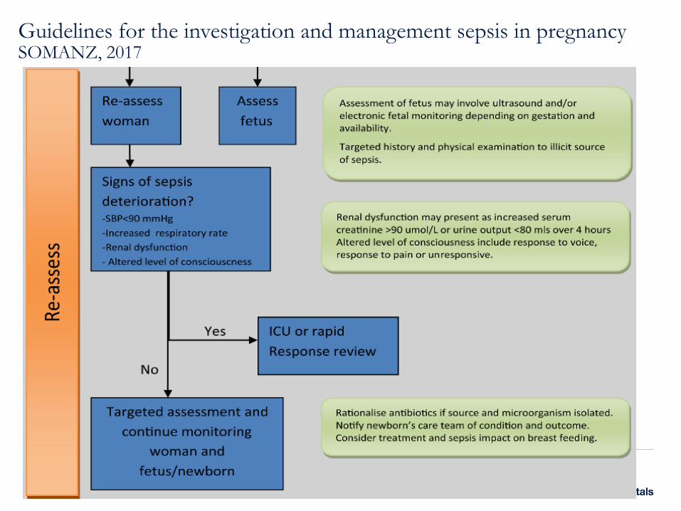
Guidelines for the investigation and management sepsis in pregnancySOMANZ, 2017
Findings:
50% of deaths from sepsis are related to Group A streptococcus
E.Coli is the most common cause of maternal bacterial infection
Sepsis can occur anytime during pregnancy & often associated with a delay in diagnosis
The normal physiological changes of pregnancy may mask early signs of sepsis
Maternal sepsis with or without hemodynamic instability may present with fetal distress as the uteroplacental circulation is not auto-regulated.
Consideration for treatment options has to be given to the impact of the maternal condition as well as the effect on the fetus.

Guidelines for the investigation and management sepsis in pregnancySOMANZ, 2017
8 Key Points
1. Screen
2. Fever
3. Etiology
4. Golden Hour
5. Timing/Mode of Delivery
6. VTE Prophylaxis
7. Anesthesia
8. ICU Transfer

Guidelines for the investigation and management sepsis in pregnancySOMANZ, 2017

Guidelines for the investigation and management sepsis in pregnancySOMANZ, 2017
Guidelines for the investigation and management sepsis in pregnancySOMANZ, 2017

BUNDLE SCIENCE
National Partnership Strategy to Enhance Maternal Safety
A "bundle" is a group of interventionsrelated to a disease process that, when
executed together, result in betteroutcomes than when implemented
individually.


CA-PAMR: Chance to Alter Outcome Grouped Cause of Death; 2002-2004 (N=145)
Grouped Cause of Death Chance to Alter Outcome
Strong /
Good (%)
Some
(%)
None
(%)
Total
N (%)
Obstetric hemorrhage 69 25 6 16 (11)
Deep vein thrombosis/
pulmonary embolism53 40 7 15 (10)
Sepsis/infection 50 40 10 10 (7)
Preeclampsia/eclampsia 50 50 0 25 (17)
Cardiomyopathy and other
cardiovascular causes25 61 14 28 (19)
Cerebral vascular accident 22 0 78 9 (6)
Amniotic fluid embolism 0 87 13 15 (10)
All other causes of death 46 46 8 26 (18)
Total (%) 40 48 12 145 7

Let’s review some physiology…
Photo from creative commons/pixabay

Normal physiologic changes
Cardiovascular
Hematologic
Pulmonary
Renal

Cardiovascular
Cardiac Output
Cardiac Changes
Stroke Volume 30-50%
Heart Rate 20% (~10-20 beats)
Anatomic Changes Uterus
Vascular Resistance
SVR PVR
Normal Cardiac Adaptation during Pregnancy
% C
ha
ng
e
pregnant Weeks of gestation postpartum

Hematologic
Cardiac Output
Total blood volume
Plasma Volume
RBC Volume
% C
ha
ng
e
Weeks of gestationpregnant postpartum
Blood Volume Changes
Total Volume
35% (~ 2,000ml)
Plasma Volume
50% (~ 1,600ml)
RBC Mass
17% (~ 350mL)
Normal Hematologic Events Associated with Pregnancy
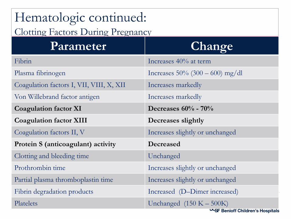
Hematologic continued:Clotting Factors During Pregnancy
Parameter ChangeFibrin Increases 40% at term
Plasma fibrinogen Increases 50% (300 – 600) mg/dl
Coagulation factors I, VII, VIII, X, XII Increases markedly
Von Willebrand factor antigen Increases markedly
Coagulation factor XI Decreases 60% - 70%
Coagulation factor XIII Decreases slightly
Coagulation factors II, V Increases slightly or unchanged
Protein S (anticoagulant) activity Decreased
Clotting and bleeding time Unchanged
Prothrombin time Increases slightly or unchanged
Partial plasma thromboplastin time Increases slightly or unchanged
Fibrin degradation products Increased (D–Dimer increased)
Platelets Unchanged (150 K – 500K)

Hematologic
Factors V, VII, VIII, IX, X, XII
Fibrinolysis
Fibrinogen
Prothrombin

Pulmonary
Diaphragm 4-7 cm –ribs flare
Functional Residual Capacity 25%
Respiratory Rate unchanged 16-20
Tidal volume from 500 – 700 ml
Compensatory Alkalemia
Not Pregnant
pH 7.35 – 7.45
pO2 90 -100
pCO2 35 – 45
HCO3 22 - 26
Pregnant
pH 7.40 – 7.45
pO2 104 -108
pCO2 27 – 32
HCO3 18 - 22

Renal AdaptationPregnancy is a high flow state
Kidneys have structural and functional changes
• 50% Increase renal blood flow
• 50% Increase glomerular filtration rate
• Physiologic hydronephorsis (right sided)
• Altered lab values
‒Creatinine
‒Proteinuria
‒Glycosuria
Abbas, et al.; Blackburn; Hacker, et al.

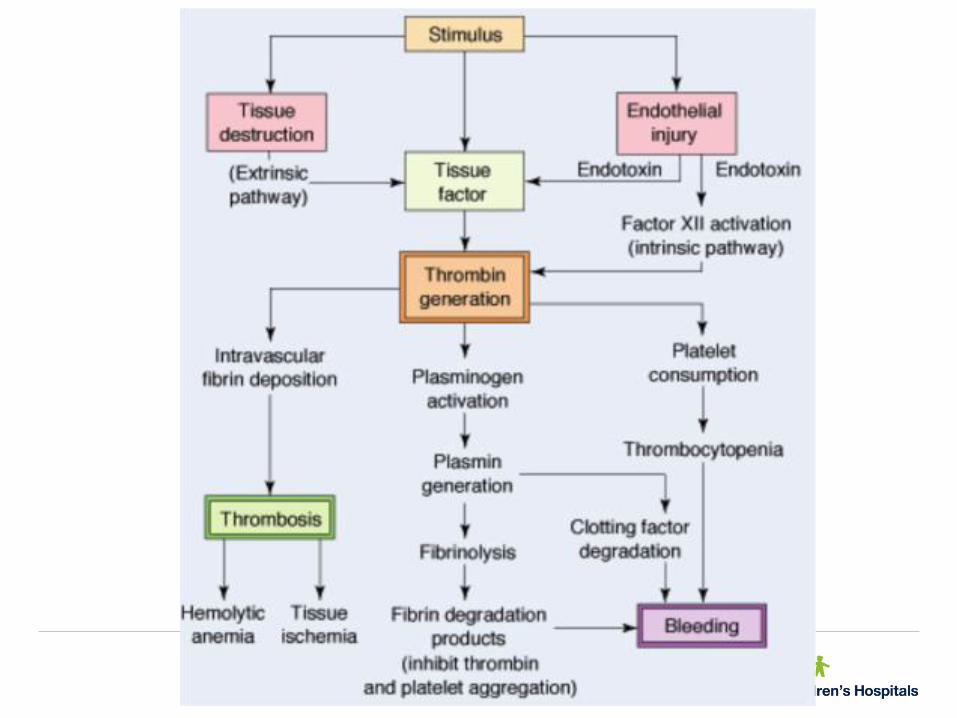
1. Tissue Damage
• Adhere to or invade cells
• Produce toxins
2. Blood Clots
• Toxins may cause coagulation
• Small clots block blood flow
• Oxygen deprivation
3. Fluid leakage from vessel
• Toxins damage vessel wall
• Fluid leaks out through holes
• Hpotensionfrom fluid loss
3 Ways Microbes cause damage
Parfitt, Sheryl E.; et, al.,MCN: 42(4):194-198, July/August 2017.

Pathogenesis of multiorgan system failure

Invasive Organism Injury
Vasodilatation occurs Inflammatory cascade is activated
Cytokines and complement system are activated
Blood coagulation system is activated
Deactivators produced + healthy anti-inflammatory response system =
inhibition of immune system reaction from occurring throughout the body
Invasive/ organism
injury
Parfitt, Sheryl E.; et, al.,MCN: 42(4):194-198, July/August 2017.

Invasive Organism Injury
Parfitt, Sheryl E.; et, al.,MCN: 42(4):194-198, July/August 2017.

Adult Respiratory Distress Syndrome
Hemorrhagic
Shock
Damage to endothelial cells in pulmonary vasculature
Fluid leaks from vascular space into alveoli
Respiratory failure

Pathophysiology of Cell Death
Tissue hypoperfusion metabolic acidosis inflammatory mediators tissue and vascular injury multiple organ failure

Secondary (Late) Causes
1. Infection/sepsis
‒ myometrial cell contractility
‒ Disrupts blood vessel endothelial lining
‒ Fever vasodilatation
2. Retained products of conception
3. Placental site sub involution
4. Coagulopathy

42
Draft 1.2

43
Draft 1.2

44
Draft 1.2

45
Draft 1.2

Tranexamic acid (TXA)
For women with established PPH
• Not responsive to medications or treatments
• Considered an adjunct treatment
• Most effective if used within first 30 minutes (3 hours from onset)
• Dose: TXA 1 gram IV over 10 minutes
• may repeat 2nd dose in 30 minutes if bleeding persists or if stopped and restarted
WOMAN Trial Collaborators. (2017) Effect of early TXA administration on mortality, hysterectomy, and
other morbidities in women with post-partum haemorrhage (WOMAN): an international, randomised,
double-blind, placebo-controlled trial. Lancet, 389(10084), 2105–2116.

Neligan PJ 2011

What is DIC?
Underlying disorder
Activates coagulation cascade
• Blood clot formation
• Coagulation factors become depleted
• Results in uncontrolled bleeding
‒Death

Disseminated Intravascular Coagulation
Accompany certain obstetrical conditions
Varied clinical presentation and prognostic course
An “effect “ of other disease processes
Treatment will be focused on removal of the causative agent
Society on Thrombosis and Hemostasis defines “DIC as:
An acquired syndrome characterized by the intravascular activation of coagulation with loss of localization arising from different causes.
It can originate from and cause damage to the microvasculature which if sufficiently severe can produce organ dysfunction.

Etiology of DICOB/Gyn
Complications
Infection
Cancer
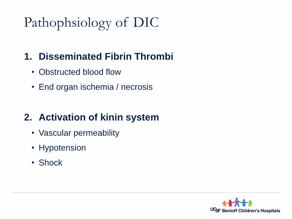
Pathophsiology of DIC
1. Disseminated Fibrin Thrombi
• Obstructed blood flow
• End organ ischemia / necrosis
2. Activation of kinin system
• Vascular permeability
• Hypotension
• Shock

Pathophsiology of DIC
3. Activation of the complement system
• Red cell and platelet lysis
• vascular permeability
• Shock
4. Release of cytokines (IL 1 & 6) and TNF
5. Plasma-induced lysis of fibrin
• FDP’s
• Depletion of Coag factors
• Hemorrhage and shock

Underlying OB conditions associated with DIC
Intrauterine Fetal Demise
Placental abruption
PPH / Hypovolemia / MBT
Severe Pre E / HELLP
Acute Fatty Liver
Amniotic Fluid Embolism
Sepsis
25%
37%
29%
14%
8%
6%
6%
100 %

Sepsis 6 %
Mechanism
• Release of TNF
‒ Endothelial injury
‒ Releases Tissue Factor
Produces Thrombin
Protein C is activated
Fibrinolysis
Diagnosis
• Clinical evidence of infection
• Lab studies
Management
Evacuation of uterusAntibiotics

Clinical Presentation
Peripheral cyanosis
Renal impairment
Drowsiness
Confusion
Coma
Cardiorespiratory failure
Large and small vessel thrombosis
Ischemia
End organ damage

Bleeding from unrelated sites
Venipuncture sites
Epistaxis
Ecchymosis
Purpura
Petechiae
Hematomas

Diagnosis of DIC
Obvious with massive hemorrhage
Lab tests
• CBC, Plts
• Fibrinogen, FDP’s
• PT, aPTT
• D Dimer
Rotem

Fluid and Blood Resuscitation
Non-pregnant guidelines pose risk of fluid overload/pulmonary edema
• Reduced colloid/oncotic pressure
OB patients 20mL/kg verses 30mL/kg
• 200 lb patient = 90 kg (90x20= 1,800 mL Normal Saline)
• Consider CVP placement
Transfuse Blood products specific to deficits
• PRBS to maintain Hgb 7.0-9.0 g/dL
• Platelets if <5,000/mm or if surgery is warranted >35,000/mm
• MTP for Stage 3 Hemorrhage/DIC

Antibiotic Administration:Delayed administration = higher mortality
Empiric Therapy with broad spectrum antibiotic ASAP
• (1 hr target)
1. Ampicillin & Gentamicin
2. Influenza Clindamycin & Vancomycin
Treat for suspected influenza
• Anti-viral therapy
De-escalate
antibiotic
once
source has
been
identified

Pressor Support
If mean arterial pressure MAP remains <65 after fluid bolus
Norepinepherine
• Appears to be safe during pregnancy in low doses
Dobutamine – rarely used
• Positive Inotrope
‒ Improve left ventricular contractility
‒ Normalize preload and improve cardiac output
‒ May restrict uterine blood flow (pregnant ewes)
• Epinephrine –safe to use after volume and Norepinepherine
‒ 2nd Line agent
• Vasopressin and May stimulate uterine contractions
• preterm birth

Timing/Mode of Delivery
Decision regarding delivery is complex
Consider the following:
• Source of infection (Chorioamnionitis – no! )
• Maternal Status
• Fetal assessment – gestational age
‒ May prolong pregnancy per maternal tolerance (not chorio)
‒ Steroids for fetal lung maturity
‒ Fetal status often will improve if maternal condition stabilizes

Respiratory Support
ICU Transfer: tachypnea, worsening hypoxia, ↑ O2 requirement
Monitor O2 saturation
• Position: Left tilt to minimize aorta/venal cava compression
Airway – delayed gastric empty
• Intubation
o Pre-oxygenation
o Functional residual capacity
o Ventilation/ perfusion (VQ) mismatch
o Customize ventilator settings for gravid patient

Risk Factors for DVT
Maternal Pregnancy Labor
Obesity Multiparity Cesarean Birth
Smoking Preeclampsia PPH Blood
Hx of VTE Physiologic changes of
PregnancyInfection
Diabetes Immobilization
Age > 35 years

Heparin Compounds
Unfractionated heparin or Low molecular weight heparin (LMWH)
Do not cross the placenta
Safe during pregnancy
• Higher dosing required during pregnancy
‒ total blood volume
‒ glomerular filtration thus in renal excretion of heparin
‒ protein binding of heparin
‒ peak plasma volume and shorter half life
Side effects
Hemorrhage, hypotension
Protamine sulfate 1mg neutralizes 100 units of heparin
Should not exceed 50 mg in single dose
Follow aPTT levels 4 hours after initiation and after dose changes

Post Birth Warning signs
• Essential Teaching for WomenVenous
Thromboembolism
• VTE is when you develop a blood clot usually in your leg (calf area) What is VTE
• Leg pain, tender to touch, burning or redness, particularly in calf areaSigns of VTE
• Call healthcare provider immediately for above signs of VTE if no response call 911 or go to nearest hospital emergency department
Obtaining Immediate Care

Call for Help Early
• Detect abnormal VS and clinical changes
• Alert the Team
• Mobilize a response
• Optimal patient outcome
I wonder why
we were
called?
Gee…she looks
pretty good to
me…

Escalation
• An abnormal parameter requires:
– Prompt reporting to a physician or other qualified clinician
– Prompt bedside evaluation by a physician or otherqualified clinical provider with the ability to activate resources in order to initiate emergencydiagnostic and therapeutic interventions as needed

House Supervisor
Nurse Manager
Staff Nurse
Medical
director
Hospital
Administer
Physician
Chain of Command / Authority
Adapted from Lyndon et al., AWHONN Fetal Heart Monitoring; Chapter 8, 2009

Simulated Multidisciplinary Drills
TJC Sentinel Event Alert, Issue 30 -July 21, 2004
Conduct
team training
in perinatal
areas to
teach staff to
work together
and
communicate
more
effectively.

OB Triage Case
A G3, P1, 38-yo woman @ 29+2 weeks arrives to OB Triage
• Hx of dry cough X’s 3 days – fever/aches past 24 hrs.
• VS: T 40.3°C (104.5°F); BP, 119/60 mm Hg; pulse 125, RR 36
• (SaO2), 95%.
FHR 175 bpm with minimal variability.
The patient had no uterine cramping or contractions
Patient reported diffuse body aches and rated her pain 10/10

How Errors Occur
Defenses Harm
Safeguards
Stop the line
Standard work
Flexible staffing
Self-checks
Culture
PoliciesResources
TrainingCommunication
Failures

Based on the AWHONN MFTI what is the priority?
a) Priority 1
b) Priority 2
c) Priority 3
d) Priority 4
e) Priority 5

Ruhl, C., Scheich, B., Onokpise, B., Bingham, DJournal of Obstetric, Gynecologic & Neonatal Nursing, Volume 44, Issue 6, 2015, 701–709
.

What needs to happen
a) Begin Early Goal Directed Therapy (EGDT)
b) Begin The 1 Hour Bundle it replaces the 3 Hour Bundle
c) Bolus with 1,000 mL NS follow with 500mL/hr untill BP is >90/50
d) The optimal fluid replacement for pregnant patients is unknown
e) Administer antibiotics once blood cultures have been obtained
f) b & d
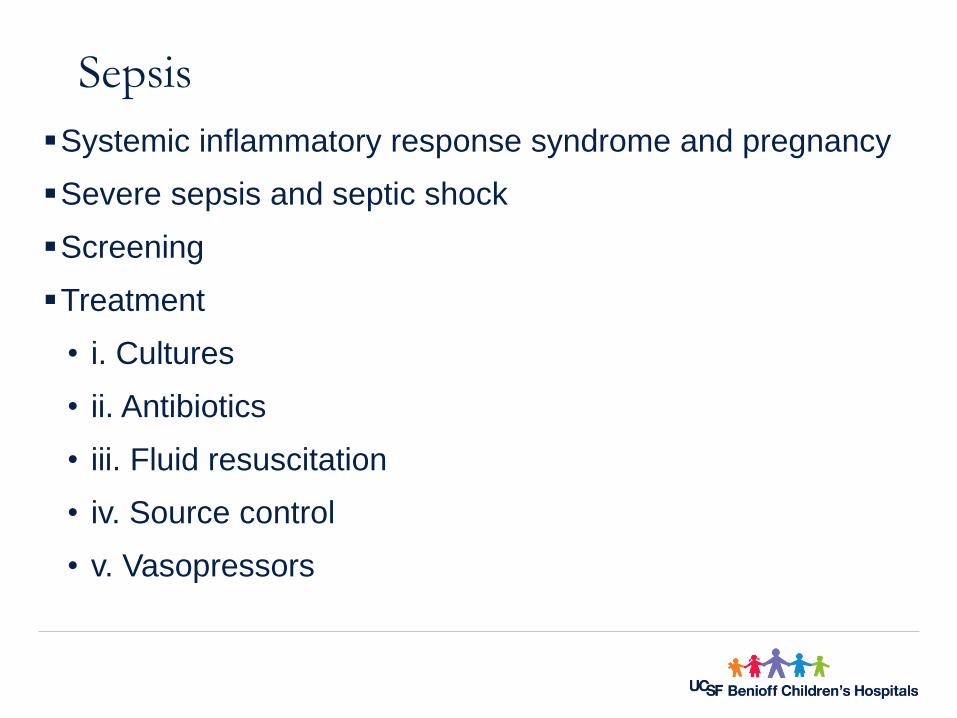
Sepsis
Systemic inflammatory response syndrome and pregnancy
Severe sepsis and septic shock
Screening
Treatment
• i. Cultures
• ii. Antibiotics
• iii. Fluid resuscitation
• iv. Source control
• v. Vasopressors

SIRS Criteria ComparisonSIRS: Systemic Inflammatory Response Syndrome
93% Sensitivity63% Specificity

Maternal Sepsis Pathway: ScreeningScreen in triage, upon admission, every shift (within first 2 hours of shift)
and PRN suspected infection (Document in EMR Flowsheet).
Lori Olvera DNP, RNC-OB, EFM-
Altered mental status (if positive and pt has suspected source of infection, immediately move forward with interventions).
Temp > 100.4°F (38°C) OR Temp < 96.8°F (36°C)HR > 110RR > 24WBC > 15,000WBC < 4,000 OR > 10% bands (found in CBC diff)
SIRS: Systemic Inflammatory Response Syndrome
2 or more positive SIRS criteria
ORALTERED MENTAL STATUS
ANDsuspected source of
infection*
= + Sepsis ScreenBegin Time Zero
SIRS CRITERIA EVALUATION : Evaluate for SIRS Criteria/ Altered Mental Status

Maternal Sepsis Pathway: ScreeningScreen in triage, upon admission, every shift (within first 2 hours of shift)
and PRN suspected infection (Document in EMR Flowsheet).
Lori Olvera DNP, RNC-OB, EFM-
SEPSIS INTERVENTIONS for 1st HOUR□ Call Sepsis Alert - NOTIFY RRT, Lab tech, and OB Provider□OB Provider places OB Severe Sepsis Order Set□Draw STAT Lactate, CBC, - close loop communication with lab□Blood Cultures (2 sets prior to antibiotics)
Draw even if patient has been treated for GBS+□Consider other labs- Chem 7, PT, PTT, (Consult with RRT)□Administer broad spectrum Antibiotic**□Obtain U/A (consider source of infection)□Chest XRAY (if suspected lung infection)□Document TIME ZERO□Vital Signs Q30 X2, Q1HX2, Q2 X2, then Q4H
Time Zero
Must complete sepsis
interventions within 60 minutes!
SIRS CRITERIA EVALUATION : Evaluate for SIRS Criteria/ Altered Mental Status

Maternal Sepsis Pathway: Increased SurveillanceScreen in triage, upon admission, every shift (within first 2 hours of shift)
and PRN suspected infection (Document in EMR Flowsheet).
Lori Olvera DNP, RNC-OB, EFM-
□Lactate ≥ 2 mmol/L – 3.9 mmol/L□SBP < 90 mmHG◊ or MAP < 65 (NOTE: ◊ Sys BP of 90 must be at least 5mm Hg lower than baseline to meet this criteria)□SBP decrease < 40mmHG from baseline□Bilirubin > 2mg/dL□Urine output < or equal to 30 ml/hr for 2 hours□Creatinine ≥ 1.5 mg/dL□Platelet count < 100,000□Coagulopathy (INR > 1.5 or PTT > 60 sec)
SEPSIS + 1 or more
positive acute
organ dysfunction
=
diagnosis of SEPSIS
ACUTE ORGAN DYSFUNTION EVALUATIONEvaluate for 1 or more ACUTE ORGAN DYSFUNCTION - Criteria due to infection

Screen in triage, upon admission, every shift (within first 2 hours of shift) and PRN suspected infection (Document in EMR Flow Sheet).
Lori Olvera DNP, RNC-OB, EFM-
SEVERE SEPSIS INTERVENTIONS□Consider IV Fluids N/S or LR 30 mL/kg;
Administer each liter over 60 min (Lactate 2-3.9)□Repeat lactate every 3 hours until lactate < 2 mmol/L□SpO2 per protocol, titrate oxygen to ≥ 92%□Consult with RRT to maximize oxygenation□Notify OB, MFM, Hospitalist□Vital signs Q30 X2, Q1H X2, Q2x2, then Q4h
ACUTE ORGAN DYSFUNTION EVALUATIONEvaluate for 1 or more ACUTE ORGAN DYSFUNCTION Criteria due to infection
Maternal Sepsis Pathway: Increased Surveillance

Maternal Sepsis Pathway: Escalate CareScreen in triage, upon admission, every shift (within first 2 hours of shift)
and PRN suspected infection (Document in EMR Flow Sheet).
Lori Olvera DNP, RNC-OB, EFM-
□LACTATE > 3.9 MMOL/L (initial lactate)□BP Systolic < 90, MAP < 65 despite fluid resuscitation□Clinical features are the same as severe sepsis
SEPTIC SHOCK INTERVENTIONS□Notify OB MD-come to bedside□RN- CALL RAPID RESPONSE TEAM-□RRT will initiate CODE SEPSIS OVERHEAD PAGE□Broad spectrum antibiotics□RRT will determine if ICU admission required□IV Fluids Normal Saline or LR bolus 30ml/kg NOW for lactate > 3.9 mmol
or hypotensive (if not previously done)□Vital signs q 30 min
SEPTIC SHOCK CRITERIAEvaluate for SEPTIC SHOCK Criteria

Maternal Sepsis Pathway: Escalate Care
Lori Olvera DNP, RNC-OB, EFM-
*Consider source of infection-Chorioamnionitis - Pyelonephritis-Endometritis - UTI-Pneumonia - Other-Intrauterine Fetal Demise
*NOTES FOR OB PROVIDER USE:•Add “Sepsis” to Problem List.•For Lactate above 3.9—PMA comes to bedside,consults with OB Doc & documents plan of care-


NICOMNoninvasive cardiac output monitor

Unplanned ICU Admission: Postoperative Course
Transfer to ICU
Weak but stable
Separation from baby
Delayed breastmilk
Hbg Hct
• Iron—IV (sucrose)
• Rh-Erythropoeitin
• Heparin
Discharge home WITH support

Surviving Sepsis: where do we go from here
Immediate post-sepsis treatment plan
• Treat anemia
• Care of newborn
‒ Breastfeeding
‒ Antibiotics/side effects
Long term patient follow-up
• Negative impact on patient
‒ Near death experience

Traumatic Childbirth
“process that involves actual or threatened serious injury or death to the mother or her infant. The
birthing woman experiences intense fear, helplessness, loss of control and horror”.
Dehumanizing experience
• High level of medical interventions, extreme painStripped of their dignityPowerlessLack of caring and support from perinatal staffFear of dying
Beck, C. Birth Trauma: In the eye of the beholder. Nursing Research (2004a).

Clinicians should be mindful of birth environment and how their behaviors influence the patient perspective of safety during birth
At least one team member should focus on emotional support during emergency birth to mitigate the potential for negative experiences that lead to emotional harm

Learning from Review
Adverse Outcome Review• Why do it?
– Finger point, blame, punish– Learn, improve future outcomes
• ACOG, AWHONN, SMFA –• Recommend all severe morbidity whether
sentinel or not: – Undergo review process:
• thorough, credible, multidisciplinary, comprehensive
Severe Maternal Morbidity

SummaryConsider normal physiologic changes of pregnancy when screening pregnant or postpartum women.
Lack of recognition and delays in treatment can result in septic shock and end organ dysfunction.
Nurses play an essential role to screen, recognize, and promptly respond to women who screen positive for sepsis.
The ability to mobilize a multidisciplinary team for early intervention, MFM, and ICU referral will promote intact survival of maternal sepsis.
Multidisciplinary review of adverse outcomes promotes learning and provides opportunity for quality improvement.

Maternal Mortality Rate,
California and United States; 1999-2013
11.1
7.7
10.0
14.6
11.8 11.7
14.0
7.4
7.3
10.9
9.7
11.6
9.2
6.2
16.9
8.9
15.1
13.1
12.19.9
9.9
9.8
13.3
12.7
15.5 16.916.6
19.3
19.9
22.0
0.0
3.0
6.0
9.0
12.0
15.0
18.0
21.0
24.0
1999 2000 2001 2002 2003 2004 2005 2006 2007 2008 2009 2010 2011 2012 2013
Year
California Rate
United States Rate
Ma
tern
al D
ea
ths
pe
r 1
00
,000
Liv
e B
irth
s
HP 2020 Objective – 11.4 Deaths per 100,000 Live Births
SOURCE: State of California, Department of Public Health, California Birth and Death Statistical Master Files, 1999-2013. Maternal mortality for California (deaths ≤
42 days postpartum) was calculated using ICD-10 cause of death classification (codes A34, O00-O95,O98-O99). United States data and HP2020 Objective use the
same codes. U.S. maternal mortality data is published by the National Center for Health Statistics (NCHS) through 2007 only. U.S. maternal mortality rates from 2008
through-2013 were calculated using CDC Wonder Online Database, accessed at http://wonder.cdc.govon March 11, 2015. Produced by California Department of
Public Health, Center for Family Health, Maternal, Child and Adolescent Health Division, March, 2015.

Nurses are a valuable source of information
and support for women and their families
Thank You!


![Antepartum haemorrhage 1 [وضع التوافق] · Antipartum haemorrhage should be taken • seriouslyyy p g and any women presenting with ahistory of fresh vaginal bleeding must](https://img.dokumen.tips/doc/110x75/5e77205f9f486d7c5a26652f/antepartum-haemorrhage-1-antipartum-haemorrhage-should-be.jpg)