Embed Size (px)
Citation preview
Original Article eISSN2465-891X The Nerve.2016.02(1):5-9http://dx.doi.org/10.21129/nerve.2016.2.1.5
www.thenerve.net
Journal of the Korean Society of Peripheral Nervous System 5
Posterior Thoracic Cage Interbody Fusion (PTCIF) as an Alternative Fusion Technique after Laminectomy in Thoracic
and Thoracolumbar Junctional Spine
Hong Kyung Shin1, Sun Kyu Oh1, Il Choi2, Sang Ryong Jeon1
1Department of Neurological Surgery, Asan Medical Center, University of Ulsan College of Medicine, Seoul,2Department of Neurological Surgery, Dongtan Sacred Heart Hospital, Hallym University College of Medicine, Hwaseong, Korea
Objective: Thoracic spine fusion is used to surgically treat various spine lesions. However, posterior-only thoracic fusion by using the pedicle screw system can be complicated by pseudoarthrosis or instrument failure. Moreover, when laminectomy is performed for decompression, thus exposing the spinal cord in the thoracic spine, the fusion bed where the bone chips are applied for posterolateral fusion may be insufficient. Therefore, we conducted interbody fusion in posterior thoracic approach.Methods: All patients underwent posterior only approach. After pedicle screw insertion at the decompressed level, the cages packed with autologous bone chips were inserted into interbody disc space and fixation was performed by rod and screw system. Four patients with thoracolumbar spinal injury and three patients with degenerative disease. Bone fusion was defined as the formation of bony continuity between the upper and lower end plates and around the fusion cages in the posterior thoracic cage interbody fusion (PTCIF) level, as determined by computed tomography (CT).Results: The average follow-up period was 15.3 (range, 8-28) months. All patients with degenerative disease exhibited neuro- logical improvement. Successful bone fusion was confirmed with CT in all patients more than 3 months after PTCIF. Opera- tion-associated complications did not occur and there was no revision operation.Conclusion: PTCIF was found to be safe and achieved good outcomes for spinal cord decompression and bone fusion in the thoracic and thoracolumbar junctional spine. Therefore, this surgical method could be considered as an alternative procedure for posterior thoracic decompression and fixation surgery.
Key Words: LaminectomyㆍSpinal cord compressionㆍSpinal fusionㆍThoracic vertebrae
Corresponding author: Sang Ryong Jeon, MD, PhDDepartment of Neurosurgery, Asan Medical Center, University of UlsanCollege of Medicine, 13, Gangdong-daero, Songpa-gu, Seoul 05505, KoreaTel: +82-2-3010-3550, Fax: +82-2-476-6738E-mail: [email protected]
INTRODUCTION
Since the thoracolumbar junctional area is highly suscep- tible to injury, the majority of spinal trauma injuries affect this area4,19), and thoracic spine is a common site for the surgical spinal diseases including infection and neoplastic disease7,8). The pathological lesions in the thoracic or thoracolumbar area that arise from trauma, neoplasm, infection, deformity, and de- generative disease are often treated by surgical removal. How- ever, this procedure often induces instability and deformity that must be treated by thoracic spine fusion.
Posterior thoracic fusion is difficult when posterior decom- pression is also conducted and the spinal cord is exposed,
which can occasionally result in pseudoarthrosis or instrument failure. Therefore, the anterior thoracic approach with inter-body fusion was developed. However, anterior thoracic appro- ach may not be familiar with spine surgeons and this procedure is associated with diverse morbidities, especially pulmonary complications5,15). For lumbar lesions, posterior lumbar inter-body fusion (PLIF) with pedicle screw fixation is used. This is not only a reliable technique for inducing bone fusion, it is also a safe method that results16,18). PLIF is particularly useful after lumbar laminectomy, which leaves a smaller fusion bed. As a result, PLIF has been widely employed in cases of lumbar spinal lesion where bone fusion is mandatory.
On the basis of these observations, we developed a novel procedure for cases of thoracic decompression and fixation sur-gery, namely, posterior thoracic cage interbody fusion (PTCIF). It was hypothesized that this procedure, which employs the posterior approach only, would yield good bony fusion and avoid the complications associated with the anterior thoracic approach. The purpose of the present study is to report this novel procedure and its surgical outcomes.
Posterior Cage Interbody Fusion in Thoracic and Thoracolumbar Spine
6 www.thenerve.net
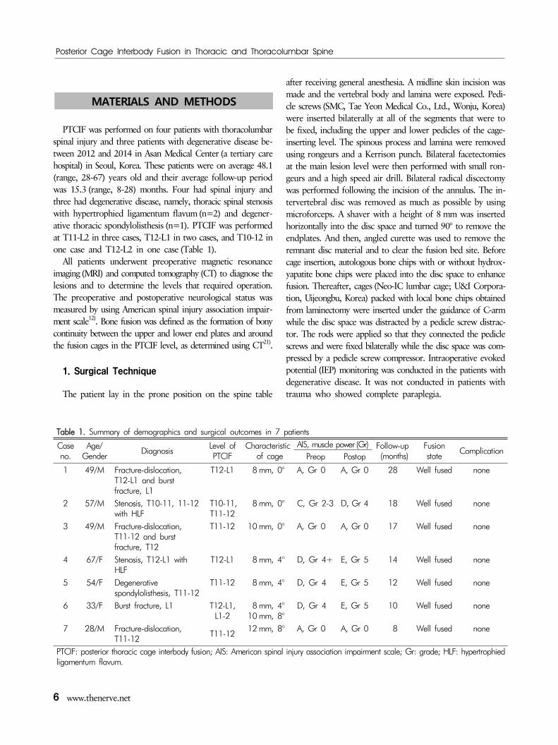
Table 1. Summary of demographics and surgical outcomes in 7 patients
Case no.
Age/Gender
DiagnosisLevel of PTCIF
Characteristic of cage
AIS, muscle power (Gr) Follow-up (months)
Fusion state
ComplicationPreop Postop
1 49/M Fracture-dislocation, T12-L1 and burst fracture, L1
T12-L1 8 mm, 0° A, Gr 0 A, Gr 0 28 Well fused none
2 57/M Stenosis, T10-11, 11-12with HLF
T10-11, T11-12
8 mm, 0° C, Gr 2-3 D, Gr 4 18 Well fused none
3 49/M Fracture-dislocation, T11-12 and burst fracture, T12
T11-12 10 mm, 0° A, Gr 0 A, Gr 0 17 Well fused none
4 67/F Stenosis, T12-L1 withHLF
T12-L1 8 mm, 4° D, Gr 4+ E, Gr 5 14 Well fused none
5 54/F Degenerative spondylolisthesis, T11-12
T11-12 8 mm, 4° D, Gr 4 E, Gr 5 12 Well fused none
6 33/F Burst fracture, L1 T12-L1, L1-2
8 mm, 4°10 mm, 8°
D, Gr 4 E, Gr 5 10 Well fused none
7 28/M Fracture-dislocation,T11-12
T11-1212 mm, 8° A, Gr 0 A, Gr 0 8 Well fused none
PTCIF: posterior thoracic cage interbody fusion; AIS: American spinal injury association impairment scale; Gr: grade; HLF: hypertrophied ligamentum flavum.
MATERIALS AND METHODS
PTCIF was performed on four patients with thoracolumbar spinal injury and three patients with degenerative disease be-tween 2012 and 2014 in Asan Medical Center (a tertiary care hospital) in Seoul, Korea. These patients were on average 48.1 (range, 28-67) years old and their average follow-up period was 15.3 (range, 8-28) months. Four had spinal injury and three had degenerative disease, namely, thoracic spinal stenosis with hypertrophied ligamentum flavum(n=2) and degener-ative thoracic spondylolisthesis (n=1). PTCIF was performed at T11-L2 in three cases, T12-L1 in two cases, and T10-12 in one case and T12-L2 in one case (Table 1).
All patients underwent preoperative magnetic resonance imaging (MRI) and computed tomography (CT) to diagnose the lesions and to determine the levels that required operation. The preoperative and postoperative neurological status was measured by using American spinal injury association impair-ment scale12). Bone fusion was defined as the formation of bony continuity between the upper and lower end plates and around the fusion cages in the PTCIF level, as determined using CT21).
1. Surgical Technique
The patient lay in the prone position on the spine table
after receiving general anesthesia. A midline skin incision was made and the vertebral body and lamina were exposed. Pedi- cle screws (SMC, Tae Yeon Medical Co., Ltd., Wonju, Korea) were inserted bilaterally at all of the segments that were to be fixed, including the upper and lower pedicles of the cage- inserting level. The spinous process and lamina were removed using rongeurs and a Kerrison punch. Bilateral facetectomies at the main lesion level were then performed with small ron-geurs and a high speed air drill. Bilateral radical discectomy was performed following the incision of the annulus. The in-tervertebral disc was removed as much as possible by using microforceps. A shaver with a height of 8 mm was inserted horizontally into the disc space and turned 90° to remove the endplates. And then, angled curette was used to remove the remnant disc material and to clear the fusion bed site. Before cage insertion, autologous bone chips with or without hydrox-yapatite bone chips were placed into the disc space to enhance fusion. Thereafter, cages (Neo-IC lumbar cage; U&I Corpora- tion, Uijeongbu, Korea) packed with local bone chips obtained from laminectomy were inserted under the guidance of C-arm while the disc space was distracted by a pedicle screw distrac- tor. The rods were applied so that they connected the pedicle screws and were fixed bilaterally while the disc space was com- pressed by a pedicle screw compressor. Intraoperative evoked potential (IEP) monitoring was conducted in the patients with degenerative disease. It was not conducted in patients with trauma who showed complete paraplegia.
Shin HK et al.
The Nerve 2(1) April 2016 7
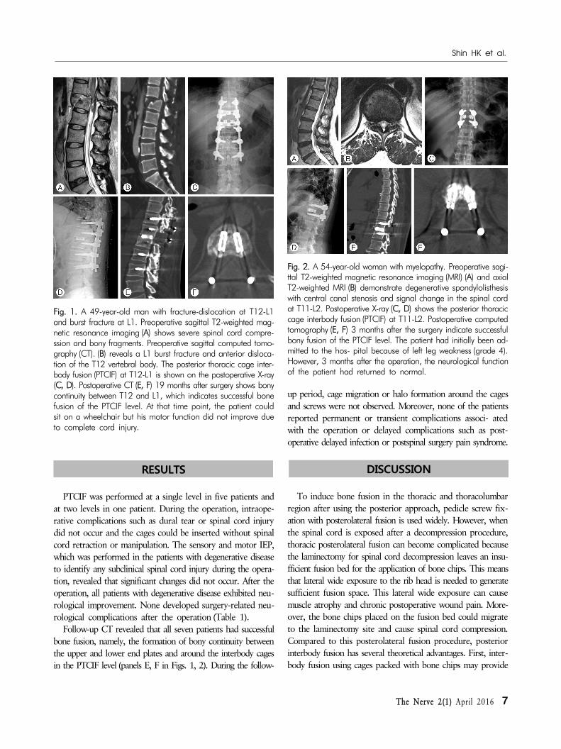
Fig. 1. A 49-year-old man with fracture-dislocation at T12-L1and burst fracture at L1. Preoperative sagittal T2-weighted mag-netic resonance imaging (A) shows severe spinal cord compre-ssion and bony fragments. Preoperative sagittal computed tomo-graphy (CT). (B) reveals a L1 burst fracture and anterior disloca-tion of the T12 vertebral body. The posterior thoracic cage inter-body fusion (PTCIF) at T12-L1 is shown on the postoperative X-ray(C, D). Postoperative CT (E, F) 19 months after surgery shows bonycontinuity between T12 and L1, which indicates successful bonefusion of the PTCIF level. At that time point, the patient couldsit on a wheelchair but his motor function did not improve dueto complete cord injury.
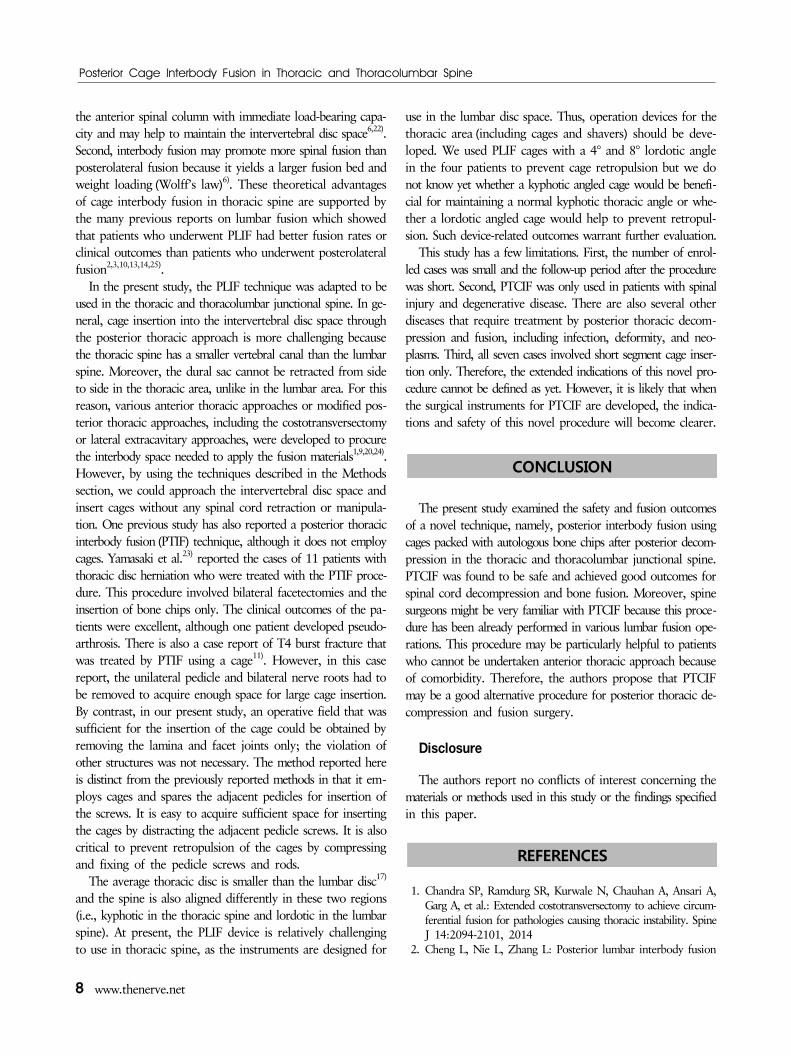
Fig. 2. A 54-year-old woman with myelopathy. Preoperative sagi-ttal T2-weighted magnetic resonance imaging (MRI) (A) and axialT2-weighted MRI (B) demonstrate degenerative spondylolisthesis with central canal stenosis and signal change in the spinal cordat T11-L2. Postoperative X-ray (C, D) shows the posterior thoraciccage interbody fusion (PTCIF) at T11-L2. Postoperative computedtomography (E, F) 3 months after the surgery indicate successfulbony fusion of the PTCIF level. The patient had initially been ad-mitted to the hos- pital because of left leg weakness (grade 4). However, 3 months after the operation, the neurological functionof the patient had returned to normal.
RESULTS
PTCIF was performed at a single level in five patients and at two levels in one patient. During the operation, intraope- rative complications such as dural tear or spinal cord injury did not occur and the cages could be inserted without spinal cord retraction or manipulation. The sensory and motor IEP, which was performed in the patients with degenerative disease to identify any subclinical spinal cord injury during the opera- tion, revealed that significant changes did not occur. After the operation, all patients with degenerative disease exhibited neu-rological improvement. None developed surgery-related neu-rological complications after the operation (Table 1).
Follow-up CT revealed that all seven patients had successful bone fusion, namely, the formation of bony continuity between the upper and lower end plates and around the interbody cages in the PTCIF level (panels E, F in Figs. 1, 2). During the follow-
up period, cage migration or halo formation around the cages and screws were not observed. Moreover, none of the patients reported permanent or transient complications associ- ated with the operation or delayed complications such as post-operative delayed infection or postspinal surgery pain syndrome.
DISCUSSION
To induce bone fusion in the thoracic and thoracolumbar region after using the posterior approach, pedicle screw fix-ation with posterolateral fusion is used widely. However, when the spinal cord is exposed after a decompression procedure, thoracic posterolateral fusion can become complicated because the laminectomy for spinal cord decompression leaves an insu- fficient fusion bed for the application of bone chips. This means that lateral wide exposure to the rib head is needed to generate sufficient fusion space. This lateral wide exposure can cause muscle atrophy and chronic postoperative wound pain. More- over, the bone chips placed on the fusion bed could migrate to the laminectomy site and cause spinal cord compression. Compared to this posterolateral fusion procedure, posterior interbody fusion has several theoretical advantages. First, inter-body fusion using cages packed with bone chips may provide
Posterior Cage Interbody Fusion in Thoracic and Thoracolumbar Spine
8 www.thenerve.net
the anterior spinal column with immediate load-bearing capa- city and may help to maintain the intervertebral disc space6,22). Second, interbody fusion may promote more spinal fusion than posterolateral fusion because it yields a larger fusion bed and weight loading (Wolff’s law)6). These theoretical advantages of cage interbody fusion in thoracic spine are supported by the many previous reports on lumbar fusion which showed that patients who underwent PLIF had better fusion rates or clinical outcomes than patients who underwent posterolateral fusion2,3,10,13,14,25).
In the present study, the PLIF technique was adapted to be used in the thoracic and thoracolumbar junctional spine. In ge- neral, cage insertion into the intervertebral disc space through the posterior thoracic approach is more challenging because the thoracic spine has a smaller vertebral canal than the lumbar spine. Moreover, the dural sac cannot be retracted from side to side in the thoracic area, unlike in the lumbar area. For this reason, various anterior thoracic approaches or modified pos-terior thoracic approaches, including the costotransversectomy or lateral extracavitary approaches, were developed to procure the interbody space needed to apply the fusion materials1,9,20,24). However, by using the techniques described in the Methods section, we could approach the intervertebral disc space and insert cages without any spinal cord retraction or manipula- tion. One previous study has also reported a posterior thoracic interbody fusion (PTIF) technique, although it does not employ cages. Yamasaki et al.23) reported the cases of 11 patients with thoracic disc herniation who were treated with the PTIF proce- dure. This procedure involved bilateral facetectomies and the insertion of bone chips only. The clinical outcomes of the pa-tients were excellent, although one patient developed pseudo- arthrosis. There is also a case report of T4 burst fracture that was treated by PTIF using a cage11). However, in this case report, the unilateral pedicle and bilateral nerve roots had to be removed to acquire enough space for large cage insertion. By contrast, in our present study, an operative field that was sufficient for the insertion of the cage could be obtained by removing the lamina and facet joints only; the violation of other structures was not necessary. The method reported here is distinct from the previously reported methods in that it em-ploys cages and spares the adjacent pedicles for insertion of the screws. It is easy to acquire sufficient space for inserting the cages by distracting the adjacent pedicle screws. It is also critical to prevent retropulsion of the cages by compressing and fixing of the pedicle screws and rods.
The average thoracic disc is smaller than the lumbar disc17) and the spine is also aligned differently in these two regions (i.e., kyphotic in the thoracic spine and lordotic in the lumbar spine). At present, the PLIF device is relatively challenging to use in thoracic spine, as the instruments are designed for
use in the lumbar disc space. Thus, operation devices for the thoracic area (including cages and shavers) should be deve- loped. We used PLIF cages with a 4° and 8° lordotic angle in the four patients to prevent cage retropulsion but we do not know yet whether a kyphotic angled cage would be benefi-cial for maintaining a normal kyphotic thoracic angle or whe- ther a lordotic angled cage would help to prevent retropul- sion. Such device-related outcomes warrant further evaluation.
This study has a few limitations. First, the number of enrol- led cases was small and the follow-up period after the procedure was short. Second, PTCIF was only used in patients with spinal injury and degenerative disease. There are also several other diseases that require treatment by posterior thoracic decom-pression and fusion, including infection, deformity, and neo- plasms. Third, all seven cases involved short segment cage inser- tion only. Therefore, the extended indications of this novel pro- cedure cannot be defined as yet. However, it is likely that when the surgical instruments for PTCIF are developed, the indica- tions and safety of this novel procedure will become clearer.
CONCLUSION
The present study examined the safety and fusion outcomes of a novel technique, namely, posterior interbody fusion using cages packed with autologous bone chips after posterior decom- pression in the thoracic and thoracolumbar junctional spine. PTCIF was found to be safe and achieved good outcomes for spinal cord decompression and bone fusion. Moreover, spine surgeons might be very familiar with PTCIF because this proce-dure has been already performed in various lumbar fusion ope- rations. This procedure may be particularly helpful to patients who cannot be undertaken anterior thoracic approach because of comorbidity. Therefore, the authors propose that PTCIF may be a good alternative procedure for posterior thoracic de- compression and fusion surgery.
Disclosure
The authors report no conflicts of interest concerning the materials or methods used in this study or the findings specified in this paper.
REFERENCES
1. Chandra SP, Ramdurg SR, Kurwale N, Chauhan A, Ansari A, Garg A, et al.: Extended costotransversectomy to achieve circum- ferential fusion for pathologies causing thoracic instability. Spine J 14:2094-2101, 2014
2. Cheng L, Nie L, Zhang L: Posterior lumbar interbody fusion
Shin HK et al.
The Nerve 2(1) April 2016 9
versus posterolateral fusion in spondylolisthesis: a prospective controlled study in the Han nationality. Int Orthop 33:1043- 1047, 2009
3. Dantas FL, Prandini MN, Ferreira MA: Comparison between posterior lumbar fusion with pedicle screws and posterior lumbar interbody fusion with pedicle screws in adult spondylolisthesis. Arq Neuropsiquiatr 65:764-770, 2007
4. DeWald RL: Burst fractures of the thoracic and lumbar spine. Clin Orthop Relat Res 189:150-161, 1984
5. Faciszewski T, Winter RB, Lonstein JE, Denis F, Johnson L: The surgical and medical perioperative complications of anterior spinal fusion surgery in the thoracic and lumbar spine in adults. A review of 1223 procedures. Spine (Phila Pa 1976) 20:1592- 1599, 1995
6. Farrokhi MR, Rahmanian A, Masoudi MS: Posterolateral versus posterior interbody fusion in isthmic spondylolisthesis. J Neuro- trauma 29:1567-1573, 2012
7. Fuentes Ferrer M, Gutiérrez Torres L, Ayala Ramírez O, Rumayor Zarzuelo M, del Prado González N: Tuberculosis of the spine. A systematic review of case series. Int Orthop 36:221-231, 2012
8. Gokaslan ZL, York JE, Walsh GL, McCutcheon IE, Lang FF, Putnam JB, Jr., et al.: Transthoracic vertebrectomy for metastatic spinal tumors. J Neurosurg 89:599-609, 1998
9. Keshavarzi S, Aryan HE: Multilevel lateral extra-cavitary corpec- tomy and reconstruction for non-contiguous metastatic lesions to the spine: case report and literature review. J Surg Oncol 99:314-317, 2009
10. Kim KT, Lee SH, Lee YH, Bae SC, Suk KS: Clinical outcomes of 3 fusion methods through the posterior approach in the lum- bar spine. Spine (Phila Pa 1976) 31:1351-1357, 2006
11. Kim SW, Lee SM, Shin H: Posterior interbody fusion using cage for T4 bursting fracture. J Korean Neurosurg Soc 37:389- 391, 2005
12. Kirshblum SC, Burns SP, Biering-Sorensen F, Donovan W, Graves DE, Jha A, et al.: International standards for neurological classi- fication of spinal cord injury (revised 2011). J Spinal Cord Med 34:535-546, 2011
13. Liu X, Wang Y, Qiu G, Weng X, Yu B: A systematic review with meta-analysis of posterior interbody fusion versus postero- lateral fusion in lumbar spondylolisthesis. Eur Spine J 23:43-56, 2014
14. Madan S, Boeree NR: Outcome of posterior lumbar interbody fusion versus posterolateral fusion for spondylolytic spondylo- listhesis. Spine (Phila Pa 1976) 27:1536-1542, 2002
15. McDonnell MF, Glassman SD, Dimar JR, 2nd, Puno RM, Johnson JR: Perioperative complications of anterior procedures on the spine. J Bone Joint Surg Am 78:839-847, 1996
16. Okuyama K, Abe E, Suzuki T, Tamura Y, Chiba M, Sato K: Posterior lumbar interbody fusion: a retrospective study of com- plications after facet joint excision and pedicle screw fixation in 148 cases. Acta Orthop Scand 70:329-334, 1999
17. Pooni JS, Hukins DW, Harris PF, Hilton RC, Davies KE: Com- parison of the structure of human intervertebral discs in the cervical, thoracic and lumbar regions of the spine. Surg Radiol Anat 8:175-182, 1986
18. Ray CD: Threaded titanium cages for lumbar interbody fusions. Spine (Phila Pa 1976) 22:667-679, 1997
19. Saboe LA, Reid DC, Davis LA, Warren SA, Grace MG: Spine trauma and associated injuries. J Trauma 31:43-48, 1991
20. Sciubba DM, Gallia GL, McGirt MJ, Woodworth GF, Garonzik IM, Witham T, et al.: Thoracic kyphotic deformity reduction with a distractible titanium cage via an entirely posterior app- roach. Neurosurgery 60:223-230, 2007
21. Shah RR, Mohammed S, Saifuddin A, Taylor BA: Comparison of plain radiographs with CT scan to evaluate interbody fusion following the use of titanium interbody cages and transpedicular instrumentation. Eur Spine J 12:378-385, 2003
22. van Dijk M, Smit TH, Sugihara S, Burger EH, Wuisman PI: The effect of cage stiffness on the rate of lumbar interbody fusion: an in vivo model using poly(l-lactic Acid) and titanium cages. Spine (Phila Pa 1976) 27:682-688, 2002
23. Yamasaki R, Okuda S, Maeno T, Haku T, Iwasaki M, Oda T: Surgical outcomes of posterior thoracic interbody fusion for tho- racic disc herniations. Eur Spine J 22:2496-2503, 2013
24. Zhang H, Huang S, Guo H, Ge L, Sheng B, Wang Y, et al.: A clinical study of internal fixation, debridement and interbody thoracic fusion to treat thoracic tuberculosis via posterior app- roach only. Int Orthop 36:293-298, 2012
25. Zhou ZJ, Zhao FD, Fang XQ, Zhao X, Fan SW: Meta-analysis of instrumented posterior interbody fusion versus instrumented posterolateral fusion in the lumbar spine. J Neurosurg Spine 15: 295-310, 2011