Embed Size (px)
Citation preview
Copyright 2005 Psychonomic Society, Inc. 144
Cognitive, Affective, & Behavioral Neuroscience2005, 5 (2), 144-155
Our ability to grab onto and hold in mind the informa-tion presented in a visual scene is extremely limited. Forinstance, subjects have great difficulty in detecting a grosschange between two similar scenes if these scenes areseparated in time or by another visual stimulus (Rensink,2002; Rensink, O’Regan, & Clark, 1997; Simons, 1996).Although the earliest stages of visual information pro-cessing are endowed with virtually unlimited processingcapacities (Pashler, 1988; Phillips, 1974; Sperling, 1960),severe limits occur when the visual information is storedinto visual working memory, which can at best accom-modate four visual items (Cowan, 2001; Luck & Vogel,1997; Pashler, 1988; Vogel, Woodman, & Luck, 2001).This bottleneck of information processing has signifi-cant real-life consequences, since it is considered to limitwhat information can be explicitly perceived and actedupon (Becker, Pashler, & Anstis, 2000; Chun & Potter,1995; Jolicœur, Dell’Acqua, & Crebolder, 2001; Rensink,2002; Shapiro, Arnell, & Raymond, 1997).
Due to the ubiquitous role of working memory in vi-sual cognition, it is not too surprising that it has been as-sociated with a large network of brain regions in both hu-mans (J. D. Cohen et al., 1997; Courtney, Ungerleider,Keil, & Haxby, 1997; Jonides et al., 1993; Linden et al.,2003; Munk et al., 2002; Pessoa, Gutierrez, Bandettini,& Ungerleider, 2002; Sala, Rämä, & Courtney, 2003)and nonhuman primates (Chafee & Goldman-Rakic, 1998;
Constantinidis & Steinmetz, 1996; Funahashi, Bruce, &Goldman-Rakic, 1989; Fuster, 1990; Goldman-Rakic,1996; Miller & Desimone, 1994; Quintana & Fuster,1999). Encompassing all the cortical lobes, this networkcan be loosely divided into regions that contribute pri-marily to Baddeley’s (1986) central executive system andthose regions involved in maintaining and storing the vi-sual information that is manipulated by the executivesystem, the so-called visuospatial sketch pad (Baddeley& Logie, 1999), or visual short-term memory (VSTM).
Executive processes, such as attentional selection,control, and manipulation of information, have been as-sociated primarily with the frontal/prefrontal cortex (Bor,Duncan, Wiseman, & Owen, 2003; Curtis & D’Esposito,2003; Sakai, Rowe, & Passingham, 2002; Smith & Jonides,1999). By contrast, the simple storage of sensory infor-mation in VSTM or in auditory short-term memory isthought to involve more posterior regions (Paulesu, Frith,& Frackowiak, 1993; Postle, Berger, & D’Esposito, 1999;Rowe & Passingham, 2001; Smith & Jonides, 1998). Inparticular, substantial evidence indicates that the poste-rior parietal and inferior temporal regions are involvedin the retention of visual information (Munk et al., 2002;Pessoa et al., 2002; Todd & Marois, 2004). However, thisanterior–posterior dissociation is likely not absolute,since the frontal/prefrontal cortex has also been involvedin the simple maintenance of information (e.g., J. D.Cohen et al., 1997; Courtney, Petit, Maisog, Ungerleider,& Haxby, 1998; Courtney et al., 1997; Curtis & D’Es-posito, 2003; Linden et al., 2003).
Thus, although there is considerable evidence thateven the simple maintenance of information in VSTMrecruits a vast network of brain regions (Linden et al.,
This work was supported by grants from the National Science Founda-tion and the National Institute of Mental Health to R.M. Correspondenceshould be addressed to R. Marois, Vanderbilt Vision Research Center,Department of Psychology, Vanderbilt University, 530 Wilson Hall, 11121st Ave. S., Nashville, TN 37203 (e-mail: [email protected]).
Posterior parietal cortex activity predictsindividual differences in visual short-term memory capacity
J. JAY TODD and RENÉ MAROISVanderbilt University, Nashville, Tennessee
Humans show a severe capacity limit in the number of objects they can store in visual short-termmemory (VSTM). We recently demonstrated with functional magnetic resonance imaging that VSTMstorage capacity estimated in averaged group data correlated strongly with posterior parietal/superioroccipital cortex activity (Todd & Marois, 2004). However, individuals varied widely in their VSTM ca-pacity. Here, we examined the neural basis of these individual differences. A voxelwise, individual-differences analysis revealed a significant correlation between posterior parietal cortex (PPC) activityand individuals’ VSTM storage capacity. In addition, a region-of-interest analysis indicated that other brainregions, particularly visual occipital cortex, may contribute to individual differences in VSTM capac-ity. Thus, although not ruling out contributions from other brain regions, the individual-differences ap-proach supports a key role for the PPC in VSTM by demonstrating that its activity level predicts indi-vidual differences in VSTM storage capacity.
INDIVIDUAL DIFFERENCES IN VSTM CAPACITY 145
2003; Munk et al., 2002; Pessoa et al., 2002; Sala et al.,2003), the extent to which different nodes of this net-work process distinct components of VSTM is still notresolved (Pessoa et al., 2002). Furthermore, the neuralbasis of arguably the most distinctive characteristic ofVSTM, its severely limited storage capacity, is poorlyunderstood. Although a parametric load manipulationhas been used in several studies to isolate brain regionsassociated with visual working memory (e.g., Braveret al., 1997; J. D. Cohen et al., 1997; Druzgal & D’Es-posito, 2003; Jha & McCarthy, 2000; Leung, Gore, &Goldman-Rakic, 2002; Linden et al., 2003), few have ex-plored the neural substrates underlying the capacity con-straints of working memory (e.g., Callicott et al., 1999;Duncan et al., 1999; Linden et al., 2003; Olesen, West-erberg, & Klingberg, 2004; Vogel & Machizawa, 2004).
In a recent study, we sought to identify the neural sub-strates associated with the capacity limit in storing a vi-sual scene in VSTM (both object identities and loca-tions; Todd & Marois, 2004). In that study, even thoughsubjects were shown from one to eight color objects (Fig-ure 1), they were successful in storing only up to three orfour of them in VSTM. More important, this VSTM stor-age capacity did not appear to be equally distributedacross the neural network subserving visual workingmemory. Rather, activity in a particular brain region, theposterior parietal/superior occipital cortex (intraparietalsulcus and intraoccipital sulcus), was found to be tightly
correlated with the limited number of objects stored inVSTM (Todd & Marois, 2004). A much weaker VSTM-capacity–related signal was observed in the ventral oc-cipital and frontal cortex. These results suggest that akey function of the posterior parietal cortex (PPC) in theneural network of visual working memory is the storageof visuospatial information.
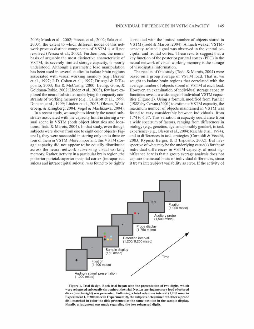
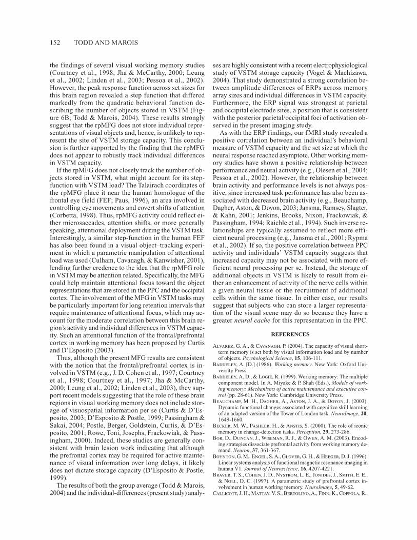
The results of this study (Todd & Marois, 2004) werebased on a group average of VSTM load. That is, wesought to isolate brain regions that correlated with theaverage number of objects stored in VSTM at each load.However, an examination of individual storage capacityfunctions reveals a wide range of individual VSTM capac-ities (Figure 2). Using a formula modified from Pashler(1988) by Cowan (2001) to estimate VSTM capacity, themaximum number of objects maintained in VSTM wasfound to vary considerably between individuals, from1.74 to 6.37. This variation in capacity could arise froma wide spectrum of factors, ranging from differences inbiology (e.g., genetics, age, and possibly gender), to taskexperience (e.g., Olesen et al., 2004; Raichle et al., 1994),and to differences in task strategies (Cornoldi & Vecchi,2003; Rypma, Berger, & D’Esposito, 2002). But irre-spective of what may be the underlying cause(s) for theseindividual differences in VSTM capacity, of most sig-nificance here is that a group average analysis does notcapture the neural basis of individual differences, sinceit treats intersubject variability as error. If the activity of
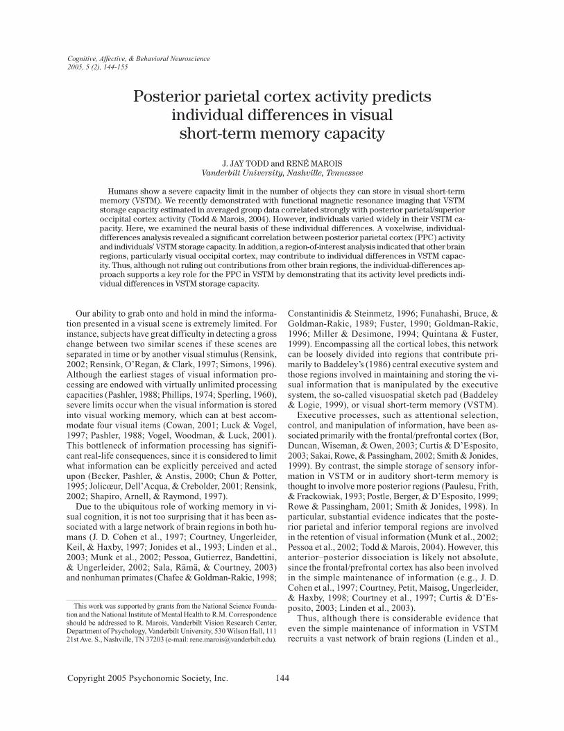
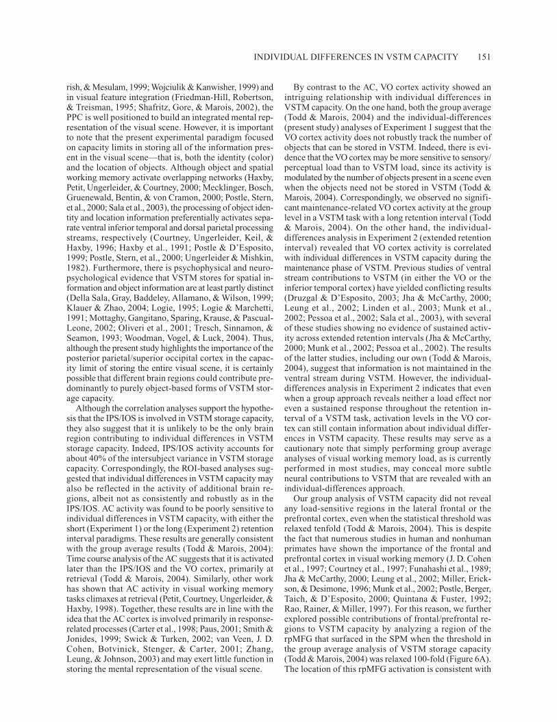
Figure 1. Trial design. Each trial began with the presentation of two digits, whichwere rehearsed subvocally throughout the trial. Next, a varying memory load of coloreddisks (one to eight) was presented. Following a brief retention interval (1,200 msec inExperiment 1, 9,200 msec in Experiment 2), the subjects determined whether a probedisk matched in color the disk presented at the same position in the sample display.Finally, a judgment was made regarding the two rehearsed digits.
Fixation(1,000 msec)
Auditory probe(1,500 msec)
Probe display(1,750 msec)
Retention interval(1,200/ 9,200 msec)
Sample display(150 msec)
Fixation(1,400 msec)
Auditory stimuli presentation(1,000 msec)
Time
3 1
+
146 TODD AND MAROIS
a brain region isolated with a group average analysisdoes not correlate with individual differences in perfor-mance, this would suggest that this brain region maycontribute only a generic component to the cognitiveprocess under investigation and that other brain regionsaccount for the diversity of performance across subjects.On the other hand, a stronger case can be made about abrain region being involved in a specific cognitive pro-cess if that brain region not only is activated in a signif-icant proportion of subjects, but also can account for thedifferences in performance between subjects. For exam-ple, in addition to demonstrating a role for the dorsal pre-frontal cortex (DPFC) in the retrieval of informationfrom auditory-verbal working memory (AVWM) in agroup average analysis, Rypma and D’Esposito (1999)showed that individual differences in speed of informa-tion retrieval from AVWM was correlated with the levelof DPFC activity. Thus, using both group and individual-differences approaches, one can isolate a brain regionthat is activated across individuals in a task and, subse-quently, identify its contribution to individual differencesin performance.
The goal of the present study was to determine howan individual-differences approach can inform us aboutthe neural basis of VSTM storage capacity. In particular,would an individual-differences approach provide con-verging evidence with the group average analysis for theimportance of the PPC in VSTM storage capacity? Orwould such individual-differences analysis instead pointto other brain regions to account for intersubject vari-ability? There is electrophysiological evidence in favorof the former possibility: Vogel and Machizawa (2004)showed that event-related potentials (ERPs) over poste-rior parietal and lateral occipital sites can predict indi-vidual subjects’ VSTM capacity. Importantly, the sourceof this ERP signal is largely compatible with the poste-
rior parietal/superior occipital locus of VSTM storagecapacity isolated in the group average fMRI study (Todd& Marois, 2004).
To determine which neural substrates might captureindividual differences in VSTM capacity, we adopted thelogic of Vogel and Machizawa (2004). That is, if a brainregion plays an important role in determining the amountof information held in VSTM, it is reasonable to expecta strong correlation between individual VSTM capacityestimates and the respective magnitude of activation inthat brain region. In other words, individuals with largeVSTM capacities would be expected to activate this brainregion more than would individuals with smaller capac-ities. Thus, in the present study, we correlated each sub-ject’s brain activity at the set size at which they reachedVSTM capacity with the number of objects they storedin VSTM at that same set size. We used this methodolog-ical approach in a voxelwise analysis in order to isolate anybrain regions whose activity correlated with individualdifferences in VSTM capacity. We also applied this tech-nique to a more sensitive region-of-interest (ROI) analysisin order to determine whether regions previously impli-cated in VSTM storage capacity on the basis of a groupaverage analysis (Todd & Marois, 2004) track individualdifferences in VSTM capacity.
METHOD
In the present study, the same data sets as those used in two extantfunctional magnetic resonance imaging (fMRI) experiments of VSTMload (Experiment 1, Todd & Marois, 2004; Experiment 2, Todd,Fougnie, & Marois, in press) were reanalyzed using an individual-differences approach.
SubjectsThe subjects were paid volunteers from the Vanderbilt commu-
nity. Seventeen young adults (9 females; all right-handed) partici-pated in Experiment 1, and 14 (8 females; 12 right-handed) volun-teered for Experiment 2. All had normal or corrected-to-normalvisual acuity and normal hearing and color vision. The experimen-tal protocol was approved by and consent was received in accor-dance with the Institutional Review Board at the Vanderbilt Uni-versity Medical Center.
Task DesignExperiment 1: Fast event-related fMRI study. A detailed de-
scription of the experimental method has been published elsewhere(Todd & Marois, 2004). The subjects performed a parametric loadmanipulation of a delayed recognition task (Figure 1). A static dis-play containing a varying number (one, two, three, four, six, oreight) of colored disks (0.38º of visual angle), each of a differentcolor (red, green, dark green, blue, light blue, black, white, brown,or yellow), was presented for 150 msec. The stimuli were presentedat nine possible locations within an invisible 3 � 3 matrix (1.38ºsquare). After a 1,200-msec retention period, a single colored diskwas presented in a position previously occupied by a disk in thesample array. The subjects indicated by buttonpress whether thecolor of the probe disk was identical to the color of the sample diskat that position (right index finger, same; right middle finger, dif-ferent). Half the trials were matched. To control for possible con-tamination of AVWM, the subjects performed a concurrent artic-ulatory suppression task (Baddeley, 1986). At trial onset, twosingle-digit numbers were presented serially through headphones
Figure 2. Individual visual short-term memory K functions.The K function for each individual (thin lines) is plotted by setsize. The group mean K function (n � 16) is represented by thethick line.
INDIVIDUAL DIFFERENCES IN VSTM CAPACITY 147
for 250 msec each, followed by a 250-msec blank interval and thenby a 250-msec auditory mask composed of superimposed forwardand reversed versions of the individual sound files. Following themask, there was a 1,400-msec period of fixation before the onset of the visual sample array. The subjects were instructed to rehearsethe two digits throughout the trial. Following the visual probe re-sponse, the subjects indicated by buttonpress whether two visuallypresented digits were the same as those rehearsed throughout thetrial.
Experiment 2: Slow event-related fMRI study. Fourteenyoung adults (8 females; 12 right-handed) volunteered for this ex-periment. The details of the experimental design have been pub-lished elsewhere (Todd et al., in press; Todd & Marois, 2004). Theexperimental design was similar to that for the fast event-relatedfMRI experiment described above, with the following exceptions:The retention interval was extended from 1,200 to 9,200 msec (trialduration, 18 sec; 7 trials/fMRI run), only two set sizes were em-ployed in order to compensate for the lower number of trials ac-quired with a slow event-related paradigm, and there were no non-event trials.
fMRI methods. fMRI methods have been described elsewhere(Todd & Marois, 2004). Low- and three-dimensional (3-D) high-resolution T1-weighted images were acquired using conventionaltechniques with a 3-T GE MRI scanner (GE Medical Systems, Mil-waukee, WI). In each functional run, 220 T2*-weighted echoplanarimages were acquired in 19 axial slices (7 mm thick, 3.75 � 3.75 mmin-plane, 0-mm skip, repetition time (TR) � 2,000 msec, echo time �25 msec, FOV � 24 cm, matrix � 64 � 64) covering the wholebrain and prescribed parallel to the AC–PC line. Trial presentationwas synchronized to TR onset by scanner trigger pulses. The stim-uli were presented on an Apple G4 Macintosh using PsychToolBoxfor Matlab. The stimuli were back-projected onto a screen viewedby the supine subject in the MR scanner through a prism mirror.The fMRI methods for Experiment 2 were the same as those forExperiment 1.
Data AnalysisBehavioral analysis. The estimated number of objects stored in
VSTM (K) was calculated using a formula developed by Pashler(1988) and modified by Cowan (2001). Here, K � (hit rate � cor-rect rejection rate �1)N, where N is the number of objects pre-sented in the sample array.
fMRI analysis. The fMRI data analysis was carried out withBrain Voyager 4.9.1 (Brain Innovation, Maastricht, The Nether-lands). Preprocessing included intrasession image realignment, 3-Dmotion correction and correction for slice scan acquisition orderusing sinc interpolation, and linear trend removal.
Group statistical parametric map analysis. VSTM load-modulated regions in Experiment 1 and for the right posterior middlefrontal gyrus (see the Results section) were identified in statisticalparametric maps (SPMs) of blood oxygen level dependent (BOLD)activation, using multiple regression analysis, with impulse regres-sors for each trial type (six set sizes and a no-event condition) con-volved with a canonical hemodynamic response function (Boynton,Engel, Glover, & Heeger, 1996). Regression coefficients wereweighted for each set size by their respective group average K value.The resulting maps from all the subjects were standardized into Ta-lairach space (Talairach & Tournoux, 1988) and were overlaid tocreate composite activation maps. The overall model fit was assessedwith an F statistic, using a random effects model, and voxels wereconsidered significant if p � .05, corrected for multiple compar-isons, using a cluster threshold of 6 voxels and spatial smoothingGaussian kernel of 8.0 mm (FWHM; Ward, 2000). ROIs were iso-lated from the SPMs, and time courses for each memory load wereextracted from these ROIs. Percentage of signal change of eachtime course was calculated using the nonevent time series as thebaseline condition (Kourtzi & Kanwisher, 2001).
Individual-differences analysis. Individual-differences analysiswas carried out using both a voxel-based and an ROI-based ap-proach. The voxelwise approach was used to identify regions thattrack individual differences in VSTM storage capacity. To isolatesuch regions, we first determined, for each subject, the maximumnumber of objects they could store (Kmax) and the percentage ofBOLD signal change (relative to no-event signal) obtained at theKmax set size. Since at set size 1 all the subjects had virtually thesame K value (mean K � 0.95) but very different BOLD signal lev-els, we standardized brain activity across subjects by first subtract-ing the percentage of BOLD signal at set size 1 from the percent-age of BOLD signal at set size Kmax. By using set size 1 activity asa baseline response for each individual, this transformation in-creased our sensitivity for detecting brain regions whose activitycorrelated with individual differences in VSTM capacity (Vogel &Machizawa, 2004). For each individual, regressors were defined forset size Kmax and set size 1 and were weighted by the individual’smaximum K value. We then probed for voxels whose activity co-varied with the magnitude of the difference between set size Kmaxand set size 1 across individuals.
The ROI approach was used to determine whether brain regionspreviously implicated in VSTM storage capacity on the basis of agroup average analysis (Todd & Marois, 2004) contributed to indi-vidual differences in VSTM capacity. For each ROI, we correlatedthe individual differences in Kmax with the individual differences inpercentage of peak activation between set size Kmax and set size 1.Specifically, the volume corresponding to the peak of the hemody-namic response collapsed across all set sizes was first defined.Then, for each individual, the activation difference between set sizeKmax and set size 1 of the peak volume was computed, and these ac-tivation differences were subsequently correlated with each indi-vidual’s Kmax. Significance for correlation analysis of the intrapari-etal sulcus/intraoccipital sulcus (IPS/IOS) ROI was set to p � .05,one-tail, on the basis of the a priori expectation of a positive linearrelationship between VSTM capacity and brain activity. For con-sistency, the same statistical criteria were employed for all the ROIs.For Experiment 2, ROI analysis of individual differences in VSTMcapacity1 was performed by first computing, for each individual,the difference in activation between set sizes 1 and 3 and regress-ing this activation difference with their corresponding K value ob-tained at set size 3 across individuals. This analysis was performedseparately for the encoding, maintenance, and retrieval phases ofVSTM, using the peak volumes acquired 5.5–7.5, 9.5–11.5, and15.5–17.5 sec after stimulus presentation, respectively. Given the9.2-sec-long retention interval, these peak time windows shouldprovide largely uncontaminated estimates of activity for each of thethree VSTM phases, particularly for the maintenance phase (Pessoaet al., 2002; Zarahn, Aguirre, & D’Esposito, 1997).
Outliers were isolated and removed in ROIs using DFFITSi witha cutoff threshold equal to �1 (J. Cohen, P. Cohen, West, & Aiken,2003). At most, two outliers were removed from any given ROI.
RESULTS
Voxel-Based AnalysesThe aim of the voxelwise analysis was to isolate brain
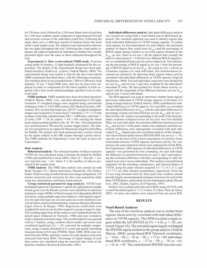
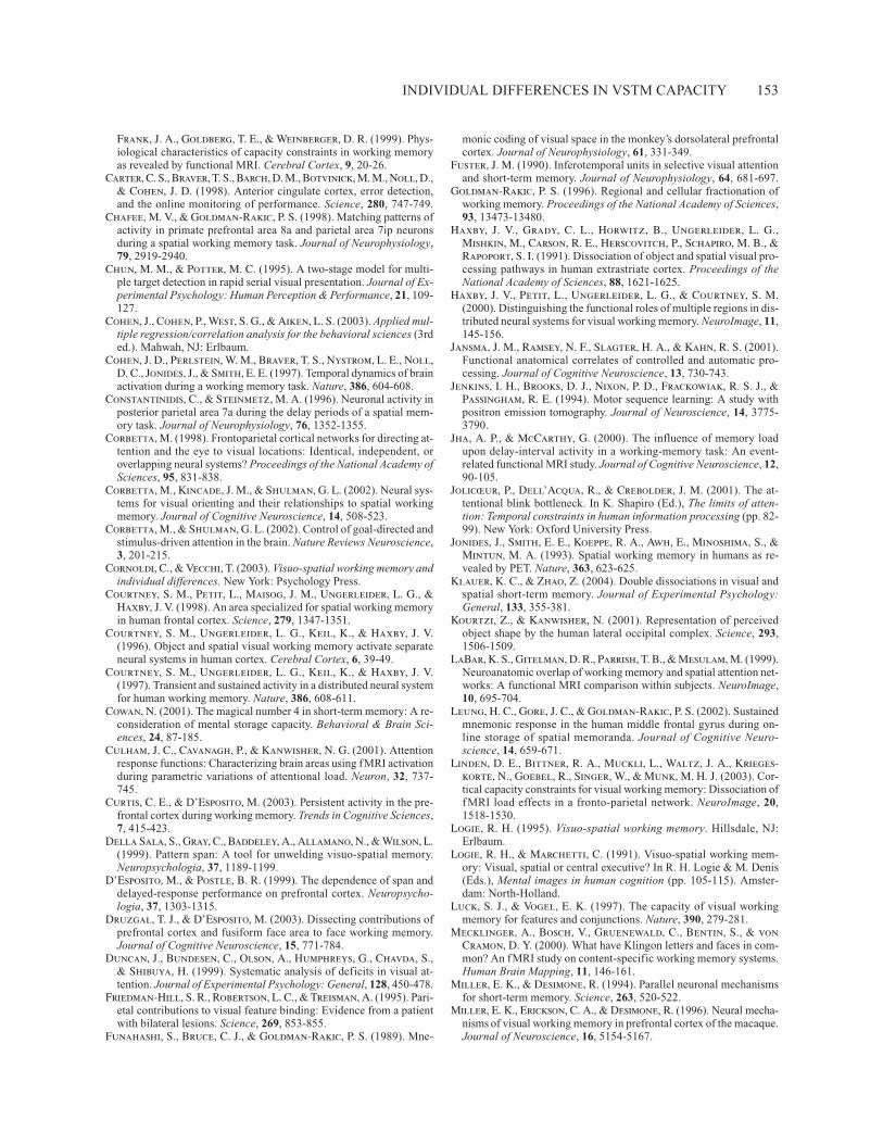
regions whose activity correlated with individual differ-ences in VSTM capacity. This SPM revealed a single re-gion within the left IPS/IOS [t(15) � 4.42, p � .05, cor-rected; Figure 3A]. The activated region largely overlappedthe IPS/IOS region isolated in the group analysis (Todd &Marois, 2004): group-based ROI Talairach coordinates,x, �14 to �30; y, �81 to �58; z, �17 to �49; individual-based ROI coordinates, x, �17 to �29; y, �81 to �61;z, �21 to �45. The contralateral IPS/IOS was also acti-
148 TODD AND MAROIS
vated when the threshold was reduced tenfold. ROI analy-sis of this IPS/IOS region confirmed that individuals’Kmax correlated with the activation difference between
set size Kmax and set size 1 [r(12) � .56, p � .05; Fig-ure 3B]. This load-modulated response was not simply dueto high-K individuals demonstrating greater overall acti-vation than low-K individuals did (Vogel & Machizawa,2004), since each subject’s BOLD signal was standardizedat set size 1. The response was also not due to scalingdifferences in activation across individuals, with subjectswith greater memory capacity simply having greateroverall activation range, since overall activation differ-ences across set sizes did not correlate with individuals’Kmax [r(14) � .36, n.s.]. Importantly, the correlation be-tween individuals’ Kmax and activity difference betweenset sizes Kmax and 1 did not extend to supracapacity setsizes. That is, there was no correlation between individ-uals’ Kmax and the activation difference between set size 8and Kmax for the individuals whose Kmax occurred at setsizes lower than 8 [r(7) � �.19, n.s.]. Analysis of thesame ROI in Experiment 2 with extended retention in-terval revealed that the activity difference between setsizes 3 and 1 was correlated with individual VSTM ca-pacities at set size 3 during maintenance [r(12) � .54, p �.05] and retrieval [r(11) � .63, p � .05], and marginallyso during encoding [r(11) � .50, p � .08; Figure 3C].
Taken together, these analyses indicate that a regionof the IPS/IOS that largely overlaps with the PPC regionisolated with a group-based approach to VSTM capacity(Todd & Marois, 2004) tracks individual differences inVSTM capacity.
ROI-Based AnalysesIn addition to the IPS/IOS, our group average study of
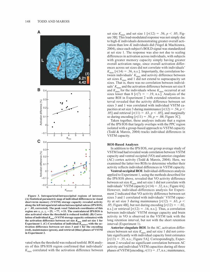
VSTM load had revealed weak correlation between VSTMcapacity and ventral occipital (VO) and anterior cingulate(AC) cortex activity (Todd & Marois, 2004). Here, weexamined the latter two ROIs to determine whether theiractivity reflects individual differences in VSTM capacity.
Ventral occipital ROI. Individual-differences analysisapplied to Experiment 1, using the methods described forthe IPS/IOS above, revealed that VO activity differencebetween set size Kmax and set size 1 did not correlate withindividuals’ VSTM capacity [r(14) � .32, n.s.; Figure 4A].However, individual-differences analysis for Experi-ment 2 indicated that VO activity difference between setsizes 3 and 1 correlated with individuals’ VSTM capac-ity at set size 3 during maintenance [r(12) � .63, p �.05; Figure 4B], but not during encoding [r(12) � �.02,n.s.] or retrieval [r(12) � .14, n.s.]. Thus, a correlationbetween individuals’ VSTM storage capacity and brainactivity in VO is observed in the VSTM task with thelong retention interval, but not with the short retentioninterval (Experiment 1).
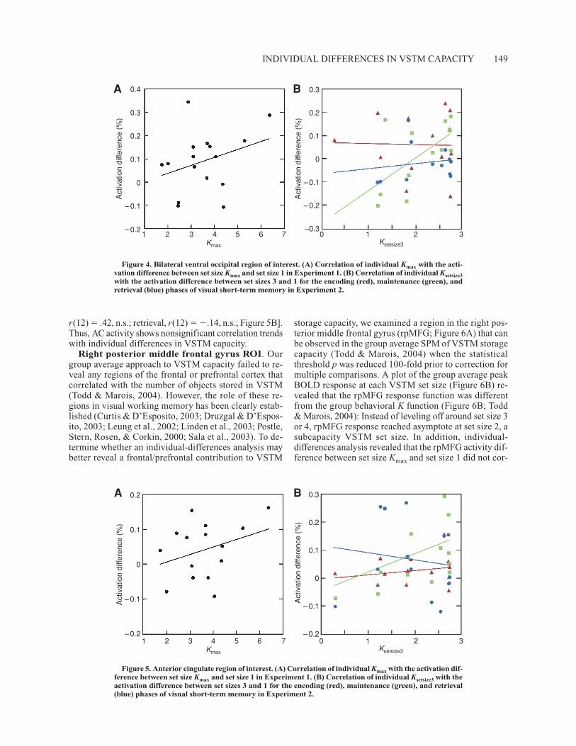
Anterior cingulate ROI. In the AC, activation differ-ences between set size Kmax and set size 1 did not corre-late significantly with individual capacity limit estimates[r(13) � .35, n.s.; Figure 5A]. Correspondingly, Exper-iment 2 revealed no significant correlation between ACactivity and individual VSTM capacities during all threephases of VSTM [encoding, r(11) � .17, n.s.; maintenance,
Figure 3. Intraparietal/intraoccipital regions of interest.(A) Statistical parametric map of individual differences in visualshort-term memory (VSTM) storage capacity revealed activityalong the left intraparietal sulcus/intraoccipital sulcus (IPS/IOS;p � .05, corrected). The peak voxel Talairach coordinates of thisregion are x, y, z, �25, �77, �33. The contralateral IPS/IOS isalso activated when the threshold is reduced tenfold. (B) Corre-lation of individual Kmax (VSTM storage capacity estimates) withthe activation difference between set size Kmax and set size 1 forExperiment 1. (C) Correlation of individual Ksetsize3 with the ac-tivation difference between set sizes 3 and 1 for the encoding(red), maintenance (green), and retrieval (blue) phases of VSTMin Experiment 2.
A
B
C
L R
(t)6.214.42
0.4
0.3
0.2
0.1
0
–0.11 2 3 4 5 6 7
Kmax
Act
ivat
ion
diffe
renc
e (%
)
0.4
0.3
0.2
0.1
0
–0.1
–0.2
Act
ivat
ion
diffe
renc
e (%
)
0 1 2 3Ksetsize3
INDIVIDUAL DIFFERENCES IN VSTM CAPACITY 149
r(12) � .42, n.s.; retrieval, r(12) � �.14, n.s.; Figure 5B].Thus, AC activity shows nonsignificant correlation trendswith individual differences in VSTM capacity.
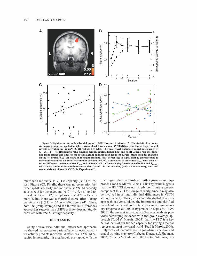
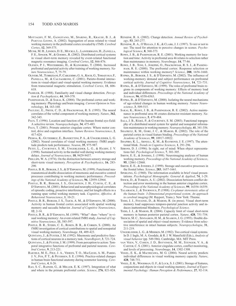
Right posterior middle frontal gyrus ROI. Ourgroup average approach to VSTM capacity failed to re-veal any regions of the frontal or prefrontal cortex thatcorrelated with the number of objects stored in VSTM(Todd & Marois, 2004). However, the role of these re-gions in visual working memory has been clearly estab-lished (Curtis & D’Esposito, 2003; Druzgal & D’Espos-ito, 2003; Leung et al., 2002; Linden et al., 2003; Postle,Stern, Rosen, & Corkin, 2000; Sala et al., 2003). To de-termine whether an individual-differences analysis maybetter reveal a frontal/prefrontal contribution to VSTM
storage capacity, we examined a region in the right pos-terior middle frontal gyrus (rpMFG; Figure 6A) that canbe observed in the group average SPM of VSTM storagecapacity (Todd & Marois, 2004) when the statisticalthreshold p was reduced 100-fold prior to correction formultiple comparisons. A plot of the group average peakBOLD response at each VSTM set size (Figure 6B) re-vealed that the rpMFG response function was differentfrom the group behavioral K function (Figure 6B; Todd& Marois, 2004): Instead of leveling off around set size 3or 4, rpMFG response reached asymptote at set size 2, asubcapacity VSTM set size. In addition, individual-differences analysis revealed that the rpMFG activity dif-ference between set size Kmax and set size 1 did not cor-
Figure 4. Bilateral ventral occipital region of interest. (A) Correlation of individual Kmax with the acti-vation difference between set size Kmax and set size 1 in Experiment 1. (B) Correlation of individual Ksetsize3with the activation difference between set sizes 3 and 1 for the encoding (red), maintenance (green), andretrieval (blue) phases of visual short-term memory in Experiment 2.
A B0.4
0.3
0.2
0.1
0
– 0.1
– 0.2
0.3
0.2
0.1
0
– 0.1
– 0.2
–0.31 2 3 4 5 6 7
Kmax
0 1 2 3Ksetsize3
Act
ivat
ion
diffe
renc
e (%
)
Act
ivat
ion
diffe
renc
e (%
)
Figure 5. Anterior cingulate region of interest. (A) Correlation of individual Kmax with the activation dif-ference between set size Kmax and set size 1 in Experiment 1. (B) Correlation of individual Ksetsize3 with theactivation difference between set sizes 3 and 1 for the encoding (red), maintenance (green), and retrieval(blue) phases of visual short-term memory in Experiment 2.
A B 0.3
0.2
0.1
0
– 0.1
– 0.20 1 2 3
Ksetsize3
Act
ivat
ion
diffe
renc
e (%
)
0.2
0.1
0
– 0.1
– 0.21 2 3 4 5 6 7
Kmax
Act
ivat
ion
diffe
renc
e (%
)
150 TODD AND MAROIS
relate with individuals’ VSTM capacity [r(14) � .25,n.s.; Figure 6C]. Finally, there was no correlation be-tween rpMFG activity and individuals’ VSTM capacityat set size 3 for the encoding [r(10) � .49, n.s.] and re-trieval [r(11) � �.42, n.s.] phases of VSTM in Experi-ment 2, but there was a marginal correlation duringmaintenance [r(11) � .53, p � .06; Figure 6D]. Thus,both the group average and the individual-differencesapproaches suggest that rpMFG activity does not tightlycorrelate with VSTM storage capacity.
DISCUSSION
Using a voxelwise individual-differences approach,we showed that posterior parietal/superior occipital cor-tex activity predicts individual differences in VSTM ca-pacity. Importantly, this area largely overlapped with the
PPC region that was isolated with a group-based ap-proach (Todd & Marois, 2004). This key result suggeststhat the IPS/IOS does not simply contribute a genericcomponent to VSTM storage capacity, since it may alsobe involved in setting individual differences in VSTMstorage capacity. Thus, just as an individual-differencesapproach has consolidated the importance and clarifiedthe role of the lateral prefrontal cortex in working mem-ory (Rypma et al., 2002; Rypma & D’Esposito, 1999,2000), the present individual-differences analysis pro-vides converging evidence with the group average ap-proach (Todd & Marois, 2004) that the PPC is a keyneural locus of our limited capacity for storing a mentalrepresentation of the visual world (Todd & Marois, 2004).
By virtue of its central role in goal-driven attention andspatial working memory (Corbetta, Kincade, & Shulman,2002; Corbetta & Shulman, 2002; LaBar, Gitelman, Par-
Figure 6. Right posterior middle frontal gyrus (rpMFG) region of interest. (A) The statistical paramet-ric map of group-averaged, K-weighted visual short-term memory (VSTM) load function in Experiment 1reveals activation in the rpMFG (threshold t � 2.12). The peak voxel Talairach coordinates are x, y,z , �26, �5, �45. (B) Behavioral K function (empty circles, dashed line) and rpMFG peak response func-tion (solid circles and line) for the group average analysis in Experiment 1. Percentage of signal change ison the left ordinate; K values are on the right ordinate. Peak percentage of signal change corresponded tothe volume acquired 5.6 sec after stimulus presentation. (C) Correlation of individual Kmax with the acti-vation difference between set size Kmax and set size 1 in Experiment 1. (D) Correlation of individual Ksetsize3with the activation difference between set sizes 3 and 1 for the encoding (red), maintenance (green), andretrieval (blue) phases of VSTM in Experiment 2.
A B
0.3
0.2
0.1
0
– 0.1
– 0.20 1 2 3
Ksetsize3
Act
ivat
ion
diffe
renc
e (%
)
0.2
0.1
0
1 2 3 4 5 6 7Kmax
Act
ivat
ion
diffe
renc
e (%
)
C D
L R
y = –7 mm
(t)5.062.12
0.2
0.1
0
Sig
nal c
hang
e (%
)
4
3
2
1
0
Num
ber of objects encoded (K)
1 2 3 4 6 8Set size
INDIVIDUAL DIFFERENCES IN VSTM CAPACITY 151
rish, & Mesulam, 1999; Wojciulik & Kanwisher, 1999) andin visual feature integration (Friedman-Hill, Robertson,& Treisman, 1995; Shafritz, Gore, & Marois, 2002), thePPC is well positioned to build an integrated mental rep-resentation of the visual scene. However, it is importantto note that the present experimental paradigm focusedon capacity limits in storing all of the information pres-ent in the visual scene—that is, both the identity (color)and the location of objects. Although object and spatialworking memory activate overlapping networks (Haxby,Petit, Ungerleider, & Courtney, 2000; Mecklinger, Bosch,Gruenewald, Bentin, & von Cramon, 2000; Postle, Stern,et al., 2000; Sala et al., 2003), the processing of object iden-tity and location information preferentially activates sepa-rate ventral inferior temporal and dorsal parietal processingstreams, respectively (Courtney, Ungerleider, Keil, &Haxby, 1996; Haxby et al., 1991; Postle & D’Esposito,1999; Postle, Stern, et al., 2000; Ungerleider & Mishkin,1982). Furthermore, there is psychophysical and neuro-psychological evidence that VSTM stores for spatial in-formation and object information are at least partly distinct(Della Sala, Gray, Baddeley, Allamano, & Wilson, 1999;Klauer & Zhao, 2004; Logie, 1995; Logie & Marchetti,1991; Mottaghy, Gangitano, Sparing, Krause, & Pascual-Leone, 2002; Oliveri et al., 2001; Tresch, Sinnamon, &Seamon, 1993; Woodman, Vogel, & Luck, 2004). Thus,although the present study highlights the importance of theposterior parietal/superior occipital cortex in the capac-ity limit of storing the entire visual scene, it is certainlypossible that different brain regions could contribute pre-dominantly to purely object-based forms of VSTM stor-age capacity.
Although the correlation analyses support the hypothe-sis that the IPS/IOS is involved in VSTM storage capacity,they also suggest that it is unlikely to be the only brainregion contributing to individual differences in VSTMstorage capacity. Indeed, IPS/IOS activity accounts forabout 40% of the intersubject variance in VSTM storagecapacity. Correspondingly, the ROI-based analyses sug-gested that individual differences in VSTM capacity mayalso be reflected in the activity of additional brain re-gions, albeit not as consistently and robustly as in theIPS/IOS. AC activity was found to be poorly sensitive toindividual differences in VSTM capacity, with either theshort (Experiment 1) or the long (Experiment 2) retentioninterval paradigms. These results are generally consistentwith the group average results (Todd & Marois, 2004):Time course analysis of the AC suggests that it is activatedlater than the IPS/IOS and the VO cortex, primarily atretrieval (Todd & Marois, 2004). Similarly, other workhas shown that AC activity in visual working memorytasks climaxes at retrieval (Petit, Courtney, Ungerleider, &Haxby, 1998). Together, these results are in line with theidea that the AC cortex is involved primarily in response-related processes (Carter et al., 1998; Paus, 2001; Smith &Jonides, 1999; Swick & Turken, 2002; van Veen, J. D.Cohen, Botvinick, Stenger, & Carter, 2001; Zhang,Leung, & Johnson, 2003) and may exert little function instoring the mental representation of the visual scene.
By contrast to the AC, VO cortex activity showed anintriguing relationship with individual differences inVSTM capacity. On the one hand, both the group average(Todd & Marois, 2004) and the individual-differences(present study) analyses of Experiment 1 suggest that theVO cortex activity does not robustly track the number ofobjects that can be stored in VSTM. Indeed, there is evi-dence that the VO cortex may be more sensitive to sensory/perceptual load than to VSTM load, since its activity ismodulated by the number of objects present in a scene evenwhen the objects need not be stored in VSTM (Todd &Marois, 2004). Correspondingly, we observed no signifi-cant maintenance-related VO cortex activity at the grouplevel in a VSTM task with a long retention interval (Todd& Marois, 2004). On the other hand, the individual-differences analysis in Experiment 2 (extended retentioninterval) revealed that VO cortex activity is correlatedwith individual differences in VSTM capacity during themaintenance phase of VSTM. Previous studies of ventralstream contributions to VSTM (in either the VO or theinferior temporal cortex) have yielded conflicting results(Druzgal & D’Esposito, 2003; Jha & McCarthy, 2000;Leung et al., 2002; Linden et al., 2003; Munk et al.,2002; Pessoa et al., 2002; Sala et al., 2003), with severalof these studies showing no evidence of sustained activ-ity across extended retention intervals (Jha & McCarthy,2000; Munk et al., 2002; Pessoa et al., 2002). The resultsof the latter studies, including our own (Todd & Marois,2004), suggest that information is not maintained in theventral stream during VSTM. However, the individual-differences analysis in Experiment 2 indicates that evenwhen a group approach reveals neither a load effect noreven a sustained response throughout the retention in-terval of a VSTM task, activation levels in the VO cor-tex can still contain information about individual differ-ences in VSTM capacity. These results may serve as acautionary note that simply performing group averageanalyses of visual working memory load, as is currentlyperformed in most studies, may conceal more subtleneural contributions to VSTM that are revealed with anindividual-differences approach.
Our group analysis of VSTM capacity did not revealany load-sensitive regions in the lateral frontal or theprefrontal cortex, even when the statistical threshold wasrelaxed tenfold (Todd & Marois, 2004). This is despitethe fact that numerous studies in human and nonhumanprimates have shown the importance of the frontal andprefrontal cortex in visual working memory (J. D. Cohenet al., 1997; Courtney et al., 1997; Funahashi et al., 1989;Jha & McCarthy, 2000; Leung et al., 2002; Miller, Erick-son, & Desimone, 1996; Munk et al., 2002; Postle, Berger,Taich, & D’Esposito, 2000; Quintana & Fuster, 1992;Rao, Rainer, & Miller, 1997). For this reason, we furtherexplored possible contributions of frontal/prefrontal re-gions to VSTM capacity by analyzing a region of therpMFG that surfaced in the SPM when the threshold inthe group average analysis of VSTM storage capacity(Todd & Marois, 2004) was relaxed 100-fold (Figure 6A).The location of this rpMFG activation is consistent with
152 TODD AND MAROIS
the findings of several visual working memory studies(Courtney et al., 1998; Jha & McCarthy, 2000; Leunget al., 2002; Linden et al., 2003; Pessoa et al., 2002).However, the peak response function across set sizes forthis brain region revealed a step function that differedmarkedly from the quadratic behavioral function de-scribing the number of objects stored in VSTM (Fig-ure 6B; Todd & Marois, 2004). These results stronglysuggest that the rpMFG does not store individual repre-sentations of visual objects and, hence, is unlikely to rep-resent the site of VSTM storage capacity. This conclu-sion is further supported by the finding that the rpMFGdoes not appear to robustly track individual differencesin VSTM capacity.
If the rpMFG does not closely track the number of ob-jects stored in VSTM, what might account for its step-function with VSTM load? The Talairach coordinates ofthe rpMFG place it near the human homologue of thefrontal eye field (FEF; Paus, 1996), an area involved incontrolling eye movements and covert shifts of attention(Corbetta, 1998). Thus, rpMFG activity could reflect ei-ther microsaccades, attention shifts, or more generallyspeaking, attentional deployment during the VSTM task.Interestingly, a similar step-function in the human FEFhas also been found in a visual object–tracking experi-ment in which a parametric manipulation of attentionalload was used (Culham, Cavanagh, & Kanwisher, 2001),lending further credence to the idea that the rpMFG rolein VSTM may be attention related. Specifically, the MFGcould help maintain attentional focus toward the objectrepresentations that are stored in the PPC and the occipitalcortex. The involvement of the MFG in VSTM tasks maybe particularly important for long retention intervals thatrequire maintenance of attentional focus, which may ac-count for the moderate correlation between this brain re-gion’s activity and individual differences in VSTM capac-ity. Such an attentional function of the frontal/prefrontalcortex in working memory has been proposed by Curtisand D’Esposito (2003).
Thus, although the present MFG results are consistentwith the notion that the frontal/prefrontal cortex is in-volved in VSTM (e.g., J. D. Cohen et al., 1997; Courtneyet al., 1998; Courtney et al., 1997; Jha & McCarthy,2000; Leung et al., 2002; Linden et al., 2003), they sup-port recent models suggesting that the role of these brainregions in visual working memory does not include stor-age of visuospatial information per se (Curtis & D’Es-posito, 2003; D’Esposito & Postle, 1999; Passingham &Sakai, 2004; Postle, Berger, Goldstein, Curtis, & D’Es-posito, 2001; Rowe, Toni, Josephs, Frackowiak, & Pass-ingham, 2000). Indeed, these studies are generally con-sistent with brain lesion work indicating that althoughthe prefrontal cortex may be required for active mainte-nance of visual information over long delays, it likelydoes not dictate storage capacity (D’Esposito & Postle,1999).
The results of both the group average (Todd & Marois,2004) and the individual-differences (present study) analy-
ses are highly consistent with a recent electrophysiologicalstudy of VSTM storage capacity (Vogel & Machizawa,2004). That study demonstrated a strong correlation be-tween amplitude differences of ERPs across memoryarray sizes and individual differences in VSTM capacity.Furthermore, the ERP signal was strongest at parietaland occipital electrode sites, a position that is consistentwith the posterior parietal/occipital foci of activation ob-served in the present imaging study.
As with the ERP findings, our fMRI study revealed apositive correlation between an individual’s behavioralmeasure of VSTM capacity and the set size at which theneural response reached asymptote. Other working mem-ory studies have shown a positive relationship betweenperformance and neural activity (e.g., Olesen et al., 2004;Pessoa et al., 2002). However, the relationship betweenbrain activity and performance levels is not always pos-itive, since increased task performance has also been as-sociated with decreased brain activity (e.g., Beauchamp,Dagher, Aston, & Doyon, 2003; Jansma, Ramsey, Slagter,& Kahn, 2001; Jenkins, Brooks, Nixon, Frackowiak, &Passingham, 1994; Raichle et al., 1994). Such inverse re-lationships are typically assumed to reflect more effi-cient neural processing (e.g., Jansma et al., 2001; Rypmaet al., 2002). If so, the positive correlation between PPCactivity and individuals’ VSTM capacity suggests thatincreased capacity may not be associated with more ef-ficient neural processing per se. Instead, the storage ofadditional objects in VSTM is likely to result from ei-ther an enhancement of activity of the nerve cells withina given neural tissue or the recruitment of additionalcells within the same tissue. In either case, our resultssuggest that subjects who can store a larger representa-tion of the visual scene may do so because they have agreater neural cache for this representation in the PPC.
REFERENCES
Alvarez, G. A., & Cavanagh, P. (2004). The capacity of visual short-term memory is set both by visual information load and by numberof objects. Psychological Science, 15, 106-111.
Baddeley, A. [D.] (1986). Working memory. New York: Oxford Uni-versity Press.
Baddeley, A. D., & Logie, R. (1999). Working memory: The multiplecomponent model. In A. Miyake & P. Shah (Eds.), Models of work-ing memory: Mechanisms of active maintenance and executive con-trol (pp. 28-61). New York: Cambridge University Press.
Beauchamp, M. H., Dagher, A., Aston, J. A., & Doyon, J. (2003).Dynamic functional changes associated with cognitive skill learningof an adapted version of the Tower of London task. NeuroImage, 20,1649-1660.
Becker, M. W., Pashler, H., & Anstis, S. (2000). The role of iconicmemory in change-detection tasks. Perception, 29, 273-286.
Bor, D., Duncan, J., Wiseman, R. J., & Owen, A. M. (2003). Encod-ing strategies dissociate prefrontal activity from working memory de-mand. Neuron, 37, 361-367.
Boynton, G. M., Engel, S. A., Glover, G. H., & Heeger, D. J. (1996).Linear systems analysis of functional magnetic resonance imaging inhuman V1. Journal of Neuroscience, 16, 4207-4221.
Braver, T. S., Cohen, J. D., Nystrom, L. E., Jonides, J., Smith, E. E.,& Noll, D. C. (1997). A parametric study of prefrontal cortex in-volvement in human working memory. NeuroImage, 5, 49-62.
Callicott, J. H., Mattay, V. S., Bertolino, A., Finn, K., Coppola, R.,
INDIVIDUAL DIFFERENCES IN VSTM CAPACITY 153
Frank, J. A., Goldberg, T. E., & Weinberger, D. R. (1999). Phys-iological characteristics of capacity constraints in working memoryas revealed by functional MRI. Cerebral Cortex, 9, 20-26.
Carter, C. S., Braver, T. S., Barch, D. M., Botvinick, M. M., Noll, D.,& Cohen, J. D. (1998). Anterior cingulate cortex, error detection,and the online monitoring of performance. Science, 280, 747-749.
Chafee, M. V., & Goldman-Rakic, P. S. (1998). Matching patterns ofactivity in primate prefrontal area 8a and parietal area 7ip neuronsduring a spatial working memory task. Journal of Neurophysiology,79, 2919-2940.
Chun, M. M., & Potter, M. C. (1995). A two-stage model for multi-ple target detection in rapid serial visual presentation. Journal of Ex-perimental Psychology: Human Perception & Performance, 21, 109-127.
Cohen, J., Cohen, P., West, S. G., & Aiken, L. S. (2003). Applied mul-tiple regression/correlation analysis for the behavioral sciences (3rded.). Mahwah, NJ: Erlbaum.
Cohen, J. D., Perlstein, W. M., Braver, T. S., Nystrom, L. E., Noll,D. C., Jonides, J., & Smith, E. E. (1997). Temporal dynamics of brainactivation during a working memory task. Nature, 386, 604-608.
Constantinidis, C., & Steinmetz, M. A. (1996). Neuronal activity inposterior parietal area 7a during the delay periods of a spatial mem-ory task. Journal of Neurophysiology, 76, 1352-1355.
Corbetta, M. (1998). Frontoparietal cortical networks for directing at-tention and the eye to visual locations: Identical, independent, oroverlapping neural systems? Proceedings of the National Academy ofSciences, 95, 831-838.
Corbetta, M., Kincade, J. M., & Shulman, G. L. (2002). Neural sys-tems for visual orienting and their relationships to spatial workingmemory. Journal of Cognitive Neuroscience, 14, 508-523.
Corbetta, M., & Shulman, G. L. (2002). Control of goal-directed andstimulus-driven attention in the brain. Nature Reviews Neuroscience,3, 201-215.
Cornoldi, C., & Vecchi, T. (2003). Visuo-spatial working memory andindividual differences. New York: Psychology Press.
Courtney, S. M., Petit, L., Maisog, J. M., Ungerleider, L. G., &Haxby, J. V. (1998). An area specialized for spatial working memoryin human frontal cortex. Science, 279, 1347-1351.
Courtney, S. M., Ungerleider, L. G., Keil, K., & Haxby, J. V.(1996). Object and spatial visual working memory activate separateneural systems in human cortex. Cerebral Cortex, 6, 39-49.
Courtney, S. M., Ungerleider, L. G., Keil, K., & Haxby, J. V.(1997). Transient and sustained activity in a distributed neural systemfor human working memory. Nature, 386, 608-611.
Cowan, N. (2001). The magical number 4 in short-term memory: A re-consideration of mental storage capacity. Behavioral & Brain Sci-ences, 24, 87-185.
Culham, J. C., Cavanagh, P., & Kanwisher, N. G. (2001). Attentionresponse functions: Characterizing brain areas using fMRI activationduring parametric variations of attentional load. Neuron, 32, 737-745.
Curtis, C. E., & D’Esposito, M. (2003). Persistent activity in the pre-frontal cortex during working memory. Trends in Cognitive Sciences,7, 415-423.
Della Sala, S., Gray, C., Baddeley, A., Allamano, N., & Wilson, L.(1999). Pattern span: A tool for unwelding visuo-spatial memory.Neuropsychologia, 37, 1189-1199.
D’Esposito, M., & Postle, B. R. (1999). The dependence of span anddelayed-response performance on prefrontal cortex. Neuropsycho-logia, 37, 1303-1315.
Druzgal, T. J., & D’Esposito, M. (2003). Dissecting contributions ofprefrontal cortex and fusiform face area to face working memory.Journal of Cognitive Neuroscience, 15, 771-784.
Duncan, J., Bundesen, C., Olson, A., Humphreys, G., Chavda, S.,& Shibuya, H. (1999). Systematic analysis of deficits in visual at-tention. Journal of Experimental Psychology: General, 128, 450-478.
Friedman-Hill, S. R., Robertson, L. C., & Treisman, A. (1995). Pari-etal contributions to visual feature binding: Evidence from a patientwith bilateral lesions. Science, 269, 853-855.
Funahashi, S., Bruce, C. J., & Goldman-Rakic, P. S. (1989). Mne-
monic coding of visual space in the monkey’s dorsolateral prefrontalcortex. Journal of Neurophysiology, 61, 331-349.
Fuster, J. M. (1990). Inferotemporal units in selective visual attentionand short-term memory. Journal of Neurophysiology, 64, 681-697.
Goldman-Rakic, P. S. (1996). Regional and cellular fractionation ofworking memory. Proceedings of the National Academy of Sciences,93, 13473-13480.
Haxby, J. V., Grady, C. L., Horwitz, B., Ungerleider, L. G.,Mishkin, M., Carson, R. E., Herscovitch, P., Schapiro, M. B., &Rapoport, S. I. (1991). Dissociation of object and spatial visual pro-cessing pathways in human extrastriate cortex. Proceedings of theNational Academy of Sciences, 88, 1621-1625.
Haxby, J. V., Petit, L., Ungerleider, L. G., & Courtney, S. M.(2000). Distinguishing the functional roles of multiple regions in dis-tributed neural systems for visual working memory. NeuroImage, 11,145-156.
Jansma, J. M., Ramsey, N. F., Slagter, H. A., & Kahn, R. S. (2001).Functional anatomical correlates of controlled and automatic pro-cessing. Journal of Cognitive Neuroscience, 13, 730-743.
Jenkins, I. H., Brooks, D. J., Nixon, P. D., Frackowiak, R. S. J., &Passingham, R. E. (1994). Motor sequence learning: A study withpositron emission tomography. Journal of Neuroscience, 14, 3775-3790.
Jha, A. P., & McCarthy, G. (2000). The influence of memory loadupon delay-interval activity in a working-memory task: An event-related functional MRI study. Journal of Cognitive Neuroscience, 12,90-105.
Jolicœur, P., Dell’Acqua, R., & Crebolder, J. M. (2001). The at-tentional blink bottleneck. In K. Shapiro (Ed.), The limits of atten-tion: Temporal constraints in human information processing (pp. 82-99). New York: Oxford University Press.
Jonides, J., Smith, E. E., Koeppe, R. A., Awh, E., Minoshima, S., &Mintun, M. A. (1993). Spatial working memory in humans as re-vealed by PET. Nature, 363, 623-625.
Klauer, K. C., & Zhao, Z. (2004). Double dissociations in visual andspatial short-term memory. Journal of Experimental Psychology:General, 133, 355-381.
Kourtzi, Z., & Kanwisher, N. (2001). Representation of perceivedobject shape by the human lateral occipital complex. Science, 293,1506-1509.
LaBar, K. S., Gitelman, D. R., Parrish, T. B., & Mesulam, M. (1999).Neuroanatomic overlap of working memory and spatial attention net-works: A functional MRI comparison within subjects. NeuroImage,10, 695-704.
Leung, H. C., Gore, J. C., & Goldman-Rakic, P. S. (2002). Sustainedmnemonic response in the human middle frontal gyrus during on-line storage of spatial memoranda. Journal of Cognitive Neuro-science, 14, 659-671.
Linden, D. E., Bittner, R. A., Muckli, L., Waltz, J. A., Krieges-korte, N., Goebel, R., Singer, W., & Munk, M. H. J. (2003). Cor-tical capacity constraints for visual working memory: Dissociation offMRI load effects in a fronto-parietal network. NeuroImage, 20,1518-1530.
Logie, R. H. (1995). Visuo-spatial working memory. Hillsdale, NJ:Erlbaum.
Logie, R. H., & Marchetti, C. (1991). Visuo-spatial working mem-ory: Visual, spatial or central executive? In R. H. Logie & M. Denis(Eds.), Mental images in human cognition (pp. 105-115). Amster-dam: North-Holland.
Luck, S. J., & Vogel, E. K. (1997). The capacity of visual workingmemory for features and conjunctions. Nature, 390, 279-281.
Mecklinger, A., Bosch, V., Gruenewald, C., Bentin, S., & vonCramon, D. Y. (2000). What have Klingon letters and faces in com-mon? An fMRI study on content-specific working memory systems.Human Brain Mapping, 11, 146-161.
Miller, E. K., & Desimone, R. (1994). Parallel neuronal mechanismsfor short-term memory. Science, 263, 520-522.
Miller, E. K., Erickson, C. A., & Desimone, R. (1996). Neural mecha-nisms of visual working memory in prefrontal cortex of the macaque.Journal of Neuroscience, 16, 5154-5167.
154 TODD AND MAROIS
Mottaghy, F. M., Gangitano, M., Sparing, R., Krause, B. J., &Pascual-Leone, A. (2002). Segregation of areas related to visualworking memory in the prefrontal cortex revealed by rTMS. CerebralCortex, 12, 369-375.
Munk, M. H., Linden, D. E., Muckli, L., Lanfermann, H., Zanella,F. E., Singer, W., & Goebel, R. (2002). Distributed cortical systemsin visual short-term memory revealed by event-related functionalmagnetic resonance imaging. Cerebral Cortex, 12, 866-876.
Olesen, P. J., Westerberg, H., & Klingberg, T. (2004). Increasedprefrontal and parietal activity after training of working memory. Na-ture Neuroscience, 7, 75-79.
Oliveri, M., Turriziani, P., Carlesimo, G. A., Koch, G., Tomaiuolo, F.,Panella, M., & Caltagirone, C. (2001). Parieto-frontal interac-tions in visual-object and visual-spatial working memory: Evidencefrom transcranial magnetic stimulation. Cerebral Cortex, 11, 606-618.
Pashler, H. (1988). Familiarity and visual change detection. Percep-tion & Psychophysics, 44, 369-378.
Passingham, D., & Sakai, K. (2004). The prefrontal cortex and work-ing memory: Physiology and brain imaging. Current Opinion in Neu-robiology, 14, 163-168.
Paulesu, E., Frith, C. D., & Frackowiak, R. S. (1993). The neuralcorrelates of the verbal component of working memory. Nature, 362,342-345.
Paus, T. (1996). Location and function of the human frontal eye-field:A selective review. Neuropsychologia, 34, 475-483.
Paus, T. (2001). Primate anterior cingulate cortex: Where motor con-trol, drive and cognition interface. Nature Reviews Neuroscience, 2,417-424.
Pessoa, K., Gutierrez, E., Bandettini, P. A., & Ungerleider, L. G.(2002). Neural correlates of visual working memory: fMRI ampli-tude predicts task performance. Neuron, 35, 975-987.
Petit, L., Courtney, S. M., Ungerleider, L. G., & Haxby, J. V.(1998). Sustained activity in the medial wall during working memorydelays. Journal of Neuroscience, 18, 9429-9437.
Phillips, W. A. (1974). On the distinction between sensory storage andshort-term visual memory. Perception & Psychophysics, 16, 283-290.
Postle, B. R., Berger, J. S., & D’Esposito, M. (1999). Functional neu-roanatomical double dissociation of mnemonic and executive controlprocesses contributing to working memory performance. Proceed-ings of the National Academy of Sciences, 96, 12959-12964.
Postle, B. R., Berger, J. S., Goldstein, J. H., Curtis, C. E., &D’Esposito, M. (2001). Behavioral and neurophysiological correlatesof episodic coding, proactive interference, and list length effects in arunning span verbal working memory task. Cognitive, Affective, &Behavioral Neuroscience, 1, 10-21.
Postle, B. R., Berger, J. S., Taich, A. M., & D’Esposito, M. (2000).Activity in human frontal cortex associated with spatial workingmemory and saccadic behavior. Journal of Cognitive Neuroscience,12, 2-14.
Postle, B. R., & D’Esposito, M. (1999). “What”–then–“where” in vi-sual working memory: An event-related fMRI study. Journal of Cog-nitive Neuroscience, 11, 585-597.
Postle, B. R., Stern, C. E., Rosen, B. R., & Corkin, S. (2000). AnfMRI investigation of cortical contributions to spatial and nonspatialvisual working memory. NeuroImage, 11, 409-423.
Quintana, J., & Fuster, J. M. (1992). Mnemonic and predictive func-tions of cortical neurons in a memory task. NeuroReport, 3, 721-724.
Quintana, J., & Fuster, J. M. (1999). From perception to action: Tem-poral integrative functions of prefrontal and parietal neurons. Cere-bral Cortex, 9, 213-221.
Raichle, M. E., Fiez, J. A., Videen, T. O., MacLeod, A. M., Pardo,J. V., Fox, P. T., & Petersen, S. E. (1994). Practice-related changesin human brain functional anatomy during nonmotor learning. Cere-bral Cortex, 4, 8-26.
Rao, S. C., Rainer, G., & Miller, E. K. (1997). Integration of whatand where in the primate prefrontal cortex. Science, 276, 821-824.
Rensink, R. A. (2002). Change detection. Annual Review of Psychol-ogy, 53, 245-277.
Rensink, R. A., O’Regan, J. K., & Clark, J. J. (1997). To see or not tosee: The need for attention to perceive changes in scenes. Psycho-logical Science, 8, 368-373.
Rowe, J. B., & Passingham, R. E. (2001). Working memory for loca-tion and time: Activity in prefrontal area 46 relates to selection ratherthan maintenance in memory. NeuroImage, 14, 77-86.
Rowe, J. B., Toni, I., Josephs, O., Frackowiak, R. S. J., & Passing-ham, R. E. (2000). The prefrontal cortex: Response selection ormaintenance within working memory? Science, 288, 1656-1660.
Rypma, B., Berger, J. S., & D’Esposito, M. (2002). The influence ofworking-memory demand and subject performance on prefrontalcortical activity. Journal of Cognitive Neuroscience, 14, 721-731.
Rypma, B., & D’Esposito, M. (1999). The roles of prefrontal brain re-gions in components of working memory: Effects of memory loadand individual differences. Proceedings of the National Academy ofSciences, 96, 6558-6563.
Rypma, B., & D’Esposito, M. (2000). Isolating the neural mechanismsof age-related changes in human working memory. Nature Neuro-science, 3, 509-515.
Sakai, K., Rowe, J. B., & Passingham, R. E. (2002). Active mainte-nance in prefrontal area 46 creates distractor-resistant memory. Na-ture Neuroscience, 5, 479-484.
Sala, J. B., Rämä, P., & Courtney, S. M. (2003). Functional topogra-phy of a distributed neural system for spatial and nonspatial informa-tion maintenance in working memory. Neuropsychologia, 41, 341-356.
Shafritz, K. M., Gore, J. C., & Marois, R. (2002). The role of theparietal cortex in visual feature binding. Proceedings of the NationalAcademy of Sciences, 99, 10917-10922.
Shapiro, K. L., Arnell, K. M., & Raymond, J. E. (1997). The atten-tional blink. Trends in Cognitive Sciences, 1, 291-296.
Simons, D. J. (1996). In sight, out of mind: When object representa-tions fail. Psychological Science, 7, 301-305.
Smith, E. E., & Jonides, J. (1998). Neuroimaging analyses of humanworking memory. Proceedings of the National Academy of Sciences,95, 12061-12068.
Smith, E. E., & Jonides, J. (1999). Storage and executive processes inthe frontal lobes. Science, 283, 1657-1661.
Sperling, G. (1960). The information available in brief visual presen-tations. Psychological Monographs: General & Applied, 74, 1-29.
Swick, D., & Turken, A. U. (2002). Dissociation between conflict de-tection and error monitoring in the human anterior cingulate cortex.Proceedings of the National Academy of Sciences, 99, 16354-16359.
Talairach, J., & Tournoux, P. (1988). Co-planar stereotaxic atlas ofthe human brain: 3-Dimensional proportional system. An approachto cerebral imaging (M. Rayport, Trans.). New York: Thieme.
Todd, J. J., Fougnie, D., & Marois, R. (in press). Visual short-termmemory load suppresses temporo-parietal junction activity and in-duces inattentional blindness. Psychological Science.
Todd, J. J., & Marois, R. (2004). Capacity limit of visual short-termmemory in human posterior parietal cortex. Nature, 428, 751-754.
Tresch, M. C., Sinnamon, H. M., & Seamon, J. G. (1993). Double dis-sociation of spatial and object visual memory: Evidence from selec-tive interference in intact human subjects. Neuropsychologia, 31,211-219.
Ungerleider, L. G., & Mishkin, M. (1982). Two cortical visual systems.In D. J. Ingle, M. A. Goodale, & R. J. W. Mansfield (Eds.), Analysis ofvisual behavior (pp. 549-586). Cambridge, MA: MIT Press.
van Veen, V., Cohen, J. D., Botvinick, M. M., Stenger, V. A., &Carter, C. S. (2001). Anterior cingulate cortex, conflict monitoring,and levels of processing. NeuroImage, 14, 1302-1308.
Vogel, E. K., & Machizawa, M. G. (2004). Neural activity predictsindividual differences in visual working memory capacity. Nature,428, 748-751.
Vogel, E. K., Woodman, G. F., & Luck, S. J. (2001). Storage of features,conjunctions and objects in visual working memory. Journal of Exper-imental Psychology: Human Perception & Performance, 27, 92-114.
INDIVIDUAL DIFFERENCES IN VSTM CAPACITY 155
Ward, B. D. (2000). Simultaneous inference for fMRI data. Availableat http://afni.nimh.nih.gov/pub/dist /doc/manuals/AlphaSim.pdf/.
Wojciulik, E., & Kanwisher, N. (1999). The generality of parietal in-volvement in visual attention. Neuron, 23, 747-764.
Woodman, G., Vogel, E., & Luck, S. J. (2004). Independent visualworking memory stores for object identity and location. Manuscriptsubmitted for publication.
Zarahn, E., Aguirre, G., & D’Esposito, M. (1997). A trial-based ex-perimental design for fMRI. NeuroImage, 6, 122-138.
Zhang, J. X., Leung, H.-C., & Johnson, M. K. (2003). Frontal activa-tions associated with accessing and evaluating information in work-ing memory: An fMRI study. NeuroImage, 20, 1531-1539.
NOTE
1. Whereas the term capacity in Experiment 1 could denote the max-imum number of objects the subjects can store, this is not the case forExperiment 2, given that only set sizes 1 and 3 were used. However, agiven individual’s capacity is not fixed, since it depends on object com-plexity (Alvarez & Cavanagh, 2004) and task specifics (e.g., durationof retention interval). Thus, the term capacity in this article refers eitherto the maximum number of objects one can store (Experiment 1; Kmax)or to the number of objects one can store at the largest set size tested(Experiment 2; Ksetsize3). K refers to the estimated number of objectsstored at a given set size and, therefore, varies with set size.
(Manuscript received October 8, 2004;revision accepted for publication March 31, 2005.)