Embed Size (px)
Citation preview

CLINICAL RESEARCH
Posterior Condylar Offset Does Not Correlate With Knee FlexionAfter TKA
Yoshinori Ishii MD, Hideo Noguchi MD,
Mitsuhiro Takeda MD, Junko Sato MD,
Shin-ichi Toyabe MD
Received: 11 January 2013 / Accepted: 12 April 2013 / Published online: 23 April 2013
� The Author(s) 2013. This article is published with open access at Springerlink.com
Abstract
Background Studies of medial and lateral femoral pos-
terior condylar offset have disagreed on whether posterior
condylar offset affects maximum knee flexion angle after
TKA.
Questions/purposes We asked whether posterior condylar
offset was correlated with knee flexion angle 1 year after
surgery in (1) a PCL-retaining meniscal-bearing TKA
implant, or in (2) a PCL-substituting mobile-bearing TKA
implant.
Methods Knee flexion angle was examined preopera-
tively and 12 months postoperatively in 170 patients who
underwent primary TKAs to clarify the effect of PCL-
retaining (85 knees) and PCL-substituting (85 knees)
prostheses on knee flexion angle. A quasirandomized
design was used; patients were assigned to receive one or
the other implant using chart numbers. A quantitative
three-dimensional technique with CT was used to examine
individual changes in medial and lateral posterior condylar
offsets.
Results In PCL-retaining meniscal-bearing knees, there
were no significant correlations between posterior condylar
offset and knee flexion at 1 year. In these knees, the mean
(± SD) postoperative differences in medial and lateral pos-
terior condylar offsets were 0.0 ± 3.6 mm and 3.8 ± 3.6 mm,
respectively. The postoperative change in maximum knee
flexion angle was �5� ± 15�. In PCL-substituting rotating-
platform knees, similarly, there were no significant correla-
tions between posterior condylar offset and knee flexion
1 year after surgery. In these knees, the mean postoperative
differences in medial and lateral posterior condylar offsets
were �0.5 ± 3.3 mm and 3.3 ± 4.2 mm, respectively. The
postoperative change in maximum knee flexion angle was
�2� ± 18�.
Conclusions Differences in individual posterior condylar
offset with current PCL-retaining or PCL-substituting
prostheses did not correlate with changes in knee flexion
1 year after TKA. We should recognize that correctly
identifying which condyle affects the results of the TKA
may be difficult with conventional radiographic techniques.
Level of Evidence Level II, therapeutic study. See
Guidelines for Authors for a complete description of levels
of evidence.
Each author certifies that he or she, or a member of his or her
immediate family, has no commercial associations (eg, consultancies,
stock ownership, equity interest, patent/licensing arrangements, etc)
that might pose a conflict of interest in connection with the submitted
article.
All ICMJE Conflict of Interest Forms for authors and Clinical
Orthopaedics and Related Research editors and board members
are on file with the publication and can be viewed on request.
Clinical Orthopaedics and Related Research neither advocates nor
endorses the use of any treatment, drug, or device. Readers are
encouraged to always seek additional information, including FDA
approval status, of any drug or device before clinical use.
Each author certifies that his institution approved the human protocol
for this investigation, that all investigations were conducted in
conformity with ethical principles of research, and that informed
consent for participation in the study was obtained.
This work was performed at Ishii Orthopaedic and Rehabilitation
Clinic, Gyoda, Saitama, Japan.
Y. Ishii (&), H. Noguchi, M. Takeda, J. Sato, S.-i. Toyabe
Ishii Orthopaedic & Rehabilitation Clinic, 1089 Shimo-Oshi,
Gyoda, Saitama 361-0037, Japan
e-mail: [email protected]
S.-i. Toyabe
Division of Information Science and Biostatistics,
Niigata University Graduate School of Medical
and Dental Sciences, Niigata, Japan
123
Clin Orthop Relat Res (2013) 471:2995–3001
DOI 10.1007/s11999-013-2999-2
Clinical Orthopaedicsand Related Research®
A Publication of The Association of Bone and Joint Surgeons®

Introduction
Postoperative maximum knee flexion is a primary func-
tional outcome measure for TKA. Previous studies
[1, 3, 11, 12, 18, 19, 23, 24] using radiographic analyses
showed contradictory findings regarding whether posterior
condylar offset has an effect on knee flexion after TKA.
Some studies reported a significant correlation [1, 3, 23, 24],
while others reported no correlation [1, 11, 12, 18, 19].
Moreover, the senior author (YI) [16] recently reported
that changes in posterior condylar offset based on radio-
graphic evaluations showed no significant correlation with
the changes observed in CT evaluated medial and lateral
posterior condylar offsets. There are three possible reasons
for this discrepancy. First, the femoral condyles are natu-
rally asymmetric in shape and dimension [7]. Second,
during TKA, it usually is necessary to remove an asym-
metric portion of bone from the posterior femoral condyles
to equalize the length of the soft tissue and properly align
rotation of the femur, particularly for rectangular flexion
gaps [13]. Third, a different magnification of the medial
condyle from that of the lateral condyle may be introduced
by radiographic evaluations. The magnification of the lateral
condyle is always less than that of the medial condyle
because the lateral side is always closer to the film plate
when taking lateral radiographs or performing videofluo-
roscopic procedures. Most studies correct for the discrep-
ancy in magnitude between preoperative and postoperative
radiographs, but not between the medial and lateral con-
dyles because of the limitations of radiographic evalua-
tions. Ideally, changes in posterior condylar offset should
be examined individually for each condyle, as noted by
previous studies [1, 3, 23, 24] that found significant cor-
relations between posterior condylar offset and knee
flexion angle after TKA. However, correctly recognizing
which condyle affects the results of the TKA may be dif-
ficult with conventional radiographic techniques. Based on
this, CT-based evaluations of medial and lateral posterior
condylar offset were recommended when assessing the
influence of posterior condylar offset on the knee flexion
angle after TKA [16].
To address the question of whether posterior condylar
offset has an effect on maximum knee flexion after TKA, it
is crucial for surgeons to accurately assess the individual
differences in each condyle. A three-dimensional (3-D)
lower extremity alignment assessment system [30] (Knee
CAS; LEXI, Inc, Tokyo, Japan) has been developed that
combines data from computed radiography and CT to
enable detection of changes in each condyle. We used this
system to try to determine the relationship between
posterior condylar offset and knee flexion after TKA with
two implant designs.
Specifically, we sought to determine whether posterior
condylar offset was correlated with knee flexion angle
1 year after surgery in (1) a PCL-retaining meniscal-
bearing TKA implant, or in (2) a PCL-substituting mobile-
bearing TKA implant.
Patients and Methods
The local institutional review board approved this study.
All patients provided informed consent. The indication for
inclusion in this study was primary osteoarthritis. Contra-
indications were revision arthroplasties, previous tibial
osteotomies, or rheumatoid arthritis. Between January
2006 and December 2011, we performed 175 TKAs in
172 patients. One hundred seventy patients were eligible
for inclusion, and all 170 agreed to participate. All knees
were implanted with the LCS1 Total Knee System
(DePuy, Warsaw, IN, USA) (Table 1).
Patients had a mean age 73 years (range, 59–86 years) at
the time of surgery. Eighty-five knees (85 patients)
received meniscal-bearing-type PCL-retaining prostheses,
and 85 knees (85 patients) received rotating-platform-
type PCL-substituting prostheses. We obtained complete
followup data from all patients in this series. Treatment
allocation was made using a quasirandomized approach,
using even chart numbers for the PCL-retaining group and
odd chart numbers for the PCL-substituting group. The two
prosthesis designs had the same geometry in the coronal
plane; however, the PCL-retaining design had noncon-
strained AP and rotational movement, while the PCL-
substituting design had only nonconstrained rotational
movement. The LCS1 femoral component had an ana-
tomic articulating surface, and the radii of curvature
posteriorly decreased. The LCS1 femoral and tibial com-
ponents were fully conforming in the sagittal plane from
full extension to 30� flexion and less conforming for
greater flexion because of the decreasing radii of curvature
of the femoral posterior condyles.
One surgeon (YI) performed all surgeries using a stan-
dardized technique as previously described [16]. In all
knees, the femoral components were fixed without cement,
and the tibial components were fixed using cement. The
patella was not resurfaced, and no lateral retinaculum
release was performed in any case.
Ligament-balancing techniques, which included the
necessary soft tissue release and removal of peripheral
osteophytes, were used and confirmed with spacer blocks
to ensure a balanced knee with equal flexion and extension
gaps. The proper intraoperative coronal and sagittal plane
laxity was confirmed manually, although no intraoperative
quantitative evaluation was performed. For femoral sizing,
2996 Ishii et al. Clinical Orthopaedics and Related Research1
123

we always placed the femoral template against the lateral
condyle to visually determine the best fit with the knee in
flexion. The femoral template used to size the femur was
fitted on the lateral border of the bone surface, rather than
on cartilage. When sizing, we maintained the anterior
flange of the femoral component in the same plane as the
anterior cortex.
Postoperative treatment included the use of a bulky
compression dressing, an intraarticular drain, and the drop
and dangle technique of Kumar et al. [21]. On the first
postoperative day, full weightbearing, as tolerated with a
cane, and exercises were allowed under the supervision of a
therapist. Beginning 1 week after surgery, passive ROM
exercises were performed every day. Patients received at
least 2 hours of daily physical therapy, including isometric
exercises, passive ROM, active-assisted ROM, quadriceps
and hamstring strengthening, and gait training (including
ascending and descending stairs). All patients received
functional electrical stimulation.
One independent physical therapist (TM) measured
flexion with a standard hand-held goniometer with 38-cm
arms. The patient rested in the supine position on the table,
and the physical therapist determined the maximum pas-
sive flexion under nonweightbearing conditions. The lateral
femoral condyle was used as a landmark to center the
goniometer. The proximal limb was directed toward the
greater trochanter and the distal limb toward the lateral
malleolus. The physical therapist measured and recorded
the amount of knee flexion to the nearest 5�. The flexion
angle was examined preoperatively and 12 months post-
operatively, which provided sufficient time to predict ROM
after TKA [5, 14, 27, 29, 32].
We used a quantitative 3-D technique developed by
Sato et al. [30, 31] to measure changes in the medial and
lateral femoral condylar offsets. This assessment required
acquisition of preoperative CT images of each patient’s
femur and tibia. In addition, biplanar computed radiography
images of the lower extremities were obtained before and
after TKA. The biplanar computed radiography images
were downloaded to a personal computer using the 3-D
lower extremity alignment assessment system Knee CAS.
The 3-D digital bone models and component models were
projected onto the biplanar computed radiography images
using the camera calibration technique. Matching the
silhouettes of these digital models to the contours of the
respective bone images and component computed radiog-
raphy images through 3-D rotation and translation allowed
computation of the 3-D position and alignment of the com-
ponents relative to the femur and tibia. After these image-
matching procedures, a 3-D view of the digital model com-
plex was displayed in which the component models were
implanted in the bone models. Any distance between points
in the 3-D digital model could be computed, and a cross-
sectional view of the 3-D digital model complex could be
displayed for any plane (Fig. 1A). More detailed information
about this system has been published [2, 20, 30, 31].
To minimize interobserver variation, an experienced
technician (HI) performed all tests. The maximum spatial
errors of this procedure were 0.5 mm when determining
distance. Regarding the reproducibility of the calculated
distance, we recognized the maximum intraobserver error,
including all analytical processes, as 0.9 mm.
Preoperative and postoperative posterior condylar off-
sets were measured from the 3-D cross-sectional views in
the sagittal plane for the preoperative femur and the fem-
oral component. The maximum thicknesses of the medial
and lateral posterior condyles were measured from the edge
of each condyle to a line tangential to the posterior cortex
of the femoral shaft (Fig. 1B).
We used Spearman’s rank correlation coefficients to
evaluate relationships between changes in each posterior
condylar offset and the post-TKA maximum knee flexion
angle. Based on a power analysis, we estimated 85 samples
would be necessary to detect a correlation coefficient of 0.3
with 80% power. The values were expressed as the
mean ± 1 SD. In all tests, a p value less than 0.05 was
considered significant. Statistical analyses were performed
using IBM1 SPSS1 Statistics, Version 19 (IBM Japan,
Inc, Tokyo, Japan).
Results
In PCL-retaining meniscal-bearing knees, there were no
significant correlations between the changes in the poster-
ior condylar offsets and the post-TKA knee flexion angles
(post-TKA knee flexion angle versus posterior condylar
offset change in medial condyle: R = 0.049, p = 0.654
[Fig. 2A]; post-TKA knee flexion angle versus posterior
condylar offset change in lateral condyle: R = �0.041,
Table 1. Patient demographics
Variable PCL-retaining
prosthesis
PCL-sacrificing
prosthesis
Number of knees/patients 85/85 85/85
Sex (male/female) 11/74 15/70
Age (years)* 72 (7) 73 (7)
BMI (kg/m2)* 26 (4) 27 (4)
Posterior slope (�)*,� 10 (2) 10 (2)
Coronal alignment (�)*,�,� 6 (3) 6 (3)
HSS score (points)* 92 (2) 91 (3)
* Values are expressed as mean with SD in parentheses; �evaluated
using radiographs; �valgus; HSS score = Hospital for Special Surgery
score of knee function.
Volume 471, Number 9, September 2013 Correlation of Posterior Condylar Offset on Knee Flexion 2997
123

p = 0.712 [Fig. 2B]). In these knees, the mean medial
posterior condylar offset was 26.1 ± 2.9 mm preopera-
tively and 26.1 ± 3.9 mm postoperatively. The difference
between preoperative and postoperative offsets for the
medial condyle was 0.0 ± 3.6 mm (same or increased in 46
joints and decreased in 39 joints). The mean lateral pos-
terior condylar offset was 25.1 ± 2.4 mm preoperatively
and 28.9 ± 3.6 mm postoperatively. The difference
between preoperative and postoperative offsets for the
lateral condyle was 3.8 ± 3.9 mm (same or increased in 73
joints and decreased in 12 joints). The maximum knee
flexion angle was 117� ± 17� preoperatively and 112� ±
15� postoperatively. The postoperative difference in max-
imum knee flexion angle for PCL-retaining meniscal-
bearing knees was �5� ± 15�.
In PCL-substituting rotating-platform knees, there were
no significant correlations between the changes in the pos-
terior condylar offsets and the post-TKA knee flexion
angles (post-TKA knee flexion angle versus posterior con-
dylar offset change in medial condyle: R = 0.065, p = 0.552
[Fig. 3A]; post-TKA knee flexion angle versus posterior
condylar offset change in lateral condyle: R = �0.159,
p = 0.147 [Fig. 3B]). In these knees, the mean medial
posterior condylar offset was 25.8 ± 2.4 mm preoperatively
and 25.4 ± 3.5 mm postoperatively. The difference between
preoperative and postoperative offsets for the medial con-
dyle was�0.5 ± 3.3 mm (same or increased in 40 joints and
decreased in 45 joints). The mean lateral posterior condylar
offset was 24.8 ± 2.4 mm preoperatively and 28.2 ± 4.3
mm postoperatively. The difference between preoperative
and postoperative offsets for the lateral condyle was
Fig. 1A–B Cross-sectional views in the sagittal plane of the femoral
and tibial components of a prosthesis used in TKA are shown. (A) A
digital model of the prosthesis complex is shown. (B) Measurement of
the maximum condyle thickness (double-headed arrow) is made from
the farthest edge of the condyle to a line drawn tangent to the
posterior femur shaft (dotted line).
Fig. 2A–B Correlations between the (A) medial and (B) lateral
posterior condylar offset difference and the change in knee flexion
with PCL-retaining prostheses are shown.
2998 Ishii et al. Clinical Orthopaedics and Related Research1
123
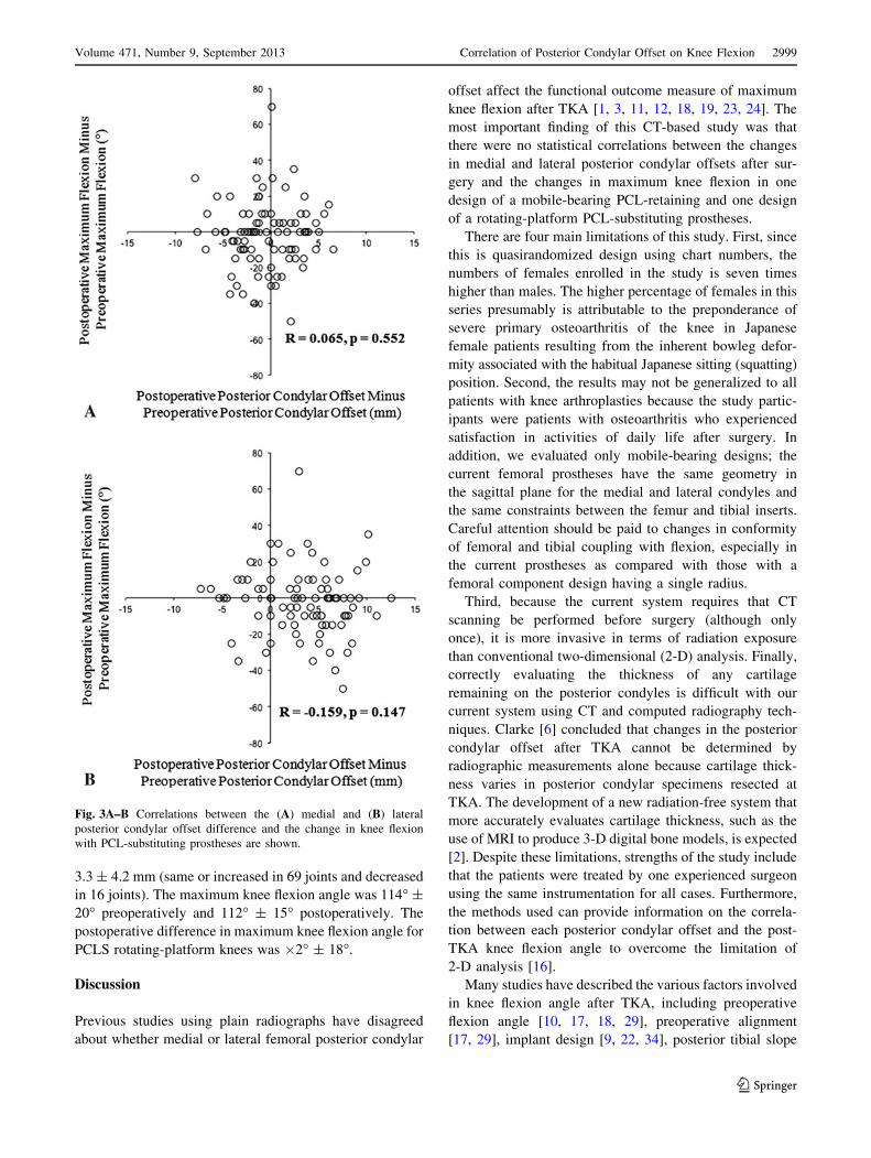
3.3 ± 4.2 mm (same or increased in 69 joints and decreased
in 16 joints). The maximum knee flexion angle was 114� ±
20� preoperatively and 112� ± 15� postoperatively. The
postoperative difference in maximum knee flexion angle for
PCLS rotating-platform knees was �2� ± 18�.
Discussion
Previous studies using plain radiographs have disagreed
about whether medial or lateral femoral posterior condylar
offset affect the functional outcome measure of maximum
knee flexion after TKA [1, 3, 11, 12, 18, 19, 23, 24]. The
most important finding of this CT-based study was that
there were no statistical correlations between the changes
in medial and lateral posterior condylar offsets after sur-
gery and the changes in maximum knee flexion in one
design of a mobile-bearing PCL-retaining and one design
of a rotating-platform PCL-substituting prostheses.
There are four main limitations of this study. First, since
this is quasirandomized design using chart numbers, the
numbers of females enrolled in the study is seven times
higher than males. The higher percentage of females in this
series presumably is attributable to the preponderance of
severe primary osteoarthritis of the knee in Japanese
female patients resulting from the inherent bowleg defor-
mity associated with the habitual Japanese sitting (squatting)
position. Second, the results may not be generalized to all
patients with knee arthroplasties because the study partic-
ipants were patients with osteoarthritis who experienced
satisfaction in activities of daily life after surgery. In
addition, we evaluated only mobile-bearing designs; the
current femoral prostheses have the same geometry in
the sagittal plane for the medial and lateral condyles and
the same constraints between the femur and tibial inserts.
Careful attention should be paid to changes in conformity
of femoral and tibial coupling with flexion, especially in
the current prostheses as compared with those with a
femoral component design having a single radius.
Third, because the current system requires that CT
scanning be performed before surgery (although only
once), it is more invasive in terms of radiation exposure
than conventional two-dimensional (2-D) analysis. Finally,
correctly evaluating the thickness of any cartilage
remaining on the posterior condyles is difficult with our
current system using CT and computed radiography tech-
niques. Clarke [6] concluded that changes in the posterior
condylar offset after TKA cannot be determined by
radiographic measurements alone because cartilage thick-
ness varies in posterior condylar specimens resected at
TKA. The development of a new radiation-free system that
more accurately evaluates cartilage thickness, such as the
use of MRI to produce 3-D digital bone models, is expected
[2]. Despite these limitations, strengths of the study include
that the patients were treated by one experienced surgeon
using the same instrumentation for all cases. Furthermore,
the methods used can provide information on the correla-
tion between each posterior condylar offset and the post-
TKA knee flexion angle to overcome the limitation of
2-D analysis [16].
Many studies have described the various factors involved
in knee flexion angle after TKA, including preoperative
flexion angle [10, 17, 18, 29], preoperative alignment
[17, 29], implant design [9, 22, 34], posterior tibial slope
Fig. 3A–B Correlations between the (A) medial and (B) lateral
posterior condylar offset difference and the change in knee flexion
with PCL-substituting prostheses are shown.
Volume 471, Number 9, September 2013 Correlation of Posterior Condylar Offset on Knee Flexion 2999
123

[4, 23, 24], anterior movement of the femur [3, 24], surgical
technique [25, 29], and rehabilitation protocol [26, 28].
Considering these studies, preoperative knee flexion angle
might be regarded as the most crucial factor for predicting
the postoperative flexion angle. Posterior condylar offset
(based on radiographic evaluation) may be an important
factor, although contradictory results have been reported
[1, 3, 11, 12, 18, 19, 23, 24]. Knee flexion is limited theo-
retically by direct impingement of the posterior aspect of the
tibial component against the posterior aspect of the femur. In
fact, Bellemans et al. [3] observed this fluoroscopically in
72% of their patients. They concluded that restoration of
posterior condylar offset was important because it allows a
greater degree of flexion before impingement occurs. How-
ever, several factors were attributable to the impingement,
including posterior condylar offset, a paradoxic anterior
movement with flexion, component design such as a high
posterior lip on the polyethylene insert, and the degree of
posterior tibial slope. The inability to maintain or restore a
functional PCL is believed to be the cause of paradoxic
forward sliding of the femur during flexion. This has been
shown to lead to early impingement and limited flexion by
videofluoroscopic studies [3, 8]. Hanratty et al. [12] used
radiographic evaluations to analyze the PCL-substituting
prosthesis used in the current study. In agreement with our
results, they found no statistical correlations between the
change in each posterior condylar offset and the changes in
knee flexion 1 year after TKA.
The in vivo kinematics and knee flexion angle have been
shown to be similar for current mobile-bearing PCL-
retaining and PCL-substituting prostheses [33], although
the PCL-retaining design has nonconstrained AP and
rotational movement and the PCL-substituting design has
only nonconstrained rotational movement. Both designs
showed approximately 1 mm of AP movement between
0� and 90� flexion. Thus, both prosthetic designs showed
almost the same positioning of the femoral component on
the tibial component during knee flexion. Another in vivo
kinematic study [15] of the PCL-retaining prosthesis used
in the current study showed that the average movement of
the medial condyle was only 1.3 mm in the anterior
direction, while the lateral condyle moved 1.5 mm in the
posterior direction from full extension to 90� knee flexion.
From full extension to maximum knee flexion, the average
amount of movement of the medial condyle was only
1.7 mm anteriorly, while the lateral condyle moved 2.6 mm
posteriorly. Although we did not perform a kinematic
analysis in our study, the current PCL-retaining prosthetic
design, with a 10� posterior tibial slope, had less of an
effect on the nonconstrained AP movement and did not
significantly affect the paradoxic anterior movement with
flexion as effectively as did the PCL-substituting design,
which has only nonconstrained rotational movement.
We investigated the influence of medial and lateral pos-
terior condylar offsets on maximum knee flexion after TKA
using CT and a 3-D lower extremity alignment assessment
system. Differences in individual posterior condylar offsets
were not correlated with changes in postoperative knee
flexion 1 year after TKA for the current PCL-retaining or
PCL-substituting prosthetic designs. We should recognize
that correctly identifying which condyle affects the results of
the TKA may be difficult with conventional radiographic
techniques. Additional studies are needed to research
whether various patterns of change in each condyle provide
significant effects on knee flexion after TKA.
Acknowledgments We thank Takanori Morozumi PT and Hisanori
Ishii RT for their contributions to gathering and analyzing the data.
Open Access This article is distributed under the terms of the
Creative Commons Attribution License which permits any use, dis-
tribution, and reproduction in any medium, provided the original
author(s) and the source are credited.
References
1. Arabori M, Matsui N, Kuroda R, Mizuno K, Doita M, Kurosaka
M, Yoshiya S. Posterior condylar offset and flexion in posterior
cruciate-retaining and posterior stabilized TKA. J Orthop Sci.
2008;13:46–50.
2. Ariumi A, Sato T, Kobayashi K, Koga Y, Omori G, Minato I,
Endo N. Three-dimensional lower extremity alignment in the
weight-bearing standing position in healthy elderly subjects.
J Orthop Sci. 2010;15:64–70.
3. Bellemans J, Banks S, Victor J, Vandenneucker H, Moemance A.
Fluoroscopic analysis of the kinematics of deep flexion in total
knee arthroplasty: influence of posterior condylar offset. J Bone
Joint Surg Br. 2002;84:50–53.
4. Bellemans J, Robijns F, Duerinckx J, Banks S, Vandenneucker H.
The influence of tibial slope on maximum flexion after total knee
arthroplasty. Knee Surg Sports Traumatol Arthrosc. 2005;13:
193–196.
5. Chaudhary R, Beaupre LA, Johnston DW. Knee range of motion
during the first two years after use of posterior cruciate-stabiliz-
ing or posterior cruciate-retaining total knee prostheses: a
randomized clinical trial. J Bone Joint Surg Am. 2008;90:2579–
2586.
6. Clarke HD. Changes in posterior condylar offset after total knee
arthroplasty cannot be determined by radiographic measurements
alone. J Arthroplasty. 2012;27:1155–1158.
7. Clarke HD, Scott WN, Insall JN, Pedersen HB, Math KR,
Vigorita VJ, Cushner FD. Anatomy. In: Insall JN, Scott WN, eds.
Surgery of the Knee. Vol 1. New York, NY: Churchill Living-
stone; 2001:13–76.
8. Dennis DA, Komistek RD, Colwell CE Jr, Ranawat CS, Scott
RD, Thornhill TS, Lapp MA. In vivo anteroposterior femorotibial
translation of total knee arthroplasty: a multicenter analysis. Clin
Orthop Relat Res. 1998;356:47–57.
9. Dennis DA, Komistek RD, Stiehl JB, Walker SA, Dennis KN.
Range of motion after total knee arthroplasty: the effect of
implant design and weight-bearing conditions. J Arthroplasty.
1998;13:748–752.
3000 Ishii et al. Clinical Orthopaedics and Related Research1
123

10. Gatha NM, Clarke HD, Fuchs R, Scuderi GR, Insall JN. Factors
affecting postoperative range of motion after total knee
arthroplasty. J Knee Surg. 2004;17:196–202.
11. Goldstein WM, Raab DJ, Gleason TF, Branson JJ, Berland K.
Why posterior cruciate-retaining and substituting total knee
replacements have similar ranges of motion: The importance of
posterior condylar offset and cleanout of posterior condylar
space. J Bone Joint Surg Am. 2006;88(suppl 4):182–188.
12. Hanratty BM, Thompson NW, Wilson RK, Beverland DE. The
influence of posterior condylar offset on knee flexion after total
knee replacement using a cruciate-sacrificing mobile-bearing
implant. J Bone Joint Surg Br. 2007;89:915–918.
13. Insall JN, Easley ME. Surgical techniques and instrumentation in
total knee arthroplasty. In: Insall JN, Scott WN, eds. Surgery of the
Knee. Vol 2. New York, NY: Churchill Livingstone; 2001:1553–
1620.
14. Insall JN, Hood RW, Flawn LB, Sullivan DJ. The total condylar
knee prosthesis in gonarthrosis: a five to nine-year follow-up of
the first one hundred consecutive replacements. J Bone Joint Surg
Am. 1983;65:619–628.
15. Ishii Y, Noguchi H, Matsuda Y, Takeda M, Walker SA, Komistek
RD. Effect of knee laxity on in vivo kinematics of meniscal-
bearing knee prostheses. Knee. 2007;14:269–274.
16. Ishii Y, Noguchi H, Takeda M, Ishii H, Toyabe S. Changes in the
medial and lateral posterior condylar offset in total knee
arthroplasty. J Arthroplasty. 2011;26:255–259.
17. Kawamura H, Bourne RB. Factors affecting range of flexion after
total knee arthroplasty. J Orthop Sci. 2001;6:248–252.
18. Kim YH, Choi Y, Kwon OR, Kim JS. Functional outcome and range
of motion of high-flexion posterior cruciate-retaining and high-flexion
posterior cruciate-substituting total knee prostheses: a prospective,
randomized study. J Bone Joint Surg Am. 2009;91:753–760.
19. Kim YH, Sohn KS, Kim JS. Range of motion of standard and
high-flexion posterior stabilized total knee prostheses: a pro-
spective, randomized study. J Bone Joint Surg Am. 2005;87:
1470–1475.
20. Kobayashi K, Sakamoto M, Tanabe Y, Ariumi A, Sato T, Omori G,
Koga Y. Automated image registration for assessing three-dimen-
sional alignment of entire lower extremity and implant position
using bi-plane radiography. J Biomech. 2009;42:2818–2822.
21. Kumar PJ, McPherson EJ, Dorr LD, Wan Z, Baldwin K. Rehabil-
itation after total knee arthroplasty: a comparison of 2 rehabilitation
techniques. Clin Orthop Relat Res. 1996;331:93–101.
22. Maloney WJ, Schurman DJ. The effect of implant design on
range of motion after total knee arthroplasty: total condylar
versus posterior stabilized total condylar designs. Clin Orthop
Relat Res. 1992;278:147–152.
23. Malviya A, Lingard EA, Weir DJ, Deehan DJ. Predicting range of
movement after knee replacement: the importance of posterior
condylar offset and tibial slope. Knee Surg Sports Traumatol
Arthrosc. 2009;17:491–498.
24. Massin P, Gournay A. Optimization of the posterior condylar
offset, tibial slope, and condylar roll-back in total knee
arthroplasty. J Arthroplasty. 2006;21:889–896.
25. Matsuda Y, Ishii Y, Noguchi H, Ishii R. Varus-valgus balance
affects the range of movement after total knee arthroplasty.
J Bone Joint Surg Br. 2005;87:804–808.
26. Mauerhan DR, Mokris JG, Ly A, Kiebzak GM. Relationship
between length of stay and manipulation rate after total knee
arthroplasty. J Arthroplasty. 1998;13:896–900.
27. Parsley BS, Engh GA, Dwyer KA. Preoperative flexion: does it
influence postoperative flexion after posterior-cruciate-retaining
total knee arthroplasty? Clin Orthop Relat Res. 1992;275:204–210.
28. Ranawat CS, Ranawat AS, Mehta A. Total knee arthroplasty
rehabilitation protocol: what makes the difference? J Arthro-
plasty. 2003;18(3 suppl 1):27–30.
29. Ritter MA, Harty LD, Davis KE, Meding JB, Berend ME. Pre-
dicting range of motion after total knee arthroplasty: clustering,
log-linear regression, and regression tree analysis. J Bone Joint
Surg Am. 2003;85:1278–1285.
30. Sato T, Koga Y, Omori G. Three-dimensional lower extremity
alignment assessment system: application to evaluation of com-
ponent positioning after total knee arthroplasty. J Arthroplasty.
2004;19:620–628.
31. Sato T, Koga Y, Sobue T, Omori G, Tanabe Y, Sakamoto M.
Quantitative 3-dimensional analysis of preoperative and postopera-
tive joint lines in total knee arthroplasty: a new concept for evaluation
of component alignment. J Arthroplasty. 2007;22:560–568.
32. Schurman DJ, Parker JN, Orstein D. Total condylar knee
replacement: a study of factors influencing range of motion as
late as two years after arthroplasty. J Bone Joint Surg Am.
1985;67:1006–1014.
33. Stiehl JB, Dennis DA, Komistek RD, Keblish PA. In vivo com-
parison of posterior cruciate ligament retention or sacrifice with a
mobile bearing total knee arthroplasty. Am J Knee Surg.
2000;13:13–18.
34. Stiehl JB, Voorhorst PE, Keblish P, Sorrells RB. Comparison of
range of motion after posterior cruciate ligament retention or
sacrifice with a mobile bearing total knee arthroplasty. Am J Knee
Surg. 1997;10:216–220.
Volume 471, Number 9, September 2013 Correlation of Posterior Condylar Offset on Knee Flexion 3001
123


![Conservative Approach to Unilateral Condylar Fracture in a … · 2016-10-09 · of condylar fractures [7]. It appears that pediatric condylar fractures could be managed by closed](https://img.dokumen.tips/doc/110x75/5f48360e47a39a42e102f2f1/conservative-approach-to-unilateral-condylar-fracture-in-a-2016-10-09-of-condylar.jpg)