Embed Size (px)
Citation preview

Gloucestershire Cellular Pathology Laboratory
Post-colectomy IBD pathology
Professor Neil A ShepherdGloucester & Cheltenham
BDIAP/BSG Lower GI Pathology SymposiumRIBA, London: 23 November 2018

Gloucestershire Cellular Pathology Laboratory
Speaker Declarations
Name of Speaker: Professor Neil A Shepherd
This presenter has the following declarations of relationship with industry: NONE
20 November 2018

Gloucestershire Cellular Pathology Laboratory
The big issues
• total (procto)colectomy for IBD – UC, IC, some cases of CD
is it UC, CD or IC?
it’s all about whether or not to undertake ileal pouch surgery
• pathology of the ileal pouch and its environs
adaptive changes, pouchitis, cuffitis and pre-pouch ileitis

Types of ileal pouch surgery
• three stage:
total colectomy, mucus fistula and ileostomyproctectomy and ileal pouch constructionileostomy reversal
• two stage:
total proctocolectomy, ileal pouch formation and ileostomyileostomy reversal
• one stage:
total proctocolectomy, ileal pouch formation and restoration of continuity

Gloucestershire Cellular Pathology Laboratory
What surgery for UC/IBDU?
• emergency/urgent presentation: bleeding, toxic megacolon, impending perforation, perforation
three stage pouch surgerymost cases of IC
• failed medical treatmentcertain diagnosis – one or two stage pouch surgery (usually)uncertain diagnosis (some cases of IC) – three stage pouch surgery
• neoplasia

Gloucestershire Cellular Pathology Laboratory
Spinelli, 2018

Spinelli, 2018

Gloucestershire Cellular Pathology Laboratory

Classical UC
“Ulcerative colitis goes up to where it stops”
Professor Bryan Warren (1958-2012)

Classical Crohn’s disease

Gloucestershire Cellular Pathology Laboratory
When does ulcerative colitis mimic Crohn’s disease?
• patchiness of disease after treatment
• resolution of histological changes after treatment
• fulminant colitis
• diversion proctitis in UCWarren et al, 1993
• SKIP LESIONScaecal patch lesionappendixsigmoid colonic diverticulosis

Defunctioned rectum excised during 3-stage pouch surgery
• combination of UC and diversion changes, in the rectal stump, produces mimicry of CD
• transmural inflammation and granulomatous inflammation
• also PMC-like changes
• if uncertainty, crucial review of the colectomy specimen for a more accurate assessment of CIBD type
Warren et al 1993; Goldstein et al, 1997;
Loughrey & Shepherd, 2017

Gloucestershire Cellular Pathology Laboratory
Discontinuous disease in UC
• appendixThe appendix as a skip lesion in ulcerative colitis.
Davison AM, Dixon MF. Histopathology, 1990.
• caecal patch lesion
• sigmoid colon with diverticular disease
• ‘rectum’
• what links these four sites of disease? Faecal stasis

The caecal patch lesion of ulcerative colitis• first defined in 1958: eight cases of UC as ‘an island’ in normal caecal mucosa, with
a tendency to occur in the lower caecum opposite the ileocaecal valveLumb & Protheroe, 1958
• classic description by d’Haens and colleaguesd’Haens et al, 1997
• more severe distal disease but better response to therapyMatsumoto et al, 2002
• commoner in younger male patients and pronounced symptomatology, especially abdominal pain, rectal bleeding and diarrhoea
Yamagishi at al, 2002; Nevin et al, 2012
• does not predict prognosis of UC, including remission rate, relapse rate, proximal disease extension and the need for proctocolectomy
Byeon et al, 2005; Bakman et al, 2011; Park et al, 2014

The isolated caecal patch lesion: a clinical, endoscopic and pathological study
Ekanayaka, Anderson, Lucarotti, Valori & Shepherd, 2018
criteria: normal colonoscopy apart from CPL;
biopsies of CPL show active IBD features; colonic & rectal biopsies normal

Gloucestershire Cellular Pathology Laboratory
The caecal patch lesion of ulcerative colitis
• important to recognise as one of the skip lesions of ulcerative colitis
• major differential diagnosis will always be Crohn’s disease
• reflects more severe distal disease in UC
• but in some studies not predictive of any known prognostic parameter in UC
• can rarely occur in isolation but then may represent other diseases, especially NSAID colopathy

Gloucestershire Cellular Pathology Laboratory
Indeterminate colitis

Gloucestershire Cellular Pathology Laboratory
Indeterminate colitis
• diagnosis made only in resection specimens
• 10-20% of colectomies, especially ‘fulminant’ colitis
• some features of UC and Crohn’s
• generally behave as UC
• cautious positive approach to pouch surgery

Gloucestershire Cellular Pathology Laboratory
Indeterminate colitis:importance of macroscopic pathology
• extensive ulceration
• involvement of transverse and right colon (more severely than distal colon)
• involvement of more than 50% of the mucosal surface
• usually diffuse disease, but may show rectal sparing
• toxic dilatation may be present

Gloucestershire Cellular Pathology Laboratory
Indeterminate colitis: histology

Gloucestershire Cellular Pathology Laboratory
Indeterminate colitis• 9-20% of colectomy specimens
Nicholls & Wells, 1992
• 1.6 to 2.4 per 100,000Stewenius et al, 1995; Moum et al, 1996
• equal sex distributionWells et al, 1991
• about 80-90% will behave like ulcerative colitisMcIntyre et al, 1995; Meucci et al, 1999; Yu et al, 2000
• 65% will be reclassified into UC or CD on further analysis of clinical, radiological and/or further histopathological evidence
Wells et al 1991; Nicholls & Wells 1992

Gloucestershire Cellular Pathology Laboratory
Indeterminate colitis & the natural history of the ileal reservoir
IC v UC % pouch failure IC v UC
% pelvic sepsisIC v UC
Atkinson et al 1994, Vancouver
16/158 19/5 25/1
Foley et al 1997, Lahey
42/499 12/2 44/23
Yu et al 2000, Mayo
82/1437 27/11 17/7
Delaney et al, 2002, Cleveland
115/1399 3.4/3.5 8.7/2.2

Gloucestershire Cellular Pathology Laboratory

Gloucestershire Cellular Pathology Laboratory
Gloucestershire Cellular Pathology Laboratory
Indeterminate colitis
• we must restrict this term to the middle ground of colectomy CIBD specimensWCOG Montreal IBD guidelines, 2005
Martland & Shepherd, 2007Langner et al, ECCO guidance, 2014
• when so restricted, it defines a group, seemingly, that mainly reflects fulminant UC
• although a small proportion will eventually be shown to have CD
• some may represent fulminant infective colitis, especially campylobacter colitis
• pouch surgery is not contraindicated (it may be too late, anyway!) but there is an increased rate of pouch failure and pelvic sepsis
• a cautious ‘yes’ to pouch surgery for IC

Gloucestershire Cellular Pathology Laboratory
The pelvic ileal reservoir/pouch
Parks AG, Nicholls RJ. Proctocolectomy without ileostomy for ulcerative colitis.
Br Med J 1978; ii: 85-88.

Gloucestershire Cellular Pathology Laboratory
Which patients get ileo-anal pouches?
ulcerative colitis
indeterminate colitis
familial adenomatous polyposis and other polyposis syndromes
Crohn's disease
colonic myopathy/neuropathy

Gloucestershire Cellular Pathology Laboratory
Restorative proctocolectomy Pouch configuration
J S W

Gloucestershire Cellular Pathology Laboratory
The ileal pouch
Mucosal adaptation
Shepherd, Jass, Duval, Moskowitz, Nicholls & Morson, 1987; de Silva at al 1990; Veress et al 1990;
Setti Carraro, Talbot & Nicholls, 1994 & 1998

The mucosal pathology of the pouch
HIDAB

Gloucestershire Cellular Pathology Laboratory
Colonic phenotypic change, colonic metaplasia or colonisation?
• morphology• mucin and lectin histochemistry and immunohistochemistry• electron microscopy
BUT
• disaccharidase activity and enteric supramucosal defence barrier maintained• not all pouches, and not all mucosa in pouches, show colonic phenotypic
change
Shepherd et al, 1987 & 1993; de Silva et al, 1990; Sylvester et al, 2000

The mucosal pathology of the pouch
Three groups:
A: 45%: UC and FAP: no active inflammation: normal or mild chronic changes/villous abnormalities
B: 42%: mainly UC but occasional FAP: chronic changes but transient active inflammation
C: 13%: always UC: severe chronic active inflammation: chronic changes constant: (chronic relapsing) pouchitis
Veress et al, 1990; Setti Carraro, Talbot & Nicholls, 1994 & 1998

Gloucestershire Cellular Pathology Laboratory
Pouch pathology: is this Crohn’s disease?

Pouchitis
• better termed ‘chronic relapsing pouchitis’
• it is NOT just any old inflammation in the pouch
• 10-20% but very variable (definitions)
• defined by clinical (diarrhoea/discharge, systemic symptoms, like UC), endoscopic and histopathological criteria/scoring


Questionnaire on activity scoring in routine GI pathology practice
• sent to UK pathologists, half specialist and half non-specialist
• 50 returns
No activity scoring system used at all
74%
pouchitis scoring 24%
coeliac disease 8%
inflammatory bowel disease
6%
reflux oesophagitis 2%
gastritis (Sydney, OLGA, etc)
0%
Shepherd, ESP/Path Soc, London, 2014
Gloucestershire Cellular Pathology Laboratory
Pouchitis
• fascinating clinical, pathological and immunological relationships with UC and with its extra-intestinal manifestations (very rare in FAP)
• role of colonic phenotypic change – is this UC in metaplastic colonic mucosa?
• BUT good remission rates with metronidazole and ciprofloxacillin, maintained with probiotics, argues for a bacterial aetiology
Mimura et al, 2004; Gionchetti et al, 2012
• microbiological research disappointing – role of anaerobes, sulphate-reducing anaerobes, etc

Gionchetti P, et al. The role of antibiotics and probiotics in pouchitis. Ann Gastroenterol 2012; 25: 100-5.

BMJ 1990; 301: 886

Gloucestershire Cellular Pathology Laboratory
Long term natural history of the pouch mucosa
• does the combination of chronic active inflammation and colonic phenotypic change predispose to an increased neoplastic risk?
• yes – in the rectal cuff with cuffitis (especially stapled anastomoses) ; cases of dysplasia and cancer reported
• yes – modest risk in FAP – adenomas and cancersNugent et al, 1993; von Herbay et al, 1996; Thompson-Fawcett et al, 2001
• probably not much – in the ileal mucosa of the pouch itself
• earlier worries from Huddinge, SwedenVeress et al, 1995; Gullberg et al, 1997
• but large reviews have shown very low rates of dysplasia: only very occasional case reports of cancer

Gloucestershire Cellular Pathology Laboratory
The natural history of the pouch mucosa and surveillance
• the rectal cuff with cuffitis probably does require surveillance
• patients with type C pathology/pouchitis merit surveillance
• can we leave the others alone?

Gloucestershire Cellular Pathology Laboratory
All looks fairly simple so what is the problem?

Site of biopsies
pre-pouch
pouch
anal cuff

Pre-pouch ileitis
• enigmatic and rare disease
• some cases represent Crohn’s disease (and likely that intestinal pathology was always Crohn’s disease) (3/19) and NSAID enteropathy (1/19)
• most a non-specific small intestinal inflammatory and ulcerative complication of UC (15/19)
• pathology similar to pouchitis, with colonic phenotypic change: 47% also had pouchitis
• many cases respond well to biologic therapy
Bell et al, 2006

Gloucestershire Cellular Pathology Laboratory
Pre-stomal ileitis
• another enigmatic and rare disease
• occurs after total colectomy and ileostomy for UC
• long segment disease with dilatation and extensive ulceration
• poor prognosis (three patients died)
• another small bowel manifestation of UC
Knill-Jones RP, Morson B, Williams R. Prestomal ileitis: clinical and pathological findings in five cases.
Quart J Med 1970; 39: 287-97.
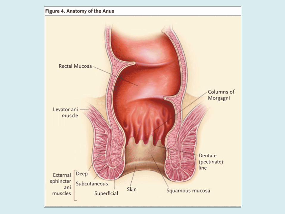

colorectal zone
(anal cuff)
transitional zone
squamous zone

Gloucestershire Cellular Pathology Laboratory
Restorative proctocolectomy Hand-sewn versus stapled anastomosis

Gloucestershire Cellular Pathology Laboratory
Cuffitis (UC involving the anal cuff)

Pouchitis vs cuffitis
• Symptoms
– frequency & urgency– incontinence– anorexia / fever – extra-intestinal
manifestations
• Diagnosis
– symptoms– endoscopic findings: site– histopathology
• Symptoms
– pain– bleeding
• Diagnosis
– symptoms– endoscopic findings; site– histopathology
Thompson-Fawcett M, Mortensen NJMcC, Warren BF. “Cuffitis” and inflammatory changes in the columnar
cuff, anal transitional zone, and ileal reservoir after stapled pouch-anal anastomosis.
Dis Colon Rectum 1999; 42: 348-355.

Gloucestershire Cellular Pathology Laboratory
Differentiating inflamed ileal pouch mucosa from inflamed cuff: any features that can help?
Relative parameters
• UACL much more common in the small
intestine
• crypt architectural distortion tends to
be more severe in cuffitis
• Paneth cells tend to be more plentiful in
the small intestine
‘Peyer’s patch pigment’
Shepherd et al, 1987

Differential diagnosis of acute inflammation/ulceration in the pouch
• Crohn’s disease
NEVER MAKE A DIAGNOSIS OF CROHN’S DISEASE BASED ON POUCH PATHOLOGY ALONE
• superinfection
• trauma
• intra-abdominal sepsis
• mucosal prolapse
• ischaemia

Gloucestershire Cellular Pathology Laboratory
Take home messages
• indeterminate colitis is a clinically and pathologically useful diagnosis when applied to colectomy specimens
• indeterminate colitis means the surgical pathology is indeterminate, not the pathologist……
• cases of CIBD, on biopsy, with unclassifiable pathology should be termed IBDU, never mind what endoscopy reporting systems call it………………
• most cases of indeterminate colitis will act like UC but with increased complications. Only a small proportion will act like Crohn’s disease

Gloucestershire Cellular Pathology Laboratory
Take home messages
• pouchitis is defined by clinical, endoscopic and pathological criteria – not just a bit of chronic inflammation and villous atrophy
• there are important mimics of Crohn’s disease in UC surgery, especially the diverted rectum and pouch granulomas and UACL
• we still don’t know the cause of pouchitis but it has important links to ulcerative colitis
• there is little evidence, currently, for significant neoplasia in the pouch mucosa but more in the anal cuff

Gloucestershire Cellular Pathology Laboratory
Acknowledgements and appreciations
Mr Tim Cook
Professor Roger Feakins
Dr Maurice Loughrey
Professor Marco Novelli
The late Professor Bryan Warren
Professor Geraint Williams