Embed Size (px)
Citation preview

Blunt Popliteal Artery Trauma David Radvinsky MD August 20, 2015
www.downstatesurgery.org
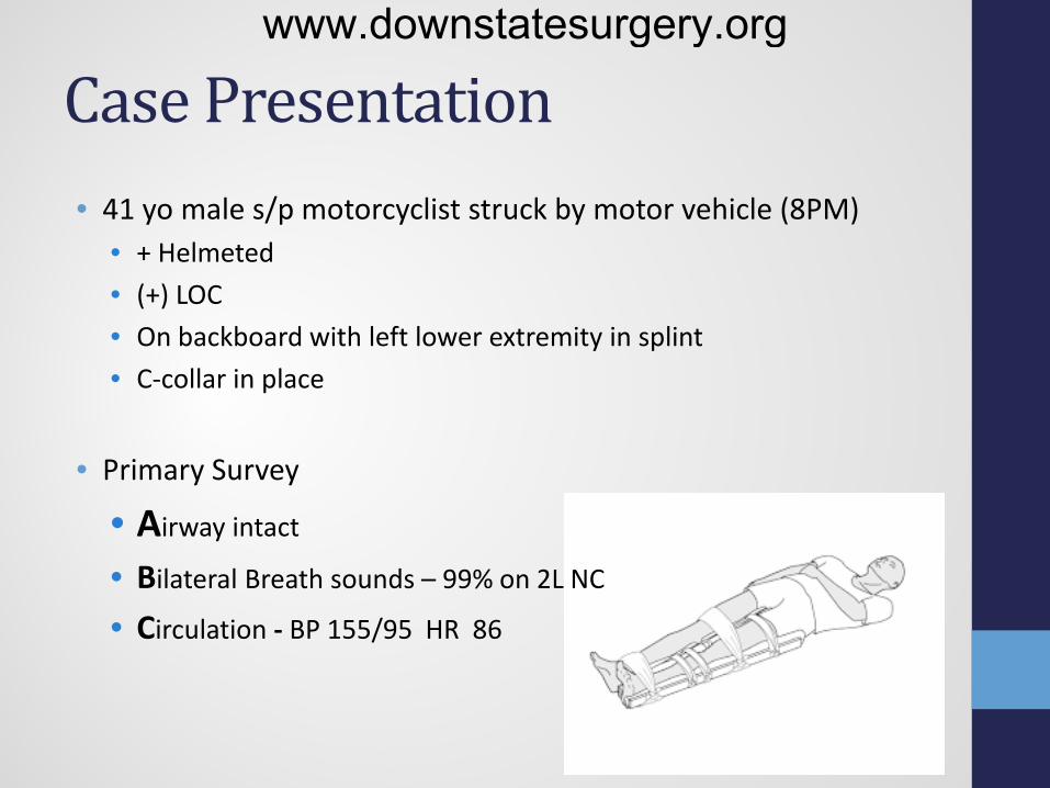
Case Presentation • 41 yo male s/p motorcyclist struck by motor vehicle (8PM)
• + Helmeted • (+) LOC • On backboard with left lower extremity in splint • C-collar in place
• Primary Survey
• Airway intact
• Bilateral Breath sounds – 99% on 2L NC
• Circulation - BP 155/95 HR 86
www.downstatesurgery.org

anterior tibial artery posterior tibial
dorsalis pedis artery
2+ 2+
2+
2+ 2+
Absent
Absent
• No evidence of expanding hematoma
• Delayed capillary refill • Cool compared to right foot
www.downstatesurgery.org

Secondary Survey • HEENT: no scalp lesions, PERRLA, EOMI, no facial tenderness,
no blood in the ear canal or nasal passages. • C-spine: no midline tenderness • Thorax: +5x5cm area of road rash over right shoulder (3rd
degree). no chest wall tenderness, with equal chest rise. RRR • Abdomen: soft, obese, NT/ND • Pelvis: stable, tenderness over left posterior pelvis/hip • T/L-spine: no midline tenderness • Extremities: (as described)
• Motor (LLE) – limited by pain • Sensory (LLE) – common peroneal nerve distribution
www.downstatesurgery.org
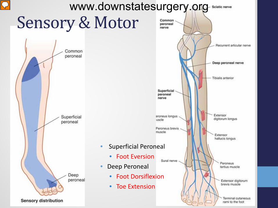
Sensory & Motor
• Superficial Peroneal • Foot Eversion
• Deep Peroneal • Foot Dorsiflexion • Toe Extension
www.downstatesurgery.org

Re-Evaluation & Adjuncts • Vitals: BP: 90/48 HR 98 SpO2: 96% on 2L NC • Transfused 1 unit pRBC
• PMHx: denied • PSHX: denied • Allergies: NKDA • Meds: None
• CXR: negative • PXR: (as shown) • FAST: negative
• Tib/Fib placed into traction • Improved capillary refill • Pulse exam unchanged • NO expanding hematoma
www.downstatesurgery.org

Imaging • CT Head: negative • CT c-spine: negative • CT chest: negative • CT abd/pel: acute displaced left ASIS fracture with
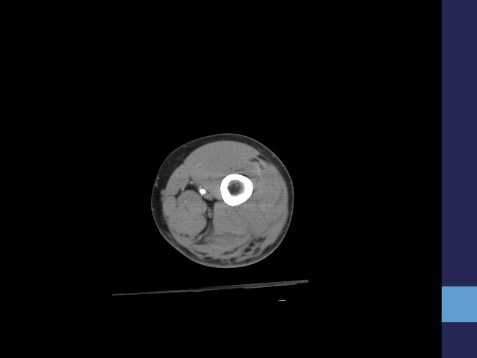
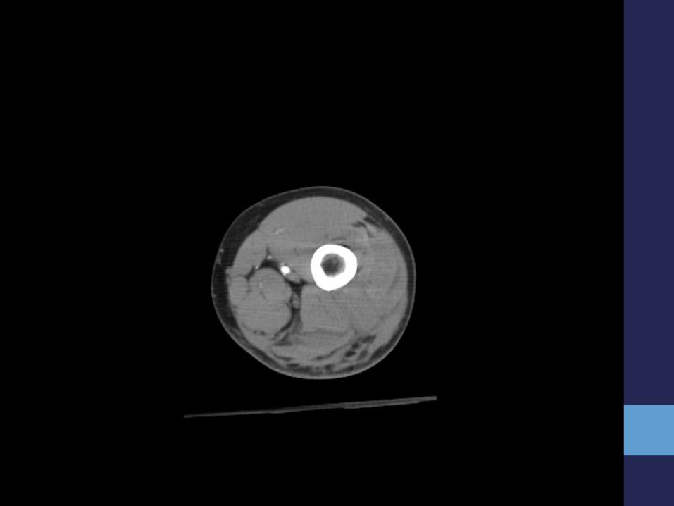
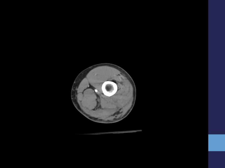
adjacent intramuscular hematoma • CTA LLE: abrupt cutoff of contrast in the popliteal artery 2cm
distal to Hunter’s canal
• Knee Dislocation?
www.downstatesurgery.org
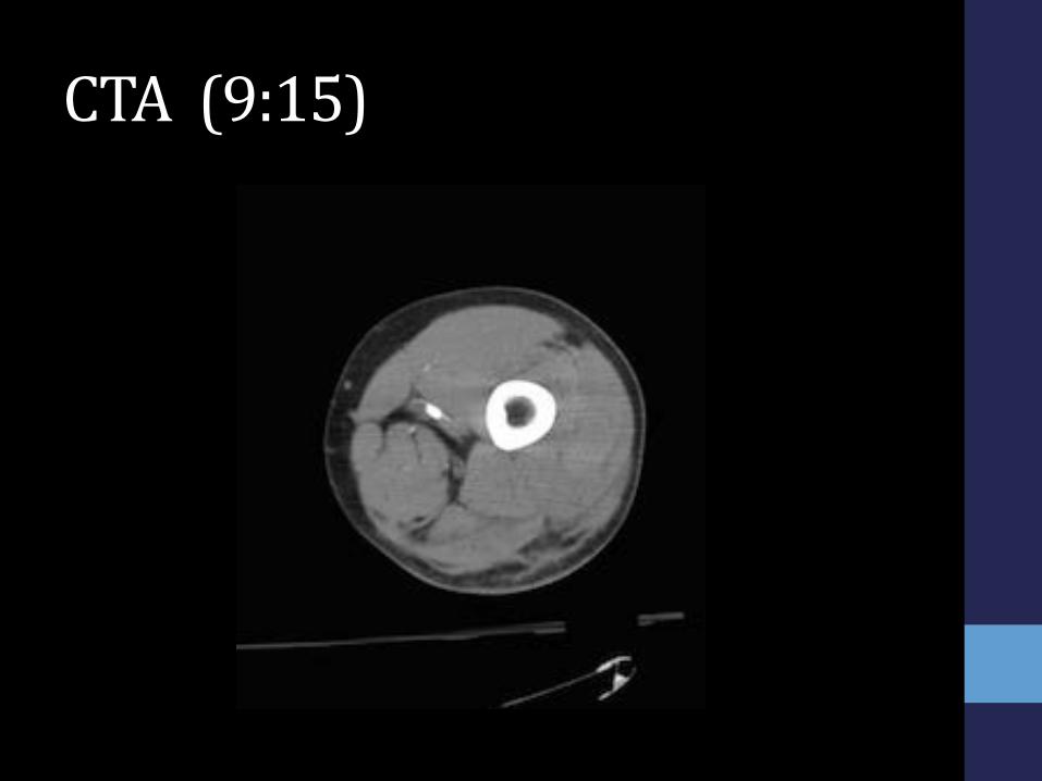
CTA (9:15) www.downstatesurgery.org
www.downstatesurgery.org
www.downstatesurgery.org
www.downstatesurgery.org

www.downstatesurgery.org

www.downstatesurgery.org
www.downstatesurgery.org

www.downstatesurgery.org

www.downstatesurgery.org

www.downstatesurgery.org

www.downstatesurgery.org

www.downstatesurgery.org

www.downstatesurgery.org

www.downstatesurgery.org

www.downstatesurgery.org

www.downstatesurgery.org

www.downstatesurgery.org

www.downstatesurgery.org

www.downstatesurgery.org

www.downstatesurgery.org

www.downstatesurgery.org

To OR • EX- FIX FIRST!?!
• (10 PM)
• Left Lower extremity angiogram (11:30 PM)
www.downstatesurgery.org

• Superficial Femoral Artery to Below Knee Popliteal Artery Bypass (12 AM) • PTFE graft (25 cm) • 4 compartment fasciotomy
PTFE graft
www.downstatesurgery.org
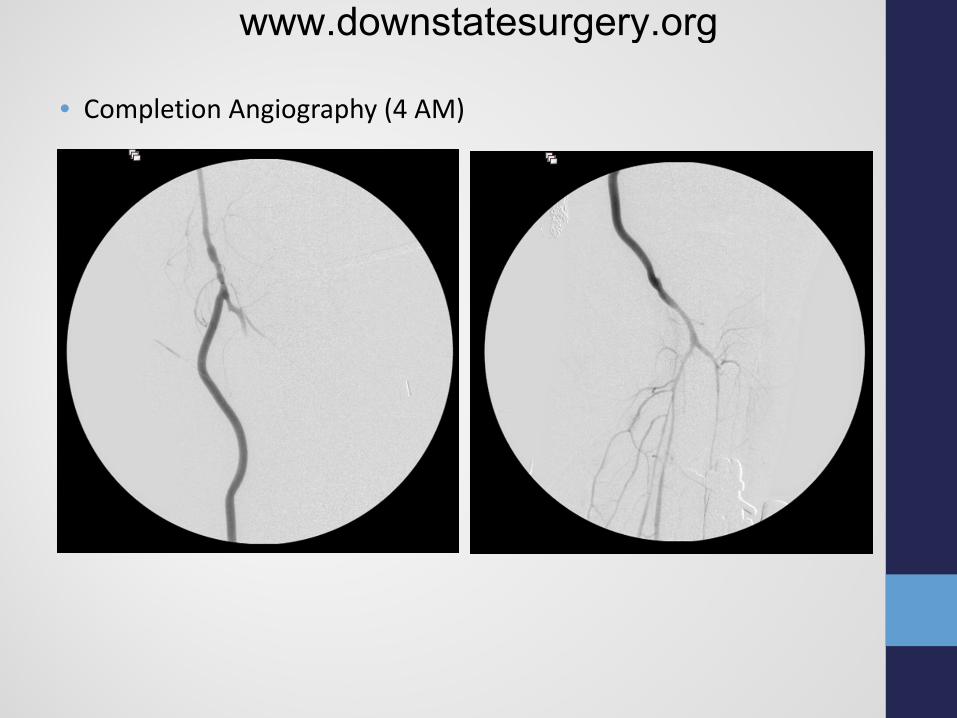
• Completion Angiography (4 AM)
www.downstatesurgery.org
Post-Op • EBL 1500 cc • Ischemia Time – 6 Hrs • Transfused 8 units pRBC, 4 FFP, 1 unit platlets, 1 cryo • VAC closure
• POD#0 - DP/PT pulse present
• 4 units pRBC, 2 units FFP • Hct 24 -> 21 (transfused additional 2 units pRBC) -> 27 • CK peaked at 34K -> bicarb drip started
• POD#2 – Hct -> 20.7 (transfused additional 1 units pRBC) -> 20.7 • POD#3 – Hct -> 19.5 (transfused additional 2 units pRBC) -> 24.8
• CTA – graft patent; iliac wing hematoma slightly larger; no collections • POD#4 – Heparin gtt started once HCT stabilized
• CK trending down -> 9K
www.downstatesurgery.org
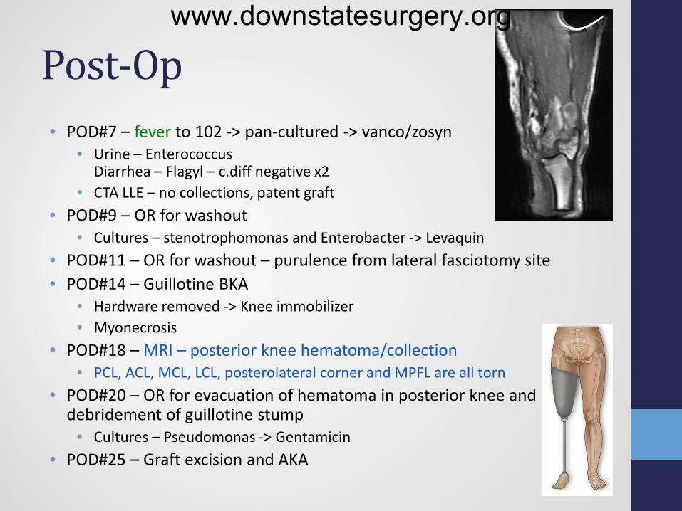
Post-Op • POD#7 – fever to 102 -> pan-cultured -> vanco/zosyn
• Urine – Enterococcus Diarrhea – Flagyl – c.diff negative x2
• CTA LLE – no collections, patent graft • POD#9 – OR for washout
• Cultures – stenotrophomonas and Enterobacter -> Levaquin • POD#11 – OR for washout – purulence from lateral fasciotomy site • POD#14 – Guillotine BKA
• Hardware removed -> Knee immobilizer • Myonecrosis
• POD#18 – MRI – posterior knee hematoma/collection • PCL, ACL, MCL, LCL, posterolateral corner and MPFL are all torn
• POD#20 – OR for evacuation of hematoma in posterior knee and debridement of guillotine stump
• Cultures – Pseudomonas -> Gentamicin • POD#25 – Graft excision and AKA
www.downstatesurgery.org

Questions?
www.downstatesurgery.org

Epidemiology • Popliteal Vessel Injuries rare – 0.2% of all traumas • High-energy mechanisms
• Pedestrian Struck • Motorcycle Accidents • Automobile Accidents
• Mechanism of blunt injury to the popliteal artery • Anterior dislocation • Posterior dislocation • Tibial plateau fracture
• Associated fractures (80% to 100%) • Associated venous injury (15% and 35%) – popliteal vein • Associated nerve injury (10%) – common peroneal nerve
www.downstatesurgery.org
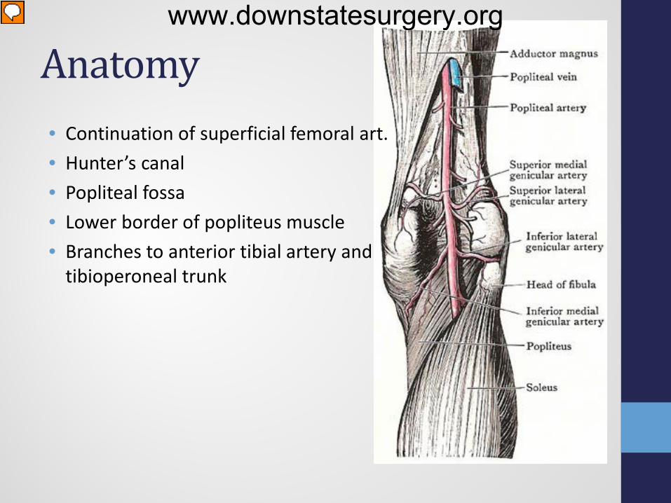
Anatomy • Continuation of superficial femoral art. • Hunter’s canal • Popliteal fossa • Lower border of popliteus muscle • Branches to anterior tibial artery and
tibioperoneal trunk
www.downstatesurgery.org
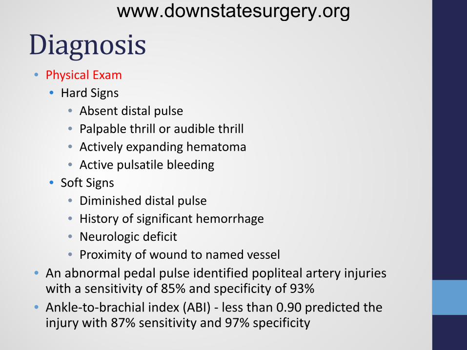
Diagnosis • Physical Exam
• Hard Signs • Absent distal pulse • Palpable thrill or audible thrill • Actively expanding hematoma • Active pulsatile bleeding
• Soft Signs • Diminished distal pulse • History of significant hemorrhage • Neurologic deficit • Proximity of wound to named vessel
• An abnormal pedal pulse identified popliteal artery injuries with a sensitivity of 85% and specificity of 93%
• Ankle-to-brachial index (ABI) - less than 0.90 predicted the injury with 87% sensitivity and 97% specificity
www.downstatesurgery.org

• Imaging • Plain radiographs to evaluate for fractures and/or dislocations • CTA to evaluate vessel integrity
• Transection • Dissection • Thrombosis
• ANGIOGRAPHY
www.downstatesurgery.org

Management • Limb salvage vs. primary amputation
• Mangled Extremity Severity Score • Gustilo III - C skeletal injuries • Transected tibial or sciatic nerve • Shock and life-threatening associated injuries • Below-knee arterial injury • Extensive soft tissue loss • Crush injury • Multiple fractures • Elderly with medical comorbidity • Severe contamination • Patient preference
www.downstatesurgery.org
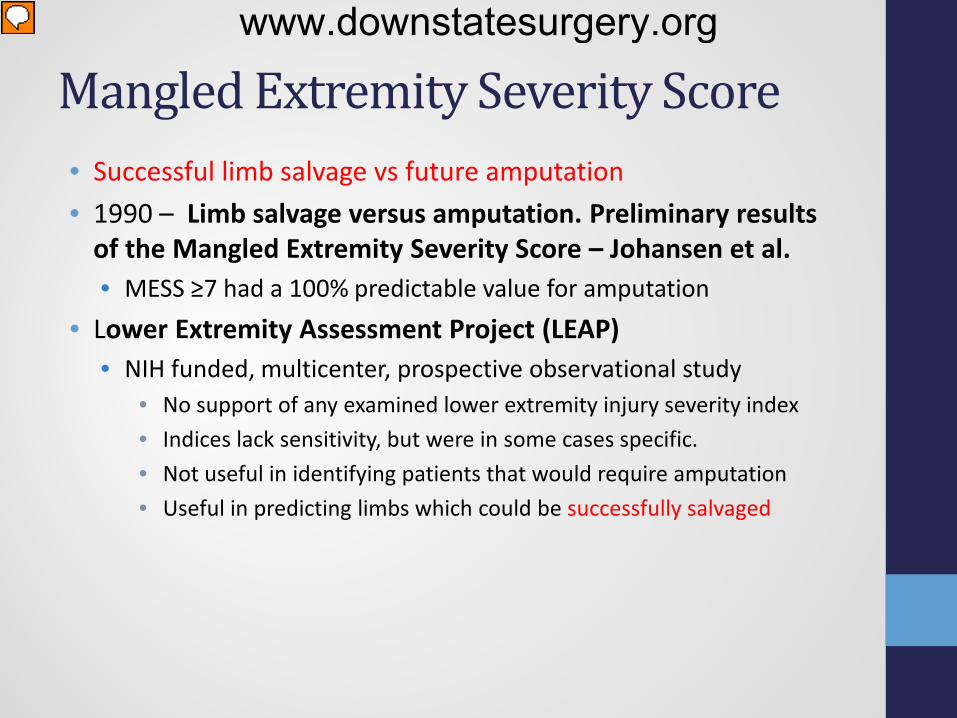
Mangled Extremity Severity Score • Successful limb salvage vs future amputation • 1990 – Limb salvage versus amputation. Preliminary results
of the Mangled Extremity Severity Score – Johansen et al. • MESS ≥7 had a 100% predictable value for amputation
• Lower Extremity Assessment Project (LEAP) • NIH funded, multicenter, prospective observational study
• No support of any examined lower extremity injury severity index • Indices lack sensitivity, but were in some cases specific. • Not useful in identifying patients that would require amputation • Useful in predicting limbs which could be successfully salvaged
www.downstatesurgery.org

Limb Salvage vs. Amputation • Mangled Extremity Severity Score
www.downstatesurgery.org

Approach www.downstatesurgery.org

www.downstatesurgery.org
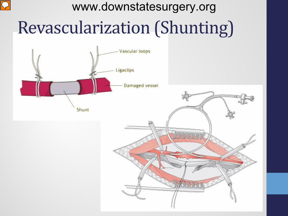
Revascularization (Shunting)
www.downstatesurgery.org
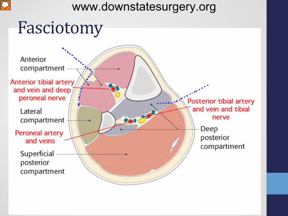
Fasciotomy www.downstatesurgery.org
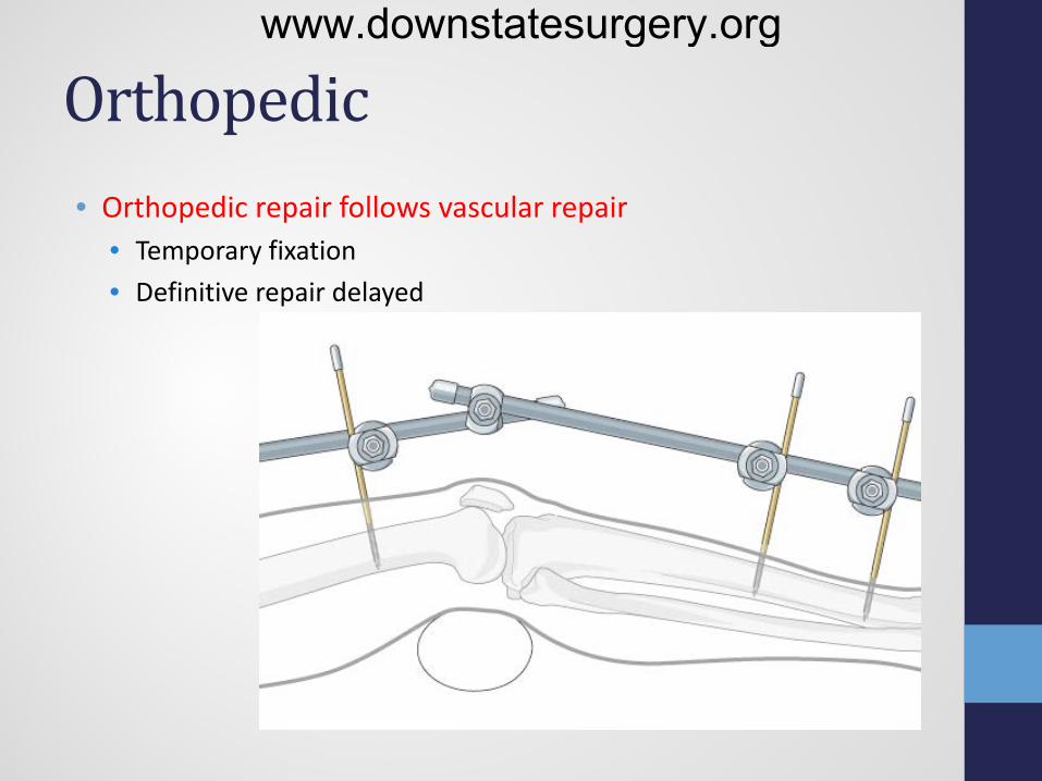
Orthopedic • Orthopedic repair follows vascular repair
• Temporary fixation • Definitive repair delayed
www.downstatesurgery.org

Options for Definitive repair • Lateral arteriorrhaphy or venorrhaphy • Patch angioplasty • Resection with end-to-end anastomosis • Resection with interposition graft • Bypass graft • Extraanatomic bypass
www.downstatesurgery.org

Autogenous vs. Graft • 1 year patency rates in infrapopliteal position
• 70-80% with autogenous vein • 30-50% for prosthetic grafts
• Autogenous – saphenous vein (ipsilateral or contralateral) • Prosthetic options
• Dacron • PTFE • Antiplatelets or anticoagulants
• Adjunctive vein cuff at the distal anastomosis of graft improves patency
www.downstatesurgery.org

Role for Endovascular? • Becoming more popular with newer techniques • Case reports in the literature
• Thrombosis • Pseudoaneurysm • AV Fistula • Dissection
www.downstatesurgery.org

Venous injury • Up to 1/3 of patients with arterial injuries have venous injury • Vein injury should be repaired
• Leg edema • Compartmental hypertension • Occlusion of arterial repair • Higher amputation rates • Allow for collateralization • Risk of acute thrombosis at the site of repair
• pulmonary embolism
www.downstatesurgery.org

Soft Tissue • Soft tissue debridement at initial operation
• Cover vascular repair with viable muscle • Decreases risk of infection and limb sepsis
• Monitor for Infection • Devitalized tissue • Hematoma • Fasciotomy sites
• Wound sepsis -> return to the OR • Open contaminated wounds -> broad spectrum abx • VAC assisted closure to promote healing of fasciotomy sites • Fasciotomy that cannot be closed primarily
• Skin graft once the muscle swelling has subsided
www.downstatesurgery.org

Nerve Injury • Common Peroneal Nerve – 10%
• Loss of function of foot - dorsiflexion • High-stepping walk (steppage gait or footdrop gait).
• Should undergo surgical exploration at emergency • Recover spontaneously
• Full recovery of partial peroneal palsies (76% to 87%) • Full recovery of complete lesions (20% to 35%)
• Repair indicated for lack of recovery after 2-5 months • Direct Repair – 84%
• Grafting (sural nerve) • <6 cm – 75% • >6cm – 16-38%
• Tendon transfer • Restoration of dorsiflexion
www.downstatesurgery.org

Post-Op • Secondary Amputation
• Failure of the arterial repair • Limb sepsis in the presence of a patent artery • Extensive muscle necrosis and nerve injury with a patent repair
• Patient’s functional status • Persistence of nerve deficit • Ankle–foot orthosis • Remedial operations to correct foot drop deformity
• REHAB
www.downstatesurgery.org

• Meta-analysis • 45 studies – lower extremity vascular trauma • Significant prognostic factors
• associated major soft tissue injury • compartment syndrome • multiple arterial injuries • duration of ischemia exceeding 6 h • associated fracture • Blunt mechanism of injury • age over 55 years • Male sex
www.downstatesurgery.org

Post-Op www.downstatesurgery.org

Summary
• Gustilo III - C skeletal injuries • Old age/severe co-morbidity • Sciatic or tibial nerve injury • Destructive soft tissue injury • Significant wound contamination • Multiple/severely comminuted fx • Elderly with medical comorbidity • Shock and life-threatening
associated injuries • Prolonged ischemia (6 hr) • Muscle Necrosis • Failed revascularization • Limb sepsis
• Limb Salvage vs. Primary Amputation? • OR – on table arteriogram • Arterial Shunt • 4 compartment fasciotomy • External Skeletal fixation • Definitive vascular repair • Soft tissue Debridement/Nerve
• Definitive Orthopedic repair • Close fasciotomy vs. skin graft • Nerve Repair vs. tendon transfer • Limb Salvage vs. Secondary
Amputation?
www.downstatesurgery.org
References • Fischer's Mastery of Surgery, 6e Edited by Josef E. Fischer, Daniel B. Jones, Frank B. Pomposelli and Gilbert R.
Upchurch. December 2011 • Rutherford's Vascular Surgery References, 8e 8th Edition by Jack L. Cronenwett MD, K. Wayne Johnston MD
FRCSC. May 5, 2014. • Evaluation and management of penetrating lower extremity arterial trauma: an Eastern Association for the
Surgery of Trauma practice management guideline. Fox N, Rajani RR, Bokhari F, Chiu WC, Kerwin A, Seamon MJ, Skarupa D, Frykberg E. J Trauma Acute Care Surg. 2012 Nov;73(5 Suppl 4):S315-20.
• Limb salvage and outcomes among patients with traumatic popliteal vascular injury: an analysis of the National Trauma Data Bank. Mullenix PS, Steele SR, Andersen CA, Starnes BW, Salim A, Martin MJ. - J. Vasc. Surg. - July 1, 2006; 44 (1); 94-100.
• Lower Extremity Assessment Project (LEAP) – The Best Available Evidence on Limb-Threatening Lower Extremity Trauma. Thomas F. Higgins MD, Joshua B. Klatt MD and Timothy C. Beals MD Orthopedic Clinics of North America, The, 2010-04-01, Volume 41, Issue 2, Pages 233-239.
• Salvage versus amputation: Utility of mangled extremity severity score in severely injured lower limbs. Kumar MK, Badole C, Patond K. - Indian J Orthop - July 1, 2007; 41 (3); 183-7.
• Characteristics and clinical outcome in patients after popliteal artery injury. Nikolaus W. Lang MD, Julian B. Joestl MD and Patrick Platzer MD, PhD. Journal of Vascular Surgery, 2015-06-01, Volume 61, Issue 6, Pages 1495-1500, Society for Vascular Surgery
• Outcome Predictors of Limb Salvage in Traumatic Popliteal Artery Injury. Anahita Dua, Sapan S. Desai, Jaecel O. Shah, Robert E. Lasky, Kristofer M. Charlton-Ouw, Ali Azizzadeh, Anthony L. Estrera, Hazim J. Safi and Sheila M. Coogan. Annals of Vascular Surgery, 2014-01-01, Volume 28, Issue 1, Pages 108-114
• Factors Associated with Amputation Following Popliteal Vascular Injuries. Jessica Keeley, Matthew Koopmann, Huan Yan, Christian DeVirgilio, Brant Putnam, David Pluradand Denis Kim. Annals of Vascular Surgery, 2015-07-01, Volume 29, Issue 5, Pages 881-881
www.downstatesurgery.org