Embed Size (px)
Citation preview

Pontifícia Universidade Católica do Rio Grande do Sul
Programa de Pós-Graduação em Biologia Celular e Molecular
Leonardo Krás Borges Martinelli
GMP redutase de Escherichia coli: mecanismos cinéticos,
catalíticos e químicos e a termodinâmica da
formação do complexo binário de ligação enzima-ligante
Porto Alegre
2011

Leonardo Krás Borges Martinelli
GMP redutase de Escherichia coli: mecanismos cinéticos, catalíticos e químicos e a
termodinâmica da formação do complexo binário de ligação enzima-ligante
Tese apresentada como requisito para obtenção de grau de Doutor pelo Programa de Pós-Graduação em Biologia Celular e Molecular da Faculdade de Biociências da Pontifícia Universidade Católica do Rio Grande do Sul
Orientador: Prof. Dr. Diógenes Santiago Santos
Co-orientador: Prof. Dr. Luiz Augusto Basso
Porto Alegre
2011

Leonardo Kras Borges Martinelli
GMP redutase de Escherichia coli: mecanismos cinéticos, catalíticos e químicos e a
termodinâmica da formação do complexo binário de ligação enzima-ligante
Tese apresentada como requisito para obtenção de grau de Doutor pelo Programa de Pós-Graduação em Biologia Celular e Molecular da Faculdade de Biociências da Pontifícia Universidade Católica do Rio Grande do Sul
Aprovada em ______de___________de_________.
BANCA EXAMINADORA
Dr Alejandro Miguel Katzin - USP
__________________________________
Dr Giancarlo Pasquali – UFRGS
__________________________________
Dra Maria Martha Campos – PUCRS (relatora)
__________________________________
Agradecimentos
Ao Prof. Diogénes Santiago Santos, agradeço pela oportunidade de integrar seu
grupo de pesquisa, pela orientação do trabalho e, por contribuir e proporcionar um
maior aprendizado e uma melhor formação acadêmica.
Ao Prof. Luiz Augusto Basso, agradeço pelo conhecimento compartilhado, pelos
ensinamentos passados e pelas correções e sugestões para que o trabalho fosse
finalizado.
Aos Drs Eraldo Batista Jr, Claudia Paiva Nunes e Gaby Renard por todo o
conhecimento, conselhos e ajudas indispensáveis para o trabalho, mas principalmente
pela amizade ao longo do tempo.
Gostaria de agradecer especialmente aos amigos Christiano, Leonardo, e
Rodrigo pela amizade ao longo dos anos, pelos momentos de diversão e descontração
que foram fundamentais para superar os momentos difíceis. A todos os colegas do
Centro de Pesquisa em Biologia Molecular e Funcional e da Quatro G, que diretamente
ou indiretamente colaboraram com o trabalho, além da amizade e companheirismo de
todos.
Gostaria de agradecer aos meus pais, que ao longo dos anos sempre me
incentivaram e me deram todo o apoio e suporte necessário para a realização dessa
etapa, e por todo carinho e compreensão ao longo do tempo.
Gostaria de agradecer em especial a uma pessoa, a minha namorada Clarissa
Medeiros, que foi fundamental para essa conquista, pelo amor, carinho, compreensão,
e paciência, ao longo do tempo.

A new type of thinking is essential if mankind is to survive and move toward higher levels.
Albert Einstein

i
Resumo A enzima guanosina monofosfato (GMP) redutase catalisa a deaminação redutiva do GMP à inosina monofosfato (IMP). GMP redutase possui um papel importante na conversão dos nucleosídeos e nucleotídeos derivados de guanina
a nucleotídeos de adenina. Além desse fato, como parte da via de salvamento de purinas, também participa da reutilização de bases livres intracelulares. Nesse trabalho, mostramos a clonagem, a expressão e a purificação da GMP redutase codificada pelo gene guaC de Escherichia coli a fim de determinar seu
mecanismo cinético; bem como as características químicas e termodinâmicas da reação catalisada. Estudos de velocidade inicial e titulação de calorimetria
isotérmica demonstraram que a GMP redutase possui um mecanismo cinético bi-bi ordenado, no qual o GMP liga primeiro à enzima, seguido pela ligação do NADPH; o NADP+ se dissocia primeiro, seguido pela liberação do IMP. A
titulação calorimétrica também mostrou que a ligação do GMP e IMP são processos termodinamicamente favoráveis. Perfis de pH demonstraram grupos com valores de pK aparentes de 6,6 e 9,6 envolvidos na catálise, valores de pK
de 7,1 e 8,6 importantes para ligação do GMP e um valor de pK de 6,4
importante para ligação do NADPH. Efeito isotópico primário demonstrou que a transferência do hidreto contribui para a etapa limitante da reação, enquanto que
os efeitos isotópicos do solvente provêm de um único local de protonação que possui um papel modesto na catálise. Efeito isotópico múltiplo sugere que as etapas de protonação e transferência do hidreto ocorrem no mesmo estado de transição, fornecendo evidência de um mecanismo concerted. Os dados de
cinética em estado pré-estacionário indicam que a liberação do produto não contribui para a etapa limitante da reação catalisada pela GMP redutase de E.
coli. Em conjunto, os resultados mostram que a reação catalisada pela GMP
redutase segue um mecanismo sequencial e ordenado, onde a transferência do hidreto e do próton ocorrem no mesmo estado de transição. Os resultados aqui
apresentados foram os primeiros a evidenciar o mecanismo cinético da GMP redutase, bem como, os resíduos fundamentais para catálise e/ou ligação, além de fornecer informações sobre o estado de transição da reação.

ii
Abstract Guanosine monophosphate (GMP) reductase catalyzes the reductive deamination of GMP to inosine monophosphate (IMP). GMP reductase plays an important role in the conversion of nucleoside and nucleotide derivatives of
guanine to adenine nucleotides. In addition, as a member of the purine salvage pathway, it also participates in the reutilization of free intracellular bases. Here we present cloning, expression and purification of Escherichia coli guaC-encoded
GMP reductase to determine its kinetic mechanism, as well as chemical and thermodynamic features of this reaction. Initial velocity studies and isothermal titration calorimetry demonstrated that GMP reductase follows an ordered bi-bi
kinetic mechanism, in which GMP binds first to the enzyme followed by NADPH binding, and NADP+ dissociates first followed by IMP release. The isothermal titration calorimetry also showed that GMP and IMP binding are
thermodynamically favorable processes. The pH-rate profiles showed groups with apparent pK values of 6.6 and 9.6 involved in catalysis, and pK values of 7.1 and 8.6 important to GMP binding, and a pK value of 6.4 important for NADPH
binding. Primary deuterium kinetic isotope effects demonstrated that hydride transfer contributes to the rate-limiting step, whereas solvent kinetic isotope effects arise from a single protonic site that plays a modest role in catalysis.
Multiple isotope effects suggest that protonation and hydride transfer steps take place in the same transition state, lending support to a concerted mechanism. Pre-steady-state kinetic data suggest that product release does not contribute to the rate-limiting step of the reaction catalyzed by E. coli GMP reductase.

iii
Lista de Siglas
RNA – ácido ribonucleico
DNA – ácido desoxirribonucleico
ATP – trifosfato de adenosina
GTP – trifosfato de guanosina
FAD – flavina adenina dinucleotídeo
NAD – nicotinamida adenina dinucleotídeo
cAMP – monofosfato de adenosina cíclico
cGMP – monofosfato de guanosina cíclico
PRPP – 5-fosforribosil-α-1-pirofosfato
10-formil-THF – 10-formiltetrahidrofolato
IMP – monofosfato de inosina
XMP – monofosfato de xantina
E. coli – Escherichia coli
APRT – adenina fosforribosiltransferase
PPi – pirofosfato
GPRT – guanina fosforribosiltransferase
HPRT – hipoxantina fosforribosiltransferase
PNP – purina nucleosídeo fosforilase
NADPH – nicotinamida adenina dinucleotídeo fosfatado
KDa – Quilodáltons
ESI-MS – do inglês electrospray ionization mass spectrometry
IPTG – isopropil β-D-tiogalactopiranosídeo

iv
SDS-PAGE – do inglês sodium dodecyl sulfate polyacrylamide gel
electrophoresis

v
SUMÁRIO
Capítulo 1
Introdução
1.1 Nucleotídeos 2
1.2 Metabolismo de purinas e pirimidinas 3
1.2.1 Via de novo de purinas 4
1.2.2 Via de salvamento de purinas 6
1.2.3 Interconversão de purinas 8
1.3 GMP redutase 9
Capítulo 2
Objetivos
2.1 Objetivo Geral 14
2.2 Objetivos Específicos 14
Capítulo 3
Artigo “Recombinant Escherichia coli GMP reductase:
kinetic, catalytic and chemical mechanisms,
and thermodynamics of enzyme-ligand binary complex formation.” 17
Capítulo 4
Considerações finais 38
Referências 41
Anexo
Clonagem, expressão, purificação e caracterização
da polinucleotídeo fosforilase (PNPase)

vi
de Mycobacterium tuberculosis para
validação como alvo para o desenvolvimento
racional de amostra atenuada 46

CAPÍTULO 1
Introdução
1.1 Nucleotídeos
1.2 Metabolismo de purinas e pirimidinas
1.2.1 Via de novo de purinas
1.2.2 Via de salvamento de purinas
1.2.3. Interconversão de purinas
1.3 GMP redutase

2
INTRODUÇÃO
1.1 Nucleotídeos
Nucleotídeos são moléculas que possuem uma diversidade estrutural
extensa, existindo oito variedades comuns de nucleotídeos, cada uma composta
por uma base nitrogenada ligada a uma pentose (açúcar de cinco carbonos), a
qual possui pelo menos um grupo fosfato. A pentose pode ser tanto uma ribose,
presente no ácido ribonucleico (RNA), quanto uma desoxirribose, componente
do ácido desoxirribonucleico (DNA) (Alberts, 2010). As bases nitrogenadas são
moléculas planares, aromáticas e heterocíclicas derivadas de purinas ou de
pirimidinas. As purinas mais comuns são a adenina (A) e guanina (G), e as
principais pirimidinas são a citosina (C), a uracila (U) e a timina (T). As purinas
são ligadas à pentose pelo átomo N9, ao contrário das pirimidinas que se ligam
à pentose pelo átomo N1 (Voet, 2006).
Além de componentes chaves dos ácidos nucléicos, portanto responsáveis
diretos pelo armazenamento e disponibilização da informação biológica, os
nucleotídeos realizam diversas outras funções bioquímicas, como o
armazenamento de energia através de moléculas como o trifosfato de adenosina
(ATP) e o trifosfato de guanosina (GTP). Os nucleotídeos também são
componentes de alguns cofatores centrais do metabolismo como flavina adenina
dinucleotídeo (FAD), nicotinamida adenina dinucleotídeo (NAD) e coenzima A,
além de regular diversas rotas metabólicas através do monofosfato de

3
adenosina cíclico (cAMP) e do monofosfato de adenosina cíclico (cGMP), que
são importantes segundos mensageiros (Voet, 2006).
1.2 Metabolismo de purinas e pirimidinas
Em Escherichia coli, purinas e pirimidinas podem ser sintetizadas a partir
de precursores simples, através da síntese de novo, ou através da reciclagem
de bases livres pela chamada via de salvamento (Smith, Marks, Lieberman,
2005). Em ambos os casos, a parte α-D-ribose-5-fosfato é derivada do 5-
fosforribosil-α-1-pirofosfato (PRPP). PRPP é necessário para a biossíntese de
purinas e pirimidinas, tanto pela via de novo quanto pela via de salvamento,
além da síntese de nucleotídeos de nicotinamida e da síntese de histidina e
triptofano (El Kouni, 2003).
A ingestão alimentar de bases purínicas e pirimidínicas é mínima, uma vez
que a dieta contém ácidos nucleicos e o pâncreas secreta desoxirribonuclease e
ribonuclease, juntamente com enzimas proteolíticas e lipolíticas. Este aspecto
permite que os ácidos nucleicos digeridos sejam convertidos em nucleotídeos e
o epitélio intestinal converta esses nucleotídeos em nucleosídeos, através da
atividade da fosfatase alcalina. Aproximadamente 5% dos nucleotídeos ingeridos
entram na circulação, tanto na forma de nucleosídeo, como na forma de base
livre. Pelo fato da ingestão mínima destas importantes moléculas, a síntese de
novo de purinas e pirimidinas é necessária (Smith, Marks, Lieberman, 2005).

4
1.2.1 Via de novo de purinas
A síntese de purinas em E. coli pela via de novo corresponde a onze
etapas enzimáticas e requer seis moléculas de ATP para cada purina sintetizada
(Figura 1). Os precursores que fornecem os componentes básicos para produzir
nucleotídeos purínicos são a glicina, α-D-ribose-5-fosfato, glutamina, aspartato,
dióxido de carbono (CO2) e 10-formiltetrahidrofolato (10-formil-THF). A síntese
de novo produz o monofosfato de inosina (IMP), primeiro intermediário da via,
que corresponde ao nucleotídeo da base hipoxantina. Através do PRPP, o anel
purínico é montado a partir de nitrogênios da amida da glutamina, do grupo
amino do aspartato, de dois átomos de carbono e do grupo amino da glicina e de
três moléculas de CO2 e dos átomos de carbono de duas moléculas de 10-
formil-THF (Neidhardt, 2005).
Figura 1: Síntese de novo de purinas em E. coli
(Adaptado de Voet, 2006)

5
A molécula de IMP serve como ponto de origem para a síntese de AMP e
GMP (Figura 2). O AMP é formado a partir do IMP em duas etapas enzimáticas,
onde primeiro o aspartato é adicionado ao IMP para formar adenilosuccinato,
numa reação catalisada pela enzima adenilosuccinato sintase (Stayton,
Rudolph, Fromm, 1983). Posteriormente, fumarato é liberado do
adenilosuccinato para formar AMP pela enzima adenilosuccinato liase (He,
Smith, Zalkin, 1983).
O GMP também é sintetizado a partir do IMP em duas etapas enzimáticas.
No primeiro passo, a base hipoxantina é oxidada pela enzima IMP
desidrogenase para produzir a base xantina e o nucleotídeo monofosfato de
xantina (XMP) (Gilbert, Lowe, Drable, 1979). O aminoácido glutamina doa a
amina ao XMP para formar GMP, numa reação catalisada pela enzima GMP
sintase (Sakamoto, Hatfield, Moyed, 1972). GMP e AMP podem ser fosforilados
aos seus respectivos nucleotídeos di e trifosfatados, através de enzimas
quinases especificadas (Smith, Marks, Lieberman, 2005).
Figura 2: Síntese de AMP e GMP a partir de IMP
(Adaptado de Voet, 2006)

6
1.2.2 Via de salvamento de purinas
A via de salvamento realiza diversas funções, como por exemplo, a
reciclagem de bases e nucleosídeos exógenos pré-formados para síntese de
nucleotídeos, bem como a reutilização de bases e nucleosídeos produzidos
endogenamente como resultado do metabolismo de nucleotídeos (Ducati, Breda,
Basso, Santos, 2010). Uma terceira função é catabólica, uma vez que a pentose
dos nucleosídeos exógenos e os grupos amino dos compostos de adenina estão
disponíveis como fontes de carbono e nitrogênio respectivamente (Neidhardt,
2005).
Em E. coli, as bases livres e os nucleosídeos presentes no meio de cultura,
quando reciclados, diminuem consideravelmente a contribuição da via de novo
para a síntese de nucleotídeos (Houlberg, Jensen, 1983). Através da via de
salvamento, bases livres como adenina, guanina e hipoxantina são convertidas
em seus nucleotídeos correspondentes em um passo metabólico (Figura 3)
correspondendo a uma economia de tempo e, principalmente, de energia em
relação à via de novo, visto que as principais enzimas da rota, adenina-
fosforribosiltransferase (APRT) e hipoxantina-guanina-fosforribosiltransferase
(HGPRT) não utilizam ATP em suas reações (Devlin, 2007). A enzima APRT
converte a base livre adenina em AMP pela transferência da parte fosforribosil
do PRPP à base, com a liberação de pirofosfato (PPi) (Hershey, Taylor, 1986).
Em E. coli, existem duas 6-oxopurinas fosforribosiltransferase correspondentes à

7
enzima HGPRT, a guanina–fosforribosiltransferase (GPRT) e a hipoxantina-
fosforribosiltransferase (HPRT), que convertem as bases livres guanina e
hipoxantina a seus respectivos nucleotídeos, pela utilização do PRPP
(Hochstadt, 1978). Além da reciclagem das bases livres, a enzima purina
nucleosídeo fosforilase (PNP) converte os nucleosídeos disponíveis nas suas
bases correspondentes (Jensen, Nygaard, 1975), que por sua vez são
convertidos aos seus respectivos nucleotídeos pelas enzimas
fosforribosiltransferases (PRT). Existe uma controvérsia em relação ao papel da
enzima PNP na via de salvamento, uma vez que ela converte os nucleosídeos
nas suas bases e por definição essa via compreende a reutilização de tais
compostos para formar nucleotídeos, papel das enzimas PRT. Portanto, o papel
da PNP ainda não foi totalmente esclarecido, podendo ser classificada como
uma enzima da via de salvamento ou simplesmente como parte do catabolismo
de nucleosídeos.

8
1.2.3. Interconversão de purinas
Os nucleotídeos de adenina e guanina podem ser interconvertidos
através de seu precursor comum, o IMP. A conversão ocorre através duas rotas
distintas, uma vez que as reações que formam AMP e GMP a partir do IMP são
irreversíveis. Compostos de adenina podem ser convertidos a nucleotídeos de
guanina por duas rotas metabólicas distintas, enquanto uma enzima catalisa a
conversão de GMP a IMP. Estas conversões servem para equilibrar os níveis de
nucleotídeos de adenina e guanina, especialmente quando esses nucleotídeos
estão presentes no meio de cultura (Neidhardt, 2005).
A conversão de AMP para IMP consiste em três passos metabólicos:
primeiro a enzima 5’ nucleotidase converte AMP em adenosina (Knöfel, Sträter,
1999), depois a enzima adenosina desaminase produz inosina a partir da
adenosina formada (Chang, Nygaard, Chinault, Kellems, 1991) e por fim a
enzima inosina quinase converte a inosina em IMP (Harlow, Nygaard, Hove-
Jensen, 1995), precursor da formação do GMP. Para os compostos de guanina
e xantina serem convertidos em IMP, primeiro eles devem ser metabolizados a
GMP, uma vez que a desaminação do GMP é a única via que leva ao IMP
(Figura 4). Essa desaminação redutiva é catalisada pela enzima caracterizada
no presente trabalho, a GMP redutase, e será mais bem detalhada no próximo
item.
Figura 3: Via de salvamento de purinas em E. coli
(Adaptado de Voet, 2006)

9
1.3 GMP redutase
A enzima GMP redutase (NADPH:GMP oxirredutase; EC 1.6.6.8) catalisa
a desaminação redutiva do GMP à IMP, com a nicotinamida adenina
dinucleotídeo fosfatado (NADPH) como doador de elétron, e é codificada pelo
gene guaC, além de possuir um papel fundamental na conversão de
nucleotídeos de guanina e adenina (Andrews, Guest, 1988). Em E. coli, participa
da via de salvamento de purinas reutilizando bases intracelulares livres,
mantendo o equilíbrio entre as bases guanina e adenina (Roberts, Lienhard,
Gaines, Smith, Guest, 1988). Esta enzima é fortemente inibida pelo ATP em
concentrações fisiológicas, uma vez que esse nucleotídeo é o produto final da
rota, porém essa inibição é completamente reversível pelo GTP (Mager,
Magasanik, 1960) além de esta enzima ser inativada sob condições de escassa
de bases púricas (Nygaard, 1983). Os níveis de GMP redutase são aumetados
até dez vezes quando se adiciona guanina ou guanosina no meio de cultura, e
esta indução é prevenida por adenina (Kessler, Gots, 1985). A enzima GMP
Figura 4: Interconversão de GMP e AMP através do IMP
(Adaptado de Voet, 2006)

10
redutase geralmente não é necessária para o eficiente crescimento bacteriano,
visto que a sua inativação não afeta a via de novo de purinas através de IMP
(Roberts, Lienhard, Gaines, Smith, Guest, 1988). Entretanto, quando a via de
IMP está bloqueada, a atividade da GMP redutase torna-se necessária para
fornecer AMP quando as fontes de bases púricas são derivadas de guanina
(Kessler, Gots, 1985).
A enzima GMP redutase já foi encontrada e caracterizada em diversos
organismos além de E. coli, como por exemplo Salmonella typhimurium (Garber,
Jochimsen, Gots, 1980), Artemia salina (Renart, Sillero, 1976), Leishmania
donavani (Spector, Jones, 1982), e Homo sapiens (Mackenzie, Sorensen, 1973).
A enzima homóloga humana representa a única etapa metabólica nas quais
nucleotídeos de guanina podem ser convertidos ao precursor chave dos
nucleotídeos de adenina e guanina (Spector, Jones, Miller, 1979). As enzimas
GMP redutases possuem uma alta similaridade entre as espécies, sendo que se
apresentam em solução na forma tetramérica, consistindo de quatro
subunidades idênticas de aproximadamente 37 KDa (Figura 5).
10Figura 5: GMP redutase de H. sapiens complexada com GMP (Li et al. 2006)

11
A caracterização enzimática da GMP redutase torna-se necessária, uma
vez que ela está envolvida na interconversão de bases púricas, sendo
responsável pela conversão e o balanceamento entre os nucleotídeos de
guanina e de adenina. A elucidação dos aspectos cinéticos, químicos e
termodinâmicos da reação catalisada pela enzima ajudará a compreender seu o
papel e sua importância no balanceamento das bases púricas.
Através da caracterização bioquímica da GMP redutase, o desenho
racional de compostos (ativadores ou inibidores) possibilitará o entendimento
mais amplo do papel da enzima na via de salvamento de E. coli, bem como a
utilização de inibidores enzimáticos como alternativa para o tratamento de
infecções por E. coli que sejam resistentes aos medicamentos de primeira
escolha. Além disso, a GMP redutase de E. coli pode servir como modelo para o
desenho de inibidores que tenham como alvo enzimas homólogas de outros
organismos patogênicos, como por exemplo, Salmonella sp., Shigella sp., e
Klebsiella sp..
A elucidação do modo de ação da GMP redutase bacteriana e a sua
disponibilidade na forma recombinante pode fornecer uma ferramenta para o
estudo das enzimas fosfodiesterases (PDEs). Essas enzimas catalisam a
hidrólise de cAMP e cGMP (Lugnier, 2006), sendo que essa reação só é
possível monitorar na presença de uma enzima acoplada, no caso GMP
redutase. As PDEs são divididas em grupos de acordo com os seus substratos e
as PDE5, PDE6 e PDE11 catalisam somente a hidrólise do cGMP e são alvos

12
molecular para tratamento de disfunção erétil, alterações visuais, regulações do
comportamento e aprendizagem (Zhang, 2009).

CAPÍTULO 2
Objetivos
2.1 Objetivo Geral
2.2 Objetivos Específicos

14
OBJETIVOS
2.1 Objetivo Geral
Este trabalho teve como objetivo a purificação e a caracterização
bioquímica e termodinâmica da enzima GMP redutase de E. coli a serem
realizados no Centro de Pesquisas em Biologia Molecular e Funcional da
PUCRS (CP-BMF) localizado no Instituto Nacional de Ciência e Tecnologia em
Tuberculose (INCT-TB).
2.2 Objetivos específicos
1- Estabelecer um protocolo de purificação eficiente a fim de se
obter a proteína recombinante GMP redutase para posterior
determinação dos mecanismos enzimáticos;
2- Confirmar a identidade e massa da proteína através de
espectrometria de massa por ESI-MS;
3- Determinar o estado oligomérico em solução da proteína
recombinante GMP reductase;
4- Determinar as constantes cinéticas verdadeiras em estado
estacionário da reação catalisada pela enzima GMP reductase;
5- Determinar o mecanismo cinético da reação catalisada pela
enzima GMP reductase;

15
6- Determinar o mecanismo químico da reação catalisada pela
enzima GMP reductase;
7- Determinar os parâmetros termodinâmicos de ligação aos
substratos e aos produtos através de titulação isotérmica de
calorimetria.

16
CAPÍTULO 3
Artigo
“Recombinant Escherichia coli GMP reductase:
kinetic, catalytic and chemical mechanisms,
and thermodynamics of enzyme-ligand
binary complex formation”
Molecular Biosystem, 2010
Impact factor, 3,859

This journal is c The Royal Society of Chemistry 2011 Mol. BioSyst.
Cite this: DOI: 10.1039/c0mb00245c
Recombinant Escherichia coli GMP reductase: kinetic, catalytic andchemical mechanisms, and thermodynamics of enzyme–ligand binarycomplex formation
Leonardo Kras Borges Martinelli,ab Rodrigo Gay Ducati,a
Leonardo Astolfi Rosado,ac Ardala Breda,ab Bruna Pelegrim Selbach,ab
Diogenes Santiago Santos*ab and Luiz Augusto Basso*ab
Received 22nd October 2010, Accepted 18th January 2011
DOI: 10.1039/c0mb00245c
Guanosine monophosphate (GMP) reductase catalyzes the reductive deamination of GMP to
inosine monophosphate (IMP). GMP reductase plays an important role in the conversion of
nucleoside and nucleotide derivatives of guanine to adenine nucleotides. In addition, as a member
of the purine salvage pathway, it also participates in the reutilization of free intracellular bases.
Here we present cloning, expression and purification of Escherichia coli guaC-encoded GMP
reductase to determine its kinetic mechanism, as well as chemical and thermodynamic features of
this reaction. Initial velocity studies and isothermal titration calorimetry demonstrated that GMP
reductase follows an ordered bi–bi kinetic mechanism, in which GMP binds first to the enzyme
followed by NADPH binding, and NADP+ dissociates first followed by IMP release. The
isothermal titration calorimetry also showed that GMP and IMP binding are thermodynamically
favorable processes. The pH-rate profiles showed groups with apparent pK values of 6.6 and 9.6
involved in catalysis, and pK values of 7.1 and 8.6 important to GMP binding, and a pK value of
6.2 important for NADPH binding. Primary deuterium kinetic isotope effects demonstrated that
hydride transfer contributes to the rate-limiting step, whereas solvent kinetic isotope effects arise
from a single protonic site that plays a modest role in catalysis. Multiple isotope effects suggest
that protonation and hydride transfer steps take place in the same transition state, lending
support to a concerted mechanism. Pre-steady-state kinetic data suggest that product release does
not contribute to the rate-limiting step of the reaction catalyzed by E. coli GMP reductase.
Introduction
Purine nucleoside and nucleotide biosynthesis is a funda-
mental and well-established pathway in the metabolism of
avian, mammalian and microbial cells.1 Guanosine mono-
phosphate (GMP) reductase (NADPH:GMP oxidoreductase;
EC 1.6.6.8) catalyzes the reductive deamination of GMP to
inosine monophosphate (IMP)2 (Fig. 1). GMP reductase plays
an important role in the conversion of nucleoside and nucleo-
tide derivatives of guanine (Gua) to adenine (Ade) nucleotides,
and in the maintenance of the intracellular balance between
Gua and Ade nucleotides.3 As part of the purine salvage
pathway, it also participates in the reutilization of free
intracellular bases.2 In Escherichia coli, the guaC-encoded
GMP reductase is induced by GMP,4 and is also regulated by
cyclic adenosine monophosphate (cAMP),5 by the intracellular
ratio of purine nucleotides, and by glutamine and its analogs.6
In addition to its role as an enzyme for the interconversion of
purine nucleotides, GMP reductase provides a nitrogen source
via its deamination activity.6 In addition, it is inhibited by
adenosine triphosphate (ATP) and is reactivated by the
presence of guanosine triphosphate (GTP).3 GMP reductase
is not normally necessary for efficient bacterial growth, since the
lack of its activity leaves the de novo synthesis of purine
nucleotides via IMP intact.4 However, when the route of IMP
is blocked, GMP reductase activity becomes necessary to
provide AMP when Gua and its derivatives are the purine
sources.7 In purine auxotroph mutants that are blocked prior to
the formation of IMP, guaC mutations prevent the use of Gua
or xanthine derivatives as sources of purine.3
aCentro de Pesquisas em Biologia Molecular e Funcional (CPBMF),Instituto Nacional de Ciencia e Tecnologia em Tuberculose (INCT-TB),Pontifıcia Universidade Catolica do Rio Grande do Sul (PUCRS),6681/92-A Av. Ipiranga, 90619-900, Porto Alegre, RS, Brazil.E-mail: [email protected], [email protected];Fax: +55 51-33203629; Tel: +55 51-33203629
bPrograma de Pos-Graduacao em Biologia Celular e Molecular,Pontifıcia Universidade Catolica do Rio Grande do Sul (PUCRS),Porto Alegre, RS, Brazil
cPrograma de Pos-Graduacao em Medicina e Ciencias da Saude,Pontifıcia Universidade Catolica do Rio Grande do Sul (PUCRS),Porto Alegre, RS, Brazil
MolecularBioSystems
Dynamic Article Links
www.rsc.org/molecularbiosystems PAPER
Do
wn
load
ed b
y P
on
tifí
cia
Un
iver
sid
ade
Cat
oli
ca d
o R
io G
ran
de
do
Su
l o
n 2
8 F
ebru
ary
20
11
Pu
bli
shed
on
07
Feb
ruar
y 2
01
1 o
n h
ttp
://p
ub
s.rs
c.o
rg |
do
i:1
0.1
03
9/C
0M
B0
02
45
C
View Online

Mol. BioSyst. This journal is c The Royal Society of Chemistry 2011
GMP reductases have been found and characterized
in a number of organisms, including E. coli,3 Salmonella
typhimurium,6 Artemia salina,8 Leishmania donovani,9 and
Homo sapiens.10 The human homologue is responsible for the
only known metabolic step by which Gua nucleotides can be
converted to the pivotal precursor of both Ade and Gua
nucleotides.11 Human deficiency of this enzyme has not been
related to any known disease, which can be explained by several
possibilities, the main one being that the lack of this enzyme is
invariably lethal.12 Human GMP reductase has been identified
as two isoenzymes, hGMPR111 and hGMPR22 (the second
being shown to promote monocytic differentiation of HL-60
leukemia cells).13 Salvatore and co-workers14 studied the role of
GMP reductase in non-shivering thermogenesis, a process
required for the survival of rodents during cold stress, and
determined that the enzyme plays a critical role in this process,
evidenced by significant increases in its expression during cold
exposure. GMP reductases have been shown to be involved in
various biological functions, including maintenance of the
balance of purine nucleotides,3 as a possible target for
antileishmanial9 and anticancer drugs,15 and involvement in
human cell differentiation.13 Moreover, GMP reductases are
similar across the species at the amino acid level.16 Accordingly,
the need to further investigate E. coli GMP reductase to
elucidate the kinetic, catalytic and chemical mechanisms to
provide a basis on which to design species-specific inhibitors
is warranted. Understanding the mode of action of E. coliGMP
reductase may also be useful to chemical biologists interested in
designing function-based chemical compounds to elucidate the
biological role of this enzyme in the context of whole E. coli
cells. In addition, availability of recombinant E. coli GMP
reductase may provide a tool for determination of substrate
specificity of cyclic nucleotide phosphodiesterases (PDEs).
Results
Amplification and cloning of the E. coli guaC gene
A PCR amplification product consistent with the expected size
for the E. coli guaC (1038 bp) coding sequence was detected by
agarose gel electrophoresis (data not shown), purified, and
cloned into the pCR-Blunt vector. The cloned sequence was
extracted from the cloning vector, purified and subcloned into
the pET-23a(+) expression vector. DNA sequence identity of
the fragment cloned in the expression vector was confirmed by
enzyme restriction analysis and automated DNA sequencing.
Expression and purification of recombinant GMP reductase
The recombinant pET-23a(+)::guaC plasmid was trans-
formed into E. coli BL21(DE3) host cells by electroporation.
Sodium dodecyl sulfate–polyacrylamide gel electrophoresis
(SDS-PAGE) analysis revealed expression of a protein in the
soluble fraction with an apparent subunit molecular mass
of B38 kDa, consistent with E. coli GMP reductase
(37 383.6 Da). The expression of the recombinant protein
was monitored at different periods of cell growth after an
OD600nm value of approximately 0.4–0.6 was reached. The best
result was achieved at 24 h of cell growth at 37 1C in
LB medium without isopropyl-b-D-thio-galactopyranoside
(IPTG) induction (data not shown). Recombinant GMP
reductase was efficiently purified to homogeneity (Fig. 2) by
a three-step purification protocol yielding 45 mg of
homogeneous recombinant GMP reductase from 2.8 g of cells
(Table 1). Homogeneous enzyme was stored atÿ80 1C with no
loss of activity.
Quaternary structure analysis of E. coli GMP reductase
The subunit molecular mass of E. coli GMP reductase was
determined as 37 382 Da by electrospray ionization mass
spectrometry (ESI-MS) and suggests that there was no
removal of the N-terminal methionine residue (theoretical mole-
cular mass of 37 383.6 Da). Amino acid sequencing of E. coli
GMP reductase confirmed approximately 85% of the sequence.
The protein native molecular mass was determined by gel
filtration chromatography and revealed a single peak with
elution volume consistent with a molecular mass of
155.98 kDa (data not shown), indicating that E. coli GMP
reductase is a tetramer in solution.
Determination of apparent steady-state kinetic constants and
initial velocity pattern
The determination of the apparent steady-state kinetic
constants was performed using either GMP or NADPH as
Fig. 1 Chemical reaction catalyzed by GMP reductase.
Fig. 2 SDS-PAGE (12%) analysis for the three chromatographic
steps of purification of recombinant E. coli GMP reductase that
yielded homogeneous protein. Lane 1, Molecular Weight Protein
Marker (Fermentas); lane 2, crude extract; lane 3, Q-Sepharose anion
exchange column; lane 4, Sephacryl S-200 size exclusion; lane 5
MonoQ High Resolution anion exchange column.
Do
wn
load
ed b
y P
on
tifí
cia
Un
iver
sid
ade
Cat
oli
ca d
o R
io G
ran
de
do
Su
l o
n 2
8 F
ebru
ary
20
11
Pu
bli
shed
on
07
Feb
ruar
y 2
01
1 o
n h
ttp
://p
ub
s.rs
c.o
rg |
do
i:1
0.1
03
9/C
0M
B0
02
45
C
View Online

This journal is c The Royal Society of Chemistry 2011 Mol. BioSyst.
the variable substrate, and the data were fitted to eqn (1)
(hyperbolic equation), which indicates that the GMP
reductase catalyzed reaction follows Michaelis–Menten kinetics.17
The apparent KM and Vmax values for GMP (at 100 mM
NADPH fixed concentration) were, respectively, 6.9 (�0.3) mM
and 0.065 (�0.001) U mg–1; and the apparent KM and
Vmax values for NADPH (at 100 mMGMP fixed concentration)
were, respectively, 11.1 (�1.2) mM and 0.40 (�0.01) U mg–1.
No E. coli GMP reductase activity could be detected for
varying NADH concentrations (up to 200 mM) in the presence
of saturating concentration of GMP. In addition, no
enzymatic activity could be detected when, in the presence of
saturating levels of NADPH, varying AMP concentrations
(up to 1000 mM) were substituted for GMP.
To both determine the true steady-state kinetic parameters
and GMP reductase enzyme mechanism, initial velocity as a
function of substrate concentration (either GMP or NADPH)
was plotted as a linear function of reciprocal of initial velocity
against the reciprocal of substrate concentration (double-
reciprocal or Lineweaver–Burk plot). The double-reciprocal
plots showed a family of lines intersecting to the left of the
y-axis (Fig. 3), which is consistent with ternary complex
formation and a sequential mechanism. Data were plotted in
reciprocal form and fitted to the equation for a sequential
initial velocity pattern (eqn (2)), yielding the following values
for the true steady-state kinetic parameters: kcat =
0.28 (�0.02) sÿ1, KGMP = 5.5 (�1.0) mM, KNADPH =
14.7 (�2.5) mM, kcat/KGMP = 5.1 (�0.9) � 104 Mÿ1 sÿ1,
and kcat/KNADPH = 1.9 (�0.3) � 104 Mÿ1 sÿ1.
Isothermal titration calorimetry (ITC)
Isothermal titration calorimetry (ITC) experiments were
carried out to both determine the relative affinities of
substrate(s)/product(s) binding to E. coli GMP reductase
and provide support for the proposed enzyme mechanism.
ITC measurements of ligand equilibrium binding to the
enzyme followed the amount of heat generated or consumed
upon formation of the binary complex, at constant temperature
and pressure. The measure of heat released (exothermic process)
upon binding of a ligand provides the binding enthalpy (DH)
Table 1 Purification protocol of recombinant E. coli GMP reductase (2.8 g wet cell paste)
Purification step Total protein (mg) Total enzyme activity (U) Specific activity (U mgÿ1) Purification fold Yield (%)
Crude extract 210 89 0.47 1.0 100Q-Sepharose FF 81 68 0.84 2 76Sephacryl S-200 47 20 0.44 1.1 23Mono-Q 45 30 0.66 1.6 34
Table 2 ITC measurements of either GMP or IMP binding to E. coli GMP reductase. DG = Gibbs free energy changes; DS = entropy changes;DH = enthalpy changes; Kd = dissociation constants
Subunit 1 Subunit 2 Subunit 3 Subunit 4
GMPDG (kcal molÿ1) ÿ5.5 (�8.6) ÿ6.6 (�5.2) ÿ6.2 (�9.1) ÿ5.1 (�6.1)DS (cal molÿ1 degÿ1) ÿ35.1 (�55.4) 133 (�105) ÿ53 (�78.4) ÿ4.1 (�4.8)DH (cal molÿ1) ÿ16 (�9) � 103 3.3 (�3.2) � 104 ÿ2.2 (�7.2) � 104 ÿ6.3 (�1.1) � 103
Kd (mM) 87 (�130) 50 (�39) 15 (�22) 160 (�198)IMP
DG (kcal molÿ1) ÿ5.6 (�8.5) ÿ5.3 (�6.6) ÿ6.9 (�7.5) ÿ6.2 (�9.0)DS (cal molÿ1 degÿ1) ÿ23 (�34) 100 (�125) ÿ115 (�125) ÿ5.4 (�7.8)DH (cal molÿ1) ÿ12 (�3) � 103 2.4 (�1.9) � 104 ÿ1.0 (�2.7) � 104 ÿ7.8 (�9.0) � 103
Kd (mM) 61 (�92) 142 (�177) 7.8 (�8.5) 26 (�37)
Fig. 3 Intersecting initial velocity patterns for GMP reductase using
either GMP (a) or NADPH (b) as the variable substrate. Each curve
represents varied-fixed levels of the cosubstrate.
Do
wn
load
ed b
y P
on
tifí
cia
Un
iver
sid
ade
Cat
oli
ca d
o R
io G
ran
de
do
Su
l o
n 2
8 F
ebru
ary
20
11
Pu
bli
shed
on
07
Feb
ruar
y 2
01
1 o
n h
ttp
://p
ub
s.rs
c.o
rg |
do
i:1
0.1
03
9/C
0M
B0
02
45
C
View Online

Mol. BioSyst. This journal is c The Royal Society of Chemistry 2011
of the process, an estimate for the stoichiometry of the
interaction (n) and the equilibrium binding constant (Keq).
These values allow the Gibbs free energy (DG) and the entropy
(DS) of the process to be calculated.
The ITC data for binding of ligands to E. coli GMP
reductase are summarized in Table 2. The ITC results showed
significant heat changes upon binding of GMP to free E. coli
GMP reductase enzyme (Fig. 4a), and data fitting to the
sequential binding sites model yielded a value of 4 for n,
consistent with the quaternary structure determined by gel
filtration. This model provides values of DH, DS, Keq for the
binding sites of each subunit. The binding of IMP to the free
enzyme also showed significant heat changes (Fig. 4c), and the
best fit was also to the sequential binding sites model yielding
an n value of 4. No heat change upon the addition of either
NADPH or NADP+ to E. coli GMP reductase could be
detected, suggesting that neither ligand can bind to free
enzyme (Fig. 4b and d).
Fig. 4 Isothermal titration calorimetry (ITC) analysis of E. coli GMP reductase titration with GMP (a), NADPH (b), IMP (c), and NADP+ (d).
The top panels show raw data of the heat pulses resulting from titration of E. coli GMP reductase. The bottom panels show the integrated heat
pulses, normalized per mole of injectant as a function of the molar ratio (ligand concentration/E. coli GMP reductase concentration). These
binding curves were best fitted to a sequential binding sites model equation.
Do
wn
load
ed b
y P
on
tifí
cia
Un
iver
sid
ade
Cat
oli
ca d
o R
io G
ran
de
do
Su
l o
n 2
8 F
ebru
ary
20
11
Pu
bli
shed
on
07
Feb
ruar
y 2
01
1 o
n h
ttp
://p
ub
s.rs
c.o
rg |
do
i:1
0.1
03
9/C
0M
B0
02
45
C
View Online

This journal is c The Royal Society of Chemistry 2011 Mol. BioSyst.
Multiple sequence alignment and E. coli GMPR structural
analysis
Sequence alignment of GMP reductases from E. coli strain
K12 (GMPR NC_010473.1) and both human isoforms
(hGMPR1 NP_006868.3 and hGMPR2 NP_001002000.1)
was performed using the CLUSTALW program.18 The
sequence alignment reveals that the homologues present high
similarity (Fig. 5). At the amino acid level, E. coli GMP
reductase is 65% identical with hGMPR1 and 68% identical
with hGMPR2. The proposed catalytic site loop that acts as a
lid that closes upon GMP binding is comprised of the
conserved residues 179–18716 (Fig. 5). Based on structural
comparison and model building for hGMPR2, it has been
suggested that the amino acid residues 129–133 are involved in
NADPH binding.16 These residues are all conserved in E. coli
GMP reductase (Fig. 5). In addition, the residues involved in
GMP binding, which are located in a flexible binding region of
hGMPRs,16 are all conserved in E. coli GMP reductase
(residues 268–290). Moreover, the N7 of the guanine moiety
of GMPmakes hydrogen bonds with Met269 and the O6 atom
with Ser270 and Gly290, which are all conserved (Fig. 5). It
has been shown for hGMPR1 that Ser288 makes hydrogen
bonds with N1 and N2 atoms of guanine base.16 However, this
residue is not conserved in E. coli GMP reductase, being
replaced by Ala288 (Fig. 5). Amino acid residues involved in
interactions with the ribose (Asp219 and Arg286) and
phosphate (Ser184, Gly221, Gly242, and Gly243) moieties of
GMP are all conserved. A disulfide bond between Cys68 and
Cys95 side chains has been observed in the crystal structure of
hGMPR2, and it has been proposed that it may play a role in
stabilization of the tetramer. Interestingly, there are no
corresponding residues in E. coli GMP reductase indicating
that such a disulfide bridge plays no part in stabilization of the
tetramer.
The three-dimensional model of E. coli GMP reductase was
built by restrained-based homology modeling implemented in
MODELLER9v1 using the human type 2 GMP reductase
structure as a template, as described in more detail in the
Experimental procedures section. The three-dimensional
model of E. coli GMPR indicates high conservation of its
tertiary structure as compared to the hGMPR2 structural
template, with a RMSD value of 0.61 A, which is in agreement
with the 68% identity at the amino acid level. The model
presented no stereochemical parameters violation, as
evaluated by PROCHECK package (data not shown), in
which 99.2% of E. coli GMP reductase amino acids are within
allowed regions of the Ramachandran plot. Amino acids
involved in GMP binding are almost all conserved in the
bacterial homologue (Fig. 5). In addition, there are minor
conformational deviations even for the conserved amino acid
residues (Fig. 6). The H-bond network of side chains and main
chains of these amino acid residues to bound GMP is
maintained in both human and E. coli GMP reductases. A
Fig. 5 Multiple sequence alignment of E. coli GMP reductase with both human enzymes (GMPR1_Hsapiens and GMPR2_Hsapiens) using the
program CLUSTALW. (*), (:), (.) and (ÿ) indicate, respectively, identity, strong similarity and weak similarity among the residues. The residues in
the proposed catalytic site (active site loop that acts as a lid that closes upon GMP binding) were shaded in light gray, the amino acid residues in the
proposed NADPH binding site were shaded in dark gray, and amino acid residues in the GMP binding site were boxed by a dashed line. Conserved
amino acid residues that are important for catalysis or binding were boxed by a solid line. Black dots above the conserved residue indicate residues
involved in catalysis, and black arrows indicate residues involved in substrate binding.
Do
wn
load
ed b
y P
on
tifí
cia
Un
iver
sid
ade
Cat
oli
ca d
o R
io G
ran
de
do
Su
l o
n 2
8 F
ebru
ary
20
11
Pu
bli
shed
on
07
Feb
ruar
y 2
01
1 o
n h
ttp
://p
ub
s.rs
c.o
rg |
do
i:1
0.1
03
9/C
0M
B0
02
45
C
View Online

Mol. BioSyst. This journal is c The Royal Society of Chemistry 2011
noticeable exception is the Gly290 amino acid residue that is
displaced by 1.3 A away from the substrate binding site in
E. coli GMP reductase (Fig. 6). Therefore, there is no H-bond
between Gly290 and GMP O6 atom since its distance raised
from 3.12 A in the template to 4.4 A in E. coliGMP reductase.
The hGMPR2 template structure indicates that the conserved
Asp219 residue makes two H-bonds between its carboxyl
oxygen atoms and O20 and O30 atoms of the GMP ribose
moiety. However, our proposed model showed just one
H-bond to the O20 atom of GMP. Although Glu289 E. coli
GMP reductase substitutes for Ser288 in hGMPR2, there is
either no gain or loss of H-bonds between the amino acid side
chain at this position and GMP. The H-bond between N1 of
the guanine moiety of GMP and the main chain oxygen atom
of Glu289 in E. coli GMP reductase replaces the same inter-
action made between the main chain oxygen atom of Ser288 in
hGMPR2. The catalytic Cys186 residue that likely plays a
critical role in E. coli GMP reductase catalysis makes an
H-bond to the C2 exocyclic amino group of the guanine
moiety of GMP (Fig. 6). Another H-bond is made between
Thr188 side chain and the C2 exocyclic amino group of GMP
(Fig. 6). Interestingly, according to the PDB coordinates of
hGMPR2, there is only one H-bond that is made between
Thr188 side chain and the C2 exocyclic amino group of GMP,
and Cys186 side chain is H-bonded to Thr188.
pH-rate profiles
To probe for acid–base catalysis, pH dependence studies of
kcat, and kcat/KM for GMP and NADPH were performed. The
pH-rate profiles are shown in Fig. 7. The bell-shaped pH-rate
data for kcat were fitted to eqn (3), yielding apparent pK values
of 6.6 (�0.6) and 9.6 (�1.2) (Fig. 7a), with slopes of +1 for the
acidic limb and ÿ1 for the basic limb. These results indicate
participation of a single ionizable group in each limb, in which
protonation of a group with an apparent pK value of 6.6 and
deprotonation of another group with a pK value of 9.6 play
critical roles in E. coli GMP reductase enzyme catalysis. The
pH-rate data for of kcat/KGMP were fitted to eqn (4) and
indicate that both protonation of two groups with apparent
pK values of 7.1 (�0.8) and deprotonation of two groups with
apparent pK values of 8.6 (�1.1) (Fig. 7b) are required for
binding of GMP. The data of the pH-rate profile for
kcat/KNADPH were fitted to eqn (5), which suggests that proto-
nation of two groups with apparent pK values of 6.2 (�1.0)
abolish NADPH binding to E. coli GMP reductase (Fig. 7c).
Energy of activation
The energy of activation for the enzyme-catalyzed chemical
reaction was assessed by measuring the dependence of kcat on
temperature (Fig. 8). These data were fitted to eqn (6), yielding
a value of 4.4 kcal mol–1, which represents the minimal
amount of energy necessary to initiate the chemical reaction
catalyzed by E. coli GMP reductase. The linearity of the
Arrhenius plot (Fig. 8) also suggests that there is no change
in the rate-limiting step over the temperature range utilized in
the assay.
Deuterium kinetic isotope effects and proton inventory
To probe for rate-limiting steps and determine the stereo-
specificity of hydride transfer, measurements of primary
Fig. 6 E. coliGMP reductase model superimposed on experimentally solved the human type 2 GMP reductase (hGMPR2) structure. Amino acid
residues of E. coliGMP reductase involved in GMP binding (light gray) and the GMPmolecule (dark gray) are shown as sticks. The corresponding
amino acids of the hGMPR2 template are depicted as thin gray sticks. It is noteworthy that Glu289 E. coliGMP reductase substitutes for Ser288 in
hGMPR2 (italics). H-bonds between amino acid residues of E. coli GMP reductase and GMP are shown as dotted lines.
Do
wn
load
ed b
y P
on
tifí
cia
Un
iver
sid
ade
Cat
oli
ca d
o R
io G
ran
de
do
Su
l o
n 2
8 F
ebru
ary
20
11
Pu
bli
shed
on
07
Feb
ruar
y 2
01
1 o
n h
ttp
://p
ub
s.rs
c.o
rg |
do
i:1
0.1
03
9/C
0M
B0
02
45
C
View Online

This journal is c The Royal Society of Chemistry 2011 Mol. BioSyst.
deuterium kinetic isotope effects were carried out (Fig. 9). The
data were fitted to eqn (7) for kinetic isotope effects on both V
and V/K, yielding values of 2.50 (�0.04) for DVNADPH and
0.40 (�0.04) for DV/KNADPH (Table 3). The corresponding
values for GMP were 1.05 (� 0.06) for DVGMP and 1.03
(� 0.07) for DV/KGMP (Table 3).
To evaluate the contribution of proton-transfer from the
solvent to the GMP reductase catalyzed reaction, solvent
kinetic isotope effects were determined and the data fitted to
eqn (7). The following values were found for the solvent
kinetic isotope effects (Table 3): D2OVNADPH = 0.99 (�0.03)
Fig. 7 Dependence of kinetic parameters on pH. (a) pH dependence
of log kcat data were fitted to eqn (3); (b) log kcat/KGMP data were fitted
to eqn (4); (c) pH dependence of log kcat/KNADPH data were fitted to
eqn (5).
Fig. 8 Temperature dependence of log kcat. Saturating concentra-
tions of NADPH and GMP substrates were employed to measure the
maximum velocity as a function of temperature ranging from 15 to
35 1C. The data were fitted to eqn (6). The linearity of the Arrhenius
plot suggests that there is no change in the rate-limiting step over the
temperature range utilized in the assay.
Fig. 9 Primary deuterium kinetic isotope effects using either NADPH
(K) or NADPD (’) as the variable substrate in the presence of
saturating concentration of GMP (100 mM). The data were fitted to
eqn (7).
Table 3 Kinetic isotope effects for E. coli GMP reductasea
Parameter Isotope effect
DV/KNADPH 0.40 � 0.04DVNADPH 2.50 � 0.04DV/KGMP 1.03 � 0.07DVGMP 1.05 � 0.06D2OV/KNADPH 0.67 � 0.09D2OVNADPH 0.99 � 0.03D2OV/KGMP 0.82 � 0.03D2OVGMP 1.30 � 0.01D2OV/KNADPD 1.50 � 0.17D2OVNADPD 1.00 � 0.02DKeq 0.82
a Value� standard error obtained upon data fitting to the appropriate
equations.
Do
wn
load
ed b
y P
on
tifí
cia
Un
iver
sid
ade
Cat
oli
ca d
o R
io G
ran
de
do
Su
l o
n 2
8 F
ebru
ary
20
11
Pu
bli
shed
on
07
Feb
ruar
y 2
01
1 o
n h
ttp
://p
ub
s.rs
c.o
rg |
do
i:1
0.1
03
9/C
0M
B0
02
45
C
View Online
Mol. BioSyst. This journal is c The Royal Society of Chemistry 2011
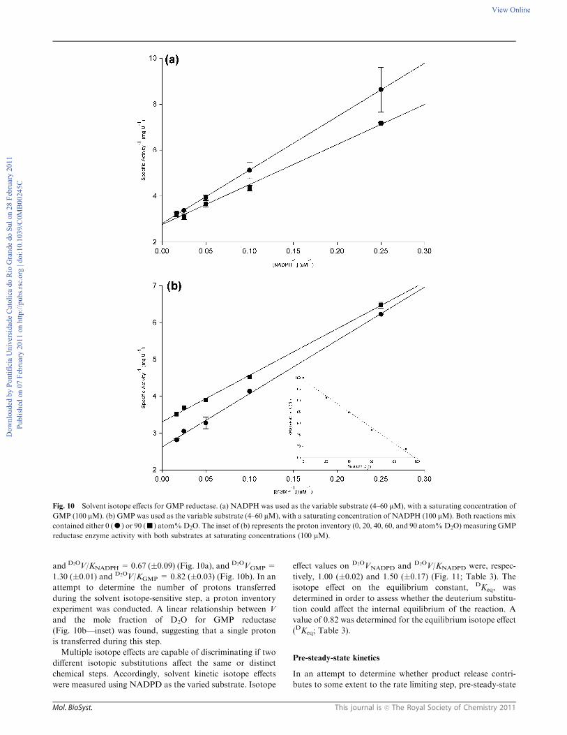
and D2OV/KNADPH =0.67 (�0.09) (Fig. 10a), and D2OVGMP =
1.30 (�0.01) and D2OV/KGMP = 0.82 (�0.03) (Fig. 10b). In an
attempt to determine the number of protons transferred
during the solvent isotope-sensitive step, a proton inventory
experiment was conducted. A linear relationship between V
and the mole fraction of D2O for GMP reductase
(Fig. 10b—inset) was found, suggesting that a single proton
is transferred during this step.
Multiple isotope effects are capable of discriminating if two
different isotopic substitutions affect the same or distinct
chemical steps. Accordingly, solvent kinetic isotope effects
were measured using NADPD as the varied substrate. Isotope
effect values on D2OVNADPD and D2OV/KNADPD were, respec-
tively, 1.00 (�0.02) and 1.50 (�0.17) (Fig. 11; Table 3). The
isotope effect on the equilibrium constant, DKeq, was
determined in order to assess whether the deuterium substitu-
tion could affect the internal equilibrium of the reaction. A
value of 0.82 was determined for the equilibrium isotope effect
(DKeq; Table 3).
Pre-steady-state kinetics
In an attempt to determine whether product release contri-
butes to some extent to the rate limiting step, pre-steady-state
Fig. 10 Solvent isotope effects for GMP reductase. (a) NADPH was used as the variable substrate (4–60 mM), with a saturating concentration of
GMP (100 mM). (b) GMP was used as the variable substrate (4–60 mM), with a saturating concentration of NADPH (100 mM). Both reactions mix
contained either 0 (K) or 90 (’) atom%D2O. The inset of (b) represents the proton inventory (0, 20, 40, 60, and 90 atom%D2O) measuring GMP
reductase enzyme activity with both substrates at saturating concentrations (100 mM).
Do
wn
load
ed b
y P
on
tifí
cia
Un
iver
sid
ade
Cat
oli
ca d
o R
io G
ran
de
do
Su
l o
n 2
8 F
ebru
ary
20
11
Pu
bli
shed
on
07
Feb
ruar
y 2
01
1 o
n h
ttp
://p
ub
s.rs
c.o
rg |
do
i:1
0.1
03
9/C
0M
B0
02
45
C
View Online

This journal is c The Royal Society of Chemistry 2011 Mol. BioSyst.
analysis of the reaction catalyzed was performed. Fitting the
pre-steady-state data to eqn (10), which describes a single
exponential decay, yielded a value of 0.204 (�0.002) sÿ1 for
the apparent first-order rate constant (Fig. 12). This result is in
agreement with the value of 0.28 (�0.02) sÿ1 for the catalytic
rate constant (kcat) determined by initial velocity study
measurements.
Discussion
Amplification, cloning, expression and purification of
recombinant E. coli GMP reductase
The guaC gene was successfully amplified from the E. coli
genome, cloned into the pCR-Blunt vector, and subcloned into
the pET-23a(+) expression vector. The automatic DNA
sequencing confirmed the integrity of the gene and the absence
of mutations. E. coli GMP reductase was expressed after 24 h
of cell growth in the absence of IPTG induction. The pET
expression system makes use of a powerful T7 polymerase,
under control of the IPTG-inducible lacUV5 promoter to
transcribe genes of interest, which are positioned downstream
of the bacteriophage T7 late promoter.19 Expression of
recombinant GMP reductase showed that even in the absence
of the inducer, high levels of protein production could be
obtained in the stationary phase, as has been previously
reported for other enzymes.20–22 It has been proposed that
uninduced expression of lac-controlled genes occurs when cells
approach the stationary phase in complex medium and that
cAMP, acetate and low pH are required to produce high level
expression in the absence of IPTG induction, which perhaps is
part of a general response to carbon-limiting conditions.23
Nevertheless, more recently, it has been shown that
unintended induction in the pET system is due to the presence
of as little as 0.0001% of lactose in the medium.24 The
recombinant E. coli GMP reductase was purified to homo-
geneity by a three-step purification protocol using standard
anionic exchange and size exclusion columns with 33% of
protein yield.
Quaternary structure analysis of E. coli GMP reductase
The results of mass spectrometry analysis combined with the
amino acid sequencing demonstrated, unequivocally, that the
homogeneous protein is, indeed, recombinant E. coli GMP
reductase. The gel filtration chromatography showed that the
enzyme from E. coli has the same tetrameric quaternary
structure of hGMPR2, demonstrated by X-ray diffraction.16
Determination of apparent steady-state kinetic constants and
initial velocity pattern
The results of the apparent steady-state kinetic data for E. coli
GMP reductase indicate a KM value for GMP of 6.9 mM,
which is 2.5-fold lower than that for hGMPR2 (17.4 mM),2
and 3-fold lower than the enzyme from L. donovani
(21.2 mM).9 The same pattern occurs for NADPH, with a
KM value of 11.1 mM, which is 2.3-fold lower than that for
hGMPR2 (26.6 mM).2 As for its human counterparts, E. coli
GMP reductase cannot catalyze conversion of GMP to IMP
using NADH as the hydride donor up to 200 mM concentra-
tion (data not shown).
Lineweaver–Burk analysis (Fig. 3) suggests that ping-pong
and rapid equilibrium ordered mechanisms can be ruled out
for E. coli GMP reductase. The former mechanism gives
double reciprocal plots displaying parallel lines and the latter
a family of lines intersecting on the y-axis. These data are
consistent with ternary complex formation and a sequential
mechanism. A sequential mechanism was also reported for
hGMPR111 and hGMPR2.2
It has been shown that the true KM values for erythrocyte
hGMPR1 are 2.6 mM for GMP and 16.9 mM for NADPH,11
which are in the same concentration range as E. coli GMP
reductase true steady-state kinetic parameters. These results
suggest that E. coli GMP reductase possesses a similar overall
Fig. 11 Multiple kinetic isotope effects using NADPD as the variable
substrate in the presence of saturating concentration of GMP
(100 mM) and either 0 (K) or 80 (’) atom% D2O. The data were
fitted to eqn (7).
Fig. 12 Representative stopped-flow trace for product formation,
measuring the decrease in absorbance at 340 nm upon conversion of
NADPH to NADP+ catalyzed by 10 mM of recombinant E. coliGMP
reductase (mixing chamber concentration) in the presence of GMP.
The data were fitted to eqn (10) for a single exponential decay, yielding
a value of 0.204 sÿ1 for the apparent first-order constant of product
formation. The top stopped-flow trace represents the control experi-
ment in the absence of the GMP substrate.
Do
wn
load
ed b
y P
on
tifí
cia
Un
iver
sid
ade
Cat
oli
ca d
o R
io G
ran
de
do
Su
l o
n 2
8 F
ebru
ary
20
11
Pu
bli
shed
on
07
Feb
ruar
y 2
01
1 o
n h
ttp
://p
ub
s.rs
c.o
rg |
do
i:1
0.1
03
9/C
0M
B0
02
45
C
View Online

Mol. BioSyst. This journal is c The Royal Society of Chemistry 2011
dissociation constant for both substrates when compared to
hGMPR1. However, the erythrocyte human enzyme presents
a bimodal GMP substrate saturation curve,11 which is usually
attributed to the existence of independent isoenzymes with
different kinetic constants or to a single multisubunit enzyme
with multiple sites that can interact in a negatively cooperative
manner.25 Deng and co-workers2 identified the hGMPR2
isoenzyme, making the bimodal substrate-saturation kinetic
found in hGMPR likely to be a result of a mixture of both
isoenzymes. At any rate, a sequential addition of substrates to
form a ternary complex has also been proposed for hGMPR2
enzyme.2
Isothermal titration calorimetry
ITC is an important and well-established technique for the
study of thermodynamics of macromolecular interactions, and
is unique in that it is capable of measuring simultaneously the
association and thermodynamic constants of binding.26
Binding experiments using ITC combined with the initial
velocity study demonstrated that the reaction catalyzed by
E. coli GMP reductase follows an ordered bi–bi kinetic
mechanism, in which GMP is the first substrate to bind to
free enzyme, followed by the binding of NADPH to form the
ternary complex capable of undergoing catalysis; and NADP+
is the first product to dissociate from the enzyme, followed by
the dissociation of IMP (Fig. 13). This mechanism is in
agreement with both human GMP reductases, for which
a sequential kinetic mechanism has been reported.2,11
Notwithstanding, the results reported here provide, to the best
of our knowledge, the first experimental evidence for both
order of addition of substrate and release of products.
The ITC measurements of GMP binding provided dissocia-
tion constant values (Kd), one for each subunit, where
Kd1 > Kd2 > Kd3, suggesting a positive homotropic
cooperativity, since the binding of one molecule of GMP
increases the affinity for the next molecule which will bind to
the next subunit. The Kd4 value is higher than the others,
indicating a lower affinity for the last subunit to bind GMP.
Interestingly, this finding is consistent with the crystal
structure of hGMPR2, as no GMP binding could be observed
for one of the subunits of this enzyme.16 However, the large
errors do not warrant to ascertain whether or not there is
cooperativity on GMP binding to E. coli GMP reductase. The
DG values for all four subunits are negative, which demon-
strate that GMP binding to E. coli GMP reductase is a
thermodynamically favorable process. The thermodynamic
analysis revealed different types of interactions between the
ligand and the enzyme subunits, ranging from favorable
hydrogen bonds and/or van der Waals interactions
(negative DH), release of ‘‘bound’’ water molecules to the
bulk solvent (positive DS) to conformational changes in either
or both of the molecules (negative DS).27 As can be seen in
Table 2, with the exception of subunit 2, the analyses of DH
and DS reveal that the binding of GMP is coupled with
favorable hydrogen bonds (negative DH) and conformational
changes (negative DS).28 This finding appears to be consistent
with the hGMPR2 structure,16 in which GMP was located on
the top of the a/b barrel surrounded by a hydrophilic surface
formed by the active site loop and the flexible binding loop
that would close upon GMP binding to the enzyme.
The IMP binding to E. coli GMP reductase yielded four
different Kd values (Table 2). However, the large errors do not
allow us to propose that there is either positive or negative
cooperativity among the subunits upon IMP binding. The
analysis of the Gibbs free energy revealed that the binding of
IMP is thermodynamically favorable for all subunits of E. coli
GMP reductase (negative DG), and the analysis of DH and DS
for IMP follows a similar pattern as observed for GMP. The
determination of the crystal structure of E. coli GMP
reductase in complex with IMP may shed light on these
thermodynamic features.
The rather large standard errors (Table 2) are a direct
consequence of the shape of the binding isotherm (C value),
which will dictate how accurately the thermodynamics para-
meters can be determined.27 The shape of the binding is
dependent on Kd and the concentration of the macromolecule.
The latter is limited by the need to obtain large quantities
and/or solubility. Another consideration is that for the experi-
mental set-up it is important to obtain an isotherm that
provides maximum data points for the fitting process,27 and
the loss of the plateau in the beginning of the titration curve
might have contributed to the large standard errors. We were
unable to determine the initial points due to rather small DH
values. At any rate, the ITC data provided clear-cut experi-
mental evidence of GMP and IMP binding to free E. coliGMP
reductase enzyme (Fig. 4a and c) that allowed the proposal of
an ordered bi–bi kinetic mechanism with GMP binding first
(Fig. 13). In addition, the ITC data provided thermodynamic
signatures of non-covalent interactions between E. coli GMP
reductase and either GMP or IMP.
pH-rate profiles
The Cys186 amino acid side chain has been shown to play a
key role in catalysis for hGMPR2 since the Cys186Ala mutant
displayed less than 5% activity as compared to wild-type
enzyme.16 The pH dependence of kcat and sequence alignment
analyses suggest that the conserved Cys186 is likely the residue
with an apparent pK value of 9.6 that plays a critical role in
E. coli GMP reductase catalysis. The cysteine thiol group
usually ionizes at slightly alkaline pH values, and the resulting
thiolate anion is the reactive species that acts as a nucleophile,
which is one of the most reactive functional groups found in
proteins.29 It is possible that the Cys186 side chain, which has
been shown to interact with GMP in the crystal structure of
hGMPR2,16 interacts with the exocyclic amino group bound
to C2 of the guanine moiety of GMP thereby facilitating both
hydride transfer to C2 and the amino leaving group. In
addition, the proposed model of E. coli GMP reductase
indicates that this residue makes an H-bond to the C2
exocyclic amino group of the guanine moiety of GMP
(Fig. 6). Notwithstanding, site-directed mutagenesis studiesFig. 13 Proposed enzyme mechanism for E. coli GMP reductase.
Do
wn
load
ed b
y P
on
tifí
cia
Un
iver
sid
ade
Cat
oli
ca d
o R
io G
ran
de
do
Su
l o
n 2
8 F
ebru
ary
20
11
Pu
bli
shed
on
07
Feb
ruar
y 2
01
1 o
n h
ttp
://p
ub
s.rs
c.o
rg |
do
i:1
0.1
03
9/C
0M
B0
02
45
C
View Online

This journal is c The Royal Society of Chemistry 2011 Mol. BioSyst.
are in progress to confirm the role of Cys186, if any, in E. coli
GMP reductase enzyme catalysis. The bell-shaped pH profile
for kcat (Fig. 7a) also showed participation of a single ionizable
group with an apparent pK value of 6.6 that has to be
deprotonated for catalysis. Sequence alignment showed
conservation of Asp129 in E. coli GMP reductase (Fig. 7) that
has been shown to be located in the active site loop in
hGMPR2.16 Although the pK value of the b-carboxyl group
of Aspartate residues are usually in the 3.9–4.0 range, it is not
unlikely that this pK value may be displaced by a neighbouring
chemical group. However, assigning a definite catalytic role to
Asp129 in E. coli GMP reductase is not warranted and site-
directed mutagenesis studies should be carried out. Although
the apparent pK value of 6.6 that plays a role in catalysis could
tentatively be ascribed to the imidazole side chain of a
Histidine residue (pK usually in the 6.0–7.0 range), His278 is
located in the GMP binding site of E. coli GMP reductase
whereas it is not present in hGMPR2 (Fig. 7).
The pH-rate data for of kcat/KGMP indicate that both
protonation of two groups with apparent pK values of 7.1
and deprotonation of two groups with apparent pK values of
8.6 (Fig. 7b) are required for GMP binding. These dissociation
constants can be in the ligand and the other in the enzyme, or
both can be in one or the other. The crystal structure of
hGMPR2 in complex with GMP demonstrated that this
substrate is surrounded by a hydrophilic surface formed by
the active site loop (residues 179–187 in hGMPR2) and
the flexible GMP binding region (residues 268–290 in
hGMPR2).16 Sequence alignment of GMP reductases
(Fig. 5) shows a relatively good conservation of these two
regions in E. coli GMP reductase (active site loop: residues
179–187; GMP binding region: 268–290). The pK value for the
N7 atom of the guanine moiety of GMP is 3.6, and the ribose
20,30-diol only loses a proton above pH 12. The guanine base
becomes protonated on one of the ring nitrogens rather than
on the exocyclic amino group since this does not interfere with
delocalization of the NH2 electron lone pair into the aromatic
system. In the case of monoesters, the phosphate group of
GMP loses one proton at pH 1 and a second proton at pH 7.
The proximity of negative charge on the phosphate residues
has a secondary effect, making the ring nitrogens more
basic (DpK E +0.4) and the amine protons less acidic
(DpK E +0.6). It is thus likely that the pH-rate profile does
not reflect any ionization of the guanine moiety of GMP. On
the other hand, it is possible that the apparent pK values
reflect change in ionization of His278 in the GMP binding site,
and change in ionization of the phosphate group of GMP and,
for instance, Asp219 that makes H-bonds with the ribose
hydroxyl groups of the pentose.
The pH-rate profile for kcat/KNADPH indicated that proto-
nation of two groups with apparent pK values of 6.2 abolish
NADPH binding to E. coli GMP reductase (Fig. 7c).
Although there was no diffraction pattern to allow identifica-
tion of the NADPH binding site in hGMPR2, structural
comparison and model building has been employed to suggest
that the amino acid residues 129–133 are involved in NADPH
binding.16 These residues are conserved in E. coli GMP
reductase (Fig. 5). The adenine-C20-ribose phosphate group
has a pK value of 6.1. It is thus tempting to suggest that
protonation of NADPH phosphate and the putative Asp129
residue located in the NADPH binding site can account for the
kcat/KNADPH pH-rate profile.
Deuterium kinetic isotope effects and proton inventory
Measurements of kinetic isotope effects in enzyme catalyzed
reactions aim at examining the contribution of proton
transfer(s) to rate-limiting step(s). However, the maximal velocity
may be dependent on several rate-contributing or partially
rate-limiting steps instead of one rate-determining step.30
Isotope effects on V report on events following the ternary
complex formation capable of undergoing catalysis
(fully loaded enzyme), which include the chemical steps,
possible enzyme conformational changes, and product release
(leading to regeneration of free enzyme). Isotope effects on
V/K report on steps in the reaction mechanism from binding of
the isotopically labeled substrate to the first irreversible step,
usually considered to be the release of the first product
(that is, all rate constants from reactant binding until the first
irreversible step).30 Any substrate can be varied; it does not
need to be the labeled one, but one obtains DV/K effect for the
varied substrate rather than for the labeled one. Although the
apparent classical limit for primary deuterium kinetic isotope
effects on V is approximately 8, values as low as 2 have
sometimes been accepted as evidence of a rate-determining
step.30,31 For reactions involving NAD(P)H oxidation,
primary deuterium isotope effects ranging from 1 to 3 have
been found.32 The magnitude of primary deuterium isotope
effect depends on the chemical nature of the transition state.33
This isotope effect reaches a maximum value when the
hydrogen is symmetrically placed between the donor atom
from which cleavage occurs and the acceptor atom to which a
new bond is formed, and decreases for reactant- or product-
like transition states. The value of 2.5 for the observed primary
deuterium kinetic isotope effect on V for E. coli GMP
reductase using either NADPH or [4S-2H]-NADPH
(DVNADPH) as a variable substrate indicates that hydride
transfer is involved in a rate-limiting step and that the transi-
tion state may be either substrate- (early) or product-like
(late). However, the symmetry of the transition state can only
be inferred from the magnitude of a deuterium kinetic isotope
effect if it is for the specific step in an enzymatically catalyzed
reaction at which the isotopically substituted bond is broken
by passing through a single transition state (that is, the
intrinsic kinetic isotope effect). Enzyme-catalyzed chemical
reactions proceed through many steps and the rate constants
for several steps are usually consequential to the composite
rate constant for the overall reaction, consequently the
observed deuterium kinetic isotope effect is less than or equal
to the intrinsic deuterium isotope effect for the step in which
the hydrogen is transferred. In any case, the observed primary
deuterium kinetic isotope effect on V for E. coli GMP
reductase indicates that hydride transfer is from C4-proS
hydrogen of NADPH and that it is partially rate-limiting.
On the other hand, the inverse primary deuterium kinetic
effect on V/K for NADPH (DV/KNADPH = 0.4) is somewhat
puzzling. Incidentally, there have been numerous reports
of inverse isotope effects on V/K of unknown origin.34,35
Do
wn
load
ed b
y P
on
tifí
cia
Un
iver
sid
ade
Cat
oli
ca d
o R
io G
ran
de
do
Su
l o
n 2
8 F
ebru
ary
20
11
Pu
bli
shed
on
07
Feb
ruar
y 2
01
1 o
n h
ttp
://p
ub
s.rs
c.o
rg |
do
i:1
0.1
03
9/C
0M
B0
02
45
C
View Online

Mol. BioSyst. This journal is c The Royal Society of Chemistry 2011
The expression of deuterium kinetic isotope effect on V/K
includes the intrinsic isotope effect, commitment factors
(forward and reverse) and equilibrium isotope effect.33 An
equilibrium isotope effect may be invoked to account for the
inverse effect on V/K, providing that the reverse commitment
factor is large and the forward commitment factor is small.
The inverse deuterium isotope effect on the equilibrium
constant (DKeq = 0.82) suggests that the deuterated product
is more stiffly bonded than the deuterated substrate36 and the
internal equilibrium might dominate the observed kinetic
isotope effect. The inverse isotope effect indicates that once
the hydride is transferred it becomes more tightly bonded to
the corresponding product (IMP) when compared to the
substrate. The magnitude of this isotope effect is a direct
consequence of the difference in the bonds of the substrate
and product, with deuterium becoming enriched in the stiffest
bond and the hydrogen in the looser one (deuterium accumu-
lates where binding is tighter).37 In addition, isotope effects on
V/K are a combination of binding and catalytic events.
Binding isotope effects can contribute in the opposite direction
of kinetic isotope effects that arise from transition state
chemistry. An inverse binding isotope effect may arise when
binding of the molecule containing the heavier isotope atom
(NADPD) is increased as bonds to the isotopic label are
tighter in the bound complex. Although it is conceivable that
the inverse DV/KNADPH value reflects an inverse binding
isotope effect, it cannot be experimentally assessed as NADPH
(enzyme mechanism proposed here) binds to the E. coli GMP
reductase:GMP binary complex to form the catalytic compe-
tent ternary complex. Accordingly, the inverse DV/KNADPH
value is likely due to the inverse deuterium isotope effect on
the equilibrium constant. Isotope effects can provide further
experimental evidence for a specific enzyme mechanism. In the
case of an ordered mechanism, the V/K for the first substrate
(GMP) to bind (at saturating concentrations of the second
substrate: NADPH) is the on-rate for the reactant binding to
enzyme, a non-isotope sensitive step.33 The value of 1.03
(� 0.07) for DV/KGMP (Table 3) provides further support for
an ordered enzyme mechanism as suggested by the initial
velocity pattern and ITC data.
To evaluate the contribution of proton transfer from solvent
to a rate-limiting step, measurements of solvent isotope effects
on V and V/K were carried out. As rule of thumb, deuterium
accumulates where binding is tighter (that is, fractionation
factor is larger than one). Transition state proton contributes
the reciprocal of its respective fractionation factor to the
solvent isotope effect, whereas the contribution of a reactant
state proton to the solvent isotope effect is equal to its
fractionation factor.32 Reactant state fractionation by enzyme
cysteine thiol groups (S–H group of cysteine) can make large
inverse contributions to solvent isotope effects.32,33 It is thus
conceivable that the value of 0.82 for D2OV/KGMP reflects
participation of Cys186 of free enzyme, which has been shown
to interact with GMP in the hGMPR2 crystal structure,16 and
the value of 0.67 for D2OV/KNADPH reflects an inverse
contribution of conserved Cys127 of free enzyme, located near
the NADPH binding site of E. coli GMP reductase (Fig. 5).
The value of 0.99 for D2OVNADPH suggests no participation of
the proton solvent in catalysis. On the other hand, solvent
proton transfer appears to play a modest role in catalysis since
a value of 1.30 was found for D2OVGMP. In addition, proton
inventory data (Fig. 10b—inset) suggest that this modest
solvent kinetic isotope effect arises from a single protonic site.
The difference found between the primary deuterium and
solvent kinetic isotope effects suggests that the hydride and
proton transfer may occur in distinct reaction steps, as
found in other NADPH-dependent reductive reactions.34
Nevertheless, it is not possible to ascertain whether or not
hydride transfer and protonation steps occur in a single
transition state (concerted mechanism) or in distinct transition
states (stepwise mechanism) based solely on primary and
solvent isotope effects.
Multiple isotope effects (double isotope effects) allow us to
determine whether two different isotopic substitutions affect
the same (concerted) or different (stepwise) chemical steps.38
This method uses the normal effect of deuterium to slow down
one step of a reaction mechanism while observing changes in
the expression of another isotope effect,38 the effects in
question being the protonation and the hydride transfer. If
the protonation and hydride transfer occur in the same
transition state, the solvent isotope effect will be larger or
unchanged with the deuterium substitution (NADPD) when
compared with NADPH. However, if the effects take place in
different steps, the solvent isotope effect will decrease with
NADPD, once the hydride transfer will become more rate-
limiting.34,35,38 As can be seen in Table 3, the results of the
multiple isotope effects showed similar values for D2OVNADPD
(1.00) and D2OVNADPH (0.99), suggesting that hydride transfer
and solvent proton transfer steps take place in the same
transition state (concerted mechanism). In addition, the value
for the solvent isotope effect on V/K with NADPD (1.50) is
larger than with NADPH (0.67), thereby lending support to a
concerted mechanism.
Pre-steady-state kinetics
In the study of the transient phase of enzyme reactions, the
data obtained are generally expressed by a single exponential
curve for the first order reaction.39 The analysis of the data
allows us to determine the apparent first order rate constant,
which was in the same range as the first order in the steady-
state kinetics. The observation of burst during a time course in
the transient phase is evidence of a significant build-up of
product formation along the reaction pathway.39 If a burst is
observed during the transient phase, and the concentration of
NADP+ produced is approximately equal to the initial E. coli
GMP reductase subunit concentration, it would indicate that
the chemical step of the reaction is much faster than the release
of the first product (NADP+). As there is no burst of NADP+
formation (Fig. 12), the stopped-flow result demonstrates that
product release does not contribute to the rate-limiting step of
the chemical reaction catalyzed by E. coli GMP reductase.
Conclusion
A rational inhibitor design relies on mechanistic and structural
information on the target enzyme. Enzyme inhibitors make up
roughly 25% of the drugs marketed in United States.40
Enzymes catalyze multistep chemical reactions to achieve rate
Do
wn
load
ed b
y P
on
tifí
cia
Un
iver
sid
ade
Cat
oli
ca d
o R
io G
ran
de
do
Su
l o
n 2
8 F
ebru
ary
20
11
Pu
bli
shed
on
07
Feb
ruar
y 2
01
1 o
n h
ttp
://p
ub
s.rs
c.o
rg |
do
i:1
0.1
03
9/C
0M
B0
02
45
C
View Online

This journal is c The Royal Society of Chemistry 2011 Mol. BioSyst.
accelerations by stabilization of transition state structure(s).40
Accordingly, mechanistic analysis should always be a top
priority for enzyme-targeted drug programs aiming at the
rational design of potent enzyme inhibitors. Moreover, ITC
has been used as an important technique for the direct
determination of thermodynamic and kinetic parameters of
enzymatic reactions.41 The recognition of the limitations
of high-throughput screening approaches in the discovery of
candidate drugs has rekindled interest in rational design
methods.42 Understanding the mode of action of E. coli
GMP reductase will inform us on how to better design
inhibitors targeting this enzyme. In addition, understanding
the mode of action of an enzyme can be used to inform
functional annotation of newly determined sequences and
structures, to select appropriate enzyme scaffolds for engineer-
ing new functions, and to refine definitions in the current EC
classifications.43 The elucidation of the mode of action of
E. coli GMP reductase and its availability in recombinant
form may also provide a tool for determination of substrate
specificity of cyclic nucleotide phosphodiesterases (PDEs).
PDEs belong to a superfamily of proteins with medical
relevance that play a major role in cell signaling by catalyzing
the hydrolysis of cyclic AMP (cAMP) and cyclic GMP
(cGMP).44 PDE enzymes are distinguished by their substrate
specificity. The PDE activity cannot be directly assayed, being
necessary to couple an accessory enzyme. As recombinant
E. coli GMP reductase is specific for GMP, it may be
employed to determine the substrate specificity of PDE
enzymes. Among the 11 families of PDEs, only PDE 5, PDE
6 and PDE 9 are specific for cGMP, which are targets for,
respectively, treatment of erectile dysfunction (sildenafil),
visual alterations, and behavioral state regulations and
learning.45 The results here presented may also help chemical
biologists to design function-based chemical compounds to
carry out either loss-of-function (inhibitors) or gain-of-
function (activators) experiments to reveal the biological role
of GMP reductase in the context of whole E. coli cells.46 It is
also hoped that the results presented here may be useful to
understand the role that GMP reductase plays in the E. coli
salvage pathway.
Experimental procedures
Materials
All chemicals were of analytical or reagent grade and were
used without further purification, unless stated otherwise.
50GMP, NADPH, IMP, and NADP+ along with lysozyme
and streptomycin sulfate were purchased from Sigma-Aldrich.
Deuterium oxide (99.9 atom% D2O) was from Cambridge
Isotope Laboratories. Fast performance liquid chromato-
graphy (FPLC) protein purification (4 1C) was carried out
using an Akta Purifier from GE Healthcare; all chromato-
graphic columns and the LMW and HMW Gel Filtration
Calibration Kit were also from GE Healthcare. Bovine serum
albumin and Bradford reagent were from Bio-Rad
Laboratories. All steady-state activity assays were performed
in an UV-2550 UV/Visible spectrophotometer (Shimadzu).
Dithiothreitol (DTT) was from Acros Organics. Pfu DNA
polymerase was from Stratagene. Restriction enzymes and T4
DNA ligase and pCR-Blunt cloning vector were from
Invitrogen and pET-23a(+) expression vector and E. coli
BL21(DE3) were from Novagen. The mass spectrometer
LTQ-XL and LTQ Orbitrap Discovery were from Thermo
and the nanoLC Ultra 1D plus was from Eksigent. An iTC200
Microcalorimeter was from MicroCal Inc (Northampton,
MA). Pre-steady-state measurements were carried out using
an Applied Photophysics SX.18MV-R stopped-flow spectro-
fluorimeter on absorbance mode.
E. coli GMP reductase comparative homology modeling
A E. coli three-dimensional model was built by comparative
homology modeling using the human type 2 GMP reductase
structure as template (PDB ID: 2A7R; hGMPR2),16 solved by
X-ray crystallography at 3.0 A in complex with GMP. The
template structure was used for E. coli GMP reductase
modeling, as well as to evaluate GMP binding mode to the
bacterial enzyme active site. Template selection was based on
high primary sequence conservation (68.5% identity and
16.47% strong similarity), and the presence of enzyme
substrate bound to its active site. Target and template
pair-wise sequence alignment required a single gap inclusion
in human GMP reductase primary sequences. The E. coli
GMP reductase model was built by restrained-based
homology modeling implemented in MODELLER9v1,47 with
the standard protocol of the comparative protein structure
modeling methodology, by satisfaction of spatial restrains.48,49
Atomic coordinates of GMP heteroatoms were copied from
the template structure into the E. coli GMP reductase model.
The best model was selected according to MODELLER
objective function50 and subjected to energy minimization
for amino acid side chain and main chain rearrangements
with the GROMACS package51 using the 43a1 force-field. The
system was submitted to an initial steepest descent energy
minimization in vacuum with a maximum number of 400
minimization steps, followed by a maximum of 3000 steps of
conjugate gradient energy minimization. The program
PROCHECK52 was employed to analyze the stereochemical
quality of the model, as previously described.53 Structural
correspondence between the E. coli model and human GMP
reductase template was evaluated by their root-mean square
deviation (RMSD). H-bond interactions were evaluated with
LIGPLOT v4.4.2,54 considering an atomic distance cut off of
3.9 A (program default values). Images were generated with
PyMOL Molecular Graphic System V1.3 (Schrodinger, LLC).
Amplification and cloning of the E. coli guaC gene
The oligonucleotide primer sequences of the E. coli guaC
structural gene were 50-AACATATGCGTATTGAAGAA-
GATCTGAAGTTAGGTTTTAAAGACG-3 0 (forward) and
3 0-AAGGATCCTCATTACAGGTTGTTGAAGATGCGG
TTTTCTTGTTCC-50 (reverse). The primers were designed to
contain, respectively, NdeI and BamHI restriction sites
(in italics). The DNA fragment (1038 bp) was amplified using
Pfu DNA polymerase, ligated into the pCR-Blunt cloning
vector, and transformed into E. coli DH10B cells. Plasmid
DNA recovered from these cells was digested with the
Do
wn
load
ed b
y P
on
tifí
cia
Un
iver
sid
ade
Cat
oli
ca d
o R
io G
ran
de
do
Su
l o
n 2
8 F
ebru
ary
20
11
Pu
bli
shed
on
07
Feb
ruar
y 2
01
1 o
n h
ttp
://p
ub
s.rs
c.o
rg |
do
i:1
0.1
03
9/C
0M
B0
02
45
C
View Online

Mol. BioSyst. This journal is c The Royal Society of Chemistry 2011
restriction enzymes NdeI and BamHI, and the isolated insert
was ligated into the pET-23a(+) expression vector, previously
treated with the same restriction enzymes. The sequence of the
E. coli guaC gene was determined by automated DNA
sequencing to confirm the identity, integrity, and absence of
PCR-introduced mutations in the cloned fragment.
Expression of recombinant GMP reductase
The recombinant plasmid pET-23a(+)::guaC was trans-
formed into E. coli BL21(DE3) cells, and these were selected
on Luria-Bertani (LB) agar plates containing 50 mg mL–1
ampicillin. LB medium (60 mL) containing 50 mg mL–1
ampicillin was inoculated with a single colony and cells were
grown overnight. The culture (10 mL) was inoculated in LB
medium (500 mL) with the same antibiotic concentration, and,
as soon as OD600 nm reached a value of 0.6, culture was grown
for additional 24 h at 180 rpm and 37 1C without IPTG
induction. Cells (15 g) were harvested by centrifugation at
7690g for 30 min at 4 1C and were stored at –20 1C. Soluble
and insoluble fractions were analyzed by 12% SDS-PAGE
and Coomassie staining.
Purification of recombinant GMP reductase
Approximately 2.8 g of frozen cells were resuspended in 14 mL
of 100 mM tris(hydroxymethyl)aminomethane (Tris; buffer A),
pH 7.8, and incubated with lysozyme (0.2 mg mL–1) for 30 min
with stirring. Cells were disrupted by sonication, and centri-
fuged at 38 900g for 30 min to remove cells debris. Strepto-
mycin sulfate was added to the supernatant up to 1% (wt/vol),
stirred for 30 min to precipitate nucleic acids, and centrifuged
at 38 900g for 30 min. The resulting supernatant was dialyzed
against buffer A (2� 2 L; 2 h each) using a dialysis tubing with
a molecular weight exclusion limit of 12–14 kDa. The sample
was clarified by centrifugation at 38 900g for 30 min and
loaded on a FPLC 2.6 cm � 8.2 cm Q-Sepharose Fast Flow
anion exchange column, pre-equilibrated with buffer A,
washed with 5 column volumes of buffer A, and the adsorbed
material was eluted with a linear gradient (0-40%) of 20
column volumes of 100 mM Tris pH 7.8 containing 500 mM
NaCl (buffer B) at 1 mL min–1. The adsorbed recombinant
GMP reductase was eluted at approximately 145 mM NaCl
concentration, with substantial removal of contaminants. All
fractions were analyzed by SDS-PAGE electrophoresis, and
the ones containing the target protein were pooled and con-
centrated to 8.5 mL using an Amicon ultrafiltration cell
(molecular weight cutoff 30 kDa). The soluble sample was
loaded on a 2.6 cm � 60 cm HiPrep Sephacryl S-200 High
Resolution size exclusion column, which was previously
equilibrated with buffer A, and isocratically eluted with buffer
A at a flow rate of 0.25 mL min–1. This step was employed to
further purify the recombinant protein and to remove salt
from the sample, thereby allowing protein preparation for the
next chromatographic step. Fractions containing the target
protein were pooled and the soluble sample was loaded on a
1.6 cm � 10 cm Mono Q High Resolution anion exchange
column, pre-equilibrated with buffer A. The column was
washed with 5 column volumes, and the adsorbed proteins
were eluted with a linear gradient (0–40%) of 20 column
volumes of buffer B, and the target protein eluted at approxi-
mately 155 mM NaCl concentration. The active fractions
containing the target protein were pooled and dialyzed against
buffer A (2 � 2 L; 2 h each) in order to remove the remaining
salt. Protein concentration was determined by Bradford’s
method using bovine serum albumin as standard.55
Quaternary structure analysis of E. coli GMP reductase
The homogeneous sample was submitted to ESI-MS in order
to confirm the identity of E. coli GMP reductase. The protein
was also digested with trypsin and the resulting peptides were
separated and analyzed by liquid chromatography associated
with mass spectrometry with fragmentation collision induced,
and the results were used to identify the amino acid sequence
through a search software (Protein Discoverer, Thermo).
The molecular mass of recombinant GMP reductase was
determined by gel filtration chromatography using a 1.0 cm �
30 cm High Resolution Superdex 200 column pre-equilibrated
with 50 mM Tris, pH 7.5, containing 200 mM NaCl at a flow
rate of 0.4 mL min–1. The LMW and HMW Gel Filtration
Calibration Kit were used as the protein molecular mass
standards in the calibration curve, measuring the elution
volumes of several standards (ferritin, catalase, aldolase,
ovalbumin, coalbumin, ribonuclease A, and blue dextran 2000),
calculating their partition coefficients (Kav), and plotting these
values against the logarithm of their molecular mass. The Kav
values were determined as Kav = (Ve ÿ Vo)/(Vt ÿ Vo), where
Ve is the sample elution volume, Vt is the total bed volume of
the column, and Vo is the column void volume, which had
been determined by loading blue dextran 2000 into the column.
Protein elution was monitored at 215, 254, and 280 nm.
Synthesis of [4S-2H]-NADPH
[4S-2H]-NADPH was synthesized according to the method of
Ottolina and co-workers56 and purified on a 1.0 cm � 10 cm
Mono Q High Resolution anion exchange column. The
column was washed with 1 column volume of 100 mM N-2-
hydroxyethylpiperazine-N0-2-ethanesulfonic acid (Hepes),
pH 7.0, and the material was eluted with a linear gradient
(0–100%) of 5 column volumes of 100 mM Hepes containing
1.0 M NaCl, pH 7.0, at a flow rate of 0.5 mL min–1. The
fractions with A260 nm/A340 nm r 2.3 were pooled.
Enzyme activity assay of E. coli GMP reductase
The recombinant GMP reductase activity was determined by a
continuous spectrophotometric assay monitoring the conver-
sion of NADPH to NADP+ at 340 nm. The standard assay
was performed at 25 1C in 100 mM Tris, pH 7.8, with 10 mM
DTT, unless stated otherwise.
Determination of apparent steady-state kinetic constants and
initial velocity pattern
In order to determine the apparent steady-state kinetic
constants, GMP reductase activity was measured (in duplicates)
in the presence of varying concentrations of GMP (5–80 mM)
and fixed NADPH concentration (100 mM), and varying
concentrations of NADPH (5–120 mM) and fixed GMP
concentration (100 mM), in 100 mM Tris, pH 7.8, buffer
Do
wn
load
ed b
y P
on
tifí
cia
Un
iver
sid
ade
Cat
oli
ca d
o R
io G
ran
de
do
Su
l o
n 2
8 F
ebru
ary
20
11
Pu
bli
shed
on
07
Feb
ruar
y 2
01
1 o
n h
ttp
://p
ub
s.rs
c.o
rg |
do
i:1
0.1
03
9/C
0M
B0
02
45
C
View Online

This journal is c The Royal Society of Chemistry 2011 Mol. BioSyst.
containing 10 mM DTT in a total volume of 500 mL. The
enzyme specificity for NADPH was evaluated measuring the
enzymatic activity in the presence of varying concentrations of
NADH (20–200 mM) at a fixed-saturating concentration of
GMP (100 mM). To determine the true steady-state kinetic
parameters and initial velocity patterns, enzymatic activity was
measured in the presence of varying concentrations of GMP
(1–60 mM) and several fixed-varied NADPH concentrations
(1–60 mM). Enzyme activity was determined in an UV-2550
UV/Visible spectrophotometer (Shimadzu) by monitoring the
decrease in absorbance at 340 nm (e340nm = 6220 Mÿ1 cmÿ1)
upon GMP reductase-catalyzed conversion of NADPH to
NADP+. One unit of enzyme activity was defined as the
amount of protein that catalyzes the consumption of 1 mmol
of NADPH per minute at 25 1C.
Isothermal titration calorimetry
ITC experiments were carried out using the iTC200 Micro-
calorimeter. The reference cell (200 mL) was loaded with water
during all experiments and the sample cell (200 mL) was filled
with E. coli GMP reductase at a concentration of 122 mM in
buffer A. The injection syringe (39.7 mL) was filled with
substrates or products at different concentrations, GMP at
0.75 mM, NADPH at 3.5 mM, NADP+ at 1.0 mM, and IMP
at 1.0 mM, and the ligand binding isotherms were measured by
direct titration (ligand into macromolecule). The same buffer
was used to prepare all ligand solutions. The stirring speed was
500 rpm at 25 1C with constant pressure for all ITC
experiments. Titration first injection (0.5 mL) was not used in
data analysis and was followed by 17 injections of 2.2 mL each
at 180 s intervals. Control titrations (ligand into buffer) were
performed in order to subtract the heats of dilution and mixing
for each experiment prior to data analysis. All data were
evaluated utilizing the Origin 7 SR4 software (Microcal, Inc.).
pH-rate profiles
The dependence of kinetic parameters on pH was determined
by measuring initial velocities in the presence of varying
concentrations of one substrate and saturating levels of the
other, in a buffer mixture of 500 mM 2-(N-morpholino)-
ethanesulfonic acid (MES)/Hepes/2-(N-cyclohexylamino)-
ethanesulfonic acid (CHES) over the following pH values:
6.0 (10–160 mM varying GMP concentration and fixed
concentration of NADPH at 240 mM, and 10–160 mM varying
NADPH concentration and fixed concentration of GMP at
240 mM), 6.5 (2–60 mM varying GMP concentration and fixed
concentration of NADPH at 100 mM, and 2–60 mM varying
NADPH concentration and fixed concentration of GMP at
100 mM), 7.0 (2–60 mM varying GMP concentration and fixed
concentration of NADPH at 100 mM, and 2–60 mM varying
NADPH concentration and fixed concentration of GMP at
100 mM), 7.5 (5–80 mM varying GMP concentration and fixed
concentration of NADPH at 100 mM, and 5–80 mM varying
NADPH concentration and fixed concentration of GMP at
100 mM), 8.0 (5–80 mM varying GMP concentration and fixed
NADPH concentration at 100 mM, and 5–80 mM varying
NADPH concentration and fixed concentration of GMP
at 100 mM), 8.5 (5–60 mM varying GMP concentration and
fixed concentration of NADPH at 100 mM, and 5–60 mM
varying NADPH concentration and fixed concentration of
GMP at 100 mM), 9.0 (5–60 mM varying GMP concentration
and fixed concentration of NADPH at 100 mM, and 5–60 mM
varying NADPH concentration and fixed concentration of
GMP at 100 mM), and 9.5 (2–60 mM varying GMP concentra-
tion and fixed concentration of NADPH at 100 mM, and
2–60 mM varying NADPH concentration and fixed concentra-
tion of GMP at 100 mM). Prior to performing the pH-rate
profiles, the recombinant enzyme was incubated over a
broader pH range and assayed under standard conditions to
identify denaturing pH values and to ensure enzyme stability
at the tested pH range.
Energy of activation
In order to determine the energy of activation of the GMP
reductase-catalyzed reaction, initial velocities were measured
in the presence of varying concentrations of one (GMP 1–60 mM)
(NADPH 1–80 mM) substrate and saturating levels of the
other (100 mM), at temperatures ranging from 15 to 35 1C.
GMP reductase was incubated for several minutes in all
temperatures tested and assayed under standard conditions
to ensure enzyme stability under all temperatures.
Deuterium kinetic isotope effects and proton inventory
Primary deuterium kinetic isotope effects were determined by
measuring initial rates using a saturating level of GMP
(100 mM) and varying concentrations of either NADPH
(4–60 mM) or [4S-2H]-NADPH (1–60 mM).
Solvent kinetic isotope effects were determined by measuring
initial velocities using a saturating level of one substrate
(100 mM) and varying concentrations of the other (GMP:
4–60 mM; NADPH: 4–60 mM) in either H2O or 90 atom%
D2O. The proton inventory was determined using saturating
concentrations of both substrates (100 mM) at various mole
fractions of D2O.
Multiple kinetic isotope effects were measured by determining
the solvent isotope effects using [4S-2H]-NADPH (NADPD)
as the varied substrate.
An equilibrium isotope effect (DKeq) was determined by
measuring the equilibrium constant in the presence of either
NADPH or [4S-2H]-NADPH. These equilibrium constants
were measured as described by Leu and Cook.57 In short, the
ratio of [NADPH]/[NADP+] was fixed at 1 and the ratio of
[IMP]/[GMP] were varied from 0.1 to 30 at fixed concentration
of NH4Cl. A plot of the change in NADPH concentration versus
[IMP]/[GMP] ratio crosses the abscissa at a value equal to Keq.57
The notation utilized to express isotope effects is that of
Northrop30 as extended by Cook and Cleland.58
Pre-steady-state kinetics
Pre-steady-state kinetic measurements of the reaction
catalyzed by E. coli GMP reductase were performed to
determine whether product release is part of the rate-limiting
step. The decrease in absorbance was monitored at 340 nm
(1 mm split width = 4.65 nm spectral band), at 25 1C, using a
split time base (2–50 s; 400 data points for each time base). The
experimental conditions were 10 mM E. coli GMP reductase,
Do
wn
load
ed b
y P
on
tifí
cia
Un
iver
sid
ade
Cat
oli
ca d
o R
io G
ran
de
do
Su
l o
n 2
8 F
ebru
ary
20
11
Pu
bli
shed
on
07
Feb
ruar
y 2
01
1 o
n h
ttp
://p
ub
s.rs
c.o
rg |
do
i:1
0.1
03
9/C
0M
B0
02
45
C
View Online

Mol. BioSyst. This journal is c The Royal Society of Chemistry 2011
10 mM DTT, 250 mM GMP and 250 mM NADPH in 100 mM
Tris-HCl, pH 7.8 (mixing chamber concentrations). The experi-
mental conditions for the control experiment were 10 mM E. coli
GMP reductase, 10 mM DTT, and 250 mM NADPH in
100 mM Tris-HCl, pH 7.8 (mixing chamber concentrations).
The dead time of the stopped-flow equipment is 1.37 ms.
Data analysis
Values of the kinetic parameters and their respective errors
were obtained by fitting the data to the appropriate equations
by using the nonlinear regression function of SigmaPlot 9.0
(SPSS, Inc.). Initial rate data at single concentration of the
fixed substrate and varying concentrations of the other were
fitted to eqn (1).17
v ¼VA
K þ Að1Þ
The family of lines intersecting to the left of the y-axis in
double-reciprocal plots was fitted to eqn (2), which describes a
mechanism involving ternary complex formation and a
sequential substrate binding.59
v ¼VAB
KiaKb þ KaBþ KbAþ ABð2Þ
For eqn (1) and (2), v is the initial velocity, V is the maximal
initial velocity, A and B are the concentrations of the
substrates (GMP and NADPH), Ka and Kb are their respective
Michaelis constants, and Kia is the dissociation constant for
enzyme–substrate A binary complex formation.
pH-rate profiles data were fitted to eqn (3), (4) or (5), where
y is the kinetic parameter, C is the pH-independent value of y,
H is the proton concentration, and Ka and Kb are, respectively,
the apparent acid and base dissociation constants for ionizing
groups, and K0 is the product of two apparent dissociation
constants.60
log y ¼ logC
1þH
Ka
þKb
H
0
B
B
@
1
C
C
A
ð3Þ
log y ¼ logC
1þH
Ka
þH2
K0
þKb
Hþ
K0
H2
0
B
B
@
1
C
C
A
ð4Þ
log y ¼ logC
1þH
Ka1
� �
1þH
Ka2
� �
0
B
B
@
1
C
C
A
ð5Þ
Eqn (3) describes a bell-shaped pH profile for a group that
must be protonated for binding/catalysis and another group
that must be unprotonated for binding/catalysis, and partici-
pation of a single ionizing group for the acidic limb
(slope value of +1) and participation of a single ionizing
group for the basic limb (slope value of ÿ1). Eqn (4) describes
a bell-shaped pH-rate profile that starts with a slope of +2 in
the acidic limb and goes to an eventual slope of ÿ2 in the basic
limb, suggesting participation of two ionizing groups in each
limb. Eqn (5) describes a pH-rate profile for two groups that
need to be unprotonated for binding (slope of +2 for the
acidic limb). Unless the pKs of the groups are at least 3 pH
units apart, there will not be a linear region with a slope of +1.
The data for temperature effects were fitted to eqn (6), where
k is the maximal reaction rate, Ea is the energy of activation,
T is the temperature in Kelvin, R is the gas constant
(1.987 cal Kÿ1 mol–1), and A is a pre-exponential factor that
correlates collision frequency and the probability of the
reaction occurring when reactant molecules collide.
log k ¼ ÿEa
2:3R
� �
1
T
� �
þ logA ð6Þ
Kinetic isotope effects data were fitted to eqn (7), which
assumes isotope effects on both V/K and V. In this equation,
EV/K and EV are the isotope effects minus 1 on V/K and V,
respectively, and Fi is the fraction of isotopic label in
substrate A.36
v ¼VA
Kð1þ FiEV=KÞ þ Að1þ FiEVÞð7Þ
ITC data were fitted to eqn (8), where DH is the enthalpy of
process given by the ITC experiment, DG is the Gibbs free
energy changes, DS is the entropy changes, T is the tempera-
ture of the experiment in Kelvin, R is the gas constant
(1.987 cal Kÿ1 mol–1), and Keq is the equilibrium binding
constant. The dissociation constant, Kd, is the inverse of the
equilibrium binding constant, Keq, described in eqn (9).
DG = DH ÿ TDS = ÿRTlnKeq (8)
Kd ¼1
Keq
ð9Þ
The pre-steady-state time course of the reaction was fitted to
eqn (10) for a single exponential decay, in which A is the
absorbance at time t, A0 is the absorbance at time zero, and k
is the apparent first-order rate constant for product
formation.39
A = A0eÿkt (10)
Acknowledgements
This work was supported by funds of Millennium Initiative
Program (CNPq) and National Institute of Science and
Technology on Tuberculosis (INCT-TB), MCT-CNPq,
Ministry of Health—Department of Science and Technology
(DECIT)—Secretary of Health Policy (Brazil) to D.S.S. and
L.A.B. D.S.S. (CNPq, 304051/1975-06) and L.A.B. (CNPq,
520182/99-5) are Research Career Awardees of the National
Research Council of Brazil (CNPq). R.G.D. is a postdoctoral
fellow of CNPq. L.A.R. acknowledges an MSc scholarship
awarded by CAPES, A.B. acknowledges her PhD scholarship
awarded by BNDES, and L.K.B.M. a PhD scholarship awarded
by CNPq.
Do
wn
load
ed b
y P
on
tifí
cia
Un
iver
sid
ade
Cat
oli
ca d
o R
io G
ran
de
do
Su
l o
n 2
8 F
ebru
ary
20
11
Pu
bli
shed
on
07
Feb
ruar
y 2
01
1 o
n h
ttp
://p
ub
s.rs
c.o
rg |
do
i:1
0.1
03
9/C
0M
B0
02
45
C
View Online

This journal is c The Royal Society of Chemistry 2011 Mol. BioSyst.
References
1 H. J. J. Nijkamp and P. G. DeHaan, Biochim. Biophys. Acta, 1967,147, 31–40.
2 Y. Deng, Z. Wang, K. Ying, S. Gu, C. Ji, Y. Huang, X. Gu,Y. Wang, Y. Xu, Y. Li, Y. Xie and Y. Mao, Int. J. Biochem. CellBiol., 2002, 34, 1035–1050.
3 S. C. Andrews and J. R. Guest, Biochem. J., 1988, 255, 35–43.4 R. E. Roberts, C. I. Lienhard, C. G. Gaines, J. M. Smith andJ. R. Guest, J. Bacteriol., 1988, 170, 463–467.
5 C. E. Benson, B. A. Brehmeyer and J. S. Gots, Biochem. Biophys.Res. Commun., 1971, 403, 1089–1094.
6 B. B. Garber, B. U. Jochimsen and J. S. Gots, J. Bacteriol., 1980,143, 105–111.
7 A. I. Kessler and J. S. Gots, J. Bacteriol., 1985, 164, 1288–1293.8 M. F. Renart and A. Silero, Biochim. Biophys. Acta, 1976, 341,178–186.
9 T. Spector and T. E. Jones, Biochem. Pharmacol., 1982, 31,3891–3897.
10 J. J. Mackenzie and L. B. Sorensen, Biochim. Biophys. Acta, 1973,327, 282–294.
11 T. Spector, T. E. Jones and R. L. Miller, J. Biol. Chem., 1979, 254,2308–2315.
12 S. Henikoff and J. M. Smith, Cell (Cambridge, Mass.), 1989, 58,1021–1022.
13 J. Zhang, W. Zhang, D. Zou, G. Chen, T. Wan, M. Zhang andX. Cao, J. Cancer Res. Clin. Oncol., 2003, 129, 76–83.
14 D. Salvatore, T. Bartha and P. R. Larsen, J. Biol. Chem., 1998,273, 31092–31096.
15 T. Page, S. J. Jacobsen, R. M. Smejkal, J. Scheele, W. L. Nyhan,J. H. Magnun and R. K. Robbins, Cancer Chemother. Pharmacol.,1985, 15, 59–62.
16 J. Li, Z. Wei, M. Zheng, X. Gu, Y. Deng, R. Qiu, F. Chen, C. Ji,W. Gong, Y. Xie and Y. Mao, J. Mol. Biol., 2006, 355, 980–988.
17 V. Henri, L. Michaelis and M. L. Menten, Biochem. Z., 1913, 49,333–369.
18 J. D. Thompson, D. G. Higgins and T. J. Gibson, Nucleic AcidsRes., 1994, 22, 4673–4680.
19 K. C. Kelley, K. J. Huestis, D. A. Austen, C. T. Sanderson,M. A. Donohue, S. K. Stickel, E. S. Kawasaki andM. S. Osburne, Gene, 1995, 156, 33–36.
20 C. Rizzi, J. Frazzon, F. Ely, P. G. Weber, I. O. Fonseca, M. Gallas,J. S. Oliveira, M. A. Mendes, B. M. Souza, M. S. Palma, D. S. Santosand L. A. Basso, Protein Expression Purif., 2005, 40, 23–30.
21 G. Biazus, C. Z. Schneider, M. S. Palma, L. A. Basso andD. S. Santos, Protein Expression Purif., 2009, 66, 185–190.
22 Z. A. Sanchez-Quitian, C. Z. Schneider, R. G. Ducati, W. F. deAzevedo, Jr., C. Bloch, Jr., L. A. Basso and D. S. Santos, J. Struct.Biol., 2010, 169, 413–423.
23 T. H. Grossman, E. S. Kawasaki, S. R. Punreddy andM. S. Osburne, Gene, 1998, 209, 95–103.
24 F. W. Studier, Protein Expression Purif., 2005, 41, 207–234.25 A. Levitzki and D. E. Koshland, Jr, Proc. Natl. Acad. Sci. U. S. A.,
1969, 62, 1121–1128.26 A. Velazquez-Campoy and E. Freire, Biophys. Chem., 2005, 115,
115–124.27 J. E. Ladbury and M. L. Doyle, Biocalorimetry II, Wiley, London,
2004, 1st edn, p. 259.28 P. Kwong, M. L. Doyle, D. J. Casper, C. Cicala, S. A. Leavitt,
S. Majeed, T. D. Steenbeke, M. Venturi, I. Chaiken, M. Fung,
H. Katinger, P. W. Parren, J. Robinson, D. Van Ryk, L. Wang,D. R. Burton, E. Freire, R. Wyatt, J. Sodroski, W. A. Hendricksonand J. Arthos, Nature, 2002, 420, 678–682.
29 G. Bulaj, T. Kortemme and D. P. Goldenberg, Biochemistry, 1998,37, 8965–8972.
30 D. B. Northrop, Biochemistry, 1975, 14, 2644–2651.31 J. H. Richards, Enzymes, Academic Press, New York, 1970, 2nd
edn, p. 321.32 P. F. Cook, Enzyme Mechanism from Isotope Effects, CRC Press,
Boca Raton, 1991, pp. 203–228.33 P. F. Cook, Isot. Environ. Health Stud., 1998, 34, 3–17.34 M. P. Patel, W. Liu, J. West, D. Tew, T. D. Meek and S. H. Thrall,
Biochemistry, 2005, 44, 16753–16765; R. G. Silva, L. P. S. deCarvalho, J. S. Blanchard, D. S. Santos and L. A. Basso,Biochemistry, 2006, 45, 13064–13073.
35 D. B. Northrop, Methods, 2001, 24, 117–124.36 P. F. Cook and W. W. Cleland, Enzyme Kinetics and Mechanism,
Garland Science Publishing, New York, 2007, ch. 9, pp. 253–323.37 P. F. Cook, J. S. Blanchard and W. W. Cleland, Biochemistry,
1980, 19, 4853–4858.38 J. G. Belasco, W. J. Albery and J. R. Knowles, J. Am. Chem. Soc.,
1983, 105, 2475–2477.39 K. Hiromi, Kinetics of Fast Enzyme Reactions: Theory and
Practice, Kodansha Ltd., Tokyo, 1979, ch. 4, pp. 188–253.40 J. G. Robertson, Biochemistry, 2005, 44, 5561–5571;
J. G. Robertson, Curr. Opin. Struct. Biol., 2007, 17, 674–679.41 M. L. Bianconi, Biophys. Chem., 2007, 126, 59–64.42 J. E. Ladbury, G. K. Klebe and E. Freire, Nat. Rev. Drug
Discovery, 2010, 9, 23–27.43 D. E. Almonacid, E. R. Yera, J. B. O. Mitchell and P. C. Babbitt,
PLoS Comput. Biol., 2010, 6(3), e1000700.44 C. Lugnier, Pharmacol. Ther., 2006, 109, 366–398.45 H. T. Zhang, Curr. Pharm. Des., 2009, 15, 1688–1698.46 A. C. Bishop and V. L. Chen, J. Chem. Biol., 2010, 2, 1–9.47 A. Sali and T. L. Blundell, J. Mol. Biol., 1993, 234, 779–815.48 M. A. Martı-Renom, A. C. Stuart, A. Fiser, R. Sanchez, F. Melo
and A. Sali, Annu. Rev. Biophys. Biomol. Struct., 2000, 29, 291–325.49 A. Sali and J. P. Overington, Protein Sci., 1994, 3, 1582–1596.50 M. Y. Shen and A. Sali, Protein Sci., 2006, 15, 2507–2524.
51 D. van der Spoel, E. Lindahl, B. Hess, G. Groenhof, A. E. Markand H. J. C. Berendsen, J. Comput. Chem., 2005, 26, 1701–1718.
52 R. A. Laskowski, M. W. MacArthur, D. S. Moss andJ. M. Thornton, J. Appl. Crystallogr., 1993, 26, 283–291.
53 I. O. Fonseca, R. G. Silva, C. L. Fernandes, O. N. de Souza,L. A. Basso and D. S. Santos, Arch. Biochem. Biophys., 2007, 457,123–133.
54 A. C. Wallace, R. A. Laskowski and J. M. Thornton, Protein Eng.,Des. Sel., 1995, 8, 127–134.
55 M. M. Bradford, R. A. Mcroire and W. L. Williams, Anal.Biochem., 1976, 72, 248–254.
56 G. Ottolina, S. Riva, G. Carrea, B. Danieli and A. F. Buckman,Biochim. Biophys. Acta, 1989, 998, 173–178.
57 L. S. Leu and P. F. Cook, Biochemistry, 1994, 33, 2667–2671.
58 P. F. Cook and W. W. Cleland, Biochemistry, 1981, 20, 1790–1796.59 I. H. Segel, Enzyme kinetics, behavior and analysis of rapid
equilibrium and steady-state enzyme systems, John Wiley & Sons,Inc., New York, 1975, p. 957.
60 P. F. Cook and W. W. Cleland, Enzyme Kinetics and Mechanism,Garland Science Publishing, New York, 2007, ch. 10, pp. 325–366.
Do
wn
load
ed b
y P
on
tifí
cia
Un
iver
sid
ade
Cat
oli
ca d
o R
io G
ran
de
do
Su
l o
n 2
8 F
ebru
ary
20
11
Pu
bli
shed
on
07
Feb
ruar
y 2
01
1 o
n h
ttp
://p
ub
s.rs
c.o
rg |
do
i:1
0.1
03
9/C
0M
B0
02
45
C
View Online

37
CAPÍTULO 4
Considerações finais

38
Considerações finais
O desenho racional de inibidores baseia-se nas informações mecanísticas
e estruturais de enzimas-alvo. Esses inibidores compreendem uma parcela de
25% do mercado de medicamentos dos Estados Unidos (Robertson, 2005;
Robertson, 2007). Desse modo, a análise mecanística do alvo deve ser sempre
uma prioridade nos programas de descoberta de novas drogas que busca o
desenho racional de potentes inibidores enzimáticos. Além do mais, a titulação
de calorimetria isotérmica tem sido usada como uma importante técnica para
determinação direta de parâmetros termodinâmicos e cinéticos de reações
enzimáticas (Bianconi, 2007). O reconhecimento das limitações da abordagem
de high-throughput screening na descoberta de novas drogas fez com que o
desenho racional de drogas tivesse uma importância maior (Ladbury, Klebe,
Freire, 2010).
O entendimento do modo de ação da enzima GMP redutase de E. coli
pode fornecer as informações necessárias para desenhar novos inibidores para
esta enzima, visto que apesar de presente na flora intestinal de indivíduos
sadios, algumas cepas de E. coli podem causar infecções intestinais ou extra
intestinais em indivíduos sadios, bem como, em pacientes imunocomprometidos
(Nataro, Bopp, Fields, Kaper, Strockbine, 2007). Infecções do trato urinário,
bacteremia, meningite e diarreia são os sintomas clínicos mais comuns,
causados primariamente por um número limitado de clones patogênicos de E.
coli (Kaper, Nataro, Mobley, 2004). A subespécie enteropatogênica de E. coli é

39
uma importante bactéria causadora de diarreia em países em desenvolvimento,
além de ter causado surtos altamente letais em enfermarias neonatais na
década de 70 nos EUA (Miqdady, Jiang, Nataro, Dupont, 2002), fatos esses que
justificam a busca de inibidores da enzima GMP redutase de E. coli.
As enzimas GMP redutase dos gêneros Salmonella e Shigella, ambos os
organismos Gram-negativos patogênicos, podem ser vistas como alvo para o
futuro desenvolvimento de inibidores, utilizando como modelo os resultados
obtidos para a enzima homóloga de E. coli, aqui apresentados, devido à alta
identidade entre as enzimas GMP reductase (99,7% com a enzima de Shigella)
(identidade de 95,1% com a enzima de Salmonella). O gênero Shigella causa
diarreia hemorrágica (disenteria) e diarreia não-hemorrágica (Nataro, Bopp,
Fields, Kaper, Strockbine, 2007); o gênero Salmonella é classificado em tifoidal e
não-tifoidal, sendo que as cepas não-tifoidais geralmente causam infecções
intestinais (diarreia, febre e cólicas) e persistem por uma semana ou mais. As
cepas tifoidais, por outro lado, causam febre tifoide, uma infecção sanguínea
grave, comum em países em desenvolvimento (Nataro, Bopp, Fields, Kaper,
Strockbine, 2007).
O desenvolvimento de um inibidor capaz de agir em Klebsiella
pneumoniae, bactéria causadora de pneumonia, torna-se crítico com o
surgimento de cepas resistentes aos carbapenêmicos imipenem e meropenem,
um grupo de antibióticos β-lactâmicos de alto espectro de ação, e um dos
agentes mais apropriados para o tratamento de infecções causadas por
bactérias Gram-negativas multirresistente (Woodford, Tierno, Young, Tysall,

40
Palepou, Ward, et al, 2004). Uma enzima que participa da via de salvamento de
purinas e responsável pelos níveis celulares de nucleotídeos púricos, como a
GMP redutase, pode ser um alvo molecular específico interessante para o
desenvolvimento de drogas que permitam o controle e profilaxia da infecção por
Klebsiella pneumoniae carbapenemase, além de infecções por K. pneumoniae
resistentes aos medicamentos de primeira escolha, como por exemplo,
penicilinas, cefalosporinas e quinolonas.
Além disso, o entendimento do modo de ação de uma enzima pode ser
utilizado para informar anotação funcional de sequências e estruturas recém-
determinadas, para selecionar modelos enzimáticos apropriados para
estabelecer novas funções, e para refinar as definições nas atuais classificações
de EC (Almonacid, Yera, Mitchell, Babbitt, 2010). Os resultados aqui
apresentados também podem ajudar o desenho de compostos baseados em
função que inibam ou ativem a enzima GMP redutase para conduzir
experimentos que revelem o papel biológico da GMP redutase no contexto
celular de E. coli (Bishop, Chen, 2010).

41
REFERÊNCIAS BIBLIOGRÁFICAS
Almonacid D E, Yera E R, Mitchell J B O, Babbitt P C, Quantitative
comparison of catalytic mechanisms and overall reactions in convergently evolved enzymes: implications for classification of enzyme function, PLoS Comput. Biol., 2010; 6(3): e10000700.
Alberts B, Johnson A, Lewis J, Raff M, Roberts K, Walter P, Biologia
Molecular da Célula, 5ª ed., Porto Alegre, Editora Artmed, 2010, 1396p.
Andrews S C, Guest J R, Nucleotide sequence of the gene encoding the GMP reductase of Escherichia coli K12, Biochem. J., 1988; 255:35-43.
Bianconi M L, Calorimetry of enzyme-catalyzed reactions Biophys.
Chem., 2007; 126(1-3): 59-64.
Bishop A C, Chen V L, Brought to life: targeted activation of enzyme
function with small molecules. J. Chem. Biol., 2010; 2(1): 1-9.
Chang Z Y, Nygaard P, Chinault A C, Kellems R E, Deduced amino acid sequence of Escherichia coli adenosine deaminase reveals evolutionarily
conserved amino acid residues: implications for catalytic function, Biochem. 1991; 30(8):2273-80.
Devlin T M, Manual de Bioquímica com correlações clínicas, 3ª ed.,
Porto Alegre, Editora Blücher, 2007, 1186p.
Ducati R G, Breda A, Basso L A, Santos D S, Purine salvage pathway in
Mycobacterium tuberculosis, Curr. Med. Chem., aceito em 2010.
el Kouni M H, Potential chemotherapeutic targets in the purine metabolism of parasites, Pharmaco Ther., 2003; 99(3):283-309.

42
Garber B B, Jochimsen B U, Gots J S, Glutamine and related analogs regulate guanosine monophosphate reductase in Salmonella typhimurium, J Bacteriol., 1980; 143(1):105-11.
Gilbert H J, Lowe C R, Drabble W T, Inosine 5’-monophosphate dehydrogenase of Escherichia coli. Purification by affinity chromatographic,
subunit structure and inhibition by guanosine 5’-monophosphate, Biochem. J., 1979; 183:481-489
Harlow K W, Nygaard P, Hove-Jensen B Cloning and characterization of the gsk gene encoding guanosine kinase of Escherichia coli J. Bacteriol., 1995;
177(8):2236-40.
He B, Smith J M, Zalkin H, Escherichia coli purB gene: cloning, nucleotide
sequence, and regulation by purR, J. Bacteriol, 1992; 174(1):130-6.
Hershey H V, Taylor M W, Nucleotide sequence and deduced amino acid sequence of Escherichia coli adenine phosphoribosyltransferase and comparison
with other analogous enzymes, Gene, 1983; 43(3):287-293.
Hochstadt J, Hypoxanthine phosphoribosyltransferase and guanine
phosphoribosyltransferase from enteric bacteria, Methods Enzymol., 1978; 51:558-567.
Houlberg U, Jensen K F, Role of hypoxanthine and guanine in regulation
of Salmonella typhimurium pur gene expression, J. Bacteriol., 1983; 155:837-
845.
Jensen K F, Nygaard P, Purine nucleoside phosphorylase from Escherichia coli and Salmonella typhimurium. Purification and some properties,
Eur. J. Biochem., 1975; 51:253-265.
Kaper J B, Nataro J P, Mobley H L, Pathogenic Escherichia coli, Nat. Rev.
Microbiol., 2004; 2:123-140. Kessler A I, Gots J S, Regulation of guaC expression in Escherichia coli,
J. Bacteriol., 1985; 164:1288-1293.

43
Knöfel T, Sträter N, X-ray structure of the Escherichia coli periplasmic 5’-
nucleotidase containing a dimetal catalytic site, Nat. Struct. Biol., 1999; 6(5):448-53.
Ladbury J E, Klebe G K, Freire E, Adding calorimetric data to decision making in lead discovery: a hot tip, Nat. Rev. Drug Discov., 2010; 9(1); 23-27.
Li J, Wei Z, Zheng M, Gu X, Deng Y, Qiu R et. al., Crystal structure of human guanosine monophosphate reductase 2 (GMPR2) in complex with GMP, J Mol. Biol. 2006; 355:980-988.
Lugnier C, Cyclic nucleotide phosphodiesterase (PDE) superfamily: a new
target for the development of specific therapeutic agents., Pharmacol. Ther. 2006, 109:366-398;
Mackenzie J J, Sorensen L B, Guanosine 5’-phosphate reductase of human erythrocytes, Biochim. Biophys. Acta, 1973; 327(2):282-94.
Mager J, Magasanik B, Guanosine 5’-phosphate reductase and its role in
the interconversion of purine nucleotides, J. Biol Chem, 1960; 235:1474-78.
Miqdady M S, Jiang Z D, Nataro J P, Dupont H L, Detection of
enteroaggregative Escherichia coli with formalin-preserved HEp-2 cells. J. Clin. Microbiol., 2002; 40:3066-3067.
Nataro J P, Bopp C A, Fields P I, Kaper J B, Strockbine, Escherichia,
Shigella, and Salmonella. In: Murray P R (Ed.). Manual of Clinical Microbiology. Washington: ASM PRESS, 2007 p. 670-683.
Neidhardt F, Escherichia coli and Salmonella, Cellular and Molecular Biology. Washington: ASM PRESS, 2005.
Nygaard P, Utilization of preformed purine bases and nucleosides. In: A.
Munch-Petersen (ed). Metabolism of nucleotides, nucleosides, and nucleobases in Microrganism. Academic Press, Inc., London, 1983, p 27-93.

44
Renart M F, Sillero A, GMP reductase in Artemia salina, Biochim.
Biophys. Acta, 1974; 341(1):178-86.
Roberts R E, Lienhard C I, Gaines C G, Smith J M, Guest J R, Genetic
and molecular characterization of the guaC-nadC-aroP region of Escherichia coli
K12, J. Bacteriol. 1988; 170(1):463-7.
Robertson J G, Mechanistic basis of enzyme-targeted drugs, Biochem., 2005; 44(15): 5561-5571.
Robertson J G, Enzymes as a special class of therapeutic target: clinical
drugs and modes of action, Curr. Opin. Struct. Biol., 2007; 17(6): 674-679.
Sakamoto N, Hatfield G W, Moyed H S, Physical properties and subunit
structure of xanthosine 5’phosphate aminase, J. Biol. Chem., 1972; 247:5880-7.
Smith C, Marks A D, Lieberman M, Marks’ Basic Medical Biochemistry, a clinical approach, 2ª ed., Estados Unidos, Editora Lippincott Williams & Wilkins., 2005, 977p.
Spector T, Jones T E, Guanosine 5’-monophosphate reductase from
Leishmania donovani. A possible chemotherapeutic target, Biochem. Pharmacol., 1982; 31(23):3891-7.
Spector T, Jones T E, Miller R L, Reaction mechanism and specificity of
human GMP reductase. Substrates, inhibitors, activators, and inactivators, J Biol. Chem. 1979; 254(7):2308-15.
Stayton M M, Rudolph F B, Fromm H J, Regulation, genetics, and properties of adenylosuccinate synthetase: a review, Curr. Top. Cell Regul., 1983; 22:104-41.
Woodford N, Tierno P M Jr, Young K, Tysall L, Palepou M F, Ward E, et
al. Outbreak of Klebsiella pneumoniae producing a new carbapenem-hydrolyzing
class A beta-lactamase, KPC-3, in a New York Medical Center, Antimicrob Agents Chemother. 2004 Dec; 48(12):4793-9.

45
Voet D, Voet J G, Bioquímica, 3ª ed., Porto Alegre, Editora Artmed, 2006, 1616p.
Zhang H T, Cyclic AMP-specific phosphodiesterase-4 as a target for the
development of antidepressant drugs, Curr. Pharm. Des. 2009, 15:1688-1698.
46
Anexo
Clonagem, expressão, purificação e caracterização da
polinucleotídeo fosforilase (PNPase) de Mycobacterium tuberculosis para
validação como alvo para o desenvolvimento racional de amostra atenuada
Introdução
Estudos epidemiológicos estimam que um terço da população mundial
esteja infectado pelo Mycobacterium tuberculosis, o principal agente etiológico
da tuberculose (TB) (Zahrt, 2003). Anualmente, ocorrem cerca de 8 a 10 milhões
de novos casos de TB e cerca de 3 milhões de mortes, de forma que o M.
tuberculosis é responsável pela maior mortalidade causada por um único agente
infeccioso (Pasqualoto, 2001). O rápido aumento no número de casos de TB, a
emergência de cepas resistentes do bacilo e o efeito devastador da co-infecção
por HIV levam à urgente necessidade do desenvolvimento de novas drogas
antimicobacterianas.
A enzima Polinucleotídeo Fosforilase (PNPase) de M. tuberculosis é
codificada pelo gene gpsl (Rv2783c) que possui 2.259 nucleotídeos que são
traduzidos em 752 aminoácidos formando uma proteína de massa molecular de
80kDa. A PNPase é uma 3´-5´-exorribonuclease que possui um papel chave no
metabolismo de RNAs de bactérias (Grunberg-Manago & Ochoa 1955,
Godefroy-Colburn & Grunberg-Manago 1972 e Colburn & Mackie 1999). A

47
enzima PNPase converte nucleotídeos difosfatados em polirribonucleotídeos em
condições de baixa concentração de fosfato inorgânico (Sulewiski, 1989 e
Littauer & Soreq, 1982). Esta enzima foi a primeira a ser descoberta que catalisa
a síntese de novo de polirribonucleotídeos com uma ligação 3´,5´-fosfodiéster
(Littauer, 2005). Estudos mostram que na reação direta, longo
polirribonucleotídeos são sintetizados de maneira progressiva a partir de
diversos ribonucleotídeos difosfatados. Cada um dos quatros ribonucleotídeos
comuns podem servir separadamente como substrato. Na reação reversa, a
PNPase é uma 3´a 5´exorribonuclease que catalisa gradualmente a fosforólise
de polirribonucleotídeos de fita simples, liberando ribonucleotídeos difosfatados
(Littauer, 2005).
Esta enzima, que possui atividade 3`-5` exonuclease e 5`-3` polimerase,
atua de forma fundamental no complexo degradossomo, sendo considerada um
regulador global de expressão gênica (Jones et al., 2003), tendo seu papel
caracterizado em diversos microorganismos como E. coli (Mohanty & Kushner,
2000), Bacillus subtillis (Mitra et. al., 1996), Salmonella enterica (Clements et al.,
2002) e Streptomyces antibioticus (Bradley & Jones, 2003). A demonstração da
atuação da PNPase como regulador global na expressão de fatores de virulência
em Salmonella enterica, onde uma única mutação no gene que codifica para a
PNPase afeta o nível de genes de transcritos envolvidos em virulência e reforça
o estabelecimento de infecção persistente, traz novas propostas para o estudo
desta enzima em M. tuberculosis, já que assim como no caso de S. entérica, o
bacilo é um patógeno intracelular facultativo que causa infecção aguda e

48
persistente. O papel dessa enzima em M. tuberculosis, ainda sem evidências
experimentais, faz necessário o estudo e caracterização desta no esforço de
elucidar os mecanismos envolvidos na virulência e invasão do bacilo.
Resultados
Inicialmente foi realizada a amplificação da região codificante do gene
gpsI de M. tuberculosis a partir do DNA genômico da cepa de M. tuberculosis
H37Rv através da técnica de reação em cadeia pela polimerase (PCR) onde foi
utilizada a enzima Pfu DNA polymerase (Stratagene), a qual possui uma alta
fidelidade na polimerização, com atividade 3’-5’ exonuclease (Figura A1).
Figura A1: Gel de agarose com 5% de brometo de etídeo, mostrando a amplificação do gene gpsI com 5 e 10% de DMSO
O produto da reação de PCR com 10% DMSO foi inserido na região de
clonagem do vetor pCR-Blunt® (Invitrogen) para a obtenção dos plasmídeos
recombinantes. Após a escolha dos plasmídeos recombinantes contendo os

49
fragmentos do gene gpsl, estes foram clivados com as enzimas de restrições
para a liberação dos insertos e posterior ligação com o vetor de expressão pET-
23a(+)® (Novagen) pré-clivado com as mesmas enzimas. O DNA plasmidial
extraído foi clivado com as endonucleases NdeI/HindIII para confirmação dos
insertos clonados (Figura A2). O sequenciamento dos fragmentos clonados, pelo
método automático confirmou a identidade do gene gpsl clonado bem como a
ausência de mutações.
Figura A2: Clivagem do plasmídeo pET 23a(+)::gpsI mostrando a liberação de um
fragmento de aproximadamente 2000bp correspondente ao gene gpsI de M. tuberculosis
Após a confirmação dos fragmentos clonados, o plasmídeo recombinante
contendo o inserto pET-23a(+)::gpsI foi transformado em diferentes cepas de
células de E. coli para obter a expressão da proteína PNPase sua forma solúvel,
onde também foram testados diferentes meios de cultivo, temperatura de
crescimento e presença de um indutor IPTG. A análise das frações solúveis e
insolúveis será realizada por meio SDS-PAGE.

50
A proteína recombinante PNPase foi expressa na sua forma solúvel
quando transformada em células de E. coli BL21pLys a 37º C durante 5h de
crescimento com indução de 1mM de IPTG (Figura A3).
Figura A3: Análise por SDS-PAGE (12%) da expressão da fração solúvel da proteína
PNPase. 1: Marcador Page Ruler; 2 e 3: 2 horas de crescimento PNPase e controle; 4 e 5: 3 horas de crescimento PNPase e controle; 6 e 7: 4 horas de crescimento PNPase e controle; 8 e 9: 5 horas de crescimento PNPase e controle; as amostras foram induzidas com 1mM de IPTG
Após obtenção da proteína na fração solúvel, as células foram cultivadas
em maior escala através do método desenvolvido por Jones e colaboradores
(2003). As células cultivadas foram ressuspendidas em tampão A (Tris-HCl
50mM pH 7.6), 10ml de tampão por grama de célula e rompidas através da ação
de lisozima combinada com um detergente zwitterionico CHAPS. A fração
solúvel foi tratada com DNase a fim de precipitar os ácidos nucléicos. Após o
rompimento e o tratamento com DNase, a amostra foi centrifugada e dialisada
contra o tampão A onde foi adicionado 10mM de fosfato de potássio, tanto
dentro quanto fora da amostra dialisada. Essa diálise foi a 37ºC sob agitação,
afim de que a enzima catalise a reação de fosforólise e libere os ácidos
nucléicos que ainda estejam complexados na enzima PNPase. Após essa
diálise, a amostra foi aplicada em uma coluna de troca iônica Q-Sepharose Fast
flow de 70ml pré-equilibrada com o tampão A, onde um gradiente linear de 0 a

51
100% de tampão B (tampão A mais 400mM de NaCl) foi aplicado. A proteína
eluída foi aplicada numa coluna de exclusão por tamanho Sephacryl S-300 pré-
equilibrada com tampão A, e posteriormente numa coluna de troca iônica de alta
resolução MonoQ 16/10, onde foi aplicado um gradiente de 0 a 100% de tampão
B e obtivemos a proteína recombinante na forma homogênea.
A fim de determinar a concentração total de proteína foi utilizado método
de Bradford (Bradford, 1976). A curva-padrão foi realizada utilizando albumina
de soro bovino, onde todos os pontos da curva foram feitos em triplicata. A
análise do método de Bradford mostrou que a partir de 5 gramas de células
iniciais foram obtidos 10 miligramas de proteína após esse protocolo de 3 etapas
cromatográficas.

52
REFERÊNCIAS BIBLIOGRÁFICAS Bradford M M, A rapid and sensitive method for the quantitation of microgram
quantities of protein utilizing the principle of protein-dye binding. Anal. Biochem., 1976; 72;248-254
Bradley P, Jones G H, Overexpression of the polynucleotide phophorilase gene
(pnp) of Streptomyces antibioticus affects mRNA stability and poly(A) tail length but not
ppGpp levels. Microbiology. 2003; 149:2173-2182.
Clements M O, et al., Polynucleotide phosphorilase is a global regulator of
virulence and persistency in Salmonella enterica. PNAS. 2002; 99:8784-8789.
Coburn G A, Mackie G A, Degradation of mRNA in Escherichia coli: and old
problem with some new twists. Prog. Nucl. Acids. Res. Mol. Biol. 1999; 62:55-105.
Godefroy-Colburn T, Grunberg-Manago M, Polynucleotide phosphrylase. The Enzymes. 1972; 7:533-574.
Grunberg-Manago M, Ochoa S, Enzymatic synthesis and breakdown of
polynucleotides: polynucleotide phosphorylase. J. Am. Chem. Soc. 1955; 77:3165-
3166.
Jones G H, Symmons M F, Hankins J S, Mackie G A, Overexpression and purification of untagged polynucleotide phosphorylases. Protein Expr Purif. 2003; 32:202-209.
Littauer U Z, Soreq H, Polynucleotide phosphorylase. The Enzymes. 1982;
15:517-553.
Littauer U Z, From polynucleotide phosphrylase to neurobiology. J Biol Chem. 2005; 280 (47):38889-38897.
53
Mitra S. et al., In vitro processing activity of Bacillus subtillis polynucleotide
phosphorilase. Mol Microbiol. 1996; 19:329-342.
Mohanty B K, Kushner S R, Polynucleotide phosphorilase both as 3`-5`
exonuclease and poly(A) polymerase in Escherichia coli. PNAS. 2000; 97:11966-11971.
Pasqualoto K F, Ferreira E I, An approach for the rational design of new antituberculose agents. Curr Drug Targets. 2001; 427-37.
Sulewski M, Marchese-Ragona S P, Johnson K A, Benkovic S J, Mechanism of
PNPase. Biochemistry (Moscow). 1989: 28:5855-5864.
Zahrt T C, Molecular mechanisms regulating persistent Mycobacterium
tuberculosis infection. Microbes Infect. 2003; 5: 159-167.