Embed Size (px)
Citation preview

Vol. 103 No. 6 June 2007
CLINICOPATHOLOGIC CONFERENCE Editor: Paul C. Edwards
Polypoid lingual lesionJose Luis Tapia, DDS, MSa, Mirdza E. Neiders, DDS, MSb, and Alfredo Aguirre, DDS, MSc,Buffalo, NYSTATE UNIVERSITY OF NEW YORK AT BUFFALO
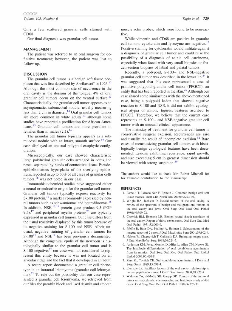
(Oral Surg Oral Med Oral Pathol Oral Radiol Endod 2007;103:727-30)A 45-year-old white woman presented with a smooth,sessile, pink, exophytic, polypoid mass on the ventralsurface of her tongue. The lesion was asymptomaticand the patient had not been aware of it. The lesionmeasured approximately 4.0 � 1.0 cm in its largestdimensions (Fig. 1). The patient’s medical history wassignificant for type II diabetes, asthma, arthritis, kidneydysfunction, hyperlipidemia, and previous lumbarspine injury. She reported a 20-year smoking historybut denied alcohol use.
DIFFERENTIAL DIAGNOSISThis slow-growing, relatively well circumscribed
soft tissue lesion was presumed to represent a benignor reactive lesion. Our differential diagnosis in-cluded fibrous hyperplasia, peripheral nerve tumor,condyloma acuminatum, and benign salivary glandneoplasia.
Fibrous hyperplasia, usually secondary to chronicirritation, is a common lesion of the tongue1; how-ever, this lesion usually presents as a solitary noduleon the lateral border of the tongue, only infrequentlyoccurring on the ventral aspect.
aAssistant Professor, Department of Oral Diagnostic Sciences, Schoolof Dental Medicine, State University of New York at Buffalo.bDistinguished Teaching Professor, Department of Oral DiagnosticSciences, School of Dental Medicine, State University of New Yorkat Buffalo.cDirector, Advanced Oral and Maxillofacial Pathology Program, andProfessor, Department of Oral Diagnostic Sciences, School of DentalMedicine, State University of New York at Buffalo.Received for publication Jul 25, 2006; returned for revision Nov 28,2006; accepted for publication Nov 29, 2006.1079-2104/$ - see front matter© 2007 Mosby, Inc. All rights reserved.
doi:10.1016/j.tripleo.2006.11.051The differential diagnosis of a presumed benign massof the tongue should also include tumors of neuralorigin. Neurilemmoma (schwannoma) is a benign, en-capsulated neoplasm that arises from Schwann cells ofthe nerve sheath. Clinically, the lesion usually appearsas a solitary, painless, nonulcerated mass. Neurilemmo-mas range in size from a few millimeters to severalcentimeters and may appear at any age. They are morecommon in adults and show no gender predilection.2,3
The tongue is the most frequent site of involvement andsome cases have been reported to arise on the ventralsurface of the tongue.2-4
Neurofibromas have also been described on thetongue. They are benign, slowly growing, nonencapsu-lated neoplasms consisting of proliferating Schwanncells and perineural fibroblasts originating from a pe-ripheral nerve. Neurofibromas can occur as solitary ormultiple lesions; the latter typically occurring in asso-ciation with neurofibromatosis type 1.2 Oral neurofi-bromas present as submucosal, nontender, discrete,small to large masses with no age or gender predilec-tion.2,3,5
Condyloma acuminatum is caused by human pap-illoma virus, most commonly HPV-6 and HPV-11subtypes.6 In addition to the genitalia and perianalregion, condyloma acuminatum can also be seen inthe mouth, where it commonly affects the labialmucosa, lingual frenum, and the dorsum of thetongue. Although oral condyloma is commonly con-sidered to represent a sexually transmitted infection,nonsexual transmission has been reported. In themouth, condyloma acuminatum is typically diag-nosed between the ages of 20 and 40 and shows amale predominance.7 Clinically, it presents most
commonly as a sessile, pink mass.8 Oral condylo-727

OOOOE728 Tapia et al. June 2007
mata may be solitary or appear as clusters that onoccasion may be confluent.7,8
Minor salivary gland tumors have been describedin the tongue,9 although this represents only 4% ofall minor salivary gland tumors. Most salivary glandneoplasms of the tongue are malignant. Among be-nign salivary gland tumors of the tongue, the mostcommon is the pleomorphic adenoma.10 This lesionoccurs between the third and fourth decades of life,typically presenting as a painless swelling withoutulceration.11
DIAGNOSISAn incisional biopsy was performed under local
Fig. 1. Polypoid lesion on the lingual surface of tongue,adjacent to the lingual frenum.
Fig. 2. Low power view of the specimen showing an intacthyperplastic stratified squamous epithelium with acanthosis.(Hematoxylin-eosin stain; original magnification �20.)
anesthesia and the tissue was fixed in 10% buffered
formalin. The specimen was soft, white in color, andmeasured 0.7 � 0.6 � 0.3 cm. Hematoxylin andeosin–stained tissue sections revealed a specimencovered by an intact hyperplastic parakeratinizedstratified squamous epithelium with acanthosis (Fig.2). The main feature of the specimen was the pres-ence of sheets of eosinophilic granular cells in thelamina propria (Fig. 3). No nerve or striated musclefibers were seen in the biopsy specimen. Immuno-peroxidase staining with S-100 (Fig. 4), neuron-spe-cific enolase (NSE), anti-lysozyme, CD68, cytoker-atin, and vimentin was performed. The tumor cellswere nonreactive to S-100, NSE, anti-lysozyme, and
Fig. 3. Sheets of eosinophilic granular cells in the laminapropria. (Hematoxylin-eosin stain; original magnification�400.)
Fig. 4. Negative staining of granular cells. Note positivestaining of dendritic cells in the epithelium. (S-100 stain;original magnification �400.)
cytokeratin, but were strongly positive to vimentin.
OOOOEVolume 103, Number 6 Tapia et al. 729
Only a few scattered granular cells stained withCD68.
Our final diagnosis was granular cell tumor.
MANAGEMENTThe patient was referred to an oral surgeon for de-
finitive treatment; however, the patient was lost tofollow-up.
DISCUSSIONThe granular cell tumor is a benign soft tissue neo-
plasm that was first described by Abrikossoff in 1926.12
Although the most common site of occurrence in theoral cavity is the dorsum of the tongue, 4% of oralgranular cell tumors occur on the ventral surface.13
Characteristically, the granular cell tumor appears as anasymptomatic, submucosal nodule, usually measuringless than 2 cm in diameter.14 Oral granular cell tumorsare more common in white adults,13 although somestudies have reported a predilection for African Amer-icans.15 Granular cell tumors are more prevalent infemales than in males (2:1).16
The granular cell tumor typically appears as a sub-mucosal nodule with an intact, smooth surface.14 Ourcase displayed an unusual polypoid exophytic config-uration.
Microscopically, our case showed characteristiclarge polyhedral granular cells arranged in cords andnests, separated by bands of connective tissue. Pseudo-epitheliomatous hyperplasia of the overlying epithe-lium, reported in up to 50% of all cases of granular cellstumors,16 was not noted in our case.
Immunohistochemical studies have suggested eithera neural or endocrine origin for the granular cell tumor.Granular cell tumors typically express reactivity forS-100 protein,17 a marker commonly expressed by neu-ral tumors such as schwannomas and neurofibromas.18
In addition, NSE,17,19 protein gene product 9.5 (PGP9.5),17 and peripheral myelin proteins20 are typicallyexpressed in granular cell tumors. Our case differs fromthe usual reactivity displayed by this tumor because ofits negative staining for S-100 and NSE. Albeit un-usual, negative staining of granular cell tumors forS-10021 and NSE17 has been previously documented.Although the congenital epulis of the newborn is his-tologically similar to the granular cell tumor and isS-100 negative,22 our case was not considered to rep-resent this entity because it was not located on analveolar ridge and the fact that it developed in an adult.
A recent report documented a granular cell pheno-type in an intraoral leiomyoma (granular cell leiomyo-ma).23 To rule out the possibility that our case repre-sented a granular cell leiomyoma, we retrieved from
our files the paraffin block and used desmin and smoothmuscle actin probes, which were found to be nonreac-tive.
While vimentin and CD68 are positive in granularcell tumors, cytokeratin and lysozyme are negative.12
Positive staining for cytokeratin would militate againsta diagnosis of granular cell tumor and could raise thepossibility of a diagnosis of acinic cell carcinoma,especially when faced with very small biopsies or fro-zen section biopsies of labial and palatal tumors.
Recently, a polypoid, S-100– and NSE-negativegranular cell tumor was described in the lower lip.24 Itwas suggested that this case represented a case ofprimitive polypoid granular cell tumor (PPGCT), anentity that has been reported in the skin.25 Although ourcase shared some similarities with the above-mentionedcase, being a polypoid lesion that showed negativereaction to S-100 and NSE, it did not exhibit cytolog-ical atypia or mitotic figures, features ascribed toPPGCT. Therefore, we believe that the current caserepresents an S-100– and NSE-negative granular celltumor with an unusual clinical appearance.
The mainstay of treatment for granular cell tumor isconservative surgical excision. Recurrences are rareand usually the result of incomplete excision.12 Rarecases of metastasizing granular cell tumors with histo-logically benign cytological features have been docu-mented. Lesions exhibiting recurrence, rapid growth,and size exceeding 5 cm in greatest dimension shouldbe viewed with strong suspicion.26
The authors would like to thank Mr. Robin Mitchell forhis valuable contribution to the manuscript.
REFERENCES1. Esmeili T, Lozada-Nur F, Epstein J. Common benign oral soft
tissue masses. Dent Clin North Am 2005;49:223-40.2. Wright BA, Jackson D. Neural tumors of the oral cavity. A
review of the spectrum of benign and malignant oral tumors ofthe oral cavity and jaws. Oral Surg Oral Med Oral Pathol1980;49:509-22.
3. Cherrick HM, Eversole LR. Benign neural sheath neoplasm ofthe oral cavity. Report of thirty-seven cases. Oral Surg Oral MedOral Pathol 1971;32:900-9.
4. Pfeifle R, Baur DA, Paulino A, Helman J. Schwannoma of thetongue: report of 2 cases. J Oral Maxillofac Surg 2001;59:802-4.
5. Nelson W, Chuprevich T, Galbraith DA. Enlarging tongue mass.J Oral Maxillofac Surg 1998;56:224-7.
6. Anderson KM, Perez-Montiel D, Miles L, Allen CM, Nuovo GJ.The histologic differentiation of oral condyloma acuminatumfrom its mimics. Oral Surg Oral Med Oral Pathol Oral RadiolEndod 2003;96:420-8.
7. Zunt SL, Tomich CE. Oral condyloma acuminatum. J DermatolSurg Oncol 1989;15:591-4.
8. Eversole LR. Papillary lesions of the oral cavity: relationship tohuman papillomaviruses. J Calif Dent Assoc 2000;28:922-7.
9. Waldron CA, el-Mofty SK, Gnepp DR. Tumors of the intraoralminor salivary glands: a demographic and histologic study of 426
cases. Oral Surg Oral Med Oral Pathol 1988;66:323-33.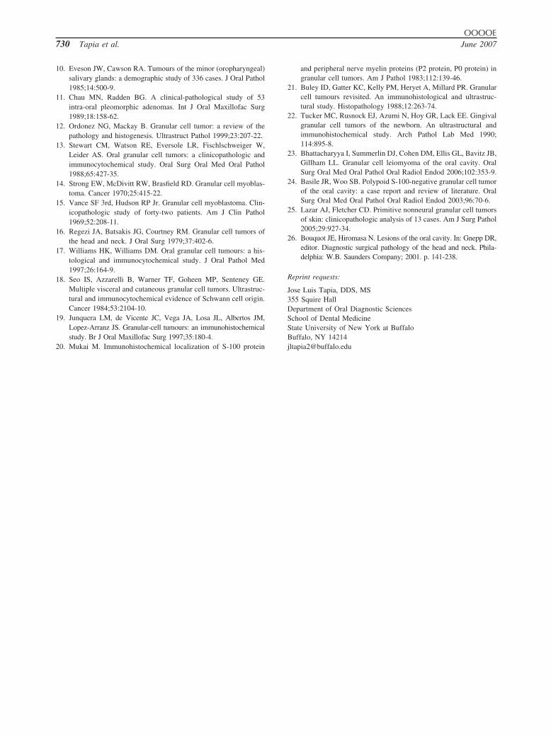
OOOOE730 Tapia et al. June 2007
10. Eveson JW, Cawson RA. Tumours of the minor (oropharyngeal)salivary glands: a demographic study of 336 cases. J Oral Pathol1985;14:500-9.
11. Chau MN, Radden BG. A clinical-pathological study of 53intra-oral pleomorphic adenomas. Int J Oral Maxillofac Surg1989;18:158-62.
12. Ordonez NG, Mackay B. Granular cell tumor: a review of thepathology and histogenesis. Ultrastruct Pathol 1999;23:207-22.
13. Stewart CM, Watson RE, Eversole LR, Fischlschweiger W,Leider AS. Oral granular cell tumors: a clinicopathologic andimmunocytochemical study. Oral Surg Oral Med Oral Pathol1988;65:427-35.
14. Strong EW, McDivitt RW, Brasfield RD. Granular cell myoblas-toma. Cancer 1970;25:415-22.
15. Vance SF 3rd, Hudson RP Jr. Granular cell myoblastoma. Clin-icopathologic study of forty-two patients. Am J Clin Pathol1969;52:208-11.
16. Regezi JA, Batsakis JG, Courtney RM. Granular cell tumors ofthe head and neck. J Oral Surg 1979;37:402-6.
17. Williams HK, Williams DM. Oral granular cell tumours: a his-tological and immunocytochemical study. J Oral Pathol Med1997;26:164-9.
18. Seo IS, Azzarelli B, Warner TF, Goheen MP, Senteney GE.Multiple visceral and cutaneous granular cell tumors. Ultrastruc-tural and immunocytochemical evidence of Schwann cell origin.Cancer 1984;53:2104-10.
19. Junquera LM, de Vicente JC, Vega JA, Losa JL, Albertos JM,Lopez-Arranz JS. Granular-cell tumours: an immunohistochemicalstudy. Br J Oral Maxillofac Surg 1997;35:180-4.
20. Mukai M. Immunohistochemical localization of S-100 protein
and peripheral nerve myelin proteins (P2 protein, P0 protein) ingranular cell tumors. Am J Pathol 1983;112:139-46.
21. Buley ID, Gatter KC, Kelly PM, Heryet A, Millard PR. Granularcell tumours revisited. An immunohistological and ultrastruc-tural study. Histopathology 1988;12:263-74.
22. Tucker MC, Rusnock EJ, Azumi N, Hoy GR, Lack EE. Gingivalgranular cell tumors of the newborn. An ultrastructural andimmunohistochemical study. Arch Pathol Lab Med 1990;114:895-8.
23. Bhattacharyya I, Summerlin DJ, Cohen DM, Ellis GL, Bavitz JB,Gillham LL. Granular cell leiomyoma of the oral cavity. OralSurg Oral Med Oral Pathol Oral Radiol Endod 2006;102:353-9.
24. Basile JR, Woo SB. Polypoid S-100-negative granular cell tumorof the oral cavity: a case report and review of literature. OralSurg Oral Med Oral Pathol Oral Radiol Endod 2003;96:70-6.
25. Lazar AJ, Fletcher CD. Primitive nonneural granular cell tumorsof skin: clinicopathologic analysis of 13 cases. Am J Surg Pathol2005;29:927-34.
26. Bouquot JE, Hiromasa N. Lesions of the oral cavity. In: Gnepp DR,editor. Diagnostic surgical pathology of the head and neck. Phila-delphia: W.B. Saunders Company; 2001. p. 141-238.
Reprint requests:
Jose Luis Tapia, DDS, MS355 Squire HallDepartment of Oral Diagnostic SciencesSchool of Dental MedicineState University of New York at BuffaloBuffalo, NY 14214
[email protected]