Embed Size (px)
Citation preview

Adrian egli, et al.: BKV and JCV After Kidney Transplantation
85
Polyomavirus BK after Kidney Transplantation – Role of Molecular and Immunologic MarkersAdrian Egli1, Alexis Dumoulin1, Sabrina Köhli and Hans H. Hirsch1,2
1Transplantation Virology, Institute for Medical Microbiology, Department of Biomedicine, University of Basel, Basel, Switzerland; 2Division of Infectious Diseases and Hospital Epidemiology, University Hospital of Basel, Basel, Switzerland
Abstract
Polyomavirus-associated nephropathy in kidney transplantation is viewed as a complication of prolonged, intense immunosuppression, which disrupts the balance between antiviral immune control and polyomavirus replication. The prevalence rate ranges from 1-10% in kidney transplant programs around the world, with graft failure and return to dialysis in > 50%. Most cases are caused by the human polyomavirus BK, which asymptomatically infects more than 80% of the general population. Reactivation of BK virus replication and high urine viral loads precede increasing plasma BK viral loads and histologically defined as well as clinically manifest nephropathy. Quantitative real-time polymerase chain reaction protocols have proven valuable as surrogate markers to follow the course of polyomavirus-associated nephropathy and to guide preemptive reduction of immunosuppression. As these assays enter clinical routine diagnostic laboratories, quality control becomes important. Testing of BK virus-specific antibodies and T-cells is currently being explored for a better characterization of the virus/host balance. The BK virus-like particles IgG in enzyme-linked immunosorbent essays are recognized as sensitive indicators of recent BK virus exposure. However, no humoral immune responses have been identified to correlate with protection from BKV viremia or disease. BK virus-specific T-cells are generally of low frequencies in the peripheral blood of both healthy donors and kidney transplant patients alike, but significantly increase at the time when plasma BK virus loads decrease, hence representing relatively late indicators of regaining control. We discuss the currently available data on molecular and immune markers regarding promises and caveats. (Trends in Transplant. 2009;3:85-102)
Corresponding author: Hans H. Hirsch, [email protected]
Key words
Transplantation. Viral infections. Polyomavirus. JC virus. BK virus. Polyomavirus associated nephrology. T-cells. Epitope mapping. Vaccine. Adaptive T-cell transfer. Quality control.
trends in transplant. 2009;3:85-102
Correspondence to:Hans H. Hirsch
transplantation Virology, Institute for medical
microbiology
Department of Biomedicine, University of Basel
Petersplatz 10, CH-4003 Basel, Switzerland
e-mail: [email protected]

trends in transplantation 2009;3
86
Introduction
The concept of co-evolutionary adapta-tion entails that both hosts and viruses are selected for genetic variants that permit coex-istence at mutually acceptable costs1. In this equilibrium, severe viral diseases must be con-sidered individual or epidemiologic accidents either of the virus changing towards more pathogenic characteristics and/or entering a new population, or, of the host not being able to respond adequately2. One kind of accident is immunosuppression, administered for trans-plantation, which disrupts the balance between virus replication and immu ne response abrupt-ly and introduces foreign HLA-type tissues, which together increase virus-associated mor-bidity and mortality3. Adequately administered antiviral drugs, if available, can counteract this failing balance by reducing the impact of viral replication. Longer-term stability, however, re-quires eventual restoration of virus-specific im-mune control. The role of the adequate mounting of an immune response is obvious when pri-mary viral infections are compared in immuno-competent and immunosuppressed individu-als. The role of established specific immune control is unveiled by the reactivation of repli-cation of persistent viruses following exposure to immunosuppressive regimens. The most im-portant examples in the setting of transplanta-tion are the human herpes viruses, especially cytomegalovirus, the hepatitis B and C virus, and the human polyomaviruses BK and JC4.
The human polyomaviruses are small, non-enveloped, icosahedral double-stranded DNA viruses of 40-45 nm and fairly resistant to environmental inactivation. To date, six polyo-mavirus have been detected in human speci-mens: BK virus (BKV)5 and JC virus (JCV)6, the related KI7 and WU virus8 in respiratory secre-tions, the MC virus in Merkel cell carcinoma9, and the simian virus 40 (SV40) in contaminated polio vaccines10. So far, compelling evidence for circulation in human populations is only
available for BKV and JCV. Both viruses are closely related (Table 1) and their epidemiol-ogy seems to suggest prototypic co-evolution: a high infection rate in the general human pop-ulation without pronounced clinical symptoms, persistence in the renourinary tract11,12, with asymptomatic reactivation and urinary shed-ding10,13,14. Polyomavirus diseases are rare in immunocompetent persons14-16. In addition to altered immune responses, specific tissue in-juries are fre quently noted. Thus, BKV can cause polyomavirus-associated nephropathy (PVAN) after kidney transplantation17 and hem-orrhagic cystitis after hematopoietic stem cell transplantation18, whereas JCV causes pro-gressive multifocal leukoencephalopathy in patients with severe immunodeficiency through HIV/AIDS, transplantation, or longstanding therapies for autoimmune diseases19-22.
In immunocompetent individuals, reacti-vation of urinary BKV and JCV replication is found in 7 and 19% of cases, respectively, with low median urine viral loads of 3.5 and 5 log genome equivalents (geq)/ml, respective-ly14,15,23-26. When extrapolated to a metropolitan area like Zurich, with one million inhabitants, the daily polyomavirus shedding would amount to 3.5 × 10¹¹ geq/ml of BKV and 3 × 10¹³ geq/ml of JCV shed into the environment. Indeed, poly-omaviruses were detected in urban sewage and investigated as indicators of water quality27-29. Earlier studies indicated that, genomic subtypes were correlated with the migration history of hu-man populations30,31. Viremia however has not been confirmed in healthy individuals14-16.
Molecular aspects of BK virus replication in kidney transplantation
In immunosuppressed individuals, BKV shedding first can be found in about 30% of urine samples, with about 3-4 Log higher BKV loads than found in healthy individuals32-37. Following kidney transplantation, high-level BKV replication is detectable in urine of 20-60%

Adrian egli, et al.: BKV and JCV After Kidney Transplantation
87
of patients, with urine viral loads of 107 to 1010 geq/ml (Fig. 1)32,38. About 30% of kidney transplant patients with BKV-positive urine samples eventually develop BKV-positive plasma samples. Progression of histologically documented PVAN is observed in about 60% of kidney transplant patients with plasma vire-mia. Hence, BKV detection and quantification serve as important markers to assess the risk of PVAN32,39. Patients with definitive PVAN have higher viral loads than viremic patients without PVAN (28,000 vs. 2,000 geq/ml)32. Ac-cording to expert recommendations, persis-tently high BKV in plasma > 4 log10 geq/ml for more than four weeks in kidney transplant pa-tients defines “presumptive PVAN”33,35,40.
Following surgical removal of PVAN-con-taining allografts, plasma BKV loads show a rapid drop, suggesting that the majority of plas-ma BKV loads is derived from replication in the graft. The calculated plasma viral half-life of 1-2 hours implies that during steady-state more than 99% of the plasma BKV loads are turned over per day, and allow estimating the tubular epithelial cell loss as in the order of 106 to 107
cells per day41. Moreover, persistent BKV rep-lication in kidney transplant patients leads to emergence of BKV variants with rearrange-ments (rr) of the non-coding control region (NCCR) containing viral promoter and enhanc-er sequences. These rr-NCCR BKV variants are first detected in plasma and later in urine, which supports the notion that intrarenal tubular epi-thelial cells and urothelial cells of ureter and bladder are independent but partially linked replication compartments. The occurrence of rr-NCCR BKV is linked to 20-fold higher plasma viral loads (median 20,000 c/ml vs. median 440,000 c/ml), and more tissue pathology both of which can be recapitulated in tissue cul-ture42. Of note, the sequences encoding the viral protein-1 (VP1) capsid and the large T-antigen were not altered, suggesting that sig-nificant immunologic pressure was not be pres-ent in patients with emerging rr-NCCR BKV.
JCV can be detected in 20-50% of urine samples and may present with shedding of “decoy cells” bearing polyomavirus particles in nuclear inclusions32,34,41. The JCV is rarely de-tected in plasma of patients with JCV-associated
Table 1. The genome structure of the human polyomaviruses BK virus and JC virus
NCCR
large T-agVP1
VP3
VP2
agno
small T-ag
Number of bp Number of Aa Homology
BKV JCV BKV JCV Gene Aa
Genome 5153* 5130† 74%
Early coding region LTag 2088 2067 695 688 78% 83%
stag 519 519 172 172 78% 78%
Late coding region VP-1 1089 1065 362 354 75% 78%
VP-2 1056 1035 351 344 81% 79%
VP-3 699 678 232 225 80% 75%
agno 201 216 66 71 72% 59%
The early genes are in red (large T antigen LTag, small T antigen stag), late genes in green (agnoprotein, VP1, VP2 and VP3). The genome length and coding sequences are given in nucleotides and amino acids (percent identity) Amount of similarity between both viruses is indicated at the genome and protein level in %. bp: base pairs; Aa: amino acids; BKV: polyomavirus BK; JCV: polyomavirus JC; NCCR: non-coding control region.*Dunlop sequence.†Mad-1 sequence.

trends in transplantation 2009;3
88
ViruriaViremiaPVAN
0 20 40 60
Weeks after transplantation
80 100 120 140
Pro
babi
lity
of B
KV
Rep
licat
ion,
Vire
mia
,an
d N
ephr
opat
hy
0.5
0.4
0.3
0.2
0.1
0.0
median 16 weeks (range 2-69)median 23 weeks (range 4-73)median 28 weeks (range 8-86)
Screening for BKV replication
Viruria
Viremia 78%
77%
PVAN 80%
• 3-monthly for 2 yrs• If graft dysfunction• If any biopsy
Recip.BKVIgG
Figure 1. Probability of BK virus viruria, viremia and nephropathy (modified after Ref. 32). BKV: BK virus; PVAN: polyomavirus-associated nephropathy; IgG: immunoglobulin G seroprevalence.
nephropathy (14%) and then of low viral loads (mean 2,000 c/ml)34. Overall, JCV-associated nephropathy seems to run a more benign course than BKV-associated nephropathy, al-though some cases with graft failure have been reported. Rearrangement of the JCV NCCR in PVAN has not been detected, which is, how-ever, a hallmark of JCV variants in progressive multifocal leukoencephalopathy.
Although the paradigm of viruria pre-ceding viremia and PVAN by about 4-8 weeks has been known for almost a decade in kid-ney transplants (Fig. 2), it is still not defined which patients progress to plasma replication and to disease. In analogy to cytomegalovirus (CMV), molecular and immunologic markers have been investigated for the potential to better assess the risk of BKV infection to prog-ress to replication and PVAN and thereby guiding intervention.
BK virus genome detection
One of the first BKV-specific polymerase chain reaction (PCR) assays was published by Arthur, et al. in 198943 to study the role of BKV in hemorrhagic cystitis after bone marrow trans-plantation. Since then, various qualitative and quantitative PCR assays have been developed (Table 2), with various targets in the 5.1 kb double-stranded DNA genome (Table 1). Most of these assays were designed to specifically detect BKV, but some cover both BKV and JCV, and include the possibility of distinguish-ing between the two. Published target sequenc-es are located in the large T gene (LT, 15 as-says), in the VP1 gene (seven assays), in the VP2 gene (three assays) or in the NCCR (three assays). Our routine in-house TaqMan® BKV PCR amplifies a conserved region of the LT gene, which was chosen to match BKV se-quences without amplifying the homologous
Adrian egli, et al.: BKV and JCV After Kidney Transplantation
89
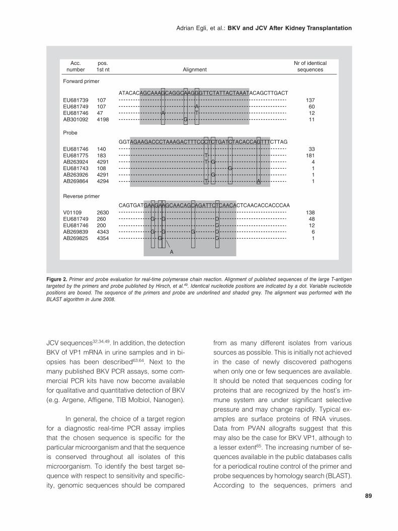
Figure 2. Primer and probe evaluation for real-time polymerase chain reaction. Alignment of published sequences of the large T-antigen targeted by the primers and probe published by Hirsch, et al.49. Identical nucleotide positions are indicated by a dot. Variable nucleotide positions are boxed. The sequence of the primers and probe are underlined and shaded grey. The alignment was performed with the BLAST algorithm in June 2008.
Acc.number
Forward primer
EU681739EU681749EU681746AB301092
107107474198
ATACACAGCAAAGCAGGCAAGGGTTCTATTACTAAATACAGCTTGACT137601211
pos.1st nt Alignment
Nr of identicalsequences
Probe
EU681746EU681775AB263924EU681743AB263926AB269864
140183429110842914294
GGTAGAAGACCCTAAAGACTTTCCCTCTGATCTACACCAGTTTCTTAG33
1814111
TT
T
G
A
Reverse primer
V01109EU681749EU681746AB269839AB269825
263026020043434354
CAGTGATGAAGAAGCAACAGCAGATTCTCAACACTCAACACCACCCAA138481261
G
GG
A
G
CCCC
G
G
AG
AT
G
G
JCV sequences32,34,49. In addition, the detection BKV of VP1 mRNA in urine samples and in bi-opsies has been descri bed63,64. Next to the many published BKV PCR assays, some com-mercial PCR kits have now become available for qualitative and quantitative detection of BKV (e.g. Argene, Affigene, TIB Molbiol, Nanogen).
In general, the choice of a target region for a diagnostic real-time PCR assay implies that the chosen sequence is specific for the particular microorganism and that the sequence is conserved throughout all isolates of this micro organism. To identify the best target se-quence with respect to sensitivity and specific-ity, genomic sequences should be compared
from as many different isolates from various sources as possible. This is initially not achieved in the case of newly discovered pathogens when only one or few sequences are available. It should be noted that sequences coding for proteins that are recognized by the host’s im-mune system are under significant selective pressure and may change rapidly. Typical ex-amples are surface proteins of RNA viruses. Data from PVAN allografts suggest that this may also be the case for BKV VP1, although to a lesser extent65. The increasing number of se-quences available in the public databases calls for a periodical routine control of the primer and probe sequences by homology search (BLAST). According to the sequences, primers and

trends in transplantation 2009;3
90
Table 2. Polymerase chain reaction assays for the detection of BK virus
First author Year Method Target region Specificity
Arthur43 1989 PCR with RFLP or hybridization
LT BK and JC (discriminatory)
Nickeleit44 1999 Semi-nested PCRDilution series
LT BK, JC(specific inner PCR)
Biel45 2000 Semi-nested PCR LT BK, JC(specific inner PCR)
Taqman LT BK and JC
Limaye46 2001 Taqman LT BK
Leung47 2001 Taqman VP1 BK
Whiley48 2001 LightCycler VP2 BK and JC (specific Tm)
PCR-ELAHA VP1 BK and JC (specific probes)
Hirsch49 2001 Taqman LT BK
Merlino50 2003 Semi-quantitative PCR LT BK
Holman51 2003 Semi-quantitative PCR LT BK
Beck52 2004 LightCycler VP2 BK
Whiley53 2004 PCR-ELAHA VP1 BK and JC (specific probes)
Randhawa54 2004 LightCycler, Sybr VP1 BK
Herman55 2004 Taqman LT BK and JC
McNees56 2005 Taqman MGB LT BK
Sehbani57 2006 LightCycler SybrGreen LT BK
Moret58 2006 PCR, hybridization, n. s. BK and JC (specific probes)
Kaigala59 2006 PCR, Microchip VP1 BK
Pal60 2006 Taqman (3 Assays) VP1, NCCR BK
Elfaitouri61 2006 Taqman VP2 BK, JC and SV40
Si-Mohamed62 2006 Taqman LT BK
PCR: polymerase chain reaction: RFLP: restriction fragment length polymorphism; LT: large T-antigen; VP: viral protein; NCCR: non-coding control region.
probes should be adapted when critical bases are altered. This can be achieved by including degenerated positions matching two or more sequence variants. Mismatches at the 3’-end of the primers have more impact than at the 5’-end, and may reduce the PCR efficiency with under-quantification by several logs. In the last decade, more sequences of naturally occurring BKV isolates have become available. Figure 2 shows the sequences of primers and probes used in our assay targeting the BKV LT. Al-
though the majority of published sequences perfectly match with both primers, about one-third of sequences show one, two and three nucleotide mismatches including some at the critical 3’-end (Fig. 2). For the probe, approxi-mately 80% of sequences bear a single nucle-otide mismatch, which are, however, located in the center and are not likely to affect detection. Nevertheless, this demonstrates the require-ment for periodical reevaluation and adaptation of diagnostic BKV assays.

Adrian egli, et al.: BKV and JCV After Kidney Transplantation
91
Diagnostic performance of BK virus real-time polymerase chain reaction assays
The amplification efficiency and linear ran ge of a PCR assay is typically established using reference plasmids containing the target sequen ce. Most assays today report a linear dynamic range from 102 to 108 per reaction. To determine the limit of detection of a PCR assay in a statistically meaningful way, we run independent replicas of five or 10 reactions of twofold dilutions of the BKV reference plasmid, starting from 100 co pies per reaction down to at least two dilutions below one copy per as-say. The results can be scored categorically as “yes-detectable” or “no-undetectable”, or by scoring the threshold cycles (Fig. 3). Since routine samples are analyzed in triplicates, we defined the limit of detection of our assays as being the concentration of target detected in at least 50% of the replicates.
The sample materials analyzed for BKV include urine, plasma, and, rarely, cerebro-spinal fluid (CSF) for suspected cases of BKV encephalitis. Tissues such as renal biopsies can also be tested for BKV DNA, but have not entered the diagnostic routine. An optimal extraction of these samples is a prerequisite for a correct quantification of the viral loads. We routinely use the MagNA Pure LC System with the Total Nucleic Acid Kit (both from Roche Diagnostics) for urine and plasma samples. A sample volume of 200 μl is ex-tracted and eluted in 100 μl. Manual extrac-tion methods are used for CSF samples (Qia-gen QIAamp Blood Kit). Fluid extractions using the Roche Kit and the Qiagen Kit yield-ed comparable results, but we observed that samples with low viral loads are occasionally under quantified with spin columns compared to beads. Biopsies are also extracted with the Qiagen Kit, with an additional lysis step by proteinase K. For the quantification of BKV
> 45
10,000
100
5,000
100
2,500
100
1,250
100
625
60
313
50
156
30
78
40
39
0
Geq/ml
% pos.
45
40
35
30
Ct-
valu
es
Figure 3. Detection limit of polymerase chain reaction assay and standard curve validation. Serial 2-fold dilution of 102 geq per PCR reac-tion obtained from the standard curve. Two independent runs of five replicates were performed. Replicates that gave no detectable results are plotted at Ct > 45. In this case, the detection limit is 50% at the 3.13 geq (in 5 µL in 25 µL total PCR assay volume). Because the DNA is 2-fold concentrated relative to the extracted sample volume, the detection threshold corresponds to 313 geq/ml. This statistical approach also allows to evaluate whether or not the standard curve is too high or too low, since the dilution series goes as low as 0.78 and 0.39 geq per assay, which should yield no more than 80 and 40% positive results per 10 assays, respectively.

trends in transplantation 2009;3
92
DNA from biopsies, quantitative PCR for a chromosomal host call target is used such as beta-actin, glyceraldehyde-3-phosphate de-hydrogenase (GAPDH), or the aspartoacy-lase gene (ACY)66. These genes are present in two copies per cell. The quantification of host cell genes allows to normalize the BKV load per 150,000 cells (i.e. equivalent to 1 µg DNA) to correct for the cellular content of the sample.
Quality controls
Internal quality controls are part of ev-ery PCR run and allow monitoring for possible contaminations Moreover, quantitation con-trols i.e. specimens with known copy number of e.g. 5000 or 50000 geq/ml are run to test for inhibition, or technical problems (positive controls). Besides these, external quality con-trols should be tested on a regular basis. Ex-ternal quality controls consist of samples whose outcome is not known to the person who analyses it. These samples should be handled in the same way as patient samples, from the extraction to the interpretation and validation of the results. For many infectious agents, such controls are commercially avail-able and are distributed in Europe for quality assurance (e.g. NEQUAS, INSTAND, QCMD). The results are collected and analyzed, and the intended results and the performance of the laboratory relative to other participants is communicated. External controls allow the laboratories to evaluate the quality of their workflow and of their assays and to compare them to those of other laboratories. A pilot study evaluating BKV and JCV external qual-ity controls was successfully conducted re-cently and such controls are now available (QCMD, Glasgow, Scotland67). Together, these methodological and quality issues are of utmost importance when BKV load testing moves from the research laboratory into the clinical diagnostic laboratory for decision making and clinical study evaluation.
Clinical value of BK virus load screening and monitoring
The sequence of BKV viruria, viremia and histologically defined PVAN (Fig. 1)32 pro-vides a rationale for screening, and scrutinizing histologic diagnostics and even preemptive intervention33,35,40. In cases of established di-agnosis, monitoring the course of plasma and urine BKV loads assists in deciding on the duration and escalation of intervention. However, as more studies are being conducted, there is considerable uncertainty about the frequency of screening and about whether or not the sample material should be urine, plasma, or both. The significance of urine BKV loads (or “decoy cells” in experienced institutions) re-sides in the high negative-predictive value which allows to exclude PVAN with > 95%. If urine BKV screening is negative, BKV viremia and PVAN are unlikely to occur within the next 4-8 weeks (Fig. 1). Conversely, the significance of plasma BKV loads resides in the high posi-tive-predictive value. However, several, mostly prospective studies have reported the absence of “definitive PVAN” in biopsies, even though the plasma BKV load was > 104 geq/ml. Poten-tial explanations for these failures are: i) the focality of PVAN, which is estimated to result in false negative biopsy results in 10-30% of cas-es, particularly in the early phase68; ii) less than four weeks of duration for increasing plasma BKV loads above 104 geq/ml according to the consensus recommendations40; and iii) over-quantification of plasma BKV loads because of shifted standard curves. Indeed, in a first multi-center study, which included Basel, differences in the quantification of patient samples were noted to be in the order of plus or minus 1 log10 geq/ml69. Since these differences of over- and under-quantification were center-specifically maintained for most of the plasma and urine samples, it had to be concluded that the refer-ence standards used to generate the BKV copy number were shifted. The results of this study69 emphasized the need for international standards that allow normalization of BKV loads

Adrian egli, et al.: BKV and JCV After Kidney Transplantation
93
by transformation into International Units. Until such normalization standards become avail-able, not only assessments of the linear dy-namic range should be made, but also valida-tion of the titer by twofold dilution series down to copy numbers of statistically less than one copy per reaction should be made (Fig. 3). In prospective studies with presumed duration of four weeks of increased risk were included, the positive predictive value was approximately 50-70%34,70,71. In table 3 we summarize recent studies reporting histologically confirmed (i.e. “definitive”) PVAN and the respective median plasma BKV loads, as well as the lowest plasma BKV load of a confirmed case (cutoff).
Humoral immune responses to BK virus
The BKV-specific antibody responses have been measured by four different assays: plaque-based assays, hemagglutination inhibi-tion assay (HIA), indirect immunofluorescence of BKV-infected cells, and enzyme-linked immuno-sorbent essay (ELISA). Neutralization assays are considered the most specific as they measure antibodies that bind to infectious BKV virions and inhibit the infection of host cells. The HIA mea-sures antibodies that inhibit the agglutination of
red blood cells mediated by the three-dimen-sional virion capsid by interfering with their bind-ing to sugar residues on type 0 erythrocytes. Indirect immunofluorescence detects intracellu-lar viral proteins including the early and late gene products. Enzyme-linked immunosorbent essays have been described using the recom-binant capsid protein VP1 in the linear form, or as three-dimensional, self-assembled, virus-like particles (VLP), the BKV-early protein large-T antigen (LT) and the BKV agnoprotein. The VP1-derived VLP are most frequently now used be-cause of their high specificity, sensitivity, and ease of handling in the ELISA format35,76-79.
Neutralizing antibodies are usually ex-amined by plaque assays80. However, this technique is too cumbersome for larger sample numbers. Neutralizing antibodies are closely correlated with antibody titers measured by HIA or by BKV VLP ELISA10,13,81,82. The VLP ELISA tests have a higher sensitivity, but rec-ognize a wider range of epitopes than the HIA82. Cross-reactivity between polyomavirus-es was examined in pre-absorption assays (pre-incubation of sera with other virus anti-gens) and revealed limited IgG cross-reactivity between SV40 and BKV, but not between BKV and JCV. When denaturing BKV VLP, the IgG activity significantly dropped, indicating the
Table 3. Polyomavirus-associated nephropathy histology and plasma BK viral load
Year Center Reference Cases (n) PVAN Median BK viral load (geq/ml) Cutoff
2002 Basel (32) 5 definitive 28,000 7,700
2004 Atlanta (70) 8 definitive 73,000 2,800
2004 Pittsburgh (54) 10 definitive 443,000 5,000
2005 Leuven (72) 21 definitive 70,800 n/a
2006 Paris (62) 5 definitive 398,000 n/a
2006 Basel (73) 16 definitive 57,000 10,000
2007 Houston (74) 8 definitive 1,427,000 63,000
2007 Rochester (71) 4 definitive n/a 16,000
2007 Baltimore / Basel (34) 75 definitive 2,900,000 10,000
2008 Milwaukee (75) 8 definitive 186,000 9,100
PVAN: polyomavirus-associated nephropathy.
trends in transplantation 2009;3
94
importance of the three-dimensional antigen structure for the majority of BKV-specific anti-bodies81. When analyzing BK-VLP and JC VLP-specific IgG activities in healthy blood donors, no statistical evidence for IgG cross-reactivity was apparent (linear regression IgG: R2 = 0.04), but some cross-reactivity for the respec-tive IgM activity (linear regression R2 = 0.32; A. Egli and H.H. Hirsch, unpublished results).
Humoral responses to BK virus in healthy individuals
Knowles, et al. used HIA to measure BKV and JCV titers10. In that study, BKV seropreva-lence of 81% was observed, with a slow de-crease of seroprevalence in older age groups, most probably due to a decrease in geometric mean titers with increasing age (8.7% reduction per 10 years). For JCV, the overall seropreva-lence was only 35%, but strongly increased with increasing age. Using BKV and JCV VLP, we found a significant decrease of BKV IgG seroprevalence and IgG activity with increasing age of healthy blood donors, while JCV sero-prevalence increased14. Given the fact that uri-nary BKV shedding was significantly less fre-quent than JCV shedding (7 vs. 19%), it was interesting to note that the activity of JCV IgG VLP antibody correlated with urine JCV load14. This suggests that higher VLP antibody levels are indicative of recent exposure to viral anti-gen and included asymptomatic virus shed-ding83. Such antibodies probably have a role in clearing and protecting from viremia, but obvi-ously don’t protect from localized polyomavirus replication in mucosal tissues. In a first study comparing antibody responses in healthy do-nors to various other recombinant BKV antigens (anti-LT, anti-VP1 and anti-agnoprotein), Leuen-berger, et al.84 found similar IgG activities for all three BKV proteins as in kidney transplant patients without recent BKV replication84. Over-all, IgG activities were higher against VP1 than against LT, and only rarely were antibodies against agnoprotein detectable84.
Humoral responses to BK and JC viruses after kidney transplantation
The role of BKV serology in kidney trans-plantation to assess potential risk of infection is controversial. The classical donor seropositive/recipient seronegative (D+R–) risk constellation is not as common. Bohl, et al. screened 198 transplant recipients, and for 66 recipients, do-nor serostatus was available. According to BKV serology, 48% were D+R+, 19.6% were D+R–, 18% were D–R+, and 13.6% were D–R–. Only donor BKV seropositivity was significantly as-sociated with the risk for BKV replication post-transplantation. The likelihood of recipients’ viru-ria in the first year posttransplantation correlated with increasing donor BKV-VLP antibody titers78. Thus, the BKV antibody activity may reflect re-cent antigen exposure and possibly higher BKV loads in the graft, which is then not met by a qualitatively and quantitatively corresponding immune response in the recipient14.
Since none of the patients studied by Bohl, et al.78 had “definitive PVAN”, the study by Hariharan, et al. is of interest. Patients with first diagnosis of PVAN had high plasma BKV loads and showed moderate to low BKV-VLP IgM and IgG levels. Cases with stabilizing PVAN with intermediate plasma BKV loads had a strong increase in IgM and intermediate IgG levels, whereas past PVAN cases had low or undetectable BKV loads and a significant increase in IgG levels. This suggest that with increasing IgG levels, plasma BKV load de-creased in part due to neutralizing antibod-ies79. However, work by Comoli, et al.83,85, Leuenberger, et al.84, and more recently by Chen, et al.77 showed that BKV-VLP IgG levels already increased during rising plasma BKV loads, indicating that neutralizing antibodies were not sufficient to prevent BKV viremia and PVAN. These observations were more recent-ly confirmed by Ginevri, et al. in a larger co-hort of 62 patients and suggest rather that BKV-VLP IgG titers reflect the patient’s current or recent history of antigen exposure35.

Adrian egli, et al.: BKV and JCV After Kidney Transplantation
95
The role BKV VLP-specific IgA was as-sessed by Randhawa, et al.86. For both do-nors and recipients, the rate of seropositivity was 80% for BKV-VLP IgG, 20% for IgA, and for 0% for IgM. Interestingly, some patients without evidence for BKV detection during follow-up showed an increase of BKV-VLP IgG seropositivity, which may therefore reflect BKV replication below detection levels in plasma or urine, or outside the sampling times. A sig-nificant increase in BKV IgA was observed (pretransplant 20% vs. posttransplant 75%), in line with the notion of mucosal exposure86. Comparison of IgG activities specific against different BKV proteins in kidney transplant pa-tients revealed that BKV LT-specific IgG ac-tivities were high in patients after clearing prolonged episodes of BKV viremia. Unlike antibody responses to linear VP1, VLP, or LT, the vast majority of kidney transplant patients, even those clearing histologically confirmed PVAN, did not mount agnoprotein-specific im-mune responses, despite abundant late-phase expression of this viral protein84.
Humoral immune responses to JCV rep-lication in kidney transplants are currently not available. Our own observations in a large in-ternational cohort of de novo kidney transplant patients indicates, however, that patients with high-level JCV replication in urine, with or with-out detectable plasma JCV loads, have a strong increase in the JCV IgG VLP activity (H.H. Hirsch, unpublished observation)38. In no cases, however, was JCV-associated neph-ropathy documented, suggesting that, akin to BKV, the JCV-VLP IgG are sensitive enough to pick up antigen exposure in the urothelial rep-lication without significant graft damage.
BK virus-specific cellular immunity in healthy donors and kidney transplanted patients
In kidney transplant patients, a diverse set of risk factors associated with BKV replication
and progression to disease has been reported, not all of which were confirmed in indepen-dent studies. These factors include older age, male gender87, seropositive donor78,88, sero-negative recipient88,89, low number of BKV-specific interferon-γ (IFNγ) production of peripheral blood mononuclear cells (PBMC)85, use of potent immunosuppressive regi-mens32,33,38,90,91, HLA mismatches32,38,92, and antirejection treatment32,38,92. Overall, the com-mon denominator of these risk factors points to an impaired BKV-specific cellular immunity akin to CMV posttransplantation4,93.
Immunologic assays for BK virus-specific cellular immune responses
In general, three immunologic assay types to quantify and qualify markers of BKV-specific immune response are commonly used: i) EliSpot assay to measure secretion of IFNγ from PBMC by detecting spot-forming units per million PBMC; ii) intracellular cytokine staining and flow cytometry to detect accumulated IFNγ in T-cells with addition of CD8 and CD4 phe-notyping; and iii) tetramer assays to detect T-cells recognizing single BKV epitopes de-pending on a specific HLA context (also flow-cytometry based) (Fig. 4). For the first two as-says, overlapping peptide libraries have been used that consist of 15 amino acid peptides overlapping by 11 amino acids that cover the entire amino acid sequence of the BKV or JCV LT, VP1, and agnoprotein. These peptide pools have been added to PBMC in order to pulse antigen-presenting cells present in the PBMC. Any peptides contained in the pool that can bind to major histocompatibility complex MHC-II, or to MHC-I after external or internal trimming, can then be recognized by the respective T-cell receptor and elicit a response such as IFNγ expression. Alternatively, antigen-presenting cells can be pulsed with BKV preparations94 or lysates from infected cell cultures95. In the lat-ter assays, slightly more spot-forming units were observed compared to stimulation by

trends in transplantation 2009;3
96
peptide pools, probably a reflection of the more abundant antigens. By contrast, tetramer assays using streptavidin-fluorophore labeled MHC-I with a single 9mer bound that can be recognized by the respective T-cell. In this case, the precise epitope for the HLA-type must be known. Provided with the co-stimula-tory signals, cytokine expression can also be assayed in this way. Otherwise, only the T-cell receptor frequency is enumerated.
Comparing the frequencies in direct ex vivo assays, it should be noted that BKV- (and JCV)-specific T-cell responses in PBMC re-sponding to BKV VP1 and LT peptides pools are 10- to 100-fold lower than the frequencies described for CMV pp65 or CMV lysate re-sponses94 (Fig. 5). Because of the low precur-sor frequency in addition to the presence of
immunosuppression, in vitro T-cell expansion for 7-9 days after a single BKV-specific stimulation has been used35,96, or longer T-cell expansion for 3-6 weeks using multiple rounds of stimulation with activated monocytes or dendritic cells77,94,97. Every expansion protocol introduces bias, by selecting responding lymphocyte populations but a common problem is that the use of 15mer peptide pools seems to favors CD4 over CD8 T-cell phenotypes. However, BKV-specific CD8 T-cells can be fostered by initial magnetic bead separation97, or by adding interleukins IL-12 and IL-7 to the expansion culture94.
Using a “direct” ex vivo IFNγ EliSpot as-say, we found low median frequencies of 24 and 25 spot-forming units of IFN-producing T-cells per 106 PBMC after stimulation with BKV LT and VP1 in healthy donors96. In a cross-sectional
Figure 4. Immunoassays for detecting BK virus-specific cellular immune responses. Peripheral blood mononuclear cells (PBMC) are either directly stimulated ex vivo or re-stimulated after in vitro pre-stimulation and expansion using viral antigens e.g. overlapping peptide pools of 15 amino acids covering the entire amino acid sequence of large T-antigen or VP1. EliSpot assay detects secreted interferon-γ (IFNγ) after captured by specific antibodies coated in well and second antibody for color reaction. Intracellular cytokine staining detects intracel-lular accumulation of IFNγ after secretory blockade with brefeldin-A and fixation/permeablization with fluorescent antibody. Tetramer stain-ing detects T-cells binding specifically to fluorescence-labeled single peptide epitopes major histocompatibility complex MHC-I tetramer complexes which can be combined with intracellular IFNγ detection.
PBmC Immunoassay*stimulation directlyex vivoor*restimulation afterin vitro expansion
IFNγ
IFNγ secretion IFNγ detection
eliSpotColor formation
eliSpot
t-cells
AntigenIFNγ IFNγ
IFNγ SecretionBlockade w. Brefeldin-A IFNγ detection
FACS
FACS
mHC-Itetramers IFNγ secretion
Blockade w. Brefeldin-AIFNγ detection

Adrian egli, et al.: BKV and JCV After Kidney Transplantation
97
SEB CMV-pp65 BKV-LT BKV-VP1 JCV-LT JCV-VP1
10,000
1000
100
10
1
INF
γ S
FU
per
10^
6 P
BM
Cs
Figure 5. Direct EliSpot assay comparing BK virus- and cytomegalovirus-specific cellular immune responses in healthy blood donors. Peripheral blood mononuclear cells (PBMC) from five healthy blood donors are directly stimulated ex vivo with the indicated antigens and interferon-γ release quantified by EliSpot assay (A. Egli, S. Köhli, H.H. Hirsch, unpublished): Staphylococcus superantigen (SEB) as positive control, overlapping peptide pools covering the cytomegalovirus pp65 (CMV-pp65); the BK virus large T-antigen (BKV-LT), capsid viral protein VP1 (BKV-VP1); JC virus large T-antigen (JCV-LT) and capsid protein VP1 (JCV-VP1). SFU: spot-forming units.
cohort of adult kidney transplant patients, we detected slightly higher median values despite immunosuppression96. This is in contrast to what has been observed for CMV-specific cellular im-mune responses, which were significantly lower in kidney transplant patients than in non-immu-nosuppressed healthy donors93,98. However, in kidney transplant patients with increasing or per-sistently high plasma BKV loads above 104 geq/ml, significantly lower median frequencies of BKV-specific IFNγ responses were found than in patients with a decrease of plasma BKV loads of more than 2 logs (BKV LT: 22 vs. 78 SFU per 106 PBMC; BKV VP1: 53 vs. 285 SFU per 106 PBMC; p < 0.05). The BKV VP1-specific re-sponses were generally higher than LT-specific responses, but the responses to LT over the cutoff of 69 spot-forming units correlated better with declining plasma BKV loads as a measure of emerging BKV immune control96. The study underlines the potential of BKV-specific cellular immunity as a monitoring assay to predict future BKV replication. It also demonstrates that risk stratification of kidney transplant patients prior to
BKV replication cannot be readily achieved by direct EliSpot assays, but may require short-term expansion and/or other more sensitive asseys.
Using in vitro expansion, BKV-specific cellular immune responses were 10-fold to 100-fold increased after single-round stimulation with BKV LT or VP1 overlapping peptide librar-ies99 for patients with decreasing rather than increasing plasma BKV loads96. However, ex-pansion could also be observed in patients with increasing or high plasma BKV loads, indicat-ing that BKV-specific T-cells may be already present in PBMC of such patients, but para-lyzed by immunosuppression and hence un-able to control BKV replication until in vitro wash out and re-stimulation. This observation is im-portant as it suggests the possibility for using such T-cell activities could be expanded for cellular immunotherapy by adoptive transfer.
Ginevri, et al.35 stimulated PBMC in culture for nine days with peptide pools en-compassing BKV LT or VP1 and retested the

trends in transplantation 2009;3
98
activities using an EliSpot assay or killing assay. Again, the IFNγ responses were higher for BKV VP1 than the LT response, but the LT-mediat-ed killing activity was higher than the respec-tive VP1 activity (median % lysis at a 5:1 ratio, BKV LT: 26% vs BKV VP1: 8%)35. A study by Zhou, et al.100 suggested that a subset of BKV LT-specific CD154 positive CD4 T-cells with TNFα and IFNγ production may carry a high potential for killing100. Thus, the currently emerging data suggest that BKV VP1-specific cellular responses may be a first sign of the recovering BKV-specific cellular immune re-sponse, whereas the BKV LT-specific activity better correlates with the specific killing activ-ity directed against BKV infected cells35,96,101.
Intracellular cytokine staining and flow cytometry have been used directly on stimu-lated PBMC samples, but were limited by the low frequency of the BKV responses102,103. Similarly, only low responses could be detect-ed by intracellular cytokine staining and flow cytometry in our cross-sectional study on kid-ney transplant patients96. However, the LT re-sponses contained a higher proportion of CD8 T-cells compared to the VP1 responses.
Tetramer staining requires prior knowl-edge of the HLA class I and the corresponding peptide epitope. Despite the increased sensi-tivity, even tetramer staining required prior ex-pansion. Studies measuring the frequency of CD8 T-cells binding to a tetramer complex loaded with VP1 epitope p44 and p106 showed frequencies > 1% in patients with declining plasma BKV loads77. Moreover, this technique has been used to examine the cross-reactive potential of identical BKV and JCV epitopes predicted by computer algorithms77,97,99,104.
Epitope mapping of BK virus
The clearance of BKV replication is as-sociated with increasing frequencies of BKV-specific T-cells. Spectra typing of in vitro ex-panding T-cell receptor β-chains indicated
that these BKV-specific T-cell responses were oligoclonal94. These experiments suggest that particular immunodominant epitopes may play a major role in the control of BKV infection. Knowledge of such epitopes might have the potential to increase the sensitivity and speci-ficity of BKV-specific immune monitoring in kidney transplant patients. In addition, vac-cines could be potentially designed involving only a small parts of the BKV proteins. How-ever, immunodominance is not simple to de-fine and may vary considerable for different BKV strains, BKV proteins, the HLA-type of the host, and possibly also the type of virus-mediated pathology. Moreover, the cellular immunity consists of different synergizing and antagonizing arms with helper, killer, and reg-ulatory functions. Interestingly, agnoprotein, an abundantly expressed late viral protein in BKV-infected cells in vitro and in vivo is im-munologically ignored by both humoral and cellular immune response84. Table 3 gives an overview of the so-far identified BKV epitopes, most of which bind to HLA-A0201.
Homology between BK virus and JC virus – impact on cellular immune assays
The high degree of homology between BKV and JCV on the protein level is potentially of importance for providing some degree of cel-lular and humoral cross-protection. The VP1 homology is 75% for the genome and 78% for amino acid sequence, which is lower compared to the LT homology of 78% for the genome and 83% for amino acid sequence (Table 1)17,99,106,107. Nevertheless, it is clear that primary infection of either virus occurs independent of the other vi-rus. According to the seroprevalence data, BKV infection occurs more frequently than JCV, and mostly prior to JCV. This is in contrast to the higher prevalence rate of JCV shedding in 19% of healthy blood donors compared to 7% of BKV shedding (A. Egli and H.H. Hirsch, unpublished). This points to differences in the epidemiology, reactivation biology, and transmission between

Adrian egli, et al.: BKV and JCV After Kidney Transplantation
99
both polyomaviruses, akin to herpes simplex virus type 1 and type 2. Due to significant dif-ferences between the BKV and JCV VP1, the major capsid protein, IgG cross-reactivity does not seem to be a major issue. By contrast, the degree of homology is significantly higher for the LT. This is also supported by EliSpot assays performed with PBMC expanded after stimula-tion with BKV LT and VP1 peptide pools, fol-lowed by re-stimulation with either BKV or JCV LT and VP1, respectively96, which indicated more cross-reactivity between cellular respons-es for LT, but less so for VP196. Several other groups focused on certain previously identified immunodominant epitopes, which were shared between BKV and JCV. For VP1, the HLA-A0201 epitopes (AITEVECFL and LLMWEAVTL) were described to have cross-reactive poten-tial77,102,104,105. For the LT protein, even more immunodominant epitopes with cross-reactive potential have been identified (LLLIWFRPV, TFSRMKYNICMGKCI, IYLRKSLQNSEFLLE, KSLQNSEFLLEKRIL)95,97,102 (Table 4).
Conclusions
Considerable progress has been made in defining the presentation of polyomavirus BK-associated nephropathy in kidney trans-plantation. Viral screening and monitoring by quantitative molecular/genetic assays such as real-time PCR have been identified as valuable surrogate markers of the risk of PVAN in kid-ney transplant patients. Several studies have shown the feasibility of preemptive intervention by reducing immunosuppression in adult and pediatric patients. Provided there is adequate design and quality control, these BKV load assays will prove indispensable in clinical practice. For a more advanced level of char-acterizing the virus/host balance in kidney transplant patients, BKV-specific immunoas-says are currently being tested. Whereas BKV VLP IgG ELISA tests are recognized as sensitive indicators of recent BKV exposure, no humoral markers of protection from BKV viremia or dis-ease have emerged. Cellular immunoassays
Table 4. T-cell epitopes in the BK virus large T-antigen and major capsid protein
BK virus large T-antigen epitopes
Position Amino acid sequence HLA type Comment Reference
362 MLTERFNHIL A0201 (95)
406 VIFDFLHCI A0201 (95, 97)
410 FLHCIVFNV A0201 (95, 97)
579 LLLIQFRPV A0201 Cross-reaction to JCV (95, 97)
25 GNLPLMRKAYLRKCK B0708 (102)
613 TFSRMKYNICMGKCI DRB1 0901 Cross-reaction to JCV (102)
57 TLYKKMEQDVKVAHQ DRB1 0301 (102)
553 IYLRKSLQNSEFLLE B08 Cross-reaction to JCV (102)
557 KSLQNSEFLLEKRIL B08 Cross-reaction to JCV (102)
157 TLACFAVYT A0201 (97)
BK virus viral protein 1 epitopes
Position Amino acid sequence HLA type Comment Reference
p44 AITEVECFL A0201 Cross-reaction to JCV (77, 102, 104, 105)
p108 LLMWEAVTL A0201 Cross-reaction to JCV (104, 105)
Position indicating the amino acid position from start of the protein (amino acid single letter code).

trends in transplantation 2009;3
100
indicate generally low frequencies in PBMC of healthy donors and kidney transplant patients alike, and only emerge in PBMC when plasma BKV loads are falling as a sign of regaining control. Further work is needed to better un-derstand BKV-specific cellular immunity in or-der to contribute to risk stratification and to dosing of immunosuppressive drugs.
Acknowledgement
The authors declare that they have no conflict of interest.
This work is supported by a Grant of the Swiss National Fund Grant No 3200B0-110040/1 and by the “Freie Akaemische Ge-sellschaft Basel”, Lichtenstein-Stiftung and Stiftung Forschung Infektionskrankheiten.
References 1. Domingo E. Virus evolution. In: Knipe, ed. Field’s Virology,
vol 1. Philadelphia, PA, USA: Lippincott Williams & Wilkins (Wolters Kluwer), 2007:389.
2. Hirsch HH. Of viruses and men: distinguishing infection, replication and disease. Future Virol. 2006;1:681.
3. Fishman JA. Infection in solid-organ transplant recipients. N Engl J Med. 2007;357:2601. **Important update of infections in solid organ transplant regarding risk factors and time table with special emphasis on the challenge of donorderived infections.
4. Egli A, Binggeli S, Bodaghi S, et al. Cytomegalovirus and polyomavirus BK posttransplant. Nephrol Dial Transplant. 2007;22(Suppl 8):viii72.** Comprehensive update on CMV and pathophysiological solid organ transplantation and in hematopoietic stem cell transplantation. Virus pathophysiological, immunobiology and intervention strategies are presented in a clinical context.
5. Gardner SD, Field AM, Coleman DV, Hulme B. New human papovavirus (BK) isolated from urine after renal transplanta-tion. Lancet. 1971;1:1253.
6. Padgett BL, Walker DL, ZuRhein GM, Eckroade RJ, Dessel BH. Cultivation of papova-like virus from human brain with progres-sive multifocal leukoencephalopathy. Lancet. 1971;1:1257.
7. Allander T, Andreasson K, Gupta S, et al. Identification of a third human polyomavirus. J Virol. 2007;81:4130.
8. Gaynor AM, Nissen MD, Whiley DM, et al. Identification of a novel polyomavirus from patients with acute respiratory tract infections. PLoS Pathol. 2007;3:e64.
9. Feng H, Shuda M, Chang Y, Moore PS. Clonal integration of a polyomavirus in human Merkel cell carcinoma. Science. 2008;319:1096.
10. Knowles WA, Pipkin P, Andrews N, et al. Population-based study of antibody to the human polyomaviruses BKV and JCV and the simian polyomavirus SV40. J Med Virol. 2003;71:115.
11. Chesters PM, Heritage J, McCance DJ. Persistence of DNA sequences of BKV and JCV in normal human tissues and in diseased tissues. J Infect Dis. 1983;147:676.
12. Dorries K, ter Meulen V. Progressive multifocal leukoenceph-alopathy: detection of papovavirus JC in kidney tissue. J Med Virol. 1983;11:307.
13. Stolt A, Sasnauskas K, Koskela P, Lehtinen M, Dillner J. Seroepidemiology of the human polyomaviruses. J Gen Vi-rol. 2003;84:1499.
14. Egli A, Infanti L, Stebler C, Sohrab B, Gosert R, Hirsch HH. Polyomavirus BKV and JCV replication in plasma and urine in healthy blood donors. Am J Transpl. 2008;8(Suppl 2):460.
15. Ling PD, Lednicky JA, Keitel WA, et al. The dynamics of herpesvirus and polyomavirus reactivation and shedding in healthy adults: a 14-month longitudinal study. J Infect Dis. 2003;187:1571.
16. Dolei A, Pietropaolo V, Gomes E, et al. Polyomavirus persis-tence in lymphocytes: prevalence in lymphocytes from blood donors and healthy personnel of a blood transfusion centre. J Gen Virol. 2000;81:1967.
17. Hirsch HH, Steiger J. Polyomavirus BK. Lancet Infect Dis. 2003;3:611.
18. Giraud G, Bogdanovic G, Priftakis P, et al. The incidence of hemorrhagic cystitis and BK-viruria in allogeneic hematopoi-etic stem cell recipients according to intensity of the condi-tioning regimen. Haematologica. 2006;91:401.
19. Khalili K, Gordon J, White MK. The polyomavirus, JCV and its involvement in human disease. Adv Exp Med Biol. 2006;577:274.
20. Berger JR, Houff S. Progressive multifocal leukoencephal-opathy: lessons from AIDS and natalizumab. Neurol Res. 2006;28:299.
21. Hirsch HH, Meylan PR, Zimmerli W, Iten A, Battegay M, Erb P. HIV-1-infected patients with focal neurologic signs: diag-nostic role of PCR for Toxoplasma gondii, EBV, and JCV. Clin Microbiol Infect. 1998;4:577.
22. Crowder CD, Gyure KA, Drachenberg CB, et al. Successful outcome of progressive multifocal leukoencephalopathy in a renal transplant patient. Am J Transplant. 2005;5:1151. *Important case study linking discontinuation of immunosuppression with survival of PML in kidney transplant patient.
23. Kitamura T, Aso Y, Kuniyoshi N, Hara K, Yogo Y. High inci-dence of urinary JCV excretion in non-immunosuppressed older patients. J Infect Dis. 1990;161:1128.
24. Rodrigues C, Pinto D, Medeiros R. Molecular epidemiology characterization of the urinary excretion of polyomavirus in healthy individuals from Portugal--a Southern European population. J Med Virol. 2007;79:1194.
25. Zhong S, Zheng HY, Suzuki M, et al. Age-related urinary excretion of BK polyomavirus by non-immunocompromised individuals. J Clin Microbiol. 2007;45:193.
26. Rossi A, Delbue S, Mazziotti R, et al. Presence, quantitation and characterization of JCV in the urine of Italian immuno-competent subjects. J Med Virol. 2007;79:408.
27. Bofill-Mas S, Girones R. Role of the environment in the trans-mission of JCV. J Neurovirol. 2003;9(Suppl 1):54.
28. Bofill-Mas S, Formiga-Cruz M, Clemente-Casares P, Calafell F, Girones R. Potential transmission of human polyomavi-ruses through the gastrointestinal tract after exposure to virions or viral DNA. J Virol. 2001;75:10290.
29. Bofill-Mas S, Pina S, Girones R. Documenting the epidemio-logic patterns of polyomaviruses in human populations by studying their presence in urban sewage. Appl Environ Mi-crobiol. 2000;66:238. *Important conceptual studies about the distribution of BKV and JCV in human waste waters.
30. Sugimoto C, Hasegawa M, Kato A, et al. Evolution of human polyomavirus JC: implications for the population history of humans. J Mol Evol. 2002;54:285.
31. Ikegaya H, Zheng HY, Saukko PJ, et al. Genetic diversity of JCV in the Saami and the Finns: implications for their popu-lation history. Am J Phys Anthropol. 2005;128:185.
32. Hirsch HH, Knowles W, Dickenmann M, et al. Prospective study of polyomavirus type BK replication and nephropathy in renal-transplant recipients. N Engl J Med. 2002;347:488. **First prospective study demonstrating the paradigm of the sequence of viruriaviremiadefinitive PVAN in kidney transplant patients and providing the rationale for screening and monitoring of plasma BKV loads.
33. Brennan DC, Agha I, Bohl DL, et al. Incidence of BK with tacrolimus versus cyclosporine and impact of preemptive immunosuppression reduction. Am J Transplant. 2005;5:582. *First prospective study using sustained BKV viremia as a surrogate marker of “presumptive” PVAN for preemptive intervention in kidney transplant patients.

Adrian egli, et al.: BKV and JCV After Kidney Transplantation
101
34. Drachenberg CB, Hirsch HH, Papadimitriou JC, et al. Polyo-mavirus BK versus JC replication and nephropathy in renal transplant recipients: a prospective evaluation. Transplanta-tion. 2007;84:323. **Systematic analysis of BKV and JCV replication and disease in kidney transplant patients, demonstrating the advantage of deco cellbased screening coupled with confirmatory molecular assays.
35. Ginevri F, Azzi A, Hirsch HH, et al. Prospective monitoring of polyomavirus BK replication and impact of preemptive intervention in pediatric kidney recipients. Am J Transplant. 2007;7:2727. ** Prospective study linking BKV surveillance and monitoring the humoral and cellular immune responses. The paper demonstrates that preemptive intervention is safe in pediatric kidney transplant patients.
36. Randhawa P, Uhrmacher J, Pasculle W, et al. A comparative study of BKV and JCV infections in organ transplant recipi-ents. J Med Virol. 2005;77:238. *Important study demonstrating the similarities of BKV viruria among SOT patients, but linking BKV viremia to kidney transplantation.
37. Randhawa P, Vats A, Shapiro R. Monitoring for polyomavirus BK and JC in urine: comparison of quantitative PCR with urine cytology. Transplantation. 2005;79:984.
38. Hirsch HH, Friman S, Tuncer M, Wiecek A, Rostaing L. Prospective study of polyomavirus BK viruria and viremia in de novo renal transplantation. Am J Transplant. 2006;6:S92 [abstract 77]. *Important preliminary data of a large prospective randomized study of de novo kidney transplant patients demonstrating higher number of BKV viremic patients with higher plasma BKV loads in the tacrolimus arm compared to cyclosporine arm.
39. Nickeleit V, Klimkait T, Binet IF, et al. Testing for polyomavi-rus type BK DNA in plasma to identify renal-allograft recipi-ents with viral nephropathy. N Engl J Med. 2000;342:1309. **A key paper reporting the discovery of BKV DNA in blood in patients with PVAN using qualitative PCR.
40. Hirsch HH, Brennan DC, Drachenberg CB, et al. Polyoma-virus-associated nephropathy in renal transplantation: inter-disciplinary analyses and recommendations. Transplanta-tion. 2005;79:1277. **A key paper setting the baseline for clinical and basic science of polyomavirus BKassociated nephropathy in kidney transplant patients.
41. Funk GA, Gosert R, Hirsch HH. Viral dynamics in transplant patients: implications for disease. Lancet Infect Dis. 2007;7:460. *Important conceptual study quantifying host cell death by BKV replication dynamics.
42. Gosert R, Rinaldo CH, Funk GA, et al. Polyomavirus BK with rearranged noncoding control region emerge in vivo in renal transplant patients and increase viral replication and cytopathology. J Exp Med. 2008;205:841. **A key paper linking for the first time the occurrence of genomic BKV variants with increased replication and cytopathology in vitro and in vivo.
43. Arthur RR, Dagostin S, Shah KV. Detection of BKV and JCV in urine and brain tissue by PCR. J Clin Microbiol. 1989;27:1174.
44. Nickeleit V, Hirsch HH, Binet IF, et al. Polyomavirus infection of renal allograft recipients: from latent infection to manifest disease. J Am Soc Nephrol. 1999;10:1080.
45. Biel SS, Held TK, Landt O, et al. Rapid quantification and differentiation of human polyomavirus DNA in undiluted urine from patients after bone marrow transplantation. J Clin Mi-crobiol. 2000;38:3689.
46. Limaye AP, Jerome KR, Kuhr CS, et al. Quantitation of BKV load in serum for the diagnosis of BKV-associated nephropathy in renal transplant recipients. J Infect Dis. 2001;183:1669.
47. Leung AY, Suen CK, Lie AK, Liang RH, Yuen KY, Kwong YL. Quantification of polyoma BK viruria in hemorrhagic cystitis complicating bone marrow transplantation. Blood. 2001;98:1971.
48. Whiley DM, Mackay IM, Sloots TP. Detection and differen-tiation of human polyomaviruses JC and BK by LightCycler PCR. J Clin Microbiol. 2001;39:4357.
49. Hirsch HH, Mohaupt M, Klimkait T. Prospective monitoring of BKV load after discontinuing sirolimus treatment in a renal transplant patient with BKV nephropathy. J Infect Dis. 2001;184:1494.
50. Merlino C, Bergallo M, Gribaudo G, et al. Polyomavirus BK DNA quantification assay to evaluate viral load in renal trans-plant recipients. J Clin Virol. 2003;28:265.
51. Holman CJ, van Burik JA, Hinrichs SH, Balfour HH. Specific detection of human BK polyomavirus in urine samples of immunocompromised patients. Clin Diagn Lab Immunol. 2003;10:66.
52. Beck RC, Kohn DJ, Tuohy MJ, Prayson RA, Yen-Lieberman B, Procop GW. Detection of polyomavirus in brain tissue of patients with progressive multifocal leukoencephalopathy by real-time PCR and pyrosequencing. Diagn Mol Pathol. 2004;13:15.
53. Whiley DM, Arden KE, Mackay IM, Syrmis MW, Sloots TP. Simultaneous detection and differentiation of human polyo-maviruses JC and BK by a rapid and sensitive PCR-ELAHA assay and a survey of the JCV subtypes within an Australian population. J Med Virol. 2004;72:467.
54. Randhawa P, Ho A, Shapiro R, et al. Correlates of quantitative measurement of BKV DNA with clinical course of BKV infection in renal transplant patients. J Clin Microbiol. 2004;42:1176.
55. Herman J, Van Ranst M, Snoeck R, Beuselinck K, Lerut E, Van Damme-Lombaerts R. Polyomavirus infection in pediat-ric renal transplant recipients: evaluation using a quantitative real-time PCR technique. Pediatr Transplant. 2004;8:485.
56. McNees AL, White ZS, Zanwar P, Vilchez RA, Butel JS. Spe-cific and quantitative detection of human polyomaviruses BKV, JCV, and SV40 by real time PCR. J Clin Virol. 2005;34:52.
57. Sehbani L, Kabamba-Mukadi B, Vandenbroucke AT, Bo-deus M, Goubau P. Specific and quantitative detection of human polyomaviruses BKV and JCV by LightCycler real-time PCR. J Clin Virol. 2006;36:159.
58. Moret H, Brodard V, Barranger C, Jovenin N, Joannes M, Andreoletti L. New commercially available PCR and mi-croplate hybridization assay for detection and differentiation of human polyomaviruses JC and BK in cerebrospinal fluid, serum, and urine samples. J Clin Microbiol. 2006;44:1305.
59. Kaigala GV, Huskins RJ, Preiksaitis J, Pang XL, Pilarski LM, Backhouse CJ. Automated screening using microfluidic chip-based PCR and product detection to assess risk of BKV-associated nephropathy in renal transplant recipients. Electrophoresis. 2006;27:3753.
60. Pal A, Sirota L, Maudru T, Peden K, Lewis AM. Real-time, quantitative PCR assays for the detection of virus-specific DNA in samples with mixed populations of polyomaviruses. J Virol Methods. 2006;135:32.
61. Elfaitouri A, Hammarin AL, Blomberg J. Quantitative real-time PCR assay for detection of human polyomavirus infec-tion. J Virol Methods. 2006;135:207.
62. Si-Mohamed A, Goff JL, Desire N, Maylin S, Glotz D, Belec L. Detection and quantitation of BKV DNA by real-time PCR in the LT-ag gene in adult renal transplant recipients. J Virol Methods. 2006;131:21.
63. Ding R, Suthanthiran M. Noninvasive diagnosis of BKV ne-phritis by measurement of messenger RNA for BK VP1 virus in urine. Transplantation. 2003;76:446.
64. Schmid H, Nitschko H, Gerth J, et al. Polyomavirus DNA and RNA detection in renal allograft biopsies: results from a European multicenter study. Transplantation. 2005;80:600.
65. Randhawa PS, Khaleel-Ur-Rehman K, Swalsky PA, et al. DNA sequencing of viral capsid protein VP-1 region in patients with BKV interstitial nephritis. Transplantation. 2002;73:1090.
66. Randhawa PS, Vats A, Zygmunt D, et al. Quantitation of viral DNA in renal allograft tissue from patients with BKV neph-ropathy. Transplantation. 2002;74:485.
67. MacKay W, Paola C, Hirsch H, Scott C, van Loon A. Nucleic acid tests for JCV and BKV in the management of immuno-compromised patients: results of an international external quality assessment study. ESCV 2008 - Clinical Virology An-nual Meeting, Saarisklä, Finnland 2008:39 [abstract P5-03].
68. Drachenberg CB, Papadimitriou JC, Hirsch HH, et al. Histo-logic patterns of polyomavirus nephropathy: correlation with graft outcome and viral load. Am J Transplant. 2004;4:2082. *Important study linking the PVAN pattern A, B and C with plasma BKV load and outcome.
69. Gordon J, Brennan D, Limaye A, et al. Multicenter validation of polyomavirus BK quantification for screening and monitor-ing of renal transplant recipients. Am J Transplant. 2005;5(Suppl 11):S381.
70. Hymes LC, Warshaw BL. Polyomavirus (BK) in pediatric renal transplants: Evaluation of viremic patients with and without BK associated nephritis. Pediatr Transplant. 2006;10:920.
trends in transplantation 2009;3
102
71. Viscount HB, Eid AJ, Espy MJ, et al. Polyomavirus PCR as a surrogate marker of polyomavirus-associated nephropa-thy. Transplantation. 2007;84:340. **Important study investigating the role of plasma BKV loads and definitive PVAN.
72. Kuypers DR, Vandooren AK, Lerut E, et al. Adjuvant low-dose cidofovir therapy for BK polyomavirus interstitial nephritis in renal transplant recipients. Am J Transplant. 2005;5:1997.
73. Hirsch HH, Drachenberg CB, Steiger J, Ramos E. Polyoma-virus-associated nephropathy in renal transplantation: criti-cal issues of screening and management. Adv Exp Med Biol. 2006;577:160.
74. Benavides CA, Pollard VB, Mauiyyedi S, Podder H, Knight R, Kahan BD. BKV-associated nephropathy in sirolimus-treated renal transplant patients: incidence, course, and clinical outcomes. Transplantation. 2007;84:83.
75. Saad ER, Bresnahan BA, Cohen EP, et al. Successful treat-ment of BK viremia using reduction in immunosuppression without antiviral therapy. Transplantation. 2008;85:850. **Important case series demonstrating successful intervention for definitive PVAN by combining screening with reducing immunosuppression.
76. Randhawa PS, Gupta G, Vats A, Shapiro R, Viscidi R. Anti-bodies to BKV-like particles as a potential monitoring tool for BKV infection after kidney transplantation. Am J Trans-plant. 2006;6:S686 [abstract 1860].
77. Chen Y, Trofe J, Gordon J, et al. Interplay of cellular and hu-moral immune responses against BKV in kidney transplant recipients with polyomavirus nephropathy. J Virol. 2006;80:3495. *Interesting study describing the mirror image of BKV replication and BKVspecific Tcells in the peripheral blood.
78. Bohl DL, Storch GA, Ryschkewitsch C, et al. Donor origin of BKV in renal transplantation and role of HLA C7 in suscep-tibility to sustained BK viremia. Am J Transplant. 2005;5:2213. *Interesting study demonstrating the role of the donor antibody status for BKV replication. Several lines of evidence support the view that PVAN is mostly donorderived.
79. Hariharan S, Cohen EP, Vasudev B, et al. BKV-specific an-tibodies and BKV DNA in renal transplant recipients with BKV nephritis. Am J Transplant. 2005;5:2719. *Interesting study demonstrating BKVspecific IgM and IgG responses throughout the course of PVAN.
80. Knowles WA. Propagation and assay of BKV. Methods Mol Biol. 2001;165:19.
81. Viscidi RP, Rollison DE, Viscidi E, et al. Serological cross-reactivities between antibodies to simian virus 40, BKV, and JCV assessed by virus-like-particle-based enzyme immuno-assays. Clin Diagn Lab Immunol. 2003;10:278.
82. Hamilton RS, Gravell M, Major EO. Comparison of antibody ti-ters determined by hemagglutination inhibition and enzyme immunoassay for JCV and BKV. J Clin Microbiol. 2000;38:105.
83. Comoli P, Binggeli S, Ginevri F, Hirsch HH. Polyomavirus-associated nephropathy: update on BKV-specific immunity. Transpl Infect Dis. 2006;8:86.
84. Leuenberger D, Andresen PA, Gosert R, et al. Human poly-omavirus type 1 (BKV) agnoprotein is abundantly expressed but immunologically ignored. Clin Vaccine Immunol. 2007;14:959. *Study demonstrating that the humoral and cellular immune responses differ to different BKV proteins.
85. Comoli P, Azzi A, Maccario R, et al. Polyomavirus BK-spe-cific immunity after kidney transplantation. Transplantation. 2004;78:1229.
86. Randhawa PS, Gupta G, Vats A, Shapiro R, Viscidi RP. Im-munoglobulin G, A, and M responses to BKV in renal trans-plantation. Clin Vaccine Immunol. 2006;13:1057.
87. Ramos E, Hirsch HH. Polyomavirus-associated nephropathy: updates on a persisting challenge. Transpl Infect Dis. 2006;8:59.
88. Smith JM, McDonald RA, Finn LS, Healey PJ, Davis CL, Li-maye AP. Polyomavirus nephropathy in pediatric kidney transplant recipients. Am J Transplant. 2004;4:2109.
89. Ginevri F, De Santis R, Comoli P, et al. Polyomavirus BK infection in pediatric kidney-allograft recipients: a single-center analysis of incidence, risk factors, and novel thera-peutic approaches. Transplantation. 2003;75:1266.
90. Mengel M, Marwedel M, Radermacher J, et al. Incidence of polyomavirus-nephropathy in renal allografts: influence of modern immunosuppressive drugs. Nephrol Dial Transplant. 2003;18:1190.
91. Binet I, Nickeleit V, Hirsch HH, et al. Polyomavirus disease under new immunosuppressive drugs: a cause of renal graft dysfunction and graft loss. Transplantation. 1999;67:918.
92. Awadallah Y, Randhawa P, Ruppert K, Zeevi A, Duquesnoy RJ. HLA mismatching increases the risk of BKV neph-ropathy in renal transplant recipients. Am J Transplant. 2004;4:1691.
93. Egli A, Binet I, Binggeli S, et al. Cytomegalovirus-specific T-cell responses and viral replication in kidney transplant recipients. J Transl Med. 2008;6:29.
94. Comoli P, Basso S, Azzi A, et al. Dendritic cells pulsed with polyomavirus BK antigen induce ex vivo polyoma BKV-spe-cific cytotoxic T-cell lines in seropositive healthy individuals and renal transplant recipients. J Am Soc Nephrol. 2003;14:3197. *The first paper addressing the BKVspecific immune response in kidney transplant patients and potential of ex vivo expansion.
95. Randhawa PS, Popescu I, Macedo C, et al. Detection of CD8+ T cells sensitized to BKV large T antigen in healthy volunteers and kidney transplant recipients. Hum Immunol. 2006;67:298.
96. Binggeli S, Egli A, Schaub S, et al. Polyomavirus BK-specif-ic cellular immune response to VP1 and large T-antigen in kidney transplant recipients. Am J Transplant. 2007;7:1131. **A key paper introducing BKVspecific immune monitoring into the clinical surveillance situation. Important observation of the differential response to BKV largeTantigen and the capsid VP1 are identified in patients without and with emerging control of BKV replication as well as the comparison of direct testing versus shortterm in vitro expansion and crossreactivity of cellular immunity to BKV and JCV.
97. Provenzano M, Bracci L, Wyler S, et al. Characterization of highly frequent epitope-specific CD45RA+/CCR7+/- T lym-phocyte responses against p53-binding domains of the hu-man polyomavirus BK large tumor antigen in HLA-A*0201+ BKVseropositive donors. J Transl Med. 2006;4:47. *Interesting paper identifying several major and minor epitopes of the BKV large Tantigen.
98. Egli A, Binet I, Binggeli S, et al. Cytomegalovirus-specific T-cell responses in healthy donors and in kidney transplant recipients with and without viral replication J Trans Med 2008;6:1479.
99. Krymskaya L, Sharma MC, Martinez J, et al. Cross-reactivi-ty of T lymphocytes recognizing a human cytotoxic T-lym-phocyte epitope within BK and JC virus VP1 polypeptides. J Virol. 2005;79:11170. ** Important paper demonstrating cellular crossreactivity of epitopes present in the BKV and JCV capsid protein VP1.
100. Zhou W, Sharma M, Martinez J, et al. Functional character-ization of BKV-specific CD4+ T-Cells with cytotoxic potential in seropositive adults. Viral Immunol. 2007;20:379.
101. Ginevri F, Basso S, Hirsch HH, et al. Reconstitution of BKV-specific immunity through immunosuppression reduction prevents BKV nephropathy in pediatric kidney recipients monitored prospectively. Transplant Int. 2007;20:80.
102. Li J, Melenhorst J, Hensel N, et al. T-cell responses to pep-tide fragments of the BKV T antigen: implications for cross-reactivity of immune response to JCV. J Gen Virol. 2006;87:2951.
103. Hammer MH, Brestrich G, Andree H, et al. HLA type-inde-pendent method to monitor polyoma BKV-specific CD4 and CD8 T-cell immunity. Am J Transplant. 2006;6:625. *Interesting paper linking strong cellular BKV responses to BKV VP1 with poor graft survival.
104. Sharma MC, Zhou W, Martinez J, et al. Cross-reactive CTL recognizing two HLA-A*02restricted epitopes within the BKV and JCV VP1 polypeptides are frequent in immunocompetent individuals. Virology. 2006;350:128.
105. Koralnik IJ, Du Pasquier RA, Kuroda MJ, et al. Association of prolonged survival in HLA-A2+ progressive multifocal leukoencephalopathy patients with a CTL response specific for a commonly recognized JCV epitope. J Immunol. 2002;168:499. *Link of expanding polyomavirusspecific cellular immunity with survival of patients with PML.
106. Yang RC, Wu R. BKV DNA: complete nucleotide sequence of a human tumor virus. Science. 1979;206:456.
107. Frisque RJ, Bream GL, Cannella MT. Human polyomavirus JCV genome. J Virol. 1984;51:458.