Embed Size (px)
Citation preview

M i c r o s c o p y f r o m C a r l Z e i s s
Polarization in Focus Axio Scope and Axio Imager
Innovative, Economical and Strain-free:
Polarization Microscopes for Education,
Routine and Research.

2
In the traditional fields of polarization microscopy – geology, mineralogy, metallo-
graphy and the exploration of fossil fuel resources – microscopes have to meet
higher standards than ever before.
and the availability of a wide range of contrasting and
measurement techniques are a must, as is the choice of
manual, motorized or encoded components. Equally im-
portant aspects are ease of use, value for money and
digital analysis options, for both routine applications and
research projects.
Future-proof, upgradeable microscopes are an essential
requirement in modern materialography as well as in the
established areas of polarization microscopy. New chal-
lenges – in industries such as construction, glass, plastics,
semiconductor, textile and fiber analysis as well as in forensic
science – call for versatile, efficient and customized sys-
tem solutions. Strain-free optics, highest optical resolution
Innovation Sets New StandardsHow to Get One Step Ahead in Polarization Microscopy
3
Table of Contents
Contrasting Techniques – Diversity in Contrast 4
Quantitative Measurements –
Precision is Our Trademark 7
Conoscopy – Designed for Conoscopy 8
Optics – Measuring Up to the Highest Standards 10
Axio Scope 12
Axio Imager 14
Axio Scope – Facts and Figures 16
Axio Imager – Facts and Figures 17
At a Glance – Tangible Benefits 18
Systems Overviews 19
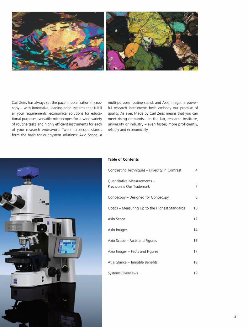
multi-purpose routine stand, and Axio Imager, a power-
ful research instrument: both embody our promise of
quality. As ever, Made by Carl Zeiss means that you can
meet rising demands – in the lab, research institute,
university or industry – even faster, more proficiently,
reliably and economically.
Carl Zeiss has always set the pace in polarization micros-
copy – with innovative, leading-edge systems that fulfill
all your requirements: economical solutions for educa-
tional purposes, versatile microscopes for a wide variety
of routine tasks and highly efficient instruments for each
of your research endeavors. Two microscope stands
form the basis for our system solutions: Axio Scope, a
4
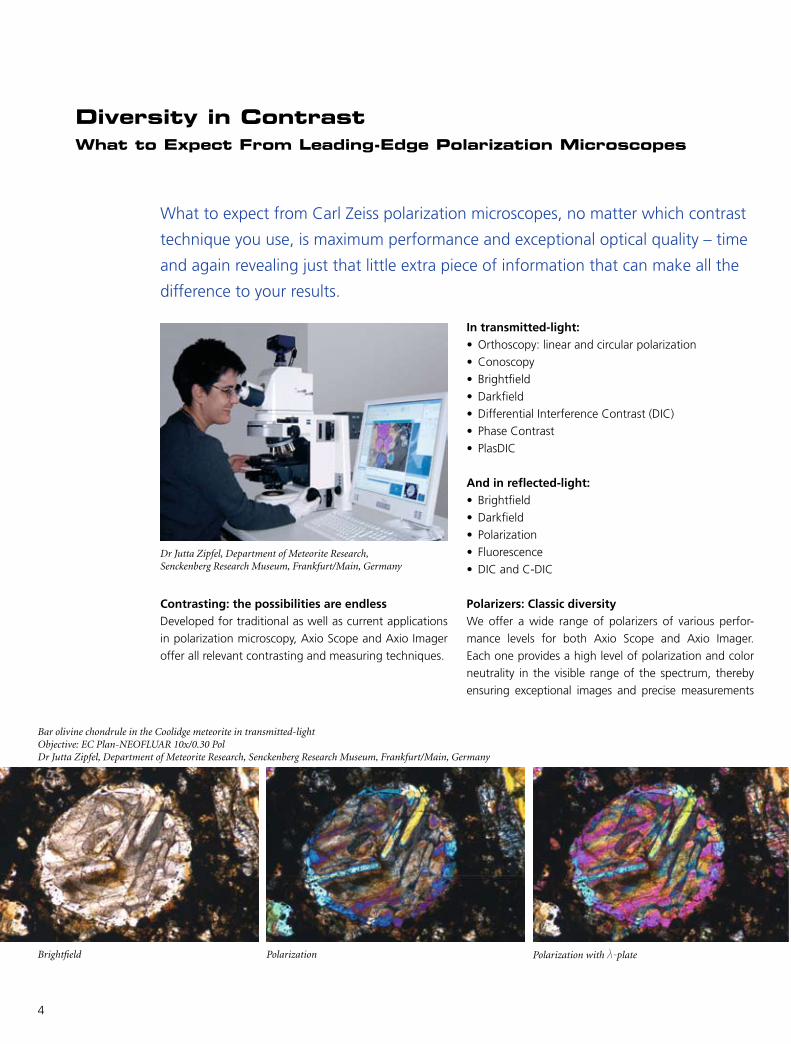
Diversity in ContrastWhat to Expect From Leading-Edge Polarization Microscopes
What to expect from Carl Zeiss polarization microscopes, no matter which contrast
technique you use, is maximum performance and exceptional optical quality – time
and again revealing just that little extra piece of information that can make all the
difference to your results.
In transmitted-light:• Orthoscopy:linearandcircularpolarization
• Conoscopy
• Brightfield
• Darkfield
• DifferentialInterferenceContrast(DIC)
• PhaseContrast
• PlasDIC
And in reflected-light:• Brightfield
• Darkfield
• Polarization
• Fluorescence
• DICandC-DIC
Polarizers: Classic diversityWe offer a wide range of polarizers of various perfor-
mance levels for both Axio Scope and Axio Imager.
Each one provides a high level of polarization and color
neutrality in the visible range of the spectrum, thereby
ensuring exceptional images and precise measurements
Contrasting: the possibilities are endlessDeveloped for traditional as well as current applications
in polarization microscopy, Axio Scope and Axio Imager
offer all relevant contrasting and measuring techniques.
Brightfield
Bar olivine chondrule in the Coolidge meteorite in transmitted-lightObjective: EC Plan-NEOFLUAR 10x/0.30 PolDr Jutta Zipfel, Department of Meteorite Research, Senckenberg Research Museum, Frankfurt/Main, Germany
Polarization Polarization withλ-plate
Dr Jutta Zipfel, Department of Meteorite Research, Senckenberg Research Museum, Frankfurt/Main, Germany

5
Bar olivine chondrule in the Coolidge meteorite in reflected-lightObjective: EC Epiplan-NEOFLUAR 10x/0.25 PolDr Jutta Zipfel, Department of Meteorite Research, Senckenberg Research Museum, Frankfurt/Main, Germany
Circular polarization: innovation in transmitted-lightCarl Zeiss polarization microscopes offer a further leading-
edge innovation focused on your everyday requirements:
the circular polarization device for transmitted-light. In
contrast to the linear polarization currently predominantly
in use, this device enables viewing and imaging devoid
of any angular-dependent extinction; all features appear
in their maximum interference colors. The benefits are
obvious – for the photomicrography of thin rock sections
as well as for structural examinations on plastics or strain
distribution in glass using digital analysis systems.
according to industrial and other standards. The range
comprises fixed and rotating polarizers for transmitted-
and reflected-light. In addition, our portfolio includes
360° rotating quantitative analyzers with 0.1° vernier
as well as combinations with a fixed or rotating lambda
plate. We also offer a dedicated temperature-resistant
polarizer module for the use in conjunction with the
high energy arc lamp HBO103 in order to guarantee a
consistent quality of polarization contrast.
Brightfield Darkfield Polarization
State of polarization of the light
Rotation of the microscope stage
0° 45° 90° 135° 180°
Spec
imen
Zirc
on
linea
rci
rcu
lar
Mu
sco
vite
linea
rci
rcu
lar
Behavior of optically anisotropic crystals in linearly and circularly polarized light, orthoscopy and conoscopy
6
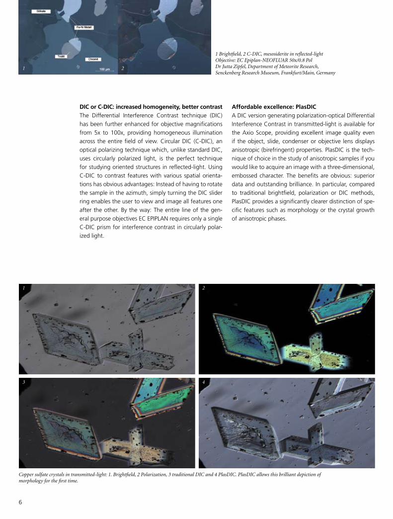
Affordable excellence: PlasDICA DIC version generating polarization-optical Differential
Interference Contrast in transmitted-light is available for
the Axio Scope, providing excellent image quality even
if the object, slide, condenser or objective lens displays
anisotropic(birefringent)properties.PlasDICisthetech-
nique of choice in the study of anisotropic samples if you
would like to acquire an image with a three-dimensional,
embossed character. The benefits are obvious: superior
data and outstanding brilliance. In particular, compared
to traditional brightfield, polarization or DIC methods,
PlasDIC provides a significantly clearer distinction of spe-
cific features such as morphology or the crystal growth
of anisotropic phases.
DIC or C-DIC: increased homogeneity, better contrastThe Differential Interference Contrast technique (DIC)
has been further enhanced for objective magnifications
from 5x to 100x, providing homogeneous illumination
across theentirefieldofview.CircularDIC (C-DIC),an
optical polarizing technique which, unlike standard DIC,
uses circularly polarized light, is the perfect technique
for studying oriented structures in reflected-light. Using
C-DIC to contrast features with various spatial orienta-
tions has obvious advantages: Instead of having to rotate
the sample in the azimuth, simply turning the DIC slider
ring enables the user to view and image all features one
after the other. By the way: The entire line of the gen-
eral purpose objectives EC EPIPLAN requires only a single
C-DIC prism for interference contrast in circularly polar-
ized light.
1 Brightfield, 2 C-DIC, mesosiderite in reflected-lightObjective: EC Epiplan-NEOFLUAR 50x/0.8 PolDr Jutta Zipfel, Department of Meteorite Research, Senckenberg Research Museum, Frankfurt/Main, Germany
Copper sulfate crystals in transmitted-light: 1. Brightfield, 2 Polarization, 3 traditional DIC and 4 PlasDIC. PlasDIC allows this brilliant depiction of morphology for the first time.
1 2
1
3
2
4
7
Precision is Our TrademarkQuantitative Methods for Your Analysis
High performance in quantitative techniques – Carl Zeiss has a tailor-made solution
for every polarization microscopy requirement
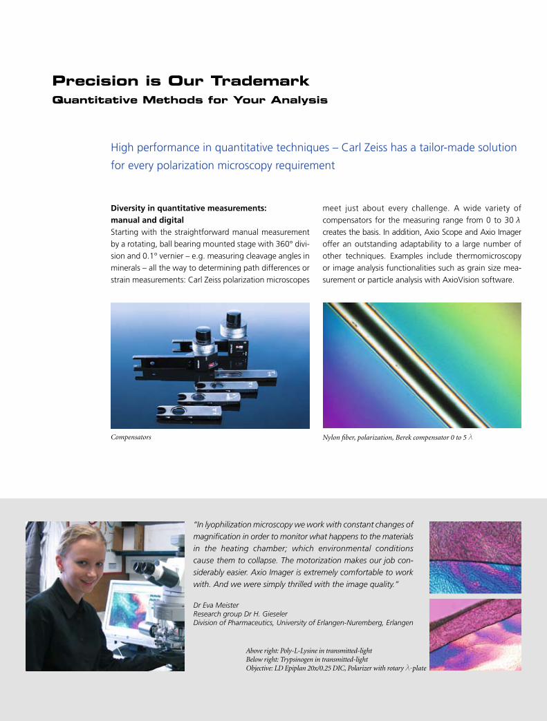
Diversity in quantitative measurements: manual and digitalStarting with the straightforward manual measurement
by a rotating, ball bearing mounted stage with 360° divi-
sion and 0.1° vernier – e.g. measuring cleavage angles in
minerals – all the way to determining path differences or
strain measurements: Carl Zeiss polarization microscopes
meet just about every challenge. A wide variety of
compensators for the measuring range from 0 to 30λ
creates the basis. In addition, Axio Scope and Axio Imager
offer an outstanding adaptability to a large number of
other techniques. Examples include thermomicroscopy
or image analysis functionalities such as grain size mea-
surement or particle analysis with AxioVision software.
“In lyophilization microscopy we work with constant changes of
magnification in order to monitor what happens to the materials
in the heating chamber; which environmental conditions
cause them to collapse. The motorization makes our job con-
siderably easier. Axio Imager is extremely comfortable to work
with. And we were simply thrilled with the image quality.”
Dr Eva MeisterResearch group Dr H. GieselerDivision of Pharmaceutics, University of Erlangen-Nuremberg, Erlangen
Nylon fiber, polarization, Berek compensator 0 to 5λCompensators
Above right: Poly-L-Lysine in transmitted-lightBelow right: Trypsinogen in transmitted-lightObjective: LD Epiplan 20x/0.25 DIC, Polarizer with rotaryλ-plate

8
Designed for ConoscopyStraightforward and Confident Mastery of a Demanding Technique
Carl Zeiss polarization microscopes provide the flexibility of fast, simple and eco-
nomical system extensions for conoscopic measurements to suit each of your needs.
without the need for tools and allows the straightfor-
ward addition of the conoscopy function for crystal
analysis to the polarization microscope at any time.
Using the objective N-ACHROPLAN 50x/0.8 Pol or
EC Plan-NEOFLUAR 40x/0.9 Pol, this makes perform-
ing conoscopy effortless and comfortable.
3. Conoscopy with the Bertrand lens slider
If the samples are uncovered or a 100x objective
magnification is desired, conoscopy with the Bertrand
lens slider is the solution of choice. The Bertrand
lens can be focused, so that you can employ a wide
range of objectives; for example EC Plan-NEOFLUAR
100x/1.30 Oil Pol or EC Epiplan-NEOFLUAR 50x/0.8 Pol.
Economical or sophisticated: five choices for conoscopyIn many cases, the analysis of an interference image will
provide even more valuable information for the classifica-
tion of anisotropic material than the image of the object
itself does. The polarization microscopes Axio Scope and
Axio Imager from Carl Zeiss are available in a number of
alternative configurations:
1. The pin-hole diaphragm or the auxiliary microscope
in the eyepiece tube
The simplest and most economical version.
2. The conoscopy module
The module is simply inserted into the reflector turret
or the reflector slider, is therefore easily exchanged
With the conoscopy module, comprising Bertrand lens, analyzer and a high aperture objective (N-ACHROPLAN 50x/0.9 Pol or EC Plan-NEOFLUAR 40x/0.9 Pol) your microscope can be upgraded to conoscopy at any time.

9
5. Pol phototube
The Pol phototube has been specifically designed for
orthoscopy and conoscopy with Axio Imager. There
is a significant advantage to this choice: Due to an
additional intermediate image plane, object, cross
hairs and iris diaphragm can be viewed concurrently.
Thanks to the adjustable iris diaphragm this is also
true for the limits of conoscopic range, down to a
minimum crystal size of 10 µm. The Bertrand optics
are pre-centered and focusable and are straightfor-
ward to turn on and off with the help of a slider. As
a result, the correlation of orthoscopic and cono-
scopic image data can be easily verified at any time.
An ideal solution for fast, reliable crystal analysis.
In addition to the economical options detailed above,
two more alternatives are available specifically for
Axio Imager to allow the upgrade of your polarization
microscope for conoscopy.
4. 5-position tube lens turret with integrated focusable
Bertrand lens
Opting for a tube lens turret in order to acquire fur-
ther magnifications will allow you to perform conos-
copy in addition, as the tube lens turret contains
an integrated Bertrand lens. The tube lens turret is
available both in an encoded and in a motorized
version.
Pol rotary stage with adjustable 45° click stops and stage clips. Vernier 0.1°, object guide for transmitted- and reflected-light applications (with and without click stops)
Small rotary stage with stage clips:360° division with 0.1° vernier
Determination of the optical characteristics of 1-axis and 2-axis minerals in linearly and circularly polarized light, the reference direction n
y of compensatorλ is aligned in NO-SW.
Un
axia
lState of polarization of the light
linear circular
compensatorλ
without with without with
Posi
tive
qu
artz
Neg
ativ
e ca
lcit
e

10
Measuring Up to the Highest StandardsCarl Zeiss Redefines the Limits of Optics
Optics of uncompromising quality form the basis for setting new standards in
polarization microscopy. The keyword here is strain-free. This principle is embodied
by the availability of a wide range of polarization objectives in various performance
classes and price levels – tailor-made for your requirements.
techniques in transmitted- and reflected-light, as well
as reflected-light applications in darkfield. Additionally,
the turret features a position to house a DIC-slider for
Differential Interference Contrast.
Polarization strain-free: the objectives Four ranges of objectives share one ambition: Only those
merit the label Pol which qualify for work in polarized
light due to exceptionally low strain. Carl Zeiss offers four
lines of strain-free objectives, varying in the extent of
correction, price level and application area.
The six-position centering nosepiece: added convenience for polarizationThe six-position Pol* centering nosepiece offers much
space for your objectives, eliminating the need for time-
consuming objective or turret changes; clearly a plus
for enhanced efficiency. The rotary stage is centered in
relation to the fixed turret opening which serves as the
reference. Subsequently, the remaining openings are
centered in the turret individually – the image position
therefore remains unaffected by each change of mag-
nification. Being equipped with M27 threads, the turret
accommodates the whole range of standard contrast
Glass fiber filled with liquid crystal, linearly polarized transmitted-lightObjective: EC Epiplan-NEOFLUAR 50x/0.80 PolThomas Tanggaard Larsen, COM Research Center, Technical University of Denmark, Lyngby, Denmark; and Peter Hansen,Crystal Fibre A/S, Birkerød, Denmark
*Axio Imager: encoded; Axio Scope: manual Pol – Polarization, DIC – Differential Interference Contrast

11
Objective lens Suitable up tofield of view
Flatness of field
Color correction
N-AChROPlAN Pol The transmitted-light lenses for samples with cover glass. With further enhancement of color correction and flattening, this is the attractively-priced, entry-level line for polarizing microscopy – ideal for education and routine.
23 Very good Very good
EC Plan-NEOFlUAR PolThe Enhanced Contrast transmitted-light objective lenses for samples with a cover glass. With their consistent minimiza-tion of stray light and contrast enhancement, these lenses meet demanding requirements. The EC Plan-NEOFLUAR lenses feature full chromatic correction for the focal plane. With their high resolving power, they offer a crisp, high-contrast and completely flat image for observation and documentation.
25 Excellent Excellent
EC EPIPlAN PolTransmitted- and reflected-light objective lenses for uncovered samples in routine applications. The Enhanced Contrast series is achromatically corrected and generates a flattened field for an intermediate image size of 23 mm. The objective lenses feature blocked pupil positions and therefore allow the C-DIC contrasting technique.
23 Very good Very good
EC Epiplan-NEOFlUAR PolThe transmitted- and reflected-light objective lenses for advanced applications for samples with or without a cover glass. Optimized for maximum contrast, their outstand-ing features include increased numerical apertures and therefore a higher resolving power. These lenses are suitable for transmitted-light examinations. However, due to the spherical aberration, objects with a cover glass can only be examined up to a magnification of 20x. There are no restric-tions for samples without a cover glass. Rigorous, object-side telecentricity makes these lenses particularly suitable for measuring purposes.
25 Excellent Excellent
In addition, the Epiplan-NEOFLUAR Pol line offers you a selection of immersion lenses.
You can get further information at www.zeiss.de/objectives.
12
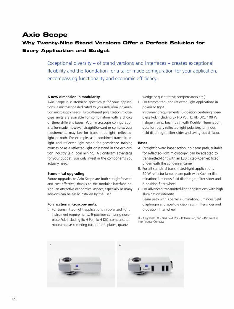
I II
Axio ScopeWhy Twenty-Nine Stand Versions Offer a Perfect Solution for
Every Application and Budget
Exceptional diversity – of stand versions and interfaces – creates exceptional
flexibility and the foundation for a tailor-made configuration for your application,
encompassing functionality and economic efficiency.
wedgeorquantitativecompensatorsetc.)
II. For transmitted- and reflected-light applications in
polarized light
Instrument requirements: 6-position centering nose-
piece Pol, including 5x HD Pol, 1x HD DIC. 100 W
halogen lamp; beam path with Koehler illumination;
slots for rotary reflected-light polarizer, luminous
field diaphragm, filter slider and swing-out diffusor.
Bases A. Straightforward base section, no beam path, suitable
for reflected-light microscopy; can be adapted to
transmitted-lightwithanLED(Fixed-Koehler)fixed
underneath the condenser carrier
B. For all standard transmitted-light applications
50 W reflector lamp, beam path with Koehler illu-
mination; luminous field diaphragm, filter slider and
6-position filter wheel
C. For advanced transmitted-light applications with high
illumination intensity
Beam path with Koehler illumination, luminous field
diaphragm and aperture diaphragm, filter slider and
6-position filter wheel
H – Brightfield, D – Darkfield, Pol – Polarization, DIC – Differential Interference Contrast
A new dimension in modularityAxio Scope is customized specifically for your applica-
tions; a microscope dedicated to your individual polariza-
tion microscopy needs. Two different polarization micros-
copy units are available for combination with a choice
of three different bases. Your microscope configuration
is tailor-made, however straightforward or complex your
requirements may be; for transmitted-light, reflected-
light or both. For example, as a combined transmitted-
light and reflected-light stand for geoscience training
courses or as a reflected-light only stand in the explora-
tionindustry(e.g.coalmining).Asignificantadvantage
for your budget: you only invest in the components you
actually need.
Economical upgradingFuture upgrades to Axio Scope are both straightforward
and cost-effective, thanks to the modular interface de-
sign: an attractive economical aspect, especially as many
add-ons can be easily installed by the user.
Polarization microscopy units:I. For transmitted-light applications in polarized light
Instrument requirements: 6-position centering nose-
piece Pol, including 5x H Pol, 1x H DIC; compensator
mountabovecenteringturret(forλ-plates, quartz

13
A B C
1
2
3
4
5
6
7
10
11 1415
16 17
18
19
20
21
13
8
9
12
From 0 to 110 mm: variable sample spaceThere are a number of options available for extending the
sample space vertically in order to accommodate taller
samples. In addition to z-travel this can be achieved by
• Loweringthestagecarrierwiththedovetail
• Removingthecondensercarrier,forexampleifthe
stage is intended to be lowered beyond the travel
range
• Insertinga30mmor60mmspacer,utilizingthecus-
tomer interface between the polarization microscopy
units(I,II)andthebases(A,B,C).The30mmand
60 mm spacer extend the maximum sample height to
80 mm and 110 mm respectively
The flexible sample space provides additional free-
dom of use and extends the range of applications for
Axio Scope.
Interface for reflector modules: infinity spaceThe interface in the infinity space is unique in this cat-
egory. Axio Scope allows you to use those reflector mod-
ules that are best suited for your applications. You have
the choice of a 2-position slider, a 4-position reflector
turret or a 6-position turret. No matter which alternative
you prefer, all of them are easily fitted with Push&Click
modules. With each option, your optics modules are held
safely and dust-free.
The variety of interfaces is a special performance featureof Axio Scope. It varies depending on which upper and lower bodies are used. In the example: upper body II and lower body B.
1 Tube for either - Intermediate plate - Tube lens turret or for2 Upper body3 Reflector space - 2-position slider - 4-position turret - 6-position turret4 Upper body for either - 30 or 60 mm spacer or for5 Lower body6 Stage carrier7 Stage8 Condenser carrier9 Condenser10 Transmitted-light filter wheel11 Compartment for 6x20 mm slider, Compensators, C-DIC slider12 Compartment for DIC slider13 Objective lenses 14 Rotary polarizer15 Luminous field diaphragm16 Aperture diaphragm17 Reflected-light filter slider18 Achromatic illumination adapter and interface for illumination HAL 100/HBO, etc.19 Transmitted-light filter slider20 Transmitted-light polarizer21 Transmitted-/reflected-light analyzer

14
Axio ImagerComfort and Convenience – Provided by an Intelligent Polarization
Microscope
The intelligent microscope assists in controlling your workflows, making them
easier and even more reliable – with Axio Imager, Carl Zeiss has implemented a concept
with regards to stand diversity, ease of use and ergonomics that will amaze you.
The touchscreen: innovation at a glanceComplex workflows made easy – the most relevant
functionalities of the motorized polarization microscope
areavailableona touchscreen (TFT). The controlof all
motorized components is at your fingertips. In addition
to the factory settings, complex processes can be pro-
grammed, saved and retrieved at the touch of a button
on the screen.
light manager and contrast manager: the automatic way to optimum settings The light manager is designed to provide reproducible
illumination settings and stable imaging conditions lead-
ing to optimum illumination and contrast. This is achieved
by automatically regulating the lamp voltage, ensuring
The stand versions: 9 times more flexibleMore economic efficiency in polarization microscopy
– Axio Imager gives you the freedom to tailor your re-
search microscope to your requirements. Nine stands are
available. You can opt for the encoded, partly motor-
ized or fully motorized version, as all key components of
Axio Imager are encoded.
The imaging cellStability is a major prerequisite for best results. The core
elements of Axio Imager – objective turret, z-guide and
stage carrier – are constructed as a compact, vibration-
free unit. This stable cell is decoupled from the remaining
stand, creating ideal conditions for imaging, particularly
for time-lapse experiments using high magnifications.
The touchscreen on the stand (left) or the docking station (right) provide clear guidance in control and configuration.
15
Operating panel: microscopy without the microscopeDesigned to provide more freedom of moving around in
the lab – Axio Imager can be controlled via an operat-
ing panel, which can be positioned separately from the
microscope stand. The panel features a focus drive and
brightness control. Other functions can be programmed
by the user. The panel offers an interface for the TFT
and for the x-, y-control of the motorized stage. A well-
conceived design for more convenience.
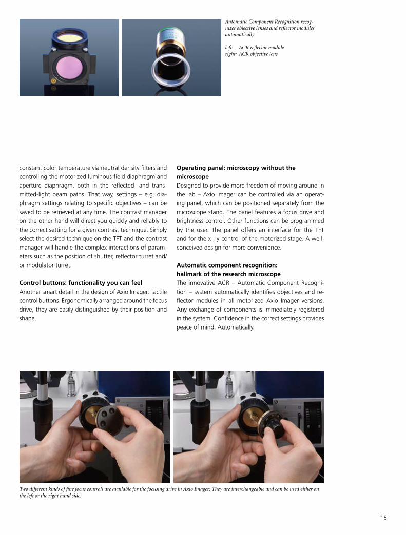
Automatic component recognition: hallmark of the research microscopeThe innovative ACR – Automatic Component Recogni-
tion – system automatically identifies objectives and re-
flector modules in all motorized Axio Imager versions.
Any exchange of components is immediately registered
in the system. Confidence in the correct settings provides
peace of mind. Automatically.
constant color temperature via neutral density filters and
controlling the motorized luminous field diaphragm and
aperture diaphragm, both in the reflected- and trans-
mitted-light beam paths. That way, settings – e.g. dia-
phragm settings relating to specific objectives – can be
saved to be retrieved at any time. The contrast manager
on the other hand will direct you quickly and reliably to
the correct setting for a given contrast technique. Simply
select the desired technique on the TFT and the contrast
manager will handle the complex interactions of param-
eters such as the position of shutter, reflector turret and/
or modulator turret.
Control buttons: functionality you can feelAnother smart detail in the design of Axio Imager: tactile
control buttons. Ergonomically arranged around the focus
drive, they are easily distinguished by their position and
shape.
Automatic Component Recognition recog-nizes objective lenses and reflector modules automatically
left: ACR reflector module right: ACR objective lens
Two different kinds of fine focus controls are available for the focusing drive in Axio Imager: They are interchangeable and can be used either on the left or the right hand side.

16
Upper body of stand
lower body of stand
Upper body for transmitted-light
with objective turret 5x H Pol / 1x H DIC, M27
Upper body for reflected- and transmitted-light
with objective turret 5x HD Pol / 1x HD DIC, M27
lower body of stand for lED illumination Microscope standAxio Scope.A1LED5x H Pol / 1x H DIC
430035-9240-000
Microscope standAxio Scope.A1LED, HAL 100 5x HD Pol / 1x HD DIC
430035-9270-000
lower body of stand for hAl 50 illumination
Microscope standAxio Scope.A1 HAL 50, 5x H Pol / 1x H DIC
430035-9250-000
Microscope standAxio Scope.A1 HAL 50, HAL 100,5x HD Pol / 1x HD DIC
430035-9280-000
lower body of stand for hAl 100 illumination
Microscope standAxio Scope.A1HAL 100, HAL 1005x H Pol / 1x H DIC
430035-9260-000
Microscope standAxio Scope.A1HAL 100, HAL 100, 5x HD Pol / 1x HD DIC
430035-9290-000
Axio Scope.A1 for polarizing microscopy – facts and figures
Dimensions in mm (inches)
17
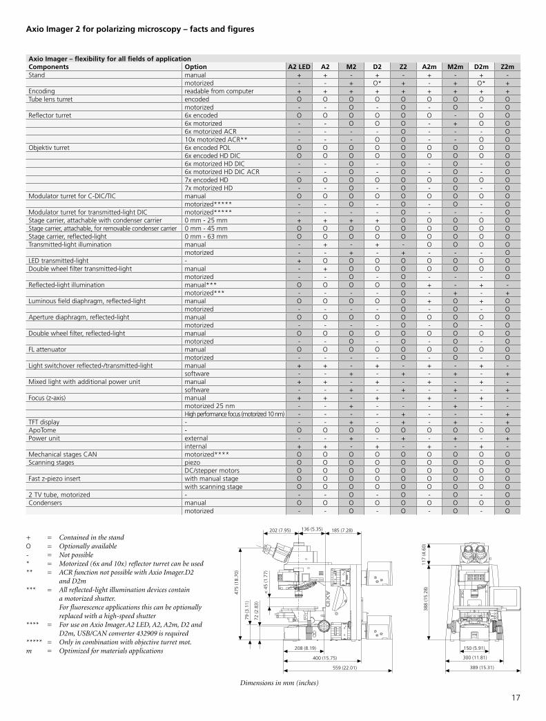
Axio Imager – flexibility for all fields of applicationComponents Option A2 lED A2 M2 D2 Z2 A2m M2m D2m Z2mStand manual + + - + - + - + -
motorized - - + O* + - + O* +Encoding readable from computer + + + + + + + + +Tube lens turret encoded O O O O O O O O O
motorized - - O - O - O - OReflector turret 6x encoded O O O O O O - O O
6x motorized - - O O O - + O O6x motorized ACR - - - - O - - - O10x motorized ACR** - - - O O - - O O
Objektiv turret 6x encoded POL O O O O O O O O O6x encoded HD DIC O O O O O O O O O6x motorized HD DIC - - O - O - O - O6x motorized HD DIC ACR - - O - O - O - O7x encoded HD O O O O O O O O O7x motorized HD - - O - O - O - O
Modulator turret for C-DIC/TIC manual O O O O O O O O Omotorized***** - - O - O - O - O
Modulator turret for transmitted-light DIC motorized***** - - - - O - - - OStage carrier, attachable with condenser carrier 0 mm - 25 mm + + + + O O O O OStage carrier, attachable, for removable condenser carrier 0 mm - 45 mm O O O O O O O O OStage carrier, reflected-light 0 mm - 63 mm O O O O O O O O OTransmitted-light illumination manual - + - + - O O O O
motorized - - + - + - - - OLED transmitted-light - + O O O O O O O ODouble wheel filter transmitted-light manual - + O O O O O O O
motorized - - O - O - - - OReflected-light illumination manual*** O O O O O + - + -
motorized*** - - - - O - + - +Luminous field diaphragm, reflected-light manual O O O O O + O + O
motorized - - - - O - O - OAperture diaphragm, reflected-light manual O O O O O O O O O
motorized - - - - O - O - ODouble wheel filter, reflected-light manual O O O O O O O O O
motorized - - O - O - O - OFL attenuator manual O O O O O O O O O
motorized - - - - O - O - OLight switchover reflected-/transmitted-light manual + + - + - + - + -
software - - + - + - + - +Mixed light with additional power unit manual + + - + - + - + -
software - - + - + - + - +Focus(z-axis) manual + + - + - + - + -
motorized 25 nm - - + - - - + - -Highperformancefocus(motorized10nm) - - - - + - - - +
TFT display - - - + - + - + - +ApoTome - O O O O O O O O OPower unit external - - + - + - + - +
internal + + - + - + - + -Mechanical stages CAN motorized**** O O O O O O O O OScanning stages piezo O O O O O O O O O
DC/stepper motors O O O O O O O O OFast z-piezo insert with manual stage O O O O O O O O O
with scanning stage O O O O O O O O O2 TV tube, motorized - - - O - O - O - OCondensers manual O O O O O O O O O
motorized - - O - O - O - O
+ = Contained in the standO = Optionally available- = Not possible * = Motorized (6x and 10x) reflector turret can be used** = ACR function not possible with Axio Imager.D2 and D2m*** = All reflected-light illumination devices contain a motorized shutter. For fluorescence applications this can be optionally replaced with a high-speed shutter**** = For use on Axio Imager.A2 LED, A2, A2m, D2 and D2m, USB/CAN converter 432909 is required***** = Only in combination with objective turret mot.m = Optimized for materials applications
Axio Imager 2 for polarizing microscopy – facts and figures
Dimensions in mm (inches)

18
Axio Scope.A1 Axio Imager 2
Stands 6 manual stand models optionally available as transmitted-light, reflected-light or transmitted-/reflected-light stands
9 encoded, partly motorized or fully motorized stand versionsoptionally available as transmitted-light, reflected-light or transmitted-/reflected-light stands
Field of view number 23 23/25
Illumination Reflected-light: 100 W HAL, HBOTransmitted-light: 100 W HAL, 50 W HAL or LED
Reflected-light: 100 W HAL, HBOTransmitted-light: 100 W HAL, LED
Optics Proven ICS optics, optionally with achromatic correction lens system Innovative IC²S infinity system for considerably more contrast in allestablished contrasting techniques
High quality and economical entry-level line of polarizing objective lenses for transmitted-light: N-ACHROPLAN Pol
Enhanced Contrast, the new generations of high contrast polarizing objective lenses:Transmitted-light: EC Plan-NEOFLUAR Pol, reflected-light: EC EPIPLAN Pol, EC Epiplan-NEOFLUAR Pol, oil immersion objective lenses
Contrasting methods Transmitted-light: qualitative and quantitative polarizing techniques, orthoscopy, linear and circular polarization, conoscopy, brightfield, dark-field, Phase Contrast, Differential Interference Contrast, PlasDIC*
Reflected-light:qualitativeandquantitativepolarization,brightfield,darkfield,DifferentialInterferenceContrast(DIC),DifferentialInterferenceContrastincircularlypolarizedlight(C-DIC),fluorescence
Contrast change
Change of modulesPush&Click withouttool
Manual contrast change via Reflector slider 2xReflector turret 4xReflector turret 6x
Contrast change via encoded or motorized Reflector turretReflector turret 6x encodedReflector turret 6x mot.Reflector turret 6x mot. ACRReflector turret 10x ACR
Objective turret 6x centering objective turret Pol, thread M27 6x centering objective turret Pol, thread M27 encoded
Polarizers Transmitted-light:Polarizer(switchable),polarizer(rotarywith0°and90°clickstops),polarizer(switchablewithlambdaplate,rotary),circularpolarizer
Reflected-light:Reflector module Pol,Reflector module Pol for HBO103,Polarizer rotary 0-90°
Reflector module Pol,Reflector module Pol for HBO103,Measuring polarizer, rotatable 360° with 0.1° vernier
Analyzers Analyzer module or analyzer slider or slider with analyzer and lambda plate, rotatable 360° or measuring analyzerwith 0.1° division, rotatable 360°
Conoscopy Bertrandlensmodule(fixedfocus)Bertrandlensslider(focusable)
Tube lens turret with focusable Bertrand lensPol phototube with focusable Bertrand lensCrosshairs and visual field diaphragm in additional intermediate image plane
Ergonomy/ease of use
Convenient intensity settingErgo tube/ergo phototube: 20° viewing angle, vertical adjustment in range of 50 mm
Ergo phototubesContrast managerLight managerTouchscreenRemote control
Software AxioVision microscope softwareBasic version, upgradeable with functional modules such as MosaiX, Panorama, particle analysis or grain size analysis
Cameras Openinterfaceforeachcameratype(videoanddigitalconsumercameras,scientificmicroscopecameras)In particular AxioCam ICc 1 and AxioCam ICc 3, AxioCam HRc, AxioCam MRc or AxioCam MRc 5
ICS – Infinity Color Corrected SystemIC²S – Infinity Contrast and Color Corrected SystemACR – Automated Component Recognition
* possible in Axio Scope only
Concrete benefitsWhat details you should know for polarizing microscopy

19
Axio Scope.A1 for polarization

20
Accessories for polarization

21
Axio Imager 2 for polarization

Today as ever, Carl Zeiss sets standards in polarization
microscopy, providing a wide range of innovative system
solutions designed to fulfill each of your requirements.
Our solutions provide economy for education purposes,
versatility for a wide variety of routine measurement
tasks and powerful efficiency and functionality for sci-
ence and research.
Two microscope stands form the basis for our system
solutions: Axio Scope and Axio Imager – the former a
robust multi-purpose routine stand, the latter a leading-
edge microscope for research and science. Both equally
signify our promise of quality in the field of polarization
microscopy – from traditional areas such as mineralogy
and geology to current advances in material applica-
tions, such as thin layer systems or solar cells. More than
just the sum of its parts, a polarization microscope from
Carl Zeiss constitutes a meticulously designed complete
system solution, perfectly integrated in the Carl Zeiss sys-
tem environment.
Info
rmat
ion
subj
ect t
o ch
ange
.Pr
inte
d on
env
ironm
enta
lly fr
iend
ly
pape
r ble
ache
d w
ithou
t cho
lorin
e.
70-2
-005
0/e
– pr
inte
d 12
.09
Carl Zeiss Microscopy GmbH07745 Jena, [email protected]/polarization
866-464-1005