Embed Size (px)
Citation preview
MICROBIOLOGY AND MOLECULAR BIOLOGY REVIEWS,1092-2172/01/$04.00�0 DOI: 10.1128/MMBR.65.3.445–462.2001
Sept. 2001, p. 445–462 Vol. 65, No. 3
Copyright © 2001, American Society for Microbiology. All Rights Reserved.
Polar Flagellar Motility of the VibrionaceaeLINDA L. MCCARTER*
Department of Microbiology, The University of Iowa, Iowa City, Iowa 52242
INTRODUCTION .......................................................................................................................................................445POLAR FLAGELLAR STRUCTURE AND THE SHEATHED FLAGELLUM..................................................447
Multiple Flagellin Genes .......................................................................................................................................447Differential Flagellin Gene Expression ................................................................................................................447Chromosomal Organization of Flagellin Genes .................................................................................................448The Basal Body .......................................................................................................................................................449The Sheath...............................................................................................................................................................449Implications of the Sheath for Filament Assembly: HAP Mutant Phenotypes..............................................449
ORGANIZATION AND REGULATION OF FLAGELLAR AND CHEMOTAXIS GENES ............................450Genes Not Found in E. coli ...................................................................................................................................450Chemotaxis Genes and Gene Organization ........................................................................................................451Regulation of Gene Expression: a Potential Hierarchy of Gene Control .......................................................452Early Gene Expression: Master Regulatory Proteins That Interact with �54 ...............................................452
THE SODIUM-DRIVEN MOTOR ...........................................................................................................................453Sodium Channel-Blocking Drugs Specifically Interfere with Polar Flagellar Rotation ...............................454The MotA-MotB Complex Translocates Sodium Ions.......................................................................................454Chimeras Composed of Sodium and Proton Parts............................................................................................454Interaction with the Switch Complex...................................................................................................................454Unique Components ...............................................................................................................................................454
CHEMOTAXIS............................................................................................................................................................455THE POLAR FLAGELLUM AS A TACTILE SENSOR .......................................................................................455THE LATERAL FLAGELLAR SYSTEM AND SWARMING MOTILITY .........................................................456SUMMARY AND PERSPECTIVES: COMPARISONS WITH FLAGELLAR SYSTEMS OF
OTHER BACTERIA ...........................................................................................................................................456ACKNOWLEDGMENTS ...........................................................................................................................................458REFERENCES ............................................................................................................................................................458
INTRODUCTION
Flagella, which act as semirigid helical propellers, providebacteria with a highly efficient means of locomotion. For ex-ample, many Vibrio and Pseudomonas species swim in liquidenvironments at speeds as fast as 60 �m/s (10, 11, 64, 189). Thepropellers are powered by reversible rotary motors embeddedin the cell membrane, which can turn the flagellum at rates ashigh as 1,700 revolutions per s (rps) (115). Energy for rotationof the motor is derived from either the sodium or protonmembrane potential (72, 117). The number and arrangementof the propellers can vary, but the mode of insertion is of twomajor types, i.e., polar or peritrichous. Flagella play other rolesin addition to swimming in liquid (reviewed in reference 132).They can enable bacteria to move over and colonize surfaces,a process called swarming (63). They also participate in adhe-sion. Attachment of bacteria to surfaces is often first mediatedby contact of the flagellum with the surface (127). As propul-sive organelles, flagella seems to aid in overcoming negativeelectrostatic interactions and thus are believed to play a keyrole in the initial steps of adsorption of bacteria to surfaces,biofilm formation, and invasion of hosts (30, 39, 154). Studiesusing Vibrio alginolyticus have demonstrated that attachment to
glass is directly proportional to swimming speed (90). Otherstudies have shown that by disabling the flagellar motor of thefish pathogen V. anguillarum, invasion into the fish host isseverely reduced (142). In addition, some flagella are sheathedby a membrane that appears to be an extension of the outercell membrane. The composition of this sheath (specifically,lipopolysaccharide and protein) may allow additional specificinteractions between the bacterium and a surface (77, 163, 164).
Extensive structural and genetic analysis of the unsheathed,peritrichous flagella of Escherichia coli and Salmonella entericaserovar Typimurium has deciphered the complexity of the or-ganelle, its assembly process, motor function, and the coor-dination of movement (i.e., chemotaxis) (reviewed in ref-erences 17, 21, 111, and 135). Many of these features areconserved in flagellar systems of other bacteria; however, novelpermutations also exist. For example, the flagella of spiro-chetes are not external to the cell but are contained within theperiplasmic space (103). These flagella also play a skeletal rolein determining the spiral shape of the cell (133). Anotherexample is the single flagellum of Caulobacter crescentus, whichis assembled at one pole during asymmetric cell division and islater ejected and replaced by a stalk (reviewed in references140 and 195). A number of bacteria are polarly flagellated, yet,aside from C. crescentus, comparatively little is known aboutpolar flagellar systems. This review uses the polar flagellarsystem of V. parahaemolyticus as a focal point to build a frame-work for describing what is known about polar systems of the
* Mailing address: Microbiology Department, University of Iowa,Iowa City, IA 52242. Phone: (319) 335-9721. Fax: (319) 335-7679.E-mail: [email protected].
445
on May 27, 2018 by guest
http://mm
br.asm.org/
Dow
nloaded from

gamma purple bacteria, most particularly Vibrio but with somereference to Pseudomonas species. The reader is also referredto the excellent review by Yorimitsu and Homma, which fo-cuses on flagellar motors of the Vibrionaceae family (202).Although our understanding of these polar motility systems isnot comprehensive, it is hoped that this review will provide ageneral context for polar systems and points of contrast withwhat is known for the well-studied peritrichous systems of E.coli and S. enterica serovar Typhimurium.
V. parahaemolyticus is a common gram-negative bacteriumin marine and estuarine environments. It is also a humanpathogen and a worldwide cause of gastroenteritis. In areas ofthe world where seafood consumption is high, such as South-east Asia, it is the primary cause of food poisoning (24, 78,193). It is a serious emerging pathogen in North America,where it is the most common Vibrio species isolated from
humans and the most frequent cause of Vibrio-associated gas-troenteritis (37, 175). Infections are usually associated withconsumption of raw or undercooked shellfish and result inacute gastroenteritis, but they can also result in wound infec-tions and septicemia. As a member of the Vibrionaceae, V. para-haemolyticus is classified as a �-proteobacterium within theenteric-vibrio branch of the �-3 subgroup (192). In phyloge-netic analyses, V. parahaemolyticus clusters most closely withV. harveyi, V. vulnificus, and V. alginolyticus. It is more distantlyrelated to V. anguillarum, V. fischeri, and V. cholerae (188).
The flagellation patterns of these members of the Vibriona-ceae family are presented in Table 1. V. parahaemolyticus andsome other members of this family exhibit mixed flagellation,possessing polar and peritrichous flagella. When grown plank-tonically, the bacteria display polar flagella (Fig. 1). The fla-gellum is sheathed by what appears to be an extension of thecell outer membrane. When grown on solid medium or me-dium of high viscosity, e.g., medium supplemented with Ficoll,the organisms produce both polar and peritrichous (also calledlateral) flagella (Fig. 2). As can be seen in Fig. 2, remarkablenumbers of peritrichously arranged flagella are produced.These flagella are unsheathed and more fragile than the polarflagellum (2). Figure 2A shows plate-grown cells that havebeen stained with phosphotungstic acid. The polar flagellum,which is produced by liquid- and surface-grown bacteria, isdistinguished from the lateral flagella by the increased thick-ness due to the sheath. Figure 2B emphasizes the strikingdifference in polar and lateral flagellar integrity: cells werestained with uranyl acetate, which causes deterioration of thelateral but not the polar flagellar structure. In liquid environ-ments, the swimming speed of the marine vibrios is approxi-mately 60 �m/s; however, as the viscosity increases, the polar
FIG. 1. Sheathed polar flagellum of V. parahaemolyticus. An electron micrograph of cells grown in liquid and stained with 1% uranyl acetateis shown. Magnification, �26,000.
TABLE 1. Flagellation of Vibrio speciesa
Organism Sheathed polarflagellab
Unsheathedperitrichous flagellac
V. parahaemolyticus Monotrichousd �e
V. alginolyticus Monotrichousd �e
V. harveyi Monotrichous �V. vulnificus Monotrichous �V. anguillarum Monotrichous �V. fischeri Lophotrichous (2–8) �V. cholerae Monotrichousd �
a Determined by electron microscopy studies by Baumann and colleagues (2,13).
b Determined when grown in liquid medium.c Determined when grown on solid medium. �, the majority of strains exam-
ined (�80%) possessed lateral flagella; �, none possessed lateral flagella.d Rotation has been demonstrated to be powered by the sodium motive force.e Rotation has been demonstrated to be powered by the proton motive force.
446 MCCARTER MICROBIOL. MOL. BIOL. REV.
on May 27, 2018 by guest
http://mm
br.asm.org/
Dow
nloaded from

flagellum is not an effective propulsive organelle and swimmingslows (10, 82). In compensation, the lateral flagellar system isinduced. These peritrichous flagella are quite functional in vis-cous environments and enable the bacterium to move over andcolonize surfaces (162). The two motility systems are geneticallydistinct (121). Thus, in possessing dual flagellar systems suited forlocomotion under different circumstances, V. parahaemolyticusseems highly adapted to survival in changing habitats, includinglife in planktonic environments and on surfaces or in biofilms.
POLAR FLAGELLAR STRUCTURE AND THESHEATHED FLAGELLUM
Multiple Flagellin Genes
Flagellar filaments, which act as propellers, consist of self-assembling protein subunits (flagellin) arranged in a helix and
forming a hollow tube (reviewed in reference 135). Subunitsmove down the hollow core and are polymerized at the tip ofthe flagellum. The V. parahaemolyticus polar organelle is acomplex flagellum. There are six polar flagellin genes, orga-nized in two loci (86, 122). The flagellins are similar to eachother: FlaB and FlaA are 78% identical, FlaB and FlaC are68% identical, FlaB and FlaD are 99% identical, FlaB andFlaF are 69% identical, and FlaB and FlaE are 50% identical.Despite the great protein similarity of FlaB and FlaD, theflagellins migrate differently on sodium dodecyl sulfate-poly-acrylamide gel electrophoresis analysis, suggesting the possi-bility of posttranslational modification, e.g., glycosylation,which has been observed for flagellins of many bacteria includ-ing some spirochetes, Campylobacter species, and P. aeruginosa(27, 56, 171, 196), or phosphorylation, which has been detectedfor flagellins of P. aeruginosa (85). Analysis of the proteincomposition of purified flagella from wild-type strains andstrains with mutations in flagellin genes suggests that all of theflagellins can be incorporated into the organelle and that FlaA,FlaB, and FlaD are the major subunits (121, 122; L. McCarter,unpublished data). Nothing is known with respect to theirspatial arrangement in the flagellum. Loss of function of asingle flagellin gene has little or no effect on swimming motilityor flagellar structure (waveform or length). Thus, none of thesix flagellin genes is essential for filament formation. Deletionof the flaFBA or the flaCD genes also has little effect onmotility, but the deletion of both loci (�flaFBA �flaCD) com-pletely abolishes motility (122).
Why are there six flagellin genes? It is not clear why theorganism possesses such an extraordinary number of flagellins.The similarity of the gene products and the dispensability ofthe genes suggest that there are no special structural require-ments, although the filament structure and function could bemore complex and adapted to specific circumstances than ourlaboratory tests can reveal. Bacteria are known to modulatethe antigenicity of their flagellar filaments by expression ofdifferent flagellin genes or by recombination and rearrange-ment of flagellin genes (reviewed in reference 190). Therefore,the capacity for immune system evasion in a host organismmight account for some of the diversity. The multiplicity offlagellin genes suggests a significant reservoir for antigenicor phase variation. Although the sheath covers the filamentand might be thought to provide a disguise, electron micros-copy suggests that it may be fragile (2, 164). Thus, the sheathmay not protect the filament against the immune responseof a host. In some respects the endoflagella of the spiro-chetes are similar, for these flagella can also be viewed asbeing sheathed, polar organelles (reviewed in reference103). The spirochete flagella are normally found in theperiplasm, between the outer membrane and the cell cylin-der and attached near each cell pole. Purification ofperiplasmic flagella demonstrated that these filaments arealso complex, with generally two to four different flagellinproteins, encoded by distinct genes, as well as containing anaccessory, nonflagellin protein.
Differential Flagellin Gene Expression
The six flagellin genes occur in five distinct transcriptionalunits; flaD and flaE appear to be cotranscribed (86, 122). The
FIG. 2. Polar and lateral flagella of surface-grown bacteria. Thehelical structure of lateral flagella, but not polar flagella, is destroyedby uranyl acetate. Electron micrographs of cells harvested from a plateand negatively stained with 0.5% phosphotungstic acid (A) or 1%uranyl acetate (B) are shown. Arrows indicate the sheathed polarflagellum. Magnification, approximately �14,000.
VOL. 65, 2001 VIBRIO POLAR FLAGELLAR MOTILITY 447
on May 27, 2018 by guest
http://mm
br.asm.org/
Dow
nloaded from

promoters for three of these operons (flaA, flaB, and flaDE)require a specialized sigma factor for transcription, �28. Primerextension mapping has defined the consensus promoter. Thefifth flagellin gene, flaF, is poorly expressed, although up-stream sequences contain a potential �28-recognition site. Thesixth flagellin gene, flaC, is the most unusual with respect totranscription. Unlike flaA, flaB, and flaD, which are expressedin E. coli in a �28-dependent manner, FlaC cannot be detectedin E. coli unless expression of the gene is driven by an isopro-pyl--D-thiogalactoside (IPTG)-inducible tac promoter. Thenucleotide sequence upstream of the start point of transcrip-tion of flaC appears unusual in that it does not contain se-quences consistent with either the �28-dependent promoter orthe �54-dependent consensus polar flagellar promoters. A ma-jor FlaC-encoding transcript can be detected in plate-growncells but not in broth-grown cultures; therefore, flaC expres-sion appears to be induced by growth on a surface. Further-more, flaC expression requires an intact lateral flagellar ge-netic pathway. A defect in the lateral flagellar hook gene(lfgE) prevents the expression of many surface-dependentgenes, including genes encoding lateral-specific flagellar �28
and lateral flagellin (125). This defect also prevents surface-induced expression of flaC. Thus, flaC appears to be aunique flagellin gene with respect to transcription. What thismeans with respect to polar flagellar function and regula-tion, particularly in the context of growth on surfaces, re-mains to be investigated.
Chromosomal Organization of Flagellin Genes
The multiple flagellin genes are found in two distinct loca-tions on the chromosome, and the organization of these genesin V. parahaemolyticus is similar to that of the loci in V. an-guillarum and V. cholerae (88, 126). However, these organismspossess only five flagellin genes and lack an equivalent of flaE.Figure 3A depicts the relationship of the flagellin genes withrespect to chromosomal organization and protein homology.The phylogram (Fig. 3B) shows that each flagellin is moreclosely related to the predicted product of its spatially equiv-alent open reading frame (ORF) in the other organisms thanto the other flagellins within the same organism. On the basisof nucleotide sequence, mutant analysis, and/or primer exten-sion analysis, the open reading frames (ORFs) indicated by theblack arrows are believed to be transcribed by �28 in all organ-isms (86, 88, 126, 150). In each of the three species, one of theflagellin genes, indicated by the gray arrow, appears differentfrom the other flagellin genes with respect to transcriptionand/or function. In V. anguillarum and V. cholerae, this gene,flaA, seems to be specifically required for motility and/or vir-ulence. Disruption of V. anguillarum flaA significantly slowsmotility, and the gene is essential for infection of the fish(129), whereas mutations in any of the other flagellin geneshave little or no effect on swimming or virulence. In V. chol-erae, mutation of flaA completely abolishes motility (88).Expression of V. cholerae flaA is directly dependent on �54,whereas the four other flagellin genes are dispensable and
FIG. 3. Comparison of multiple flagellin genes with respect to chromosomal location and predicted gene product similarity. (A) Chromosomalorganization of flagellin genes. Genes are located in two loci: flaFBA and flaCDE in V. parahaemolyticus (Vp) and flaEDB and flaAC inV. anguillarum (Va) and V. cholerae (Vc). Arrowheads indicate the direction of transcription of each ORF. Evidence suggests that the genesindicated by the black arrows are transcribed by �28. V. parahaemolyticus flaE is cotranscribed with flaD. V. anguillarum and V. cholerae lack theflaE equivalent. The gray arrow represents the ORF encoding a unique flagellin with respect to transcription and/or function. It encodes the majorflagellin of V. anguillarum and V. cholerae and is transcribed by �54. In V. parahaemolyticus, this gene is induced by growth on a surface and is notexpressed when the organism is grown in liquid. The promoter sequences of the V. parahaemolyticus flaC gene do not resemble �28- or�54-dependent polar flagellar promoters. Below each gene, the percent identities of the V. anguillarum and V. cholerae gene products withV. parahaemolyticus flagellin are shown. (B) Grow/Tree phylogram of flagellins (produced by GCG Inc. program analysis).
448 MCCARTER MICROBIOL. MOL. BIOL. REV.
on May 27, 2018 by guest
http://mm
br.asm.org/
Dow
nloaded from
require �28 (150). The critical flagellin genes of V. anguilla-rum and V. cholerae are most equivalent with respect to genelocation and predicted protein sequence to V. parahaemo-lyticus flaC.
The Basal Body
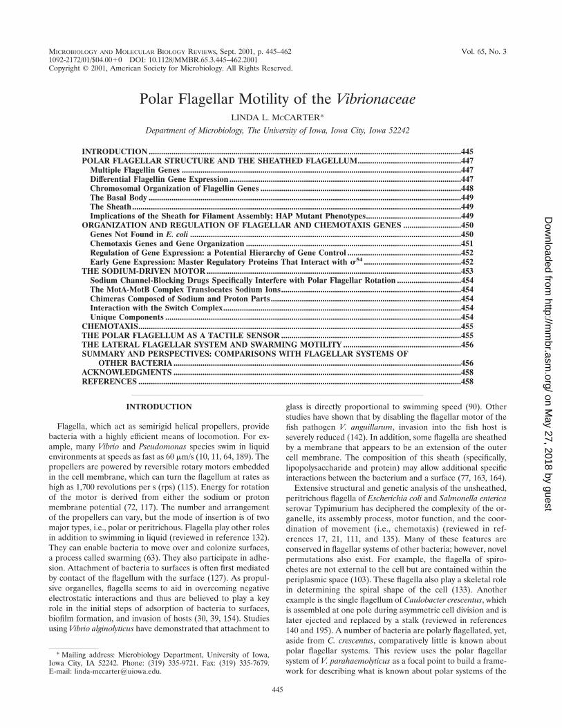
The flagellar filament is connected to a structure in themembrane known as the basal body. The basal body is com-posed of several rings and an axial structure. Some evidenceexists that the polar basal-body structure differs from that ofperitrichously inserted flagella. Two models for the basal or-ganelle of polar flagella have been derived from electron mi-croscopy studies of V. cholerae, Campylobacter fetus, Bdello-vibrio bacteriovorus, and Wolinella succinogenes (44, 46, 180). InW. succinogenes, electron micrographs display a beautiful largebasal disk described as an archimedian spiral (44). Such ele-ments, which also have been described as concentric mem-brane rings, were found associated with the basal organelle ofAquaspirillum serpens, V. cholerae, and C. fetus (36, 46). Re-gardless of whether the flagellum is sheathed or unsheathed,all of the studies report the existence in the basal body complexof a large convex disk situated below the outer membrane, andthus the large disk does not seem to be a feature specific tosheath formation. It has been hypothesized that the disk pro-vides reinforcement at the flagellar insertion site and dispersesforces that are generated by the force of flagellar rotation (44).A large basal disk is visible in V. parahaemolyticus and is shownin Fig. 4. One model for the polar basal body suggests that thedisk is the P-ring equivalent, acting as a bushing associatedwith peptidoglycan, and that for sheathed flagella the L ring,which is lipoplysaccharide or outer membrane associated, isnot present. The second model places the large disk betweenthe P and L rings. Genes for both the P and L rings exist inV. parahaemolyticus and V. cholerae. Whereas the P ring dis-plays homology to enteric P rings over the entire molecule, theVibrio L ring shows sequence divergence at the N terminus.
Perhaps the nature of this protein is one key to differences inbasal-body structure.
The Sheath
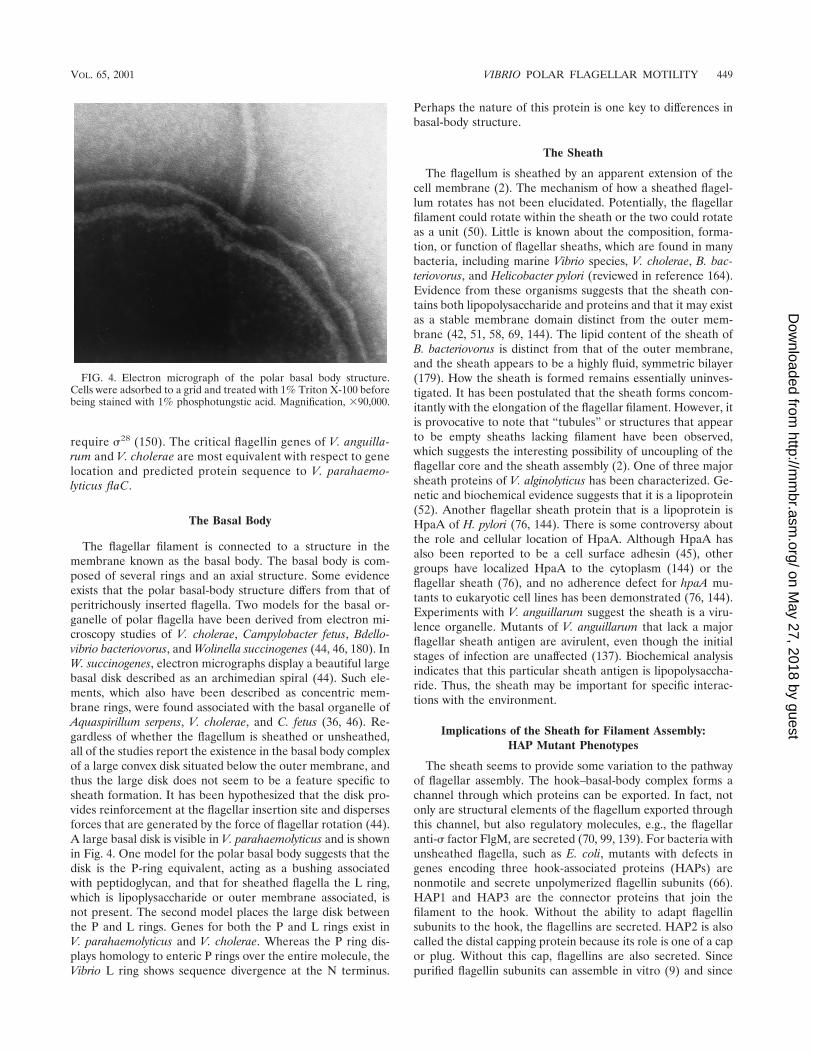
The flagellum is sheathed by an apparent extension of thecell membrane (2). The mechanism of how a sheathed flagel-lum rotates has not been elucidated. Potentially, the flagellarfilament could rotate within the sheath or the two could rotateas a unit (50). Little is known about the composition, forma-tion, or function of flagellar sheaths, which are found in manybacteria, including marine Vibrio species, V. cholerae, B. bac-teriovorus, and Helicobacter pylori (reviewed in reference 164).Evidence from these organisms suggests that the sheath con-tains both lipopolysaccharide and proteins and that it may existas a stable membrane domain distinct from the outer mem-brane (42, 51, 58, 69, 144). The lipid content of the sheath ofB. bacteriovorus is distinct from that of the outer membrane,and the sheath appears to be a highly fluid, symmetric bilayer(179). How the sheath is formed remains essentially uninves-tigated. It has been postulated that the sheath forms concom-itantly with the elongation of the flagellar filament. However, itis provocative to note that “tubules” or structures that appearto be empty sheaths lacking filament have been observed,which suggests the interesting possibility of uncoupling of theflagellar core and the sheath assembly (2). One of three majorsheath proteins of V. alginolyticus has been characterized. Ge-netic and biochemical evidence suggests that it is a lipoprotein(52). Another flagellar sheath protein that is a lipoprotein isHpaA of H. pylori (76, 144). There is some controversy aboutthe role and cellular location of HpaA. Although HpaA hasalso been reported to be a cell surface adhesin (45), othergroups have localized HpaA to the cytoplasm (144) or theflagellar sheath (76), and no adherence defect for hpaA mu-tants to eukaryotic cell lines has been demonstrated (76, 144).Experiments with V. anguillarum suggest the sheath is a viru-lence organelle. Mutants of V. anguillarum that lack a majorflagellar sheath antigen are avirulent, even though the initialstages of infection are unaffected (137). Biochemical analysisindicates that this particular sheath antigen is lipopolysaccha-ride. Thus, the sheath may be important for specific interac-tions with the environment.
Implications of the Sheath for Filament Assembly:HAP Mutant Phenotypes
The sheath seems to provide some variation to the pathwayof flagellar assembly. The hook–basal-body complex forms achannel through which proteins can be exported. In fact, notonly are structural elements of the flagellum exported throughthis channel, but also regulatory molecules, e.g., the flagellaranti-� factor FlgM, are secreted (70, 99, 139). For bacteria withunsheathed flagella, such as E. coli, mutants with defects ingenes encoding three hook-associated proteins (HAPs) arenonmotile and secrete unpolymerized flagellin subunits (66).HAP1 and HAP3 are the connector proteins that join thefilament to the hook. Without the ability to adapt flagellinsubunits to the hook, the flagellins are secreted. HAP2 is alsocalled the distal capping protein because its role is one of a capor plug. Without this cap, flagellins are also secreted. Sincepurified flagellin subunits can assemble in vitro (9) and since
FIG. 4. Electron micrograph of the polar basal body structure.Cells were adsorbed to a grid and treated with 1% Triton X-100 beforebeing stained with 1% phosphotungstic acid. Magnification, �90,000.
VOL. 65, 2001 VIBRIO POLAR FLAGELLAR MOTILITY 449
on May 27, 2018 by guest
http://mm
br.asm.org/
Dow
nloaded from

an S. enterica serovar Typhimurium mutant lacking HAP2 canpolymerize filaments if the concentration of flagellin in theexternal medium is high (67), the role of HAP2 has beenviewed as capping the flagellar tip to retard subunit secretionsufficiently to increase the local concentration of flagellin andpromote self-assembly. Recent work in Salmonella, analyzingcap-filament interactions by cryoelectron microscopy, suggestsa model for the cap as being a flat, disklike pentameric struc-ture that acts as a processive chaperone, preventing the loss offlagellin monomers and actively catalyzing folding and inser-tion into the filament (200).
HAP mutants of V. parahaemolyticus display different phe-notypes (122). The most striking difference is in the pheno-types of mutants with defects in the gene encoding HAP2.These mutants are competent for filament assembly and mo-tile. Figure 5 compares the flagella of the wild type and mu-tants with defects in the gene that encodes HAP2, and theflagella seem mostly indistinguishable. This suggests that in theabsence of HAP2 but in the presence of the flagellar sheath,the local concentrations of the flagellin monomers remain highenough to allow polymerization of subunits. HAP1 and HAP3mutants of V. parahaemolyticus are nonmotile and nonflagel-
lated; however, they produce detached, severely truncated fil-aments encased in a membrane (122). The sheath seems to actto retain flagellin monomers and allow subunit assembly. Thepolymerized flagellins cannot be connected to the hook andbleb off as abortive filaments surrounded by a membrane ves-icle. Similar filamentless mutants that produce flagellin-con-taining membrane vesicles were isolated in V. alginolyticus,although the genetic lesions in these strains were not deter-mined (136). Thus, the sheath itself appears to be able tosubstitute for the cap. The potential for the sheath to act insuch a sealing capacity raises a question about the competenceof the sheathed flagellum for secretion of the flagellar regula-tory molecule FlgM. In E. coli, FlgM is sequestered inside thecell, where it acts as an anti-� factor to prevent late flagellargene expression, until the hook is completed and FlgM is ex-ported (70). If the sheath acts as a passive barrier to export, thecheckpoint for coordinating flagellar morphogenesis and geneexpression may be somewhat different from the mechanism inE. coli.
ORGANIZATION AND REGULATION OF FLAGELLARAND CHEMOTAXIS GENES
The V. parahaemolyticus polar flagellar gene system (Fla)comprises approximately 60 genes (86). It is the default motil-ity system for the organism and is produced continuously;therefore, most of the polar flagellar genes have been namedanalogously to homologs in other bacteria. Genes in the lateralflagellar system (Laf) are expressed under particular condi-tions and have been assigned designations that are permuta-tions of the fla nomenclature. Table 2 summarizes the organi-zation, homology, and predicted function of the gene products.By comparison with E. coli, the full complement of genesencoding flagellar structural components and the export appa-ratus exist, and there are a few additions. Most of the genes arefound in two loci that contain large, predicted flagellar oper-ons. Precedence for large motility operons has been estab-lished in other bacteria, e.g., Borrelia burgdorferi (55). Theclosest homologs to many of the genes are found in V. cholerae,P. aeruginosa, and P. putida. The physical organization alsoseems highly conserved among organisms. The flagellar gene or-ganization of V. cholerae is almost identical to that of V. para-haemolyticus, with the exception of a large insertion containingnonflagellar genes positioned between flhB and flhA in V. chol-erae (65).
Genes Not Found in E. coli
One pair of genes found in V. parahaemolyticus and manyother bacteria, including other Vibrio, Bacillus, Pseudomonas,Campylobacter, and Borrelia species, but not found in E. coliis flhF and flhG. FlhF shows homology to FtsY, which is aGTP-binding protein involved in the signal recognition particletargeting pathway (134). The flhF gene was first discovered inBacillus subtilis, where it was demonstrated to be required formotility (29). A nonpolar, null mutation in flhF produced via-ble but nonmotile cells lacking flagella. Intriguingly, the V. para-haemolyticus and V. cholerae FlhF proteins contain an insertionof 170 amino acids that is not found in other FlhF sequencesderived from organisms possessing proton-driven flagella. Theinserted domain shows homology to a eukaryotic sodium chan-
FIG. 5. The flagellar sheath can act as a cap. Electron micrographsof liquid-grown cells stained with 0.5% phosphotungstic acid areshown. (A) Wild-type strain. (B) HAP2-defective strain. Magnifica-tion, �4,500.
450 MCCARTER MICROBIOL. MOL. BIOL. REV.
on May 27, 2018 by guest
http://mm
br.asm.org/
Dow
nloaded from

nel. It should be quite interesting to probe the function of thisdomain. Immediately downstream of V. parahaemolyticus flhFis flhG, which encodes a protein that shows homology to MinD.MinD is a membrane ATPase involved in septum site deter-mination (102, 118). The FlhF-FlhG pair is found in a numberof polarly flagellated bacteria. Disruption of flhF in P. putidaleads to random arrangement of flagellar insertion (147), anddisruption of fleN, the flhG homolog in P. aeruginosa, leads tomultiple polar flagella (38). Potentially, FlhF and FlhG couldwork as a pair to determine and/or control site selection offlagellar insertion as well as the number of flagella.
The gene products with the least homology to flagellar com-ponents of other bacteria are those that are candidates forflagellar chaperones, i.e., FlaI and FlgN. Perhaps this is notsurprising. Flagella are assembled via a type III export pathway(reviewed in reference 110). It seems clear that classes ofsequentially exported flagellar proteins exist (32, 130). No con-sensus flagellar export signal has been defined, although a
number of models have been suggested. It has been proposedthat the flagellar chaperones, e.g., FliT or FlgN, act to guidethe secretion of substrates by preventing cytoplasmic assemblyor degradation, by presenting substrates to the secretory ap-paratus, and by regulating translation to directly couple trans-lation to secretion (48, 80). When grown on a surface, V. para-haemolyticus assembles two distinct types of flagella. Thesubstrate signals and the export system for each flagellar sys-tem must be sufficiently divergent to exclude components fromthe second system. In some respects, the dual flagellar systemsin V. parahaemolyticus seem ideal to test hypotheses with re-spect to type III secretion determinants and the specificity ofexport.
Chemotaxis Genes and Gene Organization
The complement of chemotaxis (che) genes and their orga-nization are different from those in E. coli. One of these genes
TABLE 2. Polar flagellar and chemotaxis genes of V. parahaemolyticusa
Gene Gene product homolog orpredicted function Gene Gene product homolog or
predicted function
Region 1flgN . . . . . . . . . . . . . . . . . . . .No homolog: potential chaperoneflgM . . . . . . . . . . . . . . . . . . .Anti-�28 factorflgA . . . . . . . . . . . . . . . . . . . .Necessary for P-ring additioncheV . . . . . . . . . . . . . . . . . . .Chemotaxis CheY/CheW hybridcheR . . . . . . . . . . . . . . . . . . .Chemotaxis methyltransferaseflgB . . . . . . . . . . . . . . . . . . . .RodflgC . . . . . . . . . . . . . . . . . . . .RodflgD . . . . . . . . . . . . . . . . . . . .RodflgE . . . . . . . . . . . . . . . . . . . .HookflgF . . . . . . . . . . . . . . . . . . . .RodflgG. . . . . . . . . . . . . . . . . . . .RodflgH. . . . . . . . . . . . . . . . . . . .L ringflgI . . . . . . . . . . . . . . . . . . . . .P ringflgJ . . . . . . . . . . . . . . . . . . . . .Peptidoglycan-hydrolyzing proteinflgK . . . . . . . . . . . . . . . . . . . .HAP1flgL . . . . . . . . . . . . . . . . . . . .HAP3flaC. . . . . . . . . . . . . . . . . . . .FlagellinflaD. . . . . . . . . . . . . . . . . . . .FlagellinflaE . . . . . . . . . . . . . . . . . . . .Flagellin
Region 3motA . . . . . . . . . . . . . . . . . .Na� motor componentmotB . . . . . . . . . . . . . . . . . .Na� motor component
Region 4motX . . . . . . . . . . . . . . . . . .Na� motor component
Region 5 . . . . . . . . . . . . . . . . . . .motY . . . . . . . . . . . . . . . . . .Na� motor component
a GenBank accession numbers for each region: 1, U12817; 2, AF069392; 3, AF069391; 4, U09005; 5, U06949. Arrows indicate relative directions of transcription andpotential transcriptional units.
Region 2flaF . . . . . . . . . . . . . . . . . . . FlagellinflaB . . . . . . . . . . . . . . . . . . . . FlagellinflaA . . . . . . . . . . . . . . . . . . . . FlagellinflaG. . . . . . . . . . . . . . . . . . . . Slight homology to N terminus of flagellinsflaH. . . . . . . . . . . . . . . . . . . . HAP2flaI . . . . . . . . . . . . . . . . . . . . . No homolog; potential chapeoneflaJ. . . . . . . . . . . . . . . . . . . . . FliSflaK . . . . . . . . . . . . . . . . . . . . �54-interacting regulatorflaL . . . . . . . . . . . . . . . . . . . . Two-component sensor kinaseflaM . . . . . . . . . . . . . . . . . . . Two-component response regulatorfliE. . . . . . . . . . . . . . . . . . . . . Hook–basal-body componentfliF . . . . . . . . . . . . . . . . . . . . . M ringfliG . . . . . . . . . . . . . . . . . . . . Switch componentfliH . . . . . . . . . . . . . . . . . . . . Fla export and assemblyfliI . . . . . . . . . . . . . . . . . . . . . Fla export; ATP synthasefliJ . . . . . . . . . . . . . . . . . . . . . Fla export and assemblyfliK. . . . . . . . . . . . . . . . . . . . . Hook length controlfliL. . . . . . . . . . . . . . . . . . . . . Flagellar proteinfliM . . . . . . . . . . . . . . . . . . . . Switch componentfliN . . . . . . . . . . . . . . . . . . . . Switch componentfliO . . . . . . . . . . . . . . . . . . . . Fla export and assemblyfliP . . . . . . . . . . . . . . . . . . . . . Fla export and assemblyfliQ . . . . . . . . . . . . . . . . . . . . Fla export and assemblyfliR. . . . . . . . . . . . . . . . . . . . . Fla export and assemblyflhB . . . . . . . . . . . . . . . . . . . . Fla export and assemblyflhA . . . . . . . . . . . . . . . . . . . . Fla export and assemblyflhF . . . . . . . . . . . . . . . . . . . . Flagellar protein; also homologous to FtsY;
potential GTP-binding proteinflhG . . . . . . . . . . . . . . . . . . . Flagellar protein; MinD and other
ATP-binding proteinsfliA . . . . . . . . . . . . . . . . . . . . RNA polymerase �28 factorcheY . . . . . . . . . . . . . . . . . . . Causes change in direction of flagellar
rotationcheZ . . . . . . . . . . . . . . . . . . . Dephosphorylates CheYcheA . . . . . . . . . . . . . . . . . . . CheA kinasecheB . . . . . . . . . . . . . . . . . . . Chemotaxis methylesteraseORF 1 . . . . . . . . . . . . . . . . Soj-like and other chromosome-partitioning
ATPase proteinsORF 2 . . . . . . . . . . . . . . . . UnknowncheW . . . . . . . . . . . . . . . . . . Purine-binding chemotaxis proteinORF 3 . . . . . . . . . . . . . . . . Unknown
‘‘
’
’
’
’
’
’
’
’
’
’’
’’
’
’
’
VOL. 65, 2001 VIBRIO POLAR FLAGELLAR MOTILITY 451
on May 27, 2018 by guest
http://mm
br.asm.org/
Dow
nloaded from

is cheV, which encodes a hybrid CheY/CheW. CheV has beenfound in a number of organisms, including Bacillus species,Campylobacter jejuni, H. pylori, P. aeruginosa, and V. cholerae.In B. subtilis, genetic analysis suggests that CheV and CheWare functionally redundant (155). Three unusual ORFs occurwithin the che gene cluster of region 2. ORF1 encodes a pro-tein that resembles Soj of B. subtilis and other ATPase proteinsinvolved in chromosome partitioning (152, 161). The otherORFs encode potential polypeptides that do not resembleproteins of known function. It seems curious that a Soj-likeprotein exists within a flagellar/chemotaxis operon, and thisparticular arrangement is conserved in other bacteria, e.g.,P. aeruginosa, P. putida, and V. cholerae. Perhaps these novelORFs will prove key for understanding the linkage betweencell division and flagellation or development. It should beremarked that many of the V. parahaemolyticus che genes lo-cated in the flagellar clusters (Table 2) were discovered bymutant analysis; i.e., these genes produce defects in chemotaxiswhen mutated. The V. cholerae genome, as well as those ofPseudomonas species, indicates additional complexity with re-spect to a multiplicity of potential che genes.
Regulation of Gene Expression: a PotentialHierarchy of Gene Control
As can be seen in Table 2, a considerable number of genesare dedicated to the flagellar motility system; therefore, main-tenance of flagellation is a sizable investment with respect tocellular economy. As a result, flagellar systems are highly reg-ulated. In systems where it has been studied, the generalscheme of gene control represents a hierarchical cascade ofregulation that couples the sequential expression of specificclasses of genes to the assembly of the organelle. Genes in eachtemporal class must be functional in order for expression of thesubsequent class to occur. This also seems to be the case forV. parahaemolyticus, and a potential hierarchy is outlined inTable 3. The hierarchical scheme proposed for V. cholerae isvery similar, with some variations that are discussed below(150).
To summarize, at least three classes of transcription for the
polar flagellar hierarchy of V. parahaemolyticus have been pro-posed (86). At an early level in the transcriptional hierarchyare master regulators that potentially interact with �54, FlaKand FlaM. Flagellation in V. alginolyticus, V. cholerae, V. an-guillarum, and P. aeruginosa requires the rpoN gene, whichencodes �54 (84, 89, 145, 184). It seems probable that �54 willalso direct the transcription of V. parahaemolyticus polar genes,although this has not yet been demonstrated. Genes in themiddle class require �54-type activation and are dedicated toassembly of the hook–basal-body structure. Additionally, onefinds middle genes encoding HAP1 and HAP3, the motor genemotY, some chemotaxis genes, and fliA, which encodes �28. Inturn, this alternative � factor is specific for the other largesubset of flagellar promoters. The genes under this later levelof expression encode additional motor parts (MotA, MotB,and MotX), additional chemotaxis proteins, the distal cappingprotein HAP2, the anti-�28 factor FlgM, putative flagellar chap-erones, and five flagellins.
Early Gene Expression: Master Regulatory ProteinsThat Interact with �54
FlaK and FlaM appear to be master transcriptional regula-tors of polar genes. The genes are encoded by two linkedoperons, flaK and flaLM. Homologs of these regulators exist inV. cholerae and P. aeruginosa. The homologous gene clustersare shown in Table 4. Much is known about the role of thegene products in flagellar regulation in V. cholerae and P. ae-ruginosa (4, 35, 89, 153). Based on these studies, the followingmodel has been proposed. FlaK homologs, acting as �54-de-pendent transcription factors, activate transcription of theflaLM-like operons. FlaL homologs are histidine sensor ki-
TABLE 3. Regulatory cascade for the Vibrio polar flagellar gene systema
Early genes Middle genes (�54-dependent operons)b Late genes (�28-dependent operons)c
fliEFGHIHKLMNOPQR flhB (switch, export, andassembly proteins)
motAB (motor proteins)
flaK (�54-interacting regulator)d flhAFG fliA cheYZAB orf1 orf2 cheW orf3 (assembly,�28, and chemotaxis components)
motX (motor proteins)
flaLM (two-component sensor-responseregulator pair)
flgBCDEFGHIJ (hook and basal-body parts) flaAGHIJK (flagellin, chaperones, HAP2,and regulator K)
flgKL (HAP1 and HAP3) flaB (flagellin)flaDE (flagellins)
motY (motor protein) cheVR (chemotaxis proteins)flgMNc (anti-� factor and chaperone)
a Promoters for the polar flagellar genes have been defined in V. parahaemolyticus (86) and V. cholerae (150).b The consensus �54 recognition sequence derived from a comparison of V. parahaemolyticus promoters defined by primer extension analysis is TGGC N7
TTGC N11–13 � 1.c The consensus �28-recognition sequence derived from comparison of V. parahaemolyticus promoters defined by primer extension analysis is CTAAG N14 G(C/T)
CG(A/T)TAA N7 � 1.d In V. cholerae, the FlaK homolog is required to activate the transcription of the flaLM-like locus in a �54-dependendent manner, and genes in the middle class are
divided into FlaK- and FlaM-dependent subclasses (89, 150).e The flgMN genes are also transcribed from an upstream promoter; however, the start point of transcriptional initiation for the flgAMN operon has not been
determined.
TABLE 4. Polar flagellar regulatory genes
Organism Homologous gene clusters
V. parahaemolyticus flaK flaLMV. cholerae flrA flrBCP. aeruginosa fleQ fleSR
452 MCCARTER MICROBIOL. MOL. BIOL. REV.
on May 27, 2018 by guest
http://mm
br.asm.org/
Dow
nloaded from

nases that activate the receiver domain of the response regu-lator FlaM homologs by phosphotransfer. Phosphorylated FlaMhomologs are required for �54-dependent transcription ofgenes in the middle class of flagellar genes. For V. cholerae andP. aeruginosa, the FlaK homologue is absolutely required for�54-dependent activation of the flaLM-like operon and is es-sential for motility (4, 89). V. cholerae FlrB (the FlaL homolog)is an autokinase that can transfer phosphate to FlrC (the FlaMhomolog), specifically to the aspartate that is equivalent toAsp57 of CheY. FlrC must be phosphorylated to activateflagellar gene transcription (35).
For V. cholerae, the middle genes have been further dividedinto two classes: those that require FlrA for activation (encod-ing the MS-ring-switch-export complex) and those that requireFlrC for activation of transcription (encoding the remainder ofthe basal-body and hook structure) (150). For V. parahaemo-lyticus, like V. cholerae and P. aeruginosa, loss of function offlaM results in a completely nonmotile cell; however, disrup-tion of the gene encoding FlaK (488 amino acids) by introduc-tion of a chloramphenicol cassette at the position in the geneencoding amino acid 37 or 216 does not abolish motility (168;McCarter, unpublished). Mutants with flaK defects possess aslow-motility phenotype, which is significantly different fromthe completely nonmotile phenotype observed in the otherorganisms. One possible explanation for the difference is thatreadthrough transcription occurs in the V. parahaemolyticusflaK::Camr mutants, originating with the chloramphenicol cas-sette and continuing into flaLM, which is sufficient to circum-vent the requirement for FlaK-mediated activation of flaLM. Iftrue, this would suggest that FlaK is not required for transcrip-tion of other flagellar operons in V. parahaemolyticus and that
the middle genes of the hierarchy are not subdivided as theyare in V. cholerae. Clearly, more experiments are required.
A schematic comparison of the functional domain of theseregulators is shown in Fig. 6. The central domain of the FlaKhomologs contains a potential �54-interaction domain. TheN-terminal domains of the FlaK homologs do not contain thehallmark residues in the receiver domain of response regula-tors; this suggests that they are �54-dependent transcriptionalregulatory proteins but that they do not participate in two-component phosphorelay signaling. The FlaM homologs re-semble two-component response regulators, containing both�54-interaction domains and response regulatory regions. TheN-terminal response regulatory region of FlaM-like proteinscontains the four residues that are highly conserved in a num-ber of response regulators, i.e., residues that correspond toAsp12, Asp13, Asp57, and Lys109 of the paradigm responseregulator CheY (33). The C terminus of the FlaM homologscontains a helix-turn-helix region. The three FlaL-like proteinseach possess a histidine kinase domain; however, their N ter-mini differ. V. cholerae FlrB and P. aeruginosa FleS possessN-terminal PAS domains, whereas V. parahaemolyticus FlaLdoes not contain a recognizable PAS domain. PAS moduleshave been identified in a number of signal transduction mole-cules and are thought to be signatures of proteins that playroles in detection and adaptation to environmental change,e.g., changes in light, redox potential, and oxygen (177). Thissuggests that the ways in which these molecules act as sensorkinases may be different, specifically with respect to input.Activation of the phosphorelay cascade may be responsive todifferent signals in the different organisms. Moreover, withrespect to global control of flagellar gene expression, there aremost probably other environmental signals that contribute tothe regulation of flagellar gene expression, e.g., catabolite re-pression (199).
THE SODIUM-DRIVEN MOTOR
The flagellar filament acts as a propeller that is turned by areversible rotary motor, which is embedded in the membrane(reviewed in references 17, 19, 21, and 41). Energy to powerflagellar rotation is derived from the transmembrane electro-chemical potential of specific ions (101, 117, 128). Rotationappears to be tightly coupled to the flow of ions through themotor. Two kinds of motors, which are dependent on differentcoupling ions, have been described: H� and Na� motors (72,112). In V. parahaemolyticus and V. alginolyticus, the sodiummotive force drives polar flagellar rotation and the protonmotive force powers rotation of the lateral flagella (11, 83).The polar flagellum of V. cholerae is also sodium driven (95).
Proton-type motors of E. coli and S. enterica serovar Typhi-murium have been extensively characterized. Two cytoplasmicproteins, MotA and MotB, form the force-generating unitthrough which the protons are channeled (22, 23, 160, 169, 204,206). Torque is transmitted from the MotA-MotB complex tothe flagellar basal body. Thus, the torque generator, which isprobably fastened to the cell wall via a peptidoglycan interac-tion domain found in the C terminus of MotB (34, 40), acts asa stator to transmit force to the rotor. Critical electrostaticinteractions between MotA and FliG have been demonstrated(205). FliG is found at the base of the flagellar basal body.
FIG. 6. Comparison of the conserved domains in the master polarflagellar regulatory factors of V. parahaemolyticus (vp), V. cholerae (vc),and P. aeruginosa (pa). FlaK, FlrA, and FleQ contain a potential �54-interaction domain (Sigma-54; pfam00158). FlaL, FlrB, and FleS arepotential two-component sensors that contain the histidine kinase sig-naling domain (His kinase: pfam00512). FlrB and FleS also contain aPAS domain (Smart PAS domain or pfam00989), whereas FlaL doesnot. FlaM, FlrC, and FleR contain the �54 domain, a response regu-latory receiver domain (RRR; pfam00072), and a helix-turn-helix(HTH) region. Experiments performed with V. cholerae and P. aerugi-nosa (4, 89) support the following model for a transcriptional activa-tion cascade: FlaK, FlrA, and FleQ activate transcription of the flrBCand fleSR operons in a �54-dependent manner. FlaL, FlrB, and FleS actas sensor kinases to phosphorylate and activate the response regulatorsFlaM, FlrC, and FleR, which in turn control the transcription of other�54-dependent flagellar promoters. Evidence from V. cholerae suggeststhat FlrA directly controls the transcription of other �54-dependentflagellar genes (150).
VOL. 65, 2001 VIBRIO POLAR FLAGELLAR MOTILITY 453
on May 27, 2018 by guest
http://mm
br.asm.org/
Dow
nloaded from

Below FliG is a cytoplasmic structure called the C-ring, con-taining FliM and FliN (47). The complex containing FliG,FliM, and FliN is also known as the switch complex and isessential for torque generation, flagellar assembly, and thecontrol of the direction of flagellar rotation (73, 165, 173, 174,183, 197, 198).
Sodium Channel-Blocking Drugs Specifically Interferewith Polar Flagellar Rotation
The pioneering bioenergetic studies by Imae and coworkerson Na�-driven motility were performed using alkalophilicBacillus and marine Vibrio species (reviewed in reference 71).Imae demonstrated that sodium channel-blocking drugs, suchas amiloride, specifically inhibited sodium-driven motility andcould be used to probe motor function (170). Mutants wereisolated that could swim in the presence of the sodium chan-nel-blocking drug (92). Currently, an understanding of thearchitecture of the sodium-type flagellar motor is being devel-oped by studying V. alginolyticus, V. cholerae, and V. parahae-molyticus (reviewed in reference 202). Rotation rates for thesodium-driven flagellum are quite remarkable; e.g., using laserdark-field microscopy, the polar flagellar rotation rate for V. al-ginolyticus was recorded to average 1,100 rps and to be as highas 1,700 rps (115). Four genes have been described that arerequired for sodium-dependent flagellar rotation is V. para-haemolyticus: motA, motB, motX, and motY (7, 25, 60, 123, 124,141). In V. alginolyticus and V. cholerae, motA and motB arenamed pomA and pomB, respectively. Transposon insertionsor deletions in these genes produce flagellated but paralyzedbacteria. Loss of function of any one of the four motor genescompletely abolishes motility but does not prevent flagellarassembly.
The MotA-MotB Complex Translocates Sodium Ions
With respect to membrane topology and function, the Vibriosodium-type proteins resemble MotA and MotB of the proton-type motor (8, 93). Many data suggest that these proteinsform the Na�-conducting channel. Initial evidence implicatingVibrio MotA and MotB in Na� translocation was provided bythe isolation of mutants that could swim in the presence of thesodium channel inhibitor phenamil (74, 91). These mutantscontained alterations in motA or motB. Some of the mutationsconferring phenamil resistance altered the ion specificity of themotor, for example resulting in increased or decreased swim-ming rates in the presence of lithium ions (74). Some muta-tions, such as a substitution at residue Asp31 in motA, causeda slow-motility phenotype, suggesting that the negative chargeof this residue contributes to optimal speed or efficiency of themotor (94). Reconstitution experiments with liposomes pro-vided strong proof that purified V. alginolyticus MotA-MotBcomplexes catalyzed Na� flux (157). Furthermore, these puri-fication studies, as well as immunoprecipitation experiments,indicated that MotA and MotB physically interact and can bepurified as a complex (157, 203). Interestingly, the molar ratioof MotA to MotB was calculated to be 2:1. Intermolecularcross-linking studies using cysteine substitutions within theperiplasmic loop between the third and fourth transmembranedomains of MotA also suggested that adjacent MotA mole-cules were physically close within the motor (201). To further
probe the stoichiometry of the torque generator, a chimericconstruct was designed to produce a fused dimer of MotAmolecules (158). Such a dimer was found to support motility;however, mutational inactivation of either half of the fusiondimer caused complete loss of swimming motility and loss ofthe ability to interact with MotB, suggesting that dual MotAcomponents are essential for sodium-type motor function.
Chimeras Composed of Sodium and Proton Parts
Some intriguing experiments using chimeras of mixed so-dium and proton motor parts have been performed. E. colimotor proteins can substitute to weakly power bacterial motil-ity by using the proton motive force in a V. cholerae strain thathas been gutted for the four sodium motor genes (60). Sodium-dependent motility could be restored to a MotA-deficientV. alginolyticus strain on provision of the proton-type MotAfrom Rhodobacter sphaeroides (5). Although R. sphaeroidesMotB was not functional in V. alginolyticus, MotB chimerasthat contained portions of the N-terminal transmembrane do-main of R. sphaeroides and the C-terminal linker-peptidogly-can-binding domains of V. alginolyticus were able to reconsti-tute Na�-driven motility (6). These experiments suggest thatthe determinants of the specificity of the coupling ion do notreside exclusively within MotA and/or the transmembrane do-main of MotB, and they raise the interesting question of howion specificity is conferred.
Interaction with the Switch Complex
On the basis of extensive mutational analysis (26, 53, 54, 73,106, 107, 181, 205) and the determination of the crystal struc-ture of the C-terminal domain of FliG, a structural model forthe part of the rotor that interacts with the stator has beendeveloped for the proton-type motor (108, 205). Residuesknown to be critical for torque generation in three switchcomponents of the proton-type motor are conserved in theswitch components of the V. parahaemolyticus and V. choleraesodium-type motor. This suggests a common mechanism forenergy transfer at the rotor-stator interface regardless of thedriving force powering rotation. Such an idea is further sup-ported by the ability to productively mix sodium and protoncomponents to create functionally propulsive motors (6, 60).The sodium-type switch components FliM and FliN do possessunique charged domains not found in their proton-type ho-mologs, and it will be of interest to determine whether thesedomains are important with respect to motor, switching, orassembly functions.
Unique Components
MotX and MotY proteins are the unique components ofsodium-type flagellar systems, and their specific roles are notknown. Loss of function of either motX or motY produces aparalyzed mutant completely defective for swimming but com-petent for flagellar assembly (123, 124, 141). Both proteinspossess single membrane-spanning domains. The C terminusof MotY contains an extended domain that shows strikinghomology to a number of outer membrane proteins know tointeract with peptidoglycan, e.g., OmpA and peptidoglycan-associated lipoproteins (124). The simplest hypothesis for the
454 MCCARTER MICROBIOL. MOL. BIOL. REV.
on May 27, 2018 by guest
http://mm
br.asm.org/
Dow
nloaded from

role of MotY is that the polar flagellar motor possesses twoelements for anchoring the force generator. Perhaps extremelyprecise alignment of the stator with the rotor is required for amotor that spins as fast as 100,000 rpm. The role of MotXremains mysterious. It is known that MotX recruits MotY tothe membrane when the proteins are coexpressed in E. coli(123). Furthermore, overexpression of MotX is lethal to E. coliin proportion to the external Na� concentration, and lethalitycan be reversed by the presence of the sodium channel blockeramiloride. This suggests that the proteins may somehow par-ticipate in or modulate Na� translocation. For example, MotXcould act to modify or specify ion channel activity. Thus, it maybe that all four proteins comprise and specify the sodium-typetorque-generating unit. However, there is no existing evidencethat places MotX and MotY in the physical context of MotAand MotB. It is possible that MotX and MotY may play a moredistinct role in the generation of sodium-driven motility; e.g.,they could participate in some other aspect of the sodium cycle.
CHEMOTAXIS
Bacterial chemotaxis has been most extensively studied inorganisms with multiple, peritrichously arranged flagella, e.g.,E. coli, S. enterica serovar Typhimurium, and B. subtilis (re-viewed in reference 21). Bacteria respond to signals in theenvironment by modulating the direction of flagellar rotation.As viewed under the light microscope, the cells move in alter-nating periods of smooth, forward trajectories of swimmingand tumbling. Forward translation, or running, occurs whenthe flagellar motor rotates in a direction that results in prop-agation of the semirigid helical wave of the flagellum, whichacts like a propeller and exerts a pushing motion on the cell.Wave propagation proceeds from the cell-proximal to cell-distal end of the flagellum, and the multiple flagella coalesce torotate synchronously and form a propulsive bundle (reviewedin reference 111). Tumbling is linked to changes in the qua-ternary structure of the flagellum (113). When the filamentsare rotated in the opposite direction, structural changes areinduced within the filament; the usual transformation is from anormal left-handed helix to semicoiled to a right-handed curlyform. If most of the filaments change the direction of rotationat the same time, the bundle flies apart. However, not all of thefilaments in the bundle need to reverse the direction of rota-tion in order to elicit tumbling. Fluorescence microscopy hasrevealed that changes in the direction of rotation of a singlefilament that result in transformation of the filament structureto the semicoiled form can elicit a change in the direction ofmovement of the cell body (185). The running mode allowspositional translation, whereas the tumbling mode results inreorientation in three-dimensional space. In the absence ofchemotaxis, these bacteria move in the alternating pattern ofrunning and tumbling described as a random walk. In thepresence of chemotactic stimuli, the time spent in one mode isbiased, e.g., in the presence of a gradient of attractant, theprobability of tumbling is decreased, thus prolonging thesmooth period of swimming.
Although polarly flagellated V. parahaemolyticus cells do nottumble like peritrichously flagellated E. coli or S. enterica se-rovar Typhimurium, the motility pattern consists of alternat-ing runs in a forward direction and changes of direction. In
the light microscope, V. parahaemolyticus swims in smooth,slightly curved lines, punctuated by quick periods of directionalchange. Sometimes this change is a reversal of direction sothat the cells back up, or it can be a rapid back-and-forth mo-tion, but reversals are not usually exact and Brownian motionperturbs the cell’s trajectory. Hence at other times, only anabrupt change of direction can be observed. Similar movementhas been described for V. cholerae (formerly V. metschnikovii)(148), monoflagellated Pseudomonas citronellolis (176), and oth-er monotrichous bacteria (reviewed in reference 16).
Very little is known about chemotaxis in the Vibrionaceaefamily. In contrast to the five E. coli sensory transducing pro-teins that mediate taxis (12), the V. cholerae genome contains43 potential methyl-accepting chemotaxis proteins (MCPs)(65). Deciphering the roles of these potential MCPs will be achallenging task, particularly because there may be redundancyor overlap of signaling receptors. For example, P. aeruginosapossesses three MCPs with overlapping specificity for aminoacids and two MCPs for phosphate (172, 194). V. alginolyticusand V parahaemolyticus are attracted to serine (68, 156). Onaddition of phenol, V. alginolyticus cells rapidly move back andforth (68). MCP localization has been performed in V. para-haemolyticus by using antibody directed against the E. colichemoreceptor Trg (59). Consistent with observations in otherbacteria, the MCP localizes to both cell poles (3, 62, 87, 109,116, 166). In the elongated swarmer cell, MCPs are found atthe poles and at intervals along the cell.
The chemotaxis system is one point of integration betweenthe polar and lateral motility systems (156). Mutations in someof the central chemotaxis genes (i.e., defects in cheA or cheB)affect efficient translocation over surfaces on solidified swarm-ing agar (1.5%) and through semisolid swimming-motility agar(0.3%). These mutations also reduce migration into capillarytubes in high- and low-viscosity media, i.e., optimal conditionsunder which the lateral system and polar system operate, re-spectively (156). Thus, it is clear that some of the cytoplasmicchemotaxis components are shared by the two motility systems;however the question remains to be investigated whether thereare unique components dedicated to each system. For V. algi-nolyticus, the interesting observation has been made that thepolar and lateral flagella show differences in adapting to re-pellent, which suggests that the chemoresponses of the twosystems may not be identical (68).
THE POLAR FLAGELLUM AS A TACTILE SENSOR
The single polar flagellum efficiently propels the rod-shapedfree-living bacterium in liquid environments; however, as vis-cosity increases, the swimming speed is reduced as polar rota-tion is impeded (10, 82, 114). In addition to its role as apropulsive organelle, the polar flagellum appears to act as asensor. Growth on surfaces or in viscous environments inducesdifferentiation to the swarmer cell. Cell division ceases, thecells become transiently elongated (30 �m), and the lateralflagellar system is induced (2, 15, 162). Polar flagellar functionis coupled to expression of the swarmer cell gene system. Thepolar flagellum is produced constitutively, irrespective of liq-uid- or surface-associated growth. Physical conditions that im-pede flagellar rotation seem to act as a signal. All conditionsthat slow polar flagellar rotation lead to swarmer cell develop-
VOL. 65, 2001 VIBRIO POLAR FLAGELLAR MOTILITY 455
on May 27, 2018 by guest
http://mm
br.asm.org/
Dow
nloaded from

ment. Such conditions include increasing viscosity or usingantibodies to inhibit flagellar rotation (15, 121). Polar flagellarperformance can be perturbed in other ways. If one uses so-dium channel-blocking drugs to slow the motor, lateral flagel-lar gene induction increases proportionally to the decrease inflagellar rotation (82). Thus, the polar flagellum seems to act asa mechanosensor: interference with rotation somehow signalsswarmer cell differentiation. The mechanism by which suchsignal transduction occurs remains a mystery.
Genetic interference with polar function also affects swarmercell gene expression. All swimming-defective transposon mu-tants that have been isolated constitutively express swarmercell genes when grown in liquid (121). These mutants includeparalyzed mutants that cannot rotate the flagellum (25), whichsupports the hypothesis that sensing seems to require flagellarrotation. However, the phenotypes of some mutants are quitepuzzling in the context of such a hypothesis. These includefilamentless mutants, e.g., those with HAP1 or HAP3 defectsor with deletions of the multiple flagellin genes. One wouldpredict that for such mutants, motor rotation would be unaf-fected; however, these mutants produce lateral flagella in liq-uid. Clearly, these mutants need to be studied further.
THE LATERAL FLAGELLAR SYSTEM ANDSWARMING MOTILITY
In response to growth on surfaces, the alternate, lateral-motility system is induced. The lateral flagella are polymerizedfrom a single flagellin subunit, LafA. The lateral filaments ofV. alginolyticus and V. parahaemolyticus detach easily and formgiant coiled bundles that can be seen in the light microscope(186, 187). Other bacteria that swarm are known to produceextracellular surfactants, e.g., extracellular polysaccharide andsurfactant-like polypeptides (61, 104, 119), that are essentialfor movement; however, agents acting as swarming facilitators
have yet to be discovered for V. parahaemolyticus. Swarmingenables bacteria to colonize surfaces, coordinate behavior,and form multicellular communities, which sometimes displaythe periodic architecture shown in Fig. 7.
The gene sets for the lateral and polar flagellar systems areentirely distinct. Mutants with defects in genes encoding polarstructural or export components are competent for swarming,and swarm-defective mutants retain swimming motility. Notonly do the lateral and polar flagella differ at the structurallevel, but also they are powered by different energy sources.Lateral flagella are driven by the flow of protons through themotor (11, 83). Although all of the lateral flagellar genes havenot yet been identified, it seems that there are four classes ofgenes in the lateral hierarchy of expression (125). Class 4contains the flagellin structural gene, and it is transcribed by aspecialized �28 that recognizes a unique lateral flagellar pro-moter. Genes identified in class 3 include those that encodemotor parts, HAPs, and the lateral flagellar �28. Class 2 con-tains the hook and rod genes. Class 1 genes have not yet beendiscovered, although class 1 seems likely to contain genes en-coding master regulators similar to the flhDC operon of E. coli.
A number of bacteria are known to swarm. Some, likeV. parahaemolyticus, show mixed flagellation with distinct polarand peritrichous organelles, e.g., Rhodospirillum centenum andAzospirillum species (75, 131), and others possess single peri-trichous flagellar systems, e.g., Proteus mirabilis, Serratia spe-cies, E. coli, and S. enterica serovar Typhimurium (reviewed inreferences 49 and 63). In general, the extent of hyperflagella-tion of the swarmer cell correlates with the robustness ofswarming. Little is known about how surface contact initiatesswarmer cell development, although both chemotaxis and li-popolysaccharide have been implicated as playing importantroles in swarming in the enteric organisms (14, 28, 61, 182).Paralyzed S. enterica serovar Typhimurium motor mutantsshow normal, surface-specific regulation of flagella (182). Thisis distinctly different from the phenotype of mot mutants ofV. parahaemolyticus, which exhibit constitutive synthesis oflateral flagella (25). Results for E. coli also suggest that thechemotaxis pathway participates in surface signaling and isrequired to induce swarmer cell differentiation (28). In con-trast, V. parahaemolyticus mutants with defects in the cytoplas-mic chemotaxis components are able to fully induce swarmercell genes (121). Thus, some evidence suggests that the mech-anism of surface sensing and swarmer cell differentiation maydiffer among organisms.
SUMMARY AND PERSPECTIVES: COMPARISONS WITHFLAGELLAR SYSTEMS OF OTHER BACTERIA
A large variety of bacterial species are motile by means offlagellar propulsion. The flagella of V. parahaemolyticus are ofparticular interest because this organism possesses two flagel-lar systems. A single, sheathed polar flagellum propels the cellin liquid environments. Numerous unsheathed, lateral flagellamove the cell over surfaces. Not only are flagella organelles oflocomotion, but also they play important roles in attachment(79, 163), biofilm formation (143, 149), and pathogenesis(reviewed in reference 146). Powered by a rotary motor, theflagellum acts as semirigid helical propeller, which is attachedvia a flexible coupling known as the hook, to the basal body
FIG. 7. Swarming colonies of V. parahaemolyticus on the surface ofsolidified medium.
456 MCCARTER MICROBIOL. MOL. BIOL. REV.
on May 27, 2018 by guest
http://mm
br.asm.org/
Dow
nloaded from

(reviewed in references 21, 41, and 111). The basal body con-sists of rings and rods that penetrate the membrane and pep-tidoglycan layers. Associating with the basal body and project-ing into the cytoplasm is a structure termed the C-ring, whichcontains switch proteins and acts as the core, or rotating part,of the motor. There is coupling of the passage of protons orsodium through the flagellar motor with the generation oftorque. A number of questions remain unanswered, with re-spect to both flagellar function in general and the polar or-ganelle in particular. One is, how does the flagellar motorwork? Although many models exist, the precise mechanism ofhow the transmembrane gradient of protons or sodium ions isconverted to mechanical work is not known (18–20, 178). Thesodium-type motor is remarkably fast and attractive for studybecause its architecture can be probed with sodium channel-blocking inhibitors and because the sodium motive force canbe easily manipulated. More specific questions with regard tothe sodium-type motors include the following: what is the ar-chitecture of the motor, how is it assembled, and how is ionspecificity determined?
The polar flagellum is sheathed and produced continuously.What is the mechanism of sheath formation? Allen and Bau-mann have noted the regular appearance of tubule projectionsoriginating from the cell surface that resemble empty flagellarsheaths (2), suggesting that sheath formation can be an eventindependent of filament polymerization. In addition, very littleis known about the role or function of flagellar sheaths. Doespossession of a sheath alter the structure of the basal body? Infact, differences in basal-body structure have been observedbetween sheathed or unsheathed polar and peritrichous fla-gella. What are the determinants of site selection for polarplacement of the flagellum? Two genes, flhF and flhG, that arenot found in E. coli have recently been discovered that mayplay roles in site determination. Their products resemble com-ponents of the signal recognition particle-targeting pathwayand cell division site selection pathway, respectively. Pseudo-monas mutants with defects in these genes in show alteredpatterns of flagellation with respect to location and number(38, 147). Intriguingly, the deduced FlhF protein productsfrom both V. parahaemolyticus and V. cholerae possess a uniquedomain, which has low similarity to a eukaryotic sodium channel.
Lateral flagella are unsheathed and expressed when the bac-terium is on a surface or in viscous environments. Thus, undersome conditions the bacterium simultaneously assembles twodistinct flagellar organelles. Genetic analysis suggests that thegene systems are distinct and that no structural or assemblycomponents are shared; therefore, the independent type IIIflagellar export systems must be sufficiently specific to discrim-inate polar from lateral parts. Thus, V. parahaemolyticus shouldbe a good model organism for use in answering the question,what determines the specificity of type III flagellar export?
The chemotaxis sensory transduction system controls thedirection of flagellar rotation to modulate behavior in responseto environmental cues. The central cytoplasmic chemotaxiscomponents are conserved in many bacteria, and paralogoussystems exist for the control of other types of movement, e.g.,twitching motility. Chemotaxis has been well studied usingbacteria possessing multiple flagella that can form bundles. Itcontrols the frequency of switching of the direction of flagellarrotation. Counterclockwise motor rotation results in smooth
swimming, and clockwise rotation results in tumbling and re-orientation of the cell. One reorientation solution for a uni-flagellated bacterium is known: the single flagellum of Rhodo-bacter sphaeroides rotates unidirectionally, and rotation stopsperiodically to allow reorientation (159). However, the polarflagella of Vibrio species are reversible, and little is knownabout the mechanism or frequency of motor reversal. The ge-nomes of V. cholerae and P aeruginosa are tantalizing withrespect to chemotaxis, for they suggest additional complexityover that of E. coli due to a multiplicity of che-like genes. Forexample, five cheY-like genes can be identified in V. cholerae.
At times, V. parahaemolyticus elaborates both polar andlateral flagella. How is the flow of sensory information chan-neled to produce a coordinated response in a cell with twopropulsive systems? A partial answer can be given to this ques-tion, for it is known that some of the chemotaxis genes areshared between the two flagellar systems (156) and that mu-tations in these genes affect both swimming and swarmingmotility. It is not known whether all of the components areshared, and it is tempting to speculate that there might beseparate swim- and swarm-specific components. For example,movement on surfaces might require the integration of a socialcomponent controlling behavior similar to systems found inMyxococcus xanthus (167). One could also imagine that theremight be chemoattractants peculiar to life on surfaces. An-other unexplored area is the number and nature of the che-mosensory receptors. The V. cholerae genome (65) containsmore than 40 predicted methyl-accepting chemotaxis proteins(in comparison to the 5 known receptors in E. coli) and sug-gests a tremendous capacity for chemotactic responses.
There is some link between polar flagellar performance andsurface-induced gene expression. Inhibition of polar rotationleads to induction of expression of the genes required forswarming motility. How does the polar flagellum work as a tactilesensor? Does this mechanism of gene control extend to otherbacteria? And what genes, in addition to those that encode thelateral flagellar system, does the polar flagellum regulate?
Flagellar systems are encoded by large gene sets that arehighly regulated. A hierarchy of regulation has been elucidatedfor peritrichously flagellated E. coli and S. enterica serovarTyphimurium (96, 97, 100; reviewed in reference 31). Thiscascade of control couples the timing of gene expression toassembly of the organelle. The pyramid of expression possessesthree tiers, or classes, of genes. Genes in each class must befunctional for expression of the subsequent class to occur.Class 1 genes, flhD and flhC, encode the master transcriptionalactivators of class 2 flagellar gene expression. The flhDC op-eron is controlled by a �70 promoter and a number of globalregulatory factors (98). The majority of the class 2 flagellargenes encode components of the flagellar export system andthe basal body. One class 2 gene encodes an alternative �factor devoted to recognition of flagellar genes (138). Flagellarclass 3 operons are positively controlled by the flagellar �28
factor and negatively regulated by FlgM, an anti-� factor (139).The anti-� factor is retained within the cell until the flagellarbasal body and hook are completed (70, 99). At this time, FlgMis exported, and �28 becomes free to direct the expression ofclass 3 genes, which encode flagellin subunits and hook-asso-ciated, motor, and chemotaxis signal transduction proteins.There are additional intricacies to this cascade, e.g., transcrip-
VOL. 65, 2001 VIBRIO POLAR FLAGELLAR MOTILITY 457
on May 27, 2018 by guest
http://mm
br.asm.org/
Dow
nloaded from

tional classes within classes, translational modulation coupledto basal-body assembly, and linkage between cell division andflagellar production (1, 81, 105, 151). The regulatory hierarchyestablished for E. coli and S. enterica serovar Typhimuriumserves as the paradigm for peritrichous flagellar systems ofmany other bacteria.
The other well-characterized set of flagellar genes andscheme of flagellar control are those of C. crescentus (reviewedin references 140 and 195). In this organism, flagellation andcell division are strikingly coupled. On cell division, the daugh-ter cell is motile and propelled by a polar flagellum whereasthe mother cell is nonmotile and stalked. DNA replication isrepressed in the motile cell until the point in the cell cyclewhen differentiation to a new stalked cell occurs. Many of thegenes required for flagellar biosynthesis are homologs of E.coli and S. enterica serovar Typhimurium genes; however, theflagellar hierarchy differs between C. crescentus and the entericbacteria. The flagellar genes of C. crescentus are organized infour levels of expression with two assembly checkpoints, com-pletion of the MS-ring-switch export complex and completionof the basal body-hook structures. Genes at the lowest level ofthe hierarchy are transcriptionally regulated by �54-dependentpromoters and the two-component pair FlbE-FlbD (191). Themaster transcriptional regulator at the top of the hierarchy isalso a member of the response regulator family of two-com-ponent signal transduction systems, and this regulator, CtrA,controls the initiation of DNA replication, DNA methylation,cell division, and flagellar biogenesis (43).
The polar flagellar hierarchy of members of the Vibrionaceaeis different from the peritrichous system of the enteric bacteriaor the polar system of C. crescentus. It also differs from what isknown about the regulation of spirochete flagellar motility, forwhich transcription of an operon containing 26 flagellar genesis initiated by a �70-type promoter (57). The system of genecontrol is relevant for a number of polarly flagellated bacteriaincluding Pseudomonas species. Similar to E. coli, the flagellargenes at the lowest level of the Vibrio hierarchy are transcribedby a polar-flagellum-specific �28. The gene encoding the �28
factor is in an operon controlled by �54-dependent promoter.Other genes in this middle level of control include those thatencode the hook-basal body and export apparatus. The timingof the expression of certain motor and HAP components ap-pears to be split among different levels of the hierarchy, whichmay reveal something about specific, ordered requirements inthe morphogenesis pathway. Also, evidence obtained in exper-iments with V. cholerae suggests that the genes in the middlelevel may be divided into subclasses (150). Similar to C. cres-centus, near the top of the hierarchy are genes encoding �54-dependent regulatory factors including one member of a two-component, sensor-response regulatory pair. Thus, there arecommon themes in flagellar gene control and assembly, butthere also appear to be variations among organisms. Some ofthe most interesting differences remain to be explored, e.g., thecellular and environmental factors that influence flagellar-geneexpression and motile behavior.
ACKNOWLEDGMENTS
I thank those who have provided data and discussion, most partic-ularly past and present members of my laboratory and Michael Silver-man.
Work from my laboratory was supported by the funding of grantGM43196 from the National Institutes of Health.
REFERENCES
1. Aizawa, S. I., and T. Kubori. 1998. Bacterial flagellation and cell division.Genes Cells 3:625–634.
2. Allen, R. D., and P. Baumann. 1971. Structure and arrangement of flagellain species of the genus Beneckea and Photobacterium fischeri. J. Bacteriol.107:295–302.
3. Alley, M. R., J. R. Maddock, and L. Shapiro. 1992. Polar localization of abacterial chemoreceptor. Genes Dev. 6:825–836.
4. Arora, S. W., B. W. Ritchings, E. C. Almira, S. Lory, and R. Ramphal. 1997.A transcriptional activator FleQ, regulates mucin adhesion and flagellargene expression in Pseudomonas aeruginosa in a cascade manner. J. Bac-teriol. 179:5574–5581.
5. Asai, Y., I. Kawagishi, R. Sockett, and M. Homma. 1999. Hybrid motor withH�- and Na�-driven components can rotate Vibrio polar flagella by usingsodium ions. J. Bacteriol. 181:6332–6338.
6. Asai, Y., I. Kawagishi, R. E. Sockett, and M. Homma. 2000. Coupling ionspecificity of chimeras between H(�)- and Na(�)-driven motor proteins,MotB and PomB, in Vibrio polar flagella. EMBO J. 19:3639–3648.
7. Asai, Y., S. Kojima, H. Kato, N. Nishioka, I. Kawagishi, and M. Homma.1997. Putative channel components for the fast-rotating sodium-drivenflagellar motor of a marine bacterium. J. Bacteriol. 179:5104–5110.
8. Asai, Y., T. Shoji, I. Kawagishi, and M. Homma. 2000. Cysteine-scanningmutagenesis of the periplasmic loop regions of PomA, a putative channelcomponent of the sodium-driven flagellar motor in Vibrio alginolyticus.J. Bacteriol. 182:1001–1007.
9. Asakura, S., G. Eguchi, and T. Iino. 1968. Unidirectional growth of Salmo-nella flagella in vitro. J. Mol. Biol. 35:227–236.
10. Atsumi, T., Y. Maekawa, T. Yamada, I. Kawagishi, Y. Imae, and M.Homma. 1996. Effect of viscosity on swimming by the lateral and polarflagella of Vibrio alginolyticus. J. Bacteriol. 178:5024–5026.
11. Atsumi, T., L. McCarter, and Y. Imae. 1992. Polar and lateral flagellarmotors of marine Vibrio are driven by different ion-motive forces. Nature355:182–184.
12. Barnakov, A. N., L. A. Barnakova, and G. L. Hazelbauer. 1998. Comparisonin vitro of a high- and a low-abundance chemoreceptor of Escherichia coli:similar kinase activation but different methyl-accepting activities. J. Bacte-riol. 180:6713–6718.
13. Baumann, P., and L. Baumann. 1981. The marine Gram-negative eubac-teria: genera Photobacterium, Beneckea, Alteromonas, and Alcaligenes, p.1302–1331. In M. Starr, H. Stolp, H. Truper, A. Balows, and H. Schlegel(ed.), The prokaryotes. Springer-Verlag, New York, N.Y.
14. Belas, R., M. Goldman, and K. Ashliman. 1995. Genetic analysis of Proteusmirabilis mutants defective in swarmer cell elongation. J. Bacteriol. 177:823–828.
15. Belas, R., M. Simon, and M. Silverman. 1986. Regulation of lateral flagellagene transcription in Vibrio parahaemolyticus. J. Bacteriol. 167:210–218.
16. Berg, H. C. 1975. Chemotaxis in bacteria. Annu. Rev. Biophys. Bioeng.4:119–136.
17. Berg, H. C. 2000. Constraints on models for the flagellar rotary motor.Philos. Trans. R. Soc. London Ser. B. 355:491–501.
18. Berg, H. C., and L. Turner. 1993. Torque generated by the flagellar motorof Escherichia coli. Biophys. J. 65:2201–2216.
19. Berry, R. M. 2000. Theories of rotary motors. Philos. Trans. R. Soc. Lond.Ser. B 355:503–509.
20. Berry, R. M., and J. P. Armitage. 1999. The bacterial flagellar motor. Adv.Microb. Physiol. 41:291–337.
21. Blair, D. F. 1995. How bacteria sense and swim. Annu. Rev. Microbiol.49:489–522.
22. Blair, D. F., and H. C. Berg. 1990. The MotA protein of E. coli is aproton-conducting component of the flagellar motor. Cell 60:439–449.
23. Blair, D. F., and H. C. Berg. 1991. Mutations in the MotA protein ofEscherichia coli reveal domains critical for proton conduction. J. Mol. Biol.221:1433–1442.
24. Blake, P. A., R. E. Weaver, and D. G. Hollis. 1980. Diseases of humans(other than cholera) caused by Vibrios. Annu. Rev. Microbiol. 34:341–367.
25. Boles, B. R., and L. L. McCarter. 2000. Insertional inactivation of genesencoding components of the sodium-type flagellar motor and switch ofVibrio parahaemolyticus. J. Bacteriol. 182:1035–1045.
26. Braun, T. F., S. Poulson, J. B. Gully, J. C. Empey, S. Van Way, A. Putnam,and D. F. Blair. 1999. Function of proline residues of MotA in torquegeneration by the flagellar motor of Escherichia coli. J. Bacteriol. 181:3542–3551.
27. Brimer, C. D., and T. C. Montie. 1998. Cloning and comparison of fliCgenes and identification of glycosylation in the flagellin of Pseudomonasaeruginosa a-type strains. J. Bacteriol. 180:3209–3217.
28. Burkart, M., A. Toguchi, and R. M. Harshey. 1998. The chemotaxis system,but not chemotaxis, is essential for swarming motility in Escherichia coli.Proc. Natl. Acad. Sci. USA 95:2568–2573.
29. Carpenter, P. B., D. W. Hanlon, and G. W. Ordal. 1992. flhF, a Bacillus
458 MCCARTER MICROBIOL. MOL. BIOL. REV.
on May 27, 2018 by guest
http://mm
br.asm.org/
Dow
nloaded from

subtilis flagellar gene that encodes a putative GTP-binding protein. Mol.Microbiol. 6:2705–2713.
30. Characklis, W. G. 1990. Biofilm processes, p. 195–231. In W. G. Characklisand K. C. Marshall (ed.), Biofilms. John Wiley & Sons, Inc., New York, N.Y.
31. Chilcott, G. S., and K. T. Hughes. 2000. Coupling of flagellar gene expres-sion to flagellar assembly in Salmonella enterica serovar Typhimurium andEscherichia coli. Microbiol. Mol. Biol. Rev. 64:694–708.
32. Chilcott, G. S., and K. T. Hughes. 1998. The type III secretion determinantsof the flagellar anti-transcription factor, FlgM, extend from the amino-terminus into the anti-sigma28 domain. Mol. Microbiol. 30:1029–1040.
33. Cho, H. S., S. Y. Lee, D. Yan, X. Pan, J. S. Parkinson, S. Kustu, D. E.Wemmer, and J. G. Pelton. 2000. NMR structure of activated CheY. J. Mol.Biol. 297:543–551.
34. Chun, S. Y., and J. S. Parkinson. 1988. Bacterial motility: membranetopology of the Escherichia coli MotB protein. Science 230:276–277.
35. Correa, N. E., C. M. Lauriano, R. McGee, and K. E. Klose. 2000. Phos-phorylation of the flagellar regulatory protein FlrC is necessary for Vibriocholerae motility and enhanced colonization. Mol. Microbiol. 35:743–755.
36. Coulton, J. W., and R. G. E. Murray. 1978. Cell envelope associations ofAquaspirillum serpens flagella. J. Bacteriol. 136:1037–1049.
37. Daniels, N. A., L. MacKinnon, R. Bishop, S. Altekruse, B. Ray, R. M.Hammond, S. Thompson, S. Wilson, N. H. Bean, P. M. Griffin, and L.Slutsker. 2000. Vibrio parahaemolyticus infections in the United States,1973–1998. J. Infect. Dis. 181:1661–1666.
38. Dasgupta, N., S. K. Arora, and R. Ramphal. 2000. fleN, a gene that regu-lates flagellar number in Pseudomonas aeruginosa. J. Bacteriol. 182:357–364.
39. Davey, M., and G. O’Toole. 2000. Microbial biofilms: from ecology tomolecular genetics. Microbiol. Mol. Biol. Rev. 64:847–867.
40. De Mot, R., and J. Vanderleyden. 1994. The C-terminal sequence conser-vation between OmpA-related outer membrane proteins and MotB sug-gests a common function in both gram-positive and gram-negative bacteria,possibly in the interaction of these domains with peptidoglycan. Mol. Mi-crobiol. 12:333–334.
41. DeRosier, D. J. 1998. The turn of the screw the bacterial flagellar motor.Cell 93:17–20.
42. Doig, P., and T. J. Trust. 1994. Identification of surface-exposed outermembrane antigens of Helicobacter pylori. Infect. Immun. 62:4526–4533.
43. Domian, I. J., A. Reisenauer, and L. Shapiro. 1999. Feedback control of amaster bacterial cell-cycle regulator. Proc. Natl. Acad. Sci. USA 96:6648–6653.
44. Engelhardt, H., S. C. Schuster, and E. Baeuerlein. 1993. An archimedianspiral: the basal disk of the Wolinella flagellar motor. Science 262:1046–1048.
45. Evans, D. G., T. K. Karjalainen, D. J. Evans, Jr., D. Y. Graham, and C. H.Lee. 1993. Cloning, nucleotide sequence, and expression of a gene encodingan adhesin subunit protein of Helicobacter pylori. J. Bacteriol. 175:674–683.
46. Ferris, F. G., T. J. Beveridge, M. L. Marceau-Day, and A. D. Larson. 1984.Structure and cell envelope associations of flagellar basal complexes ofVibrio cholerae and Campylobacter fetus. Can. J. Microbiol. 30:322–333.
47. Francis, N. R., G. E. Sosinsky, D. Thomas, and D. J. DeRosier. 1994.Isolation, characterization and structure of bacterial flagellar motors con-taining the switch complex. J. Mol. Biol. 235:1261–1270.
48. Fraser, G. M., J. C. Bennett, and C. Hughes. 1999. Substrate-specific bind-ing of hook-associated proteins by FlgN and FliT, putative chaperones forflagellum assembly. Mol. Microbiol. 32:569–580.
49. Fraser, G. M., and C. Hughes. 1999. Swarming motility. Curr. Opin. Mi-crobiol. 2:630–635.
50. Fuerst, J. A. 1980. Bacterial sheathed flagella and the rotary motor modelfor the mechanism of bacterial motility. J. Theor. Biol. 4:761–774.
51. Fuerst, J. A., and J. W. Perry. 1988. Demonstration of lipopolysaccharideon sheathed flagella of Vibrio cholerae O:1 by protein A-gold immunoelec-tron microscopy. J. Bacteriol. 170:1488–1494.
52. Furuno, M., K. Sato, I. Kawagishi, and M. Homma. 2000. Characterizationof a flagellar sheath component, PF60, and its structural gene in marineVibrio. J. Biochem. (Tokyo) 127:29–36.
53. Garza, A. G., R. Biran, J. A. Wohlschlegel, and M. D. Manson. 1996.Mutations in motB suppressible by changes in stator or rotor components ofthe bacterial flagellar motor. J. Mol. Biol. 258:270–285.
54. Garza, A. G., L. W. Harris-Haller, R. A. Stoebner, and M. D. Manson. 1995.Motility protein interactions in the bacterial flagellar motor. Proc. Natl.Acad. Sci. USA 92:1970–1974.
55. Ge, Y., and N. W. Charon. 1997. Identification of a large motility operon inBorrelia burgdorferi by semi-random PCR chromosome walking. Gene 189:195–201.
56. Ge, Y., C. Li, L. Corum, C. A. Slaughter, and N. W. Charon. 1998. Structureand expression of the FlaA periplasmic flagellar protein of Borrelia burg-dorferi. J. Bacteriol. 180:2418–2425.
57. Ge, Y., I. G. Old, I. Saint Girons, and N. W. Charon. 1997. Molecularcharacterization of a large Borrelia burgdorferi motility operon which isinitiated by a consensus sigma70 promoter. J. Bacteriol. 179:2289–2299.
58. Geis, G., S. Suerbaum, B. Forsthoff, H. Leying, and W. Opferkuch. 1993.
Ultrastructure and biochemical studies of the flagellar sheath of Helicobac-ter pylori. J. Med. Microbiol. 38:371–377.
59. Gestwicki, J. E., A. C. Lamanna, R. M. Harshey, L. L. McCarter, L. L.Kiessling, and J. Adler. 2000. Evolutionary conservation of methyl-accept-ing chemotaxis protein location in Bacteria and Archaea. J. Bacteriol. 182:6499–6502.
60. Gosink, K. K., and C. C. Hase. 2000. Requirements for conversion of theNa�-driven flagellar motor of Vibrio cholerae to the H�-driven motor ofEscherichia coli. J. Bacteriol. 182:4234–4240.
61. Gygi, D., M. M. Rahman, H. C. Lai, R. Carlson, J. Guard-Petter, and C.Hughes. 1995. A cell-surface polysaccharide that facilitates rapid popula-tion migration by differentiated swarm cells of Proteus mirabilis. Mol. Mi-crobiol. 17:1167–1175.
62. Harrison, D. M., J. Skidmore, J. P. Armitage, and J. R. Maddock. 1999.Localization and environmental regulation of MCP-like proteins inRhodobacter sphaeroides. Mol. Microbiol. 31:885–892.
63. Harshey, R. M. 1994. Bees aren’t the only ones: swarming in Gram-negativebacteria. Mol. Microbiol. 13:389–394.
64. Harwood, C., K. Fosnaugh, and M. Dispensa. 1989. Flagellation of Pseudo-monas putida and analysis of its motile behavior. J. Bacteriol. 171:4063–4066.
65. Heidelberg, J. F., J. A. Eisen, W. C. Nelson, R. A. Clayton, M. L. Gwinn,R. J. Dodson, D. H. Haft, E. K. Hickey, J. D. Peterson, L. Umayam, S. R.Gill, K. E. Nelson, T. D. Read, H. Tettelin, D. Richardson, M. D. Ermo-laeva, J. Vamathevan, S. Bass, H. Qin, I. Dragoi, P. Sellers, L. McDonald,T. Utterback, R. D. Fleishmann, W. C. Nierman, and O. White. 2000. DNAsequence of both chromosomes of the cholera pathogen Vibrio cholerae.Nature 406:477–483.
66. Homma, M., H. Fujita, S. Yamaguchi, and T. Iino. 1984. Excretion ofunassembled flagellin by Salmonella typhimurium mutants deficient inhook-associated proteins. J. Bacteriol. 159:1056–1059.
67. Homma, M., T. Iino, K. Kutsukake, and S. Yamaguchi. 1986. In vitroreconstitution of flagellar filaments onto hooks of filamentless mutants ofSalmonella typhimurium by addition of hook-associated proteins. Proc.Natl. Acad. Sci. USA 83:6169–6173.
68. Homma, M., H. Oota, S. Kojima, I. Kawagishi, and Y. Imae. 1996. Che-motactic responses to an attractant and a repellent by the polar and lateralflagellar systems of Vibrio alginolyticus. Microbiology 142:2777–2783.
69. Hranitzky, K. W., A. Mulholland, A. D. Larson, E. R. Eubanks, and L. T.Hart. 1980. Characterization of a flagellar sheath protein of Vibrio cholerae.Infect. Immun. 27:597–603.
70. Hughes, K. T., K. L. Gillen, M. J. Semon, and J. E. Karlinsey. 1993. Sensingstructural intermediates in bacterial flagellar assembly by export of a neg-ative regulator. Science 262:1277–1280.
71. Imae, Y., and T. Atsumi. 1989. Na�-driven bacterial flagellar motors.J. Bioenerg. Biomembr. 21:705–716.
72. Imae, Y., H. Matsukura, and S. Kobayashi. 1986. Sodium-driven flagellarmotors motors of alkalophilic Bacillus. Methods Enzymol. 125:582–592.
73. Irikura, V. M., M. Kihara, S. Yamaguchi, H. Sockett, and R. M. Macnab.1993. Salmonella typhimurium fliG and fliN mutations causing defects inassembly, rotation, and switching of the flagellar motor. J. Bacteriol. 175:802–810.
74. Jaques, S., Y. K. Kim, and L. L. McCarter. 1999. Mutations conferringresistance to phenamil and amiloride, inhibitors of sodium-driven motilityof Vibrio parahaemolyticus. Proc. Natl. Acad. Sci. USA 96:5740–5745.
75. Jiang, Z. Y., B. G. Rushing, H. Bai, H. Gest, and C. E. Bauer. 1998.Isolation of Rhodospirillum centenum mutants defective in phototactic col-ony motility by transposon mutagenesis. J. Bacteriol. 180:1248–1255.
76. Jones, A. C., R. P. Logan, S. Foynes, A. Cockayne, B. W. Wren, and C. W.Penn. 1997. A flagellar sheath protein of Helicobacter pylori is identical toHpaA, a putative N-acetylneuraminyllactose-binding hemagglutinin, but isnot an adhesin for AGS cells. J. Bacteriol. 179:5643–5647.
77. Jones, G. W., and R. Freter. 1976. Adhesive properties of Vibrio cholerae:nature of the interaction with isolated rabbit brush border membranes andhuman erythrocytes. Infect. Immun. 14:240–245.
78. Joseph, S. W., R. R. Colwell, and J. B. Kaper. 1982. Vibrio parahaemolyticusand related halophilic Vibrios. Crit. Rev. Microbiol. 10:77–124.
79. Kaneko, T., and R. R. Colwell. 1975. Adsorption of V. parahaemolyticusonto chitin and copepods. Appl. Microbiol. 29:269–274.
80. Karlinsey, J., J. Lonner, K. Brown, and K. Hughes. 2000. Translation/secretion coupling by type III secretion systems. Cell 102:487–497.
81. Karlinsey, J. E., H. C. Tsui, M. E. Winkler, and K. T. Hughes. 1998. Flkcouples flgM translation to flagellar ring assembly in Salmonella typhi-murium. J. Bacteriol. 180:5384–5397.
82. Kawagishi, I., M. Imagawa, Y. Imae, L. McCarter, and M. Homma. 1996.The sodium-driven polar flagellar motor of marine Vibrio as the mech-anosensor that regulates lateral flagellar expression. Mol. Microbiol. 20:693–699.
83. Kawagishi, I., Y. Maekawa, T. Atsumi, M. Homma, and Y. Imae. 1995.Isolation of the polar and lateral flagellum-defective mutants in Vibrioalginolyticus and identification of their flagellar driving energy sources.J. Bacteriol. 177:5158–5160.
VOL. 65, 2001 VIBRIO POLAR FLAGELLAR MOTILITY 459
on May 27, 2018 by guest
http://mm
br.asm.org/
Dow
nloaded from

84. Kawagishi, I., M. Nakada, N. Nishioka, and M. Homma. 1997. Cloning ofa Vibrio alginolyticus rpoN gene that is required for polar flagellar forma-tion. J. Bacteriol. 179:6851–6854.
85. Kelly-Wintenberg, K., S. L. South, and T. C. Montie. 1993. Tyrosine phos-phate in a- and b-type flagellins of Pseudomonas aeruginosa. J. Bacteriol.175:2458–2461.
86. Kim, Y. K., and L. L. McCarter. 2000. Analysis of the polar flagellar genesystem of Vibrio parahaemolyticus. J. Bacteriol. 182:3693–3704.
87. Kirby, J. R., T. B. Niewold, S. Maloy, and G. W. Ordal. 2000. CheB isrequired for behavioural responses to negative stimuli during chemotaxis inBacillus subtilis. Mol. Microbiol. 35:44–57.
88. Klose, K. E., and J. J. Mekalanos. 1998. Differential regulation of multipleflagellins in Vibrio cholerae. J. Bacteriol. 180:303–316.
89. Klose, K. E., and J. J. Mekalanos. 1998. Distinct roles of an alternativesigma factor during both free-swimming and colonizing phases of the Vibriocholerae pathogenic cycle. Mol. Microbiol. 28:501–520.
90. Kogure, K., E. Ikemoto, and H. Morisaki. 1998. Attachment of Vibrioalginolyticus to glass surfaces is dependent on swimming speed. J. Bacteriol.180:932–937.
91. Kojima, S., Y. Asai, T. Atsumi, I. Kawagishi, and M. Homma. 1999. Na�-driven flagellar motor resistant to phenamil, an amiloride analog, caused bymutations in putative channel components. J. Mol. Biol. 285:1537–1547.
92. Kojima, S., T. Atsumi, K. Muramoto, S. Kudo, I. Kawagishi, and M.Homma. 1997. Vibrio alginolyticus mutants resistant to phenamil, a specificinhibitor of the sodium-driven flagellar motor. J. Mol. Biol. 265:310–318.
93. Kojima, S., M. Kuroda, I. Kawagishi, and M. Homma. 1999. Randommutagenesis of the pomA gene encoding a putative channel component ofthe Na(�)-driven polar flagellar motor of Vibrio alginolyticus. Microbiology145:1759–1767.
94. Kojima, S., T. Shoji, Y. Asai, I. Kawagishi, and M. Homma. 2000. Aslow-motility phenotype caused by substitutions at residue Asp31 in thePomA channel component of a sodium-driven flagellar motor. J. Bacteriol.182:3314–3318.
95. Kojima, S., K. Yamamoto, I. Kawagishi, and M. Homma. 1999. The polarflagellar motor of Vibrio cholerae is driven by an Na� motive force. J. Bac-teriol. 181:1927–1930.
96. Komeda, Y. 1982. Fusions of flagellar operons to lactose genes on a Mu lacbacteriophage. J. Bacteriol. 150:16–26.
97. Komeda, Y. 1986. Transcriptional control of flagellar genes in Escherichiacoli K-12. J. Bacteriol. 168:1315–1318.
98. Kutsukake, K. 1997. Autogenous and global control of the flagellar masteroperon, flhDC, in Salmonella typhimurium. Mol. Gen. Genet. 24:440–448.
99. Kutsukake, K., and T. Iino. 1994. Role of the FliA-FlgM regulatory systemon the transcriptional control of the flagellar regulon and flagellar forma-tion in Salmonella typhimurium. J. Bacteriol. 176:3598–3605.
100. Kutsukake, K., Y. Ohya, and T. Iino. 1990. Transcriptional analysis of theflagellar regulon of Salmonella typhimurium. J. Bacteriol. 172:741–747.
101. Larsen, S. H., J. Adler, J. J. Gargus, and R. W. Hogg. 1974. Chemo-mechanical coupling without ATP the source of energy for motility andchemotaxis in bacteria. Proc. Natl. Acad. Sci. USA 71:1239–1243.
102. Levin, P. A., J. J. Shim, and A. D. Grossman. 1998. Effect of minCD on FtsZring position and polar septation in Bacillus subtilis. J. Bacteriol. 180:6048–6051.
103. Li, C., A. Motaleb, M. Sal, S. F. Goldstein, and N. W. Charon. 2000.Spirochete periplasmic flagella and motility. J. Mol. Microbiol. Biotechnol.2:345–354.
104. Lindum, P., W. U. Anthoni, C. Christophersen, L. Eberl, S. Molin, and M.Givskov. 1998. N-Acyl-L-homoserine lactone autoinducers control produc-tion of an extracellular lipopeptide biosurfactant required for swarmingmotility of Serratia liquefaciens MG1. J. Bacteriol. 180:6384–6388.
105. Liu, X., and P. Matsumura. 1996. Differential regulation of multiple over-lapping promoters in flagellar class II operons in Escherichia coli. Mol.Microbiol. 21:613–620.
106. Lloyd, S. A., and D. F. Blair. 1997. Charged residues of the rotor proteinFliG essential for torque generation in the flagellar motor of Escherichiacoli. J. Mol. Biol. 266:733–744.
107. Lloyd, S. A., H. Tang, X. Wang, S. Billings, and D. F. Blair. 1996. Torquegeneration in the flagellar motor of Escherichia coli: evidence of a directrole for FliG but not for FliM or FliN. J. Bacteriol. 178:223–231.
108. Lloyd, S. A., F. G. Whitby, D. F. Blair, and C. P. Hill. 1999. Structure of theC-terminal domain of FliG, a component of the rotor in the bacterialflagellar motor. Nature 400:472–475.
109. Lybarger, S. R., and J. R. Maddock. 2000. Differences in the polar cluster-ing of the high- and low-abundance chemoreceptors of Escherichia coli.Proc. Natl. Acad. Sci. USA 97:8057–8062.
110. Macnab, R. M. 1999. The bacterial flagellum: reversible rotary propellorand type III export apparatus. J. Bacteriol. 181:7149–7153.
111. Macnab, R. M. 1996. Flagella and motility, p. 123–146. In F. C. Neidhardt,R. Curtiss III, J. L. Ingraham, E. C. C. Lin, K. B. Low, Jr., B. Magasanik,W. Reznikoff, M. Riley, M. Schaechter, and H. E. Umbarger (ed.), Esch-erichia coli and Salmonella: cellular and molecular biology, 2nd ed. ASMPress, Washington, D.C.
112. Macnab, R. M. 1986. Proton-driven bacterial flagellar motor. MethodsEnzymol. 125:563–579.
113. Macnab, R. M., and M. K. Ornston. 1977. Normal-to-curly flagellar tran-sitions and their role in bacterial tumbling. Stabilization of an alternativequaternary structure by mechanical force. J. Mol. Biol. 112:1–30.
114. Magariyama, Y., S. Sugiyama, K. Muramoto, I. Kawagishi, Y. Imae, and S.Kudo. 1995. Simultaneous measurement of bacterial flagellar rotation rateand swimming speed. Biophys. J. 69:2154–2162.
115. Magariyama, Y., S. Sugiyama, K. Muramoto, Y. Maekawa, I. Kawagishi, Y.Imae, and S. Kudo. 1994. Very fast flagellar rotation. Nature 371:752.
116. Maki, N., J. E. Gestwicki, E. M. Lake, L. L. Kiessling, and J. Adler. 2000.Motility and chemotaxis of filamentous cells of Escherichia coli. J. Bacteriol.182:4337–4342.
117. Manson, M. D., P. Tedesco, H. C. Berg, F. M. Harold, and C. Van der Drift.1977. A protonmotive force drives bacterial flagella. Proc. Natl. Acad. Sci.USA 74:3060–3064.
118. Marston, A. L., H. B. Thomaides, D. H. Edwards, M. E. Sharpe, andJ. Errington. 1998. Polar localization of the MinD protein of Bacillussubtilis and its role in selection of the mid-cell division site. Genes Dev.12:3419–3430.
119. Matsuyama, T., K. Kaneda, Y. Nakagawa, K. Isa, H. Hara-Hotta, and I.Yano. 1992. A novel extracellular cyclic lipopeptide which promotes flagel-lum-dependent and -independent spreading growth of Serratia marcescens.J. Bacteriol. 174:1769–1776.
120. McCarter, L. 1999. The multiple identities of Vibrio parahaemolyticus.J. Mol. Microbiol. Biotechnol. 1:51–57.
121. McCarter, L., M. Hilmen, and M. Silverman. 1988. Flagellar dynamometercontrols swarmer cell differentiation of V. parahaemolyticus. Cell 54:345–351.
122. McCarter, L. L. 1995. Genetic and molecular characterization of the polarflagellum of Vibrio parahaemolyticus. J. Bacteriol. 177:1595–1609.
123. McCarter, L. L. 1994. MotX, the channel component of the sodium-typeflagellar motor. J. Bacteriol. 176:5988–5998.
124. McCarter, L. L. 1994. MotY, a component of the sodium-type flagellarmotor. J. Bacteriol. 176:4219–4225.
125. McCarter, L. L., and M. E. Wright. 1993. Identification of genes encodingcomponents of the swarmer cell flagellar motor and propeller and a sigmafactor controlling differentiation of Vibrio parahaemolyticus. J. Bacteriol.175:3361–3371.
126. McGee, K., P. Horstedt, and D. L. Milton. 1996. Identification and char-acterization of of additional flagellin genes from Vibrio anguillarum. J. Bac-teriol. 178:5188–5198.
127. Meadows, P. S. 1971. The atachment of bacteria to solid surfaces. Arch.Mikrobiol. 75:374–381.
128. Meister, M., G. Lowe, and H. C. Berg. 1987. The proton flux through thebacterial flagellar motor. Cell 49:643–650.
129. Milton, D., R. O’Toole, P. Horstedt, and H. Wolf-Watz. 1996. Flagellin A isessential for virulence of Vibrio anguillarum. J. Bacteriol. 178:1310–1319.
130. Minamino, T., H. Doi, and K. Kutsukake. 1999. Substrate specificity switch-ing of the flagellum-specific export apparatus during flagellar morphogen-esis in Salmonella typhimurium. Biosci. Biotechnol. Biochem. 63:1301–1303.
131. Moens, S., M. Schloter, and J. Vanderleyden. 1996. Expression of thestructural gene, laf1, encoding the flagellin of the lateral flagella in Azospi-rillum brasilense Sp7. J. Bacteriol. 178:5017–5019.
132. Moens, S., and J. Vanderleyden. 1996. Functions of bacterial flagella. Crit.Rev. Microbiol. 22:67–100.
133. Motaleb, M. A., L. Corum, J. L. Bono, A. F. Elias, P. Rosa, D. S. Samuels,and N. W. Charon. 2000. Borrelia burgdorferi periplasmic flagella have bothskeletal and motility functions. Proc. Natl. Acad. Sci. USA 97:10899–10904.
134. Muller, M., H. G. Koch, K. Beck, and U. Schafer. 2000. Protein traffic inbacteria multiple routes from the ribosome to and across the membrane.Prog. Nucleic Acid Res. Mol. Biol. 66:107–157.
135. Namba, K., and F. Vonderviszt. 1997. Molecular architecture of bacterialflagellum. Q. Rev. Biophys. 30:1–65.
136. Nishioka, N., M. Furuno, I. Kawagishi, and M. Homma. 1998. Flagellin-containing membrane vesicles excreted from Vibrio alginolyticus mutantslacking a polar-flagellar filament. J. Biochem. (Tokyo) 123:1169–1173.
137. Norqvist, A., and H. Wolf-Watz. 1993. Characterization of a novel chromo-somal virulence locus involved in expression of a major surface flagellarsheath antigen of the fish pathogen Vibrio anguillarum. Infect. Immun.61:2434–2444.
138. Ohnishi, I., K. Kutsukake, H. Suzuki, and T. Iino. 1990. Gene fliA encodesan alternative sigma factor specific for flagellar operons in Salmonellatyphimurium. Mol. Gen. Genet. 221:1139–1147.
139. Ohnishi, I., K. Kutsukake, H. Suzuki, and T. Iino. 1992. A novel transcrip-tional regulation mechanism in the flagellar regulon of Salmonella typhi-murium: an antisigma factor inhibits the activity of the flagellum-specificsigma factor, sigma F. Mol. Microbiol. 6:3149–3157.
140. Ohta, M., and A. Newton. 1996. Signal transduction in the cell cycle regu-lation of Caulobacter differentiation. Trends Microbiol. 8:326–332.
141. Okunishi, I., I. Kawagishi, and M. Homma. 1996. Cloning and character-ization of motY, a gene coding for a component of the sodium-driven
460 MCCARTER MICROBIOL. MOL. BIOL. REV.
on May 27, 2018 by guest
http://mm
br.asm.org/
Dow
nloaded from

flagellar motor in Vibrio alginolyticus. J. Bacteriol. 178:2409–2415.142. Ormonde, P., P. Horstedt, R. O’Toole, and D. L. Milton. 2000. Role of
motility in adherence to and invasion of a fish cell line by Vibrio anguilla-rum. J. Bacteriol. 182:2326–2328.
143. O’Toole, G. A., and R. Kolter. 1998. Flagellar and twitching motility arenecessary for Pseudomonas aeruginosa biofilm development. Mol. Micro-biol. 30:295–304.
144. O’Toole, P. W., L. Janzon, P. Doig, J. Huang, M. Kostrzynska, andT. J. Trust. 1995. The putative neuraminyllactose-binding hemagglutininHpaA of Helicobacter pylori CCUG 17874 is a lipoprotein. J. Bacteriol.177:6049–6057.
145. O’Toole, R., D. L. Milton, P. Horstedt, and H. Wolf-Watz. 1997. RpoN ofthe fish pathogen Vibrio (Listonella) anguillarum is essential for flagellumproduction and virulence by the water-borne but not intraperitoneal routeof inoculation. Microbiology 143:3849–3859.
146. Ottemann, K. M., and J. F. Miller. 1997. Roles for motility in bacterial-hostinteractions. Mol. Microbiol. 24:1109–1117.
147. Pandza, S., M. Baetens, C. H. Park, T. Au, M. Keyhan, and A. Matin. 2000.The G-protein FlhF as a role in polar flagellar placement and general stressresponse induction in Pseudomonas putida. Mol. Microbiol. 36:414–423.
148. Pijper, A., and A. J. Nunn. 1949. Flagella and motility of Vibrio metschni-kovii. J. R. Microsc. Soc. 69:138–142.
149. Pratt, L. A., and R. Kolter. 1998. Genetic analysis of Escherichia coli biofilmformation roles of flagella, motility, chemotaxis and type I pili. Mol. Mi-crobiol. 30:285–293.
150. Prouty, M., N. Correa, and K. Klose. 2001. The novel �54- and �28- depen-dent flagellar gene transcription hierarchy of Vibrio cholerae. Mol. Micro-biol. 39:1595–1609.
151. Pruss, B. M., and P. Matsumura. 1997. Cell cycle regulation of flagellargenes. J. Bacteriol. 179:5602–5604.
152. Quisel, J. D., and A. D. Grossman. 2000. Control of sporulation geneexpression in Bacillus subtilis by the chromosome partitioning proteins Soj(ParA) and Spo0J (ParB). J. Bacteriol. 182:3446–3451.
153. Ritchings, B. W., E. C. Almira, S. Lory, and R. Ramphal. 1995. Cloning andphenotypic characterization of fleS and fleR, new response regulators ofPseudomonas aeruginosa which regulate motility and adhesion to mucin.Infect. Immun. 63:4868–4876.
154. Rodgers, H. J. 1979. Adhesion of microroganisms to surfaces: some generalconsiderations of the role of the envelope, p. 28–55. In D. C. Ellwood,J. Melling, and P. Rutter (ed.). Adhesion of microroganisms to surfaces.Academic Press, Inc., New York. N.Y.
155. Rosario, M. M. L., K. L. Fredrick, G. Ordal, and J. Helmann. 1994.Chemotaxis in Bacillus subtilis requires either of two functionally redundantCheW homologs. J. Bacteriol. 176:2736–2739.
156. Sar, N., L. McCarter, M. Simon, and M. Silverman. 1990. Chemotacticcontrol of the two flagellar systems of Vibrio parahaemolyticus. J. Bacteriol.172:334–341.
157. Sato, K., and M. Homma. 2000. Functional reconstitution of the Na(�)-driven polar flagellar motor component of Vibrio alginolyticus. J. Biol.Chem. 275:5718–5722.
158. Sato, K., and M. Homma. 2000. Multimeric structure of PomA, a compo-nent of the Na�-driven polar flagellar motor of Vibrio alginolyticus. J. Biol.Chem. 275:20223–20228.
159. Shah, D. S. H., S. L. Porter, A. C. Martin, P. A. Hamblin, and J. P.Armitage. 2000. Fine tuning bacterial chemotaxis analysis of Rhodobactersphaeroides behaviour under aerobic and anaerobic conditions by mutationof the major chemotaxis operons and cheY genes. EMBO J. 19:4602–4613.
160. Sharp, L. L., J. Zhou, and D. F. Blair. 1995. Features of MotA protonchannel structure revealed by tryptophan-scanning mutagenesis. Proc. Natl.Acad. Sci. USA 92:7946–7950.
161. Sharpe, M. E., and J. Errington. 1996. The Bacillus subtilis soj-spo0J locusis required for a centromere-like function involved in prespore chromo-some partitioning. Mol. Microbiol. 21:501–509.
162. Shinoda, S., and K. Okamoto. 1977. Formation and function of Vibrioparahaemolyticus lateral flagella. J. Bacteriol. 129:1266–1271.
163. Sjoblad, R. D., and R. N. Doetsch. 1982. Adsorption of polarly flagellatedbacteria to surfaces. Curr. Microbiol. 7:191–194.
164. Sjoblad, R. D., C. W. Emala, and R. N. Doetsch. 1983. Bacterial sheaths:structures in search of function. Cell Motil. 3:93–103.
165. Sockett, H., S. Yamaguchi, M. M. Kihara, V. M. Irikura, and R. M. Mac-nab. 1992. Molecular analysis of the flagellar switch protein FliM of Sal-monella typhimurium. J. Bacteriol. 174:793–806.
166. Sourjik, V., and H. C. Berg. 2000. Localization of components of thechemotaxis machinery of Escherichia coli using fluorescent protein fusions.Mol. Microbiol. 37:740–751.
167. Spormann, A. M. 1999. Gliding motility in bacteria: insights from studies ofMyxococcus xanthus. Microbiol. Mol. Biol. Rev. 63:621–641.
168. Stewart, B. J., and L. L. McCarter. 1996. Vibrio parahaemolyticus FlaJ, ahomologue of FliS, is required for production of a flagellin. Mol. Microbiol.20:137–149.
169. Stolz, B., and H. C. Berg. 1991. Evidence for interactions between MotA
and MotB, torque-generating elements of the flagellar motor of Escherichiacoli. J. Bacteriol. 173:7033–7037.
170. Sugiyama, S. J., E. J. Cragoe, and Y. Imae. 1988. Amiloride, a specificinhibitor for the Na�-driven flagellar motors of alkalophilic Bacillus. J. Biol.Chem. 263:8215–8219.
171. Szymanski, C. M., R. Yao, C. P. Ewing, T. J. Trust, and P. Guerry. 1999.Evidence for a system of general protein glycosylation in Campylobacterjejuni. Mol. Microbiol. 32:1022–1030.
172. Taguchi, K., H. Fukutomi, A. Kuroda, J. Kato, and H. Ohtake. 1997.Genetic identification of chemotactic transducers for amino acids inPseudomonas aeruginosa. Microbiology 143:3223–3229.
173. Tang, H., S. Billings, X. Wang, L. Sharp, and D. F. Blair. 1995. Regulatedunderexpression and overexpression of the FliN protein of Escherichia coliand evidence for an interaction between FliN and FliM in the flagellarmotor. J. Bacteriol. 177:3496–3503.
174. Tang, H., and D. F. Blair. 1995. Regulated underexpression of the FliMprotein of Escherichia coli and evidence for a location in the flagellar motordistinct from the MotA/MotB torque generators. J. Bacteriol. 177:3485–3495.
175. Tauxe, R. V. 1997. Emerging foodborne diseases an evolving public healthchallenge. Emerg. Infect. Dis. 3:425–434.
176. Taylor, B. L., and D. E. Koshland, Jr. 1974. Reversal of flagellar rotation inmonotrichous and peritrichous bacteria: generation of changes in direction.J. Bacteriol. 119:640–642.
177. Taylor, B. L., and I. B. Zhulin. 1999. PAS domains: internal sensors ofoxygen, redox potential, and light. Microbiol. Mol. Biol. Rev. 63:479–506.
178. Thomas, D. R., D. G. Morgan, and D. J. DeRosier. 1999. Rotational sym-metry of the C ring and a mechanism for the flagellar rotary motor. Proc.Natl. Acad. Sci. USA 96:10134–10139.
179. Thomashow, L. S., and S. C. Rittenberg. 1985. Isolation and composition ofsheathed flagella from Bdellovibrio bacteriovorus 109J. J. Bacteriol. 163:1047–1054.
180. Thomashow, L. S., and S. C. Rittenberg. 1985. Waveform analysis andstructure of flagella and basal complexes from Bdellovibrio bacteriovorus109J. J. Bacteriol. 163:1038–1046.
181. Togashi, F., S. Yamaguchi, M. Kihara, S.-I. Aizawa, and R. M. Macnab.1997. An extreme clockwise switch bias mutation in fliG of Salmonellatyphimurium and its suppression by slow-motile mutations in motA andmotB. J. Bacteriol. 179:2994–3003.
182. Toguchi, A., M. Siano, M. Burkart, and R. M. Harshey. 2000. Genetics ofswarming motility in Salmonella enterica serovar Typhimurium: critical rolefor lipopolysaccharide. J. Bacteriol. 182:6308–6321.
183. Toker, A. S., and R. M. Macnab. 1997. Distinct regions of bacterial flagellarswitch protein FliM interact with FliG, FliN, and Che Y. J. Mol. Biol.273:623–634.
184. Totten, P. A., J. C. Lara, and S. Lory. 1990. The rpoN gene product ofPseudomonas aeruginosa is required for expression of diverse genes, includ-ing the flagellin gene. J. Bacteriol. 172:389–396.
185. Turner, L., W. S. Ryu, and H. C. Berg. 2000. Real-time imaging of fluores-cent flagellar filaments. J. Bacteriol. 182:2793–2801.
186. Ulitzur, S. 1974. Induction of swarming in Vibrio parahaemolyticus. Arch.Microbiol. 101:357–363.
187. Ulitzur, S., and M. Kessel. 1973. Giant flagellar bundles of Vibrio algino-lyticus (NCMB1803). Arch. Mikrobiol. 94:331–339.
188. Urakawa, H., K. Kita-Tsukamoto, and K. Ohwada. 1997. 16s rDNA geno-typing using PCR/RFLP (restriction fragment length polymorphism) anal-ysis among the family Vibrionaceae. FEMS Microbiol. Lett. 152:125–132.
189. Vaituzis, Z., and R. Doetsch. 1969. Motility tracks: technique for quantita-tive study of bacterial movement. Appl. Microbiol. 17:584–588.
190. Wilson, D. R., and T. J. Beveridge. 1993. Bacterial flagellar filaments andtheir component flagellins. Can. J. Microbiol. 39:451–472.
191. Wingrove, J. A., and J. W. Gober. 1996. Identification of an asymmetricallylocalized sensor histidine kinase responsible for temporally and spatiallyregulated transcription. Science 274:597–601.
192. Woese, C. R., W. G. Weisburg, C. M. Hahn, C. M. Paster, L. B. Zablen,B. J. Lewis, T. J. Macke, W. Ludwig, and E. Stackebrandt. 1985. Thephylogeny of purple bacteria of the gamma subdivision. Syst. Appl. Micro-biol. 6:25–33.
193. Wong, H. C., C. C. Liu, C. M. Yu, and Y. S. Lee. 1996. Utilization of ironsources and its possible roles in the pathogenesis of V. parahaemolyticus.Microbiol. Immunol. 40:791–798.
194. Wu, H., J. Kato, A. Kuroda, T. Ikeda, N. Takiguchi, and H. Ohtake. 2000.Identification and characterization of two chemotactic transducers for in-organic phosphate in Pseudomonas aeruginosa. J. Bacteriol. 182:3400–3404.
195. Wu, J., and A. Newton. 1997. Regulation of the Caulobacter flagellar genehierarchy: not just for motility. Mol. Microbiol. 24:233–239.
196. Wyss, C. 1998. Flagellins, but not endoflagellar sheath proteins, of Trepo-nema pallidum and of pathogen-related oral spirochetes are glycosylated.Infect. Immun. 66:5751–5754.
197. Yamaguchi, S., S.-I. Aizawa, M. Kihara, M. Isomura, C. J. Jones, and R. M.Macnab. 1986. Genetic evidence for a switching and energy-transducing
VOL. 65, 2001 VIBRIO POLAR FLAGELLAR MOTILITY 461
on May 27, 2018 by guest
http://mm
br.asm.org/
Dow
nloaded from

complex in the flagellar motor of Salmonella typhimurium. J. Bacteriol. 168:1172–1179.
198. Yamaguchi, S., H. Fujita, A. Ishihara, S.-I. Aizawa, and R. M. Macnab.1986. Subdivision of flagellar genes of Salmonella typhimurium into regionsresponsible for assembly, rotation, and switching. J. Bacteriol. 166:187–193.
199. Yokota, T., and S. Kuwahara. 1974. Adenosine 3�,5�-cyclic monophosphate-deficient mutants of Vibrio cholerae. J. Bacteriol. 120:106–113.
200. Yonekura, K., S. Maki, D. G. Morgan, D. J. DeRosier, F. Vonderviszt, K.Imada, and K. Namba. 2000. The bacterial flagellar cap as the rotarypromoter of flagellin self-assembly. Science 290:2148–2152.
201. Yorimitsu, T., Y. Asai, K. Sato, and M. Homma. 2000. Intermolecularcross-linking between the periplasmic loop3–4 regions of PomA, a compo-nent of the Na�-driven flagellar motor of Vibrio alginolyticus. J. Biol. Chem.275:31387–31391.
202. Yorimitsu, T., and M. Homma. 2001. Na�-driven flagellar motor of Vibrio.
Biochim. Biophys. Acta 1505:82–93.203. Yorimitsu, T., K. Sato, Y. Asai, I. Kawagishi, and M. Homma. 1999. Func-
tional interaction between PomA and PomB, the Na�-driven flagellar mo-tor components of Vibrio alginolyticus. J. Bacteriol. 181:5103–5106.
204. Zhou, J., and D. F. Blair. 1997. Residues of the cytoplasmic domain ofMotA essential for torque generation in the bacterial flagellar motor.J. Mol. Biol. 273:428–439.
205. Zhou, J., S. A. Lloyd, and D. F. Blair. 1998. Electrostatic interactionsbetween rotor and stator in the bacterial flagellar motor. Proc. Natl. Acad.Sci. USA 95:6436–6441.
206. Zhou, J., L. L. Sharp, H. L. Tang, S. A. Lloyd, S. Billings, T. F. Braun, andD. F. Blair. 1998. Function of protonatable residues in the flagellar motorof Escherichia coli: a critical role for Asp 32 of MotB. J. Bacteriol. 180:2729–2735.
462 MCCARTER MICROBIOL. MOL. BIOL. REV.
on May 27, 2018 by guest
http://mm
br.asm.org/
Dow
nloaded from