Embed Size (px)
Citation preview

A Neural Computation for Visual Acuityin the Presence of Eye MovementsXaq Pitkow
1¤, Haim Sompolinsky
2,3, Markus Meister
3,4*
1 Program in Biophysics, Harvard University, Cambridge, Massachusetts, United States of America, 2 Racah Institute of Physics and Center for Neural Computation, Hebrew
University, Jerusalem, Israel, 3 Center for Brain Science, Harvard University, Cambridge, Massachusetts, United States of America, 4 Department of Molecular and Cellular
Biology, Harvard University, Cambridge, Massachusetts, United States of America
Humans can distinguish visual stimuli that differ by features the size of only a few photoreceptors. This is possibledespite the incessant image motion due to fixational eye movements, which can be many times larger than thefeatures to be distinguished. To perform well, the brain must identify the retinal firing patterns induced by thestimulus while discounting similar patterns caused by spontaneous retinal activity. This is a challenge since thetrajectory of the eye movements, and consequently, the stimulus position, are unknown. We derive a decision rule forusing retinal spike trains to discriminate between two stimuli, given that their retinal image moves with an unknownrandom walk trajectory. This algorithm dynamically estimates the probability of the stimulus at different retinallocations, and uses this to modulate the influence of retinal spikes acquired later. Applied to a simple orientation-discrimination task, the algorithm performance is consistent with human acuity, whereas naive strategies that neglecteye movements perform much worse. We then show how a simple, biologically plausible neural network couldimplement this algorithm using a local, activity-dependent gain and lateral interactions approximately matched to thestatistics of eye movements. Finally, we discuss evidence that such a network could be operating in the primary visualcortex.
Citation: Pitkow X, Sompolinsky H, Meister M (2007) A neural computation for visual acuity in the presence of eye movements. PLoS Biol 5(12): e331. doi:10.1371/journal.pbio.0050331
Introduction
People with normal visual acuity are able to resolve visualfeatures that subtend a single arc minute of visual angle. Forthe letters ‘‘F’’ and ‘‘P’’ on a Snellen eye chart, thiscorresponds to a difference of just a few photoreceptors(Figure 1). As we try to resolve these tiny features, fixationaleye movements jitter them across the retina over distancessubstantially greater than the features themselves (Figure 1).How can we have such fine acuity when our eyes are movingso much?
If the brain knew the complex eye movement trajectory,then it could realign the retinal responses before processingthem further. However, central visual circuits probably donot have access to the eye movement trajectory at asufficiently fine scale. Fixational eye movements arise fromimperfect compensation for head and body movements [1,2]and motor noise [3], so it is unlikely that the visual system hasa reliable estimate of the resultant image motion. Althoughthere are both efference copies of eye movement signals andproprioceptive feedback, they have a limited accuracy ofseveral degrees [4,5], which is inadequate for tracking themuch smaller movements during fixation. Thus, any estimatethe brain makes about fine fixational eye movements isprobably driven by visual input alone [6,7].
Unfortunately, visual processing in the retina introducesnoise, leaving the brain with uncertainty both about thestimulus shape itself and about the precise trajectory thestimulus traces on the retina. The retina’s output neurons—the retinal ganglion cells—are not perfectly reliable in theirresponse to stimulation, and even without stimulation, theyfire action potentials at a substantial rate. For brief, smallstimuli on a featureless background, the total stimulated
retinal response may consist of just a few tens of spikes. Thebrain must distinguish these spikes from the many hundredsof spontaneous spikes that reflect only noise. The usualremedy would be to accumulate many spikes over time untilthe signal emerges from the noise; but this is difficult becausethe fixational eye movements scatter the desired responsesacross space.Thus we recognize a challenge for visual acuity in the
presence of eye movements: To identify the stimulus, thebrain needs to know the precise stimulus trajectory; yet totrack the stimulus trajectory, the brain needs to identifywhich neural spikes are stimulated and which are only noise.Presented with this challenge, what strategy could the brain
use to achieve the visual acuity that humans exhibit? We willshow that naive decodings of retinal spike trains that neglectthe eye movements perform poorly at discriminating finevisual features. We derive a significantly better strategy thatexploits the fact that eye movements are continuous toestimate the stimulus position on the retina and give greaterweight to retinal spikes originating near this position.Surprisingly, we found that this strategy is attainable by a
Academic Editor: David Burr, Istituto di Neurofisiologia, Italy
Received March 27, 2007; Accepted November 9, 2007; Published December 27,2007
Copyright: � 2007 Pitkow et al. This is an open-access article distributed under theterms of the Creative Commons Attribution License, which permits unrestricteduse, distribution, and reproduction in any medium, provided the original authorand source are credited.
Abbreviations: V1, primary visual cortex
* To whom correspondence should be addressed. E-mail: [email protected]
¤ Current address: Center for Theoretical Neuroscience, Columbia University, NewYork, New York, United States of America
PLoS Biology | www.plosbiology.org December 2007 | Volume 5 | Issue 12 | e3312898
PLoS BIOLOGY

simple neural network whose properties are consistent withfunctional and anatomical features of primary visual cortex.
Results
PsychophysicsFor concreteness, we choose a simple task to analyze: An
observer is asked to discriminate between two tiny orientedbars that span 1 or 2 arcmin of visual angle. In the retina’sfovea, this stimulus affects just a few cone photoreceptors,each collecting light from a region about 0.5 arcmin indiameter. Each cone drives approximately one On-type andone Off-type ganglion cell, and conversely, each ganglion cellreceives its input from just one cone [8]. This means that atany given instant, the brain receives information about thestimulus from spiking in a small cluster of retinal ganglioncells, but the identity of those cells changes continually as thestimulus jitters across the retina. We tested human subjectson this discrimination task and found that despite thesechallenges, many human subjects can actually perform wellabove chance (Figure 2, see also [9,10]).
It is plausible that the finest human acuity might be limitedprimarily by the information available in the retina ratherthan by later constraints or losses. For example, our ability todetect dim lights in absolute darkness is ultimately limited byphoton shot noise at the rod photoreceptor. In bright light—the condition considered here—noise introduced by retinalprocessing greatly exceeds photon shot noise [11–13].Correspondingly, human thresholds on fine acuity tasks areworse by a factor of ten than expected from ideal processingof photon counts [9,10]. Instead, human performance onsimple visual tasks is more compatible with the limitationsfrom noisy retinal ganglion cell spikes [14,15]. If acuity is infact limited by the retinal spike trains, then the brain mustmake efficient use of these spikes to extract the relevantinformation.
Markov Decoder ModelWe now present a strategy for accumulating information
about position and orientation of the small stimulus bar onthe retina. This strategy decodes the observed spike trainsfrom retinal ganglion cells using prior knowledge about thestatistics of those spikes and the statistics of eye movements.The output of the decoder is a moment-to-moment estimateof the bar’s orientation.
The decoder assumes a model of retinal ganglion cell spikegeneration, shown in Figure 3A, which includes random eyemovements, optical blur, spatial receptive fields, temporalfiltering, rectification, and probabilistic spiking. Each stim-ulus is a small, dark, oriented rectangle that jitters across theretina. The eye’s optics introduce a spatial blur, implementedby a Gaussian filter with a 0.5 arcmin diameter. We assumethis image is sensed by photoreceptors arranged on a squarelattice, each activating one Off-type ganglion cell. We neglectthe On-type cells because they will generate only a weakresponse to the small, dark stimulus (see Discussion). For thesame reason, we neglect the broad, but shallow, surrounds ofOff-cells, which are usually approximately 50 times weakerthan the receptive field center [16]. Furthermore, we firstassume for simplicity that ganglion cells report on theinstantaneous light intensity in their receptive field center;later, we will consider implications of including a temporalfilter like that in Figure 3D. Under these assumptions, when astimulus with orientation S is at position x, a model retinalganglion cell at position y fires action potentials with Poissonstatistics at the instantaneous time-dependent rate rS(y � x)depicted in Figure 3B, ranging from a peak value rmax atpositions near the stimulus to the background firing rate r0 atlarge distances. In bright conditions, retinal ganglion cellsrespond to a contrast of 100% (black on white) with a spikerate of rmax ; 100 Hz [17]. Far from the stimulus, we assumeneurons fire spontaneously with rates on the order of r0 ; 10Hz [18,19].In weighting the retinal responses properly, the decoder
takes into account the statistics of the trajectories that aretraced by the fixed stimulus on the moving retina. Fixationaleye movements are classified into three types of motion:microsaccades, drift, and tremor [20]. Microsaccades are notthought to play a role in fine visual tasks [21–23], though they
Figure 1. The Neighboring Letters ‘‘F’’ and ‘‘P’’ on the 20/20 Line of the
Snellen Eye Chart, Blurred by a Gaussian of Diameter 0.5 arcmin and
Projected onto an Image of the Foveal Cone Mosaic (Photoreceptor
Image Modified from [92])
The 1-arcmin features that distinguish the letters extend over only a fewphotoreceptors. Also shown is a sample fixational eye movementtrajectory for a standing subject (courtesy of [25]), sampled every 2 msfor a duration of 500 ms and then smoothed with a 4-ms boxcar filter.doi:10.1371/journal.pbio.0050331.g001
PLoS Biology | www.plosbiology.org December 2007 | Volume 5 | Issue 12 | e3312899
Visual Acuity Despite Eye Movements
Author Summary
Like a camera, the eye projects an image of the world onto ourretina. But unlike a camera, the eye continues to execute small,random movements, even when we fix our gaze. Consequently, theprojected image jitters over the retina. In a camera, such jitter leadsto a blurred image on the film. Interestingly, our visual acuity ismany times sharper than expected from the motion blur.Apparently, the brain uses an active process to track the imagethrough its jittering motion across the retina. Here, we propose analgorithm for how this can be accomplished. The algorithm usesrealistic spike responses of optic nerve fibers to reconstruct thevisual image, and requires no knowledge of the eye movementtrajectory. Its performance can account for human visual acuity.Furthermore, we show that this algorithm could be implementedbiologically by the neural circuits of primary visual cortex.

may contribute to peripheral vision [24]. Tremor has very lowamplitude, less than a photoreceptor diameter. We thereforeconcentrate on the drift component, which has the propertiesof a random walk [3], with modest deviations on short andlong timescales [25]. For simplicity, we assume that thefixational eye movements are described by a spatially discreterandom walk across the photoreceptor lattice with aneffective diffusion constant of D ; 100 arcmin2/s (seeMaterials and Methods).
For a random walk trajectory, the probability of thecurrent position depends only on its most recent previousposition. This attribute, in combination with the assumptionthat retinal responses are memoryless, allows us to write adifferential equation for the probability distribution PðS; x; tÞof the stimulus orientation S and current location x, given allthe spikes observed before time t:
@
@tPðS; x; tÞ ¼
Xy
kyðtÞfSðy� xÞPðS; x; tÞ � rtotS ðxÞPðS; x; tÞ
þDr2PðS; x; tÞ ð1Þ
(see Protocol S1 for a derivation). In this equation,kyðtÞ ¼
Pty dðt� tyÞ stands for the observed spike train
of the retinal neuron y at time t; fSðy� xÞ ¼ lnðrSðy� xÞ=r0Þreflects the expected firing-rate profile generatedby the stimulus; rtotS ðxÞ ¼
Py rSðy� xÞ denotes the total
expected firing rate of the retinal ganglion cell array; andr2 represents a discrete version of a second-order spatialderivative operator. On a square lattice, r2PðS; x; tÞ ¼1a2 ðP
Dx PðS; xþ Dx; tÞ � 4PðS; x; tÞÞ, where x þ Dx ranges overthe four nearest neighbors of x on the lattice (Figure 3C), anda is the distance between lattice points.
Equation 1, also known as a Fokker-Planck equation,describes a reaction-diffusion system [26]. There are threesources of changes in the stimulus posterior probabilitiesPðS; x; tÞ. The first term,
Xy
kyðtÞfSðy� xÞPðS; x; tÞ; ð2Þ
implies that each spike of a retinal neuron y results in a
multiplicative update of the stimulus posterior probabilitiesPðS; x; tÞ by a factor rSðy� xÞ=r0 (as shown in Materials andMethods), thus increasing the likelihoods of stimulus posi-tions x near the firing retinal neuron, where rSðy� xÞ=r0 islarge. The second term,
�rtotS ðxÞPðS; x; tÞ; ð3Þ
represents the ‘‘negative’’ evidence accumulating duringquiescent periods. In between retinal spikes, PðS; x; tÞ decaysexponentially with a decay rate that equals the total expectedfiring rate of the retinal array with the stimulus S at positionx. In the present case, in which the total activation of theretina is the same regardless of the orientation and positionof the stimulus, we ignore this term since it does not affectthe relative values of the posterior distribution for differentorientations S or positions x. These first two terms representthe local ‘‘reaction’’ terms. The last term,
Dr2PðS; x; tÞ; ð4Þ
is the ‘‘diffusion’’ term; it describes the lateral spread of theposterior probability across the retina during the timebetween retinal spikes. This spread accounts for the expectedstimulus movements due to the fixational eye movements.The rate of spread is given by D, the diffusion constant of thefixational eye movements. The initial condition for solvingEquation 1 is specified by PðS; x; 0Þ, which is the initialprobability distribution of the stimulus orientation andposition prior to observing any spikes. We will assume thatit is uniform over the entire range of positions andorientations. Finally, we note that Equation 1 technicallyyields the posterior probability only up to an overallnormalization factor (see Protocol S1 for a strictly normal-ized version). This is unimportant for discrimination, sinceonly the relative values of P for different orientations matter.However, in numerical work, one must supplement Equation1 by a divisive normalization, periodically dividing allcomponents of P by the sum
PS;x PðS; x; tÞ over space and
orientation (see Materials and Methods).This decoder of retinal spike trains can be applied to a
Figure 2. The Discrimination Task
(A) Tiny horizontal and vertical stimuli, sized to subtend 0.5 3 1, 0.75 3 1.5, and 1 3 2 arcmin2 when viewed at a distance of 88 cm.(B) Performance of nine human participants on this task, measured by the fraction of correct guesses out of 32 trials. Error bars represent the 68%confidence interval.doi:10.1371/journal.pbio.0050331.g002
PLoS Biology | www.plosbiology.org December 2007 | Volume 5 | Issue 12 | e3312900
Visual Acuity Despite Eye Movements

variety of tasks. For instance, in a localization task witha stimulus of known orientation, S, the estimate of thestimulus position x is given by xestimate ¼ arg m
xax PðS; x; tÞ. In
a discrimination task in which only the orientation needs tobe determined, a sum over the irrelevant position variableyields Sestimate ¼ arg m
SaxP
x PðS; x; tÞ.The first-order differential equation (Equation 1) implies
that the posterior probability can be updated in a way thatdepends only on the current posterior probability and thecurrent evidence from spikes. This is possible because theassumed process of generating spikes depends only on thecurrent stimulus location. This is an example of what isknown as a Markov process, more specifically, a hiddenMarkov process because the location variable is not observeddirectly. We will call this decoder of the spike trains the‘‘Markov decoder.’’ It will yield optimal decisions if theMarkov assumptions accurately describe the spike generationprocess.
Visualizing the Markov Decoder AlgorithmWe illustrate the performance of the decoder in Figure 4A–
4E, using spike trains from a one-dimensional model retina.In the first task (Figure 4A–4C), the stimulus shape is known,so the only uncertainty is its location. The stimulus follows arandom walk trajectory, generating the instantaneous firing-rate pattern (Figure 4A), and eliciting extra spikes forneurons along its path while other neurons produce sponta-neous spikes at a lower rate (Figure 4B). The Markov decodercollects all the retinal spikes and solves Equation 1 to estimatethe posterior probability distribution over positions (seeMaterials and Methods for numerical details). The result isdisplayed in Figure 4C.
In the particular trial depicted, the task of localizing thestimulus appears quite difficult, even with only one spatialdimension: In any given time slice, the evidence provided byretinal spikes is rather weak. Nonetheless, the accumulated
evidence over time provides a good estimate of the stimulustrajectory. As evidence from the spiking neurons accumu-lates, the decoder locks onto and tracks the true stimuluslocation.In a second task, the decoder must discriminate between
two possible stimulus shapes moving on a one-dimensionalretina (Figure 4D and 4E). Because one dimension does notallow for horizontal and vertical bars, we take the shapevariable S to refer to two stimuli related by reflection (Figure4D, inset). Again, these probabilities evolve according to thereaction-diffusion dynamics of Equation 1, where incomingspikes lead the probability distributions to track the stimulus,but now there is a competition for probability between twostimulus shapes. The Markov decoder may make errors inposition, stimulus identity, or both, depending on theparticular spike trains it observed, but on average, itdiscriminates between the two stimulus shapes with anaccuracy well above chance.
Non-Markovian Spike Generation with Temporal FilteringSo far, we have assumed that the retinal ganglion cells
report on the instantaneous light intensity, but this is notrealistic. Primate photoreceptors react slowly, with integra-tion times on the order of 25 ms [27], yet the eye movements’diffusion constant of 100 arcmin2/s implies that the stimulustypically moves one photoreceptor diameter in under amillisecond. Therefore, the firing of retinal ganglion cellscannot track the light intensity as it fluctuates on this rapidtimescale. More realistically, the ganglion cells respond to thelight intensity in their receptive field averaged by a biphasictemporal filter like that shown in Figure 3D [28].This temporal filtering has an important implication: Since
the spiking probability depends on an extended history ofstimulus positions, the spikes cannot be interpreted optimallyby the Markov decoder. One can generalize Equation 1 toderive the optimal decoder in this situation. The posterior
Figure 3. Models of Spike Generation and Decoding
(A) A block diagram of the features in the model visual system; see text for details.(B) Firing-rate profiles rS(y) induced by horizontal and vertical stimuli on the model foveal lattice. Left: 0.5 3 1 arcmin2. Right: 1 3 2 arcmin2.(C) A graphical representation of the discrete second-derivative operator used to calculate diffusion rates.(D) The temporal filters that model retinal ganglion cells use to convert the time-varying light intensity into the instantaneous firing rate.doi:10.1371/journal.pbio.0050331.g003
PLoS Biology | www.plosbiology.org December 2007 | Volume 5 | Issue 12 | e3312901
Visual Acuity Despite Eye Movements

probability distribution now extends over all possible randomwalk trajectories within the temporal range of the filter.There are approximately 108 such trajectories leading up toeach stimulus location, and propagating their probabilitydistribution is numerically unwieldy. It also seems improb-able that the brain takes such an approach. These argumentsapply strictly to the optimal decoder, but there may existuseful and efficient nonoptimal decoders. In fact, we foundthat the simple Markov decoder still performs well at thediscrimination task, despite the mismatch between theencoding process and the decoder’s assumptions.To explore this, we generated retinal ganglion cell spikes
(Figure 4F and 4G) with a model that includes a biphasictemporal filter (Figure 3D). The filtering adds a motion smearto the stimulus, which renders the output spike trains moreambiguous. Despite its ignorance of the temporal filtering,the decoder can still track the stimulus location, with a smalldelay due to the filter (Figure 4H). Furthermore, the decodersuccessfully accumulates information about the stimulusshape (Figure 4I and 4J).
Performance of the Markov DecoderWe now evaluate the Markov decoder’s performance on the
original visual task: to discriminate whether a small jitteringbar is oriented horizontally or vertically. Here, we modeledthe retina and the decoder using two spatial dimensions andsimulated many trials of the discrimination task. For everytrial, we selected a random stimulus orientation andtrajectory, filtered the instantaneous light intensity with abiphasic temporal filter, rectified the result to calculate theexpected firing rates for all retinal neurons over time, andgenerated Poisson spike trains with these firing rates (Figure3). We then applied the decoder algorithm to these spiketrains by numerically solving Equation 1 and selecting theorientation estimated to be more probable. Performance wasquantified as the fraction of trials in which the decoderguessed correctly.The results of these simulations show that the Markov
Figure 4. Simulations of the Markov Decoder (Equation 1) for a Small
Stimulus Moving on a One-Dimensional Model Retina
(A–E) Spike generation by a Markov process.(F–J) Spike generation by a non-Markov process that includes thebiphasic temporal filter from Figure 3D.(A and F) Firing rate induced by a stimulus moving on the retina with arandom walk diffusion constant of 100 arcmin2/s. The stimulus shapeactivates three neurons in the pattern shown in the inset. Thebackground rate is 10 Hz, and the peak stimulated rate is 100 Hz.(B and G) Poisson retinal spike trains drawn from this instantaneous firingrate. Each row corresponds to a neuron, spaced every 0.5 arcmin.(C and H) Evolution of the location probability Pðx; tÞ for a knownstimulus shape S (inset in [A]), but an unknown location x, derived fromthe spike trains shown in the previous panel.(D and I) Decoder behavior when the stimulus can instead take one oftwo possible shapes, but the true shape is unknown. The two stimulieach activate three retinal neurons, in mirror-image patterns (inset). Thespike trains now induce two spatial distributions of the posteriorprobability PðS; x; tÞ, plotted in shades of red and blue.(E and J) Shape probability PðS; tÞ ¼
Px PðS; x; tÞ, colored red for the
correct stimulus identity and blue for the incorrect one. In these trials, wesee that once the decoder coalesces around the stimulus location, it firstattributes a greater probability to the wrong stimulus (leftmost arrow in[D] and [I]) before accumulating enough evidence for the correctstimulus (middle arrow). The decoder can lose track of the stimulusbriefly (e.g., at rightmost arrow) but continues to favor the correctstimulus until the end of the trial. Note that (E) reflects the true posteriorprobabilities, whereas in (J), the Markov decoder can only estimate thembecause the spike generation process includes temporal filtering that thedecoder neglects.doi:10.1371/journal.pbio.0050331.g004
PLoS Biology | www.plosbiology.org December 2007 | Volume 5 | Issue 12 | e3312902
Visual Acuity Despite Eye Movements

decoder’s performance is generally compatible with humanperformance. The decoder is able to reliably discriminatehorizontal from vertical within a few hundred milliseconds(Figure 5A) using spikes generated at biologically realisticrates around 100 Hz (Figure 5B). Like humans, the Markovdecoder finds discrimination very challenging with thesmallest stimuli, and fairly routine for the largest (compareFigures 2B and 5C).
Importance of Accounting for Fixational Eye MovementsThe Markov decoder can be used to evaluate the
importance of accounting for fixational eye movements inestimating the stimulus shape or orientation. Specifically, weask the question: how much better does the Markov decoderperform compared to strategies that ignore the eye move-ment statistics?
Two naive strategies can be proposed: The first assumesthat there are no eye movements. This amounts to using aMarkov decoder, but setting its presumed diffusion constantto zero. Another strategy recognizes that the eye movesapproximately every 0.6 ms (the average time betweenrandom walk steps on the square lattice), but is otherwiseignorant of the eye movement statistics; it conservativelyassumes that jumps to all stimulus positions are equally likely.
Naturally, the decoder that uses the correct diffusionstatistics works best, but simulations reveal that it outper-forms the two naive decoders by a large margin (Figure 5). Forvery brief stimuli of the same duration as the transient retinalresponse (;30 ms), the decoder that assumes a fixed stimulusand the decoder that knows the correct movement statisticsperform equally well, because temporal filtering does notallow the responses to track the stimulus movements. Yet,under typical viewing conditions, such a duration is too brieffor human subjects to discriminate the stimulus shapes. Asthe decoder integrates information beyond the temporalfilter’s persistence time, the movements become relevant and
the naive algorithm essentially blurs the stimulus even more.The decoder giving equal odds to all locations at all timesrelies only on the rare coincidences when multiple stimulatedneurons spike in tight synchrony. Eventually, this naivedecoder can manage to discriminate the stimuli, but itrequires a much longer time or many more spikes than theMarkov decoder.
RobustnessHow robust is the algorithm to imperfections in imple-
mentation? The key parameter that incorporates the statisticsof the eye movements is the assumed diffusion constant. Asshown above, if the decoder assumes that the eye movementsare much faster or much slower than they really are, then theperformance degrades substantially. However, between thesetwo extremes, there is a broad range of assumed diffusionconstants that causes only a few percent of extra mistakes(Figure 6A). In fact, the decoder benefits slightly fromassuming a lower diffusion constant, probably due to theapparent stimulus persistence caused by temporal blurring.This demonstrates that it is essential to account for eyemovements, but the algorithm proposed here is robust tomisestimates of the movement statistics.Every time the decoder receives a retinal spike, the
estimated stimulus probability rises locally by a factorproportional to the expected stimulated firing rate dividedby the background rate (Materials and Methods, Equation 10),which reflects the confidence in the new information broughtby a retinal spike. Changing this factor in the Markov decoderwould be expected to alter its performance. However, wefound that performance is remarkably insensitive to thisvariable over a wide range of values (Figure 6B).Finally, we may ask whether the decoder performance is
sensitive to the assumed stimulus shapes. Each retinal spikeincreases the estimated stimulus probability at all thoselocations where a stimulus could potentially have caused that
Figure 5. Model Performance on the Horizontal versus Vertical Discrimination Task Shown in Figure 2
Performance is measured by simulating retinal responses, calculating decisions based on those responses, and computing the fraction of correctdecisions (see Materials and Methods). When fixational eye movements jitter the stimulus, the Markov decoder is able to perform well on the task byaccounting for the eye movement statistics (black curves). Two naive decoders are also applied to this task, one that assumes the stimulus is fixed (red)and one that assumes maximum uncertainty about those movements (blue). Performance is shown as a function of stimulus duration (A), peakstimulated firing rate (B), and stimulus size (C). Where not otherwise specified, the parameters for these simulations are background firing rate of 10 Hz,a peak stimulated rate of 100 Hz, a stimulus of 1 3 2 arcmin2, a duration of 500 ms, and a diffusion constant of 100 arcmin2/s.doi:10.1371/journal.pbio.0050331.g005
PLoS Biology | www.plosbiology.org December 2007 | Volume 5 | Issue 12 | e3312903
Visual Acuity Despite Eye Movements

spike. If the expected stimuli differ from the true stimuli,then this probability increases over the wrong set oflocations, leading to suboptimal performance. To explorethis, we set the decoder’s expected stimulus shape to be largerthan the true shape by various amounts (Figure 6C).Enlargement up to about 1 arcmin produced no noticeablechange in the decoder’s performance, but larger discrep-ancies of about 2 arcmin led to significant decline. Thisbehavior can be understood as follows: a misestimate of thestimulus size effectively leads to excessive smearing of thepositional information. This must be compared to thediffusional smearing that occurs as the stimulus moves inthe typical time between informative spikes, which amountsto approximately 1 arcmin. Thus the Markov decoder ishardly affected by misestimates in stimulus shape smallerthan this amount.
In summary, the Markov decoder is robust to variousparameters that encompass its a priori assumptions about thestimulus. If the decoder allows activity to diffuse at anapproximately correct rate, and expects shapes not dramat-ically larger than the true stimuli, then it can achieve gooddiscrimination performance.
Network ImplementationDespite the apparent complexity of the differential
equation governing the Markov decoder, its dynamics mapdirectly onto a simple neural network with a structureconsistent with many known properties of visual cortex. Forclarity, we will first introduce a network that estimates thelocation probabilities for a given stimulus shape, and thenshow the extension required for shape discrimination.
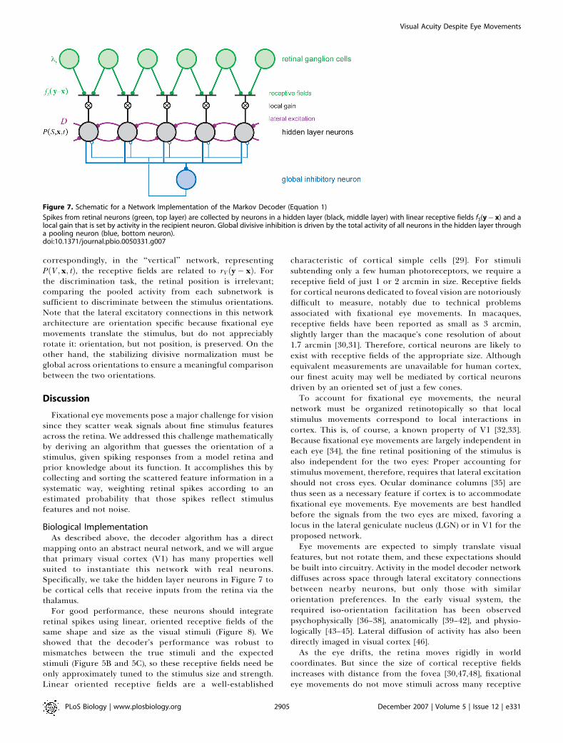
Figure 7 depicts a network that implements the Markovdecoder algorithm for estimating the location of a stimuluswith a known orientation S. The network has three types ofneurons: the retinal neurons, a hidden layer of decoder
neurons, and an inhibitory neuron. Each neuron in thehidden layer is associated with a spatial location, x, and itsactivity at time t represents the estimated posterior proba-bility (up to a normalization factor) that the stimulus ispresent at that location, PðS; x; tÞ. The feedforward input toeach hidden layer neuron x consists of spikes from retinallocations y, weighted by a spatial receptive fieldfSðy� xÞ ¼ lnðrSðy� xÞ=r0Þ, which ranges from zero far fromthe stimulus to a peak of lnðrmax=r0Þ. The weighted retinalinput is then multiplied by a variable gain proportional to theactivity of the postsynaptic neuron, PðS; x; tÞ. This gatedretinal input implements the contribution of Equation 2 tothe update of the estimated posterior probability. Theneurons in the network interact through lateral connectionsmimicking the diffusion operator (Equation 4 and Figure 3C).Recall that the diffusion operator takes the summedprobability of the nearest-neighbors of a given location,P
Dx PðS; xþ Dx; tÞ, and subtracts 4PðS; x; tÞ from this inorder to conserve probability. In the network, conservationof activity is not required, so the subtraction can be omitted:when the change in P is simply proportional to P, the solutionis an exponential decay that scales P uniformly at alllocations, leaving the relative values of the activity unaltered.Thus, lateral excitatory connections are sufficient to imple-ment the diffusion term in the network. For the same reason,the network does not need any representation of the localdecay term, Equation 3, which also scales all activities equally.Finally, the network includes a global divisive inhibition tomaintain network activity at a stable level despite the variousexcitatory interactions.To extend this framework to the discrimination task, we
need two copies of the network that differ by their orientationtuning (Figure 8). In the ‘‘horizontal’’ network, representingPðH; x; tÞ, the neurons are tuned to horizontal stimuli, hencetheir receptive fields are determined by rH(y� x) (Figure 3B);
Figure 6. Markov Decoder Robustness to Mismatched Parameters
(A) Discrimination performance when the decoder’s estimate for the trajectory statistics is wrong: The stimulus is known to perform a random walk onthe retina, but the diffusion constant is misestimated. The performance is optimal for estimated values close to the actual diffusion constant anddeclines gently on either side.(B) Performance as a function of the expected stimulated firing rate, parameterized as ðrest
max � r0Þ=ðrmax � r0Þ.(C) Performance as a function of the expected stimulus size, obtained by convolving the true stimulus shape with a spatial Gaussian of the specifiedradius. In each of these plots, parameters are the same as in Figure 5.doi:10.1371/journal.pbio.0050331.g006
PLoS Biology | www.plosbiology.org December 2007 | Volume 5 | Issue 12 | e3312904
Visual Acuity Despite Eye Movements
correspondingly, in the ‘‘vertical’’ network, representingPðV ; x; tÞ, the receptive fields are related to rV ðy� xÞ. Forthe discrimination task, the retinal position is irrelevant;comparing the pooled activity from each subnetwork issufficient to discriminate between the stimulus orientations.Note that the lateral excitatory connections in this networkarchitecture are orientation specific because fixational eyemovements translate the stimulus, but do not appreciablyrotate it: orientation, but not position, is preserved. On theother hand, the stabilizing divisive normalization must beglobal across orientations to ensure a meaningful comparisonbetween the two orientations.
Discussion
Fixational eye movements pose a major challenge for visionsince they scatter weak signals about fine stimulus featuresacross the retina. We addressed this challenge mathematicallyby deriving an algorithm that guesses the orientation of astimulus, given spiking responses from a model retina andprior knowledge about its function. It accomplishes this bycollecting and sorting the scattered feature information in asystematic way, weighting retinal spikes according to anestimated probability that those spikes reflect stimulusfeatures and not noise.
Biological ImplementationAs described above, the decoder algorithm has a direct
mapping onto an abstract neural network, and we will arguethat primary visual cortex (V1) has many properties wellsuited to instantiate this network with real neurons.Specifically, we take the hidden layer neurons in Figure 7 tobe cortical cells that receive inputs from the retina via thethalamus.
For good performance, these neurons should integrateretinal spikes using linear, oriented receptive fields of thesame shape and size as the visual stimuli (Figure 8). Weshowed that the decoder’s performance was robust tomismatches between the true stimuli and the expectedstimuli (Figure 5B and 5C), so these receptive fields need beonly approximately tuned to the stimulus size and strength.Linear oriented receptive fields are a well-established
characteristic of cortical simple cells [29]. For stimulisubtending only a few human photoreceptors, we require areceptive field of just 1 or 2 arcmin in size. Receptive fieldsfor cortical neurons dedicated to foveal vision are notoriouslydifficult to measure, notably due to technical problemsassociated with fixational eye movements. In macaques,receptive fields have been reported as small as 3 arcmin,slightly larger than the macaque’s cone resolution of about1.7 arcmin [30,31]. Therefore, cortical neurons are likely toexist with receptive fields of the appropriate size. Althoughequivalent measurements are unavailable for human cortex,our finest acuity may well be mediated by cortical neuronsdriven by an oriented set of just a few cones.To account for fixational eye movements, the neural
network must be organized retinotopically so that localstimulus movements correspond to local interactions incortex. This is, of course, a known property of V1 [32,33].Because fixational eye movements are largely independent ineach eye [34], the fine retinal positioning of the stimulus isalso independent for the two eyes: Proper accounting forstimulus movement, therefore, requires that lateral excitationshould not cross eyes. Ocular dominance columns [35] arethus seen as a necessary feature if cortex is to accommodatefixational eye movements. Eye movements are best handledbefore the signals from the two eyes are mixed, favoring alocus in the lateral geniculate nucleus (LGN) or in V1 for theproposed network.Eye movements are expected to simply translate visual
features, but not rotate them, and these expectations shouldbe built into circuitry. Activity in the model decoder networkdiffuses across space through lateral excitatory connectionsbetween nearby neurons, but only those with similarorientation preferences. In the early visual system, therequired iso-orientation facilitation has been observedpsychophysically [36–38], anatomically [39–42], and physio-logically [43–45]. Lateral diffusion of activity has also beendirectly imaged in visual cortex [46].As the eye drifts, the retina moves rigidly in world
coordinates. But since the size of cortical receptive fieldsincreases with distance from the fovea [30,47,48], fixationaleye movements do not move stimuli across many receptive
Figure 7. Schematic for a Network Implementation of the Markov Decoder (Equation 1)
Spikes from retinal neurons (green, top layer) are collected by neurons in a hidden layer (black, middle layer) with linear receptive fields fS(y� x) and alocal gain that is set by activity in the recipient neuron. Global divisive inhibition is driven by the total activity of all neurons in the hidden layer througha pooling neuron (blue, bottom neuron).doi:10.1371/journal.pbio.0050331.g007
PLoS Biology | www.plosbiology.org December 2007 | Volume 5 | Issue 12 | e3312905
Visual Acuity Despite Eye Movements

fields in the periphery. Accordingly, there is no need tocompensate for fixational eye movements in the periphery.We expect, therefore, to see some aspects of the corticalnetwork that are specialized for foveal vision. Consistent withthis, more of striate cortex is dedicated to responses from thefovea than can be explained by the density of retinal ganglioncells [49–51], and lateral suppression and facilitation differbetween central and peripheral vision [37].
The Markov decoder requires that the lateral facilitatoryinteractions induce localized changes in the gain for newinput spikes. Such multiplicative gain modulations haveindeed been observed in the visual cortex [52,53]. A numberof neural mechanisms have been invoked to create neuralmultipliers [54–60]. One potential mechanism involves thepostsynaptic NMDA (n-methyl-d-aspartic acid) receptor, aglutamate-gated ion channel with a voltage sensitivity thatcauses it to open only when the postsynaptic potential issufficiently large. In the visual cortex, NMDA activation hasbeen shown to produce a multiplicative effect on input gain[61]. Synapses between cortical layers and within layers havedifferent NMDA and AMPA (alpha-amino-3-hydroxy-5-meth-yl-4-isoxazole propionic acid) receptor distributions, so thatlateral inputs may be simply additive, whereas feedforwardinput may experience a variable gain [62], as required by theMarkov decoder architecture.
With an accelerating nonlinearity and excitatory interac-tions, this network has a positive feedback loop that would
cause the activity to quickly diverge. Normalization willmaintain stability, but the normalization must be global andorientation independent so that neural activities can becompared on the same scale. Previously described wide-fielddivisive normalization [63–66] can serve this purpose,although other global homeostatic mechanisms would func-tion as well.In our forced-choice task, the accumulated evidence for the
horizontal and vertical stimuli must be compared. This can beaccomplished downstream by a final winner-take-all compu-tation in which the total activity in each subnetwork is pooledand then compared [67]. This type of computation must takeplace somewhere in the brain any time a decision must bereached, and various biological implementations have beenproposed for this operation [68,69].Whereas the input to the network consists of discrete
spikes, the network units themselves represent the stimulusprobability, which is a continuous variable. This variablemight be most simply encoded by the collective firing rate ofa cluster of neurons [70], especially given that the number ofcells representing the visual field expands dramatically fromthe retina to the visual cortex [71]. Alternatively, thecomputation might well proceed with discrete spikes: modelnetworks of spiking neurons tend to produce similar behavioras rate models with continuous variables, so long as the spikesare not too strongly correlated [72].In summary, all the key elements of a Markov decoder for
short line segments are present in the neural circuitry ofprimary visual cortex. One essential feature, namely monoc-ular processing, is no longer available beyond V1. Wetherefore propose that V1 functions as a dynamic networkto accumulate information on fine stimulus features in theface of fixational eye movements.
Human Performance versus Model PerformanceWe presented psychophysical results indicating that human
subjects could reliably discriminate between horizontal andvertical stimuli measuring 1 3 2 arcmin (100% accuracy;Figure 2), but that the task was barely achievable when thestimulus was half that size (70% accuracy). Using biologicallyreasonable parameters, a Markov decoder of retinal spiketrains attains comparable, but slightly weaker, performance(90% and 60%, respectively; Figure 5). What additionalinformation do humans have that might account for thisdiscrepancy? Here, we consider several aspects of realisticvisual processing that were ignored by the Markov decoder.We treated only Off-type retinal ganglion cells, but there
are equally many On-type cells in the fovea, and in principle,they could also contribute to discrimination. An On cell issuppressed when a small, dark stimulus on a light backgroundenters its receptive field, and is then excited when thestimulus exits. These responses are unreliable because thereduction in firing rate from the background of 10 Hz isdetectable only after 100 ms of silence, and the excitatoryresponse is slow and weak. We explored this further withexplicit simulation of both On and Off cells: The decoderperformance improved very little (unpublished data), lessthan required to fully account for human acuity.Human fixational eye movements are not exactly random
walks. Instead, they exhibit some small persistence of velocityon a timescale of 2 ms and antipersistence on a timescale of100 ms [25,73]. To explore how these details affect the Markov
Figure 8. Two Independent but Competing Subnetworks, Each
Structured as in Figure 7, Receive Input from the Same Retinal Ganglion
Cells, but Use Different Receptive Fields
The total activity in each subnetwork is pooled by two readout neurons.The more active readout neuron indicates the network’s estimate of thestimulus orientation.doi:10.1371/journal.pbio.0050331.g008
PLoS Biology | www.plosbiology.org December 2007 | Volume 5 | Issue 12 | e3312906
Visual Acuity Despite Eye Movements
decoder’s discrimination performance, we performed addi-tional simulations. Antipersistence at long times can beexplained by occasional microsaccades that periodicallydeflect the eye toward its starting position. Such occasionaljerks of the image hardly affected a Markov decoder ignorantof microsaccades (unpublished data): After each stimulusjump, there was only a slight delay until the tails of thediffusing posterior distribution encountered the elevatedspike rate at the new stimulus location. The persistence of eyemovements at short times is consistent with velocitycorrelations lasting just a few milliseconds [73]. For a givendiffusion constant, a persistent random walk lingers longer ateach retinal location than a pure Markov random walk,leading to slightly stronger responses. Correspondingly,simulations showed that the Markov decoder’s performanceimproves modestly with the introduction of a short persis-tence time (unpublished data).
As discussed above, the Markov decoder is suboptimalbecause of the temporal blurring of the stimulus before spikegeneration. The optimal decoder must keep track of allpossible histories affecting the current firing rate, rather thanonly the last stimulus position, and the computational effortrapidly becomes prohibitive. Strategies have been proposedto simplify the decoding of such processes [74,75], but theserequire complicated learning algorithms and do not lendthemselves to straightforward neural implementation. Asimpler strategy for improving the Markov decoder mightbe to first process the retinal spike trains with a temporalfilter designed to ‘‘undo’’ temporal integration in the retina.This could plausibly take place in the thalamus [76].
Finally, the real visual system enjoys two additional benefitsthat were not available to the Markov decoder. The first isglobal image motion: Our human observers viewed the tinybar stimuli on a white sheet posted within a laboratory scene.As the eye moves, this peripheral background image movescoherently upon the retina, providing additional globalmotion cues that the brain could perhaps incorporate to
improve perception. Second, our model for retinal responsesused the most-random spike pattern for a given firing rate,namely a Poisson process. By contrast, real retinal ganglioncells fire more precisely [11,12] and could thus be moreinformative, even for a Markov decoder.
Are Fixational Eye Movements Helpful or Harmful?One commonly held view is that fixational eye movements
actually improve vision by preventing the decay of retinalresponses that occurs under static stimuli [20]. For example,Rucci and Desbordes have demonstrated that for moderatelylarge, noisy stimuli, orientation discrimination is worse whenthe image is stabilized on the retina, a result they attribute toa loss of the image motion that would otherwise refresh, andpossibly structure, neural activity [77]. In contrast, here wehave described these eye movements as a hindrance ratherthan a help. The transient nature of retinal ganglion cellresponses does imply that a fixed stimulus will elicit fewerspikes than a moving stimulus, diminishing the signal that thebrain receives. But if the eye movements are too large, thenthe light intensity is spread thinly over many cells, decreasingeach individual response while increasing the positionaluncertainty and thus the noise [78]. Between the limits ofno eye movement and very large eye movements, an optimumexists. This should occur with eye movements that shift thestimulus to a new set of retinal ganglion cells just as the initialresponse starts to truncate, and no sooner. For a stimulusarea s and transient response duration s, this occurs when thediffusion constant is D; s=4s. For tiny stimuli (s ¼ 0.5 3 1arcmin2) and biphasic temporal kernels with s ¼ 35 ms, thepredicted optimum of D ; 3 arcmin2/s is more than a fullorder of magnitude smaller than the naturally occurring eyemovements of approximately 100 arcmin2/s.To explore this further, we computed the Markov decoder’s
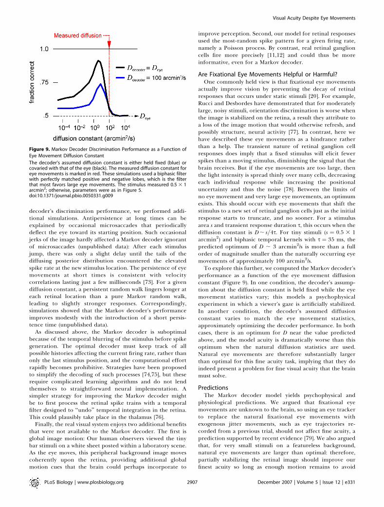
performance as a function of the eye movement diffusionconstant (Figure 9). In one condition, the decoder’s assump-tion about the diffusion constant is held fixed while the eyemovement statistics vary; this models a psychophysicalexperiment in which a viewer’s gaze is artificially stabilized.In another condition, the decoder’s assumed diffusionconstant varies to match the eye movement statistics,approximately optimizing the decoder performance. In bothcases, there is an optimum for D near the value predictedabove, and the model acuity is dramatically worse than thisoptimum when the natural diffusion statistics are used.Natural eye movements are therefore substantially largerthan optimal for this fine acuity task, implying that they doindeed present a problem for fine visual acuity that the brainmust solve.
PredictionsThe Markov decoder model yields psychophysical and
physiological predictions. We argued that fixational eyemovements are unknown to the brain, so using an eye trackerto replace the natural fixational eye movements withexogenous jitter movements, such as eye trajectories re-corded from a previous trial, should not affect fine acuity, aprediction supported by recent evidence [79]. We also arguedthat, for very small stimuli on a featureless background,natural eye movements are larger than optimal: therefore,partially stabilizing the retinal image should improve ourfinest acuity so long as enough motion remains to avoid
Figure 9. Markov Decoder Discrimination Performance as a Function of
Eye Movement Diffusion Constant
The decoder’s assumed diffusion constant is either held fixed (blue) orcovaried with that of the eye (black). The measured diffusion constant foreye movements is marked in red. These simulations used a biphasic filterwith perfectly matched positive and negative lobes, which is the filterthat most favors large eye movements. The stimulus measured 0.5 3 1arcmin2; otherwise, parameters were as in Figure 5.doi:10.1371/journal.pbio.0050331.g009
PLoS Biology | www.plosbiology.org December 2007 | Volume 5 | Issue 12 | e3312907
Visual Acuity Despite Eye Movements

prematurely truncating retinal responses (Figure 9). Althoughdiscrimination of larger stimuli does benefit from eyemovements [79], there are indications that fine acuity isimproved by stabilization [80].
There are two major physiological predictions. First,activity in V1 neurons should locally modulate the gain forfeedforward input originating from the retina. Without thismodulation, the advantage of using prior expectations is lost.Second, if the neural interactions in V1 are to correctlyencode the probabilistic expectations given by random walkeye movement statistics, then the interactions should imple-ment a diffusion operator, which entails that the time delayto reach maximal interaction strength should scale as thesquare of the interaction distance. This should be observableboth directly, as lateral excitatory currents, and indirectly,through the time course of the resulting gain modulation.
The Bayesian FrameworkThe essential aspect of the Markov decoder we have
described is that information of one type attunes the observerto other, related information. In the present context, thedecoder expects that responses to oriented line segments arecorrelated across space and time due to fixational eyemovements, and thus these expected responses are enhanced.Other statistical regularities produce expectations as well.For example, strings of line segments often occur together incontours. Correspondingly, collinear iso-orientation facilita-tion has been hypothesized to subserve contour integration[41,81], and can be viewed as another instance of theprinciple of enhancing responses to expected signals. Moregenerally, expectations should increase the gain for informa-tion that is relevant to the current task, but when thatinformation is irrelevant, then expectations may insteadreduce the gain.
The probabilistic processing of information has generatedsubstantial interest as a general framework for neuralcomputation, often designated ‘‘Bayesian computation’’ dueto the use of Bayes’ rule in calculating probabilities. Humanperception has been shown in several conditions to behaveaccording to this rule [82–84]. Experimental evidence alsohints that the cortex may be implementing Bayesianinference on a neural level [85]. Modeling studies havesuggested how networks of neurons could make theseprobabilistic inferences [75,86–89]. One study of particularrelevance also describes a neural network for approximatelyBayesian decoding of arbitrary hidden Markov processes [90].
Although our mathematical formalism is closely related toprevious work, we have made several advances in applying theBayesian paradigm. First, we identified a concrete biologicalpuzzle of considerable practical importance: how can humanssee with high acuity when fixational eye movements rapidlyjitter the stimulus over a large area? Second, previousBayesian computations treated neural signals that werepoorly constrained by experiment, so the performance ofthese computations could be characterized only qualitatively.In contrast, retinal signals are well studied, enabling us tomake quantitative comparisons between model and humanperformance. Third, previous studies predominantly de-scribed the formal structure of Bayesian computations,whereas we identified a simple and biologically plausiblemapping of the probabilistic calculations onto corticalcircuitry.
OutlookThe decoder we have described is optimized for discrim-
inating the orientation of line segments, but human acuityextends to more complex tasks, such as telling ‘‘F’’ from ‘‘P.’’Within our formalism, optimal discrimination of arbitraryshapes would require receptive fields tuned to those shapes,whereas the early visual system appears to encode orientededges, with more complex feature selectivity arising only laterin higher brain regions. Therefore, this Markov decoder byitself cannot account for discrimination in complex acuitytasks. However, we propose that it functions as a usefulpreprocessor that reduces the confounding effects of fixa-tional eye movements before passing signals to subsequentcortical regions for high-level processing.If the stimulus contains several lines of multiple orienta-
tions, the decoder’s output will have several peaks thatcorrespond to the individual oriented segments. These peakswill track the stimulus pattern as it is scanned over the retina.This output can then be processed by subsequent networkstuned to more complex patterns. Simulations show that sucha pattern detector identifies an arrangement of oriented barsbetter when it is provided with the output of a Markovdecoder than with signals from similar decoders that fail toproperly account for eye movements (see figure in ProtocolS1). Thus, the Markov decoder elaborates the conventionalmodel of V1 as extracting oriented image elements, andimproves over this static model through dynamic processingthat partially corrects for eye movements.In real-world acuity tasks, we do not perceive the incessant
motion of the image upon our retinas, but rather perceive astable image in world coordinates. Nonetheless, our internalrepresentation early in the visual pathway stores visualinformation in a retinal coordinate system [91]. This movingframe of reference must eventually be superseded before ourstable perceptions arise and decisions are reached. Thenetwork proposed here could be viewed as creating anintermediate coordinate system: the most current informa-tion is represented in retinal coordinates, but the nonlinearoperations of the network effectively shift the past retinalcoordinates into improved alignment. We may view thisneural computation as a step towards invariant worldcoordinates.
Materials and Methods
Psychophysics. Three groups of small horizontal and verticalstimuli like those in Figure 2 were presented at a distance of 4 m andwere scaled to subtend the angles 0.5 3 1, 0.75 3 1.5, and 1 3 2arcmin2. Stimuli were printed in black ink on white paper. Ambientlighting generated a luminance of 86 candelas/m2 for the whitebackground, and 20-fold dimmer for the black stimuli. Room featuresprovided global motion cues, which we did not seek to eliminate.Nine subjects were asked to discriminate between the stimuli whilestanding, and were not provided with error feedback. Subjects werefree to view the stimuli as long as they liked, typically taking a fewseconds per stimulus. Performance was reported as the fraction ofcorrect answers out of 32 attempts for each condition. Error barswere given as 68% confidence interval around the mean, assuming abinomial distribution of correct guesses and a uniform prior over thefraction correct. Other experiments with briefly flashed stimulishowed that reliable discrimination was already achieved within 500ms (unpublished data).
Simulations. We generated model retinal responses for thediscrimination task in the following steps: the stimulus orientationS was chosen randomly to be either horizontal or vertical, a randomwalk trajectory was constructed, and the stimulus light intensityprofile was moved along this random walk trajectory; the dynamic
PLoS Biology | www.plosbiology.org December 2007 | Volume 5 | Issue 12 | e3312908
Visual Acuity Despite Eye Movements

light intensity at each retinal position was filtered by a temporalkernel, then passed through a threshold rectifier to yield theinstantaneous firing rate; this rate drove an inhomogeneous Poissongenerator to produce the spike train for the retinal neuron at thatlocation. We passed these spikes to the Markov decoder implement-ing Equation 1, which returned a guess of the stimulus identity. Thesesteps are depicted in Figure 3A and described in detail below.
In both the simulations of retinal spike trains and in the Markovdecoder, we modeled the fovea as a square lattice of conephotoreceptors. In the human retina, cones are spaced every 0.5arcmin, and the receptive fields of retinal ganglion cells each consistof a single cone. Correspondingly, the model ganglion cells hadsquare receptive fields separated by 0.5 arcmin. For numerical work,we simulated a 16 3 16 arcmin2 array with wraparound boundaryconditions, which was sufficiently large for the relevant values of thediffusion constant and the diffusion time, yet small enough for fastsimulations.
The stimulus itself consisted of a rectangle with size z and a 1 3 2aspect ratio oriented in either the vertical or horizontal direction.Optical blur was produced by convolving the stimulus with aGaussian modulation transfer function of diameter 2r ¼ 0.5 arcmin[10]. The stimulus at location x induced an instantaneous spatial lightabsorption profile at retinal positions y of
ISðy; xÞ ¼ e�jyj2=2r2
*U1;1ðyÞ*Uz;2zðx� yÞ; ð5Þ
where * denotes a convolution operation, and Ua;bðxÞ represents atwo-dimensional box profile with dimensions a and b. The resultantstimulus profile is shown in Figure 3B.
We modeled fixational eye movements as a random walk thatshifts the stimulus across the retina. The one-sided power spectrumof a one-dimensional random walk is given by D=p2f 2, where f is thetemporal frequency. Eizenman et al. [3] reported one-sided powerspectra with f �2 dependence for the horizontal component offixational eye movements, from which we inferred a two-dimen-sional diffusion constant of D¼100 arcmin2/s. Corroborating resultscome from direct measurements of squared eye displacement as afunction of time lag [25]; fitting these data with a straight line ofslope 4D expected from a random walk yielded diffusion constantsof the same magnitude, 100 arcmin2/s.
We simulated the trajectory of the stimulus as a random walk on adiscrete spatial lattice, but continuous in time. After an infinitesimaltime interval dt, the probability of stepping to a nearest neighborlocation is dt � D=a2, where D is the diffusion constant, and a thedistance between lattice points. After many such time steps over afinite interval Dt, the probability that the walker has moved a distanceDx horizontally and Dy vertically can be expressed in series form:
PðDx;Dy;DtÞ ¼ FðDx;DtÞ � FðDy;DtÞ
FðDx;DtÞ ¼ 1N
XN�1j¼0
exp i2pjDxNa
� �exp � 2DDt
a21� cos2p
jN
� �� �; ð6Þ
where N is the number of points on a side of the square lattice. Forspeedy simulations, we chose a constant sampling interval Dt¼ 0.7 msand drew independent random walk steps from this distribution;finer temporal sampling produced nearly identical results (unpub-lished data).
The spatial stimulus profile was moved around the model retinaaccording to the random walk. This produced a temporal sequence oflight intensities within each retinal ganglion cell’s receptive field,which was then convolved with the parameterized biphasic temporalfilter (Figure 3D)
hðtÞ ¼ tn
snþ11
e�t=s1 � qtn
snþ12
e�t=s2 ð7Þ
to produce a temporally blurred stimulus (Figure 4F). The parameterswere chosen as s1 ¼ 5 ms, s2 ¼ 15 ms, n ¼ 3, and q ¼ 0.8 for allsimulations [28] except Figure 9, for which q ¼ 1 to maximize theperformance improvement attributable to eye movements. Finally,this spatiotemporal profile was offset by the background firing rate r0,half-wave rectified to prevent negative firing rates, and scaled so thatthe maximum possible firing rate was given by rmax. The typical firing-rate parameters we used were r0 ¼ 10 Hz and rmax ¼ 100 Hz unlessotherwise specified.
The Markov decoder operated on one trial of all ganglion cell spiketrains to produce a guess for the stimulus identity, according to thedifferential equation (Equation 1). This equation can be solved
iteratively, moving from spike to spike. When neuron y produces aspike at time ty, the diffusion term (Equation 4) is negligiblecompared to the spiking term (Equation 2), so we have only
@
@tPðS; x; tÞ ¼ dðt� tyÞfSðy� xÞP S; x; tð Þ ð8Þ
Dividing both sides by PðS; x; tÞ and substituting fSðxÞ ¼ lnðrSðx=r0Þ,we see that
@
@tlnPðS; x; tÞ ¼ dðt� tyÞln
rSðy� xÞr0
: ð9Þ
Integrating the delta function over the spike from time t�y to timetþy we find that the log-probability jumps at spike times by lnðrSðx=r0Þ,which means that the probability itself is multiplied:
PðS; x; tþy Þ ¼rSðy� xÞ
r0PðS; x; t�y Þ ð10Þ
In the absence of spikes, only the terms of Equations 3 and 4contribute to the differential equation (Equation 1), so theprobability distribution PðS; x; tÞ both decays and diffuses laterallyacross space. Because the two oriented stimuli both produce the sametotal spike rate from the retinal array regardless of position, thedecay term (Equation 3) does not alter the relative probabilities, andwe therefore neglect it. The diffusion term (Equation 4) can beimplemented most efficiently in the spatial frequency domain~PðS; k; tÞ, where the diffusion operator Dr~ 2 simply multiplies itsoperand. The solution to
@
@t~PðS; k; tÞ ¼ Dr~ 2 ~PðS; k; tÞ ð11Þ
during a spike-free interval [t, t þ Dt] is
~PðS; k; tþ DtÞ ¼ exp DDtr~ 2h i
~PðS; k; tÞ: ð12Þ
For computational speed, we sampled the decoder’s activity every0.7 ms. Between samples, the probability distribution was multipliedin the Fourier domain according to Equation 12, and at the sampletimes, the probabilities were multiplied in the spatial domainfollowing Equation 10: once for each spike that occurred since thelast sample time. Thus we were able to execute the ideal observeralgorithm by multiplication alternately in the spatial domain and thefrequency domain. To ensure stability in the absence of the decayterm (Equation 3), at every sampling time, we rescaled the posteriorprobability by its sum,
PS;x PðS; x; tÞ, recovering a properly normal-
ized probability.These estimated posterior probabilities can be displayed as a
function of space and time, as in Figure 4. Or to reach a decision inthe discrimination task, we summed the probabilities over allpositions after the specified stimulus duration T to obtain theposterior probability for orientation, PðS;TÞ; the orientation with thegreatest probability counted as the decoder’s guess. By repeating thisprocess many times (104 iterations) and calculating the fraction ofcorrect trials, we quantified the performance for this ideal strategyfor various parameter sets, as plotted in Figures 5, 6, and 9.
Supporting Information
Protocol S1. The Derivation of the Markov Decoder Equation
Found at doi:10.1371/journal.pbio.0050331.sd001 (1.2 MB PDF).
Acknowledgments
The authors thank Ralf Engbert and Reinhold Kliegl for their eyemovement data, and Daniel Fisher, Maneesh Sahani, and ananonymous referee for helpful conversations and suggestions.
Author contributions. XP, HS, and MM conceived and designed theexperiments, analyzed the data, and wrote the paper. XP performedthe experiments.
Funding. XP and MM were supported by a National Institutes ofHealth grant. The work of HS was partially supported by a grant ofthe US-Israel Binational Science Foundation.
Competing interests. The authors have declared that no competinginterests exist.
PLoS Biology | www.plosbiology.org December 2007 | Volume 5 | Issue 12 | e3312909
Visual Acuity Despite Eye Movements

References1. Skavenski AA, Hansen RM, Steinman RM, Winterson BJ (1979) Quality of
retinal image stabilization during small natural and artificial body rotationsin man. Vision Res 19: 675–683.
2. Tomlinson RD (1990) Combined eye-head gaze shifts in the primate. III.Contributions to the accuracy of gaze saccades. J Neurophysiol 64: 1873–1891.
3. Eizenman M, Hallett PE, Frecker RC (1985) Power spectra for ocular driftand tremor. Vision Res 25: 1635–1640.
4. Guthrie BL, Porter JD, Sparks DL (1983) Corollary discharge providesaccurate eye position information to the oculomotor system. Science 221:1193–1195.
5. Donaldson IM (2000) The functions of the proprioceptors of the eyemuscles. Philos Trans R Soc Lond B Biol Sci 355: 1685–1754.
6. Murakami I, Cavanagh P (1998) A jitter after-effect reveals motion-basedstabilization of vision. Nature 395: 798–801.
7. Murakami I, Cavanagh P (2001) Visual jitter: evidence for visual-motion-based compensation of retinal slip due to small eye movements. Vision Res41: 173–186.
8. Schein SJ (1988) Anatomy of macaque fovea and spatial densities ofneurons in foveal representation. J Comp Neurol 269: 479–505.
9. Geisler WS (1984) Physical limits of acuity and hyperacuity. J Opt Soc Am A1: 775–782.
10. Geisler WS, Davila KD (1985) Ideal discriminators in spatial vision: two-point stimuli. J Opt Soc Am A 2: 1483–1497.
11. Berry MJ 2nd, Meister M (1998) Refractoriness and neural precision. JNeurosci 18: 2200–2211.
12. Uzzell VJ, Chichilnisky EJ (2004) Precision of spike trains in primate retinalganglion cells. J Neurophysiol 92: 780–789.
13. Chichilnisky EJ, Rieke F (2005) Detection sensitivity and temporalresolution of visual signals near absolute threshold in the salamanderretina. J Neurosci 25: 318–330.
14. Hennig MH, Worgotter F (2004) Eye micro-movements improve stimulusdetection beyond the nyquist limit in the peripheral retina. Adv Neural InfProcess Syst 16: 1475–1482.
15. Wachtler T, Wehrhahn C, Lee BB (1996) A simple model of human fovealganglion cell responses to hyperacuity stimuli. J Comput Neurosci 3: 73–82.
16. Croner L, Kaplan E (1995) Receptive fields of P and M ganglion cells acrossthe primate retina. Vision Res 35: 7–24.
17. Shapley RM, Victor JD (1978) The effect of contrast on the transferproperties of cat retinal ganglion cells. J Physiol 285: 275–298.
18. Frechette ES, Sher A, Grivich MI, Petrusca D, Litke AM, et al. (2005) Fidelityof the ensemble code for visual motion in primate retina. J Neurophysiol94: 119–135.
19. Troy JB, Lee BB (1994) Steady discharges of macaque retinal ganglion cells.Vis Neurosci 11: 111–118.
20. Martinez-Conde S, Macknik SL, Hubel DH (2004) The role of fixational eyemovements in visual perception. Nat Rev Neurosci 5: 229–240.
21. Winterson BJ, Collewijn H (1976) Microsaccades during finely guidedvisuomotor tasks. Vision Res 16: 1387–1390.
22. Kowler E, Steinman RM (1979) Miniature saccades: eye movements that donot count. Vision Res 19: 105–108.
23. Bridgeman B, Palca J (1980) The role of microsaccades in high acuityobservational tasks. Vision Res 20: 813–817.
24. Martinez-Conde S (2006) Fixational eye movements in normal andpathological vision. Prog Brain Res 154: 151–176.
25. Engbert R, Kliegl R (2004) Microsaccades keep the eyes’ balance duringfixation. Psychol Sci 15: 431–436.
26. Turing AM (1952) The chemical basis of morphogenesis. Phil Trans RoyalSoc Lon B 237: 37–72.
27. Schneeweis DM, Schnapf JL (1999) The photovoltage of macaque conephotoreceptors: adaptation, noise, and kinetics. J Neurosci 19: 1203–1216.
28. Chichilnisky EJ, Kalmar RS (2002) Functional asymmetries in ON and OFFganglion cells of primate retina. J Neurosci 22: 2737–2747.
29. Hubel DH, Wiesel TN (1959) Receptive fields of single neurones in the cat’sstriate cortex. J Physiol 148: 574–591.
30. Dow BM, Snyder AZ, Vautin RG, Bauer R (1981) Magnification factor andreceptive field size in foveal striate cortex of the monkey. Exp Brain Res 44:213–228.
31. Snodderly DM, Gur M (1995) Organization of striate cortex of alert, trainedmonkeys (Macaca fascicularis): ongoing activity, stimulus selectivity, andwidths of receptive field activating regions. J Neurophysiol 74: 2100–2125.
32. Talbot SA, Marshall WH (1941) Physiological studies on neural mechanismsof visual localization and discrimination. Amer J Ophthal 24: 1255–1263.
33. Tootell RB, Switkes E, Silverman MS, Hamilton SL (1988) Functionalanatomy of macaque striate cortex. II. Retinotopic organization. J Neurosci8: 1531–1568.
34. Steinman RM, Collewijn H (1980) Binocular retinal image motion duringactive head rotation. Vision Res 20: 415–429.
35. Hubel DH, Wiesel TN, Stryker MP (1978) Anatomical demonstration oforientation columns in macaque monkey. J Comp Neurol 177: 361–380.
36. Polat U, Sagi D (1993) Lateral interactions between spatial channels:suppression and facilitation revealed by lateral masking experiments.Vision Res 33: 993–999.
37. Xing J, Heeger DJ (2000) Center-surround interactions in foveal andperipheral vision. Vision Res 40: 3065–3072.
38. Adini Y, Sagi D, Tsodyks M (1997) Excitatory-inhibitory network in thevisual cortex: psychophysical evidence. Proc Natl Acad Sci U S A 94: 10426–10431.
39. Bosking WH, Zhang Y, Schofield B, Fitzpatrick D (1997) Orientationselectivity and the arrangement of horizontal connections in tree shrewstriate cortex. J Neurosci 17: 2112–2127.
40. Gilbert CD, Wiesel TN (1989) Columnar specificity of intrinsic horizontaland corticocortical connections in cat visual cortex. J Neurosci 9: 2432–2442.
41. Sincich LC, Blasdel GG (2001) Oriented axon projections in primary visualcortex of the monkey. J Neurosci 21: 4416–4426.
42. Malach R, Amir Y, Harel M, Grinvald A (1993) Relationship betweenintrinsic connections and functional architecture revealed by opticalimaging and in vivo targeted biocytin injections in primate striate cortex.Proc Natl Acad Sci U S A 90: 10469–10473.
43. Ts’o DY, Gilbert CD, Wiesel TN (1986) Relationships between horizontalinteractions and functional architecture in cat striate cortex as revealed bycross-correlation analysis. J Neurosci 6: 1160–1170.
44. Kapadia MK, Ito M, Gilbert CD, Westheimer G (1995) Improvement invisual sensitivity by changes in local context: parallel studies in humanobservers and in V1 of alert monkeys. Neuron 15: 843–856.
45. Polat U, Mizobe K, Pettet MW, Kasamatsu T, Norcia AM (1998) Collinearstimuli regulate visual responses depending on cell’s contrast threshold.Nature 391: 580–584.
46. Grinvald A, Lieke EE, Frostig RD, Hildesheim R (1994) Cortical point-spread function and long-range lateral interactions revealed by real-timeoptical imaging of macaque monkey primary visual cortex. J Neurosci 14:2545–2568.
47. Hubel DH, Wiesel TN (1974) Uniformity of monkey striate cortex: aparallel relationship between field size, scatter, and magnification factor. JComp Neurol 158: 295–305.
48. Wilson JR, Sherman SM (1976) Receptive-field characteristics of neurons incat striate cortex: changes with visual field eccentricity. J Neurophysiol 39:512–533.
49. Van Essen DC, Newsome WT, Maunsell JH (1984) The visual fieldrepresentation in striate cortex of the macaque monkey: asymmetries,anisotropies, and individual variability. Vision Res 24: 429–448.
50. Azzopardi P, Cowey A (1993) Preferential representation of the fovea in theprimary visual cortex. Nature 361: 719–721.
51. Azzopardi P, Cowey A (1996) The overrepresentation of the fovea andadjacent retina in the striate cortex and dorsal lateral geniculate nucleus ofthe macaque monkey. Neuroscience 72: 627–639.
52. McAdams CJ, Maunsell JH (1999) Effects of attention on the reliability ofindividual neurons in monkey visual cortex. Neuron 23: 765–773.
53. Treue S, Martinez Trujillo JC (1999) Feature-based attention influencesmotion processing gain in macaque visual cortex. Nature 399: 575–579.
54. Koch C, Poggio T (1992) Multiplying with synapses and neurons. In:McKenna T, Davis J, Zornetzer S, editors. Single neuron computation.Boston: Academic Press. pp. 315–345.
55. Mel BW (1993) Synaptic integration in an excitable dendritic tree. JNeurophysiol 70: 1086–1101.
56. Koch C, Segev I (2000) The role of single neurons in informationprocessing. Nat Neurosci 3: 1171–1177.
57. Chance FS, Abbott LF, Reyes AD (2002) Gain modulation from backgroundsynaptic input. Neuron 35: 773–782.
58. Murphy BK, Miller KD (2003) Multiplicative gain changes are induced byexcitation or inhibition alone. J Neurosci 23: 10040–10051.
59. Mehaffey WH, Doiron B, Maler L, Turner RW (2005) Deterministicmultiplicative gain control with active dendrites. J Neurosci 25: 9968–9977.
60. Gabbiani F, Krapp HG, Koch C, Laurent G (2002) Multiplicativecomputation in a visual neuron sensitive to looming. Nature 420: 320–324.
61. Fox K, Sato H, Daw N (1990) The Effect of varying stimulus intensity onNMDA-receptor activity in cat visual cortex. J Neurophysiol 64: 1413–1428.
62. Rivadulla C, Sharma J, Sur M (2001) Specific roles of NMDA and AMPAreceptors in direction-selective and spatial phase-selective responses invisual cortex. J Neurosci 21: 1710–1719.
63. Heeger DJ (1992) Normalization of cell responses in cat striate cortex. VisNeurosci 9: 181–197.
64. Carandini M, Heeger DJ, Movshon JA (1997) Linearity and normalization insimple cells of the macaque primary visual cortex. J Neurosci 17: 8621–8644.
65. Cavanaugh JR, Bair W, Movshon JA (2002) Nature and interaction of signalsfrom the receptive field center and surround in macaque V1 neurons. JNeurophysiol 88: 2530–2546.
66. Webb BS, Dhruv NT, Solomon SG, Tailby C, Lennie P (2005) Early and latemechanisms of surround suppression in striate cortex of macaque. JNeurosci 25: 11666–11675.
67. Lee DK, Itti L, Koch C, Braun J (1999) Attention activates winner-take-allcompetition among visual filters. Nat Neurosci 2: 375–381.
68. Coultrip R, Granger RH, Lynch G (1992) A cortical model of winner-take-all competition via lateral inhibition. Neural Netw 5: 47–54.
69. Anton PS, Granger RH, Lynch G (1992) Temporal information processing
PLoS Biology | www.plosbiology.org December 2007 | Volume 5 | Issue 12 | e3312910
Visual Acuity Despite Eye Movements

in synapses, cells, and circuits. In: McKenna T, Davis J, Zornetzer S, editors.Single neuron computation. Boston: Academic Press. pp. 291–313.
70. Shadlen MN, Newsome WT (1998) The variable discharge of corticalneurons: implications for connectivity, computation, and informationcoding. J Neurosci 18: 3870–3896.
71. Callaway EM (2005) Structure and function of parallel pathways in theprimate early visual system. J Physiol 566: 13–19.
72. Shriki O, Hansel D, Sompolinsky H (2003) Rate models for conductance-based cortical neuronal networks. Neural Comput 15: 1809–1841.
73. Mergenthaler K, Engbert R (2007) Modeling the control of fixational eyemovements with neurophysiological delays. Phys Rev Lett 98: 138104.
74. Huys QJM, Zemel RS, Natarajan R, Dayan P (2007) Fast population coding.Neural Comput 19: 460–497.
75. Zemel RS, Huys QJM, Natarajan R, Dayan P (2005) Probabilisticcomputation in spiking populations. In: Saul L, Weiss Y, Bottou L, editors.Advances in neural information processing systems 17. Cambridge(Massachusetts): MIT Press. pp. 1609–1616.
76. Dong DW, Atick JJ (1995) Temporal decorrelation: a theory of lagged andnonlagged responses in the lateral geniculate nucleus. Network 6: 159–178.
77. Rucci M, Desbordes G (2003) Contributions of fixational eye movements tothe discrimination of briefly presented stimuli. J Vis 3: 852–864.
78. Pelli DG (1985) Uncertainty explains many aspects of visual contrastdetection and discrimination. J Opt Soc Am A 2: 1508–1532.
79. Rucci M, Iovin R, Poletti M, Santini F (2007) Miniature eye movementsenhance fine spatial detail. Nature 447: 851–854.
80. Riggs LA, Ratliff F, Cornsweet JC, Cornsweet TN (1953) The disappearanceof steadily fixated visual test objects. J Opt Soc Am 43: 495–501.
81. Sigman M, Cecchi GA, Gilbert CD, Magnasco MO (2001) On a commoncircle: natural scenes and Gestalt rules. Proc Natl Acad Sci U S A 98: 1935–1940.
82. Brainard DH, Longere P, Delahunt PB, Freeman WT, Kraft JM, et al. (2006)Bayesian model of human color constancy. J Vis 6: 1267–1281.
83. Kording KP, Ku SP, Wolpert DM (2004) Bayesian integration in forceestimation. J Neurophysiol 92: 3161–3165.
84. Stocker AA, Simoncelli EP (2006) Noise characteristics and prior expect-ations in human visual speed perception. Nat Neurosci 9: 578–585.
85. Shadlen MN, Newsome WT (2001) Neural basis of a perceptual decision inthe parietal cortex (area LIP) of the rhesus monkey. J Neurophysiol 86:1916–1936.
86. Deneve S, Latham PE, Pouget A (2001) Efficient computation and cueintegration with noisy population codes. Nat Neurosci 4: 826–831.
87. Deneve S (2005) Bayesian inference in spiking neurons. Adv Neural InfProcess Syst 17: 353–360.
88. Rao RP (2005) Hierarchical Bayesian inference in networks of spikingneurons. Adv Neural Inf Process Syst 17: 1113–1120.
89. Ma WJ, Beck JM, Latham PE, Pouget A (2006) Bayesian inference withprobabilistic population codes. Nat Neurosci 9: 1432–1438.
90. Rao RP (2004) Bayesian computation in recurrent neural circuits. NeuralComput 16: 1–38.
91. Gur M, Snodderly DM (1997) Visual receptive fields of neurons in primaryvisual cortex (V1) move in space with the eye movements of fixation. VisionRes 37: 257–265.
92. Roorda A, Williams DR (1999) The arrangement of the three cone classes inthe living human eye. Nature 397: 520–522.
PLoS Biology | www.plosbiology.org December 2007 | Volume 5 | Issue 12 | e3312911
Visual Acuity Despite Eye Movements