Embed Size (px)
Citation preview

IOSR Journal of Dental and Medical Sciences (IOSR-JDMS)
e-ISSN: 2279-0853, p-ISSN: 2279-0861.Volume 13, Issue 8 Ver. I (Aug. 2014), PP 83-89 www.iosrjournals.org
www.iosrjournals.org 83 | Page
Pleomorphic Adenoma of Palate- A Case Report with CT and
CBCT Features.
Anuraag B. Choudhary1, Abhilasha O. Yadav
2, Mukta B. Motwani
3,
Mayur B.Chaudhary4, Shweta M. Chaudhary
5, Pankaj J. Banode
5
1Senior Lecturer, Department of Oral Medicine and Radiology, VSPM’s Dental College & Research Centre,
Digdoh hills, Nagpur Maharashtra (India). 2Reader, Department of Oral and Maxillo-facial Surgery, Sharad Pawar Dental College and Hospital,
DMIMSU, Sawangi (M), Wardha Maharashtra (India). 3Professor & Head, Department of Oral Medicine and Radiology, VSPM’s Dental college & research Centre,
Digdoh hills, Nagpur Maharashtra (India). 4Reader, Department of Oral Pathology and Microbiology, Bharti Vidyapeeth Dental College Pune
Maharashtra (India). 5Reader, Department of Pedodontia, Bharti Vidyapeeth Dental College Pune Maharashtra (India).
6Associate Professor, Department of Radiodiagnosis, AVBRH, co-ordinator TIFAC CORE Cathetrization Lab
Sawangi, Wardha Maharashtra (India).
Abstract: Pleomorphic adenoma (PA) can be defined as a benign mixed tumor composed of epithelial and
myoepithelial cells arranged with wide cytomorphologic and architectural diversity and morphological
patterns, demarcated from surrounding tissues by fibrous capsule. PA is one of the salivary gland tumors
affecting both major and minor salivary glands. Parotid gland is the most commonly affected of the major
group, and palate is the most common site of the minor salivary glands affected. The tumor has 3 components: an epithelial cell component; a myoepithelial cell component; and a mesenchymal component. The
identification of these 3 components, which may vary quantitatively from one tumor to another, is essential to
the recognition of pleomorphic adenoma. The tumor is also known as a benign mixed tumor. When these lesions
affects the minor salivary glands on palate and if there is any involvement of the surrounding structures like
maxillary antrum and nasal floor, then pre-operative imaging of these lesions is mandatory by 3-dimensional
imaging modalities like CT or CBCT to evaluate the exact depth and the dimensions of the lesion. Following
case of pleomorphic adenoma of palatal salivary glands which was evaluated with CT as well as CBCT pre-
operatively.
I. Introduction Salivary gland tumors are rare; comprising less than 3% of all neoplasm’s of the head and neck region
and are known by their complex microscopical features. 1, 2 Pleomorphic adenoma (PA) is the most common salivary gland tumor and represents 60% to 73% of the parotid gland tumors, 40% to 60% of the submandibular
and minor salivary glands tumors. 3, 4, 5 It is a benign neoplasm composed of epithelial and myoepithelial cells
arranged in a great variety of morphological patterns, with areas of mesenchymal differentiation. 6, 7 Epithelial
cells typically form duct-like structures associated with non-ductal cells presenting variable shapes and forms.
The stromal element demonstrates varying degrees of myxoid, hyaline, cartilaginous, or osseous differentiation. 6 Since the tumor is involving primarily the minor salivary glands, it does not show any radiographic features
initially when size of the lesion is small. Over a period of time when tumor grows large enough, it may cause
pressure resorption and ultimately destruction and perforation of the palatal bone.
Here we present a case of slow growing pleomorphic adenoma of minor salivary glands of palate,
which caused pressure resorption and perforation of palatal bone which was well visualized by Cone Beam
Computed Tomography (CBCT) technique, which gave an appropriate 3-dimensional images as well as the extent of the lesion alongwith the evidence of palatal perforation in left posterior palatal region.
II. Case report A 56 year female patient visited to the Department of Oral Medicine and Radiology, Sharad Pawar
Dental College and Hospital, Sawangi (Meghe), Wardha, Maharashtra, India with the chief complaint of painful
slow growing swelling on left posterior part since 1year. Her medical history was non-contributory. She gave
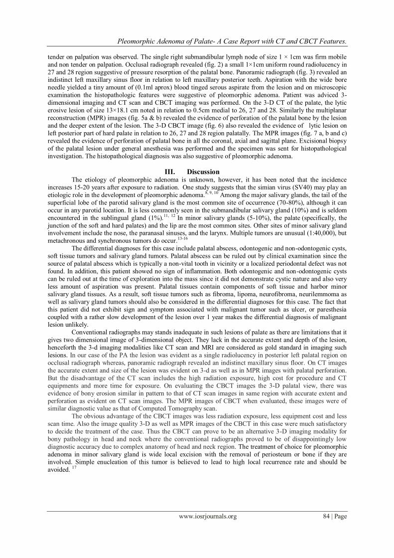
history that the swelling had been there for 1 year, but grew slowly in the past 6 months. Intraoral examination
(fig. 1) revealed a soft tissue mass 3×3 cm. in greatest diameter at the posterior left side of the hard palate. The
oral mucosa covering the lesion was intact and bluish pink in colour. The lesion was rubbery in consistency and
Pleomorphic Adenoma of Palate- A Case Report with CT and CBCT Features.
www.iosrjournals.org 84 | Page
tender on palpation was observed. The single right submandibular lymph node of size 1 × 1cm was firm mobile
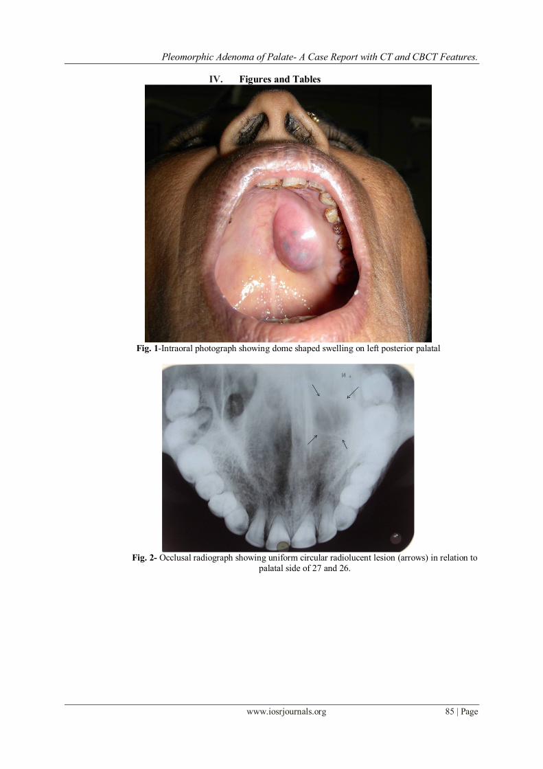
and non tender on palpation. Occlusal radiograph revealed (fig. 2) a small 1×1cm uniform round radiolucency in
27 and 28 region suggestive of pressure resorption of the palatal bone. Panoramic radiograph (fig. 3) revealed an indistinct left maxillary sinus floor in relation to left maxillary posterior teeth. Aspiration with the wide bore
needle yielded a tiny amount of (0.1ml aprox) blood tinged serous aspirate from the lesion and on microscopic
examination the histopathologic features were suggestive of pleomorphic adenoma. Patient was adviced 3-
dimensional imaging and CT scan and CBCT imaging was performed. On the 3-D CT of the palate, the lytic
erosive lesion of size 13×18.1 cm noted in relation to 0.5cm medial to 26, 27 and 28. Similarly the multiplanar
reconstruction (MPR) images (fig. 5a & b) revealed the evidence of perforation of the palatal bone by the lesion
and the deeper extent of the lesion. The 3-D CBCT image (fig. 6) also revealed the evidence of lytic lesion on
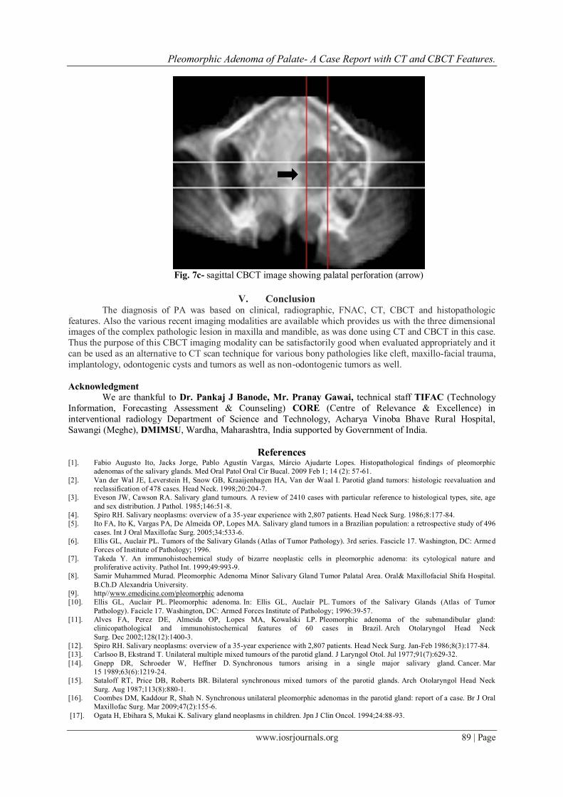
left posterior part of hard palate in relation to 26, 27 and 28 region palatally. The MPR images (fig. 7 a, b and c)
revealed the evidence of perforation of palatal bone in all the coronal, axial and sagittal plane. Excisional biopsy
of the palatal lesion under general anesthesia was performed and the specimen was sent for histopathological
investigation. The histopathological diagnosis was also suggestive of pleomorphic adenoma.
III. Discussion The etiology of pleomorphic adenoma is unknown, however, it has been noted that the incidence
increases 15-20 years after exposure to radiation. One study suggests that the simian virus (SV40) may play an
etiologic role in the development of pleomorphic adenoma.8, 9, 10 Among the major salivary glands, the tail of the
superficial lobe of the parotid salivary gland is the most common site of occurrence (70-80%), although it can
occur in any parotid location. It is less commonly seen in the submandibular salivary gland (10%) and is seldom
encountered in the sublingual gland (1%).11, 12 In minor salivary glands (5-10%), the palate (specifically, the
junction of the soft and hard palates) and the lip are the most common sites. Other sites of minor salivary gland involvement include the nose, the paranasal sinuses, and the larynx. Multiple tumors are unusual (1:40,000), but
metachronous and synchronous tumors do occur.13-16
The differential diagnoses for this case include palatal abscess, odontogenic and non-odontogenic cysts,
soft tissue tumors and salivary gland tumors. Palatal abscess can be ruled out by clinical examination since the
source of palatal abscess which is typically a non-vital tooth in vicinity or a localized periodontal defect was not
found. In addition, this patient showed no sign of inflammation. Both odontogenic and non-odontogenic cysts
can be ruled out at the time of exploration into the mass since it did not demonstrate cystic nature and also very
less amount of aspiration was present. Palatal tissues contain components of soft tissue and harbor minor
salivary gland tissues. As a result, soft tissue tumors such as fibroma, lipoma, neurofibroma, neurilemmoma as
well as salivary gland tumors should also be considered in the differential diagnoses for this case. The fact that
this patient did not exhibit sign and symptom associated with malignant tumor such as ulcer, or paresthesia
coupled with a rather slow development of the lesion over 1 year makes the differential diagnosis of malignant lesion unlikely.
Conventional radiographs may stands inadequate in such lesions of palate as there are limitations that it
gives two dimensional image of 3-dimensional object. They lack in the accurate extent and depth of the lesion,
henceforth the 3-d imaging modalities like CT scan and MRI are considered as gold standard in imaging such
lesions. In our case of the PA the lesion was evident as a single radiolucency in posterior left palatal region on
occlusal radiograph whereas, panoramic radiograph revealed an indistinct maxillary sinus floor. On CT images
the accurate extent and size of the lesion was evident on 3-d as well as in MPR images with palatal perforation.
But the disadvantage of the CT scan includes the high radiation exposure, high cost for procedure and CT
equipments and more time for exposure. On evaluating the CBCT images the 3-D palatal view, there was
evidence of bony erosion similar in pattern to that of CT scan images in same region with accurate extent and
perforation as evident on CT scan images. The MPR images of CBCT when evaluated, these images were of similar diagnostic value as that of Computed Tomography scan.
The obvious advantage of the CBCT images was less radiation exposure, less equipment cost and less
scan time. Also the image quality 3-D as well as MPR images of the CBCT in this case were much satisfactory
to decide the treatment of the case. Thus the CBCT can prove to be an alternative 3-D imaging modality for
bony pathology in head and neck where the conventional radiographs proved to be of disappointingly low
diagnostic accuracy due to complex anatomy of head and neck region. The treatment of choice for pleomorphic
adenoma in minor salivary gland is wide local excision with the removal of periosteum or bone if they are
involved. Simple enucleation of this tumor is believed to lead to high local recurrence rate and should be
avoided. 17
Pleomorphic Adenoma of Palate- A Case Report with CT and CBCT Features.
www.iosrjournals.org 85 | Page
IV. Figures and Tables
Fig. 1-Intraoral photograph showing dome shaped swelling on left posterior palatal
Fig. 2- Occlusal radiograph showing uniform circular radiolucent lesion (arrows) in relation to
palatal side of 27 and 26.

Pleomorphic Adenoma of Palate- A Case Report with CT and CBCT Features.
www.iosrjournals.org 86 | Page
Fig. 3- Cropped panoramic radiograph showing indistinct floor of left maxillary sinus (arrows)
Fig. 4- 3-D CT of hard palate showing erosion of palatal bone with measurement of size

Pleomorphic Adenoma of Palate- A Case Report with CT and CBCT Features.
www.iosrjournals.org 87 | Page
Fig. 5a & 5b- Coronal and axial CT scan images showing posterior extension and palatal perforation
(arrows)
Fig. 6- 3-D CBCT image of palatal bone showing round erosive lesion

Pleomorphic Adenoma of Palate- A Case Report with CT and CBCT Features.
www.iosrjournals.org 88 | Page
Fig. 7a- coronal CBCT image showing palatal bone perforation and soft tissue shadow of the lesion
Fig. 7b- axial CBCT image showing uniform round radiolucent lesion (arrow)
Pleomorphic Adenoma of Palate- A Case Report with CT and CBCT Features.
www.iosrjournals.org 89 | Page
Fig. 7c- sagittal CBCT image showing palatal perforation (arrow)
V. Conclusion The diagnosis of PA was based on clinical, radiographic, FNAC, CT, CBCT and histopathologic
features. Also the various recent imaging modalities are available which provides us with the three dimensional images of the complex pathologic lesion in maxilla and mandible, as was done using CT and CBCT in this case.
Thus the purpose of this CBCT imaging modality can be satisfactorily good when evaluated appropriately and it
can be used as an alternative to CT scan technique for various bony pathologies like cleft, maxillo-facial trauma,
implantology, odontogenic cysts and tumors as well as non-odontogenic tumors as well.
Acknowledgment
We are thankful to Dr. Pankaj J Banode, Mr. Pranay Gawai, technical staff TIFAC (Technology
Information, Forecasting Assessment & Counseling) CORE (Centre of Relevance & Excellence) in
interventional radiology Department of Science and Technology, Acharya Vinoba Bhave Rural Hospital,
Sawangi (Meghe), DMIMSU, Wardha, Maharashtra, India supported by Government of India.
References [1]. Fabio Augusto Ito, Jacks Jorge, Pablo Agustín Vargas, Márcio Ajudarte Lopes. Histopathological findings of pleomorphic
adenomas of the salivary glands. Med Oral Patol Oral Cir Bucal. 2009 Feb 1; 14 (2): 57-61.
[2]. Van der Wal JE, Leverstein H, Snow GB, Kraaijenhagen HA, Van der Waal I. Parotid gland tumors: histologic reevaluation and
reclassification of 478 cases. Head Neck. 1998;20:204-7.
[3]. Eveson JW, Cawson RA. Salivary gland tumours. A review of 2410 cases with particular reference to histological types, site, age
and sex distribution. J Pathol. 1985;146:51-8.
[4]. Spiro RH. Salivary neoplasms: overview of a 35-year experience with 2,807 patients. Head Neck Surg. 1986;8:177-84.
[5]. Ito FA, Ito K, Vargas PA, De Almeida OP, Lopes MA. Salivary gland tumors in a Brazilian population: a retrospective study of 496
cases. Int J Oral Maxillofac Surg. 2005;34:533-6.
[6]. Ellis GL, Auclair PL. Tumors of the Salivary Glands (Atlas of Tumor Pathology). 3rd series. Fascicle 17. Washington, DC: Armed
Forces of Institute of Pathology; 1996.
[7]. Takeda Y. An immunohistochemical study of bizarre neoplastic cells in pleomorphic adenoma: its cytological nature and
proliferative activity. Pathol Int. 1999;49:993-9.
[8]. Samir Muhammed Murad. Pleomorphic Adenoma Minor Salivary Gland Tumor Palatal Area. Oral& Maxillofacial Shifa Hospital.
B.Ch.D Alexandria University.
[9]. http//www.emedicine.com/pleomorphic adenoma
[10]. Ellis GL, Auclair PL. Pleomorphic adenoma. In: Ellis GL, Auclair PL. Tumors of the Salivary Glands (Atlas of Tumor
Pathology). Facicle 17. Washington, DC: Armed Forces Institute of Pathology; 1996:39-57.
[11]. Alves FA, Perez DE, Almeida OP, Lopes MA, Kowalski LP. Pleomorphic adenoma of the submandibular gland:
clinicopathological and immunohistochemical features of 60 cases in Brazil. Arch Otolaryngol Head Neck
Surg. Dec 2002;128(12):1400-3.
[12]. Spiro RH. Salivary neoplasms: overview of a 35-year experience with 2,807 patients. Head Neck Surg. Jan-Feb 1986;8(3):177-84.
[13]. Carlsoo B, Ekstrand T. Unilateral multiple mixed tumours of the parotid gland. J Laryngol Otol. Jul 1977;91(7):629-32.
[14]. Gnepp DR, Schroeder W, Heffner D. Synchronous tumors arising in a single major salivary gland. Cancer. Mar
15 1989;63(6):1219-24.
[15]. Sataloff RT, Price DB, Roberts BR. Bilateral synchronous mixed tumors of the parotid glands. Arch Otolaryngol Head Neck
Surg. Aug 1987;113(8):880-1.
[16]. Coombes DM, Kaddour R, Shah N. Synchronous unilateral pleomorphic adenomas in the parotid gland: report of a case. Br J Oral
Maxillofac Surg. Mar 2009;47(2):155-6.
[17]. Ogata H, Ebihara S, Mukai K. Salivary gland neoplasms in children. Jpn J Clin Oncol. 1994;24:88-93.


![[PAPER] Pleomorphic Adenoma Print.docx](https://img.dokumen.tips/doc/110x75/56d6bd9b1a28ab30168ea546/paper-pleomorphic-adenoma-printdocx.jpg)