Embed Size (px)
Citation preview

OR I G INA L ART I C L E
PLEKHM2 mutation leads to abnormal localizationof lysosomes, impaired autophagy flux and associateswith recessive dilated cardiomyopathy and leftventricular noncompactionEmad Muhammad1,†, Aviva Levitas2,†, Sonia R. Singh3,4, Alex Braiman1,Rivka Ofir5, Sharon Etzion5, Val C. Sheffield6, Yoram Etzion5,7, Lucie Carrier3,4
and Ruti Parvari1,8,*1Department ofMicrobiology, ImmunologyandGenetics, Faculty ofHealth Sciences, Ben-GurionUniversityof theNegev, Beer-Sheva 84105, Israel, 2Department of Pediatric Cardiology, Soroka University Medical Center andFaculty of Health Sciences, Ben-Gurion University of the Negev, Beer-Sheva 84101, Israel, 3Department ofExperimental Pharmacology and Toxicology, Cardiovascular Research Center, University Medical CenterHamburg-Eppendorf, Hamburg 20246, Germany, 4DZHK (German Centre for Cardiovascular Research), partnersite Hamburg/Kiel/Lübeck, Germany, 5Regenerative Medicine and Stem Cell Research Center, Beer-Sheva 84105,Israel, 6Department of Pediatrics, Division of Medical Genetics and Hughes Medical Institute, University of Iowa,Iowa City, IA 52242, USA, 7Department of Physiology and Cell Biology, Faculty of Health Sciences, Ben-GurionUniversity of the Negev, Beer-Sheva 84105, Israel and 8National Institute for Biotechnology in the Negev, Ben-Gurion University of the Negev, Beer-Sheva, Israel
*To whom correspondence should be addressed. Tel: +972 86479967; Fax: +972 86472983; Email: [email protected]
AbstractGene mutations, mostly segregating with a dominant mode of inheritance, are important causes of dilated cardiomyopathy(DCM), a disease characterized by enlarged ventricular dimensions, impaired cardiac function, heart failure and high risk ofdeath. Anothermyocardial abnormality often linked to genemutations is left ventricular noncompaction (LVNC) characterizedby a typical diffuse spongy appearance of the left ventricle. Here, we describe a large Bedouin family presenting with a severerecessive DCM and LVNC. Homozygosity mapping and exome sequencing identified a single gene variant that segregated asexpected and was neither reported in databases nor in Bedouin population controls. The PLEKHM2 cDNA2156_2157delAGvariant causes the frameshift p.Lys645AlafsTer12 and/or the skipping of exon 11 that results in deletion of 30 highly conservedamino acids. PLEKHM2 is known to interact with several Rabs andwith kinesin-1, affecting endosomal trafficking. Accordingly,patients’ primary fibroblasts exhibited abnormal subcellular distribution of endosomesmarked by Rab5, Rab7 and Rab9, as wellas the Golgi apparatus. In addition, lysosomes appeared to be concentrated in the perinuclear region, and autophagy flux wasimpaired. Transfection of wild-type PLEKHM2 cDNA into patient’s fibroblasts corrected the subcellular distribution of thelysosomes, supporting the causal effect of PLEKHM2 mutation. PLEKHM2 joins LAMP-2 and BAG3 as a disease gene altering
†The authors wish it to be known that, in their opinion, the first 2 authors should be regarded as joint First Authors.Received: May 7, 2015. Revised: September 28, 2015. Accepted: October 5, 2015
© The Author 2015. Published by Oxford University Press. All rights reserved. For Permissions, please email: [email protected]
Human Molecular Genetics, 2015, Vol. 24, No. 25 7227–7240
doi: 10.1093/hmg/ddv423Advance Access Publication Date: 12 October 2015Original Article
7227
at Bibliothekssystem
Universitaet H
amburg on D
ecember 2, 2015
http://hmg.oxfordjournals.org/
Dow
nloaded from

autophagy resulting in an isolated cardiac phenotype. The association of PLEKHM2mutation with DCM and LVNC supports theimportance of autophagy for normal cardiac function.
IntroductionDilated cardiomyopathy (DCM) refers to a group of heterogeneousmyocardial disorders that are characterized by ventricular dila-tion and depressed myocardial contractility in the absence ofabnormal loading conditions (1). It is an important cause ofheart failure (HF) and sudden cardiac death, and represents∼30–40% of patients in multicenter HF trials (2). DCM accountsfor >10 000 deaths in the USA annually and for more than halfof heart transplantations in adult and children worldwide (1,3).In patients with idiopathic DCM, a common estimation is that25–50% of cases are familial with genetic origin (4,5), althoughhitherto reported mutations explain only part of the familialDCM cases (6). Autosomal-dominant inheritance is the mostcommon mode of inheritance (7), presenting usually in the se-cond or third decade of life (8,9). Most of the known mutatedgenes [MIM 115200 (10)] encode structural components of theheart muscle, implying that the disease is caused by impairmentof force generation, sensing and/or transmission. Autosomal-recessive mutations leading to DCM are far less common.Among the few existing cases, there are two genes encoding car-diac structural proteins (TNNI3 and TTN; [MIM 611880 and 611705,respectively]), which are also linked to dominant DCM forms ofinheritance. Other described genes encode FUKUTIN (FKTN[MIM 611615]), important for properly transmitting the generatedforce, and TAFAZZIN (TAZ [MIM 300395]), involved in cardiolipinmetabolism and ATP production (11). Similarly, we have identi-fied a mutation in SDHA encoding the flavoprotein subunit ofcomplex II of the mitochondrial respiratory chain (12). Mutationsin SDHA, TAZ and FKTN cause additional syndromes (Leigh, Barthandmuscular dystrophydystroglycanopathy [MIM256000; 30206;236670]); for reviews, see (13,14).
A unique form of cardiomyopathy, that is gaining attention inregard to its genetic background, is left ventricular noncompaction(LVNC [MIM 604169]) (15,16). LVNC is a cardiomyopathy character-ized by a distinctive spongy appearance of the myocardium, dueto increased trabeculation and deep intertrabecular recesses inhypertrophied and hypokinetic segments of the left ventricle (LV)that communicate with the LV cavity (17). However, there is nocurrent ‘gold standard’ for the diagnosis, which can be alsochallenging according to the extent of trabeculation and deep in-tertrabecular recesses (18,19). Clinical manifestations are highlyvariable, ranging from no symptoms to disabling congestive HF,arrhythmias and systemic thromboembolism (20–22). There is nospecific histological finding in LVNC, although fibrosis has beendescribed (23,24). Up to 44% of patients have a family history of car-diomyopathy (25–29). In children X-linked, autosomal-dominantand mitochondrial inheritance have been reported, whereas inadults autosomal-dominant inheritance has been described (20).Mutations in several genes that have previously been associatedwith hypertrophic cardiomyopathy and DCM are reported (30–36),including sarcomeric protein-encoding genes (36). These findingssupport the concept that LVNC is part of a diverse spectrum ofcardiac pathologies triggered by sarcomeric protein gene defects,and that there is a shared molecular etiology of different cardio-myopathic phenotypes. However, genetic studies have been lim-ited, and the genetic basis of LVNC is still unresolved inmost cases.
We identified here a novel gene, in which a genetic variantleads to recessively inherited DCM and LVNC that is limited to a
few segments, by using genetic mapping and exome sequencingof a highly inbred family of Bedouin ancestry.
ResultsClinical findings in affected patients
The study was initiated following several independent hospitali-zations of children from a single large Bedouin family. A total offour patients at age of 7(years)–16 yearswhowere diagnosedwithDCM, focal areas of spongy LV myocardium and sustainedventricular tachycardia (VT) were examined (Fig. 1A and Table 1).Two additional family members who died prior to the study(siblings of patients II-1 and II-2, Fig. 1A) were reported to havedied from sudden death.
In terms of cardiac morphology, two-dimensional (2D) andDoppler echocardiography (TTE) in the apical four-chamber andparasternal short-axis images at the level of the ventriclesshowed dilatation of both ventricles with global severe depres-sion of LV function (Table 1). In addition, 4–5 trabeculae and inter-trabecular recesses in inferior and lateral walls of the LV werenoted with normal origins of the coronary arteries. In these lacu-nar regions, the ratio of compacted versus noncompacted myo-cardium was 1 : 2. Cardiac magnetic resonance imaging (CMRI)revealed, in short-axis axial and four-chamber images, severedilatation of the LV with severely depressed function. Clinically,the two older patients (II1 and II7, Table 1) were frequently suffer-ing from VT and, on multiple occasions, underwent recurrentelectrical cardioversion and intravenous administration of amio-darone. They have undergone implantable cardioverter-defibril-lator (ICD) implantation and one patient had a heart transplant(Table 1).
Homozygosity mapping and identificationof the mutation in PLEKHM2
Topursue amolecular diagnosis in this family, genomicDNAwasisolated from blood of all numbered individuals (Fig. 1A), lym-phoblastoid cells were established for all patients and fibroblastcultures were established from patients II1 and II2. Assuminghomozygosity by descent of a recessive mutation as the likelycause of the disorder, we have genotyped three patients (II-1,II-2 and II-7) and identified the autozygosity regions shared by thethree patients as: chr1:15528487–18566851 and chr11:116305597–128546634 (GRCh37/Hg 19, total of 15.3 Mb; Fig. 1B). Exome se-quencing was performed on patient II-1. Analysis of the varia-tions in the homozygous regions revealed 14 homozygousvariants. Six of them were reported in public databases (ExAcbrowser, 1000 Genomes, dbSNP, Exome Variant Server) with ahigh frequency, and seven were present in homozygosity in ourcollection of Bedouin exomes and were not associated with anytype of cardiac abnormality. Only one variant was novel, a dele-tion of two nucleotides in Chr 1:16055171_16055172delAG(GRCh37/Hg 19) in exon 12 and in the coding region of PLEKHM2(pleckstrin homology domain containing, family M member 2)cDNA2156_2157delAG, causing the frameshift p.Lys645Alaf-sTer12. This novel variant segregated as expected in the largefamily, namely homozygous in the patients but not in theparents or in any of the eight healthy siblings (Fig. 1A and C).
7228 | Human Molecular Genetics, 2015, Vol. 24, No. 25
at Bibliothekssystem
Universitaet H
amburg on D
ecember 2, 2015
http://hmg.oxfordjournals.org/
Dow
nloaded from

The variant was excluded as a population-specific variation bynot being present in 102 control Bedouins of the Negev, com-posed of our Bedouin exome collection and additional samplesthat were analyzed by restriction analysis with MwoI, a site cre-ated by the variation (Fig. 1A). Using RT-qPCR analysis of severaltissues, we confirmed that PLEKHM2 is transcribed in the humanheart at intermediate levels in the myocardium, although tran-scription is most prominent in brain and testis (SupplementaryMaterial, Fig. S1).
PLEKHM2, also known as SKIP (SifA and kinesin-interactingprotein), was originally identified as a target of the Salmonella ef-fector protein SifA and found to bind the light chain of kinesin-1to activate the motor on the bacteria’s replicative vacuole (37). Ininfected cells, SifA contributed to the fission of vesicles from thebacterial vacuole and the SifA/PLEKHM2 complex was requiredfor the formation and/or the anterograde transport of kinesin-1-enriched vesicles (38). PLEKHM2 has an N-terminal RUNdomain, two WD domains (39) and a carboxy-terminal PH do-main. The position of the frameshift deletion is in the middleof the protein, and thus, truncation of the protein would elimin-ate the PH domain (Fig. 2C).
The frameshift mutation in exon 12, which is an inner exon ofthe gene, may lead to either nonsense-mediated decay of themRNA (40) or to rescue of translation by exon skipping. Thus,we verified the effect of the gene variant on mRNA level. RNAwas extracted from lymphoblastoid cells and fibroblasts ofpatient II-1 and from comparable cells of control individuals,and cDNA was prepared. PCR was performed on the cDNAusing primers in exons 10 and 15, flanking exon 12 that contains
the mutation. The RT-PCR products of the patient-derived cellsdemonstrate two fragments in contrast to the one expected frag-ment of normal splicing shown in control cells (Fig. 2A). Sangersequencing of the PCR products demonstrated that the large frag-ment of the patient (579 bp) contains exon 12with the deleted AGand the smaller fragment of 488 bp that appears only in patientcells reflecting skipping of exon 11 (Fig. 2B). The expected pro-teins are presented in Figure 2C. Conventional splicing leadingto the large fragment would produce the truncated protein p.Ly-s645AlafsTer12, whereas the mutation-induced splicing with theskipping of exon 11 would result in a smaller fragment rescuingthe reading frame of translation. However, the later protein prod-uct is missing 30 highly conserved amino acids encoded by exon11 and 4 amino acids of exon 12 that are replaced by three novelamino acids. Figure 2D represents the evolutionary conservationand Supplementary Material, Figure S2 represents the 3D struc-ture of PLEKHM2 using the modeller v9.14 program showing thelocation of the deleted amino acids. To further ascertain that thePLEKHM2 protein missing exon 11 is expressed in patients II-1and II-2, we performed western blot on protein lysates of fibro-blasts of these patients in comparison with control fibroblastsof two control individuals aswell as commercially available fibro-blasts (ScienCell-HDF-a cat. no. 2320). Using antibodies directedagainst the C-terminal region (Fig. 2E) and the N-terminal region(Fig. 2F) of the protein, PLEKHM2 was detected in patients’ cells.The 30 amino acids difference (encoded by exon 11) could notbe distinguished from the 1019 amino acids protein while sepa-rated on 12% polyacrylamide gel. The reduction in the total pro-tein quantity is, however, in agreement with the RT-PCR result,
Figure 1. Family pedigree and identification of the homozygous intervals and themutation. (A) Pedigree: three patients (II-1, II-2 and II-7) were genotyped. The segregation
of the PLEKHM 2 mutation that creates a MwoI site is presented by the restriction analysis using this enzyme, under each individual. Digestion of the 687 bp amplicon
resulted in 25, 73, 75, 83, 190 and 241 bp fragments; if the mutation is present, the 239 bp fragment is cleaved into 135 and 104 bp fragments. All patients were
homozygous for the mutation; all available parents and all healthy siblings were heterozygotes, except sibling II-3 who was homozygous normal. (B)AgileMultiIdeogram showing the homozygous regions of chromosomes 1 and 11 shared among patients II-1, II-2 and II-7. The total length of the chromosomal
homozygous regions was 15.3 Mb. (C) Sequence chromatogram of the PCR product of genomic DNA showing the single variant compatible as a causing mutation, a
deletion of two nucleotides 1:16055171_16055172delAG (GRCh37/Hg 19) in exon 12. The chromatograms present the homozygous normal sequence (individual II-3),
heterozygotes (Het) and mutant (Mut).
Human Molecular Genetics, 2015, Vol. 24, No. 25 | 7229
at Bibliothekssystem
Universitaet H
amburg on D
ecember 2, 2015
http://hmg.oxfordjournals.org/
Dow
nloaded from

Table 1. Clinical evaluation of patients
Patient Age atonset(years)
Presenting symptomsat onset
Echo at onset Age at follow-up andpresent situation
Echo atfollow-up
CMRIcine-SSFPLVEDV (ml/sqs)LVESV (ml/sqs)EF (%)
CMRI descriptionCine-SSFP four chambersshort axis
Late gadolinium enhancement(LGE)
II-1 16 Ventriculartachycardia
LVESD 59 mmLVEDD 68 mmEF—35%
18ICD followed by hearttransplantation,presently 22 years
LVESD60 mm
LVEDD 73EF—35%
Severe dilatation LVSevere global LVdysfunction
LVEDV—181LVESV—122EF—30
Noncompaction on mid-lateral anterior andposterior segments
Compact—3.4 mmNoncompact—10 mm
Large patches of diffuse/transmural and longitudinalstriae of mid-wall enhancement
II-2 13 Asymptomatic familialcardiomyopathycheckout
LVESD 34 mmLVEDD 51 mmEF—58%
18ICD at 17.5 years due toVPBS
Asymptomatic
LVESD57 mm
LVEDD70 mm
EF—35%
Severe dilatation LVSevere global LVdysfunction
LVEDV—176LVESV—114EF—35
Noncompaction on mid-lateral anterior andposterior segments andapex
Compact—6.9 mmNoncompact—14 mm
Large transmural patches in apicaland mid-anterior lateralsegments. Longitudinal striae inthe mid-wall of the septum
II-7 16 Exercise inducedventriculartachycardia
LVESD 38 mmLVEDD 54 mmEF 56%Mid-lateraltrabeculations
21ICD at 17.5 yearsDied at 22 while waitingfor hearttransplantation
LVESD59 mm
LVEDD65 mm
EF < 11%
Not done
II-12 7 Asymptomatic familialcardiomyopathycheckout
LVESD 24 mmLVEDD 43 mmEF—70%
14asymptomatic, fullyactive
LVESD32 mm
LVEDD59 mm
EF—70%
Moderate dilatation LVNormal global LVfunction
LVEDV 91 mmLVESV 39 mmEF—65
Noncompaction on mid-lateral anterior andposterior segments
Compact—6.4 mmNoncompact—16 mm
No enhancement
The annotation of the patients is according to Figure 1A.
LVESD: left ventricular end-systolic diameter; LVEDD: left ventricular end-diastolic diameter; EF: ejection fraction; ICD: implantable cardioverter-defibrillator; VPBS: ventricular prematur beats.
7230|
Hum
anMolecular
Genetics,2015,V
ol.24,No.25
at Bibliothekssystem Universitaet Hamburg on December 2, 2015 http://hmg.oxfordjournals.org/ Downloaded from

showing that at least half the transcriptswould produce the trun-cated protein (Fig. 2E and F).
Effect of the mutation on patients’ cells
Various lines of evidence may support the notion that PLEKHM2is vital for endocytotic trafficking. A specific and direct inter-action between the late endosomal GTPase Rab9 and the PHdomain of PLEKHM2 was demonstrated in a GTP-dependentmanner. This interaction was required for the peripheral distri-bution of lysosomes (41). Rab7 also seems to interact withPLEKHM2, being pulled down by SifA in cellular lysates, thoughno direct interaction with the PH domain was demonstrated
(41,42). In addition, PLEKHM2was demonstrated to directly inter-act with Rab1a by its RUN domain (43). In regard to endocytotictrafficking, the Rab family members of proteins have been impli-cated as critical regulators of vesicle formation, intracellulartransport (including binding to motor proteins or motoradaptors), vesicle tethering and vesicle fusion (44). They actthrough the GTP-dependent recruitment of protein ligands atthe appropriate time and place. In addition to its direct inter-action with the Rab proteins, PLEKHM2 was suggested to be in-volved in the movement of endosomes through its interactionwith kinesin since its overexpression induced a microtubule-and kinesin-1-dependent anterogrademovement of late endoso-mal/lysosomal compartments, whereas the opposite was found
Figure 2. Expression of PLEKHM2 gene in patients’ cells. (A) RT-PCR products. RNA was extracted from lymphoblastoid cells and fibroblasts of patient II-1 and from
comparable cells of control individuals. PCR was performed on the cDNA using primers in exons 10 and 15, flanking exon 12 that contains the mutation and the PCR
products were separated on 2% agarose gel. The patient cells exhibited two fragments in contrast to control cells that had only one. The large PCR product of the
patient (579 bp) contains the deletion of AG in exon 12 and the smaller fragment of 488 bp that appears only in patients’ cells results from skipping of exon 11. (B)Sequence chromatogram of the 581 bp PCR product of the control cells (upper), the 579 bp PCR product of the patient (middle) and the 448 bp PCR product showing the
skipping of exon 11 (exon 12 joining directly to exon 10 is marked by darker letters). (C) Diagram of the domains of the protein and the location of themutation. RUN-PH-
WD detail of names of domains is according to the predictions by UniProt and literature (39). Themutant diagram presents the result of the frameshift mutation that will
be created by the PLEKHM2mRNA that produces the 579 bpRT-PCRproduct. The 12 amino acidsmarked in darker are created by the frameshift translation. The diagramof
themutant lacking exon 11 presents the amino acidsflanking the deletion and in darker are the amino acids that are produced by the frameshift that is created by splicing
exon 10–12 until the deletion of the AGnucleotide rescues the frameshift. (D) Sequence conservation of exon 11 and the first amino acids of exon 12 that aremissing in the
patients. The amino acids that aredeleted by themutation are shaded by gray. (E) PLEKHM2protein is expressed infibroblasts cells of patients II-1 and II-2 as demonstrated
by western blot using an antibody to the C-terminal region. Control cells (C) are from fibroblasts from two control individuals as well as commercial fibroblasts. PLEKHM2
protein sample produced by expression of plasmid Pcmv-Myc-PLEKHM2 was loaded in parallel for use as size migration control (PLEKHM2). (F) Same as (E) with an
antibody to the N-terminal region.
Human Molecular Genetics, 2015, Vol. 24, No. 25 | 7231
at Bibliothekssystem
Universitaet H
amburg on D
ecember 2, 2015
http://hmg.oxfordjournals.org/
Dow
nloaded from

for siRNA-induced knockdown of PLEKHM2 levels (37,38). Thus,we evaluatedwhether themutation affects the subcellular distri-bution of the Rabs reported to interact with PLEKHM2. Compareddistribution of Rabs in fibroblasts of patients relative to control fi-broblasts of comparable passages (15) by immunofluorescent la-beling demonstrated that this is indeed the case. The qualitativeand quantitative results presenting the subcellular distributionof Rab5, Rab7 and Rab9 for patient II-1 relative to commercialfibroblast are presented in Figure 3. The distribution of the endo-somes labeled with different Rab antibodies presents a peri-nuclear localization in patients’ fibroblasts relative to controls.Similar results were observed in cells from patient II-2 incomparison with a healthy control (Supplementary Material,Fig. S3). In contrast,we couldnot identify differences in the subcel-lular distribution of Rab1, Rab4a and Rab11 (not shown). The Golgiapparatus, detected using an antibody against GM130, exhibited asignificant dispersion in patients when compared with controls(Fig. 3, for patient II-1 and Supplementary Material, Fig. S3 forpatient II-2). This is in agreement with the findings in the litera-ture using siRNA-induced knockdown of PLEKHM2 levels (37).
The interaction of PLEKHM2 with lysosomes is mediatedthrough the binding of the constitutive lysosomal membraneprotein Arl8. Both PLEKHM2 and Arl8 are required for lysosomesto distribute away from the microtubule-organizing center (39).Thus, we further evaluated the effect of the mutation on the dis-tribution of lysosomes using LAMP1 antibodies and found thatlysosomes of patient cells are more concentrated near the nu-cleus comparedwith controls (Fig. 4A and Supplementary Mater-ial, Fig. S4). This finding was supported by quantitative analysisas shown in Figure 4B and Supplementary Material, Figure S4,and is in agreement with the findings in the literature usingsiRNA-induced knockdown of PLEKHM2 levels (37,38). In add-ition, the size of the lysosomes in patient cells was larger(Fig. 4A and C), suggesting a possible disruption of function.
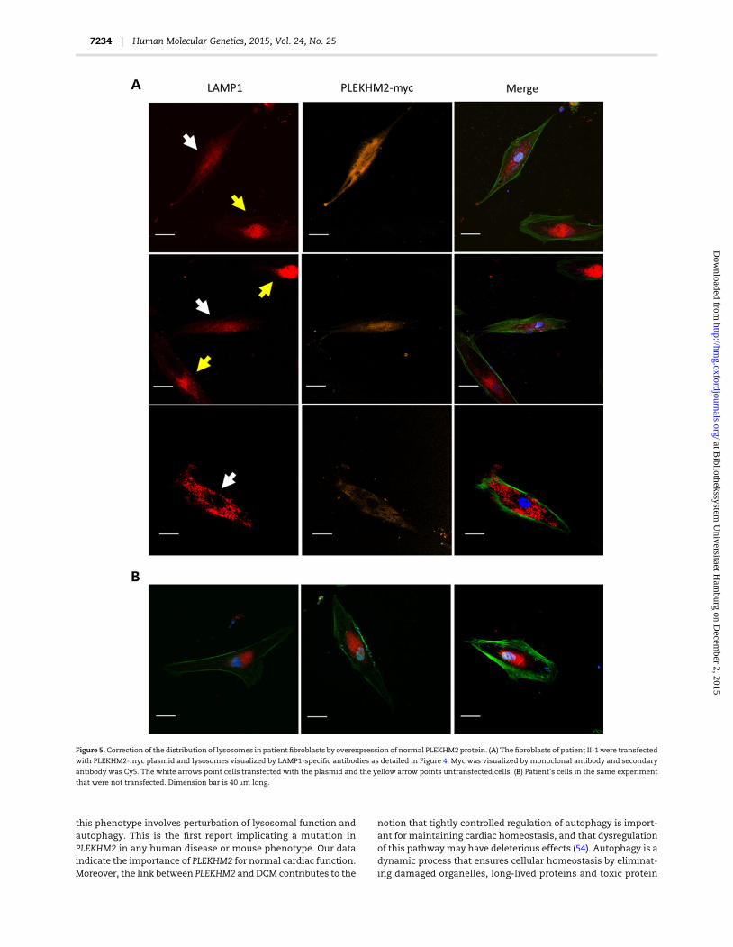
To further evaluate the cause and effect relationship betweenthe PLEKHM2 mutation and lysosomal distribution, we testedwhether exogenous expression of wild-type PLEKHM2 in the pa-tient fibroblasts may correct the abnormality of lysosome distri-bution. For that purpose, the fibroblasts of patient II-1 weretransfected with the PLEKHM2-myc plasmid and lysosomes
Figure 3. Subcellular distribution of organelles stained with Rabs and Golgi. Representative photographs of the distribution of the endosomes in primary fibroblasts of
patient II-1 relative to control fibroblasts labeled with Rab 5, Rab7 and Rab9. Note perinuclear localization of the various rab in the patient’ cells relative to control cells
and fragmentation and spreading of the Golgi apparatus in patient’ cells relative to control cells (comparable results were obtained for patient II-2, Supplementary
Material, Fig. S3). Blue is DAPI staining for the nuclei, and green is AlexaFluor 488 Phalloidin used to mark the cells’ borders. Dimension bar is 40 μm long. Relative
distribution around nuclei, presented at the right side of the respective images, was calculated for a set intensity around the nucleus, and the area giving this
intensity was calculated for each cell. The intensity was measured using ImageJ software (NIH) with a constant threshold. At least 20 cells of each patient and control
with the specific staining were measured. Data are expressed as mean ± SD, and P-values are indicated at the right of the bars (Student’s t-test).
7232 | Human Molecular Genetics, 2015, Vol. 24, No. 25
at Bibliothekssystem
Universitaet H
amburg on D
ecember 2, 2015
http://hmg.oxfordjournals.org/
Dow
nloaded from

were visualized by LAMP1-specific antibodies. Indeed, the resultsindicated that transfected cells which expressed the normalPLEKHM2 converted their distribution of LAMP1 toward the dis-persed distribution normally seen in healthy fibroblasts. In con-trast, cells which were not transfected demonstrated abnormaldistribution of LAMP1 (Fig. 5). Cells transfectedwith a control vec-tor expressing GFP only also demonstrated abnormal distributionof LAMP1 (not shown).
Autophagy is highly dependent on Rab proteins at variousstages. Rab1, Rab5, Rab7, Rab9A, Rab11, Rab23, Rab32 andRab33B participate in autophagosome formation (45), whereasRab9 is required in noncanonical autophagy. Rab7, in additionto Rab8B and Rab24, has a key role in autophagosomematuration(45). Additionally, autophagy depends on lysosomal function(46,47). Thus, we evaluated the autophagic flux in fibroblastswith mutated PLEKHM2 derived from two of the patients in com-parison with those from two controls. The accumulation of twoproteins widely used for monitoring autophagy flux LC3-II andp62 (48) upon treatment of the cells with leupeptinwasmeasuredby Western blotting using specific antibodies. The result showedamarked decrease in the ability of fibroblasts from the patients to
accumulate these proteins (Fig. 6), suggesting marked impair-ment in autophagic flux.
Inhibition of autophagy was reported by several studies to en-hance migration of both normal (49–51) and tumor cells (50–53).Thus, we aimed to determine whether the fibroblasts with mu-tated PLEKHM2 demonstrate changes in cell migration. To thisend, fibroblasts of identical passages (16–18) from patient II-1and control fibroblasts were plated with culture inserts of500 μm. When the cell density reached ∼80%, the inserts were re-moved, thus creating a ∼500 μm-wide gap within the culture.Fibroblast movement toward closing the gap was traced by time-lapse live cellmicroscopy. Indeed, the patient’s fibroblasts bridgedthe gap within 29 ± 2 h in comparison with 46 ± 6 h for control fi-broblasts (Fig. 7, P = 0.002), indicating fastermovement of the fibro-blasts with mutated PLEKHM2 compared with controls.
DiscussionOur results demonstrate that a mutation in PLEKHM2 causes re-cessive DCM with LVNC that is limited to a few segments in ourpatients, and indicates that a likely mechanistic explanation for
Figure 4. Subcellular and size distribution of lysosomes. Lysosomes were visualized using antibodies to LAMP1. Acquirement of the images is as detailed in Figure 3. (A) A
representative photograph of patient’s cells shows a perinuclear localization of lysosomes, and the relative distribution around the nuclei is presented at the right.
Dimension bar shows 40 μm. (B) Relative distribution around nuclei was analyzed as in Figure 3. (C) Analysis of lysosomal size indicates a higher proportion of large
lysosomes in patient compared with control fibroblasts. Analysis was done using the ‘analyze particles’ plugin of ImageJ with size settings from 0–infinity and
circularity 0–1.
Human Molecular Genetics, 2015, Vol. 24, No. 25 | 7233
at Bibliothekssystem
Universitaet H
amburg on D
ecember 2, 2015
http://hmg.oxfordjournals.org/
Dow
nloaded from
this phenotype involves perturbation of lysosomal function andautophagy. This is the first report implicating a mutation inPLEKHM2 in any human disease or mouse phenotype. Our dataindicate the importance of PLEKHM2 for normal cardiac function.Moreover, the link between PLEKHM2 and DCM contributes to the
notion that tightly controlled regulation of autophagy is import-ant for maintaining cardiac homeostasis, and that dysregulationof this pathwaymay have deleterious effects (54). Autophagy is adynamic process that ensures cellular homeostasis by eliminat-ing damaged organelles, long-lived proteins and toxic protein
Figure 5.Correction of the distribution of lysosomes in patientfibroblasts by overexpression of normal PLEKHM2protein. (A) Thefibroblasts of patient II-1were transfected
with PLEKHM2-myc plasmid and lysosomes visualized by LAMP1-specific antibodies as detailed in Figure 4. Myc was visualized by monoclonal antibody and secondary
antibody was Cy5. The white arrows point cells transfected with the plasmid and the yellow arrow points untransfected cells. (B) Patient’s cells in the same experiment
that were not transfected. Dimension bar is 40 μm long.
7234 | Human Molecular Genetics, 2015, Vol. 24, No. 25
at Bibliothekssystem
Universitaet H
amburg on D
ecember 2, 2015
http://hmg.oxfordjournals.org/
Dow
nloaded from

aggregates, as well as recycling nutrients during starvation andstress (47). Tightly controlled regulation of autophagy is import-ant for maintaining cardiac homeostasis, and dysregulation ofthis pathway resulting in either excessive or insufficient autop-hagy is suggested to have deleterious effects (54). Altered autop-hagy has been observed in many cardiovascular diseases inresponse to pathological stimuli, including cardiac hypertrophyand HF (55). Animal models suggest that impaired autophagymay lead to cardiac dysfunction over time (56). PLEKHM2 joinstwo other genes (LAMP2 and BAG3) with a role in autophagy ascauses of cardiomyopathy. LAMP2 is an important constituentof the lysosomal membrane; mutations in LAMP2 were demon-strated to cause Danon disease, a condition of severe and pro-gressive myopathy and cardiomyopathy with multisystemglycogen-storage disease (OMIM 300257) associated with accu-mulation of autophagicmaterial in striatedmyocytes (57), hyper-trophic cardiomyopathy with skeletal muscle weakness (58) andprominent cardiac hypertrophy and electrophysiological abnor-malities (52). Mutations in BAG3 can cause childhood onset ofrapidly progressive myofibrillar myopathy (MFN) (59,60) or auto-somal-dominant DCM (61–63). However, human BAG3mutationshave not been characterized sufficiently, and two DCM-asso-ciated mutations rather fall into the category of stress-inducedapoptosis (61). After gene transfer of BAG3mutations in neonatalrat cardiomyocytes or H9c9 cells, the assembly and integrity ofZ-discs and nuclear localization of BAG3 were disturbed and
Figure 6. Autophagic flux was impaired in patient fibroblasts. (A) Representative immunoblot performed on crude proteins from fibroblasts of two patients [VT1 (II-1) and
VT4 (II-2)] and two controls. Cells were treated or not with 100 µM leupeptin for 24 h. Immunoblots were stained with antibodies against LC3 or p62. (B) Quantification of
LC3-II and p62 normalized to Ponceau as a loading control. Treated samples were related to untreated controls for each cell line to compare the autophagic flux
independent of varying basal expression levels among cell lines. Bars represent mean ± SEM with *P < 0.05 and ***P < 0.001 versus untreated control, one-way ANOVA
with Bonferroni’s post-test (n = 4 per group).
Figure 7. PLEKHM2 patient’s fibroblasts migrate faster than control fibroblasts.
Fibroblasts of patient II-1 and control fibroblasts were plated with 500 μm
culture inserts. When the density reached ∼80%, the inserts were removed and
fibroblasts movement was monitored using time-lapse live cell microscopy for
65–70 h. The DIC images were acquired every 15 min. The bridging time was
defined as the time passed from the insert removal till formation of a
continuous string of cells across the gap. The results represent four
independent experiments each in quadruplicates. Data are expressed as
mean ± SEM, P = 0.002 (Student’s t-test).
Human Molecular Genetics, 2015, Vol. 24, No. 25 | 7235
at Bibliothekssystem
Universitaet H
amburg on D
ecember 2, 2015
http://hmg.oxfordjournals.org/
Dow
nloaded from

cells exhibited a higher susceptibility to stress-induced apoptosis(61). On the contrary, myotube formation was disturbed aftergene transfer of MFN-associated, but not DCM-associated BAG3,mutations in C2C12 myoblasts (61).
LVNC has been considered to be a developmental failure ofthe heart to fully form the compact myocardium during thelater stages of cardiac development resulting in persistence ofmultiple prominent ventricular trabeculations and deep intertra-becular recesses (64). Several genetically engineeredmousemod-els that have defects in cardiac trabeculation and compactionindicate that cellular growth and differentiation signaling path-ways are keys in these ventricular morphogenetic events andpoint to several important genes participating in the NOTCHpathway (65). One study reported that inactivating mutations inthe NOTCH pathway regulator MIB1 cause LVNC in humans(66). However, the etiology and pathogenesis causing LVNC arenot known (64). Although LVNC is classified as a primary geneticcardiomyopathy by the American Heart Association (1), it is stillconsidered unclassified in the European Society of Cardiologyclassification, as it remains unclear whether it represents a dis-tinct disease process or a morphological trait shared by manyphenotypically different cardiomyopathies (2). Mutations in 24nuclear genes, in addition to mitochondrial disorders andchromosome abnormalities, present with LVNC together withmultiple cardiac disorders. The extensive genetic heterogeneityof LVNC suggests that the uniformmorphology of LVNC is attrib-utable to embryonic noncompaction, as well as from induction ofhypertrabeculation as a compensatory reaction of an impairedmyocardium (67). This suggestion is supported by the observa-tions that in some patients, LVNC develops later in life, can be ac-quired by intense athletic training or as an adaptive mechanismof an impaired myocardium, such as ischemic heart disease ortrauma, and may even disappear (24,67,68). Cardiac fibroblastsappear to be of critical importance in heart repair and cell migra-tion is essential inwoundhealing (69). In this regard, inhibition ofautophagy was reported by several studies to enhance migrationof both normal (49–51) and tumor cells (50–53). This mechanismis suggested as ‘signalphagy’, where active signaling proteins arenot degraded by autophagy, for example RHOA-GTP (70) or theoncogenic transcription factor, Twist1 (49). Since derangementsin cell movement could contribute to the noncompaction ob-served in patients, we tested the migration of fibroblasts of a pa-tient compared with fibroblasts of a control. Our data clearlyindicate enhanced movement of the patient’s fibroblast relativeto controls. Thus, although noncompaction was observed atage 7 in our youngest patient (II-12, Table 1) and thus could becaused by a defect in development, it is plausible that this abnor-mally facilitatedmigrationmay contribute to the noncompactionmorphology.
The current study associates a mutation disrupting lyso-somes and autophagy with pathology selectively affecting thehumanheart. Thus, these resultsmay pave theway for better un-derstanding of the apparently critical link between autophagyand normal cardiac function.
Materials and MethodsPatients
The study was approved by the Soroka Medical Center institu-tional review board, and all participants gave written informedconsent prior to participation. Genomic DNA was extractedfrom white blood cells by using standard procedures. Medical re-cords were carefully reviewed, and details of somatic growth,
psychomotor development, clinical course, hospitalizationsand laboratory results were obtained. Parents and siblings wereinterviewed and underwent a complete physical examination,which particularly focused on the cardiac and neuromuscularfindings. Cardiac evaluation included standard transthoracic2D and Doppler echocardiography (TTE) using a Vivid 7 system(GE Medical Systems, Saskatchewan, Canada) and CMRI using a1.5 T scanner (Achieva, Philips Medical Systems, Best, TheNetherlands), which included phase contrast images. TTE mea-surements of LV end-diastolic dimension (LVED) and LV end-systolic dimension (LVES) were obtained in accordance with therecommendations of the American Society of Echocardiography(71). Dimensions were corrected for age and bovine serumalbumin (BSA) according to the formula of Henry et al. (72):LVED (percent predicted) = (measured LVED/predicted LVED) ×100; predicted LVED = (45.3 × BSA0.3)− (0.03 × Age) − 7.2. LV abnor-malities were classified as follows: DCM, LVED ≥117% predictedand fractional shortening <25% in the absence of known causesof ventricular dilatation (72,73). LV noncompaction evaluationwas performed according to the echocardiographic criteria de-scribed by Jenni et al. (17,74).
Genomic DNA was isolated from blood by standard proce-dures. Lymphoblastoid cells and fibroblast cultures were estab-lished by standard procedures.
Genotyping and exome sequencing
Genotyping was performed using Affymetrix (CA, USA) Gene-Chip Human SNP5 array. We determined the genotype calls byusing Affymetrix GeneChip Genotyping Console AnalysisSoftware (GTYPE) and identified the autozygosity using Agile-MultiIdeogram (http://dna.leeds.ac.uk/agile/AgileMultiIdeogram/).Whole exome sequencing was performed using NimbleGen v2exome capture followed by 50 bp paired-end sequencing usingan Illumina HiSeq Sequencing System. The resulting sequencedata were analyzed as described (75).
Verification of a mutation
PCR amplification of exon 12 was performed using primers for-ward: TTGTAGACGAGGCTGACTCTCA and reverse: CAGAAACCACACCGTGACAT (annealing temperature 61°C). Direct sequencingthe PCR products was performed on anABI PRISM 3100 DNAAna-lyzer with the BigDye Terminator v.1.1 Cycle Sequencing Kit (Ap-plied Biosystems, CA, USA) according to the manufacturer’sprotocol. MwoI (NEB) digestion was done according to the manu-facturer’s instruction. Restriction digest of the 687 bp ampliconresulted in 25, 73, 75, 83, 190 and 241 bp fragments; if the muta-tion is present, the 239 bp fragment is cleaved into 135 and104 bp fragments. Digested products were separated by electro-phoresis on 2% agarose gel.
RT-qPCR analysis
About 500 ng of total RNA from different human tissues obtainedfrom Clontech (Takara Bio Europe/Clontech) was primed withrandom hexamers and reverse-transcribed with the SuperScriptReverse Transcription Kit from Invitrogen, following the manu-facturer’s instructions. cDNAs were PCR-amplified in the ABI7500 Software V2.0.3 (Applied Biosystems) with the SYBR Green(Applied Biosystems). The ubiquitously expressed GAPDH genewas used as an internal control to normalize the data. The resultsrepresent one experiment done in duplicates, means ± sem (notvisible). The sequences of the primers used in the experiment
7236 | Human Molecular Genetics, 2015, Vol. 24, No. 25
at Bibliothekssystem
Universitaet H
amburg on D
ecember 2, 2015
http://hmg.oxfordjournals.org/
Dow
nloaded from

are the following: PLEKHM2 (Ex11–Ex12 + 13): forward: 5′TGCTGCTCACAGACTGCTAT 3′; reverse: 5′ AAGGCCAACCGACACATA 3′. GAPDH forward: 5′ AGAAGGCTGGGGCTCATTTG 3′; re-verse: 5′ GGGGCCATCCACAGTCTTC 3′.The PLEKHM2 primersyielded a linear standard curve with an R2 = 0.99.
Analysis of splicing
RNA was extracted from lymphoblastoid cells and fibroblasts ofpatient II-1 and from comparable cells of control individualsusing the EZ-RNA II Kit (Biological Industries, Israel). cDNA wasprepared using the SuperScript Kit (Invitrogen), PCR was per-formed on the cDNA using primers in exons 10 and 15, forward:TCGGAGTTCAGAGTAGACAACAA and reverse: ATGCCTTCTTTGGTGATGGT flanking exon 12 that contains the mutation andthe PCR products were separated on 2% agarose gel.
Protein structure
The predictions of the domains of PLEKHM2 were retrieved fromUniProt (http://www.uniprot.org/uniprot/). The 3D structuremodeling was created by using the modeller v9.14 program(http://toolkit.tuebingen.mpg.de/modeller) as described in themanual of the manufacturer. The amino acid sequence ofhuman PLEKHM2 was retrieved from the NCBI protein database(NP_055979.2). A sequence similarity search was performedusing the NCBI BLASTP server (https://blast.ncbi.nlm.nih.gov/Blast.cgi?PROGRAM=blastp&PAGE_TYPE=BlastSearch&BLAST_SPEC=&LINK_LOC=blasttab&LAST_PAGE=blastn). Based on thesequence identity, 3CXB and 3HW2were chosen as themost suit-able templates for homologymodeling. Colormodifications on theprotein was changed using RasMol v2.7.5.2 (http://rasmol.org/).
Western blot analyses
Cells were lysed in RIPA lysis buffer (20 m Tris pH 7.5, 150 m
NaCl, 1% Triton, 1 m EDTA, 1 m EGTA, 2.5 m sodium pyro-phosphate and 1 mNa3VO4)with 1 : 100 PMSFand a protease in-hibitor (Sigma cat. No. S8830). Prior to separation, Laemmlisample buffer (62.5 m Tris–HCl, pH 6.8; 25% glycerol; 2% SDS;0.01% bromophenol blue and β-mercaptoethanol for a final con-centration of 5% was added) was added in a 1 : 1 ratio. Subse-quently, the samples were heated for 5 min at 95°C. About35 µg of protein tissue extractwas loaded on 12%SDS–PAGE. Blot-ting was done into the nitrocellulose membrane, and the blotswere blocked with 5% nonfat dry milk in TBST (Tris-bufferedsaline, 0.05% Tween) and incubated with the primary antibody(at 4°C, for overnight). For PLEKHM2 C-terminal region (ThermoPA5-20850) and for the N-terminal region (Santa Cruz sc-136808) and respective HRP-conjugated secondary antibodyanti-rabbit IgG (Jackson, USA). Signal intensities of PLEKHM2bands were normalized against the internal control GAPDH(Millipore MAB374). Commercial fibroblasts were purchasedfrom ScienCell-HDF-a (cat. no. 2320).
Immunofluorescence
Fibroblast cells were grown in an eight-well slide (ibidi 80826) in10% DMEM. Cells were fixed with 4% formaldehyde solution, in-cubated with 0.1% Triton X-100 for 5 min and blocked with 1%(BSA; Sigma-Aldrich) in PBS for 30 min. Samples were incubatedat room temperature for 1 h with the primary antibody. Allprimary antibodies except Rab4a (Santa Cruz biotechnology Absc-312) were from Cell Signaling technology: Rab1 Ab #13075,Rab5 #3547, Rab7 #9367, Rab9 #5118, Rab11 #5589, LAMP1 #9091
and GM130 #12480 secondary antibody Cy3 (Thermo SA5-10169). Blue is DAPI (Vectorlabs H-1200) staining for the nuclei,and green is AlexaFluor 488 Phalloidin (Molecular probesA12379) to mark the cells’ borders. Images were acquired withthe confocal FluoView 1000 fluorescencemicroscope (OLYMPUS),with a 40× objective and with the manufacturer’s softwareFV1000.
Overexpression of normal PLEKHM2 protein
Fibroblast cells of patient II-1 were transfected using Turbofect(Thermo #R0531) with PLEKHM2-myc plasmid (pcDNA3.1, giftof Dr Stéphane Méresse). Myc was visualized by primary anti-myc antibody (monoclonal, gift of DrNoah Isakov) and secondarycy5 anti-mouse antibody (Thermo SA5-10169), and lysosomes vi-sualized by LAMP1 antibody and cy3 anti-rabbit antibody.
Autophagic flux
Patients’ fibroblasts were plated at a density of 30 000 cells per 24-well platewell. The next day, cells were treated or not with 100 µMleupeptin (Sigma, L-8511) and harvested 24 h after treatment inlysis buffer (3% SDS, 30 m Tris-base, pH 8.8, 5 m EDTA, 30 m
NaF, 10% glycerol and 1 m DTT). About 10 µg of protein lysatewas used for immunoblots, which were stained with antibodiesagainst LC3 (Novus Biologicals, NB100-2331) or p62 (Sigma, P0067).
Cell migration assay
Fibroblasts of identical passages (16–18) of patient II-1 and con-trol fibroblasts were plated in eight-wells μ-slides (ibidi GmBhGermany, Cat #80826) with culture inserts of 500 μm (ibidiGmBh Germany, cat. #80209). When the density reached ∼80%,the inserts were removed and fibroblast movement was moni-tored using time-lapse live cell microscopy for 65–70 h. The dif-ferential interference contrast (DIC) images were acquired every15 min. The bridging time was defined as the time passed fromthe insert removal until formation of a continuous string ofcells across the gap.
Supplementary MaterialSupplementary Material is available at HMG online.
AcknowledgementsWe are grateful to the affected persons and their families, whosecooperation made this study possible. We are very grateful toDr Stéphane Méresse from Centre d’Immunologie de Marseille-Luminy for his gift of the PLEKHM2-myc plasmid.
Conflict of Interest statement. None declared.
FundingThis work was partly supported by an internal grant fromBen- Gurion University of the Negev, Faculty of Health Sciences.This work was partly supported by the Leducq Foundation(research grant no. 11, CVD O4 to L.C.). V.C.S. was supported bythe Howard Hughes Medical Institute.
References1. Yancy, C.W., Jessup, M., Bozkurt, B., Butler, J., Casey, D.E. Jr,
Drazner, M.H., Fonarow, G.C., Geraci, S.A., Horwich, T., Januz-zi, J.L. et al. (2013) 2013 ACCF/AHA guideline for the manage-ment of heart failure: a report of the American College of
Human Molecular Genetics, 2015, Vol. 24, No. 25 | 7237
at Bibliothekssystem
Universitaet H
amburg on D
ecember 2, 2015
http://hmg.oxfordjournals.org/
Dow
nloaded from

Cardiology Foundation/American Heart Association TaskForce on practice guidelines. Circulation, 128, e240–e327.
2. McMurray, J.J., Adamopoulos, S., Anker, S.D., Auricchio, A.,Bohm, M., Dickstein, K., Falk, V., Filippatos, G., Fonseca, C.,Gomez-Sanchez, M.A. et al. (2012) ESC guidelines for the diag-nosis and treatment of acute and chronic heart failure 2012:the task force for the diagnosis and treatment of Acute andChronic Heart Failure 2012 of the European Society of Cardi-ology. Developed in collaboration with the Heart Failure As-sociation (HFA) of the ESC. Eur. J. Heart Fail., 14, 803–869.
3. Piran, S., Liu, P., Morales, A. and Hershberger, R.E. (2012)Where genome meets phenome: rationale for integratinggenetic and protein biomarkers in the diagnosis andmanage-ment of dilated cardiomyopathy and heart failure. J. Am. Coll.Cardiol., 60, 283–289.
4. Gregori, D., Rocco, C., Miocic, S. and Mestroni, L. (2001) Esti-mating the frequency of familial dilated cardiomyopathy inthe presence of misclassification errors. J. Appl. Stat., 28,53–62.
5. Petretta, M., Pirozzi, F., Sasso, L., Paglia, A. and Bonaduce, D.(2011) Review and metaanalysis of the frequency of familialdilated cardiomyopathy. Am. J. Cardiol., 108, 1171–1176.
6. Karkkainen, S. and Peuhkurinen, K. (2007) Genetics of dilatedcardiomyopathy. Ann. Med., 39, 91–107.
7. McNally, E.M., Golbus, J.R. and Puckelwartz, M.J. (2013) Genet-ic mutations and mechanisms in dilated cardiomyopathy.J. Clin. Invest., 123, 19–26.
8. Grunig, E., Tasman, J.A., Kucherer, H., Franz, W., Kubler, W.and Katus, H.A. (1998) Frequency and phenotypes of familialdilated cardiomyopathy. J. Am. Coll. Cardiol., 31, 186–194.
9. Mestroni, L., Rocco, C., Gregori, D., Sinagra, G., Di Lenarda, A.,Miocic, S., Vatta, M., Pinamonti, B., Muntoni, F., Caforio, A.L.et al. (1999) Familial dilated cardiomyopathy: evidence forgenetic and phenotypic heterogeneity. Heart Muscle DiseaseStudy Group. J. Am. Coll. Cardiol., 34, 181–190.
10. OMIM. Online Mendelian Inheritance in Man. McKusick-NathansInstitute of Genetic Medicine, Johns Hopkins University, Bal-timore, MD and National Center for Biotechnology Informa-tion, National Library of Medicine, Bethesda, MD; http://www.ncbi.nlm.nih.gov/OMIM, in press.
11. He, Q. (2010) Tafazzin knockdown causes hypertrophyof neo-natal ventricular myocytes. Am. J. Physiol.. Heart Circ. Physiol.,299, H210–H216.
12. Levitas, A., Muhammad, E., Harel, G., Saada, A., Caspi, V.C.,Manor, E., Beck, J.C., Sheffield, V. and Parvari, R. (2010) Famil-ial neonatal isolated cardiomyopathy caused by a mutationin the flavoprotein subunit of succinate dehydrogenase.Eur. J. Hum. Genet., 18, 1160–1165.
13. Parvari, R. and Levitas, A. (2012) The mutations associatedwith dilated cardiomyopathy. Biochem. Res. Int., 2012, 639250.
14. Friedrich, F.W. and Carrier, L. (2012) Genetics of hypertrophicand dilated cardiomyopathy. Curr. Pharmaceut. Biotechnol., 13,2467–2476.
15. Moric-Janiszewska, E. and Markiewicz-Loskot, G. (2008) Gen-etic heterogeneity of left-ventricular noncompaction cardio-myopathy. Clin. Cardiol., 31, 201–204.
16. Zhang, W., Chen, H., Qu, X., Chang, C.P. and Shou, W. (2013)Molecular mechanism of ventricular trabeculation/compac-tion and the pathogenesis of the left ventricular noncompac-tion cardiomyopathy (LVNC). Am. J. Med. Genet. C Semin. Med.Genet., 163c, 144–156.
17. Jenni, R., Oechslin, E.N. and van der Loo, B. (2007) Isolatedventricular non-compaction of the myocardium in adults.Heart, 93, 11–15.
18. Paterick, T.E. and Tajik, A.J. (2012) Left ventricular noncom-paction: a diagnostically challenging cardiomyopathy. Circ.J., 76, 1556–1562.
19. Arbustini, E.,Weidemann, F. andHall, J.L. (2014) Left ventricu-lar noncompaction: a distinct cardiomyopathy or a traitshared by different cardiac diseases? J. Am. Coll. Cardiol., 64,1840–1850.
20. Maron, B.J., Towbin, J.A., Thiene, G., Antzelevitch, C., Corrado,D., Arnett, D., Moss, A.J., Seidman, C.E. and Young, J.B. (2006)Contemporary definitions and classification of the cardiomy-opathies: an American Heart Association Scientific State-ment from the Council on Clinical Cardiology, Heart Failureand Transplantation Committee; Quality of Care and Out-comes Research and Functional Genomics and TranslationalBiology Interdisciplinary Working Groups; and Council onEpidemiology and Prevention. Circulation, 113, 1807–1816.
21. Fazio, G., Corrado, G., Pizzuto, C., Zachara, E., Rapezzi, C., Su-lafa, A.K., Sutera, L., Stollberger, C., Sormani, L., Finsterer, J.et al. (2008) Supraventricular arrhythmias in noncompactionof left ventricle: is this a frequent complication? Int. J. Cardiol.,127, 255–256.
22. Stollberger, C., Winkler-Dworak, M., Blazek, G. and Finsterer,J. (2007) Prognosis of left ventricular hypertrabeculation/non-compaction is dependent on cardiac and neuromuscular co-morbidity. Int. J. Cardiol., 121, 189–193.
23. Burke, A., Mont, E., Kutys, R. and Virmani, R. (2005) Left ven-tricular noncompaction: a pathological study of 14 cases.Hum. Pathol., 36, 403–411.
24. Finsterer, J., Stollberger, C. and Feichtinger, H. (2002) Histo-logical appearance of left ventricular hypertrabeculation/noncompaction. Cardiology, 98, 162–164.
25. Chin, T.K., Perloff, J.K., Williams, R.G., Jue, K. and Mohrmann,R. (1990) Isolated noncompaction of left ventricular myocar-dium. A study of eight cases. Circulation, 82, 507–513.
26. Ritter, M., Oechslin, E., Sutsch, G., Attenhofer, C., Schneider, J.and Jenni, R. (1997) Isolated noncompaction of the myocar-dium in adults. Mayo Clin. Proceed., 72, 26–31.
27. Oechslin, E.N., Attenhofer Jost, C.H., Rojas, J.R., Kaufmann, P.A.and Jenni, R. (2000) Long-term follow-up of 34 adults withisolated left ventricular noncompaction: a distinct cardiomy-opathy with poor prognosis. J. Am. Coll. Cardiol., 36, 493–500.
28. Ichida, F., Hamamichi, Y., Miyawaki, T., Ono, Y., Kamiya, T.,Akagi, T., Hamada, H., Hirose, O., Isobe, T., Yamada, K. et al.(1999) Clinical features of isolated noncompaction of the ven-tricular myocardium: long-term clinical course, hemo-dynamic properties, and genetic background. J. Am. Coll.Cardiol., 34, 233–240.
29. Sasse-Klaassen, S., Gerull, B., Oechslin, E., Jenni, R. and Thier-felder, L. (2003) Isolatednoncompaction of the left ventricularmyocardium in the adult is an autosomal dominant disorderin themajority of patients.Am. J. Med. Genet. A, 119A, 162–167.
30. Ichida, F., Tsubata, S., Bowles, K.R., Haneda,N., Uese, K.,Miya-waki, T., Dreyer, W.J., Messina, J., Li, H., Bowles, N.E. et al.(2001) Novel gene mutations in patients with left ventricularnoncompaction or Barth syndrome. Circulation, 103, 1256–1263.
31. Vatta, M., Mohapatra, B., Jimenez, S., Sanchez, X., Faulkner,G., Perles, Z., Sinagra, G., Lin, J.H., Vu, T.M., Zhou, Q. et al.(2003) Mutations in Cypher/ZASP in patients with dilated car-diomyopathy and left ventricular non-compaction. J. Am.Coll. Cardiol., 42, 2014–2027.
32. Kenton, A.B., Sanchez, X., Coveler, K.J., Makar, K.A., Jimenez,S., Ichida, F., Murphy, R.T., Elliott, P.M., McKenna, W., Bowles,N.E. et al. (2004) Isolated left ventricular noncompaction is
7238 | Human Molecular Genetics, 2015, Vol. 24, No. 25
at Bibliothekssystem
Universitaet H
amburg on D
ecember 2, 2015
http://hmg.oxfordjournals.org/
Dow
nloaded from
rarely caused by mutations in G4.5, alpha-dystrobrevin andFK binding protein-12. Mol. Genet. Metab., 82, 162–166.
33. Chen, R., Tsuji, T., Ichida, F., Bowles, K.R., Yu, X.,Watanabe, S.,Hirono, K., Tsubata, S., Hamamichi, Y., Ohta, J. et al. (2002)Mu-tation analysis of the G4.5 gene in patients with isolated leftventricular noncompaction. Mol. Genet. Metab., 77, 319–325.
34. Xing, Y., Ichida, F., Matsuoka, T., Isobe, T., Ikemoto, Y., Higaki,T., Tsuji, T., Haneda, N., Kuwabara, A., Chen, R. et al. (2006)Genetic analysis in patientswith left ventricular noncompac-tion and evidence for genetic heterogeneity. Mol. Genet.Metab., 88, 71–77.
35. Hermida-Prieto, M., Monserrat, L., Castro-Beiras, A., Laredo,R., Soler, R., Peteiro, J., Rodriguez, E., Bouzas, B., Alvarez, N.,Muniz, J. et al. (2004) Familial dilated cardiomyopathyand iso-lated left ventricular noncompaction associated with laminA/C gene mutations. Am. J. Cardiol., 94, 50–54.
36. Klaassen, S., Probst, S., Oechslin, E., Gerull, B., Krings, G.,Schuler, P., Greutmann, M., Hurlimann, D., Yegitbasi, M.,Pons, L. et al. (2008) Mutations in sarcomere protein genesin left ventricular noncompaction.Circulation, 117, 2893–2901.
37. Boucrot, E., Henry, T., Borg, J.P., Gorvel, J.P. and Meresse, S.(2005) The intracellular fate of Salmonella depends on the re-cruitment of kinesin. Science, 308, 1174–1178.
38. Dumont, A., Boucrot, E., Drevensek, S., Daire, V., Gorvel, J.P.,Pous, C., Holden, D.W. and Meresse, S. (2010) SKIP, the hosttarget of the Salmonella virulence factor SifA, promotes kine-sin-1-dependent vacuolar membrane exchanges. Traffic, 11,899–911.
39. Rosa-Ferreira, C. and Munro, S. (2011) Arl8 and SKIP act to-gether to link lysosomes to kinesin-1.Dev. Cell, 21, 1171–1178.
40. Maquat, L.E. (2004) Nonsense-mediated mRNA decay: spli-cing, translation and mRNP dynamics. Nat. Rev. Mol. CellBiol., 5, 89–99.
41. Jackson, L.K., Nawabi, P., Hentea, C., Roark, E.A. andHaldar, K.(2008) The Salmonella virulence protein SifA is a G protein an-tagonist. Proc. Natl. Acad. Sci. USA, 105, 14141–14146.
42. Harrison, R.E., Brumell, J.H., Khandani, A., Bucci, C., Scott, C.C.,Jiang, X., Finlay, B.B. and Grinstein, S. (2004) Salmonellaimpairs RILP recruitment to Rab7 during maturation of inva-sion vacuoles. Mol. Biol. Cell, 15, 3146–3154.
43. Fukuda, M., Kobayashi, H., Ishibashi, K. and Ohbayashi, N.(2011) Genome-wide investigation of the Rab binding activityof RUNdomains: development of a novel tool that specificallytraps GTP-Rab35. Cell Struct. Funct., 36, 155–170.
44. Grosshans, B.L., Ortiz, D. and Novick, P. (2006) Rabs and theireffectors: achieving specificity inmembrane traffic. Proc. Natl.Acad. Sci. USA, 103, 11821–11827.
45. Bento, C.F., Puri, C., Moreau, K. and Rubinsztein, D.C. (2013)The role of membrane-trafficking small GTPases in the regu-lation of autophagy. J. Cell Sci., 126, 1059–1069.
46. Levine, B. and Kroemer, G. (2008) Autophagy in the pathogen-esis of disease. Cell, 132, 27–42.
47. Xie, M., Morales, C.R., Lavandero, S. and Hill, J.A. (2011) Tun-ing flux: autophagy as a target of heart disease therapy.Curr. Opin. Cardiol., 26, 216–222.
48. Klionsky, D.J., Abdalla, F.C., Abeliovich, H., Abraham, R.T.,Acevedo-Arozena, A., Adeli, K., Agholme, L., Agnello, M.,Agostinis, P., Aguirre-Ghiso, J.A. et al. (2012) Guidelines forthe use and interpretation of assays for monitoring autop-hagy. Autophagy, 8, 445–544.
49. Qiang, L., Zhao, B., Ming, M., Wang, N., He, T.C., Hwang, S.,Thorburn, A. and He, Y.Y. (2014) Regulation of cell prolifer-ation and migration by p62 through stabilization of Twist1.Proc. Natl. Acad. Sci. USA, 111, 9241–9246.
50. Belaid, A., Cerezo, M., Chargui, A., Corcelle-Termeau, E., Ped-eutour, F., Giuliano, S., Ilie, M., Rubera, I., Tauc, M., Barale, S.et al. (2013) Autophagy plays a critical role in the degradationof active RHOA, the control of cell cytokinesis, and genomicstability. Cancer Res., 73, 4311–4322.
51. Belaid, A., Ndiaye, P.D., Cerezo, M., Cailleteau, L., Brest, P.,Klionsky, D.J., Carle, G.F., Hofman, P. and Mograbi, B. (2014)Autophagy and SQSTM1 on the RHOA(d) again: emergingroles of autophagy in the degradation of signaling proteins.Autophagy, 10, 201–208.
52. Arad, M., Maron, B.J., Gorham, J.M., Johnson,W.H. Jr, Saul, J.P.,Perez-Atayde, A.R., Spirito, P., Wright, G.B., Kanter, R.J.,Seidman, C.E. et al. (2005) Glycogen storage diseases present-ing as hypertrophic cardiomyopathy. N Engl. J. Med., 352,362–372.
53. Bai, H., Li, H., Li,W., Gui, T., Yang, J., Cao, D. and Shen, K. (2015)The PI3 K/AKT/mTOR pathway is a potential predictor of dis-tinct invasive and migratory capacities in human ovariancancer cell lines. Oncotarget, 22, 25520–25532.
54. Gustafsson, A.B. and Gottlieb, R.A. (2009) Autophagy in ische-mic heart disease. Circ. Res., 104, 150–158.
55. Ma, S.,Wang, Y., Chen, Y. and Cao, F. (2014) The role of the au-tophagy in myocardial ischemia/reperfusion injury. Biochim.Biophys. Acta, 1852, 271–276.
56. Taneike, M., Yamaguchi, O., Nakai, A., Hikoso, S., Takeda, T.,Mizote, I., Oka, T., Tamai, T., Oyabu, J., Murakawa, T. et al.(2010) Inhibition of autophagy in the heart induces age-re-lated cardiomyopathy. Autophagy, 6, 600–606.
57. Nishino, I., Fu, J., Tanji, K., Yamada, T., Shimojo, S., Koori, T.,Mora, M., Riggs, J.E., Oh, S.J., Koga, Y. et al. (2000) PrimaryLAMP-2 deficiency causes X-linked vacuolar cardiomyopathyand myopathy (Danon disease). Nature, 406, 906–910.
58. Charron, P., Villard, E., Sebillon, P., Laforet, P., Maisonobe, T.,Duboscq-Bidot, L., Romero, N., Drouin-Garraud, V., Frebourg,T., Richard, P. et al. (2004) Danon’s disease as a cause ofhypertrophic cardiomyopathy: a systematic survey. Heart,90, 842–846.
59. Selcen, D., Muntoni, F., Burton, B.K., Pegoraro, E., Sewry, C.,Bite, A.V. and Engel, A.G. (2009) Mutation in BAG3 causes se-vere dominant childhood muscular dystrophy. Ann. Neurol.,65, 83–89.
60. Lee,H.C., Cherk, S.W.,Chan, S.K.,Wong, S., Tong, T.W.,Ho,W.S.,Chan, A.Y., Lee, K.C. and Mak, C.M. (2012) BAG3-relatedmyofibrillar myopathy in a Chinese family. Clin. Genet., 81,394–398.
61. Arimura, T., Ishikawa, T., Nunoda, S., Kawai, S. andKimura, A.(2011) Dilated cardiomyopathy-associated BAG3 mutationsimpair Z-disc assembly and enhance sensitivity to apoptosisin cardiomyocytes. Hum. Mutat., 32, 1481–1491.
62. Norton, N., Li, D., Rieder, M.J., Siegfried, J.D., Rampersaud, E.,Zuchner, S., Mangos, S., Gonzalez-Quintana, J., Wang, L.,McGee, S. et al. (2011) Genome-wide studies of copy numbervariation and exome sequencing identify rare variants inBAG3 as a cause of dilated cardiomyopathy. Am. J. Hum.Genet., 88, 273–282.
63. Villard, E., Perret, C., Gary, F., Proust, C., Dilanian, G., Heng-stenberg, C., Ruppert, V., Arbustini, E., Wichter, T., Germain,M. et al. (2011) A genome-wide association study identifiestwo loci associated with heart failure due to dilated cardio-myopathy. Eur. Heart J., 32, 1065–1076.
64. Towbin, J.A. (2010) Left ventricular noncompaction: a newform of heart failure. Heart Fail. Clin., 6, 453–469, viii.
65. Chen, H., Zhang, W., Li, D., Cordes, T.M., Mark Payne, R. andShou, W. (2009) Analysis of ventricular hypertrabeculation
Human Molecular Genetics, 2015, Vol. 24, No. 25 | 7239
at Bibliothekssystem
Universitaet H
amburg on D
ecember 2, 2015
http://hmg.oxfordjournals.org/
Dow
nloaded from

and noncompaction using genetically engineered mousemodels. Pediatr. Cardiol., 30, 626–634.
66. Luxan, G., Casanova, J.C., Martinez-Poveda, B., Prados, B.,D’Amato, G., MacGrogan, D., Gonzalez-Rajal, A., Dobarro, D.,Torroja, C., Martinez, F. et al. (2013) Mutations in the NOTCHpathway regulator MIB1 cause left ventricular noncompac-tion cardiomyopathy. Nat. Med., 19, 193–201.
67. Finsterer, J. (2009) Cardiogenetics, neurogenetics, and patho-genetics of left ventricular hypertrabeculation/noncompac-tion. Pediatr. Cardiol., 30, 659–681.
68. Hussein, A., Karimianpour, A., Collier, P. and Krasuski, R.A.(2015) Isolated noncompaction of the left ventricle in adults.J. Am. Coll. Cardiol., 66, 578–585.
69. Diaz-Araya, G., Vivar, R., Humeres, C., Boza, P., Bolivar, S. andMunoz, C. (2015) Cardiac fibroblasts as sentinel cells in car-diac tissue: receptors, signaling pathways and cellular func-tions. Pharmacol. Res., doi: 10.1016/j.phrs.2015.07.001.
70. Belaid, A., Ndiaye, P.D., Klionsky, D.J., Hofman, P. andMograbi, B. (2013) Signalphagy: scheduled signal terminationby macroautophagy. Autophagy, 9, 1629–1630.
71. Schiller, N.B., Shah, P.M., Crawford, M., DeMaria, A., Dever-eux, R., Feigenbaum, H., Gutgesell, H., Reichek, N., Sahn, D.,Schnittger, I. et al. (1989) Recommendations for quantitation
of the left ventricle by two-dimensional echocardiogra-phy. American Society of Echocardiography Committeeon Standards, Subcommittee on Quantitation of Two-Dimensional Echocardiograms. J. Am. Soc. Echocardiogr., 2,358–367.
72. Henry, W.L., Gardin, J.M. and Ware, J.H. (1980) Echocardio-graphic measurements in normal subjects from infancy toold age. Circulation, 62, 1054–1061.
73. Richardson, P., McKenna, W., Bristow, M., Maisch, B., Maut-ner, B., O’Connell, J., Olsen, E., Thiene, G., Goodwin, J.,Gyarfas, I. et al. (1996) Report of the 1995World Health Organ-ization/International Society and Federation of CardiologyTask Force on the Definition and Classification of cardiomy-opathies. Circulation, 93, 841–842.
74. Jenni, R., Oechslin, E., Schneider, J., Attenhofer Jost, C. andKaufmann, P.A. (2001) Echocardiographic and pathoan-atomical characteristics of isolated left ventricular non-compaction: a step towards classification as a distinctcardiomyopathy. Heart, 86, 666–671.
75. Muhammad, E., Reish, O., Ohno, Y., Scheetz, T., Deluca, A.,Searby, C., Regev, M., Benyamini, L., Fellig, Y., Kihara, A.et al. (2013) Congenital myopathy is caused by mutation ofHACD1. Hum. Mol. Genet., 22, 5229–5236.
7240 | Human Molecular Genetics, 2015, Vol. 24, No. 25
at Bibliothekssystem
Universitaet H
amburg on D
ecember 2, 2015
http://hmg.oxfordjournals.org/
Dow
nloaded from