Embed Size (px)
Citation preview

Kidney International, Vol. 62 (2002), pp. 2043–2054
Platelet-derived growth factor-D expression in developing andmature human kidneys
SIRIBHA CHANGSIRIKULCHAI, KELLY L. HUDKINS, TRACY A. GOODPASTER, JOHN VOLPONE,STAVROS TOPOUZIS, DEBRA G. GILBERTSON, and CHARLES E. ALPERS
Department of Medicine, Srinakharinwirot University, Bangkok, Thailand; Department of Pathology, University of Washington,and ZymoGenetics, Inc., Seattle, Washington, USA
sequence of which corresponded to human PDGF-D (GenbankPlatelet-derived growth factor-D expression in developing andnumber AF336376).mature human kidneys.
Conclusions. To our knowledge, these are the first studiesBackground. Platelet-derived growth factor (PDGF) is a fam-to localize PDGF-D in human kidneys and suggest that PDGF-Dily of growth regulatory molecules composed of sulfide-bondedmay have a role in kidney development. PDGF-D was showndimeric structures. Two well-studied PDGF peptides (PDGF-A
and PDGF-B) have been shown to mediate a wide range of to bind to PDGF � receptor, which localizes to mesangial cells,biological effects. PDGF-D is a newly recognized member of parietal epithelial cells, and interstitial fibroblasts, suggestingthe PDGF family. Initial studies of the PDGF-D gene found potential paracrine interactions between those cells and theits expression in cells of the vascular wall, suggesting that it could visceral epithelium.participate in vascular development and pathology. However,its localization in human kidney tissues has never been studied.
Methods. PDGF-D expression in fetal (N � 30) and adultPlatelet-derived growth factor (PDGF) is a family of(N � 25) human kidney tissues was examined by immunohisto-
growth regulatory molecules composed of sulfide-bondedchemistry using an affinity-purified antibody raised to humanPDGF-D. Antibody absorption with the immunizing peptide was dimeric structures. It was originally discovered as a factoremployed to confirm the specificity of this antibody. PDGF-D promoting the proliferation of fibroblasts and smoothprotein and gene expression in human kidneys also were dem- muscle cells [1, 2]. Until recently, two PDGF peptides,onstrated by Western blotting and reverse transcription-poly-
PDGF-A and PDGF-B, which are able to form three di-merase chain reaction (RT-PCR).Results. In the developing kidney, PDGF-D was first ex- meric isoforms as PDGF-AA, PDGF-AB, and PDGF-BB,
pressed by epithelial cells of comma- and S-shaped structures have been thought to comprise all of the members of theof the developing nephron, and most consistently in the visceral PDGF signaling system [3, 4]. These PDGF ligands exertepithelial cells in the later stages of glomerular differentiation.
their action on responsive cells by binding to the cellIn addition, PDGF-D could be found in mesenchymal, pre-membrane tyrosine kinase receptors consisting of �- andsumptively fibroblast cells in the interstitium of developing renal
pelvis and in fetal smooth muscle cells in arterial vessels. In the �-isoforms that dimerize upon ligand binding to form theadult normal kidney, PDGF-D was expressed by the visceral PDGFR-��, -��, or -�� isoforms [5–12]. The biologicalepithelial cells. There was persistent expression in arterial smooth activity of PDGF-A and PDGF-B chains and PDGF �muscle cells as well as in some neointimal smooth muscle cells
and � receptors have been extensively studied in bothof arteriosclerotic vessels, and expression in smooth muscleexperimental models and human settings [13–26].cells of vasa rectae in the medulla. PDGF-D could be identified
at the basolateral membrane of some injured tubules in areas As a result of analysis of the Biotechnology Informa-of chronic tubulointerstitial injury routinely encountered in tion EST databases, two additional isoforms of PDGFaging kidneys. Western blotting of homogenates of adult kid-
termed PDGF-C and PDGF-D have been recently iden-neys demonstrated monospecific bands at 50 kD correspondingtified [27–29]. Human PDGF-D encodes a protein of 370to previously established size parameter for this protein. RT-
PCR of human kidney RNA resulted in a 918 basepair band, the amino acids and has three domain structures: an amino-terminal CUB domain, a C-terminal PDGF/vascular endo-thelial growth factor (VEGF)-homology domain (growth
Key words: growth regulatory molecule, platelet-derived growth factor,factor domain or core domain), and an intermediarydeveloping nephron, vascular development, protein PDGF-D, fibro-
blasts, glomerulogenesis arteriosclerosis. bridge region with no homology to other protein do-mains. There is also a single putative site for N-linked
Received for publication March 26, 2002glycosylation in the core domain. The amino acid se-and in revised form June 13, 2002
Accepted for publication July 11, 2002 quence of human PDGF-D is closely related to humanPDGF-C, demonstrating 50% and 43% identity in the 2002 by the International Society of Nephrology
2043

Changsirikulchai et al: PDGF-D expression in human kidneys2044
core domain and the full-length structure, respectively antibody is non-reactive with either full-length or growthfactor domain of PDGF-C, or with PDGF-A and PDGF-B[38]. In addition, PDGF-D has approximately 25% over-
all amino acid identity with PDGF-A and PDGF-B [28]. chains.PDGFRb. Rabbit polyclonal antibody (lot no. PR7649;A PDGF-D expression profile in different human organs
including kidneys was reported by using real-time quan- provided by Dr. D.F. Bowen-Pope) recognizes PDGFR�by Western blotting and stains mesangial cells by immu-titative polymerase chain reaction (PCR) and Northern
blot analysis [27, 28]. It also has been demonstrated that nohistochemistry as previously described [14]. It detectspatterns of PDGFR� similar to those detected by anti-PDGF-D is able to bind with PDGF receptor � [27, 28].
PDGF-D could be expressed in human fibroblasts and body PR7212 as previously used and referenced by ourgroup [16], but has the advantage of working well incells of the vascular wall and could stimulate the prolifer-
ation of human smooth muscle cells, suggesting that it methacarn fixed tissues.Muscle-specific actin. Murine monoclonal antibodymight play a role in vascular development and disease
[29]. PDGF-D protein expression in mouse embryos us- (clone 1A4; Dako Corp., Carpinteria, CA, USA) has beenwell characterized by Western blotting [30]. It has beening immunohistochemistry demonstrated its staining in
developing heart, lung, kidneys and some muscle deriva- previously demonstrated to recognize alpha-smooth mus-cle actin in methyl Carnoy’s fixed tissues [16], and wastives [27].
Although PDGF-D has been reported to be expressed used as a marker of mesangial cells as previously de-scribed [31].in human kidney, its precise expression in specific cell
types is controversial. Uutela et al could not find PDGF-D WT-1. Rabbit polyclonal antibody (cat no. sc-192, lotno. D301; Santa Cruz Biotechnology Inc., Santa Cruz,mRNA expression in a human kidney cell line [29]. To
address this issue, our current study was designed to CA, USA) was raised against a peptide of the carboxyterminus of Wilms’ tumor (WT) protein of human origin.demonstrate PDGF-D expression in specific cell types
by performing immunohistochemistry in developing and It has been characterized by tissue immunohistochemis-try and Western blotting [32–35], and was used as amature human kidneys. We also utilized Western blot-
ting and reverse transcription (RT)-PCR in order to es- phenotypic marker of visceral epithelial cells (podocytes)as previously described [35, 36].tablish the existence of this molecule in human kidneys
at protein and RNA levels. Ulex europaeus agglutinin I. A lectin glycoprotein (lotno. 91129; Vector Laboratories, Burlingame, CA, USA)established to be a marker of endothelial cells by lectin
METHODSbinding was used to identify glomerular and vascular
Tissue selection endothelial cells as previously described [37, 38].Human fetal kidneys (N � 30) with estimated gesta-
Immunohistochemistrytional ages ranging from 54 to 122 days were obtainedfresh from tissue examined after therapeutic abortions. Immunohistochemistry was performed on four-micron
sections of methyl Carnoy’s fixed, paraffin-embeddedThese studies have received human subjects approval byan institutional IRB review panel (UW 96-1826-A-08). tissues following a standard avidin-biotin complex (ABC)
method. Briefly, sections were deparaffinized in xyleneTissues were fixed in methyl Carnoy’s (methacarn) solu-tion (60% methanol, 30% chloroform, 10% acetic acid) and rehydrated with graded ethanols. Endogenous per-
oxidase was blocked with 3% hydrogen peroxide and theand processed and embedded in paraffin according toconventional techniques. samples were then rinsed in phosphate-buffered saline
(PBS). For reduction of background labeling, the sec-Normal-appearing human adult kidney tissues (N �25) were obtained fresh from uninvolved portions of tions were blocked by using the avidin-biotin blocking
kit (Vector Labs) per the manufacturer’s instruction andkidneys surgically resected for localized renal cell carci-noma, or from cadaver donor kidneys unable to be uti- Normal Goat Serum (Kirkegaard & Perry Laboratories,
Gaithersberg, MD, USA) for 30 minutes. The sectionslized for transplantation, under an IRB approved proto-col (M-1183). Tissues were fixed in methacarn solution were incubated overnight at 4�C with rabbit anti-human
PDGF-D polyclonal antibody diluted 1:100 in PBS con-and processed as stated earlier.taining 1% bovine serum albumin (BSA) and 5% non-
Antibodies fat dry milk. After washing in PBS, the sections weresequentially incubated with biotinylated goat anti-rabbitPDGF-D–chain. Rabbit polyclonal antibody (lot no.
E0259, provided by ZymoGenetics Inc., Seattle, WA, IgG (Vector Labs) and the ABC-Elite reagent (VectorLabs) Finally, 3,3�-diaminobenzidine (DAB; with nickelUSA) is an affinity-purified antibody raised against the
middle part of PDGF-D molecule. It recognizes the full- chloride enhancement) was used as the chromogen andsections were counterstained with methyl green, dehy-length human PDGF-D peptide but does not recognize
the isolated growth factor domain (GFD) peptide. This drated and cover slipped.

Changsirikulchai et al: PDGF-D expression in human kidneys 2045
For all samples, a negative antibody control consisted (SDS) and �-mercaptoethanol was added, and then theprotein sample was boiled and the pellet discarded.of substitution of the primary antibody with normal rab-
The protein samples were loaded on to 8 to 16% poly-bit IgG. Transfected BHK-570 cells (expressing PDGF-C)acrylamide gels, electrophoretically size-separated, andor transfected Chinese hamster ovary (CHO) cells (ex-then transferred to nitrocellulose membranes. The mem-pressing PDGF-D) as well as their parenteral BHK andbranes were rinsed with Tris-buffered saline (TBS; pHCHO cells (kindly provided by ZymoGenetics) were7.6) and were blocked with 5% non-fat dry milk in TBSused as positive and negative controls to establish thecontaining 1% BSA at 37�C for two hours. The mem-sensitivity and specificity of the antibody to PDGF-Dbranes were then incubated overnight with a 1/1500 dilu-for immunohistochemical procedures.tion of rabbit polyclonal anti-PDGF-D antibody at 4�C.
Double labeling immunohistochemistry After thoroughly washing in TBS with 0.3% Tween-20,the membranes were sequentially incubated with goatFour-micron sections of methyl Carnoy’s-fixed, paraf-anti-rabbit IgG alkaline phosphatase conjugated for 30fin embedded tissues were prepared for immunohisto-minutes, washed, developed with a chemiluminescentchemistry as described above. The slides were incubatedsubstrate (CSPD�; Applied Biosystems, Foster City, CA,with murine monoclonal antibody against �-smooth mus-USA) and exposed to film. The full-length PDGF-Dcle actin, rabbit polyclonal anti-PDGFR� antibody, rab-protein (kindly provided by ZymoGenetics) was usedbit polyclonal WT-1 antibody, or Ulex europaeus aggluti-as the positive protein control. As a negative antibodynin I. These were followed by biotinylated anti-mousecontrol, the primary antibody was replaced by normalIgG, biotinylated goat anti-rabbit IgG, or biotinylatedrabbit IgG at an equivalent concentration. The full-Ulex, and then the ABC-Elite reagent, and DAB to givelength and growth factor domain PDGF-C protein alsoa brown reaction. Peroxidase activity was blocked againwere used to confirm the non-reactive of anti-PDGF-Dwith 3% hydrogen peroxide. Then, the sections wereantibody to PDGF-C. In addition (not shown) this anti-sequentially incubated with Normal Goat Serum to re-body does not cross-react with PDGF-A or PDGF-Bduce the background labeling, rabbit polyclonal anti-protein.human PDGF-D antibody with an overnight incubation,
biotinylated goat anti-rabbit IgG, the ABC-alkaline phos- RT-PCRphatase reagent (Vector Labs), and finally a blue sub-
To demonstrate PDGF-D gene expression in kidneystrate kit (Vector� Blue Alkaline Phosphatase Substratetissues, total cellular RNA from normal and transplantKit III; Vector Labs) to yield a blue reaction product.nephrectomies was extracted with RNAqueous-Midi kit
The slides were counterstained with Nuclear Fast Red (RNAqueous-Midi�; Ambion Inc., Austin, TX, USA)(Vector Labs), dehydrated and cover slipped. and stored at �70�C until use. Approximately two micro-
grams of RNA was mixed with the RT mixture per in-Antibody absorptionstructions in the OneStep RT-PCR kit (Qiagen Inc., Chats-
The immunizing PDGF-D peptide used to generate worth, CA, USA) and then reverse-transcribed withanti-PDGF-D antibody was resuspended in PBS, incu- PDGF-D primers (Life Technologies, Grand Island, NY,bated with rabbit polyclonal anti-PDGF-D antibody at USA) for 30 minutes at 50�C. The sequences of the4�C overnight (the excess concentration of the peptide PDGF-D primers were 5�-GTGCAGAGTCCTAGAto antibody was 100:1), and then used as the absorbed TTCCC-3� for the forward primer and 5�-GAGGTGGTprimary antibody in a standard ABC immunohistochem- CTTGAGCTGCAG-3� for the reverse primer. After theistry procedure in kidney sections and BHK-570 cell lines hot start at 95�C for 15 minutes, 32 cycles of amplificationexpressing the full-length PDGF-D protein. Concurrent were carried out with the following processes: denatur-controls included repeating the procedure in an identical ation at 94�C for one minute, annealing at 59�C for onefashion but without the PDGF-D peptide absorption minute, extension at 72�C for one minute in each cyclestep. and 10 minutes at the last cycle. RT-PCR products were
resolved on a 1% agarose gel containing ethidium bro-Protein preparation and Western blotting mide and were visualized under ultraviolet illumination.
Approximately 250 milligrams of frozen adult kidney RNA was replaced by nuclease free water as the negativewere minced and washed thoroughly in PBS. After cen- control for RT-PCR assays. The RT-PCR product wastrifugation and removing the tissue debris, the suspen- cloned and sequenced to confirm its identity with PDGF-D.sion buffer [0.1 mol/L NaCl, 0.01 mol/L Tris-HCl pH 7.6,0.001 mol/L ethylenediaminetetraacetic acid (EDTA) pH
RESULTS8.0, 1 �g/mL aprotinin, and 100 �g/mL phenylmethylsul-Immunohistochemistryfonyl fluoride (PMSF)] was added and the tissue was
dispersed in Tissuemizer. For Western reducing condi- Human fetal kidney. Expression of PDGF-D was notidentified in the metanephric blastema or the vesicletions, 2� sample buffer with sodium dodecyl sulfate
Changsirikulchai et al: PDGF-D expression in human kidneys2046
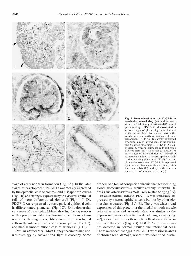
Fig. 1. Immunolocalization of PDGF-D indeveloping human kidney. (A) In a low powerview of a fetal kidney of estimated 83 days ofgestational age, PDGF-D is demonstrated invarious stages of glomerulogenesis, but notin the metanephric blastema (arrows) or thevesicle developing as the earliest stage of glom-erulogenesis. (B) PDGF-D is weakly expressedby epithelial cells (arrowheads) in the comma-and S-shaped structures. (C ) PDGF-D is ex-pressed by visceral epithelial cells and someparietal epithelial cells of the glomerulus atearly stages of differentiation. (D) PDGF-Dexpression confined to visceral epithelial cellsof the maturing glomerulus. (E, F ) In extra-glomerular structures, PDGF-D is expressedby fibroblast-like mesenchymal cells withinthe renal pelvis (E), and by medial smoothmuscle cells of muscular arteries (F).
stage of early nephron formation (Fig. 1A). In the later of them had foci of nonspecific chronic changes includingglobal glomerulosclerosis, tubular atrophy, interstitial fi-stages of development, PDGF-D was weakly expressed
by the epithelial cells of comma- and S-shaped structures brosis and arteriosclerosis most likely related to aging [39].In adult normal kidneys, PDGF-D was uniformly ex-(Fig. 1B) and strongly expressed by the visceral epithelial
cells of more differentiated glomeruli (Fig. 1 C, D). pressed by visceral epithelial cells but not by other glo-merular structures (Fig. 2 A, B). There was widespreadPDGF-D was expressed by some parietal epithelial cells
in differentiated glomeruli (Fig. 1C). Extraglomerular expression of this protein in the medial smooth musclecells of arteries and arterioles that was similar to thestructures of developing kidney showing the expression
of this protein included the basement membrane of im- expression pattern identified in developing kidney (Fig.2C), as well as in smooth muscle cells of vasa rectae inmature collecting ducts, fibroblast-like mesenchymal
cells in the interstitial area of the renal pelvis (Fig. 1E), the medullary area (Fig. 2D). PDGF-D expression wasnot detected in normal tubular and interstitial cells.and medial smooth muscle cells of arteries (Fig. 1F).
Human adult kidney. Most kidney specimens had nor- There were focal changes in PDGF-D expression in areasof chronic renal damage, where it was identified in scle-mal histology by conventional light microscopy. Some

Changsirikulchai et al: PDGF-D expression in human kidneys 2047
Fig. 2. Immunolocalization of PDGF-D inmature human kidney. (A, B show higherpower view) PDGF-D is expressed by visceralepithelial cell of the glomerulus. (C ) PDGF-Dis expressed by medial smooth muscle cells ofarteries, similar to that shown in Fig. 1F. (D)PDGF-D expression is present in the medul-lary area (arrows). PDGF-D staining struc-tures in these areas are vasa rectae (inset).(E ) PDGF-D is expressed at the basolateralmembrane of some injured tubules (arrows),but not by normal tubules (arrowheads). (F )There is expression of PDGF-D by some neo-intimal cells (arrow) in arteriosclerotic vessels.
Table 1. Double labeling immunohistochemical staining pattern in developmental kidney
Metanephric Metanephric Differentiated Mature Uretericblastema vesicle Comma-S-stage glomerulus glomerulus bud
PDGF-D (�) (�) () faintly in EP () VEP and PEP () PEP (�)PDGFR� () (�) (�) () mesangium () mesangium (�)�-smooth muscle actin (�)a (�)b (�)c () mesangium () mesangium (�)WT-1 (�) () focal () EP () nucleus of VEP () nucleus of VEP (�)Ulex europaeus (�)a (�) () EC () EC () EC (�)
Abbrevations are: EP, epithelial cells; VEP, visceral epithelial cells; PEP, parietal epithelial cells; EC, endothelial cells; WT-1, Wilm’s tumor. Symbols are: (�), nega-tive; (), positive.
a Components of vasculature in blastema and interstitium demonstrate the expressionb Perivascular mesenchymal cells are positive staining for alpha-smooth muscle actinc Mesenchymal cells within the glomerular clefts demonstrate positive staining for alpha-smooth muscle actin

Changsirikulchai et al: PDGF-D expression in human kidneys2048
Fig. 3. Double immunolabeling of PDGF-D with antibody markers of different cell types in developing human kidney. (A) Alpha-smooth muscleactin (brown) is localized to mesangial cells, while PDGF-D (blue) is localized to visceral epithelial cells of the glomerulus. (B) PDGFR� (brown)is expressed by mesangial cells, while PDGF-D (blue) is expressed by visceral epithelial cells of the glomerulus. (C ) Co-localization of WT-1(brown, nuclear stain), a phenotypic marker of visceral epithelial cells, and PDGF-D (blue, cytoplasmic stain) in the same cells in the differentiatedglomerulus confirms that PDGF-D is expressed by visceral epithelial cells. PDGF-D also is expressed by some parietal epithelial cells. (D) Ulexeuropaeus agglutinin I (brown) stains endothelial cells, while PDGF-D (blue) is consistently expressed by visceral epithelial cells of the maturingglomeruli. (E ) Ulex europaeus agglutinin I (brown) stains endothelial cells of muscular arteries, while PDGF-D (blue) stains medial smoothmuscle cells. (F ) Serial tissue section of that shown in E, demonstrating co-localization of alpha-smooth muscle actin (brown) and PDGF-D (blue)in the same cells confirms that PDGF-D positive cells are arterial smooth muscle cells. (Publication of this figure in color was made possible bya grant from ZymoGenetics, Seattle, Washington.)

Changsirikulchai et al: PDGF-D expression in human kidneys 2049
Table 2. Double labeling immunohistochemical staining pattern in mature kidney
Glomerular Glomerular Interstitialcapillary EC mesangium VEP PEP Tubule Vessel cells
PDGF-D (�) (�) () (�) (�)a ()b SMC (�)PDGFR� (�) () (�) () (�) () vascular adventitial cells ()�-smooth muscle actin (�) () (�) (�) (�) () SMC ()WT-1 (�) (�) () (�) (�) (�) (�)Ulex europaeus agglutinin I () (�) (�) (�) (�)c () EC (�)
Abbrevations are: EC, endothelial cells; VEP, visceral epithelial cells; PEP, parietal epithelial cells; SMC, smooth muscle cells. Symbols are: (�), negative; (),positive.
a The staining is positive at the cellular basement membrane of the damaged tubulesb The staining also is positive in neointimal cells of arteriosclerotic vessels and vasa rectae in the medullary areac The staining is positive in peritubular capillary endothelial cells
rotic glomeruli, in basolateral membranes of injured tu- (Fig. 5 A-D). The immunostaining of the basement mem-bules (Fig. 2E), and in some neointimal cells of arterio- brane of immature collecting ducts and that of fibroblast-sclerotic vessels (Fig. 2F). like mesenchymal cells in the renal pelvis of developing
kidney could be abolished also by the same procedureDouble labeling immunohistochemistry (Fig. 5 E and F). These findings helped establish the
The localization of PDGF-D expression in developing specificity of the antibody for the immunohistochemicaland normal adult kidneys was confirmed by double-label- procedures reported.ing immunohistochemical techniques using additional The presence of PDGF-D protein in human kidneyantibodies to identify phenotypic markers of different was confirmed by Western blot analysis of tissue-extractedcell types. Ulex europaeus agglutinin I was used to iden- protein from normal and transplant kidneys using thetify endothelial cells. Expression of WT-1 identified vis- polyclonal anti-PDGF-D antibody. The 50 kD monospe-ceral epithelial cells. While no specific markers of mesan- cific bands corresponding to the size of the PDGF-Dgial cells in tissue sections have been identified, PDGFR� monomer as indicated by using a control preparation ofis expressed in the mesangium of differentiated glomer- PDGF-D full-length protein were identified in the kidneyuli [16] and �-smooth muscle actin, a marker for smooth tissue extract running under reducing condition (Fig.muscle cells, has been identified in mesangial cells of 6). The anti-PDGF-D antibody recognized PDGF-D full-differentiated glomeruli [16]. Furthermore, PDGFR� length protein at approximately 90 kD running under non-and alpha-smooth muscle actin have been shown to be reducing condition (data not shown).These results wereexpressed also in the activated mesangial cells in disease consistent with the biochemical properties of PDGF-Dstates of both experimental and human models [13, 14, in other reports (90 kD unreduced and 49 to 55 kD re-31, 40]. Therefore, PDGFR� and �-smooth muscle actin duced) [27, 28]. The specificity of this antibody also waswere used as markers of mesangial cells in this study. confirmed by immunoblotting, which demonstrated that
In developing kidney, the results of double immuno- neither PDGF-C growth factor domain nor full-lengthstaining for PDGFR�, �-smooth muscle actin, WT-1, protein reacted with anti-PDGF-D antibody.Ulex europaeus agglutinin I, and PDGF-D are summa-
RT-PCRrized in Table 1 and shown in Figure 3. In mature normalkidney, the results of double immunostaining for these Platelet-derived growth factor-D mRNA expressionantibodies are summarized in Table 2 and shown in Fig- in the kidney was analyzed by RT-PCR. A PDGF-Dure 4. The double immunolabeling studies confirmed nucleotide fragment of the expected length (918 bp)that PDGF-D was expressed by visceral epithelial cells could be detected in kidney tissue extracts obtained fromof glomeruli in developing and mature kidneys. In addi- normal and transplant nephrectomies by using RT-PCRtion, the co-expression of �-smooth muscle actin and primers specific for PDGF-D (Fig. 7). The specificity ofPDGF-D by neointimal cells of arteriosclerotic vessels RT-PCR product was confirmed by DNA sequencing.indicates that PDGF-D expressing cells in this site are The DNA sequence was matched with human PDGF-Dlikely to be smooth muscle cells. nucleotides 365-1283 of Genbank number AF336376
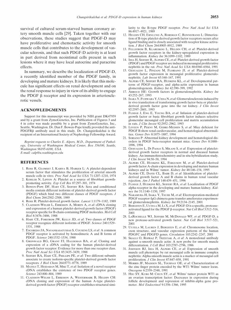
(data not shown).Antibody specificity and Western blotting
Preincubation of anti-PDGF-D antibody with the im-DISCUSSIONmunizing peptide completely abolished the specific im-
This study demonstrates PDGF-D protein and genemunostaining of glomerular epithelial cells, and arterialsmooth muscle cells of developing and mature kidneys expression in human kidneys by Western blotting and

Changsirikulchai et al: PDGF-D expression in human kidneys2050
Fig. 4. Double immunolabeling of PDGF-D with antibody markers of different cell types in mature human kidney. (A) Alpha-smooth muscleactin (brown) is expressed by mesangial cells and PDGF-D (blue) is expressed by visceral epithelial cells in the glomerulus. Alpha-smooth muscleactin also stains extraglomerular interstitial fibroblasts. (B) PDGFR� (brown) stains mesangial cells, while PDGF-D (blue) stains visceral epithelialcells in the glomerulus. (C ) Ulex europaeus agglutinin I (brown) binds endothelial cells, while PDGF-D (blue) stains visceral epithelial cells inthe glomerulus. (D) Co-localization of PDGF-D (blue) with WT-1 (brown) in the glomerulus confirms that PDGF-D is expressed by visceralepithelial cells. (E, F ) In arteriosclerotic vessels, PDGF-D (blue) is co-localized with alpha-smooth muscle actin (brown, in E) in medial smoothmuscle cells and neointimal cells and is distinct from the double immunolabeling pattern with ulex europaeus agglutinin I (brown, in F) whichbinds endothelial cells. (Publication of this figure in color was made possible by a grant from ZymoGenetics, Seattle, Washington.)
Changsirikulchai et al: PDGF-D expression in human kidneys 2051
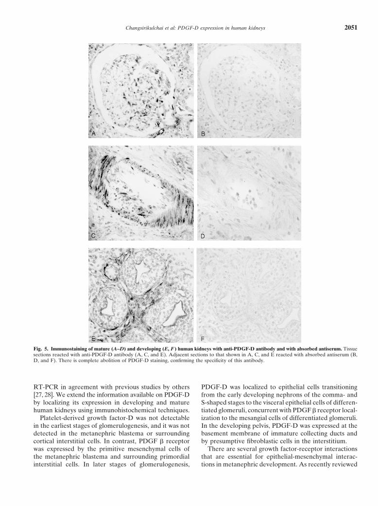
Fig. 5. Immunostaining of mature (A–D) and developing (E, F ) human kidneys with anti-PDGF-D antibody and with absorbed antiserum. Tissuesections reacted with anti-PDGF-D antibody (A, C, and E). Adjacent sections to that shown in A, C, and E reacted with absorbed antiserum (B,D, and F). There is complete abolition of PDGF-D staining, confirming the specificity of this antibody.
RT-PCR in agreement with previous studies by others PDGF-D was localized to epithelial cells transitioningfrom the early developing nephrons of the comma- and[27, 28]. We extend the information available on PDGF-D
by localizing its expression in developing and mature S-shaped stages to the visceral epithelial cells of differen-tiated glomeruli, concurrent with PDGF � receptor local-human kidneys using immunohistochemical techniques.
Platelet-derived growth factor-D was not detectable ization to the mesangial cells of differentiated glomeruli.In the developing pelvis, PDGF-D was expressed at thein the earliest stages of glomerulogenesis, and it was not
detected in the metanephric blastema or surrounding basement membrane of immature collecting ducts andby presumptive fibroblastic cells in the interstitium.cortical interstitial cells. In contrast, PDGF � receptor
was expressed by the primitive mesenchymal cells of There are several growth factor-receptor interactionsthat are essential for epithelial-mesenchymal interac-the metanephric blastema and surrounding primordial
interstitial cells. In later stages of glomerulogenesis, tions in metanephric development. As recently reviewed
Changsirikulchai et al: PDGF-D expression in human kidneys2052
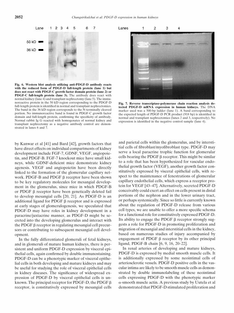
Fig. 6. Western blot analysis utilizing anti-PDGF-D antibody reactswith the reduced form of PDGF-D full-length protein (lane 1) butdoes not react with PDGF-C growth factor domain protein (lane 2) orPDGF-C full-length protein (lane 3). The antibody does react withnormal kidney (lane 4) and transplant nephrectomy (lane 5). The immu-noreactive protein in the 50 kD region corresponding to the PDGF-D Fig. 7. Reverse transcriptase-polymerase chain reaction analysis de-full-length protein is identified in normal and transplant nephrectomies. tected PDGF-D mRNA expression in human kidneys. The DNAThe band in the 36 kD region corresponds to the N-terminally cleaved marker used was a 500-bp ladder (lane 1). A band corresponding toportion. No immunoreactive band is found in PDGF-C growth factor the expected length of PDGF-D PCR product (918 bp) is identified indomain and full-length protein, confirming the specificity of antibody. normal and transplant nephrectomies (lanes 2 and 3, respectively). NoNormal rabbit Ig G reacted with homogenates of normal kidney and expression is identified in the negative control sample (lane 4).transplant nephrectomy as a negative antibody control are demon-strated in lanes 6 and 7.
and parietal cells within the glomerulus, and by intersti-by Kanwar et al [41] and Bard [42], growth factors thattial cells of fibroblast/myofibroblast type. PDGF-D mayhave direct effects on individual compartments of kidneyserve a local paracrine trophic function for glomerulardevelopment include FGF-7, GDNF, VEGF, angiopoie-cells bearing the PDGF � receptor. This might be similartin, and PDGF-B. FGF-7 knockout mice have small kid-to a role that has been hypothesized for vascular endo-neys, while GDNF-deficient mice demonstrate kidneythelial growth factor (VEGF), another growth factor con-agenesis. VEGF and angiopoietin have been directlystitutively expressed by visceral epithelial cells, with re-linked to the formation of the glomerular capillary net-spect to the maintenance of fenestrations of glomerularwork. PDGF-B and PDGF � receptor have been showncapillary endothelial cells, which express a receptor pro-to be key regulatory molecules for mesangial develop-tein for VEGF [43–47]. Alternatively, secreted PDGF-Dment in the glomerulus, since mice in which PDGF-Bconceivably could exert an effect on cells present in distalor PDGF � receptor have been genetically deleted failportions of the nephron and in the lower urinary tract,to develop mesangial cells [20, 21]. As PDGF-D is anor perhaps systemically. Since so little is currently knownadditional ligand for PDGF � receptor and is expressedabout the regulation of PDGF-D release from variousat early stages of glomerulogenesis, we speculated thatcell types, we are unable to offer a more specific schemaPDGF-D may have roles in kidney development in afor a functional role for constitutively expressed PDGF-D.paracrine/juxtacrine manner, as PDGF-D might be se-Its ability to engage the PDGF � receptor strongly sug-creted into the developing glomerulus and interact withgests a role for PDGF-D in promoting proliferation andthe PDGF � receptor in regulating mesangial cell precur-migration of mesangial and interstitial cells in the kidney,sors or contributing to subsequent mesangial cell devel-based on numerous studies of injury accompanied byopment.engagement of PDGF � receptor by its other principalIn the fully differentiated glomeruli of fetal kidneys,ligand, PDGF-B chain [6, 9, 16, 20–22].and in glomeruli of mature human kidneys, there is per-
In renal arteries of developing and mature kidneys,sistent and uniform PDGF-D expression by visceral epi-PDGF-D is expressed by medial smooth muscle cells. Itthelial cells, again confirmed by double immunostaining.is additionally expressed by some neointimal cells ofPDGF-D can be a phenotypic marker of visceral epithe-arteriosclerotic vessels. PDGF-D positive cells in the vas-lial cells in both developing and mature kidneys and maycular intima are likely to be smooth muscle cells as demon-be useful for studying the role of visceral epithelial cellsstrated by double immunolabeling of these neointimalin kidney diseases. The significance of widespread ex-cells expressing PDGF-D with the phenotypic markerpression of PDGF-D by visceral epithelial cells is un-�-smooth muscle actin. A previous study by Uutela et alknown. The principal receptor for PDGF-D, the PDGF �
receptor, is constitutively expressed by mesangial cells demonstrated that PDGF-D stimulated proliferation and
Changsirikulchai et al: PDGF-D expression in human kidneys 2053
larity to the B-type PDGF receptor. Proc Natl Acad Sci USAsurvival of cultured serum-starved human coronary ar-86:4917–4921, 1989
tery smooth muscle cells [29]. Taken together with our 12. Heldin CH, Ernlund A, Rorsman C, Ronnstrand L: Dimeriza-tion of B-type platelet-derived growth factor receptors occurs afterobservations, these studies suggest that PDGF-D mayligand binding and is closely associated with receptor kinase activa-have proliferative and chemotactic activity for smoothtion. J Biol Chem 264:8905–8912, 1989
muscle cells that contributes to the development of vas- 13. Fellstrom B, Klareskog L, Heldin CH, et al: Platelet-derivedgrowth factor receptors in the kidney-upregulated expression incular sclerosis, and that such PDGF-D activity is at leastinflammation. Kidney Int 36:1099–1102, 1989in part derived from neointimal cells present in such
14. Iida H, Seifert R, Alpers CE, et al: Platelet-derived growth factorlesions where it may have local autocrine and paracrine (PDGF) and PDGF receptor are induced in mesangial proliferative
nephritis in the rat. Proc Natl Acad Sci USA 88:6560–6564, 1991effects.15. Gesualdo L, Pinzani M, Floriano JJ, et al: Platelet-derivedIn summary, we describe the localization of PDGF-D,
growth factor expression in mesangial proliferative glomerulo-a recently identified member of the PDGF family, in nephritis. Lab Invest 65:160–167, 1991
16. Alpers CE, Seifert RA, Hudkins KL, et al: Developmental pat-developing and mature kidneys. It is likely that this mole-terns of PDGF-receptor, and alpha-actin expression in humancule has significant effects on renal development and onglomerulogenesis. Kidney Int 42:390–399, 1992
the renal response to injury in view of its ability to engage 17. Abboud HE: Growth factors in glomerulonephritis. Kidney Int43:252–267, 1993the PDGF � receptor and its expression in arterioscle-
18. Isaka Y, Fujiwara Y, Ueda N, et al: Glomerulosclerosis induced byrotic vessels. in vivo transfection of transforming growth factor-beta or platelet-derived growth factor gene into the rat kidney. J Clin Invest92:2597–2601, 1993ACKNOWLEDGMENTS
19. Floege J, Eng E, Young BA, et al: Infusion of platelet-derivedSupport for this manuscript was provided by NIH grant DK47959 growth factor or basic fibroblast growth factor induces selective
and by a grant from ZymoGenetics, Inc. Publication of Figures 3 and glomerular mesangial cell proliferation and matrix accumulation4 in color was made possible by a grant from ZymoGenetics, Inc., in rats. J Clin Invest 92:2952–2962, 1993Seattle, Washington. Dr. Dan F. Bowen-Pope kindly provided the anti- 20. Leveen P, Pekny M, Gebre-Medhin S, et al: Mice deficient for
PDGF B show renal cardiovascular, and hematological abnormali-PDGFR� antibody used in this study. Dr. Changsirikulchai is theties. Genes Dev 8:1875–1887, 1994recipient of an International Society of Nephrology Fellowship Award.
21. Soriano P: Abnormal kidney development and hematological dis-orders in PDGF beta-receptor mutant mice. Genes Dev 8:1888–Reprint requests to Charles E. Alpers, M.D., Department of Pathol-1896, 1994ogy, University of Washington Medical Center, Box 356100, Seattle,
22. Gesualdo L, Di Paolo S, Milani S, et al: Expression of platelet-Washington 96195-6100, USA.derived growth factor receptors in normal and diseased humanE-mail: [email protected]. An immunohistochemistry and in situ hybridization study.J Clin Invest 94:50–58, 1994
REFERENCES 23. Alpers CE, Hudkins KL, Ferguson M, et al: Platelet-derivedgrowth factor A-chain expression in developing and mature human
1. Ross R, Glomset J, Kariya B, Harker L: A platelet-dependent kidneys and in Wilms’ tumor. Kidney Int 48:146–154, 1995serum factor that stimulates the proliferation of arterial smooth 24. Alpers CE, Davis CL, Barr D, et al: Identification of platelet-muscle cells in vitro. Proc Natl Acad Sci USA 71:1207–1210, 1974 derived growth factor A and B chains in human renal vascular
2. Kohler N, Lipton A: Platelet as a source of fibroblast growth- rejection. Am J Pathol 148:439–451, 1996promoting activity. Exp Cell Res 87:297–301, 1974 25. Floege J, Hudkins KL, Seifert RA, et al: Localization of PDGF
3. Bowen-Pope DF, Hart CE, Seifert RA: Sera and conditioned alpha-receptor in the developing and mature human kidney. Kid-media contain different isoforms of platelet-derived growth factor ney Int 51:1140–1150, 1997(PDGF) which bind two different classes of PDGF receptors. J 26. Nakamura H, Isaka Y, Tsujie M, et al: Electroporation-mediatedBiol Chem 264:2502–2508, 1989 PDGF receptor-IgG chimera gene transfer ameliorates experimen-
4. Ross R: Platelet-derived growth factor. Lancet 1:1179–1182, 1989 tal glomerulonephritis. Kidney Int 59:2134–2145, 20015. Claesson-Welsh L, Eriksson A, Moren A, et al: cDNA cloning 27. Bergsten E, Uutela M, Li X, et al: PDGF-D is a specific, protease-
and expression of a human platelet-derived growth factor (PDGF) activated ligand for the PDGF �-receptor. Nat Cell Biol 3:512–516,receptor specific for B-chain-containing PDGF molecules. Mol Cell 2001Biol 8:3476–3486, 1988 28. LaRochelle WJ, Jeffers M, McDonald WF, et al: PDGF-D, a
6. Hart CE, Forstrom JW, Kelly JD, et al: Two classes of PDGF new protease-activated growth factor. Nat Cell Biol 3:517–521,receptor recognize different isoforms of PDGF. Science 240:1529– 20011531, 1988 29. Uutela M, Lauren J, Bergsten E, et al: Chromosome location,
7. Escobedo JA, Navankasatussas S, Cousens LS, et al: A common exon structure, and vascular expression patterns of the humanPDGF receptor is activated by homodimeric A and B forms of PDGFC and PDGFD genes. Circulation 103:2242–2247, 2001PDGF. Science 240:1532–1534, 1988 30. Skalli O, Ropraz P, Trzeciak A, et al: A monoclonal antibody
8. Gronwald RG, Grant FJ, Haldeman BA, et al: Cloning and against �-smooth muscle actin: A new probe for smooth muscleexpression of a cDNA coding for the human platelet-derived differentiation. J Cell Biol 103:2787–2796, 1986growth factor receptor: Evidence for more than one receptor class. 31. Johnson RJ, Iida H, Alpers CE, et al: Expression of smoothProc Natl Acad Sci USA 85:3435–3439, 1988 muscle cell phenotype by rat mesangial cells in immune complex
9. Seifert RA, Hart CE, Phillips PE, et al: Two different subunits nephritis: Alpha-smooth muscle actin is a marker of mesangial cellassociate to create isoform-specific platelet-derived growth factor proliferation. J Clin Invest 87:847–858, 1991receptors. J Biol Chem 264:8771–8778, 1989 32. Morris JF, Madden SL, Tournay OE, et al: Characterization of
10. Matsui T, Heidaran M, Miki T, et al: Isolation of a novel receptor the zinc finger protein encoded by the WT1 Wilms’ tumor locus.cDNA establishes the existence of two PDGF receptor genes. Oncogene 6:2339–2348, 1991Science 243:800–804, 1989 33. Hsu SY, Kubo M: Chun SY, et al: Wilms’ tumor protein WT1 as
11. Claesson-Welsh L, Eriksson A, Westermark B, Heldin CH: an ovarian transcription factor: Decreases in expression duringcDNA cloning and expression of the human A-type platelet- follicle development and repression of inhibin-alpha gene pro-
moter. Mol Endocrinol 9:1356–1366, 1995derived growth factor (PDGF) receptor establishes structural simi-

Changsirikulchai et al: PDGF-D expression in human kidneys2054
34. Silberstein GB, Horn KV, Strickland P, Roberts CT Jr: Altered 41. Kanwar KS, Carone FA, Kumar A, et al: Role of extracellularexpression of the WT1 Wilms tumor suppressor gene in human matrix, growth factors and proto-oncogenes in metanephric devel-breast cancer. Proc Natl Acad Sci USA 94:8132–8137, 1997 opment. Kidney Int 52:589–606, 1997
35. Yang Y, Jeanpierre C, Dressler GR, et al: WT1 and PAX-2 42. Bard JB: Growth and death in the developing mammarian kidney:podocyte expression in Denys-Drash syndrome and isolated diffuse Signals, receptors and conversation. Bioessays 24:72–82, 2002mesangial sclerosis. Am J Pathol 154:181–192, 1999 43. Carmeliet P, Ferreira V, Breier G, et al: Abnormal blood vessel
36. Mundlos S, Pelletier J, Darveau A, et al: Nuclear localization development and lethality in embryos lacking a single VEGF allele.of the protein encoded by the Wilms’ tumor gene WT1 in embry- Nature 380:435–439, 1996onic and adult tissues. Development 119:1329–1341, 1993 44. Abrahamson DR, Robert B, Hyink DP, et al: Origins and forma-
37. Holthofer H, Virtanen I, Kariniemi AL, et al: Ulex europaeus tion of microvasculature in the developing kidney. Kidney IntI lectin as a marker for vascular endothelium in human tissues. 54(Suppl 67):S7–S11, 1998Lab Invest 47:60–66, 1982 45. Eichmann A, Corbel C, Jaffredo T, et al: Avian VEGF-C: Clon-38. Alpers CE, Beckstead JH: Monocyte/macrophage-derived cells
ing, embryonic expression pattern and stimulation of the differenti-in normal and transplanted human kidneys. Clin Immunol Immu-ation of VEGFR2-expressing endothelial cell precursors. Develop-nopathol 36:129–140, 1985ment 125:743–752, 199839. Kaplan C, Pasternack B, Shah H, Gallo G: Age-related inci-
46. Esser S, Wolburg K, Wolburg H, et al: Vascular endothelialdence of sclerotic glomeruli in human kidneys. Am J Pathol 80:227–growth factor induces endothelial fenestrations in vitro. J Cell Biol234, 1975140:947–959, 199840. Alpers CE, Hudkins KL, Gown AM, Johnson RJ: Enhanced
47. Tufro A: VEGF spatially directs angiogenesis during metanephricexpression of “muscle-specific” actin in glomerulonephritis. KidneyInt 41:1134–1142, 1992 development in vitro. Dev Biol 227:558–566, 2000