Embed Size (px)
Citation preview
DOI: 10.1542/peds.2003-0668-F 2004;114;970Pediatrics
B. Lynne Hutchison, Luke A.D. Hutchison, John M.D. Thompson and Ed A. MitchellCohort Study
Plagiocephaly and Brachycephaly in the First Two Years of Life: A Prospective
http://pediatrics.aappublications.org/content/114/4/970.full.html
located on the World Wide Web at: The online version of this article, along with updated information and services, is
of Pediatrics. All rights reserved. Print ISSN: 0031-4005. Online ISSN: 1098-4275.Boulevard, Elk Grove Village, Illinois, 60007. Copyright © 2004 by the American Academy published, and trademarked by the American Academy of Pediatrics, 141 Northwest Pointpublication, it has been published continuously since 1948. PEDIATRICS is owned, PEDIATRICS is the official journal of the American Academy of Pediatrics. A monthly
by guest on May 23, 2013pediatrics.aappublications.orgDownloaded from

Plagiocephaly and Brachycephaly in the First Two Years of Life:A Prospective Cohort Study
B. Lynne Hutchison, DipHSc, PG DipSc*; Luke A.D. Hutchison, BSc (Hons), MS‡;John M.D. Thompson, PhD*; and Ed A. Mitchell, DCH, FRACP, FRCPCH, DSc (Med)*
ABSTRACT. Objectives. Although referrals for non-synostotic plagiocephaly (NSP) have increased in recentyears, the prevalence, natural history, and determinantsof the condition have been unclear. The objective of thisstudy was to assess the prevalence and natural history ofNSP in normal infants in the first 2 years of life and toidentify factors that may contribute to the developmentof NSP.
Methods. Two hundred infants were recruited atbirth. At 6 weeks, 4 months, 8 months, 12 months, and 2years, the head circumference shape was digitally photo-graphed, and head shape was quantified using custom-written software. At each age, infants were classified ascases when the cephalic index was >93% and/or theoblique cranial length ratio was >106%. Neck rotationand a range of infant, infant care, socioeconomic, andobstetric factors were assessed.
Results. Ninety-six percent of infants were followedto 12 months, and 90.5% were followed to 2 years. Prev-alence of plagiocephaly and/or brachycephaly at 6 weeksand 4, 8, 12, and 24 months was 16.0%, 19.7%, 9.2%, 6.8%,and 3.3% respectively. The mean cephalic index by 2years was 81.6% (range: 72.0%–102.6%); the mean obliquecranial length ratio was 102.6% (range: 100.1%–109.4%).Significant univariate risk factors of NSP at 6 weeksinclude limited passive neck rotation at birth, preferen-tial head orientation, supine sleep position, and headposition not varied when put to sleep. At 4 months, riskfactors were male gender, firstborn, limited passive neckrotation at birth, limited active head rotation at 4 months,supine sleeping at birth and 6 weeks, lower activity level,and trying unsuccessfully to vary the head position whenputting the infant down to sleep.
Conclusions. There is a wide range of head shapes ininfants, and prevalence of NSP increases to 4 months butdiminishes as infants grow older. The majority of caseswill have resolved by 2 years of age. Limited head rota-tion, lower activity levels, and supine sleep positionseem to be important determinants. Pediatrics 2004;114:970–980; plagiocephaly, brachycephaly, anthropometry,cohort studies, infant care, supine position.
ABBREVIATIONS. NSP, nonsynostotic plagiocephaly; SIDS, sud-den infant death syndrome; OCLR, oblique cranial length ratio;
PDQ-II, Revised Denver II Prescreening Questionnaire; PAT, Pic-torial Assessment of Temperament; OR, odds ratio; CI, confidenceinterval.
The rising incidence of nonsynostotic plagio-cephaly (NSP) has been documented in theliterature since 1992 and has been attributed to
the adoption of the supine sleep position in accor-dance with sudden infant death syndrome (SIDS)prevention recommendations.1 A critical review ofthe literature in 1998 concluded that the actual prev-alence is unknown and that the condition was beingrecognized more frequently as a result of increasedawareness.2
Many facets of the plagiocephalic condition areunclear.3 However, it has been shown that NSP ismore likely to occur in boys,4–7 firstborns,8,9 prema-ture infants,10 and those who sleep in the supineposition.10,11 A preferred head orientation12,13 andlimited head rotation6 may be important determi-nants. Varying the head position and tummy timeseem to be protective, whereas developmental delayand lower activity levels may be associated withcases.11 Although positional preference has been fol-lowed prospectively,13 no prospective cohort studiesof normal infants that have investigated the preva-lence of NSP and quantified the development ofhead shape in the first 2 years have been undertaken.
Strictly speaking, brachycephaly, or a high ce-phalic index without noticeable skewness of thehead, is not true plagiocephaly, which means“oblique head.” In NSP, a high cephalic index issometimes but not always associated with asymme-try. However, as our clinical experience indicatesthat central occipital flattening is as concerning toparents as the skewed head shape, we have chosen tocombine them. It is possible that the mechanisms arethe same, the flat area merely corresponding to thepreferred resting position, and any associated boss-ing reflecting different areas of displacement of headvolume. Alternatively, it has been postulated thatplagiocephaly with asymmetry may more commonlyoriginate from neck muscle dysfunction, whereasbrachycephaly results from compression.14
We have developed a new head shape measuringtechnique, HeadsUp, which involves an elastic headcircumference band that is photographed digitallyfrom above the head. The photograph is analyzedusing a custom-written computer program to obtainmeasurements to quantify the head shape. The
From the *Department of Paediatrics, University of Auckland, Auckland,New Zealand; and ‡Computer Science Department, Brigham Young Uni-versity, Provo, Utah.Accepted for publication Mar 16, 2004.doi:10.1542/peds.2003-0668-FReprint requests to (L.H.) Department of Paediatrics, University of Auck-land, Private Bag 92019, Auckland, New Zealand. E-mail: [email protected] (ISSN 0031 4005). Copyright © 2004 by the American Acad-emy of Pediatrics.
970 PEDIATRICS Vol. 114 No. 4 October 2004 by guest on May 23, 2013pediatrics.aappublications.orgDownloaded from
method has been used successfully in a pilot study of60 infants and demonstrated much greater reliabilityand acceptability compared with measurements ob-tained using a flexible measuring strip.15 We haveidentified cutoff points for both cephalic index andoblique cranial length ratio (OCLR; ie, the ratio of thelong cross-diagonal measurement to the shortercross-diagonal measurement). Beyond these thresh-olds, central occipital flattening and head shapeasymmetry, respectively, are deemed to be abnor-mal. In practice, we believe that these cutoffs approx-imate the points at which head deformity becomesvisually obvious, particularly where the infant haslittle hair. The points, 93% for cephalic index and106% for OCLR, allow for the allocation of the cohortinfants to either case or control. We aimed to deter-mine the prevalence, natural history, and risk factorsof NSP in the first 2 years of life.
METHODSThe cohort infants were born at the delivery unit at North Shore
Hospital, Auckland, a community maternity unit that deals withlow-risk deliveries. On admission to the unit, the mother had theopportunity to opt out of being approached regarding research.The researcher was given a birth list each day, with the names ofthose who opted out deleted from this list. Selection of everyfourth infant on the list yielded a cohort of 238 born betweenSeptember 2001 and February 2002. Infants with congenital defor-mities, those who were not domiciled in the Waitemata HealthDistrict, those who were planning to move out of the region in thenext year, and those who could not be seen in the first week wereexcluded.
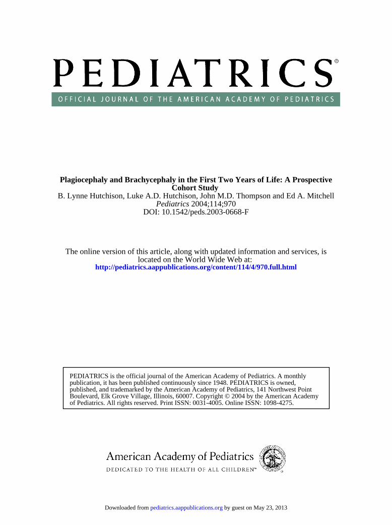
Of the eligible mothers who were invited to participate, 200(88%) were enrolled in the study (Fig 1). The initial interview wasconducted either in the postnatal ward or in the mother’s homewithin the first week after delivery. At this interview, the motherwas asked about sociodemographic factors (parents’ ages, ethnic-ity, occupation, and mother’s education), obstetric factors (parity,gestation, presentation, method of delivery, length of labor, andmultiple birth), and infant details such as date of birth, gender,Apgar scores, and birth measurements. The mother’s highest ed-ucational level was classified into 1) no qualifications or 1 or moreSchool Certificate (year 11) subjects, 2) sixth form or Bursaryqualification (years 12–13), and 3) tertiary education or profes-sional certification. The parents’ occupations were rated in accor-dance with the New Zealand Socio-economic Index of Occupa-tional Status16 classifications into high, medium, and lowsocioeconomic status, the highest rating of either parent beingused for the classification.
The interviewer assessed the infant’s head shape for anythingunusual, such as a caput or area of flattening. When possible,when the infant was in a relaxed state, passive head rotation was
assessed. This was accomplished by standing behind the supine-lying infant, holding the head between the hands, and gentlyrotating the head from side to side. Any restriction or tightness in1 or both directions was noted. The head circumference wasmeasured around the maximum fronto-occipital circumference.
At 6 weeks, 4 months, 8 months, 12 months, and 24 months, themother and the infant were visited at home, and a set of digitalphotographs of the infant’s head using the HeadsUp band weretaken to document the head shape, as follows. While seated on themother’s knee (or on the floor for older infants), the infant wasgiven 1 or 2 toys to play with if necessary while a close-fitting,nylon stocking–type cap was placed on the head to flatten the hair.The infant’s identification was written on a yellow sticker andattached to the stocking cap. A small yellow cape was placed overthe shoulders to mask any competing colors in the clothing orsurroundings. A soft, elastic, blue headband made of a narrowstrip of 7-mm-thick covered neoprene was then placed over thehead circumference. On the headband are sliding green ear mark-ers and a red marker to indicate the middle of the nose. The redmarker is also a known length (50 mm), which is used to deter-mine scale. After positioning the band and the markers, digitalphotographs were taken from �800 mm above the vertex of thehead, using a Sony DSC-S50 digital still camera with pivoting LCDviewing screen. Several photographs were taken, and the 3 bestwere kept for analysis by the HeadsUp computer program. Themean measurements taken from the 3 photographs were used inthe final analysis. We did not use the HeadsUp photographicmeasure on the newborns because of possible birth molding of thehead.
The main measure of asymmetry from one side of the head tothe other was the OCLR. These lines were taken from pointslocated 40 degrees either side of the posterior midline, obliquelyacross the head to derived frontozygomatic points in the frontalarea of the head circumference. The other important measure usedwas the cephalic index, a measure of central posterior flattening orbrachycephaly, calculated from (head breadth/head length) �100. Criteria to allocate case definition were made previouslyusing analyses of mean cephalic indices and OCLR measurementsobtained from a pilot photograph study (unpublished data). Thecriteria so defined require that the cephalic index be 93% or aboveand/or OCLR be 106% or above. Other measurements assessedwere head circumference, head area bounded by the blue band,angles of each ear relative to the nose position, and transcranialdifference in millimeters.
At 6 weeks, the infant’s passive head rotation was assessed asfor the newborns. At 4, 8, 12, and 24 months, active head rotationwas checked by seating the infant facing outward on the mother’sknee, then holding a bright musical toy in front of the infant. Thetoy was brought around to each side to encourage the infant tofollow the toy with the eyes until they were looking across theshoulder, without moving the body around with the head. Anylimitation or difficulty in head rotation was recorded.
At each of the follow-up interviews, the mother was askedabout such factors as weight, length, and head circumferencemeasurements as recorded in the child’s Well Child Record Book(a parent-held community child health nursing record), healthproblems, hair loss, and the presence of preferential head turningor neck dysfunction. In addition, she was asked about infant carepractices such as breastfeeding; pacifier use; sleep position; headvarying; total daily duration of tummy time and upright time; bedtype; mattress type; underbedding; pillow use; preferred maternalholding positions; and amount of time spent in supine in cots, carseats, bouncers, and other places per day on average.
At the 6-week interview, the mother was given the RevisedDenver II Prescreening Questionnaire (PDQ-II)17 and was asked togo through the items until 3 “no” responses were recorded. Ateach subsequent interview, she was asked to reassess the previous“no” responses and then to continue the items until 3 “no” re-sponses were recorded again. The 0- to 9-month and 9- to 24-month forms were used. The number of delays and/or cautionsfor the child’s age was recorded. Children with no delays and 1 orno cautions were rated normal. If a child had 1 delay or 2 cautions,then the rating was slightly abnormal, and 2 or more delays or 3or more cautions were rated abnormal.
Temperament assessment using the Pictorial Assessment ofTemperament (PAT)18 was conducted at 4 months. This consists ofa 10-item measure of temperament based on Carey’s RevisedInfant Temperament Questionnaire and consists of vignettes de-Fig 1. Enrollment of subjects.
ARTICLES 971 by guest on May 23, 2013pediatrics.aappublications.orgDownloaded from

picting 3 different types of response to 10 different situations, suchas getting dressed, waking up, loud noises, etc. The response typesare “easy,” “average or slow-to-warm-up,” and “difficult.” Ascore of 1 is given for “easy” responses, 2 for “average or slow-to-warm-up” responses, and 3 for “difficult” responses, giving atotal possible score range of between 10 and 30.
Temperament was also assessed at each age by showing themother a plastic gauge with a slider on it. At 1 end is a happy faceand the words “very settled/easy-going,” and at the other end isan unhappy face and the words “very unsettled/difficult.” Themother was asked to position the slider to indicate the child’soverall temperament at that age. The other side of the gauge ismarked with a scale from 0 to 10, and the researcher thus was ableto assign a score for the mother’s assessment. Activity level at eachage was similarly assessed by the mother, with the gauge scoringbetween “very inactive” (score � 0) and “very active” (score � 10).
The interviewer assessed mattress softness using a subjectiverating of soft, medium, or firm. A weight (10.5 g/cm2) on a6-mm-diameter aluminum plunger gauged in 1-mm increments,which was dropped onto the mattress through a hole in a smallcircle of wood, also gave an objective assessment of the mattressfirmness. If the mattress was extremely hard, then the weightdropped very little, giving a reading close to 0 mm, whereas verysoft mattresses gave a higher reading of up to 30 mm. When themattress was not available, the mother was asked to rate it as soft,medium, or firm.
Baseline data were collected at the first interview. The datawere analyzed as a standard case-control analysis using univariateand multivariate logistic regression, using SAS (Release 8.2; SASInstitute, Cary, NC).
All interviews were conducted by the principal investigator,with the exception of some 6-week and 4-month interviews thatwere done by 1 other trained interviewer. No advice was offeredto parents of children who developed a misshapen head shape;however, if at any time a mother became concerned about headshape problems developing in her infant, she was advised to talkto her family doctor or Plunket nurse (community child healthnurse) for advice. The Auckland Ethics Committee approved thestudy.
RESULTS
Cohort CharacteristicsThe infants who were enrolled in this study were
healthy infants who were mostly full term. Ninety-one percent of the initial interviews were completedin the first 36 hours. Of the 200 infants who wereenrolled in the study, 100% were seen at 6 weeks, 198(99%) were seen at 4 months, 196 (98%) were seen at8 months, 192 (96%) were seen at 12 months, and 181(90.5%) were followed to 2 years. The characteristicsof the cohort are listed in Table 1.
Head MeasurementsNewborn head shape was normal on visual assess-
ment for 63.0% of the cohort. Cone-shaped headswere seen in 13.5% of the cohort; prominent or in-dented sutures in 12%; and caputs, flat areas, or other“bumps or dents” in 16%.
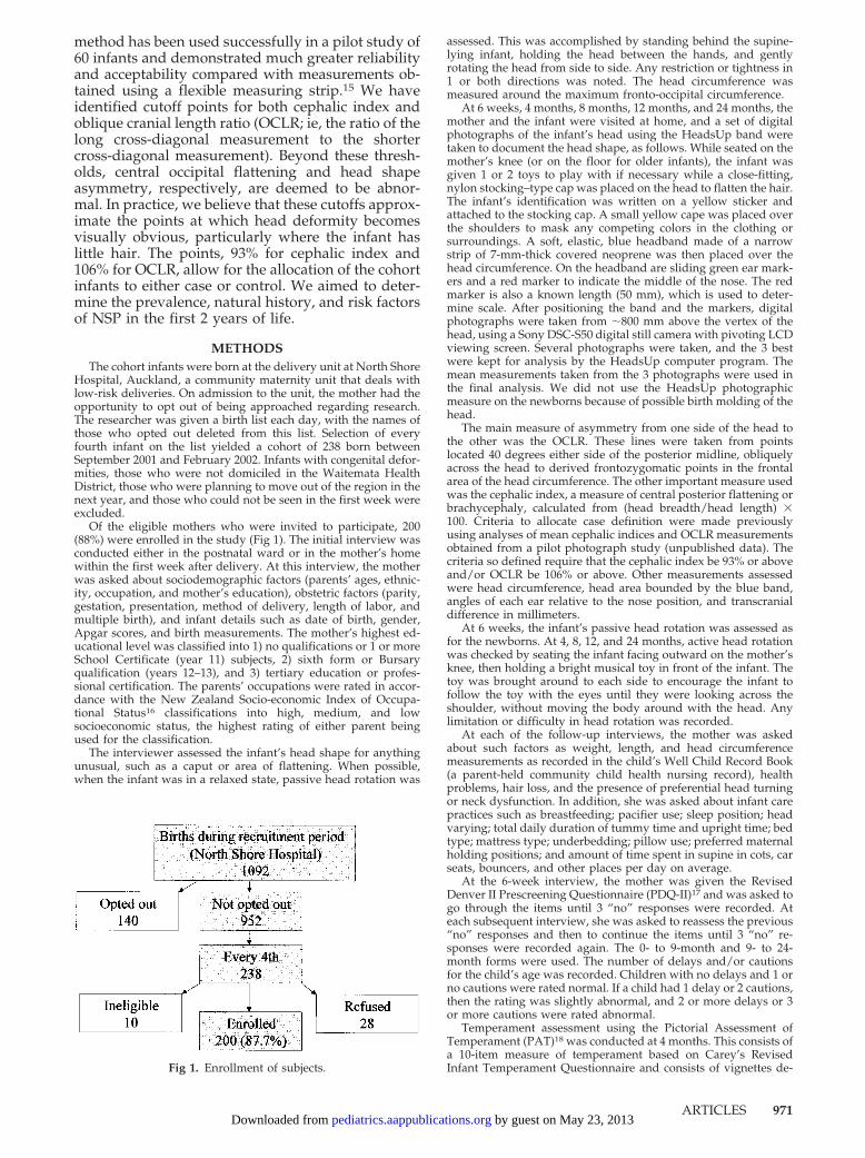
At the follow-up interviews, there was a widerange for cephalic index and OCLR (Table 2). Thewidest range for both occurred at 6 weeks, butwhereas the maximum OCLR reduced thereafter, themaximum cephalic index was recorded at 12 months.There was also a wide range of ear angle positionsseen particularly at the 6-week period, although nodifference was detected between cases and controlsubjects at each age for ear angles or head circum-ference. Head circumference as measured by tapemeasure versus that measured by HeadsUp showeda high correlation between the 2 types of measure (r
� 0.98) over all ages. At follow-up and using theHeadsUp measure, boys’ head circumferences weresignificantly larger than girls’, being �1 cm larger ateach age. There was no difference detected betweengenders for cephalic index, OCLR, or ear angles.
The mean difference between the transcranial di-ameters in the plagiocephalic infants was 9.8 mm(SD: 2.0), 11.3 mm (SD: 0.6), 10.3 mm (SD: 0.9), 11.1mm (SD: 1.2), and 12.0 mm (SD: 2.4), at 6 weeks and4, 8, 12, and 24 months respectively.
Prevalence and Natural HistoryAfter each follow-up interview, any infant who
had a cephalic index of �93% and/or an OCLR of�106% was classified as a case (see Fig 2). In Fig 2,cases that were defined as normal for the purposes ofthis study are in the lower left-hand quadrant,brachycephalic cases are in the top left-hand quad-rant, plagiocephalic cases are in the lower right-handquadrant, and those with both are in the top right-hand quadrant.
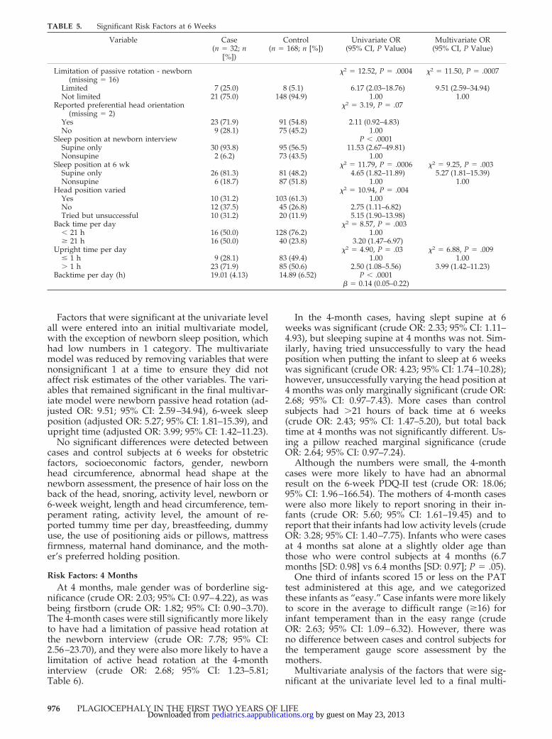
Using the cutoff points of 93% for cephalic indexand 106% for OCLR, the numbers of cases and non-cases at each age are illustrated in Figs 3 and 4.Although half of the 6-week cases had resolved by 4months, 23 new cases had occurred at 4 months.Thereafter, few new cases occurred and the totalnumber of cases started diminishing so that by 12months, there were only one third as many cases asat 4 months. By the age of 2 years, only 6 (3.3%)children recorded values outside the set parameters:3 were brachycephalic and 3 were plagiocephalic.
TABLE 1. Cohort Description (n � 200)
Variable n (%)
GenderMale 106 (53.0)Female 94 (47.0)
ParityFirstborn 90 (45.0)Later born 110 (55.0)
Multiple birthSingletons 199 (99.5)Twin 1 (0.5)
Gestation�37 wk 4 (2.0)�37 wk 196 (98.0)
DeliveryNormal vaginal 133 (66.5)Cesarean 41 (20.5)Assisted vaginal 26 (13.0)
5-Min Apgar score�7 1 (0.5)�7 199 (99.5)
Maternal age�25 20 (10.0)25–29 38 (19.0)30–34 87 (43.5)35� 55 (27.5)
Mother’s highest qualificationNone or school certificate 44 (22.3)Sixth form/bursary 45 (22.9)Tertiary/professional 108 (54.8)
Socioeconomic statusLow 22 (11.0)Medium 111 (55.5)High 67 (33.5)
972 PLAGIOCEPHALY IN THE FIRST TWO YEARS OF LIFE by guest on May 23, 2013pediatrics.aappublications.orgDownloaded from
One of the 12-month cases was unable to be followedto 2 years.
The overall prevalence rates for the cohort were16% at 6 weeks, 19.7% at 4 months, 9.2% at 8 months,6.8% at 12 months, and 3.3% at 24 months. More than
twice as many infants were classified as having pla-giocephaly alone (OCLR �106%) than havingbrachycephaly alone (cephalic index �93%) at 6weeks, but at both 4 and 8 months, more infants wereclassified as brachycephalic than plagiocephalic. This
Fig 2. Mean cephalic index and OCLR scores for all infants at each age (controls [normal] are in the lower left quadrants of each scatterplot).
TABLE 2. Head Measurements (All Infants)
Variable 6 Weeks(n � 200)
4 Months(n � 198)
8 Months(n � 196)
12 Months(n � 192)
2 Years(n � 181)
Mean(SD)
Min Max Mean(SD)
Min Max Mean(SD)
Min Max Mean(SD)
Min Max Mean(SD)
Min Max
Cephalicindex
83.5 (5.7) 72.1 103.8 84.9 (6.2) 72.5 101.3 83.8 (5.7) 73.5 103.2 82.9 (5.4) 72.7 104.1 81.6 (4.8) 72.0 102.6
OCLR 103.2 (2.5) 100.2 112.2 102.9 (2.1) 100.2 109.5 102.5 (1.7) 100.1 108.1 102.5 (1.7) 100.2 108.8 102.6 (1.7) 100.1 109.4HC 38.2 (1.3) 34.1 41.6 41.7 (1.2) 38.9 44.8 44.8 (1.4) 41.4 49.0 46.5 (1.4) 42.8 49.6 48.6 (1.3) 45.3 51.8L ear
angle90.2 (4.6) 75.7 99.5 90.6 (3.1) 82.3 98.3 89.5 (3.5) 80.7 102.0 89.3 (3.1) 80.0 97.3 88.1 (3.3) 80.3 97.0
R earangle
89.9 (4.6) 80.5 104.7 89.5 (3.1) 81.7 97.7 90.5 (3.5) 78.0 99.3 90.7 (3.1) 83.0 100.0 91.9 (3.3) 83.0 99.7
ARTICLES 973 by guest on May 23, 2013pediatrics.aappublications.orgDownloaded from
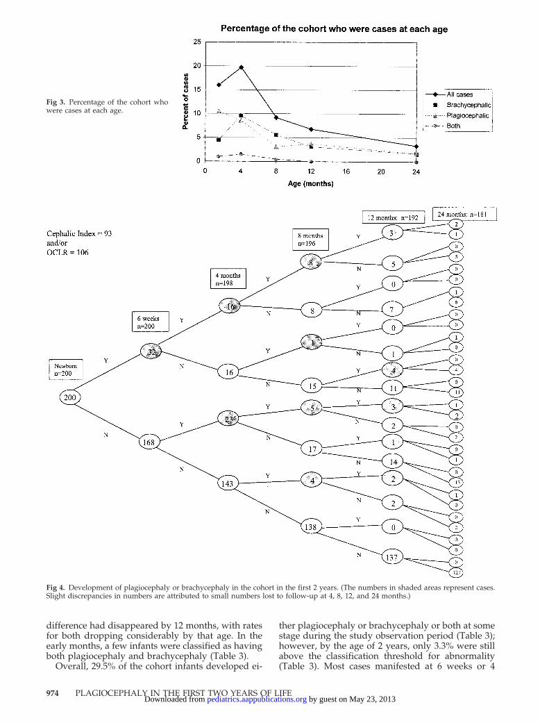
difference had disappeared by 12 months, with ratesfor both dropping considerably by that age. In theearly months, a few infants were classified as havingboth plagiocephaly and brachycephaly (Table 3).
Overall, 29.5% of the cohort infants developed ei-
ther plagiocephaly or brachycephaly or both at somestage during the study observation period (Table 3);however, by the age of 2 years, only 3.3% were stillabove the classification threshold for abnormality(Table 3). Most cases manifested at 6 weeks or 4
Fig 3. Percentage of the cohort whowere cases at each age.
Fig 4. Development of plagiocephaly or brachycephaly in the cohort in the first 2 years. (The numbers in shaded areas represent cases.Slight discrepancies in numbers are attributed to small numbers lost to follow-up at 4, 8, 12, and 24 months.)
974 PLAGIOCEPHALY IN THE FIRST TWO YEARS OF LIFE by guest on May 23, 2013pediatrics.aappublications.orgDownloaded from

months; only 4 new cases had developed at 8months, and thereafter no infant developed deforma-tion. If the cutoff points were different, then theprevalence of cases at each age would be found to bedifferent. Table 4 shows how prevalence wouldchange if the limits were set at lower or higher levels.
Fifteen case mothers consulted their Plunket nurseor general practitioner regarding their infants’ headdeformity. One case was also seen by a physiother-apist. Treatment consisted of positioning advice in 9cases; reassurance only was given in the other 6cases. Although helmet treatment is available in NewZealand, no case was considered severe enough to bereferred for such treatment by their health profes-sional.
Of the 6 2-year cases, there were 3 boys and 3 girls,and 5 were firstborns. All slept supine in the earlymonths. Two of the 3 brachycephalic cases wereabove the cephalic index cutoff at all ages; the mostsevere case, a girl born at 36 weeks’ gestation, wasjust below 93% at 6 weeks and thereafter was con-sistently over 100%. Although her parents had beenconcerned at 4, 8, and 12 months, by 2 years, theywere not particularly worried. Head rotation testswere normal at all ages. In the plagiocephalic cases,1 case progressively worsened to 8 months, and theOCLR thereafter remained about the same, at�109%. She showed evidence of head tilt and limitedrotation at 4 and 8 months. The mother remarked at2 years that she still leaned to 1 side when she wastired. This mother was also unconcerned at 2 years.The other 2 plagiocephalic cases’ OCLRs tended tovacillate around the cutoffs. One had mild unilateralflattening of the forehead at age 2 years but other-wise looked almost normal. No parent of a 2-yearcase was concerned about the head shape; 1 controlparent expressed mild concern about her son, whosehead had improved since 8 months but was stillslightly flat in a small area above the band.
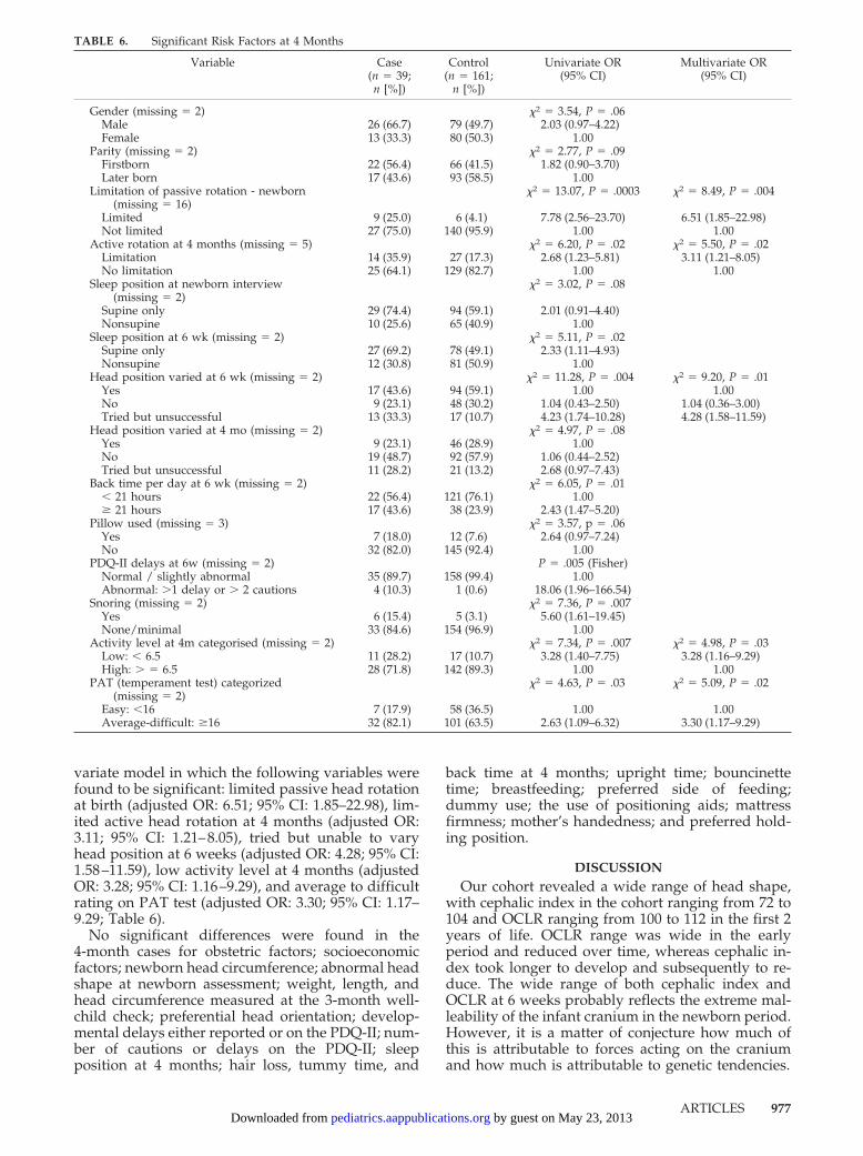
Risk Factors: 6 WeeksOwing to the small number of cases at 8, 12, and 24
months, analysis of risk factors was restricted to 6weeks and 4 months. Six-week cases were signifi-cantly more likely to have had a limitation of passiverotation at the newborn interview (crude odds ratio[OR]: 6.17; 95% confidence interval [CI]: 2.03–18.76;Table 5), although no difference was detected in thepassive head rotation test performed at 6 weeks.More case mothers reported a preferential head ori-entation at 6 weeks, although this was of borderlinesignificance.
Ninety-four percent of 6-week cases had been po-sitioned for supine sleep at the newborn interview,compared with 56.5% of control subjects (crude OR:11.53; 95% CI: 2.67–49.81). By 6 weeks, 81.3% of caseswere still sleeping supine, compared with 48.2% ofcontrol subjects (crude OR: 4.65; 95% CI: 1.82–11.89).The mothers of cases were less likely to be varyingthe head position when putting the infant down tosleep (crude OR: 2.75; 95% CI: 1.11–6.82), or theywere trying to vary it but were not able to owing tothe infant’s turning to his or her own preferred po-sition (crude OR: 5.15; 95% CI: 1.90–13.98).
When the total amount of back time was added up,the cases were spending a mean of 19.0 (SD: 4.1)hours a day on their back, compared with controlsubjects at 14.9 (SD: 6.5) hours (P � .0001). Half of thecase infants were spending �21 hours a day supine,compared with 23.8% of control infants (crude OR:3.20; 95% CI: 1.47–6.97), although cases were morelikely than control subjects to be spending �1 hour aday upright (crude OR: 2.50; 95% CI: 1.08–5.56). Caseinfants spent more time than control infants lying inbouncy seats or rockers, although this reached onlymarginal significance (P � .08). Two (6.3%) cases and3 (1.8%) control subjects had 2 or more developmen-tal delays or 3 or more cautions on the PDQ-II.
TABLE 3. New Cases at Each Age
Condition 6 Weeks(n [%])
4 Months(n [%])
8 Months(n [%])
12 Months(n [%])
2 Years(n [%])
Total(% of
originalcohort)
Either 32 (16.0) 23 (11.6) 4 (2.0) 0 (0.0) 0 (0.0) 59 (29.5)Plagiocephaly only 21 (10.5) 12 (6.1) 3 (1.5) 0 (0.0) 0 (0.0) 36 (18)Brachycephaly only 9 (4.5) 11 (5.6) 1 (0.5) 0 (0.0) 0 (0.0) 21 (10.5)Both 2 (1.0) 0 (0.0) 0 (0.0) 0 (0.0) 0 (0.0) 2 (1.0)
TABLE 4. Prevalence of Plagiocephaly Using Different Cutoff Criteria
Cutoff 6-Week Cases(n [%])
4-Month Cases(n [%])
8-Month Cases(n [%])
12-Month Cases(n [%])
2-Year Cases(n [%])
Cephalic index �91% and/orOCLR40 �105%
51 (25.5) 56 (28.0) 37 (18.9) 36 (18.7) 23 (12.7)
Cephalic index �92% and/orOCLR40 �105.5%
39 (19.5) 49 (24.7) 26 (13.3) 20 (10.4) 12 (6.6)
Cephalic index �93% and/orOCLR40 �106%
32 (16.0) 39 (19.7) 18 (9.2) 13 (6.8) 6 (3.3)
Cephalic index �94% and/orOCLR40 �106%
30 (15.0) 36 (18.2) 18 (9.2) 12 (6.3) 6 (3.3)
Cephalic index �95% and/orOCLR40 �107%
24 (12.0) 19 (9.6) 10 (5.1) 8 (4.2) 5 (2.8)
ARTICLES 975 by guest on May 23, 2013pediatrics.aappublications.orgDownloaded from
Factors that were significant at the univariate levelall were entered into an initial multivariate model,with the exception of newborn sleep position, whichhad low numbers in 1 category. The multivariatemodel was reduced by removing variables that werenonsignificant 1 at a time to ensure they did notaffect risk estimates of the other variables. The vari-ables that remained significant in the final multivar-iate model were newborn passive head rotation (ad-justed OR: 9.51; 95% CI: 2.59–34.94), 6-week sleepposition (adjusted OR: 5.27; 95% CI: 1.81–15.39), andupright time (adjusted OR: 3.99; 95% CI: 1.42–11.23).
No significant differences were detected betweencases and control subjects at 6 weeks for obstetricfactors, socioeconomic factors, gender, newbornhead circumference, abnormal head shape at thenewborn assessment, the presence of hair loss on theback of the head, snoring, activity level, newborn or6-week weight, length and head circumference, tem-perament rating, activity level, the amount of re-ported tummy time per day, breastfeeding, dummyuse, the use of positioning aids or pillows, mattressfirmness, maternal hand dominance, and the moth-er’s preferred holding position.
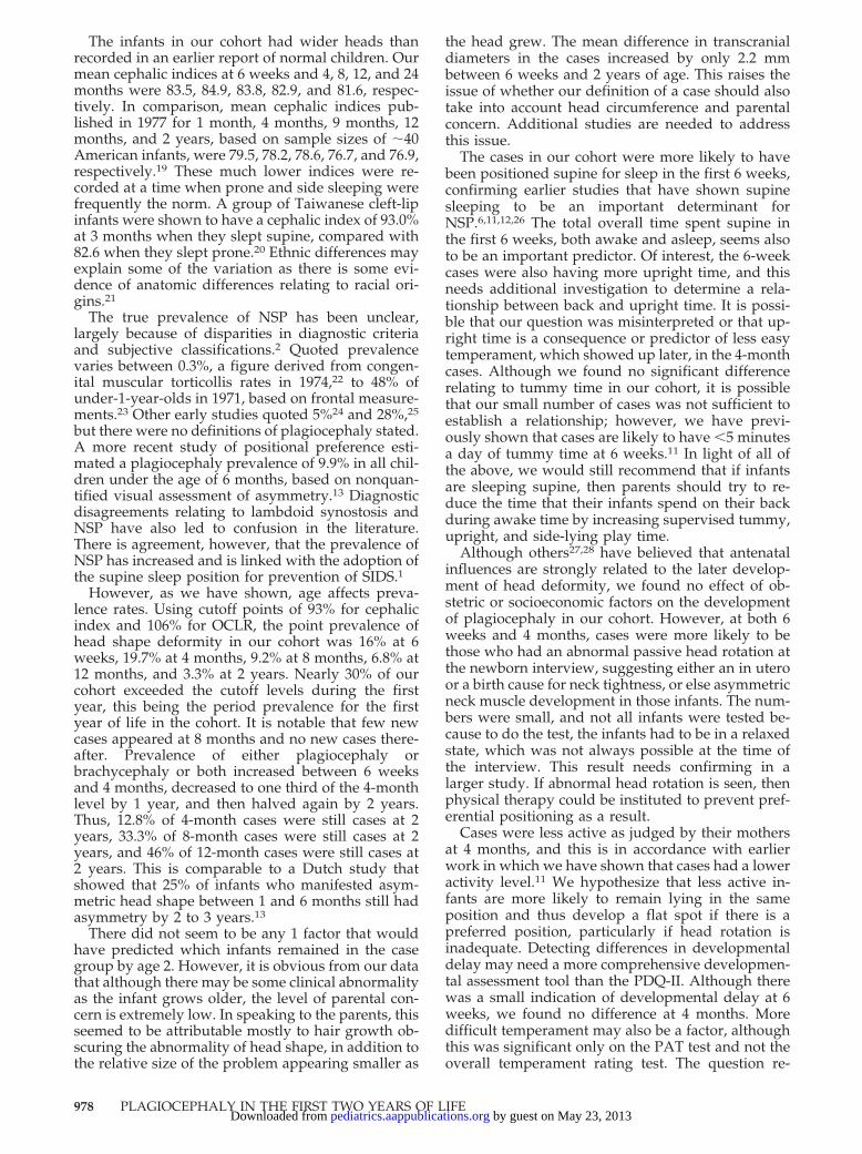
Risk Factors: 4 MonthsAt 4 months, male gender was of borderline sig-
nificance (crude OR: 2.03; 95% CI: 0.97–4.22), as wasbeing firstborn (crude OR: 1.82; 95% CI: 0.90–3.70).The 4-month cases were still significantly more likelyto have had a limitation of passive head rotation atthe newborn interview (crude OR: 7.78; 95% CI:2.56–23.70), and they were also more likely to have alimitation of active head rotation at the 4-monthinterview (crude OR: 2.68; 95% CI: 1.23–5.81;Table 6).
In the 4-month cases, having slept supine at 6weeks was significant (crude OR: 2.33; 95% CI: 1.11–4.93), but sleeping supine at 4 months was not. Sim-ilarly, having tried unsuccessfully to vary the headposition when putting the infant to sleep at 6 weekswas significant (crude OR: 4.23; 95% CI: 1.74–10.28);however, unsuccessfully varying the head position at4 months was only marginally significant (crude OR:2.68; 95% CI: 0.97–7.43). More cases than controlsubjects had �21 hours of back time at 6 weeks(crude OR: 2.43; 95% CI: 1.47–5.20), but total backtime at 4 months was not significantly different. Us-ing a pillow reached marginal significance (crudeOR: 2.64; 95% CI: 0.97–7.24).
Although the numbers were small, the 4-monthcases were more likely to have had an abnormalresult on the 6-week PDQ-II test (crude OR: 18.06;95% CI: 1.96–166.54). The mothers of 4-month caseswere also more likely to report snoring in their in-fants (crude OR: 5.60; 95% CI: 1.61–19.45) and toreport that their infants had low activity levels (crudeOR: 3.28; 95% CI: 1.40–7.75). Infants who were casesat 4 months sat alone at a slightly older age thanthose who were control subjects at 4 months (6.7months [SD: 0.98] vs 6.4 months [SD: 0.97]; P � .05).
One third of infants scored 15 or less on the PATtest administered at this age, and we categorizedthese infants as “easy.” Case infants were more likelyto score in the average to difficult range (�16) forinfant temperament than in the easy range (crudeOR: 2.63; 95% CI: 1.09–6.32). However, there wasno difference between cases and control subjects forthe temperament gauge score assessment by themothers.
Multivariate analysis of the factors that were sig-nificant at the univariate level led to a final multi-
TABLE 5. Significant Risk Factors at 6 Weeks
Variable Case(n � 32; n
[%])
Control(n � 168; n [%])
Univariate OR(95% CI, P Value)
Multivariate OR(95% CI, P Value)
Limitation of passive rotation - newborn(missing � 16)
�2 � 12.52, P � .0004 �2 � 11.50, P � .0007
Limited 7 (25.0) 8 (5.1) 6.17 (2.03–18.76) 9.51 (2.59–34.94)Not limited 21 (75.0) 148 (94.9) 1.00 1.00
Reported preferential head orientation(missing � 2)
�2 � 3.19, P � .07
Yes 23 (71.9) 91 (54.8) 2.11 (0.92–4.83)No 9 (28.1) 75 (45.2) 1.00
Sleep position at newborn interview P � .0001Supine only 30 (93.8) 95 (56.5) 11.53 (2.67–49.81)Nonsupine 2 (6.2) 73 (43.5) 1.00
Sleep position at 6 wk �2 � 11.79, P � .0006 �2 � 9.25, P � .003Supine only 26 (81.3) 81 (48.2) 4.65 (1.82–11.89) 5.27 (1.81–15.39)Nonsupine 6 (18.7) 87 (51.8) 1.00 1.00
Head position varied �2 � 10.94, P � .004Yes 10 (31.2) 103 (61.3) 1.00No 12 (37.5) 45 (26.8) 2.75 (1.11–6.82)Tried but unsuccessful 10 (31.2) 20 (11.9) 5.15 (1.90–13.98)
Back time per day �2 � 8.57, P � .003� 21 h 16 (50.0) 128 (76.2) 1.00� 21 h 16 (50.0) 40 (23.8) 3.20 (1.47–6.97)
Upright time per day �2 � 4.90, P � .03 �2 � 6.88, P � .009� 1 h 9 (28.1) 83 (49.4) 1.00 1.00� 1 h 23 (71.9) 85 (50.6) 2.50 (1.08–5.56) 3.99 (1.42–11.23)
Backtime per day (h) 19.01 (4.13) 14.89 (6.52) P � .0001� � 0.14 (0.05–0.22)
976 PLAGIOCEPHALY IN THE FIRST TWO YEARS OF LIFE by guest on May 23, 2013pediatrics.aappublications.orgDownloaded from
variate model in which the following variables werefound to be significant: limited passive head rotationat birth (adjusted OR: 6.51; 95% CI: 1.85–22.98), lim-ited active head rotation at 4 months (adjusted OR:3.11; 95% CI: 1.21–8.05), tried but unable to varyhead position at 6 weeks (adjusted OR: 4.28; 95% CI:1.58–11.59), low activity level at 4 months (adjustedOR: 3.28; 95% CI: 1.16–9.29), and average to difficultrating on PAT test (adjusted OR: 3.30; 95% CI: 1.17–9.29; Table 6).
No significant differences were found in the4-month cases for obstetric factors; socioeconomicfactors; newborn head circumference; abnormal headshape at newborn assessment; weight, length, andhead circumference measured at the 3-month well-child check; preferential head orientation; develop-mental delays either reported or on the PDQ-II; num-ber of cautions or delays on the PDQ-II; sleepposition at 4 months; hair loss, tummy time, and
back time at 4 months; upright time; bouncinettetime; breastfeeding; preferred side of feeding;dummy use; the use of positioning aids; mattressfirmness; mother’s handedness; and preferred hold-ing position.
DISCUSSIONOur cohort revealed a wide range of head shape,
with cephalic index in the cohort ranging from 72 to104 and OCLR ranging from 100 to 112 in the first 2years of life. OCLR range was wide in the earlyperiod and reduced over time, whereas cephalic in-dex took longer to develop and subsequently to re-duce. The wide range of both cephalic index andOCLR at 6 weeks probably reflects the extreme mal-leability of the infant cranium in the newborn period.However, it is a matter of conjecture how much ofthis is attributable to forces acting on the craniumand how much is attributable to genetic tendencies.
TABLE 6. Significant Risk Factors at 4 Months
Variable Case(n � 39;n [%])
Control(n � 161;
n [%])
Univariate OR(95% CI)
Multivariate OR(95% CI)
Gender (missing � 2) �2 � 3.54, P � .06Male 26 (66.7) 79 (49.7) 2.03 (0.97–4.22)Female 13 (33.3) 80 (50.3) 1.00
Parity (missing � 2) �2 � 2.77, P � .09Firstborn 22 (56.4) 66 (41.5) 1.82 (0.90–3.70)Later born 17 (43.6) 93 (58.5) 1.00
Limitation of passive rotation - newborn(missing � 16)
�2 � 13.07, P � .0003 �2 � 8.49, P � .004
Limited 9 (25.0) 6 (4.1) 7.78 (2.56–23.70) 6.51 (1.85–22.98)Not limited 27 (75.0) 140 (95.9) 1.00 1.00
Active rotation at 4 months (missing � 5) �2 � 6.20, P � .02 �2 � 5.50, P � .02Limitation 14 (35.9) 27 (17.3) 2.68 (1.23–5.81) 3.11 (1.21–8.05)No limitation 25 (64.1) 129 (82.7) 1.00 1.00
Sleep position at newborn interview(missing � 2)
�2 � 3.02, P � .08
Supine only 29 (74.4) 94 (59.1) 2.01 (0.91–4.40)Nonsupine 10 (25.6) 65 (40.9) 1.00
Sleep position at 6 wk (missing � 2) �2 � 5.11, P � .02Supine only 27 (69.2) 78 (49.1) 2.33 (1.11–4.93)Nonsupine 12 (30.8) 81 (50.9) 1.00
Head position varied at 6 wk (missing � 2) �2 � 11.28, P � .004 �2 � 9.20, P � .01Yes 17 (43.6) 94 (59.1) 1.00 1.00No 9 (23.1) 48 (30.2) 1.04 (0.43–2.50) 1.04 (0.36–3.00)Tried but unsuccessful 13 (33.3) 17 (10.7) 4.23 (1.74–10.28) 4.28 (1.58–11.59)
Head position varied at 4 mo (missing � 2) �2 � 4.97, P � .08Yes 9 (23.1) 46 (28.9) 1.00No 19 (48.7) 92 (57.9) 1.06 (0.44–2.52)Tried but unsuccessful 11 (28.2) 21 (13.2) 2.68 (0.97–7.43)
Back time per day at 6 wk (missing � 2) �2 � 6.05, P � .01� 21 hours 22 (56.4) 121 (76.1) 1.00� 21 hours 17 (43.6) 38 (23.9) 2.43 (1.47–5.20)
Pillow used (missing � 3) �2 � 3.57, p � .06Yes 7 (18.0) 12 (7.6) 2.64 (0.97–7.24)No 32 (82.0) 145 (92.4) 1.00
PDQ-II delays at 6w (missing � 2) P � .005 (Fisher)Normal / slightly abnormal 35 (89.7) 158 (99.4) 1.00Abnormal: �1 delay or � 2 cautions 4 (10.3) 1 (0.6) 18.06 (1.96–166.54)
Snoring (missing � 2) �2 � 7.36, P � .007Yes 6 (15.4) 5 (3.1) 5.60 (1.61–19.45)None/minimal 33 (84.6) 154 (96.9) 1.00
Activity level at 4m categorised (missing � 2) �2 � 7.34, P � .007 �2 � 4.98, P � .03Low: � 6.5 11 (28.2) 17 (10.7) 3.28 (1.40–7.75) 3.28 (1.16–9.29)High: � � 6.5 28 (71.8) 142 (89.3) 1.00 1.00
PAT (temperament test) categorized(missing � 2)
�2 � 4.63, P � .03 �2 � 5.09, P � .02
Easy: �16 7 (17.9) 58 (36.5) 1.00 1.00Average-difficult: �16 32 (82.1) 101 (63.5) 2.63 (1.09–6.32) 3.30 (1.17–9.29)
ARTICLES 977 by guest on May 23, 2013pediatrics.aappublications.orgDownloaded from
The infants in our cohort had wider heads thanrecorded in an earlier report of normal children. Ourmean cephalic indices at 6 weeks and 4, 8, 12, and 24months were 83.5, 84.9, 83.8, 82.9, and 81.6, respec-tively. In comparison, mean cephalic indices pub-lished in 1977 for 1 month, 4 months, 9 months, 12months, and 2 years, based on sample sizes of �40American infants, were 79.5, 78.2, 78.6, 76.7, and 76.9,respectively.19 These much lower indices were re-corded at a time when prone and side sleeping werefrequently the norm. A group of Taiwanese cleft-lipinfants were shown to have a cephalic index of 93.0%at 3 months when they slept supine, compared with82.6 when they slept prone.20 Ethnic differences mayexplain some of the variation as there is some evi-dence of anatomic differences relating to racial ori-gins.21
The true prevalence of NSP has been unclear,largely because of disparities in diagnostic criteriaand subjective classifications.2 Quoted prevalencevaries between 0.3%, a figure derived from congen-ital muscular torticollis rates in 1974,22 to 48% ofunder-1-year-olds in 1971, based on frontal measure-ments.23 Other early studies quoted 5%24 and 28%,25
but there were no definitions of plagiocephaly stated.A more recent study of positional preference esti-mated a plagiocephaly prevalence of 9.9% in all chil-dren under the age of 6 months, based on nonquan-tified visual assessment of asymmetry.13 Diagnosticdisagreements relating to lambdoid synostosis andNSP have also led to confusion in the literature.There is agreement, however, that the prevalence ofNSP has increased and is linked with the adoption ofthe supine sleep position for prevention of SIDS.1
However, as we have shown, age affects preva-lence rates. Using cutoff points of 93% for cephalicindex and 106% for OCLR, the point prevalence ofhead shape deformity in our cohort was 16% at 6weeks, 19.7% at 4 months, 9.2% at 8 months, 6.8% at12 months, and 3.3% at 2 years. Nearly 30% of ourcohort exceeded the cutoff levels during the firstyear, this being the period prevalence for the firstyear of life in the cohort. It is notable that few newcases appeared at 8 months and no new cases there-after. Prevalence of either plagiocephaly orbrachycephaly or both increased between 6 weeksand 4 months, decreased to one third of the 4-monthlevel by 1 year, and then halved again by 2 years.Thus, 12.8% of 4-month cases were still cases at 2years, 33.3% of 8-month cases were still cases at 2years, and 46% of 12-month cases were still cases at2 years. This is comparable to a Dutch study thatshowed that 25% of infants who manifested asym-metric head shape between 1 and 6 months still hadasymmetry by 2 to 3 years.13
There did not seem to be any 1 factor that wouldhave predicted which infants remained in the casegroup by age 2. However, it is obvious from our datathat although there may be some clinical abnormalityas the infant grows older, the level of parental con-cern is extremely low. In speaking to the parents, thisseemed to be attributable mostly to hair growth ob-scuring the abnormality of head shape, in addition tothe relative size of the problem appearing smaller as
the head grew. The mean difference in transcranialdiameters in the cases increased by only 2.2 mmbetween 6 weeks and 2 years of age. This raises theissue of whether our definition of a case should alsotake into account head circumference and parentalconcern. Additional studies are needed to addressthis issue.
The cases in our cohort were more likely to havebeen positioned supine for sleep in the first 6 weeks,confirming earlier studies that have shown supinesleeping to be an important determinant forNSP.6,11,12,26 The total overall time spent supine inthe first 6 weeks, both awake and asleep, seems alsoto be an important predictor. Of interest, the 6-weekcases were also having more upright time, and thisneeds additional investigation to determine a rela-tionship between back and upright time. It is possi-ble that our question was misinterpreted or that up-right time is a consequence or predictor of less easytemperament, which showed up later, in the 4-monthcases. Although we found no significant differencerelating to tummy time in our cohort, it is possiblethat our small number of cases was not sufficient toestablish a relationship; however, we have previ-ously shown that cases are likely to have �5 minutesa day of tummy time at 6 weeks.11 In light of all ofthe above, we would still recommend that if infantsare sleeping supine, then parents should try to re-duce the time that their infants spend on their backduring awake time by increasing supervised tummy,upright, and side-lying play time.
Although others27,28 have believed that antenatalinfluences are strongly related to the later develop-ment of head deformity, we found no effect of ob-stetric or socioeconomic factors on the developmentof plagiocephaly in our cohort. However, at both 6weeks and 4 months, cases were more likely to bethose who had an abnormal passive head rotation atthe newborn interview, suggesting either an in uteroor a birth cause for neck tightness, or else asymmetricneck muscle development in those infants. The num-bers were small, and not all infants were tested be-cause to do the test, the infants had to be in a relaxedstate, which was not always possible at the time ofthe interview. This result needs confirming in alarger study. If abnormal head rotation is seen, thenphysical therapy could be instituted to prevent pref-erential positioning as a result.
Cases were less active as judged by their mothersat 4 months, and this is in accordance with earlierwork in which we have shown that cases had a loweractivity level.11 We hypothesize that less active in-fants are more likely to remain lying in the sameposition and thus develop a flat spot if there is apreferred position, particularly if head rotation isinadequate. Detecting differences in developmentaldelay may need a more comprehensive developmen-tal assessment tool than the PDQ-II. Although therewas a small indication of developmental delay at 6weeks, we found no difference at 4 months. Moredifficult temperament may also be a factor, althoughthis was significant only on the PAT test and not theoverall temperament rating test. The question re-
978 PLAGIOCEPHALY IN THE FIRST TWO YEARS OF LIFE by guest on May 23, 2013pediatrics.aappublications.orgDownloaded from
mains as to whether these factors are causative orresultant of the head deformity.
There are some limitations to this study. Both theprevalence and the severity of the cases are probablyconservative as a result of mothers’ being highlyaware of head shape by participating in the studyand possibly taking preventive steps to avoid headflattening. Head shape is a continuous measure rang-ing from the perfectly symmetrical to the severelyabnormal. The cutoff is somewhat arbitrary and wasused to carry out logistic regression and to establishORs for risk factors. The cutoff points of 93% and106% were derived from a small sample, and prev-alence rates would be different given different cut-offs. A receiver-operator characteristic analysis of alarge case-control study would be needed to deter-mine optimum cutoff points to resolve this issue. Wehave opted for a fairly conservative level that webelieve adequately reflects visual assessment of ab-normality.
Although brachycephaly and plagiocephaly couldhave been considered as 2 different outcomes, webelieved that factors for both were similar and thatby excluding brachycephalic cases, they would havebeen included with the control infants, thus makingour estimates of ORs falsely conservative.
Some infants varied in and out of abnormal bybeing close to the cutoff points, and this in combina-tion with possible measurement error would explainthose in the NYNY, YNYN and YNNY categories inFig 4, although it also possibly reflects that the skullis still malleable and subject to subtle changes inshape. We also recognize that we have small num-bers of cases at 6 weeks and 4 months, and thereforeour case versus control results need to be treatedwith caution. Failure to find a significant result maybe attributable to sample size. However, excellentretention rates enabled us to track the prevalencethrough the first 2 years of life, allowing for greaterunderstanding of the dynamics of head shape devel-opment as it relates to environmental and other fac-tors and providing reassurance that the majority ofcases resolve by 2 years of age.
CONCLUSIONSHead shape varied to a great extent in this group
of normal infants in the first 2 years of life. The first4 months seems to be an important time for theinitiation of plagiocephaly and brachycephaly. Riskfactors are particularly associated with early limita-tion of head rotation and early resting positions. Alimitation in neck function should be checked for inthe early weeks and neck motion exercises com-menced if necessary to encourage full head turningto both sides. Supine sleeping also plays a major part.Although it is vital that the supine sleep position bemaintained for SIDS protection, varying the headposition in the first 6 weeks may be important forplagiocephaly prevention. Not being able to achievethis should alert parents to the possibility of limitedneck mobility. Infants with lower activity levels maybe more susceptible to developing plagiocephaly.
Although the maximum range of head shape de-formity was seen at 6 weeks, the greatest point prev-
alence of plagiocephaly in our cohort was seen at 4months. Almost 30% of the cohort exceeded the cho-sen cutoffs for classification of cases at some point inthe first 8 months, but most cases improved withtime, leaving a point prevalence of NSP of 3.3% at 2years.
ACKNOWLEDGMENTSThis study was funded by the Cot Death Association, a division
of the Child Health Research Foundation, Auckland, New Zea-land. Dr Thompson and Professor Mitchell are supported by theChild Health Research Foundation. Recruitment of subjects wasmade possible by help from the staff at North Shore Hospital andBirthcare. We thank Melanie Hayes for assistance in data collec-tion.
REFERENCES1. Persing JA, James H, Swanson J, et al. Prevention and management of
positional skull deformities in infants—American Academy of Pediat-rics Clinical Report. Pediatrics. 2003;112:199–202
2. Rekate HL. Occipital plagiocephaly: a critical review of the literature.J Neurosurg. 1998;89:24–30
3. Mitchell E, Hutchison BL. Plagiocephaly—more questions than an-swers. N Z Med J. 2003;116:580
4. Chadduck WM, Kast J, Donahue DJ. The enigma of lambdoid positionalmolding. Pediatr Neurosurg. 1997;26:304–311
5. David DJ, Menard RM. Occipital plagiocephaly. Br J Plast Surg. 2000;53:367–377
6. Golden KA, Beals SP, Littlefield TR, et al. Sternocleidomastoid imbal-ance versus congenital muscular torticollis: their relationship to posi-tional plagiocephaly. Cleft Palate Craniofac J. 1999;36:256–261
7. Mulliken JB, Vander Woude DL, Hansen M, et al. Analysis of posteriorplagiocephaly: deformational versus synostotic. Plast Reconstr Surg.1999;103:371–380
8. Clarren SK. Plagiocephaly and torticollis: etiology, natural history, andhelmet treatment. J Pediatr. 1981;98:92–95
9. Bruneteau RJ, Mulliken JB. Frontal plagiocephaly: synostotic, compen-sational, or deformational. Plast Reconstr Surg. 1992;89:21–31; discussion2–3
10. Kane AA, Mitchell LE, Craven KP, et al. Observations on a recentincrease in plagiocephaly without synostosis. Pediatrics. 1996;97:877–885
11. Hutchison BL, Thompson JM, Mitchell E. The determinants of nonsyn-ostotic plagiocephaly: a case-control study. Pediatrics. 2003;112(4).Available at: www.pediatrics.org/cgi/content/full/112/4/e316
12. Pollack IF, Losken HW, Fasick P. Diagnosis and management of poste-rior plagiocephaly. Pediatrics. 1997;99:180–185
13. Boere-Boonekamp MM, van der Linden-Kuuiper LT. Positionalpreference: prevalence in infants and follow-up after two years. Pediat-rics. 2001;107:339–343
14. Captier G, Leboucq N, Birgorre M, et al. Clinico-radiological study ofthe skull deformation in plagiocephaly without synostosis. Arch Pediatr.2003;10:208–214
15. Hutchison BL, Hutchison LAD, Thompson JM, Mitchell EA. Quantifi-cation of plagiocephaly and brachycephaly in infants using a digitalphotographic technique. Cleft Palate Crainofac J. In press
16. Davis P, McLeod K, Ransom M, et al. The New Zealand SocioeconomicIndex: developing and validating an occupationally-derived indicatorof socio-economic status. Aust N Z J Public Health. 1999;23:27–33
17. Revised Denver II Prescreening Questionnaire. Denver, CO: Denver Devel-opmental Materials, Inc; 1998
18. Clarke-Stewart KA, Fitzpatrick MJ, Allhusen VD, et al. Measuring dif-ficult temperament the easy way. J Dev Behav Pediatr. 2000;21:207–220
19. Dekaban AS. Tables of cranial and orbital measurements, cranial vol-ume, and derived indexes in males and females from 7 days to 20 yearsof age. Ann Neurol. 1977;2:485–491
20. Huang CS, Cheng HC, Lin WY, et al. Skull morphology affected bydifferent sleep positions in infancy. Cleft Palate Craniofac J. 1995;32:413–419
21. Bass WM. Human Osteology: A Laboratory and Field Manual. 3rd ed.Columbia, MO: Missouri Archeological Society; 1987
22. Dunn P. Congenital sternomastoid torticollis: an intrauterine posturaldeformity. Arch Dis Child. 1974;49:824
23. Watson GH. Relation between side of plagiocephaly, dislocation of hip,scoliosis, bat ears, and sternomastoid tumours. Arch Dis Child. 1971;46:203–210
ARTICLES 979 by guest on May 23, 2013pediatrics.aappublications.orgDownloaded from
24. Danby PM. Plagiocephaly in some 10-year-old children. Arch Dis Child.1962;37:500–504
25. Wynne-Davies R. Infantile idiopathic scoliosis. Causative factors, particu-larly in the first six months of life. J Bone Joint Surg Br. 1975;57:138–141
26. Argenta LC, David LR, Bell WO. An increase in infant cranial deformitywith supine sleeping position. J Craniofac Surg. 1996;7:5–11
27. Littlefield TR, Kelly KM, Pomatto JK, et al. Multiple-birth infants athigher risk for development of deformational plagiocephaly: II. is onetwin at greater risk? Pediatrics. 2002;109:19–25
28. Peitsch WK, Keefer CH, LaBrie RA, et al. Incidence of cranialasymmetry in healthy newborns. Pediatrics. 2002;110(6). Available at:www.pediatrics.org/cgi/content/full/110/6/e72
THE STATUS SYNDROME
“The alarming message here is that status has become a lethal threat. In therelatively prosperous, industrialized West, Michael Marmot, an epidemiologist atUniversity College, London, writes, ‘Where you stand in the social hierarchy isintimately related to your chances of getting ill and your length of life.’ And thehigher your status, the better your prospects. . . . [A] numbing arsenal of facts andfigures serves to show that it is social rank—and not suspiciously similar-soundingfactors like income or education—that makes the crucial difference. There’s thestudy of Oscar winners that found they live 4 years longer than their co-stars andfellow nominees, and the fact that with each mile along the subway line fromdowntown Washington to suburban Montgomery County, MD, life expectancyincreases by a year and a half. There is also a mountain of suggestive evidence fromprimate research: low-status rhesus macaques with heart disease; low-status ba-boons with soaring cortisol levels and unwholesome amounts of HDL cholesterol.It is not our social position per se that does us in, all this implies, but rather thestress that comes from having less control over our work and lives than people ofhigher rank. Not that this is exactly news.”
Eakin E, review of Marmot M. The Status Syndrome. New York Times Book Review. August 22, 2004
Noted by JFL, MD
980 PLAGIOCEPHALY IN THE FIRST TWO YEARS OF LIFE by guest on May 23, 2013pediatrics.aappublications.orgDownloaded from
DOI: 10.1542/peds.2003-0668-F 2004;114;970Pediatrics
B. Lynne Hutchison, Luke A.D. Hutchison, John M.D. Thompson and Ed A. MitchellCohort Study
Plagiocephaly and Brachycephaly in the First Two Years of Life: A Prospective
ServicesUpdated Information &
mlhttp://pediatrics.aappublications.org/content/114/4/970.full.htincluding high resolution figures, can be found at:
References
ml#ref-list-1http://pediatrics.aappublications.org/content/114/4/970.full.htat:This article cites 23 articles, 8 of which can be accessed free
Citations
ml#related-urlshttp://pediatrics.aappublications.org/content/114/4/970.full.htThis article has been cited by 19 HighWire-hosted articles:
Subspecialty Collections
_and_newbornhttp://pediatrics.aappublications.org/cgi/collection/prematurePremature & Newbornthe following collection(s):This article, along with others on similar topics, appears in
Permissions & Licensing
mlhttp://pediatrics.aappublications.org/site/misc/Permissions.xhttables) or in its entirety can be found online at: Information about reproducing this article in parts (figures,
Reprints http://pediatrics.aappublications.org/site/misc/reprints.xhtml
Information about ordering reprints can be found online:
rights reserved. Print ISSN: 0031-4005. Online ISSN: 1098-4275.Grove Village, Illinois, 60007. Copyright © 2004 by the American Academy of Pediatrics. All and trademarked by the American Academy of Pediatrics, 141 Northwest Point Boulevard, Elkpublication, it has been published continuously since 1948. PEDIATRICS is owned, published, PEDIATRICS is the official journal of the American Academy of Pediatrics. A monthly
by guest on May 23, 2013pediatrics.aappublications.orgDownloaded from