Embed Size (px)
Citation preview

Research ArticlePKM2 Is Required to Activate Myeloid Dendritic Cells fromPatients with Severe Aplastic Anemia
Chunyan Liu ,1 Mengying Zheng ,1 Ting Wang ,1 Huijuan Jiang ,1 Rong Fu,1
Huaquan Wang ,1 Kai Ding,1 Qiufan Zhou,1 and Zonghong Shao 1,2
1The Department of Hematology, General Hospital of Tianjin Medical University, Tianjin, China2The Department of Hematology, The Second Hospital of Tianjin Medical University, Tianjin, China
Correspondence should be addressed to Zonghong Shao; [email protected]
Received 26 September 2017; Revised 22 December 2017; Accepted 10 January 2018; Published 15 February 2018
Academic Editor: Massimo Collino
Copyright © 2018 Chunyan Liu et al. This is an open access article distributed under the Creative Commons Attribution License,which permits unrestricted use, distribution, and reproduction in any medium, provided the original work is properly cited.
Severe aplastic anemia (SAA) is an autoimmune disease in which bone marrow failure is mediated by activated myeloid dendriticcells (mDCs) and T lymphocytes. Recent research has identified a strong immunomodulatory effect of pyruvate kinase M2 (PKM2)on dendritic cells in immune-mediated diseases. In this study, we aimed to explore the role of PKM2 in the activation of mDCs inSAA.We observed conspicuously higher levels of PKM2 in mDCs from SAA patients compared to normal controls at both the geneand protein levels. Concurrently, we unexpectedly discovered that after the mDC-specific downregulation of PKM2, mDCs frompatients with SAA exhibited weakened phagocytic activity and significantly decreased and shortened dendrites relative to theircounterparts from normal controls. The expression levels of the costimulatory molecules CD86 and CD80 were also reduced onmDCs. Our results also suggested that PKM2 knockdown in mDCs reduced the abilities of these cells to promote theactivation of CD8+ T cells (CTLs), leading to the decreased secretion of cytotoxic factors by the latter cell type. Thesefindings demonstrate that mDC activation requires an elevated intrinsic PKM2 level and that PKM2 improves the immunestatus of patients with SAA by enhancing the functions of mDCs and, consequently, CTLs.
1. Introduction
Severe aplastic anemia (SAA) is a hematologic disease char-acterized by pancytopenia with severe bone marrow failure.To date, an increasing number of studies have recognizedSAA as an autoimmune disease in which bone marrow fail-ure is mediated by activated T lymphocytes [1, 2]. Myeloiddendritic cells (mDCs) have recently been recognized asimportant players in the primary immune responses relatedto SAA. Our previous research demonstrated increases inboth the immature and activated mDC populations in thebone marrow of SAA patients, indicating that immuneimbalances might originate from an early stage in the antigenrecognition process [3]. Stimulated mDCs secrete IL-12 andthus act as major stimulators of the polarization of Th0 cellsto Th1 cells, a process that leads to excessive T lymphocytefunction and ultimately to the apoptosis of hematopoietic
cells. Although knowledge about the immunopathogenesisof SAA has improved gradually after years of research, thespecific mechanism by which activated mDCs and evenT cells are involved requires further validation. Conse-quently, the immune etiology of SAA has become the focusof further research.
Within the glycolytic pathway, pyruvate kinase M2(PKM2) catalyzes the dephosphorylation of phosphoenol-pyruvate to pyruvate, a rate-limiting step [4, 5]. PKM2 there-fore acts as a key regulator of metabolic activities in bothcancer and activated immune cells, with critical roles in cellgrowth, proliferation, apoptosis, and many other physiologi-cal activities [6, 7]. PKM2 can be allosterically regulated bymetabolites and intracellular signaling pathways, and previ-ous observations have indicated that PKM2 may interactwith some pathogen-related proteins at the chromatin level(e.g., staphylococcal Opa, human immunodeficiency virus,
HindawiOxidative Medicine and Cellular LongevityVolume 2018, Article ID 1364165, 9 pageshttps://doi.org/10.1155/2018/1364165
and hepatitis C virus) to enhance their pathogenicity andsubsequently promote disease progression [8–10]. Addition-ally, recent research has shown that PKM2 has a stronglyimmunomodulatory effect on the antigen-presenting abilitiesof dendritic cells [11]. However, the relationship betweenPKM2 and mDCs in the context of SAA remains unclear.In this study, we aimed to investigate the role of PKM2 inmDC activation in SAA patients and to provide data to sup-port a potential mechanism of mDC activation and theimmune process in this population.
2. Materials and Methods
2.1. Study Subjects. Thirty patients with SAA, including 12males and 18 females with a median age of 37 years (range,10–58 years), were enrolled in the present study. All patients,including 15 newly diagnosed cases and 15 cases in remissionafter immunosuppressive therapy (IST), had been diagnosedaccording to International AA Study Group criteria at theDepartment of Hematology, Tianjin Medical UniversityGeneral Hospital, Tianjin, between September 2014 andNovember 2015. The disease was considered severe (i.e.,SAA) if at least two of the following parameters were met: aneutrophil count< 0.5× 10⁹/L, platelet count< 20× 109/L,and reticulocyte count< 20× 109/L with hypocellular bonemarrow. Cases with a neutrophil count< 0.2× 10⁹/L werediagnosed as very SAA (VSAA). Patients were excluded ifthey had congenital AA or other autoimmune diseases.All patients were screened for paroxysmal nocturnal hemo-globinuria (PNH) by flow cytometry with anti-CD55 andanti-CD59 antibodies, and no PHN clones were identified.Remission was defined as improvement of AA aftertreatment with immunosuppressive therapies (e.g., anti-thymocyte globulin, cyclosporine, and glucocorticoid) andhematopoietic-stimulating factors (e.g., granulocyte colony-stimulating factor, recombinant human erythropoietin,recombinant human thrombopoietin, and/or IL-11). Allpatients in remission achieved a bone marrow hematopoieticrecovery and became transfusion-independent, althoughsome patients with normal peripheral blood cell counts con-tinued to require drug therapy.
Eighteen healthy volunteers (10 males, 8 females) with amedian age of 26 years (range, 23–40 years) were selectedas normal controls. This study was approved by the EthicsCommittee of Tianjin Medical University. Informed writtenconsent was obtained from all patients in accordance withthe Declaration of Helsinki.
2.2. Cell Culture and Purification. The targeted bone marrowmononuclear cells (BMMNCs) were extracted from SAApatients and healthy volunteers by density gradient centrifu-gation using a Ficoll-Paque PLUS solution (Amersham Bio-sciences, Uppsala, Sweden). Cells from each subject werecultured separately at a density of 2× 10⁶ cells/mL in com-plete medium [RPMI 1640 culture medium supplementedwith 10% fetal bovine serum (FBS, Hyclone, Logan, UT,USA), 60mg/L penicillin and 100mg/L streptomycin (GibcoBRL, Grand Island, NY, USA)] containing 100ng/mLrecombinant human (rh) granulocyte-monocyte colony-
stimulating factor (GM-CSF) and 40ng/mL rhIL-4 (Pepro-Tech Inc., Rocky Hill, NJ, USA) at 37°C in an atmospherecontaining 5% CO2. Freshmedium and cytokines were addedevery 2 days. On day 6, rh tumor necrosis factor (TNF)(1000μg/mL) (PeproTech Inc.) was added for 24 h to inducemDC maturation. On day 7, suspended mature mDCs werecollected from the culture supernatant and stained withFITC-conjugated human leukocyte antigen- (HLA-) DRFITC and APC-conjugated CD11c-APC-specific monoclo-nal antibodies. HLA-DR+CD11c+ cells were sorted andcollected using a FACSAria flow cytometer (BD Biosci-ences, San Jose, CA, USA), and the purity of the sortedcells was determined.
2.3. RNA Isolation and qPCR. Total RNA was extracted usingTRIzol reagent (Invitrogen, Carlsbad, CA, USA) according tothe manufacturer’s protocol. Equal amounts of RNA werereverse transcribed using the iScript cDNA Synthesis kit(Bio-Rad, Hercules, CA, USA). Real-time PCR wasperformed using 1μL of each cDNA working solution in afinal volume of 25μL containing 12.5μL of SYBR solution(Invitrogen) and sense and antisense primers (300 nM each).β-Actin was used as a housekeeping gene with which to stan-dardize the expression levels of targeted mRNAs. PCRswere performed on a Bio-Rad PCR iQ5 device (Bio-Rad),using the following thermal cycling profile for all genesof interest: 95°C for 2min, followed by 40 amplificationcycles (95°C for 10 s, the indicated annealing temperaturefor 35 s, 72°C for 30 s, and 65°C for 10 s). The primersequences and annealing temperatures are listed in Table 1.The relative expression levels of all genes of interest were cal-culated using the 2−ΔΔCt method.
2.4. Western Blotting. Cultured mDC cells were collected andlysed directly in RIPA buffer supplemented with a completeprotease inhibitor (Roche, Basel, Switzerland) and phospha-tase inhibitors (Roche). Protein levels in the lysates werequantified using a BCA kit. Proteins were separated via10% sodium dodecyl sulfate polyacrylamide gel electrophore-sis (SDS-PAGE) and transferred to nitrocellulose (NC)membranes (Millipore Corp., Billerica, MA, USA). Themembranes were blocked with 10% skimmed milk (BD Bio-sciences) and subsequently incubated with anti-PKM2 (R&DSystems, Minneapolis, MN, USA) and anti-β-actin antibod-ies (Cell Signaling Technology, Danvers, MA, USA) at adilution of 1 : 2000. The antibodies were dissolved in a solu-tion containing 5% dried milk in Tris-buffered saline withTween 20 (TBS-T) (20mmol/L Tris–HCl buffer, pH7.4,150mmol/L NaCl, 0.05% Tween 20). After extensivewashing with phosphate-buffered saline (PBS), the mem-branes were then incubated with relevant horseradishperoxidase-conjugated secondary antibodies (1 : 1000 dilu-tion; Zhongshan Biotech, Beijing, China). The labeledprotein bands were detected using Super ECL Plus DetectionReagent. All protein levels were normalized to β-actin.
2.5. siRNA Transfection. Once purified, mDC cells from SAApatients were transfected with control (siControl) or PKM2siRNA via Lipofectamine (Invitrogen) for 72h. The siRNA
2 Oxidative Medicine and Cellular Longevity

oligos were synthesized by Sigma (St. Louis MO, USA). Thetransfected cells were collected and functionally evaluatedby western blotting. The ultrastructures on mDCs wereobserved using a scanning electron microscope (SEM).
2.6. Phagocytosis. To study phagocytosis, mDCs were incu-bated in the dark with carboxylate-modified yellow-green(YG) microspheres (9× 10⁶ beads; diameter, 2μm) for 1.5hours at 37°C. After washing with PBS, the cells were ana-lyzed on a FACSCalibur flow cytometer (BD Biosciences)with excitation/emission wavelength settings of 488/530nmto count the microsphere-containing mDCs by detectingthe wavelength emissions of the fluorescent microspheres.The percentage of phagocytosis (PP) was determined usingthe following formula: PP=M1+M2+M3+M4, where M1,M2, M3, and M4 corresponded to the numbers of cells withone, two, three, and four microspheres, respectively. Thephagocytosis index (PI) was determined as follows: (intracel-lular cpms/(intracellular + extracellular cpms))× 100%.
2.7. Detection of CD80 and CD86 Molecular Expression byFlow Cytometry. Surface expression of the costimulatorymolecules CD80 and CD86 on mDC cells was evaluated byflowcytometry toverify the effect ofPKM2siRNAtransfectionon the activation and function of mDCs from patients withSAA. HLA-DR-FITC, CD11c-APC, CD86-PE, and CD80-PEantibodies and appropriate isotypic controls (BD PharMin-gen, San Diego, CA, USA) were added to the sample tubesaccording to the manufacturer’s instructions. All sample datawere acquired using a FACSCalibur flow cytometer and ana-lyzed using CellQuest software, version 3.1 (BD Biosciences).
2.8. Cell Coculture Assays. CD8+ T cells (i.e., CTLs) fromSAA patients were sorted by immunomagnetic separation.To verify the effects of PKM2 on the ability of mDCs frompatients with SAA to activate CD8+ T cells, mDCs treatedwith PKM2-siRNA or siControl were cocultured with CTLsat a ratio of 1 : 4 for 72 hours. All culture supernatants werecollected, and the IFN-γ levels were quantified using com-mercial enzyme-linked immunosorbent assay (ELISA) kitsaccording to the manufacturer’s instructions (R&D Systems).Additionally, the expression of perforin and granzyme BmRNA in cocultured CTL cells was analyzed by quantitativeRT-PCR as described above. The primers used to amplify
perforin, granzyme B, and β-actin are listed in Table 2. Anapoptosis analysis of cocultured CTLs was performed usinga FITC Annexin V apoptosis detection kit I (BD Biosciences,Franklin Lakes, NJ, USA), followed by analysis on a FACSCa-libur flow cytometer.
2.9. Statistical Analysis. Data are presented as means± standard deviations. The significance of differencesbetween groups was assessed using Student’s t-test. A P valueof <0.05 was considered statistically significant. All statisticalanalyses were performed using the SPSS 21.0 statistical pack-age (SPSS Inc., Chicago, IL, USA).
3. Results
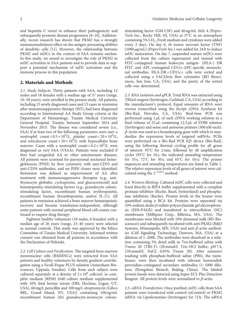
3.1. Elevated PKM2 mRNA and Protein Expression in mDCCells from SAA Patients. Both western blotting and qPCRwere used to evaluate the expression of PKM2 mRNA andprotein in mDC cells from untreated SAA patients, patientsin remission, and normal controls. As shown in Figure 1(a),significantly higher expression of PKM2mRNAwas observedinmDC cells from untreated SAA patients (1.50± 0.84), com-pared to cells from patients in remission (0.81± 0.24) andcontrols (0.32± 0.11, P < 0 05). Western blotting revealed asimilar trend in protein expression (Figure 1(b)).
3.2. Downregulation of PKM2 Strongly Impairs mDCFunction. To verify whether mDC-specific PKM2 overex-pression was responsible for the activation of mDCs inSAA, we successfully silenced PKM2 gene expression in thiscell population via siRNA transfection. This process resultedin significantly lower levels of PKM2 protein expressionrelative to cells transfected with siControl.
We used a SEM to evaluate differences in the ultrastruc-tures on mDCs from the PKM2-siRNA and siControlgroups. Following PKM2-siRNA transfection, the mDCswere round-shaped, with a typical morphology of small,short protrusions. Conversely, the mDCs from the siControlgroup had long, branched protrusions (Figure 2).
Antigen uptake and processing is a typical hallmark ofmDCs. Notably, we observed that mDCs transfected withPKM2 siRNA had a lower phagocytic ability (Figure 3).The PP and PI of mDCs in the siControl group were46.37%± 7.6% and 1.57± 0.34, respectively, whereas thecorresponding levels in mDCs transfected with PKM2 siRNAwere 38.02%± 4.4% and 1.43± 0.18, respectively. This differ-ence in PP was significant (P < 0 05). Although the inter-group difference in PI was not significant, we observed adecreasing trend after PKM2 siRNA transfection (P > 0 05).These results suggest that PKM2 downregulation decreasedthe antigen uptake capacities of mDCs.
Next, we functionally examined the expression of thecostimulatory molecules CD80 and CD86 on the surfacesof mDCs by flow cytometry. We observed a significantlylower frequency of CD86 expression in the siPKM2 group(41.89± 5.54%), compared to the siControl group (55.76± 7.08%) (P < 0 05). Similarly, we observed a statistical dif-ference in the expression of CD80 between the siControl
Table 1: Primers used for quantitative real-time PCR detection.
Targetgene
Primer sequencesAnnealing
temperature (°C)
PKM2
F:5′-GACCTGAATGCCAGCGTATC-3′
F:5′-ACCTACACCTCCAAGCCATC-3′
58
β-Actin
F:5′-TTGCCGACAGGATGCAGAA-3′
R:5′-GCCGATCCACACGGAGTACT-3′
56
3Oxidative Medicine and Cellular Longevity
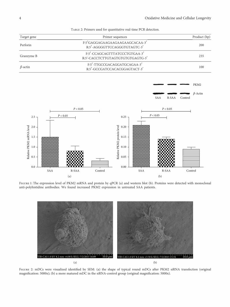
Table 2: Primers used for quantitative real-time PCR detection.
Target gene Primer sequences Product (bp)
PerforinF:5′GAGGAGAAGAAGAAGAAGCACAA-3′R:5′-AGGGGTTCCAGGGTGTAGTC-3′ 200
Granzyme BF:5′-CCAGCAGTTTATCCCTGTGAA-3′
R:5’-CACCTCTTGTAGTGTGTGTGAGTG-3′ 235
β-actinF:5′-TTGCCGACAGGATGCAGAA-3′R:5′-GCCGATCCACACGGAGTACT-3′ 100
P < 0.05
P < 0.05
0.0
0.5
1.0
1.5
2.0
2.5
Relat
ive P
KM2
mRN
A le
vel
R-SAASAA Control
(a)
P < 0.05
P < 0.05
SAA R-SAA Control�훽-Actin
PKM2
ControlR-SAASAA0.00
0.05
0.10
0.15
0.20
0.25Re
lativ
e PKM
2 pr
otei
n le
vel
(b)
Figure 1: The expression level of PKM2 mRNA and protein by qPCR (a) and western blot (b). Proteins were detected with monoclonalanti-polyhistidine antibodies. We found increased PKM2 expression in untreated SAA patients.
TIB-CAS 1.0 kV 8.2 mm ×4.00 k SE(L) 7/2/2015 16:09 10.0 �휇m
(a)
TIB-CAS 1.0 kV 8.2 mm ×3.50 k SE(L) 7/2/2015 15:34 10.0 �휇m
(b)
Figure 2: mDCs were visualized identified by SEM: (a) the shape of typical round mDCs after PKM2 siRNA transfection (originalmagnification: 5000x); (b) a more matured mDC in the siRNA-control group (original magnification: 5000x).
4 Oxidative Medicine and Cellular Longevity

group (43.79± 9.67%) and siPKM2 group (31.92± 7.41%)(P < 0 05, Figure 4).
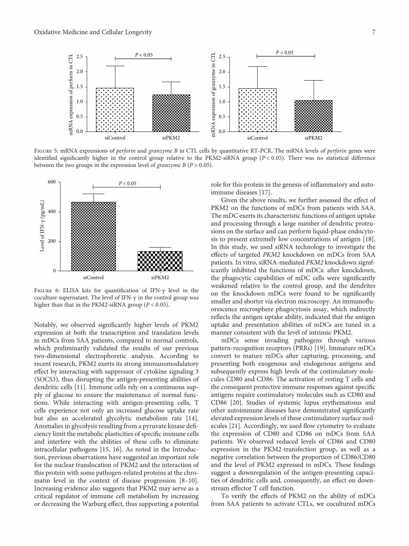
3.3. PKM2-Mediated mDC Hyperfunction Is Required toActivate CD8+ T Cells from SAA Patients. The purity ofsorted (see Materials and Methods) CD8+ T cells (CTLs)from SAA patients exceeded 90% in a flow cytometric analy-sis. To address the effect of PKM2 on the mDC-mediatedactivation of CTLs, mDCs from the PKM2-siRNA andsiControl groups were cocultured individually with CTLcells, and the expression of perforin and granzyme B mRNAin CTL cells was analyzed by quantitative RT-PCR. As shownin Figure 5, the level of perforin mRNA was significantlyhigher in the siControl group (1.23± 0.43) relative to thePKM2-siRNA group (0.84± 0.39) (P < 0 05). The level ofgranzyme B mRNA was also higher (1.45± 0.74) in CTLsexposed to the siControl group relative to the PKM2-siRNA group (1.05± 0.67), although this difference was notsignificant (P > 0 05).
Next, we used commercial ELISA kits to quantify thelevels of IFN-γ in the coculture supernatants. Notably, a
significantly higher IFN-γ concentration was detected insupernatants from the control group (466.3± 53.9 pg/mL),compared to the PKM2-siRNA group (134.2± 25.1 pg/mL)(Figure 6, P < 0 05).
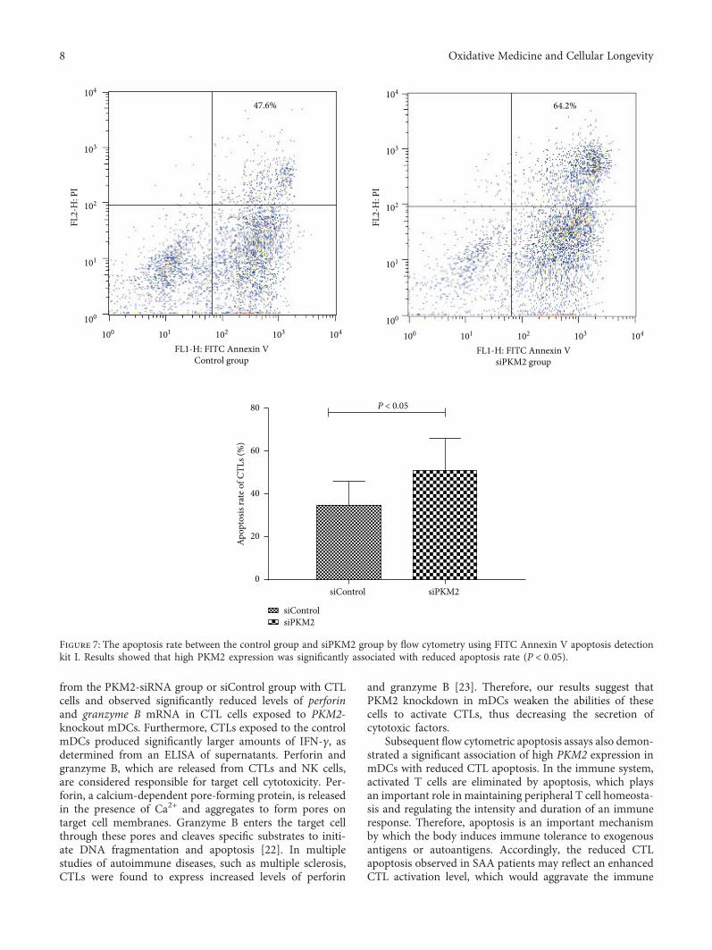
To evaluate the apoptosis of CTLs activated by mDCsafter PKM2-siRNA transfection, we analyzed FITC AnnexinV and propidium iodide (PI) staining via flow cytometry.Notably, high PKM2 expression in mDCs correlated signifi-cantly with reduced CTL apoptosis. The mDCs inducedCTL apoptosis in a manner consistent with the intrinsic levelof PKM2 (Figure 7).
4. Discussion
SAA is characterized by severe pancytopenia and a bonemarrow hematopoietic failure, with clinical symptomsincluding fatal anemia, infection, and bleeding. In recentyears, the pathogenesis of SAA has been attributed to thedysregulation of immune cell subsets, particularly T lympho-cytes, and the subsequently excessive apoptosis of hemato-poietic stem cells [12]. Numerous studies have confirmed
R2
R3 R4 R5 R6 R7
101 102 103 104100
Tubshi
0
200
400
600
800
1000SS
C-he
ight
(a)
R2
R3 R4 R5 R6 R7
101 102 103 104100
Tunshi
0
200
400
600
800
1000
SSC-
heig
ht
(b)
P > 0.05
0
20
40
60
PP (%
)
siPKM2siControl
(c)
P > 0.05
0.0
0.5
1.0
1.5
2.0
2.5
PI (%
)
siControl siPKM2
(d)
Figure 3: FACS detection on antigen uptake capacity of mDCs. (a) Antigen uptake capacity of mDCs in the siControl group. (b) Antigenuptake capacity of mDCs in the siPKM2 group. (c) The level of PP of mDCs from the siControl group was higher than that of thesiPKM2 group (P < 0 05). (d) There was no statistical difference of PI between the two groups (P > 0 05).
5Oxidative Medicine and Cellular Longevity

an abnormal cellular immune status in SAA patients, whichincludes the overactivation of CTLs, an imbalance of Th1/Th2 subsets resulting from the enhancement of Th1 and typeI lymphoid factors, and insufficiencies in the regulatory T celland NK cell population. Our previous study also found thatthe mDC populations in SAA patients were increased andoveractivated [3]. However, the mDCs gradually returnedto a normal state once disease recovery was achieved afterIST [3]. As IST allows many SAA patients to achieve a good
curative effect [13], we speculated that mDCs, as the mostpowerful professional antigen-presenting cells, might beabnormally stimulated during the primary stages of theimmune response. In this study, we further subjected mDCsfrom SAA patients and normal controls to protein expressionanalyses and observed enhanced expression of PKM2 in cellsisolated from SAA patients at an early stage after onset.
We used both western blotting and qPCR technology todetect PKM2 protein and mRNA expression in mDCs.
54.04% 43.97%
100
101
102
103
104
CD86
PE
101 102 103 104100
HLA-DR FITCControl group
100
101
102
103
104
CD86
PE
101 102 103 104100
HLA-DR FITCsiPKM2 group
(a)
60.31% 26.94%
100
101
102
103
104
CD80
PE
101 102 103 104100
HLA-DR FITCsiPKM2 group
101 102 103 104100
HLA-DR FITCControl group
100
101
102
103
104
CD80
PE
(b)
P < 0.05
P < 0.05
0
20
40
60
80
Expr
essio
n of
CD
86 o
n m
DCs
(%)
siPKM2siControl0
20
40
60
Expr
essio
n of
CD
80 o
n m
DCs
(%)
siPKM2siControl
(c)
Figure 4: The levels of costimulatory molecules CD80 and CD86 on mDCs in the siControl group and siPKM2 group by flow cytometry. Thelevels of CD86 and CD80 on mDCs from the siControl group were higher than those from the siPKM2 group (P < 0 05).
6 Oxidative Medicine and Cellular Longevity
Notably, we observed significantly higher levels of PKM2expression at both the transcription and translation levelsin mDCs from SAA patients, compared to normal controls,which preliminarily validated the results of our previoustwo-dimensional electrophoretic analysis. According torecent research, PKM2 exerts its strong immunomodulatoryeffect by interacting with suppressor of cytokine signaling 3(SOCS3), thus disrupting the antigen-presenting abilities ofdendritic cells [11]. Immune cells rely on a continuous sup-ply of glucose to ensure the maintenance of normal func-tions. While interacting with antigen-presenting cells, Tcells experience not only an increased glucose uptake ratebut also an accelerated glycolytic metabolism rate [14].Anomalies in glycolysis resulting from a pyruvate kinase defi-ciency limit the metabolic plasticities of specific immune cellsand interfere with the abilities of these cells to eliminateintracellular pathogens [15, 16]. As noted in the Introduc-tion, previous observations have suggested an important rolefor the nuclear translocation of PKM2 and the interaction ofthis protein with some pathogen-related proteins at the chro-matin level in the context of disease progression [8–10].Increasing evidence also suggests that PKM2 may serve as acritical regulator of immune cell metabolism by increasingor decreasing the Warburg effect, thus supporting a potential
role for this protein in the genesis of inflammatory and auto-immune diseases [17].
Given the above results, we further assessed the effect ofPKM2 on the functions of mDCs from patients with SAA.The mDC exerts its characteristic functions of antigen uptakeand processing through a large number of dendritic protru-sions on the surface and can perform liquid-phase endocyto-sis to present extremely low concentrations of antigen [18].In this study, we used siRNA technology to investigate theeffects of targeted PKM2 knockdown on mDCs from SAApatients. In vitro, siRNA-mediated PKM2 knockdown signif-icantly inhibited the functions of mDCs: after knockdown,the phagocytic capabilities of mDC cells were significantlyweakened relative to the control group, and the dendriteson the knockdown mDCs were found to be significantlysmaller and shorter via electron microscopy. An immunoflu-orescence microsphere phagocytosis assay, which indirectlyreflects the antigen uptake ability, indicated that the antigenuptake and presentation abilities of mDCs are tuned in amanner consistent with the level of intrinsic PKM2.
mDCs sense invading pathogens through variouspattern-recognition receptors (PRRs) [19]. Immature mDCsconvert to mature mDCs after capturing, processing, andpresenting both exogenous and endogenous antigens andsubsequently express high levels of the costimulatory mole-cules CD80 and CD86. The activation of resting T cells andthe consequent protective immune responses against specificantigens require costimulatory molecules such as CD80 andCD86 [20]. Studies of systemic lupus erythematosus andother autoimmune diseases have demonstrated significantlyelevated expression levels of these costimulatory surface mol-ecules [21]. Accordingly, we used flow cytometry to evaluatethe expression of CD80 and CD86 on mDCs from SAApatients. We observed reduced levels of CD86 and CD80expression in the PKM2-transfection group, as well as anegative correlation between the proportion of CD86/CD80and the level of PKM2 expressed in mDCs. These findingssuggest a downregulation of the antigen-presenting capaci-ties of dendritic cells and, consequently, an effect on down-stream effector T cell function.
To verify the effects of PKM2 on the ability of mDCsfrom SAA patients to activate CTLs, we cocultured mDCs
P < 0.05
0.0
0.5
1.0
1.5
2.0
2.5
mRN
A ex
pres
sion
of g
ranz
yme i
n CT
L
siPKM2siControlsiControl siPKM20.0
0.5
1.0
1.5
2.0
2.5
mRN
A ex
pres
sion
of p
erfo
rin in
CTL
P > 0.05
Figure 5: mRNA expressions of perforin and granzyme B in CTL cells by quantitative RT-PCR. The mRNA levels of perforin genes wereidentified significantly higher in the control group relative to the PKM2-siRNA group (P < 0 05). There was no statistical differencebetween the two groups in the expression level of granzyme B (P > 0 05).
0
200
400
600
Leve
l of I
FN-�훾
(pg/
mL)
siPKM2siControl
P < 0.05
Figure 6: ELISA kits for quantification of IFN-γ level in thecoculture supernatant. The level of IFN-γ in the control group washigher than that in the PKM2-siRNA group (P < 0 05).
7Oxidative Medicine and Cellular Longevity
from the PKM2-siRNA group or siControl group with CTLcells and observed significantly reduced levels of perforinand granzyme B mRNA in CTL cells exposed to PKM2-knockout mDCs. Furthermore, CTLs exposed to the controlmDCs produced significantly larger amounts of IFN-γ, asdetermined from an ELISA of supernatants. Perforin andgranzyme B, which are released from CTLs and NK cells,are considered responsible for target cell cytotoxicity. Per-forin, a calcium-dependent pore-forming protein, is releasedin the presence of Ca2+ and aggregates to form pores ontarget cell membranes. Granzyme B enters the target cellthrough these pores and cleaves specific substrates to initi-ate DNA fragmentation and apoptosis [22]. In multiplestudies of autoimmune diseases, such as multiple sclerosis,CTLs were found to express increased levels of perforin
and granzyme B [23]. Therefore, our results suggest thatPKM2 knockdown in mDCs weaken the abilities of thesecells to activate CTLs, thus decreasing the secretion ofcytotoxic factors.
Subsequent flow cytometric apoptosis assays also demon-strated a significant association of high PKM2 expression inmDCs with reduced CTL apoptosis. In the immune system,activated T cells are eliminated by apoptosis, which playsan important role in maintaining peripheral T cell homeosta-sis and regulating the intensity and duration of an immuneresponse. Therefore, apoptosis is an important mechanismby which the body induces immune tolerance to exogenousantigens or autoantigens. Accordingly, the reduced CTLapoptosis observed in SAA patients may reflect an enhancedCTL activation level, which would aggravate the immune
47.6% 64.2%
101 102 103 104100
FL1-H: FITC Annexin VControl group
100
101
102
103
104
FL2-
H: P
I
101 102 103 104100
FL1-H: FITC Annexin VsiPKM2 group
100
101
102
103
104
FL2-
H: P
I
0
20
40
60
80
Apop
tosis
rate
of C
TLs (
%)
siPKM2
siPKM2
siControl
siControl
P < 0.05
Figure 7: The apoptosis rate between the control group and siPKM2 group by flow cytometry using FITC Annexin V apoptosis detectionkit I. Results showed that high PKM2 expression was significantly associated with reduced apoptosis rate (P < 0 05).
8 Oxidative Medicine and Cellular Longevity
imbalance. PKM2 might therefore improve the immune sta-tus of an SAA patient by supporting the functions of mDCs.
In conclusion, the pathogenesis of SAA involves abnor-malities of the immune system. Our preliminary studyobserved increased PKM2 expression in the mDCs of SAApatients, and this may be an important contributor to theoveractivation of mDCs and downstream CD8+ T cells inthis population. In the future, we aim to confirm the specificmechanism by which PKM2 affects the functions of mDCsand thus alters the immune processes in SAA.
Disclosure
An abstract version of the manuscript is presented at the58th American Society of Hematology (ASH) annual meet-ing as per the following URL: http://www.bloodjournal.org/content/128/22/1497.
Conflicts of Interest
The authors have no competing financial interests to declare.
Authors’ Contributions
Chunyan Liu, Mengying Zheng, and Ting Wang contributedequally to this work.
Acknowledgments
This work was supported by the National Natural ScienceFoundation of China (81400085, 81570106, 81600093,81570111, and 81400088), Tianjin Municipal Natural Sci-ence Foundation (14JCYBJC25400, 15JCYBJC24300, and12JCZDJC21500), and Tianjin Science and Technology sup-port key project plan (20140109).
References
[1] H. Yamazaki, “Acquired aplastic anemia,” Rinshō Ketsueki,vol. 57, no. 2, pp. 91–97, 2016.
[2] M. Miano and C. Dufour, “The diagnosis and treatment ofaplastic anemia: a review,” International Journal of Hematol-ogy, vol. 101, no. 6, pp. 527–535, 2015.
[3] S. Zonghong, T. Meifeng, W. Huaquan et al., “Circulatingmyeloid dendritic cells are increased in individuals with severeaplastic anemia,” International Journal of Hematology, vol. 93,no. 2, pp. 156–162, 2011.
[4] W. Yang, Y. Xia, D. Hawke et al., “PKM2 phosphorylates his-tone H3 and promotes gene transcription and tumorigenesis,”Cell, vol. 150, no. 4, pp. 685–696, 2012.
[5] B. Chaneton and E. Gottlieb, “Rocking cell metabolism:revised functions of the key glycolytic regulator PKM2 incancer,” Trends in Biochemical Sciences, vol. 37, no. 8,pp. 309–316, 2012.
[6] J. C. Alves-Filho and E. M. Palsson-McDermott, “Pyruvatekinase M2: a potential target for regulating inflammation,”Frontiers in Immunology, vol. 7, p. 145, 2016.
[7] W. Yang and Z. Lu, “Pyruvate kinase M2 at a glance,” Journalof Cell Science, vol. 128, no. 9, pp. 1655–1660, 2015.
[8] X. Wu, Y. Zhou, K. Zhang, Q. Liu, and D. Guo, “Isoform-spe-cific interaction of pyruvate kinase with hepatitis C virusNS5B,” FEBS Letters, vol. 582, no. 15, pp. 2155–2160, 2008.
[9] S. Sen, S. L. Deshmane, R. Kaminski, S. Amini, and P. K. Datta,“Non-metabolic role of PKM2 in regulation of the HIV-1LTR,” Journal of Cellular Physiology, vol. 232, no. 3, pp. 517–525, 2017.
[10] J. M. Williams, G. C. Chen, L. Zhu, and R. F. Rest, “Using theyeast two-hybrid system to identify human epithelial cell pro-teins that bind gonococcal Opa proteins: intracellular gono-cocci bind pyruvate kinase via their Opa proteins and requirehost pyruvate for growth,” Molecular Microbiology, vol. 27,no. 1, pp. 171–186, 1998.
[11] Z. Zhang, Q. Liu, Y. Che et al., “Antigen presentation by den-dritic cells in tumors is disrupted by altered metabolism thatinvolves pyruvate kinase M2 and its interaction with SOCS3,”Cancer Research, vol. 70, no. 1, pp. 89–98, 2010.
[12] Y. Zeng and E. Katsanis, “The complex pathophysiology ofacquired aplastic anaemia,” Clinical and Experimental Immu-nology, vol. 180, no. 3, pp. 361–370, 2015.
[13] Red Blood Cell Disease (Anemia) Group, Chinese Society ofHematology, Chinese Medical Association, “Chinese expertconsensus on the diagnosis and treatment of aplastic anemia(2017),” Zhonghua Xue Ye Xue Za Zhi, vol. 38, pp. 1–5, 2017.
[14] E. M. Palsson-McDermott and L. A. O'Neill, “The Warburgeffect then and now: from cancer to inflammatory diseases,”BioEssays, vol. 35, no. 11, pp. 965–973, 2013.
[15] A. Schurich, L. J. Pallett, D. Jajbhay et al., “Distinct metabolicrequirements of exhausted and functional virus-specific CD8T cells in the same host,” Cell Reports, vol. 16, no. 5,pp. 1243–1252, 2016.
[16] M. P. Keppel and M. A. Cooper, “Assessment of NK cellmetabolism,” Methods in Molecular Biology, vol. 1441,pp. 27–42, 2016.
[17] E. M. Palsson-McDermott, A. M. Curtis, G. Goel et al., “Pyru-vate kinase M2 regulates Hif-1α activity and IL-1β inductionand is a critical determinant of the warburg effect in LPS-activated macrophages,” Cell Metabolism, vol. 21, no. 1,pp. 65–80, 2015.
[18] Z. Liu and P. A. Roche, “Macropinocytosis in phagocytes: reg-ulation of MHC class-II-restricted antigen presentation indendritic cells,” Frontiers in Physiology, vol. 6, p. 1, 2015.
[19] C. Arnold-Schrauf, L. Berod, and T. Sparwasser, “Dendriticcell specific targeting of MyD88 signalling pathways in vivo,”European Journal of Immunology, vol. 45, no. 1, pp. 32–39,2015.
[20] E. C. de Jong, H. H. Smits, and M. L. Kapsenberg, “Dendriticcell-mediated T cell polarization,” Springer Seminars in Immu-nopathology, vol. 26, no. 3, pp. 289–307, 2005.
[21] J. Dieker, J. Tel, E. Pieterse et al., “Circulating apoptotic micro-particles in systemic lupus erythematosus patients drive theactivation of dendritic cell subsets and prime neutrophils forNETosis,” Arthritis & Rhematology, vol. 68, no. 2, pp. 462–472, 2016.
[22] A. Saeidi, M. Buggert, K. F. Che et al., “Regulation of CD8+ T-cell cytotoxicity in HIV-1 infection,” Cellular Immunology,vol. 298, no. 1-2, pp. 126–133, 2015.
[23] M. Salou, A. Garcia, L. Michel et al., “Expanded CD8 T-cellsharing between periphery and CNS in multiple sclerosis,”Annals of Clinical Translational Neurology, vol. 2, no. 6,pp. 609–622, 2015.
9Oxidative Medicine and Cellular Longevity

Stem Cells International
Hindawiwww.hindawi.com Volume 2018
Hindawiwww.hindawi.com Volume 2018
MEDIATORSINFLAMMATION
of
EndocrinologyInternational Journal of
Hindawiwww.hindawi.com Volume 2018
Hindawiwww.hindawi.com Volume 2018
Disease Markers
Hindawiwww.hindawi.com Volume 2018
BioMed Research International
OncologyJournal of
Hindawiwww.hindawi.com Volume 2013
Hindawiwww.hindawi.com Volume 2018
Oxidative Medicine and Cellular Longevity
Hindawiwww.hindawi.com Volume 2018
PPAR Research
Hindawi Publishing Corporation http://www.hindawi.com Volume 2013Hindawiwww.hindawi.com
The Scientific World Journal
Volume 2018
Immunology ResearchHindawiwww.hindawi.com Volume 2018
Journal of
ObesityJournal of
Hindawiwww.hindawi.com Volume 2018
Hindawiwww.hindawi.com Volume 2018
Computational and Mathematical Methods in Medicine
Hindawiwww.hindawi.com Volume 2018
Behavioural Neurology
OphthalmologyJournal of
Hindawiwww.hindawi.com Volume 2018
Diabetes ResearchJournal of
Hindawiwww.hindawi.com Volume 2018
Hindawiwww.hindawi.com Volume 2018
Research and TreatmentAIDS
Hindawiwww.hindawi.com Volume 2018
Gastroenterology Research and Practice
Hindawiwww.hindawi.com Volume 2018
Parkinson’s Disease
Evidence-Based Complementary andAlternative Medicine
Volume 2018Hindawiwww.hindawi.com
Submit your manuscripts atwww.hindawi.com