Embed Size (px)
Citation preview

Pitfalls in diagnosis of amyloidosis
Ashutosh WechalekarProfessor of Medicine and Haematology
National Amyloidosis CentreUniversity College London
Department Of HaematologyUniversity College London Hospitals

Disclosure of Conflict of Interest
Nature of relationship(s)Name of for-profit or not-for-profit organization(s)
Description of relationship(s)
Any direct financial payments including receipt of honoraria
GSK, Janssen, Celgene, Takeda Honorarium
Membership on advisory boards or speakers’ bureaus
Prothena
Funded grants or clinical trials
Amgen Research funding
Patents on a drug, product or device
All other investments or relationships that could be seen by a reasonable, well-informed participant as having the potential to influence the content of the educational activity
❑ I do not have a relationship with a for-profit and/or a not-for-profit organization to disclose❑ I have a relationship with a for-profit and/or a not-for-profit organization to disclose

Pitfalls in diagnosis:
• Early recognition
• Correct identification of fibril type
• Appropriate use of diagnostic methods

Amyloidosis is a difficult diagnosis
➢ Can involve any organ or organ system in any combination ➢ Presents with “common” symptoms - Physicians don’t “think” of
amyloidosis ➢ There is no single diagnostic blood test for amyloidosis
Lousada et al Adv Ther. 2015; 32(10): 920–928.

Fontana M et al, Circulation 2015;132:1570-9
Amount of amyloid deposition determines outcomes

Patients with minimal cardiac symptoms have better outcomes
Wechalekar – ALChemy 2016 - unpublished
NYHA 0-2
NYHA 3-4

Late diagnosis remains a problem – the heart
Manwani et al ALChemy IMW 2017

Late diagnosis remains a problem – the kidneys
Dispenzieri, Blood. 2014 Oct 9;124(15):2315-6

What can we do: Suspect, Suspect, Suspect….Educate Educate, Educate!
• CKD with Albuminuria > 0.5g/day
• Unexplained fatigue, weight loss, gut symptoms
• Bleeding or soft tissue symptoms
• Heart failure with preserved ejection fraction (HFPEF)
• A patient with “LVH” on echocardiogram and normal or low voltage ECG
• A combination of peripheral and autonomic neuropathy
CKD prevalence by age (UK)
This is simple

Help is on the way….. A blood test for AL?

Finding amyloid deposits

Where to look for amyloid
• Screening biopsy
– Abdominal fat aspirate (FA)
• Muchtar et al in 612 pts with AL amyloidosis a BM + FA was diagnostic in 89%
• Quarta et al in 600 pts with cardiac amyloid found positive FA in
– AL : 181/216 (84%)
– ATTRmt: 51/113 (45%)
– ATTRwt: 42/271 (15%)
0
10
20
30
40
50
60
70
80
90
100
0
100
200
300
400
500
600
Total specimens CR-stained
Sensitivity (%)
N
Ann Med. 2017 Mar Muchtar et al. Overuse of organ
biopsies in immunoglobulin light
chain amyloidosis (AL): the consequence of failure of
early recognition.
Eur Heart J. 2017 Jun Quarta et al Diagnostic sensitivity of
abdominal fat aspiration in cardiac amyloidosis.
1. Abdominal fat aspirates useful only if
laboratory set up to test
2. Cannot rule out amyloid deposition if
negative
Congo Red Staining: national problem
Improvement in amyloid staining leading to accurate diagnosis
UK National External Quality Assurance Scheme for Congo Red
*2014 Amyloid Run 89* → 85% pass, 15% fail (~45 labs)
*2015 Amyloid Run 94* → 90% pass, 10% fail (~30 labs)
*2016 Amyloid Run 105 → 71% pass, 26% poor and suboptimal
*
NEQAS Assessors report for amyloid –
remains 13% who “failed to clearly
demonstrate amyloid”
no change from 2013!

Problem of Congo red interpretation
Fat aspirates 329
Renal 244
Bone marrow 317
Cardiac 70
Rectal 50
Skin 58
Other 216
Total 1284 cases
Fat Aspirates21%
Connective Tissues/muscle /fat
2%
BMT21%
GI11%
Head and Neck3%
Renal/ urology19%
SKIN4%
Lung3%
Carpel tunnel2%
Soft Tissue2% Cardiac
7%
Spleen /Lymph node1%
2015
False positive rate 24%
False Negative rate 12%
2016
False positive rate 17.5%
False Negative rate 10%
2017
False positive rate 24%
False Negative rate 9%
2018
False positive rate 24%
False Negative rate 12%

Even on targeted biopsies

Nephrology Dialysis Transplantation, Volume 29, Issue 11, November 2014, Pages 2120–2126, https://doi.org/10.1093/ndt/gfu242
The content of this slide may be subject to copyright: please see the slide notes for details.
Remember to request Congo red staining: The story of Minimal change disease
The next challenge is typing:All patients or selected patients

Amyloidosis is not just AL or ATTR
AL; 5203; 58%
LECT2; 34; 0%
AA; 856; 10%
Localised; 962; 11%
Insulin; 18; 0%
Hered; 961; 11%
wtTTR, 929, 10%
ApoA1; 52; 6%
FibAalpha; 134; 14%
CysC; …
Gel; 20; 2%
Lyso; 23; 2%
TTR; 724; 76%
Types of Hereditary Amyloid in 961 patients

Organ involvement and amyloid type
Amyloidosis
Renal involvement
AA
LECT2
ApoA1/Afib
Liver involvementApoA1
Lysozyme
Heart involvementTransthyretin
ApoA1
NeuropathyTransthyretin
ApoA1
Soft Tissue Nil
AL am
yloid
osis

What is and is not AL amyloidosis
• M-protein + amyloid deposits on biopsy AL Amyloidosis
Hereditary amyloidosis:
10% of NAC cases
No Family history in ~ 50%
Variable phenotype
Incidental MGUS in 20%+
Difficult immunohistochemistry
Multiple mutations – often novel
10% misdiagnosis rate
30% in isolated renal
Amyloidosis

Changing demographics of amyloidosis
0%
10%
20%
30%
40%
50%
60%
70%
2004 2005 2006 2007 2008 2009 2010 2011 2012 2013 2014 2015 2016 2017
Percentage Breakdown
AL AA Hered wtTTR Localised
0
50
100
150
200
250
300
350
400
450
500
2004 2005 2006 2007 2008 2009 2010 2011 2012 2013 2014 2015 2016 2017
Number of New Cases by Type
AL AA Hered wtTTR Localised

99mTc-DPD in the heart – very sensitive in ATTR (~100%)
CardiacBone
Grade 1 Grade 2 Grade 3Grade 0

99mTc-DPD for cardiac amyloidosis
Cardiac uptake on DPD ATTR amyloidosis
Cardiac uptake on DPD Amyloidosis
No Cardiac uptake on DPD No amyloidosis
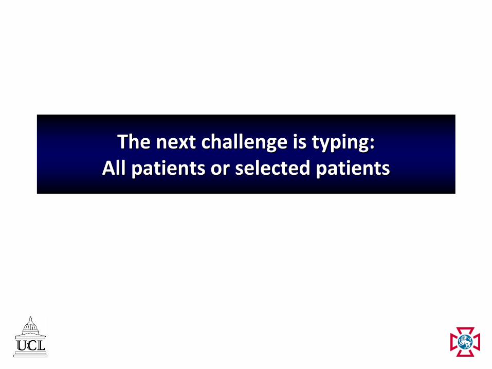
ATTR diagnosis using imaging
Isolated cardiac AL in
Elderly on imaging
Monoclonal Gammopathy
YES
Endomyocardialbiopsy
Monoclonal Gammopathy
NO
99mTcDPD/PYP
scan
Positive =>gr2
ATTR type
Negative or Grade 1
Consider cardiac Bx
FLC/IFE/SPEP
Modified from Gillmore et al Circulation 2016

Patients with wtATTR may have abnormal sFLC or
IFE
Phull et al Amyloid. 2018 Mar;25(1):62-67.

Diagnosis of AA amyloidosis is (usually) simple
Amyloid in the medulla, staining
positive for DAKO Monoclonal
amyloid AA immuno.
But the history is unusual as is the
strictly medullary pattern without any
glomerular involvement and with
minimal proteinuria.

Commercial monoclonal antibodies against: ➢ AA amyloid (1:600)Commercial polyclonal antibodies against: ➢ Amyloid P-component (1:5000), Fibrinogen (1:2000)➢ Lysozyme (1:3000)➢ Transthyretin (1:4000)➢ λ-light chain (1:160000)➢ κ-light chain (1:160 000)Noncommercial polyclonal antibodies directed against: ➢ apolipoprotein AI (1:1000)➢ λ-light chain-derived amyloid proteins (AL1, 1:3000)
High sensitivity and specificity of IHC
Schoenland SO Blood. 2012 Jan 12;119(2):488-93.
Gold standard of IHC – the German experience

Antibody Panel Used at NAC
Reporting:
Two persons – blinded reporting

Diagnosis of AA amyloidosis is (usually) simple - at NAC
AA Lect2

A tale of two diseases
Anti-TTR
Anti-λ

Two types of amyloidosis in one patient
kappa
Apo A1
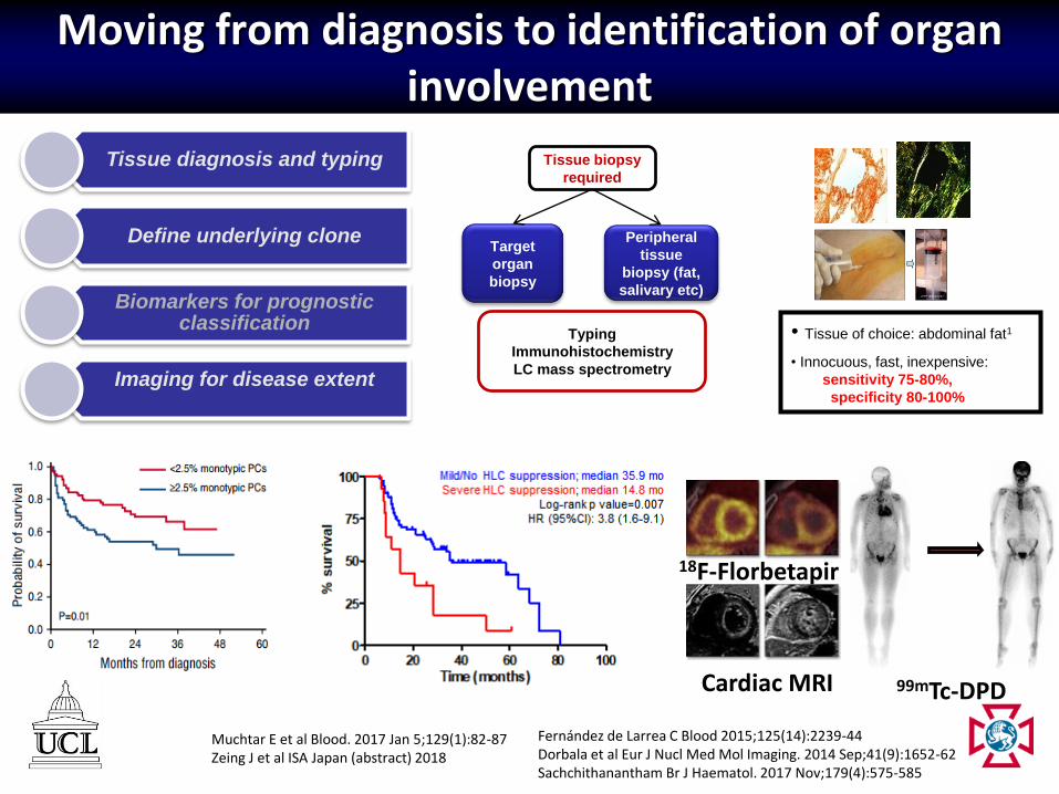
Moving from diagnosis to identification of organ involvement
Tissue diagnosis and typing
Define underlying clone
Biomarkers for prognostic classification
Imaging for disease extent
Tissue biopsy
required
Target
organ
biopsy
Peripheral
tissue
biopsy (fat,
salivary etc)
Typing
Immunohistochemistry
LC mass spectrometry
• Tissue of choice: abdominal fat1
• Innocuous, fast, inexpensive:
sensitivity 75-80%,
specificity 80-100%
18F-Florbetapir
99mTc-DPDCardiac MRI
Fernández de Larrea C Blood 2015;125(14):2239-44Dorbala et al Eur J Nucl Med Mol Imaging. 2014 Sep;41(9):1652-62Sachchithanantham Br J Haematol. 2017 Nov;179(4):575-585
Muchtar E et al Blood. 2017 Jan 5;129(1):82-87Zeing J et al ISA Japan (abstract) 2018

Cardiac amyloidosis may be present with “low” cardiac biomarkers
• Current thresholds for definition: NT-proBNP >332 ng/L
• We reviewed all patients with NT-proBNP <332 ng/L
This has crucial implication for goals of treatment

Cardiac MRI in amyloidosis

Problem of cardiac MRI interpretation
• Typical patient: 40-60 yr old, history of hypertension and renal impairment

Patients with low presenting dFLC should not be missed
• ~20% of all patients have presenting dFLC <50 mg/L
• They have ‘low-risk’ clinical phenotype and better outcomes
Dittrich et al Blood. 2017 Aug 3;130(5):632-642.
Milani et al Blood. 2017 Aug 3;130(5):625-631. 0,10
1,00
10,00
100,00
1000,00
10000,00
100000,00
0 100 200 300 400 500 600 700 800 900 1000
dFLC
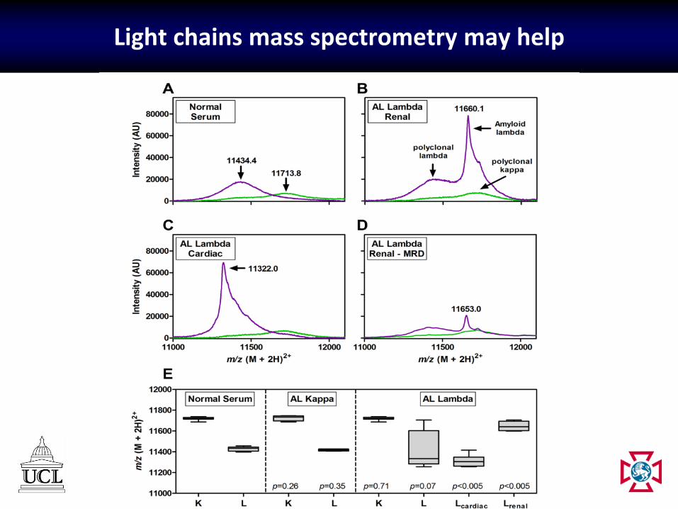
Light chains mass spectrometry may help
Conclusions
• Challenges to diagnosis of amyloidosis persist
• Physician education and early suspicion is crucial
• High prevalence of CKD and HfPEf in the “target” population make screening difficult
• Novel blood tests may allow easier detection
• Typing and correctly following published diagnostic algorithms remain key
• Review in referral laboratories and typing is important
• We have recognise overlap between different types and more patients with mixed amyloid types are being recognised
• Remember to the limitation of imaging and blood tests when diagnosing amyloidosis