Embed Size (px)
Citation preview

Mechanisms of Photoreceptor Death and Survival inMammalian Retina
Jonathan Stone*, Juliani Maslim, Krisztina Valter-Kocsi, Kyle Mervin,Felicity Bowers, Yi Chu, Nigel Barnett, Jan Provis, Geo� Lewis, Steven K. Fisher,
Silvia Bisti, Claudia Gargini, Luigi Cervetto, Saul Merin and Jacob Pe'erNSW Retinal Dystrophy Research Centre, Department of Anatomy and Histology, University of Sydney,Sydney, Australia; Department of Anatomy and Histology, University of Sydney, Sydney, Australia;
Institute for Neuroscience, University of California, Santa Barbara, USA; Dipartimento STBB,Universita' dei L'Aquila, L'Aquila, Italy; Istituto di Neuro®siologia, CNR, Pisa, Italy; Dipartimento di
Psichiatria, Neurobiologia, Farmacologia e Biotecnologie, Universita' di Pisa, Pisa, Italy and Departmentof Ophthalmology, Hadassah Faculty of Medicine, Hadassah University Hospital, The Hebrew University
of Jerusalem, Israel
CONTENTS
Abstract . . . . . . . . . . . . . . . . . . . . . . . . . . . . . . . . . . . . . . . . . . . . . . . . . . . . . . . . . . . . . . . . . . . . . . . . . . . . . . . . . . . . . . 691
1. Durability and fragility. . . . . . . . . . . . . . . . . . . . . . . . . . . . . . . . . . . . . . . . . . . . . . . . . . . . . . . . . . . . . . . . . . . . . . . 6911.1. Sources of fragility . . . . . . . . . . . . . . . . . . . . . . . . . . . . . . . . . . . . . . . . . . . . . . . . . . . . . . . . . . . . . . . . . . . . . . 691
1.1.1. The fragility of the outer segment membrane . . . . . . . . . . . . . . . . . . . . . . . . . . . . . . . . . . . . . . . . 6921.1.2. The need to invert the retina . . . . . . . . . . . . . . . . . . . . . . . . . . . . . . . . . . . . . . . . . . . . . . . . . . . . . . . 692
1.1.3. The need for energy and the need to be avascular . . . . . . . . . . . . . . . . . . . . . . . . . . . . . . . . . . . 692
1.1.4. The choroidal circulation: Achilles' heel of the eye? . . . . . . . . . . . . . . . . . . . . . . . . . . . . . . . . . . 6931.2. The normality and abnormality of photoreceptor death . . . . . . . . . . . . . . . . . . . . . . . . . . . . . . . . . . . . 693
2. Photoreceptor development: overproduction, culling and a critical period. . . . . . . . . . . . . . . . . . . . . . . . 694
2.1. Developmental death of photoreceptors. . . . . . . . . . . . . . . . . . . . . . . . . . . . . . . . . . . . . . . . . . . . . . . . . . . 6942.1.1. Evidence of overproduction and culling. . . . . . . . . . . . . . . . . . . . . . . . . . . . . . . . . . . . . . . . . . . . . 6942.1.2. The culling of photoreceptors occurs late . . . . . . . . . . . . . . . . . . . . . . . . . . . . . . . . . . . . . . . . . . . 694
2.2. Correlates of photoreceptor culling . . . . . . . . . . . . . . . . . . . . . . . . . . . . . . . . . . . . . . . . . . . . . . . . . . . . . . . 696
2.2.1. Morphology: growth of outer segments . . . . . . . . . . . . . . . . . . . . . . . . . . . . . . . . . . . . . . . . . . . . . 6962.2.2. Function: development of the ERG . . . . . . . . . . . . . . . . . . . . . . . . . . . . . . . . . . . . . . . . . . . . . . . . 696
2.2.3. Metabolism: sharp acceleration . . . . . . . . . . . . . . . . . . . . . . . . . . . . . . . . . . . . . . . . . . . . . . . . . . . . 696
2.2.4. Molecular correlate. . . . . . . . . . . . . . . . . . . . . . . . . . . . . . . . . . . . . . . . . . . . . . . . . . . . . . . . . . . . . . . . 6962.2.5. Vasculogenesis: physiological hypoxia and hypoglycaemia . . . . . . . . . . . . . . . . . . . . . . . . . . . 697
2.3. The regulation of culling . . . . . . . . . . . . . . . . . . . . . . . . . . . . . . . . . . . . . . . . . . . . . . . . . . . . . . . . . . . . . . . . . 697
2.3.1. Competition for oxygen, glucose . . . . . . . . . . . . . . . . . . . . . . . . . . . . . . . . . . . . . . . . . . . . . . . . . . . 697
2.3.2. Culling by energy competition is speci®c to photoreceptors. . . . . . . . . . . . . . . . . . . . . . . . . . . 6992.3.3. What ends the lethality of competition? . . . . . . . . . . . . . . . . . . . . . . . . . . . . . . . . . . . . . . . . . . . . . 699
2.4. The critical period: programmed vulnerability . . . . . . . . . . . . . . . . . . . . . . . . . . . . . . . . . . . . . . . . . . . . . 699
2.4.1. The purpose of vulnerability . . . . . . . . . . . . . . . . . . . . . . . . . . . . . . . . . . . . . . . . . . . . . . . . . . . . . . . 699
2.4.1.1. Matching energy demand to supply by lethal competition . . . . . . . . . . . . . . . . . . . . 699
2.4.1.2. Energy matching after the critical period . . . . . . . . . . . . . . . . . . . . . . . . . . . . . . . . . . . . 700
Progress in Retinal and Eye Research Vol. 18, No. 6, pp. 689 to 735, 1999# 1999 Elsevier Science Ltd. All rights reservedPrinted in Great Britain1350-9462/99/$ - see front matter
PII: S1350-9462(98)00032-9
*Corresponding author. Fax: +0061-2-9351-6556; E-mail: [email protected]

2.4.2. Dangers of vulnerability: photoreceptor depletion . . . . . . . . . . . . . . . . . . . . . . . . . . . . . . . . . . . 700
2.4.2.1. Genetic dystrophies begin with the critical period. . . . . . . . . . . . . . . . . . . . . . . . . . . . 700
2.4.2.2. Genetic and environmental factors can summate . . . . . . . . . . . . . . . . . . . . . . . . . . . . 702
2.5. The critical period in humans . . . . . . . . . . . . . . . . . . . . . . . . . . . . . . . . . . . . . . . . . . . . . . . . . . . . . . . . . . . . 702
3. Mechanisms of adult stability: adaptability and self-protection: . . . . . . . . . . . . . . . . . . . . . . . . . . . . . . . . . 702
3.1. Adaptability of energy sourcing . . . . . . . . . . . . . . . . . . . . . . . . . . . . . . . . . . . . . . . . . . . . . . . . . . . . . . . . . . 702
3.1.1. The high aerobic metabolism of photoreceptors . . . . . . . . . . . . . . . . . . . . . . . . . . . . . . . . . . . . . 703
3.1.2. The high anaerobic capacity of the retina . . . . . . . . . . . . . . . . . . . . . . . . . . . . . . . . . . . . . . . . . . . 704
3.1.3. Photoreceptors can switch energy sources . . . . . . . . . . . . . . . . . . . . . . . . . . . . . . . . . . . . . . . . . . . 704
3.2. Physiological stresses induce expression of protective cytokines . . . . . . . . . . . . . . . . . . . . . . . . . . . . . 704
3.2.1. Normal light experience induces cytokine and antioxidant expression . . . . . . . . . . . . . . . . . 705
3.2.1.1. Circadian light, damage and survival . . . . . . . . . . . . . . . . . . . . . . . . . . . . . . . . . . . . . . . 705
3.2.1.2. Mechanisms of light-induced protection: antioxidants and trophic factors . . . . . 705
3.2.2. Skylight and sidelight: cytokine protection is a local retinal response. . . . . . . . . . . . . . . . . . 707
3.2.3. Surviving at the edge: a ring of protection?. . . . . . . . . . . . . . . . . . . . . . . . . . . . . . . . . . . . . . . . . . 709
3.2.4. How many factors?. . . . . . . . . . . . . . . . . . . . . . . . . . . . . . . . . . . . . . . . . . . . . . . . . . . . . . . . . . . . . . . . 709
3.3. Pathological stresses induce expression of protective cytokines. . . . . . . . . . . . . . . . . . . . . . . . . . . . . . 711
3.3.1. Continuous light . . . . . . . . . . . . . . . . . . . . . . . . . . . . . . . . . . . . . . . . . . . . . . . . . . . . . . . . . . . . . . . . . . 711
3.3.2. Incision and laser burn . . . . . . . . . . . . . . . . . . . . . . . . . . . . . . . . . . . . . . . . . . . . . . . . . . . . . . . . . . . . 711
3.3.3. Heat . . . . . . . . . . . . . . . . . . . . . . . . . . . . . . . . . . . . . . . . . . . . . . . . . . . . . . . . . . . . . . . . . . . . . . . . . . . . . 711
3.3.4. Hypoxia . . . . . . . . . . . . . . . . . . . . . . . . . . . . . . . . . . . . . . . . . . . . . . . . . . . . . . . . . . . . . . . . . . . . . . . . . . 711
3.3.5. Genetic stressÐthe RCS rat and rd mouse . . . . . . . . . . . . . . . . . . . . . . . . . . . . . . . . . . . . . . . . . . 711
3.4. The price of protection . . . . . . . . . . . . . . . . . . . . . . . . . . . . . . . . . . . . . . . . . . . . . . . . . . . . . . . . . . . . . . . . . . 712
3.4.1. Metabolic suppression . . . . . . . . . . . . . . . . . . . . . . . . . . . . . . . . . . . . . . . . . . . . . . . . . . . . . . . . . . . . . 712
3.4.2. Endogenous upregulation . . . . . . . . . . . . . . . . . . . . . . . . . . . . . . . . . . . . . . . . . . . . . . . . . . . . . . . . . . 713
3.4.3. Exogenous application. . . . . . . . . . . . . . . . . . . . . . . . . . . . . . . . . . . . . . . . . . . . . . . . . . . . . . . . . . . . . 713
3.4.4. By what mechanism? . . . . . . . . . . . . . . . . . . . . . . . . . . . . . . . . . . . . . . . . . . . . . . . . . . . . . . . . . . . . . . 713
4. Mechanisms of adult instability: dearth of energy and excess of oxygen. . . . . . . . . . . . . . . . . . . . . . . . . . 715
4.1. The replete retina detached: starvation . . . . . . . . . . . . . . . . . . . . . . . . . . . . . . . . . . . . . . . . . . . . . . . . . . . . 715
4.1.1. Death, survival and proliferation: reactions to detachment . . . . . . . . . . . . . . . . . . . . . . . . . . . 715
4.1.2. Mitigating the retina's reaction to detachment. . . . . . . . . . . . . . . . . . . . . . . . . . . . . . . . . . . . . . . 716
4.2. The depleted retina attached: the ``oxygen toxicity'' hypothesis . . . . . . . . . . . . . . . . . . . . . . . . . . . . . 716
4.2.1. The RCS rat . . . . . . . . . . . . . . . . . . . . . . . . . . . . . . . . . . . . . . . . . . . . . . . . . . . . . . . . . . . . . . . . . . . . . . 717
4.2.2. The rd mouse . . . . . . . . . . . . . . . . . . . . . . . . . . . . . . . . . . . . . . . . . . . . . . . . . . . . . . . . . . . . . . . . . . . . . 718
4.2.3. After light-induced depletion . . . . . . . . . . . . . . . . . . . . . . . . . . . . . . . . . . . . . . . . . . . . . . . . . . . . . . . 718
4.2.4. A general mechanism of late-stage dystrophies? . . . . . . . . . . . . . . . . . . . . . . . . . . . . . . . . . . . . . 719
4.3. The continuing enigma of damage by continuous light . . . . . . . . . . . . . . . . . . . . . . . . . . . . . . . . . . . . . 719
5. A two stage model of retinal dystrophies . . . . . . . . . . . . . . . . . . . . . . . . . . . . . . . . . . . . . . . . . . . . . . . . . . . . . . 723
5.1. Proposal . . . . . . . . . . . . . . . . . . . . . . . . . . . . . . . . . . . . . . . . . . . . . . . . . . . . . . . . . . . . . . . . . . . . . . . . . . . . . . . . 723
5.2. Comparative note: how epithelia self-protect . . . . . . . . . . . . . . . . . . . . . . . . . . . . . . . . . . . . . . . . . . . . . . 724
6. Future directions: predictions of therapy . . . . . . . . . . . . . . . . . . . . . . . . . . . . . . . . . . . . . . . . . . . . . . . . . . . . . . 725
6.1. Retinal detachment: oxygen supplementation will improve outcomes. . . . . . . . . . . . . . . . . . . . . . . . 725
6.2. Preventing photoreceptor depletion . . . . . . . . . . . . . . . . . . . . . . . . . . . . . . . . . . . . . . . . . . . . . . . . . . . . . . . 725
6.2.1. Improved perinatal care will prevent some RP . . . . . . . . . . . . . . . . . . . . . . . . . . . . . . . . . . . . . . 725
6.2.2. Other approaches to preventing depletion must be applied early . . . . . . . . . . . . . . . . . . . . . . 726
6.2.2.1. Gene therapy . . . . . . . . . . . . . . . . . . . . . . . . . . . . . . . . . . . . . . . . . . . . . . . . . . . . . . . . . . . . . 726
6.2.2.2. Trophic factor therapy. . . . . . . . . . . . . . . . . . . . . . . . . . . . . . . . . . . . . . . . . . . . . . . . . . . . . 726
J. Stone et al.690

6.3. Therapy for the depleted retina. . . . . . . . . . . . . . . . . . . . . . . . . . . . . . . . . . . . . . . . . . . . . . . . . . . . . . . . . . . 7266.3.1. Predictions from the hypothesis of oxygen toxicity. . . . . . . . . . . . . . . . . . . . . . . . . . . . . . . . . . . 726
6.3.1.1. Hypoxia will slow late stage dystrophy. . . . . . . . . . . . . . . . . . . . . . . . . . . . . . . . . . . . . . 7266.3.1.2. Antioxidants will slow late stage dystrophy . . . . . . . . . . . . . . . . . . . . . . . . . . . . . . . . . 727
6.3.1.3. Prevention of light adaptation will slow dystrophies . . . . . . . . . . . . . . . . . . . . . . . . . 727
6.3.2. Therapy for the depleted retina: other approaches . . . . . . . . . . . . . . . . . . . . . . . . . . . . . . . . . . . 728
6.3.2.1. Dietary vitamin A . . . . . . . . . . . . . . . . . . . . . . . . . . . . . . . . . . . . . . . . . . . . . . . . . . . . . . . . . 728
6.3.2.2. Trophic factors. . . . . . . . . . . . . . . . . . . . . . . . . . . . . . . . . . . . . . . . . . . . . . . . . . . . . . . . . . . . 7286.3.2.3. Hyperbaric hyperoxia. . . . . . . . . . . . . . . . . . . . . . . . . . . . . . . . . . . . . . . . . . . . . . . . . . . . . . 729
6.3.3. Treatment of late stage RP will provide a limited improvement in vision . . . . . . . . . . . . . . 729
6.3.4. The senescence of the retina can be slowed . . . . . . . . . . . . . . . . . . . . . . . . . . . . . . . . . . . . . . . . . . 729
References . . . . . . . . . . . . . . . . . . . . . . . . . . . . . . . . . . . . . . . . . . . . . . . . . . . . . . . . . . . . . . . . . . . . . . . . . . . . . . . . . . . . 730
AbstractÐThe mammalian retina, like the rest of the central nervous system, is highly stable and can maintain its struc-ture and function for the full life of the individual, in humans for many decades. Photoreceptor dystrophies are instancesof retinal instability. Many are precipitated by genetic mutations and scores of photoreceptor-lethal mutations have nowbeen identi®ed at the codon level. This review explores the factors which make the photoreceptor more vulnerable tosmall mutations of its proteins than any other cell of the body, and more vulnerable to environmental factors than anyother retinal neurone. These factors include the highly specialised structure and function of the photoreceptors, their highappetite for energy, their self-protective mechanisms and the architecture of their energy supply from the choroidal circu-lation. Particularly important are the properties of the choroidal circulation, especially its fast ¯ow of near-arterial bloodand its inability to autoregulate. Mechanisms which make the retina stable and unstable are then reviewed in three di�er-ent models of retinal degeneration, retinal detachment, photoreceptor dystrophy and light damage. A two stage model ofthe genesis of photoreceptor dystrophies is proposed, comprising an initial ``depletion'' stage caused by genetic or environ-mental insult and a second ``late'' stage during which oxygen toxicity damages and eventually destroys any photoreceptorswhich survive the initial depletion. It is a feature of the model that the second ``late'' stage of retinal dystrophies is drivenby oxygen toxicity. The implications of these ideas for therapy of retinal dystrophies are discussed. # 1999 ElsevierScience Ltd. All rights reserved
1. DURABILITY AND FRAGILITY
It is easy to be impressed by both the durability
and the fragility of mammalian photoreceptors.
In phylogenetic terms their designÐeach is a
neurone of the central nervous system in which
the cilium is specialised to bear photosensitive
pigments and signal their breakdownÐis con-
sidered to have been conserved throughout the
evolution of vertebrates, thus over hundreds of
millions of years. In ontogenetic terms the photo-
receptor shares the ability of all central nervous
system neurones to survive and function for the
full life of the whole animal, in long-lived species
like Homo sapiens for seven, eight or nine dec-
ades.
The mechanisms which give the photoreceptor
its evolutionary and individual durability would
seem therefore to deserve close study. In practice
however photoreceptors have proved more fragile
than other CNS neurones, more vulnerable tosmall mutations in their proteins, more sensitiveto environmental damage. It has therefore beenthe fragility of photoreceptors which has attractedmost study, particularly because their degener-ation leaves the major sensory de®cit of blindnessin diseases such as retinitis pigmentosa and macu-lar degeneration.
1.1. Sources of Fragility
In seeking the sources of this fragility, attentionturns quickly to the specialisations of the photo-receptor. Its durability it shares with other CNSneurones. It is specialisationsÐthe building, themaintenance and the energy demands of thephotoreceptive outer segmentÐwhich equip thephotoreceptor for its function and make it vulner-able.
Mechanisms of photoreceptor death and survival in mammalian retina 691

1.1.1. The fragility of the outer segment membrane
The membrane of the outer segment appears tobe less stable than the membranes of other CNSneurones, and damage to it is a sensitive index ofstress, for example by bright light (Penn andAnderson, 1991). Another index of the instabilityof outer segment membrane is its fast turnover; itis continuously replaced from its cilial end anddiscarded from its tip (Young, 1974; Anderson etal., 1978; LaVail, 1983). The rate of this turnover(the entire outer segment of rat rods is replacedevery two weeks) seems to be unique to thephotoreceptor and it requires the tips of photo-receptors to be apposed to an e�ciently phagocy-totic epithelium. This requirement poses a majorproblem for retinal architecture in the vertebrateeye, and the solution which has evolved to thisproblem is a major source of the vulnerability ofphotoreceptors.
1.1.2. The need to invert the retina
The requirement that the photoreceptive outersegments of photoreceptors be apposed to a pha-gocytotic membrane is met in the vertebrate eyeby a combination of evolutionary and ontogenetic``decisions'' which invert the retina, locating thephotoreceptive elements on its outer surface,away from the pupil. A more logical retinal struc-ture is found in the one invertebrate group withan optical eye, the cephalopods (squids, cuttle®sh,octopi), whose photoreceptors have evolved aphotoreceptive element (the rhabdome) quitedi�erent from the vertebrate outer segment. Therhabdome does not require servicing by a phago-cytotic membrane and the photoreceptors lie atthe inner surface of the retina with their rhab-domes pointed towards the pupil, directly accessi-ble to light (Young, 1989). In the vertebrateretina, by contrast, the photoreceptors form alayer at the outer aspect of the retina, and orienttheir outer segments outwards, so that they can``stick into'' and be serviced by the retinal pig-ment epithelium (RPE) (Steinberg et al., 1976;Anderson et al., 1978; LaVail, 1983). The ver-tebrate eye forms images on the outer segments,and light forming the image must pass through
all the layers of the retina, from axons to innersegments, before reaching that image plane. Ane�ective phagocytotic relation is thus establishedbetween the tips of the outer segments and theRPE, but at the ``cost'' of inverting the retina.
The organisation of the vertebrate retina ishighly successful but it also fails, as in the photo-receptor dystrophies. We argue here that theinversion of vertebrate retina required the evol-ution of extreme patterns of energy supply, inwhich lies one cause of the failures.
1.1.3. The need for energy and the need to be avascular
For reasons reviewed elsewhere (Ames et al.,1992; Demontis et al., 1997), the function andmaintenance of the outer segment require prodi-gious amounts of energy in the form of phos-phorylated nucleotides, particularly ATP.Photoreceptors generate ATP principally(Winkler, 1995) from glucose (as do other neur-ones), but their consumption of oxygen for theoxidative metabolism of glucose and their con-sumption of glucose by the anaerobic process ofglycolysis are both extremely high (Section 3.1).The function of photoreceptors (Section 3.1.1,Section 3.1.2) and eventually their survival(Section 2.3) depend on the supply of glucose andoxygen in large quantities.
If the photoreceptors were supplied with nutri-ents by vessels coursing among them close totheir inner segments (their great energy sinks), thevessels would (because of the inversion of retinalstructure) create gaps in the photoreceptor arrayand cast shadows on the outer segments.Presumably to prevent such shadows and gaps,blood vessels are strictly excluded by a stillunknown mechanism from the outer half of theretina, creating the paradox that the most energy-hungry region of the central nervous system is theonly region to lack intrinsic vessels. Their energyneeds have been met by the evolution of a dedi-cated vascular bed, the choroid, located immedi-ately external to the RPE. Glucose and oxygenreach the photoreceptors by di�usion from thechoroidal circulation, across the RPE and its pro-minent basement membrane (Bruch's membrane).The logistical challenge of meeting their high
J. Stone et al.692

energy demand by di�usion from a distance isextreme however, and to meet it the choroidal cir-culation has evolved extreme properties.
1.1.4. The choroidal circulation: Achilles' heel of the
eye?
The choroid is an anatomically profuse plexusof vessels whose capillaries (collectively termedthe choriocapillaris) are the most permeable ofany circulation, being 30 times more permeablethan capillaries of skeletal muscle, and ®ve timesmore permeable than the capillaries of the kidney(Bill et al., 1980). Supplied by several ciliaryarteries and drained by several ``vortex'' veins,the choroid's throughput of blood is (volume forweight of tissue supplied) 40±50 times that of theretinal circulation (which supplies the inner halfof the retina) or cerebral circulation, and fourtimes faster than the ¯ow through the kidney cor-tex (Alm and Bill, 1972). Flow is so fast throughthe choroid that the blood in the vortex veinscontains near-arterial levels of oxygen (Bill et al.,1983). The teleologically valuable result of thisarrangement is that the choriocapillaris bathesthe outer aspect of the retina with near-arteriallevels of nutrients, which di�use to the photo-receptors to fuel their prodigious metabolism. Billet al. (1983) further suggested that the fast ¯ow ofthe choroidal circulation also serves to removeheat generated by that metabolism.
The problem with the choroidal circulation isthat it does not regulate itself well. Presumablybecause its capillaries do not lie in the tissue theysupply, they cannot ``autoregulate'', i.e., they can-not adjust their delivery of blood to what is hap-pening in the tissue they are supplying (Bill andSperber, 1990). One consequence is that when theoxygen consumption of the photoreceptors falls,as when the rods light adapt, oxygen tension inthe ONL rises sharply, from near hypoxia to 30mmHg (Linsenmeier, 1986; Wolbarsht et al.,1987; Brown et al., 1996), so that photoreceptorsexperience a marked daily ``swing'' in oxygenlevels which does not occur in any other tissue.
The inability of the choroidal circulation toautoregulate is the source of much pathology ofthe retina. If blood oxygen levels rise, for example
in a baby given oxygen to relieve respiratory dis-tress, oxygen ¯oods from the choroid across theretina. Oxygen in de®cit (i.e., hypoxia) is a power-ful and normal developmental stimulus, e.g., forangiogenesis in neonatal retina (Section 2.2.5),and the (unintended) elimination of retinal hy-poxia in neonates, by the uncontrolled ¯ow ofoxygen from the choroid, was the major cause ofthe disease retinopathy of prematurity (reviewedChan-Ling and Stone, 1993; Stone and Maslim,1997). Oxygen in excess is also a powerful toxinand we argue below (Section 4.2.4) that thechronically unregulated ¯ow of oxygen from thechoroid to outer retina is a major cause of thelate stages of photoreceptor dystrophies.
1.2. The Normality and Abnormality of Photoreceptor
Death
Although millions of photoreceptors in eacheye last the full length of human life, millions donot. Even in normal human eyes photoreceptorsdie throughout life (Dorey et al., 1989; Gao andHolly®eld, 1992). The data of Gao and Holly®eldindicate that at least from the late teen years andprobably earlier, every human eye loses hundredsof thousands of photoreceptors every year.Correspondingly, our vision slowly fades. Evenafter allowing for age-related changes in opticalclarity, thresholds in sensitivity, acuity and colourtasks rise with age after the sixth decade (Sloaneet al., 1988; Knoblauch et al., 1987). Because ofthe very high numbers of photoreceptors initiallygenerated (over 100 million in each young humaneye) good retinal performance can persist intoadvanced age, but if we live long enough, blind-ness becomes part of the ``oblivion'' of advancedageÐ`` . . .sans teeth, sans eyes, sans everything''(As You Like It II, vii, 139).
This continuing normal photoreceptor loss isaccelerated by ``scores of mutations in dozens ofgenes'' (Dryja and Berson, 1995), causing theblindness of conditions known as retinitis pig-mentosa (RP). No other neurone is so vulnerableto so many small mutations, and the large cohortof RP cases with no family history suggest thatthe normal rate of photoreceptor loss can also beaccelerated by environmental factors. We next
Mechanisms of photoreceptor death and survival in mammalian retina 693

review some of the factors that make photo-receptors both durable and vulnerable.
2. PHOTORECEPTOR DEVELOPMENT:OVERPRODUCTION, CULLING AND A
CRITICAL PERIOD
2.1. Developmental Death of Photoreceptors
The generation of photoreceptors and otherretinal neurones has been reviewed elsewhere(Stone, 1987; Robinson, 1991; Rapaport andVietri, 1991; LaVail et al., 1991). Emphasis isgiven here to a period of death among photo-receptors which appears to be a normal part oftheir development. During this period (wesuggest) the population of photoreceptors isculled from an initial excess to a level appropriatefor adult life. Evidence of a cull is reviewed herebecause regulation of the cull is a central factor inthe genesis of photoreceptor dystrophies.
2.1.1. Evidence of overproduction and culling
Throughout the central nervous system the gen-esis of cells is followed by a period of deathamong the newly born cells. Within many sub-populations of CNS neurones, the death beginsbefore the genesis is complete and extends forsome days after its end. Most reviewers, forexample Cowan (1973), interpret this close link-age of birth and death as a sequence of overpro-duction and culling to adult levels. In the CNS(including the retina) the culling is seen as a scat-tering of degenerating cells among probably allsubpopulations of neurones, typically becomingprominent in late foetal life and disappearingduring early postnatal development (e.g., Ferreret al., 1990, 1994; Young, 1984; Provis andPenfold, 1988; Harman et al., 1989; Hamburgerand Oppenheim, 1982). All cellular layers of theretina, and probably all classes of neurones foundin those layers, are a�ected (reviewed inRobinson, 1991).
Several non-exclusive ideas of the signi®canceof developmental overproduction and cullinghave been proposed. Overproduction could serve
an evolutionary role, providing a reserve of neur-one numbers whose modulation could provideselective advantage (Williams et al., 1993).Alternatively or as well, the culling of overpro-duced neurones by speci®c rules could createsome of the speci®city of adult neural circuitry(Saunders, 1966; Voyvodic, 1996). Hamburgerand Oppenheim (1982) suggested ``the unifyingprinciple that competition for a trophic agent isthe overriding factor in explaining natural neur-onal death''. Ra� (1992) and Ra� et al. (1993)broadened this concept to one of ``social con-trols'' of cell death, reviewing several forms ofcell-cell interactions which determine neuronaldeath and survival. Other authors have stressedthe importance of the timing of expressionof growth factors in controlling culling(Cunningham et al., 1981; Dreher and Robinson,1988; Dreher et al., 1996). Still other workershave stressed evidence of intrinsic time limits tothe survival of developing neurones (Galli-Restaand Ensini, 1996; Galli-Resta and Resta, 1992).
A signi®cant step in our recent work was therealisation that cell death occurs signi®cantlylater among photoreceptors than among otherneurones of the retina and CNS (Section 2.1.2)and is regulated by di�erent factors (Section 2.3).
2.1.2. The culling of photoreceptors occurs late
Prior to the development of the TUNEL tech-nique (Gavrieli et al., 1992) for the detection ofdying cells in situ by the fragmentation of thenuclear DNA, dying cells were detected as pykno-tic nuclei. Writing in 1991 Robinson, (1991, hisTable 2.3), reviewed available data on retinal celldeath from many authors for eight mammalianspecies. For three of these species cell death in theINL and ONL has been re-examined with theTUNEL technique (Maslim et al., 1997). That re-examination con®rmed the timing of cell deathdescribed previously for the INL, but suggestedthat cell death in the ONL lasts longer and peakslater than estimated from observation of pyknoticbodies. For the rat, ONL death appeared to peakat P16.5 when estimated from pyknotic bodies,but at P25 when estimated from TUNEL label-ling (Fig. 1B). For the rabbit the corresponding
J. Stone et al.694
Fig. 1. (Caption overleaf)
Mechanisms of photoreceptor death and survival in mammalian retina 695

estimates were P3 (from pyknosis) and P13(TUNEL); and for the cat were P±5 (5d beforebirth) and P20. It also emerged that death amongphotoreceptors occurs later than among CNSneurones in general. In the rat for example, celldeath in the inner layers of retina, which peaks atabout P11 and is at very low levels by P13(Maslim et al., 1995, 1997; Fig. 1B), is contem-poraneous with cell death in the neocortex, wheredeath is largely complete by P12 (Ferrer et al.,1990).
These later estimates of death among photo-receptors may be the more accurate because theampli®cation steps in the TUNEL techniquemake it more sensitive (Fig. 1A). If so, develop-mental death occurs signi®cantly later amongphotoreceptors than among other retinal andCNS neurones, raising the possibility that it isdetermined by di�erent factors. When we soughtto identify those factors we were led to issues ofphotoreceptor metabolism.
2.2. Correlates of Photoreceptor Culling
2.2.1. Morphology: growth of outer segments
In the rat (the species for which data on photo-receptor culling are most detailed), the inner andouter segments of photoreceptors can be detectedby light microscopy at about P7 and grow untilP16 (Braekevelt and Hollenberg, 1970; Fig. 1C±E), thus approaching their adult length as natu-rally occurring cell death begins to become promi-nent (Fig. 1B). Similarly in the cat, inner and
outer segments approach their adult length byP25 (Donovan, 1967), as developmental death ofphotoreceptors becomes prominent. In the rabbit,inner and outer segments grow between P2 andP17 (Reichenbach et al., 1991), again precedingand during the onset of the period of develop-mental photoreceptor death.
2.2.2. Function: development of the ERG
The ERG, a gross measure of retinal function,develops as the inner and outer segments grow(reviewed in Maslim et al., 1997), its amplitude inthe cat and rat increasing towards adult levelsbefore and then during the period of developmen-tal photoreceptor death.
2.2.3. Metabolism: sharp acceleration
The onset of retinal function is accompanied byand presumably fuelled by a sharp acceleration inthe glycolytic and oxidative metabolism of glu-cose (Fig. 2) (Graymore, 1959, 1960; Cohen andNoell, 1960). The developmental death of photo-receptors thus occurs shortly after the onset oftheir function and metabolism.
2.2.4. Molecular correlate
Many genes upregulate during the onset of thecritical period, those relating to outer segmentconstruction, photopigments and membrane
Fig. 1. Developmental death of photoreceptors. A: The outer part of the ONL of an adult rat labelled with a green ¯uor-escent DNA-speci®c dye (SYTO 12, Molecular Probes) and with the TUNEL technique (red ¯uophore) for DNA frag-mentation. In this ®eld a single nucleus is TUNEL-labelled. The inset shows the labelled cell with the green DNA signal``blown'' away in part, to show that the TUNEL labelling is limited to the external surface of the nucleus. This is con-sidered an early stage in the apoptotic death, caused by activation of cytoplasmic nucleases (Gavrieli et al., 1992). B:Developmental cell death in the INL and ONL of the non-dystrophic (albino) rat (from Maslim et al., 1997). Death in theONL begins, peaks and ends relatively later. The ONL counts have been multiplied by ®ve, for purposes of presentation.C±E: Development of two key molecules in photoreceptor di�erentiation. An antibody to rod opsin (green, antibody gen-erously provided by Dr. R. Molday) shows that between P6 and P17 the outer segments (os) of rods grow markedly. Atthe same time the concentration of opsin in somas in the ONL (seen in rectangular areas marked by asterisks in which thered ¯uophore has been deleted) reduces. Over the same period the concentration of cytochrome oxidase (red ¯uophore)grows in intensity, particularly in the inner segments (is) (seen in rectangular areas in which the green ¯uophore has been
J. Stone et al.696

channels. One protein particularly relevant to theonset of photoreceptor function is the enzymecytochrome oxidase (CO). CO plays a key role inthe oxidative metabolism of glucose and its con-centration in the mitochondria of the inner seg-ments of photoreceptors is particularly intense(Kageyama and Wong-Riley, 1984). As Fig. 1C±E shows, CO appears in the inner segments of ratphotoreceptors between P8 and P17, coincidingwith the onset of photoreceptor function anddeath (Bowers and Stone, 1998).
2.2.5. Vasculogenesis: physiological hypoxia and hypo-
glycaemia
In the rat the onset of photoreceptor metab-olism induces a physiological episode of hypoxiain the middle and inner layers of retina (Stone etal., 1995; Stone and Maslim, 1997). This episodeinduces retinal glial cells (astrocytes, MuÈ ller cells)to express the hypoxia-inducible (Shweiki et al.,1992) angiogenic factor VEGF. Their expressionof VEGF induces formation of vasculature whichsupplies oxygen to the retina, relieving the hy-
poxia and limiting the formation of new vessels.Hypoxia is thus an important and normal stimu-lus for some developmental events, and it wasknowledge of this role of hypoxia as a develop-mental signal that led us to test whether hypoxiais a factor in regulating photoreceptor death (nextSection).
VEGF expression can also be induced by hypo-glycaemia (Stein et al., 1995; Shweiki et al., 1995)and it seems likely (though the point has yet to betested) that the acceleration of photoreceptormetabolism which makes the retina hypoxic alsomakes it hypoglycaemic. Since glucose is themajor source of retinal ATP, whether by glycoly-sis or the oxidative TCA cycle (Section 3.1.3), itwill be important to test the role of hypoglycae-mia in the regulation of photoreceptor death.
2.3. The Regulation of Culling
2.3.1. Competition for oxygen, glucose
Evidence that competition for oxygen is a fac-tor in the developmental death among photo-
Fig. 2. Metabolic correlates of developmental photoreceptor death. Graymore (Graymore, 1959, 1960) quanti®ed a rapidincrease in the metabolism of the rat retina in early postnatal life. The increase was apparent as lactate/pyruvate pro-duction both in the absence of oxygen ``anaerobic glycolysis'' and in the presence of oxygen ``aerobic glycolysis'', andoccurred between days 14 and 19, thus coinciding with the onset of developmental photoreceptor death (Fig. 1B). A sim-ultaneous increase was detected in the rate of oxidative metabolism of glucose. The data shown here are for the RCS rat,and show a rise in oxidative metabolism between day 16 and day 20 followed by a fall caused by the dystrophy. This ex-periment was done to establish that the sharp acceleration of retinal metabolism occurs principally in the photo-
receptors.
Mechanisms of photoreceptor death and survival in mammalian retina 697

receptors of the rat was reported by Maslim et al.(1997) and Valter et al. (1998a,b). In brief,increasing the oxygen available to albinoSprague-Dawley (non-dystrophic) rat pups duringthe period of developmental death reduces therate of death in a dose-related manner, whiledecreasing the oxygen available increases the rateof death markedly (Fig. 3B). Similarly, hyperoxiaslows and hypoxia accelerates photoreceptordeath in the dystrophic RCS rat. Further, Valteret al. (1998b) reported evidence that, at least inthe RCS rat, these e�ects are not mediated by theneuroprotective factor bFGF (basic ®broblastgrowth factor); that is, hyperoxia (which slowsdevelopmental death of photoreceptors) did notcause an upregulation of bFGF. Mervin andStone (1997) extended these observations to thenon-dystrophic C57 black mouse. Hyperoxia
during the period of developmental cell death (ap-proximately P8±16) slowed and hypoxia greatlyincreased the rate of photoreceptor death(Fig. 3A).
As the period of developmental photoreceptordeath comes to an end, between P25 and P30 inthe rat, the rescue e�ect of hyperoxia can nolonger be tested. The e�cacy of hypoxia in indu-cing photoreceptor death has been tested in oldermice and rats (Mervin and Stone, 1997; Valter etal., 1998; Fig. 3). In both species hypoxia killsadult photoreceptor cells but in much smallernumbers than in the juvenile.
There are no published data on whether bloodglucose levels in¯uence developmental photo-receptor death. Because glucose is the major diet-ary substrate for ATP generation in the retina(whether by oxidative or glycolytic metabolism),
Fig. 3. Response of photoreceptors in adult and juvenile retina to hypoxia. In both the pigmented C57 black mouse (A)and the albino rat (B), hypoxia (breathing air containing 10% oxygen) accelerated the rate of death among photo-receptors. The adults were kept in hypoxia for up to 7 days, the juveniles from P8±P15 (mouse) or from P15±24 (rat). Theincrease was much greater in juvenile animals than in adults. The smaller response in adults was statistically signi®cant in
both species.
J. Stone et al.698

the potential for blood glucose levels to play arole in photoreceptor culling seems high.
2.3.2. Culling by energy competition is speci®c to photo-
receptors
Two observations suggest that competition forenergy is not important in the culling of otherretinal neurones, at least during the period whenthat competition is important for photoreceptors.Maslim et al. (1997) tested whether hyperoxiacould slow developmental cell death in the INL,and reported no in¯uence. Conversely the accel-eration of cell death caused by hypoxia in theirexperiments was con®ned almost completely tophotoreceptors. Neurones of the inner nuclearlayer and ganglion cell layers were little a�ected,if at all.
2.3.3. What ends the lethality of competition?
The death of photoreceptors during normaldevelopment is thus regulated at least in part bymetabolic stress and their death slows and thenends, in part, because the death of some photo-receptors reduces metabolic stress on the survi-vors. The competition among photoreceptors forenergy presumably extends into adult life how-ever. The outer layers of retina become hypoxiceach night, for example, as the metabolism ofrods increases when they dark adapt(Linsenmeier, 1986). This nightly episode of hy-poxia is not known to induce photoreceptordeath and, even when the severity of hypoxia ofadult retina is experimentally increased, thephotoreceptor death which occurs is much lessthan in the juvenile (Fig. 3). What has changed?
Because of evidence (Section 3.2) that the retinacan express cytokines with protective properties,such as bFGF, we examined the distribution ofbFGF protein in the retina during the period ofdevelopmental photoreceptor death. Early in thisperiod bFGF protein is detectable in the somasof MuÈ ller cells and in blood vessels (Fig. 1F) andin the RPE (apparent in Fig. 1H), and the level ofbFGF in these structures seems to increase to P29(Fig. 1F±H). Among neurones, bFGF was detect-
able in the somas of ganglion cells and the innersegments of photoreceptors, but at relatively lowlevels (not shown). At P31 and older (Fig. 1I,J)bFGF protein became detectable, and in speci®cregions of the retina became prominent in thesomas of photoreceptors. We argue in Section 3.2and Section 3.3 that the appearance of bFGF inphotoreceptor somas is induced by stress (Fig. 4)and is associated with photoreceptor resistance tostress. Cao et al. (1997a) reported that the abilityof the rat retina to upregulate its expression ofbFGF in response to damage is minimal as lateas P22 (well into the critical period) and reachesadult levels at about P90, well after the period ofdevelopmental photoreceptor death. It seemspossible that the appearance of bFGF in photo-receptor somas at about P30 is a factor in endingthe period of their culling.
A feature of the development sequence inFig. 1F±J is that bFGF does not appear in photo-receptor somas until well after it has appeared inother retinal neurones, in the RPE and in bloodvessels. It seems possible that the expression ofbFGF, and perhaps of other protective cytokinesin photoreceptors is delayed in order to create aperiod of vulnerability.
2.4. The Critical Period: Programmed Vulnerability
2.4.1. The purpose of vulnerability
The idea just suggested, that the end of theperiod of developmental death among photo-receptors is ``programmed'' as a delay in the ex-pression of protective cytokines inphotoreceptors, led us to speculate that the periodhas a teleological purpose. Given program andpurpose, it would be appropriate to term theperiod a ``critical period''.
2.4.1.1. Matching energy demand to supply bylethal competition
One possible purpose of a critical period ofphotoreceptor vulnerability to metabolic stress isto match the population of photoreceptors to thesupply of metabolic substrates (glucose, oxygen).The retina (we suggest) samples the levels of sub-strates available in early postnatal life, adjusts the
Mechanisms of photoreceptor death and survival in mammalian retina 699

photoreceptor population to that availability andthen, assuming that any future interruption tothat supply will be temporary, makes the survi-vors resistant to metabolic stress.
2.4.1.2. Energy matching after the critical period
Whatever the purpose of the period of photo-receptor vulnerability, its end marks a strategicshift in the response of the retina to a de®cit inenergy supply. During the period, the retina's re-sponse is to reduce the photoreceptor populationtowards the energy available. After the criticalperiod the photoreceptors respond by survivalmechanisms, such as the switching of energysources (Section 3.1) or the upregulation of pro-tective cytokines (Section 3.3.4, Section 3.4.1), ifnecessary at the cost of retinal sensitivity (Section3.4). Photoreceptors then survive energy star-
vation except in quite extreme conditions, such asretinal detachment (Section 4.1).
2.4.2. Dangers of vulnerability: photoreceptor depletion
Episodes of stress during the critical periodmight be expected to cause unusual levels ofphotoreceptor death which (it is proposed inSection 5 below) can precipitate total destructionof the photoreceptor population. Conversely itmight be expected that many photoreceptor dys-trophies will commence during the critical period.
2.4.2.1. Genetic dystrophies begin with the criticalperiod
The time of commencement of human dystro-phies is not well established (Section 6.2.1). Of
Fig. 4. E�ect of bright light stress on bFGF protein expression in adult photoreceptors. A: In adult rat retina unstressedby exposure to bright continuous light (BCL) bFGF (green) is prominent in MuÈ ller cell somas in the INL, in the walls ofvessels (v) and in the nuclei of astrocytes in the axon layer. Astrocytes are labelled red by an antibody to GFAP; oneastrocyte nucleus can be seen detected as a yellow spot among the astrocytes. The blue label is a DNA dye. The dye used(TOTO3, Molecular Probes) works in the infrared range and is placed by software in the blue channel of these images. B:After 24 h exposure to BCL, two signs of stress are apparent. Processes of MuÈ ller cells are labelled for GFAP (red) andbFGF protein (green) is prominent not only in vessels (v) but also in the somas of photoreceptors in the ONL. C: bFGF
seems localised to the cytoplasm of the somas. The nuclei of these cells (n) are free of label.
J. Stone et al.700

the dystrophies much studied in rodents, how-ever, many start in the critical period. In the rat,for example, the autosomal recessive RCS dystro-phy begins at the same time as the period ofdevelopmental cell death in this species (aboutP15, Maslim et al., 1997, Fig. 5B), then acceler-ates and proceeds to exhaustion. In dystrophiescreated by implantation into rats of opsin mu-tations which generate RP in humans (the P23Hand S334ter mutations) the dystrophy also beginsin the critical period, and in some strains is near-complete by P30 (Nishikawa et al., 1997). In therd mouse, in which a rod-cone dystrophy is preci-pitated by an autosomal recessive mutation of anenzyme important in the phototransduction cas-cade (cGMP phosphodiesterase, Farber et al.,1994; Farber, 1995), the degeneration of rods
again begins early (at about P8, Chang et al.,1993; Lolley et al., 1994), coinciding with theonset of the critical period in the genetically nor-mal C57 mouse (Mervin and Stone, 1998;Fig. 5A).
The rds mouse is an interesting exception tothis rule. The ``s'' in rds stands for ``slow''; the de-generation is so termed because it begins laterand occurs more slowly than, for example, the rddystrophy. The excess degeneration of photo-receptors is not apparent until P21 (Chang et al.,1993), thus after the critical period of non-dys-trophic mice. Arguably, the rds exception con-®rms the rule. In the homozygous rds mouse theouter segments of photoreceptors do not developbeyond rudiments (Travis et al., 1992). The sharpincrease in retinal metabolism which normally
Fig. 5. When dystrophies begin. A: Data are shown for the frequency of TUNEL-labelled (dying) photoreceptors in thenormally developing retina of the C57 black mouse, and for the loss of DNA during the rd dystrophy, using data fromLolley et al. (1994). B: Data are shown for the frequency of dying photoreceptors in the normally developing retina of thenon-dystrophic albino rat, and for the dystrophic RCS rat. For purposes of comparison, all values are normalised to theirmaximum in the data sets used. The point of these graphs is the evidence they provide that the dystrophies begin at the
time of naturally occurring photoreceptor death.
Mechanisms of photoreceptor death and survival in mammalian retina 701

accompanies the growth and onset of function inouter segments (Section 2.2.3) presumably doesnot occur in this strain, so that the critical perioddoes not begin. Travis et al. (1991) suggested thatthe slow degeneration which eventually destroysthe photoreceptors of this strain may result froma quite di�erent cause, oxygen toxicity (Section4.2).
2.4.2.2. Genetic and environmental factors cansummate
In the RCS rat the dystrophy of photoreceptorsis caused by a mutation which disturbs the nor-mal process by which the RPE phagocytoses thediscarded tips of photoreceptors (Li and Turner,1988; LaVail et al., 1992a). The dystrophy is alsocaused by hypoxia (Valter et al., 1998). These twolines of evidence are not contradictory, for thereis evidence (Valter et al., 1998) that the failure ofphagocytosis by the RPE causes an accumulationof debris in the subretinal space of this strain,and that the debris blocks the ¯ow of oxygenfrom the choroid to the photoreceptors, creatinga severe hypoxia during the critical period. Inshort, genetic and environmental factors combinein this strain to drive the initial stages of the dys-trophy. It is possible that environmental factorsplay a role in genesis of other genetically precipi-tated dystrophies, including those seen inhumans.
2.5. The Critical Period in Humans
In rodents the critical period of vulnerabilityhas morphological correlates which can bechecked in human material. In the human the ret-ina develops over a much longer period than inthe rat, and with a longer centro-peripheral delayof weeks instead of days (Provis et al., 1985).Nevertheless, except for the specialised events re-lated to the formation of the fovea, the majorevents of retinal development occur in the humanin a sequence common to all mammals(Robinson, 1991).
The population of photoreceptors di�erentiatesover a prolonged period, cones di�erentiatingbefore rods. The rods become evident on the rimof the incipient fovea by week 13 (Diaz-Araya
and Provis, 1992) but do not become evident inperipheral retina until as late as week 30. The tim-ing of the development of rod outer segments inhuman retina has not been studied in detail butlabelling for rod opsin indicates that rod outersegments in the central area are relatively undeve-loped at week 35 (Fig. 6A). A wave of cell deatha�ects the INL between week 15 and about week30, and by week 35 only low levels of cell deathare seen in the INL or ONL, in either central orperipheral retina (Fig. 6B,C). A similar pattern ofcell death is seen in animal models, such as therat and cat before the period of developmentalphotoreceptor death (Section 2.1). At week 41, inperipheral retina, the inner and outer segmentshave begun to grow but are short and incomple-tely di�erentiated (Fig. 6D) and cell death is lowin both the INL and ONL (Fig. 6F,H). However,in the more central (developmentally moreadvanced) regions of the retina at this age, theinner and outer segments of rod photoreceptorsare longer and more di�erentiated (Fig. 6E); celldeath is virtually absent from the INL but evidentin the ONL (Fig. 6G,I). This pattern suggeststhat by week 41 a period of naturally occurringphotoreceptor death, comparable to thatobserved in rodents, has begun in the centralregion. Material is not available to enable identi®-cation of the end of this period.
In rodents, the period of naturally occurringcell death corresponds approximately to the criti-cal period, raising the possibility that in humansthe critical period begins in the central retinabetween gestational weeks 35 and 41 and extendsfor a still unde®ned period. Since humans areborn at about week 40 it is possible that the criti-cal period of human photoreceptor developmentis ``on'' at the time of birth and persists into earlyneonatal life.
3. MECHANISMS OF ADULT STABILITY:ADAPTABILITY AND SELF-PROTECTION:
3.1. Adaptability of Energy Sourcing
``The . . .retina has three unusual properties: it converts glucoseto lactic acid with prodigious speed; it consumes oxygen more
J. Stone et al.702

readily than other tissues; and the formation of lactic acid israpid even in the presence of oxygen . . . .'' Cohen and Noell(1965)
3.1.1. The high aerobic metabolism of photoreceptors
The central nervous system (the retinaincluded) uses ATP as its energy currency andgenerates that ATP from glucose, rather thanfrom protein or fats (Winkler, 1995). The retinametabolises glucose to pyruvate, producing 2MATP per mole glucose in a process termed gly-colysis. The pyruvate is then either converted to
lactate and removed by the circulation or oxidisedin the TCA cycle, producing another 36 moleATP per mole of glucose (Winkler, 1995). Workwith oxygen microelectrodes has provided evi-dence that photoreceptors use 3±4 times moreoxygen than other retinal and CNS neurones(Alder et al., 1990; Braun et al., 1995) and twiceas much again in dark adapted conditions(Linsenmeier, 1986; Brown et al., 1996) and areprobably the cells of the body with the highest rateof oxidative metabolism. The site of this very highoxidative metabolism is the concentration of mito-chondria in the inner segments. The ATP pro-duced there goes to meet the very high energy cost
Fig. 6. Evidence of developmental photoreceptor death in humans. Images are from human retinas aged 35 (A±C) and 41weeks post-conception (D±I). At week 35 the inner and outer nuclear layers (inl, onl) are well separated but the outer seg-ments (labelled red with an antibody to rod opsin) are very short (arrowed in A). At this age, cell death (red, TUNEL) isprominent in the INL but not the ONL (B, C). At week 41, the outer segments (labelled red with the antibody to opsin inD and E) are longer than at week 35, even in peripheral retina (arrow in D) and more markedly so in central retina(arrows in E). The inner segments, or at least their location (is), are now apparent. At this age, cell death (red in F±I)) ispersisting in the INL in peripheral retina (F, H). In central retina, however, death in the INL is close to zero, and some
photoreceptors are dying (G, H). The scale in B refers to A, B, C. The scale in E refers to D, E, F, G, I.
Mechanisms of photoreceptor death and survival in mammalian retina 703

of maintaining the structure and ionic polarisationof the outer segment (Ames et al., 1992; Demontiset al., 1997), particularly of Na+ transport.
3.1.2. The high anaerobic capacity of the retina
Despite the high rate of oxidative metabolismof glucose in photoreceptors, most (90%,Winkler, 1995; >80%, Wang et al., 1997) of theglucose utilised by photoreceptors is not oxidised,but is metabolised to pyruvate, which is then con-verted to lactate and leaves the tissue by enteringthe bloodstream. This non-oxidative metabolismof glucose to pyruvate is termed glycolysis.Because glycolysis is so much less e�cient thanoxidative metabolism, this 90% of glucose metab-olised glycolytically produces about 33% of theATP used by photoreceptors, while the 10% ofglucose metabolised oxidatively produces 66% ofthe ATP (Winkler, 1995).
The experiments which established the retina'shigh rate of aerobic glycolysis were done on thewhole retina, and could not identify which cellclasses are responsible. By comparing the metab-olism of the normal retina with that of retinasdepleted of photoreceptors by the RCS dystrophy(Graymore, 1960) or by iodoacetate poisoning(Graymore and Tansley, 1959), evidence wasgained (Fig. 2) that the high metabolic capacitiesof the retinaÐboth oxidative and glycolyticÐliein the photoreceptors.
3.1.3. Photoreceptors can switch energy sources
The photoreceptors' high requirement for ATP,and their high rates of oxidative and anaerobicmetabolism to produce that ATP, could makethem doubly vulnerable, to both hypoxia andhypoglycaemia. Instead the photoreceptors haveevolved a protective ability to upregulate aerobicmetabolism in the face of hypoglycaemia and an-aerobic metabolism in the face of hypoxia.
One early measure of the retina's ability toswitch energy sources came from wartime workof McFarland and colleagues (McFarland andForbes, 1940; McFarland et al., 1945) on visionin dark-adapted hypoxic conditions which
mimicked those experienced by pilots at night.They showed that lowering atmospheric oxygentension causes a rise in dark-adapted thresholds(explaining the dimming of vision at altitudereported by pilots). They went on to show thatthe threshold rise induced by hypoxia was swiftlyreversed by providing the subjects with eithermore oxygen (restoring oxygen levels to normal)or more glucose (80 gm by mouth, probablyÐsince the subjects were air force recruitsÐa candybar).
At the cellular level, Fig. 7 shows one of manyapproaches used by Winkler (1995) to assess theinteraction between glycolytic and oxidative pro-duction of ATP in the whole retina. In theabsence of glucose and oxygen, the retina canproduce little ATP. With glucose plentiful (20mM) however, ATP production was high, reach-ing 75% of control levels even in the completeabsence of oxygen. Conversely, with glucoselevels 20 times lower (1 mM) but in normal levelsof oxygen, ATP production reached 85% of con-trol levels by the e�cient oxidative metabolism ofthe limited glucose available. So, if pushed, theretina can produce 75% of the ATP it needs byglycolysis and 85% by oxidative metabolism;when both oxygen and glucose are available, theretina has excess capacity for ATP production. Atthe functional level, Winkler (1983, 1995), showedin a range of tests of the ERG that the responseof photoreceptors (the a-wave) is more durablethan the response of inner retina (the b-wave) inthe face of both hypoxia and hypoglycaemia.Kang and Linsenmeier (1998) have recently con-®rmed the robustness of the a-wave in the face ofhypoxia.
Overall, the energy adaptability of the photo-receptor is a strongly protective adaptation to therisks of starvation posed by its high appetite.
3.2. Physiological Stresses Induce Expression of
Protective Cytokines
In the studies which established the ability ofneurotrophic factors to protect photoreceptorsfrom genetic and light damage (Faktorovich etal., 1990, 1992; LaVail et al., 1992b) candidatefactors were selected by their ability to protect
J. Stone et al.704

neurones in other systems. They were introducedas factors exogenous to the retina but with thera-peutic potential. It subsequently became clear,however, that the retina can express protectivefactors endogenously, that it does so in responseto damaging stimuli and that endogenouslyexpressed factors are strongly protective to photo-receptors. It has emerged moreover that thesemechanisms function in physiological as well aspathological conditions, that the genetically nor-mal retina experiencing natural levels of daylightin a normal day/night cycle continuously usesthese factors to condition itself by recent experi-ence for the stresses of the future.
3.2.1. Normal light experience induces cytokine and anti-
oxidant expression
3.2.1.1. Circadian light, damage and survival
Penn and colleagues (Penn et al., 1987; Pennand Anderson, 1987; Penn et al., 1989: reviewedin Penn and Anderson, 1991), developed twomajor insights into environmental in¯uences onthe death and survival of photoreceptors. First,they showed that the genetically normal retina isdamaged by normal light experience. They raisedgenetically normal rats in cyclic light in three
groups, with the brightness of the light phase setat one of three levels: dim (5 lux), bright (300 lux)or very bright (800 lux). At the age of 3 months,all animals had functional, intact retinas but theretinas of rats raised in brighter light had fewerphotoreceptors, with shorter outer segments,more damaged membranes and less rhodopsinthan those raised in dim light and correspond-ingly yielded a weaker ERG. This observationsuggests that light-induced damage in photo-receptors is as normal and adaptive as tread-induced roughening of the skin on the sole of thefoot.
Their second ®nding was a paradox rich in im-plication. When exposed to potentially damaginglight, the highly intact dim-raised retinas weredevastated, while retinas that were depleted andscarred because they had been raised in brightlight emerged unscathed. Thus the retina usesdamaging experience to upregulate self-protectivemechanisms. Without the experience of brightlight, the retina is beautifully intact, highly sensi-tive and defenceless.
3.2.1.2. Mechanisms of light-induced protection:antioxidants and trophic factors
Two responses have been identi®ed in the ret-ina which are light-induced and protective. Penn
Fig. 7. The ability of the retina to switch energy sources. Data, redrawn from Figure 3 of Winkler (1995) show the abilityof the isolated retina to produce ATP (normalised) in two conditions of oxygen availability (100% nitrogen or 100% oxy-gen in atmosphere over the medium) and four levels of glucose availability (mM in the incubation medium). The datashow (see text) that in the absence of oxygen, the retina can produce 75% of its ATP needs by glycolysis; and that in thepresence of minimal (1 mM) glucose and 100% oxygen, the retina can produce 85% of its ATP needs. The totals of thesetwo capacities (160%) is a measure of the capacity of the retina to switch between glycolysis and oxidative metabolism of
glucose.
Mechanisms of photoreceptor death and survival in mammalian retina 705

Fig. 8. (Caption opposite)
J. Stone et al.706

et al. (1987) tested antioxidant levels in the retinasof three groups of rats (dim-, bright-, very bright-reared) described in the previous Section. Thelevels of all antioxidants tested (vitamin E,ascorbic acid, glutathione) were higher in the ret-inas raised in the brighter light conditions. Thisimplies that the damage caused by bright circa-dian light is caused at least partly by an oxygentoxicity, perhaps (Section 6.3.1.3) by the daily risein oxygen tension of the outer retina caused bythe light adaptation of rods (Linsenmeier, 1986;Brown et al., 1996). In con®rmation, Organisciaket al. (1998a) have recently shown the e�ective-ness of an antioxidant delivered systemically inlimiting light-induced damage to the retina.
Second, Liu et al. (1998) showed that con-ditioning the retina with an episode of very brightlight protects the retina against subsequent chal-lenge by the same light, and that the rescue e�ectis associated with an upregulation of expressionof two trophic factors, bFGF and CNTF (ciliaryneurotrophic factor). We noted in our exper-iments that in rats raised in physiological levels(40 lux) of circadian (12 h on/12 h o�) light,bFGF levels in the ONL are higher in inferior ret-ina than superior retina (Fig. 8A,B) (probablybecause the light sources were above the rearingcage, see next Section) and that photoreceptordeath induced by a very bright, continuous lightwas less in inferior retina (Fig. 8C,D). We nextrepeated Penn and Anderson (1991) model, con-ditioning albino rats for at least three weeks incircadian (12 h on, 12 h darkness) light of twobrightnesses (200 lux and 5 lux). The brighter cir-cadian light induced a higher concentration ofbFGF in photoreceptors in inferior retina(Fig. 8D,E). We then challenged rats from thesetwo groups with very bright (1400 lux) continu-ous (48 h) light and con®rmed Penn andAnderson's (1991) observation that the retina
conditioned to brighter circadian light su�eredless damage (Fig. 8G,H).
Thus, both brief exposure to intensities capableof destroying the retina (Liu et al., 1998) andlonger-term physiological, circadian exposureinduce bFGF concentration in the retina, and inphotoreceptors in particular. However induced,the concentration of bFGF (and presumably ofother factors) appears to be protective.
3.2.2. Skylight and sidelight: cytokine protection is a
local retinal response
The relative vulnerability of superior retina tolight damage described in the previous Sectionhad been reported previously (Duncan andO'Steen, 1986). At least in our experiments therats had been raised in rooms where the lightswere located in the ceiling and it seemed possiblethat the inferior retina contained more bFGF(Fig. 8) and was more resistant because it hadbeen more exposed to the conditioning light. Totest the point we placed adult rats which hadbeen initially raised in rooms with ceiling lights inboxes with black ceilings and a light source toone side. Over three weeks the concentration ofbFGF in the ONL in the inferior retina faded,and a new concentration appeared in the superiorretina 1±3 mm above the optic disc (Fig. 9B,lower diagrams). Further, when tested with thesame damaging light stimulus which produced theextensive degeneration of superior retina shownin Fig. 8C, retinas conditioned to ``sidelight''showed almost no degeneration at all in the su-perior retina (Fig. 9C). Sidelight conditioning hadended the vulnerability of superior retina.
These results suggest that the retina continu-ously regulates the levels of protective cytokinessuch as bFGF in the ONL at a local level and
Fig. 8. E�ect of normal light rearing on retinal bFGF levels and vulnerability. Rats were raised conventionally, in a 12 h/12 h light dark cycle, with the lights in the ceiling. The lights were either 200 lux (bright) or 5 lux (dim). In the brightreared animals bFGF protein levels were higher in both inner and outer nuclear layers in the inferior mid-peripheral retinathan in superior retina (compare B and F). A similar but weaker di�erence was apparent in the dim-raised retinas (com-pare A and E, inner nuclear layer); importantly, bFGF levels in inferior retina were lower in the dim-raised animals (com-pare E and F). The double headed arrows mark the width of the ONL. Onl designates the ONL, inl the inner nuclearlayer. When littermates similarly raised were exposed to 48 h BCL, photoreceptors were induced to die, and becameTUNEL+ (C, D, G, H). Death rates were less in the bright reared animals than in dim-reared, and less in inferior retina
than in superior.
Mechanisms of photoreceptor death and survival in mammalian retina 707

Fig. 9. (Caption opposite)
J. Stone et al.708

does so on a continuing basis, in response to day-to-day (or perhaps week-to-week) variations inlight experience. The slow time course of bFGFregulation suggests the function of the continuousregulation of bFGF is prospective, using currentexperience to protect against future exposure.
3.2.3. Surviving at the edge: a ring of protection?
The retina has two edges, its peripheral anterioredge and a much shorter edge around the opticdisc. It is a consistent feature of adult rat retinathat bFGF is strongly upregulated in the ONL atboth edges (con®rming Xiao et al., 1998) and thatthe protein GFAP is upregulated in MuÈ ller cellsat the same sites (Fig. 10). Upregulation ofGFAP in MuÈ ller cells is a sign of environmental(Eisenfeld et al., 1984; Burns and Robles, 1990;Raad et al., 1996), genetic (Eisenfeld et al., 1984;EkstroÈ m et al., 1988) or traumatic (BjoÈ rklund etal., 1985; Erickson et al., 1987; Humphrey et al.,1993a; Cao et al., 1997a) stress to the retina, anda comparable upregulation of GFAP in astrocytesof the brain has been reported in hypoxia-stressedbrain (Zimmer et al., 1991). Upregulation ofbFGF in the ONL is also a feature of normalmouse retina (Fig. 10C) and of normal humanretina (Li et al., 1997).
The stress which induces upregulation of bFGFand GFAP at the edges of the retina has not beenidenti®ed. One possibility is that the edges of theretina are exposed to relatively high oxygen ten-sion. Oxygen reaches all areas of the retina by dif-fusion across the RPE from the choroid. This¯ow may be supplemented at the peripheral edgeby ¯ow of oxygen ``around the edge'' (Valter etal., 1998) or, at the optic disc, by oxygen ¯owingfrom the large vessels which traverse the nerve
head. Whatever its cause, the concentration ofbFGF in photoreceptors at the edges of the retinais much greater than the concentrations inducedby circadian light in mid-peripheral retina andis presumably one factor making photoreceptorsat the edges resistant to damage from hypoxia(Valter et al., 1998), from bright continuous light(Bowers et al., 1998b), from the oxygen toxicitywhich follows light damage (Figs 15 and 16) andfrom the late stages of the RCS dystrophy(LaVail and Battelle, 1975). It may prove usefulto view the ``rings'' of high bFGF levels in theONL around each of the two edges of the retinaas barriers against external stress.
3.2.4. How many factors?
The work of LaVail and colleagues (LaVail etal., 1992b) emphasised how many cytokines canrescue photoreceptors when applied exogenously(by intravitreal injection). Several studies (Kostyket al., 1994; Wen et al., 1995; Wen et al., 1996;Cao et al., 1997b; Liu et al., 1998) have shownthat the retina expresses two of these factors(bFGF and CNTF) endogenously in response tostimuli which induce retinal self-protection and inspatial and temporal patterns which correlatewith protection. Conversely, Campochiaro et al.(1996) showed that in the retinas of transgenicmice with mutant receptors for bFGF the photo-receptors undergo a slow but eventually devastat-ing degeneration. The evidence that at leastCNTF and bFGF are self-protective factors en-dogenous to the retina seems compelling.
Others, using di�erent tools, have noted thatphotoreceptor rescue correlates with the upregula-tion of quite di�erent genes, the heat shock pro-teins (Section 3.3.3) and endogenous antioxidant
Fig. 9. Cytokine-mediated protection is continuously adjusted by the retina on a local basis. In rats raised in ``skylight''(lights in the ceiling) of conventional brightness (200 lux) and period (12 h on, 12 h o�) bFGF protein levels in the ONLare higher in inferior retina than superior (compare A and D, con®rming Fig. 8). The double-headed arrows mark thewidth of the ONL. When rats raised in skylight were raised for a further three weeks in ``sidelight'', with the ceiling dar-kened and the light at the height of the cage ¯oor, on one side of the cage, the prior concentration of bFGF in the inferiormid-periphery of the retina faded and a new concentration appeared in superior retina (B and E). The fading and the newappearance of bFGF extended over approximately three weeks. When rats raised ®rst in skylight and then in sidelight forthree weeks were exposed to BCL (parameters as in Fig. 8) with the light source above the cage, the superior retina provedhighly resistant (compare Fig. 9C with Fig. 8C). Figure 9C and D are labelled with the TUNEL reaction (red) and with a
PNA lectin which shows cone sheaths (green).
Mechanisms of photoreceptor death and survival in mammalian retina 709

Fig. 10. (Caption opposite)
J. Stone et al.710

mechanisms (Section 3.2.1.2). Self-protectionseems to involve several mechanisms and morethan several factors.
3.3. Pathological Stresses Induce Expression of
Protective Cytokines
3.3.1. Continuous light
Continuous light damages the photoreceptorsof the rat's retina (Noell et al., 1966). Thisdamage is associated with an increase in the levelsof bFGF protein in photoreceptors(Fig. 11B,D,F). The strong increases seen inFig. 11B,D seem to be an exaggerated form ofthe relationship noted above (Section 3.2.1.2)between the daily light experience of the retinaand the levels of bFGF protein in the ONL. Liuet al. (1998) showed that a 12 h exposure tobright light, which if continued caused major reti-nal damage, caused minimal photoreceptor deathbut upregulated both bFGF and CNTF (thoughtheir data did not indicate in which layers), andmade the retina resistant to subsequent challengeby the same light.
3.3.2. Incision and laser burn
Trauma to a patch of retina makes photo-receptors around the wound resistant to damage.This was shown by Faktorovich et al. (1992) incontrol observations for their study of the protec-tive e�ects of growth factors injected into the eyeand by Behbehani et al. (1984) and Humphrey etal. (1993b) who studied the e�ect of small laserlesions on the viability of surrounding retina inthe dystrophic RCS rat. Both needlestick and
laser injuries upregulate the expression of trophicfactors in photoreceptors near the wound (Wen etal., 1995; Xiao et al., 1998; Chu et al., 1998)suggesting that the protection of surroundingphotoreceptors is mediated by these factors.
3.3.3. Heat
Barbe et al. (1988) reported that stressing theretina with heat (418C for 15 min) caused the ex-pression of heat shock proteins (HSPs) in the ret-ina. If the retina was challenged with potentiallydamaging light 4 h after heat stress, when HSPsynthesis was maximal, the damage caused wasless than in retinas not prestressed by heat.Hyperthermia coincident with damaging light ex-acerbated the damage caused (Organisciak et al.,1995), suggesting that heat per se is damaging tothe retina. Barbe et al. suggest that the HSPsupregulated by the heat stress are a mechanism ofretinal self-protection.
3.3.4. Hypoxia
We have reported (Valter et al., 1998) thatlevels of hypoxia which increase the rate ofphotoreceptor death in the RCS rat also increasethe levels of bFGF protein in the retina and ofbFGF mRNA in the ONL.
3.3.5. Genetic stressÐthe RCS rat and rd mouse
Gao and Holly®eld (1995) described the upre-gulation of bFGF mRNA in the ONL of theRCS rat and rd mouse, two strains in which gen-etic defects drive the photoreceptor dystrophy.
Fig. 10. Edge e�ects. The green label is an antibody to bFGF protein, the red label is an antibody to GFAP. All retinasare from animals raised conventionally, in dim cyclic light with lights located in the ceiling. A, B: bFGF protein levels inthe ONL (onl) were consistently very high at the edge of adult retina. In A, the retinal edge has folded during processing,enabling a gradient in bFGF labelling from the extreme edge centrally to be seen in a single image. GFAP levels inMuÈ ller cells were also raised towards the edge of the retina. At higher power (B), bFGF is seen to concentrate in ONLsomas and to colocalise with GFAP expression in MuÈ ller cell processes. C: A similar colocalisation of bFGF and GFAPexpression was apparent at the edge of the adult C57 mouse retina. D: At the optic nerve head, bFGF labels astrocytes in
Mechanisms of photoreceptor death and survival in mammalian retina 711

Whether this upregulation occurs because of en-vironmental sequelae of gene malfunction (suchas hypoxia in the juvenile RCS rat, Valter et al.,1998) or by some more direct action of the mu-tation is not established.
3.4. The Price of Protection
The experiments reviewed by Penn andAnderson (1991) created much new understand-ing, but raised an enigma. It is easy to maketeleological sense of their observation that lightconditioning upregulates protective mechanismsin the retina. But their data make clear that thatupregulation requires levels of light which induceirreversible damage (the loss of photoreceptors)and that the upregulation is reversible. Whymight mechanisms of retinal protection evolvewith a threshold such that irreversible damageoccurs before the protection is e�ective? Whyallow the retina to become vulnerable after it hasbeen made resistant to damage? Why not justturn on the protection available and leave it on?We surmised that the protective mechanismscome at a price, that the price is a loss of sensi-tivity and that the regulation of the retina's self-protective mechanisms has evolved as a compro-mise between sensitivity and survival.
3.4.1. Metabolic suppression
In approaching the question of how moleculessuch as bFGF protect neurones from stress wewere in¯uenced by the concept of ``metabolic sup-pression''. Hochachka (1986) reviewed a body ofliterature which concerned the response of aerobictissues to hypoxia. All aerobic tissues, it wasargued, show a common response to hypoxia:they reduce their oxidative metabolism. Themechanisms of this reduction were considered toinclude ``metabolic arrest'', the shutdown of oxi-dative metabolism with a concurrent reduction infunction, and ``membrane stabilisation'', e�ec-tively a reduction in the leakiness of membranes.Both mechanisms seem available and e�ective innon-mammals, but are less available in mamma-
Fig. 11. Damaging light induces an upregulation of bFGFwhich is related to the damage caused, not to the intensityof the light. A: Bright (1400 lux) continuous light (BCL)for 24 h induced fragmentation of DNA in photoreceptorsof adult rat retina, seen as red nuclei among those labelledgreen by a DNA dye. The dying cells are all in the ONL(onl). B: The same exposure to BCL induced high levelsof bFGF in the retina, most prominently in the ONL. Thered vimentin label has been removed over a rectangularpatch to show the bFGF labelling more clearly. C, D:BCL for 48 h caused TUNEL labelling of apparently allcells in the ONL in superior retina and a massive concen-tration of bFGF protein in the ONL. The green label in Cis the G. simplicifolia lectin, which labels vessels andmicroglia. E, F: BCL for 48 h induced death of someONL cells in inferior retina (E) and a limited and patchy
upregulation of bFGF in the ONL (F).
J. Stone et al.712

lian tissues, which are as a consequence much lesstolerant of hypoxia.
The retina's ability to upregulate glycolysis inhypoxia (Section 3.1.3) gives it considerable re-sistance to hypoxia, in terms at least of the survi-val of photoreceptors. Nevertheless Steinberg(1987) argued that metabolic suppression doesoccur in the retina and we have asked whetherbFGF, which protects the photoreceptors fromdamage in many circumstances (Section 3.2) pro-tects by inducing metabolic suppression.
Since much of the high oxygen consumption ofphotoreceptors goes to fuel the Na+/K+ pumpsof the inner segments, it seemed possible thatbFGF might induce a reduction in the load onthat pump by reducing the number of activecGMP-gated channels in the outer segment mem-brane, thereby reducing its leakiness. This wouldbe a form of the ``membrane stabilisation'' reac-tion postulated by Hochachka (1986) as a mech-anism of metabolic suppression. Since the Na+/K+ pumps function to maintain the dark currentof photoreceptors, it seemed possible that bFGFmight mediate a reduction in dark current andthereby in the ERG. The idea was tested in twomodels of bFGF upregulation and the resultswere not entirely those predicted.
3.4.2. Endogenous upregulation
Bush and Williams (1991) described an initiallypuzzling phenomenon. They showed that sec-tion of the optic nerve of the rat eye makes thephotoreceptors highly resistant to light damage,the resistance taking 2±3 weeks to develop afterthe section. As the protective e�ects of bFGFbecame established (Section 3.3) Kostyk et al.(1994) tested whether bFGF regulation wasa�ected by nerve section; they showed a strongupregulation of bFGF protein in the ONL fol-lowing optic nerve section. These observationsprovide a model of endogenous upregulation ofbFGF in photoreceptors, associated with a strongprotection of the photoreceptors from lightdamage.
Our observations (Stone et al., 1997) were thatnerve section reduces the b-wave of the ERGmarkedly (Fig. 12A,B) without damaging the gen-
eral morphology of outer retina (Fig. 12H±J), orreducing its content of key molecules, such ascytochrome oxidase and opsin (Fig. 12J), andcon®rmed that nerve section upregulates bFGFexpression in the ONL (Fig. 12K,L). Nerve sec-tion reduced the b-wave recorded in light- as wellas dark-adapted conditions (data not shown).The traces in 12A and B were elicited with an 8msec ¯ash of moderate (R28 cd/m2) intensity.When a briefer (1.5 msec) and brighter ¯ash gen-erated by a photo¯ash unit was used, the earlynegative a-wave was more apparent (Fig. 12D).Nerve section again caused a major reduction inthe b-wave, but the a-wave was not reduced inamplitude.
3.4.3. Exogenous application
Nerve section may of course upregulate mol-ecules and mechanisms other than bFGF. To testwhether bFGF could account for the ERGchanges caused by nerve section, we injectedbFGF into the vitreous humour using doses andapproaches devised by Faktorovich et al. (1990,1992). Initial experiments were done in the cateye, whose large size makes delivery easier; inboth animals tested a major reduction in photopicERG was observed (data not shown). The exper-iment was then extended to the rat, with similarresults (Fig. 12C). Again, when a brief intense¯ash was used to optimise the a-wave it wasapparent that injected bFGF suppressed the b-wave but not the a-wave. In our hands the e�ectsof exogenously applied bFGF on the ERG werevery similar to those of endogenous upregulationof bFGF.
3.4.4. By what mechanism?
The evidence just presented that bFGF sup-presses the b-wave but not the a-wave was a sur-prise, for the idea of metabolic suppression(Section 3.4.1) led us to expect a downregulationof the metabolic activity of the photoreceptors,and therefore presumably of the a-wave. Insteadthe data indicate that bFGF produces a selectivesuppression of the b-wave, thus mimicking the
Mechanisms of photoreceptor death and survival in mammalian retina 713

Fig. 12. (Caption opposite)
J. Stone et al.714
e�ects of both hypoxia and hypoglycaemia(Winkler, 1983, 1995; Section 3.1.3).
It is possible that the selective loss of the b-wave in the energy-deprived retina represents nota passive response to the deprivation but anactive response of the retina, with a protectivepurpose. bFGF, known to be highly protective tophotoreceptors (Section 3.2, Section 3.3) producesthe same ERG change, i.e., a selective reductionof the b-wave. Perhaps bFGF, hypoxia and hypo-glycaemia all activate the same, still enigmatic,protective mechanism(s).
4. MECHANISMS OF ADULTINSTABILITY: DEARTH OF ENERGY AND
EXCESS OF OXYGEN
What mechanisms make the adult retina un-stable, despite the natural longevity of mamma-lian CNS neurones and the self-protectivemechanisms the retina has evolved? We reviewhere three distinct forms of retinal destabilisation.
4.1. The Replete Retina Detached: Starvation
4.1.1. Death, survival and proliferation: reactions to
detachment
Detachment of even a small part of the neuralretina from the choroid threatens vision in severalways. The detachment may expand from its initialsite, displacing large areas of retina; clinically this
risk is limited by prompt surgery (Michels et al.,1990, Chapter 15). Even if the detachment isreduced promptly, however, the re-attached areadoes not fully recover function. Further, prolif-erative changes (proliferative vitreoretinopathy)can develop in the re-attached area and spread toinvolve surrounding, healthy retina, sometimesdestroying the whole retina (Glaser and Michels,1989). What is happening in the detached retina?
Fisher and colleagues have described the reac-tion of the retina to detachment in cellular andmolecular terms, in a feline model (Anderson etal., 1983; Erickson et al., 1983; Cook et al., 1995;Fisher et al., 1991; Lewis et al., 1996; Lewis et al.,1998).These studies showed that when the retinais detached from the choroid:
. There ensues a wave of death among retinalneurones which is largely speci®c to photo-receptors (Erickson et al., 1983).
. The surviving photoreceptors dismantle theirouter segments, and the organelle concen-trations of the inner segment become sparse(Anderson et al., 1983).
. The surviving photoreceptors appear to with-draw the terminals of their axons from theOPL (Erickson et al., 1983; Lewis et al., 1998).
. Glial cells of the retina proliferate (Fisher etal., 1991) and hypertrophy, and their processesspread along both outer and inner surfaces ofthe retina (Fisher et al., 1991).
The changes which occur in human retina whendetached have not been established in such detail;Wilson and Green (1987) and Chang et al. (1996)
Fig. 12. bFGF caused a reduction in the b-wave of the ERG, whether applied exogenously or upregulated endogenously.A, B: Dark-adapted ERGs recorded from the vitreous humour in a rat in which one optic nerve had been sectioned threeweeks previously, to an 8 msec ¯ash of 5 intensities (0.11, 0.56, 5.04, 14 and 28 cd/m2). The response was consistentlysmaller from the nerve sectioned eye (A vs B). These responses, and those in C, are dominated by the positive (upward-going) b-wave of the ERG. C: The injection of bFGF (1 mg) into the vitreous humour produced a similar reduction of theERG, apparent 1±10 days later. C compares responses from bFGF-injected and vehicle- (PBS-) injected eyes of the sameanimal to an 8msec ¯ash of intensity 28 cd/m2. D: When a brief (1.5 msec), bright photo¯ash was used, the early negativea-wave was more apparent. Nerve section again caused a major reduction in the b-wave but an apparent increase in theamplitude of the a-wave. The increase may only be apparent, the result of the reduction of the b-wave. E: A similar re-duction of the b-wave, and apparent increase in the a-wave, were seen 1±2 days after injection of bFGF into the vitreoushumour. In F±L, g denotes the ganglion cell layer, i the inner nuclear layer, o the outer nuclear layer, is the inner seg-ments, os outer segments. F, G: Nerve section reduced the size of somas in the ganglion cell layer (g), presumably by caus-ing retrograde degeneration of ganglion cells. H, I: Except for this e�ect on ganglion cells, the histology of the retina,including the inner and outer segments (is, os) appeared normal in the nerve-sectioned eye. J: An antibody to cytochromeoxidase (red) con®rms that nerve section did not reduce the concentration of this enzyme in inner segments. An antibodyto rod opsin (green) indicates that the outer segments appeared of normal length. K, L: Nerve section increased bFGF
concentration in the retina (con®rming Kostyk et al., 1994), most prominently in the cells of the ONL.
Mechanisms of photoreceptor death and survival in mammalian retina 715

have shown however that, as in the cat, detach-ment of the human retina causes the death ofphotoreceptors. Berglin et al. (1997) have shownthat in the rabbit, as well as the cat and human,detachment causes the death of photoreceptors.
Death of and damage to photoreceptors willdegrade the visual function of the retina inobvious ways. The glial proliferation may be thebasis of proliferative vitreoretinopathy and maytherefore be the most threatening aspect of theretina's response to detachment.
4.1.2. Mitigating the retina's reaction to detachment
Detachment separates the outer retina (thephotoreceptors) from the source of its primenutrient (glucose) and from its source of oxygenwhich, via the TCA cycle, greatly increases thee�ciency of glucose metabolism (Section 3.1).Detached retina is presumably both hypoxic andhypoglycaemic. We have tested whether thee�ects of detachment can be mitigated by increas-ing the ¯ow of oxygen from the choroid to theretina. Such an increase can be achieved by pla-cing the animal in oxygen-enriched air (Alder andCringle, 1985; Linsenmeier and Yancey, 1989).To test this idea we created (following Andersonet al., 1986) retinal detachments in cat eyes andobserved the state of the retina after three days.Some were kept for those three days in normoxia,others in 70% oxygen (hyperoxia). Hyperoxia
. reduced the death of photoreceptors (Fig. 13A±C). This reduction was evident in ®ve of sevenexperiments, and was the least reliable of thee�ects of hyperoxia.
. slowed the disintegration of synaptic structuresin the OPL (D±F);
. stabilised bFGF distribution in the detachedretina (G±I);
. stabilised the morphology of the outer seg-ments of photoreceptors, (J±L);
. reduced the loss of cytochrome oxidase fromthe inner segments, (J±I);
. reduced the accumulation of glutamate in reti-nal neurones and glia (M±O); and
. suppressed the proliferative glial response todetachment.
Speci®cally, detachment without oxygensupplementation caused a glial proliferation,seen as nuclei labelled with an antibody tothe nuclear antigen Ki-67 (following Gelleret al., 1995). Oxygen supplementationreduced the density of mitotic nuclei from32.5 per mm of section (20 mm thick) to 5.5with supplementation. (Mitotic nuclei wererare in normal, attached retina).
These results suggest that the detached retina isboth hypoxic and hypoglycaemic, because it isdetached from its source of both and cannotswitch (Section 3.1) to oxidative metabolism in re-sponse to the hypoglycaemia, nor to glycolysis inresponse to the hypoxia. Oxygen supplementationis protective presumably because it allows anupregulation of the oxidative metabolism of theresidual glucose available in the detached retina.Glucose supplementation should prove to be simi-larly protective, but the point has yet to be tested.Present results also suggest (Section 6.1) thato�ering patients oxygen (and glucose) betweendiagnosis of a detachment and surgery shouldimprove the function of re-attached retina andreduce the possibility of proliferative vitreoretino-pathy.
4.2. The Depleted Retina Attached: the ``Oxygen
Toxicity'' Hypothesis
In their analysis of the rds dystrophy, Travis etal. (1991) suggested that in the absence of outersegments (which fail to form in the rds homozy-gote) ``the photoreceptors may . . .be degeneratingfrom prolonged O2 toxicity''. Two of our own ob-servations have led us to adopt and broaden thissuggestion to predict that any substantial de-pletion of photoreceptors threatens the survivorsby exposure to toxic levels of oxygen. First, in ananalysis of the RCS dystrophy, we noted that hy-poxia during the critical period accelerates thedystrophy but hypoxia after the critical period isprotective (Valter et al., 1998). Second, weobserved that hypoxia is protective against lightdamage to adult retina (Section 4.3).
Seeking to understand how hypoxia could be atoxin for the developing retina and a tonic for the
J. Stone et al.716

depleted adult retina, we reasoned as follows.Any toxic e�ect of hypoxia on photoreceptorsshould be self-limiting because the death of somewould reduce oxygen consumption in the outerretina and relieve the hypoxia. Prolonged hypoxiashould produce a retina in which the photo-receptor population is depleted but stable. In aretina depleted of photoreceptors, by contrast,the outer retina is (we argued) hyperoxic. Theoxygen comes from its normal source, the chor-oid, and is in excess because many photo-receptors, having died, have stopped usingoxygen and the choroid does not autoregulate(Section 1.1.4). Further, the toxic e�ect of hyper-oxia on those survivors should be self-reinforcing.The loss of some photoreceptors would reduceoxygen consumption in the outer retina, add tothe hyperoxic stress on the survivors and drivethe dystrophy to completion. The idea was testedin three models.
4.2.1. The RCS rat
RCS rats were raised in normoxia to the age ofP27, thus through the critical period in whichthere is a major depletion of rods. Some werethen raised in normoxia, others in 10% oxygen,for up to 43 days. Hypoxia slowed the rate of
Fig. 13. Oxygen supplementation mitigates the damagecaused by retinal detachment. A±C: Detachment induceddeath (red labelling) among cells of the ONL (B). The rateof death was reduced by oxygen supplementation (C).This result was obtained in ®ve of seven experiments.Death is zero in attached retina (A). The scale in A refersalso to B and C. D±F: Detachment reduced the labellingof synaptophysin in the OPL (compare D and E). Thelevel of synaptophysin in the ONL was stabilised by oxy-gen supplementation (F). The scale in E applies also to F.G±I: Detachment caused a downregulation of bFGF pro-
tein, normally found in all layers of the retina (compareD and E). Some bFGF appears to have concentrated inthe inner segments (is) of the detached retina. Oxygensupplementation stabilised bFGF distribution within thedetached retina (F). However, the blobs of bFGF in theinner segments (is in I) are abnormal. The scale in G alsorefers to H. J±L: In attached retina (J) cone opsin (red)concentrates in the outer segment of a cone, with a blushof staining in its soma. The green ¯uophore marks cyto-chrome oxidase in the inner segments. Detachmentcaused a dismantling of the outer segments of cones andthe appearance of cone opsin in cone somas (compare Jand K). Oxygen supplementation stabilised cone opsin inouter segments and reduced the dispersion of cytochromeoxidase from the inner segments (L). M±O: Glutamatewas detected by a rabbit polyclonal antibody donated byR. E. Marc, following Kalloniatis et al. (1996).Detachment caused a rise in glutamate levels in all layersof retina, most prominently in the processes of MuÈ llercells (M, N). Oxygen supplementation reduced this rise
(O).
Mechanisms of photoreceptor death and survival in mammalian retina 717

ERG loss in this strain (Fig. 14A±C). Three mor-phological features of the RCS dystrophy werereduced by hypoxia: the loss of cells from theONL (Fig. 14D±F) was slowed; the loss of opsinbearing outer segments (both rod and cone) wasslowed; and the upregulation of bFGF whichoccurs during the dystrophy was reduced. Allthree morphological trends could contribute inde-pendently to the preservation of the ERG.
The e�ect of hypoxia in reducing the upregula-tion of bFGF in the ONL which normally occursduring the RCS dystrophy suggests that the hy-poxia relieves stress on the photoreceptors(Section 3.3), i.e., that hypoxia acts ``upstream''from the regulation of bFGF, as well as of photo-receptor damage and death.
4.2.2. The rd mouse
rd mice were raised in normoxia until P15, atwhich age the rod population of the photo-receptors is severely depleted, and then in eithernormoxia or 10% oxygen for up to 18 more days.Hypoxia slowed the loss of cone opsin bearingouter segments, slowed the loss of the last row ofcells from the ONL and slowed the deteriorationof the ERG.
4.2.3. After light-induced depletion
Following Noell et al. (1966) we used brightcontinuous light (BCL) to deplete the photo-
Fig. 14. Hypoxia slowed the late stages of the RCS dystrophy. A±C: The dark-adapted ERG (recorded from the cornea,in response to a stroboscopic ¯ash) was larger in pups raised in hypoxia from P25. The ERG declined with age (B and C)but was consistently larger in the hypoxia-raised pups. D±F: Hypoxia from P25 also slowed the reduction of the thickness
of the ONL in this strain. The arrows and the asterisks mark the ONL.
J. Stone et al.718
receptor population in adult, genetically normalretinas. Exposure of young adult albino rats tobright (1400 lux) illumination from above for 48h induced death of all photoreceptors in a ``sensi-tive patch'' of superior retina (also apparent inthe results of Duncan and O'Steen 1985 andWiechmann and O'Steen, 1992) and caused thedeath of lesser numbers of photoreceptorsthroughout the rest of the retina (Fig. 8).
Most photoreceptors were TUNELÿ at the endof BCL, suggesting that BCL killed only a min-ority of cells. Nevertheless, con®rming many pre-vious reports, the retina examined 14 days laterwas devoid of photoreceptors except for a few atthe edges of the retina (Fig. 15E). Among thesesurvivors, moreover, marked degenerativechanges were evident, including shortening anddistortion of the outer segment and opsin ac-cumulation in their somas. When the pattern ofdegeneration between 0 and 14 days after BCLwas mapped (Fig. 15C±E) it was apparent thatthe depletion spread peripherally from the ``sensi-tive area''. At 3 days, the sensitive area wasdenuded of photoreceptors, surrounding areas ofthe ONL were thinned and opsin labelled outersegments had also disappeared from a wide sur-rounding area. After 7 days these degenerativechanges had spread more peripherally until, at 14days, photoreceptors persisted only at the retinamargin.
The spread of degeneration identi®ed in Fig. 15suggested that a toxin spreads from the sensitiveregion into surrounding retina. We hypothesisedthat the toxin was oxygen, unused by the photo-receptors destroyed by BCL. To test this we keptsome animals post-BCL in 10% oxygen, some in70% oxygen. In those kept in hyperoxia, the de-generation was greatly accelerated, reaching theedge of the retina by 3 days instead of 14 days(Fig. 16A±D). Conversely, in those kept in hy-poxia, post-BCL degeneration was markedly slo-wed (Fig. 16E±I). ERG recordings 5 days post-BCL showed apparently complete loss of theresponse in animals kept in normoxia, but aclear response from animals kept in hypoxia(Fig. 16G).
Two novel conclusions about light damage, atleast in the present model, would seem to follow.First, most photoreceptor death precipitated by
BCL is caused, not by the BCL, but by a toxinwhich attacks the surviving photoreceptors fromthe region of greatest BCL-induced damage.Second, that toxin is oxygen.
4.2.4. A general mechanism of late-stage dystrophies?
The observations just presented support the hy-pothesis that photoreceptors killed by a mutationor environmental stress leave behind a factortoxic to surviving photoreceptors, and that thefactor is oxygen. It seems possible, arguablylikely, that this mechanism operates in the latestages of all photoreceptor dystrophies, includinghuman RP. The present suggestion was antici-pated by two previous studies. Travis et al. (1991)suggested that oxygen toxicity drives the rds dys-trophy (Section 4.2). Fain and Lisman (1993),Lisman and Fain (1995) proposed a general``equivalent light'' hypothesis of photoreceptordystrophies, central to which is the suggestionthat in many dystrophies photoreceptors diebecause they are chronically activated. They dis-cussed several mechanisms which might lead fromchronic activation to death, among them that``degeneration could result from a change in theoxygen tension in the outer retina''. In broadstroke and close detail however their equivalentlight hypothesis is distinct from the presentsuggestion that dystrophies occur by depletionof some photoreceptors followed by a deple-tion-induced oxygen toxicity to the survivors.The present suggestion is set out formally inSection 5.
4.3. The Continuing Enigma of Damage by Continuous
Light
Since the vulnerability of genetically normalretina to bright continuous light (BCL) wasdescribed by Noell (Noell et al., 1966), thephenomenon has attracted great interest andmuch experiment, yet its mechanism remains elu-sive (for recent reviews see Organisciak andWinkler (1994) and Reme et al. (1996)). We havetested the e�ect of oxygen levels during BCL on
Mechanisms of photoreceptor death and survival in mammalian retina 719
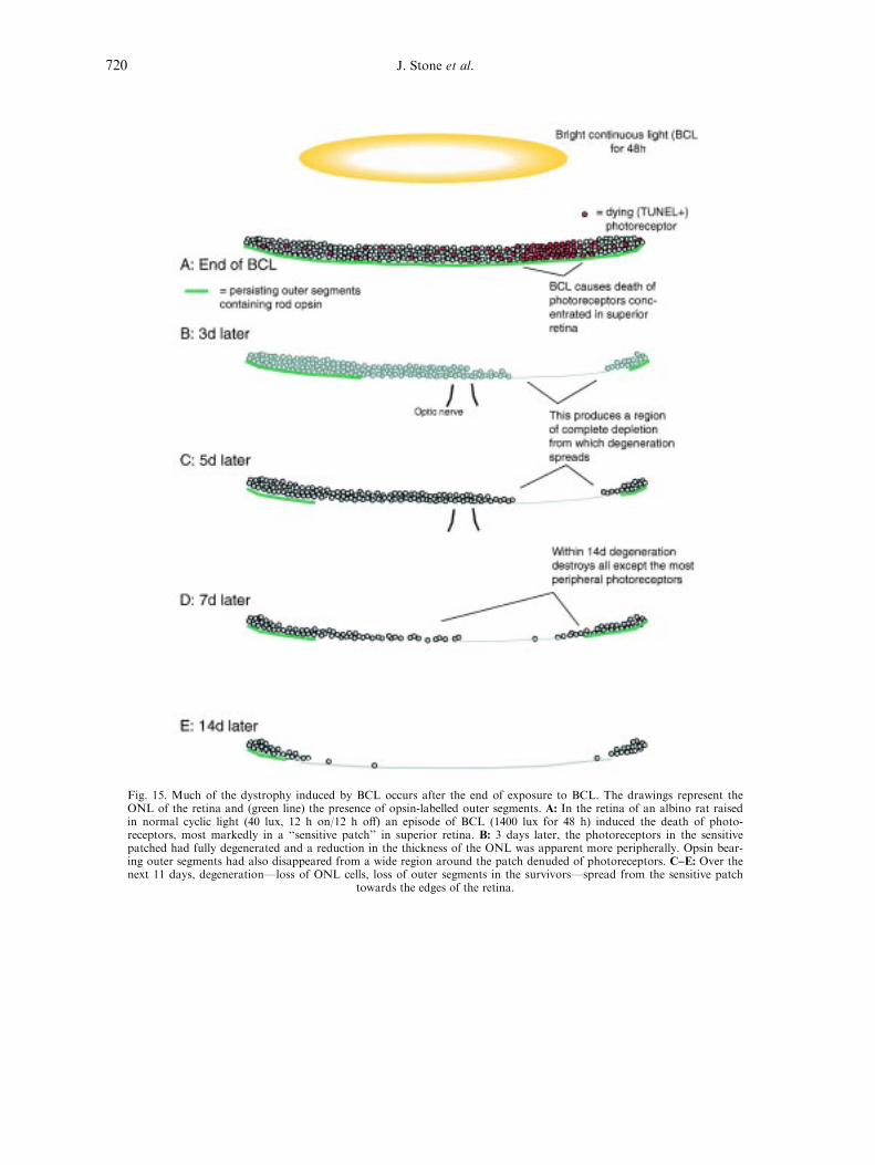
Fig. 15. Much of the dystrophy induced by BCL occurs after the end of exposure to BCL. The drawings represent theONL of the retina and (green line) the presence of opsin-labelled outer segments. A: In the retina of an albino rat raisedin normal cyclic light (40 lux, 12 h on/12 h o�) an episode of BCL (1400 lux for 48 h) induced the death of photo-receptors, most markedly in a ``sensitive patch'' in superior retina. B: 3 days later, the photoreceptors in the sensitivepatched had fully degenerated and a reduction in the thickness of the ONL was apparent more peripherally. Opsin bear-ing outer segments had also disappeared from a wide region around the patch denuded of photoreceptors. C±E: Over thenext 11 days, degenerationÐloss of ONL cells, loss of outer segments in the survivorsÐspread from the sensitive patch
towards the edges of the retina.
J. Stone et al.720

Fig. 16. Hyperoxia accelerates and hypoxia slows post-BCL degeneration. A±D: In animals kept in 70% oxygen afterBCL, the spread of degeneration from the sensitive patch towards the edges of the retina was accelerated, as shown sche-matically in C and D. A and B compare the histology of the retina in the inferior mid-periphery. This region was largelyintact in the animal kept in normoxia; in the animal kept in hyperoxia the ONL had degenerated. E±I: In animals kept inhypoxia after BCL exposure, the spread of degeneration from the sensitive patch was slowed, as shown schematically inH and I. E and F compare the histology of the retina in the inferior mid-periphery. The ONL in this region was largelydegenerate in the animal kept in normoxia (E). In the animal kept in hypoxia after P25 the ONL in the same region waslargely intact (F). At 7 days post-BCL, no ERG was detectable in the normoxic animal (blue trace in G) but an ERG per-
sisted (red trace, b-wave maximum of 25 mv) in the hypoxic animal (red trace).
Mechanisms of photoreceptor death and survival in mammalian retina 721

Fig. 17. Dual rescue e�ect of oxygen during BCL. A±C: In rats raised in dim cyclic skylight and then exposed to BCL for48 h in normoxia, degeneration was intense in the superior mid-periphery (B, red label of TUNEL+ cells). Both hypoxia(A) and hyperoxia (C) reduced the degeneration induced by BCL. D±F: In the inferior mid-periphery, the degenerationinduced by BCL was less than in the superior mid-periphery (E), as previously noted (Fig. 8, Fig. 10 and Fig. 15). Againthe degeneration caused by BCL was reduced by both hypoxia (D) and hyperoxia (F). G±I: BCL induced a strong upregu-lation of bFGF protein in the ONL of superior mid-peripheral retina (H), as previously noted (Fig. 10). That upregulation
was reduced by both hypoxia (G) and hyperoxia (I). Thus the rescue e�ects of oxygen were not mediated by bFGF.
J. Stone et al.722

the photoreceptor death induced in the albino rat
retina by bright continuous light.
The e�ects were surprising. Both hypoxia and
hyperoxia reduced the level of cell death induced
by BCL (Fig. 17A±C, D±F) and both slowed the
upregulation of bFGF also induced by light
damage (Fig. 17G±I).This dual protective e�ect
suggests that light damage results from two pro-
cesses, one involving hypoxic toxicity and the
other hyperoxic toxicity. It seems unlikely that
both mechanisms could operate simultaneously;
one possibility is that one process acts during
part of the day-night cycle, the other during the
other half of the cycle.
The importance of circadian rhythms in the
phenomenon of light damage has been demon-
strated by Duncan and O'Steen (1985) and
Organisciak et al. (1998b), who presented evi-
dence that the retina is most sensitive to light
damage in dark half of the day/night cycle. The
retina has strong circadian and light-driven
rhythms in its secretion of local neurohormones,
in particular dopamine (released by amacrine
cells during ``day'') and melatonin (released by
photoreceptors during ``night'') (reviewed in
Zawilska, 1994; Morgan and Boelen, 1996a,b).
Wiechmann and O'Steen (1992) reported that
exogenously applied melatonin exacerbates light
damage and Bush et al. (1997) reported that
selective blocking of melatonin receptors pro-
tects against light damage. Given the vulner-
ability of the retina in the presence of
melatonin, it can be readily understood that the
light-induced release of dopamine protects the
retina from light damage, because dopamine
suppresses melatonin secretion. What is pre-
sently mysterious is the role of melatonin. The
vertebrate retina evolved in a reliable day-night
cycle of light; somewhere in the mechanisms it
evolved to adapt to that cycle lies the key to
the damage caused by BCL.
Finally we stress that this dual protective e�ect
of oxygen is found only when hypoxia or hyper-
oxia are applied during BCL. The degeneration
which follows BCL appears to be driven by
oxygen toxicity only; in that period hypoxia is
protective, and hyperoxia is highly toxic (Section
4.2.3).
5. A TWO STAGE MODEL OF RETINALDYSTROPHIES
The ideas and data presented in Section 4 leadto the following model of photoreceptor dystro-phies (other than the dystrophy which followsdetachment).
5.1. Proposal
It is proposed that dystrophies occur in twostages, labelled (1) and (2) in Fig. 18. First alethal mutation and/or environmental factor,such as excess light or stress during the criticalperiod, causes depletion of the photoreceptorpopulation. This depletion reduces oxygen con-sumption in the ONL, causing oxygen levels inthe layer to rise. The hyperoxia is toxic, exacer-bating the depletion ((2) in Fig. 18). The continu-ing death of photoreceptors exacerbates thehyperoxia, driving the dystrophy to exhaustion.The mechanisms which cause photoreceptordeath at the two stages are thus very di�erent.The ®rst stage may be speci®c to the individual, aparticular mutation or a particular environmentalepisode. The second stage is driven by a commonmechanism, oxygen toxicity.
The model includes one further element.Photoreceptors which survive the oxygen toxicityare damaged by it and in response upregulate(Section 3.3) their expression of protective fac-tors, such as bFGF (Section 3.2±4), or CNTF(LaVail et al., 1992b, 1998; Wen et al., 1995),which slow the degeneration. The rate of photo-receptor loss is the net result of damage and self-protection.
The model has evident limitations. In particu-lar, its central tenet that outer retina becomeshyperoxic after photoreceptor depletion has yetto be tested by direct measurement. It is usefulbecause it provides testable explanations. It canexplain, for example, why cones die in dystro-phies caused by mutations in rod-speci®c genes;why many dystrophies progress relentlessly; whythe progress of dystrophies can be slow (the pro-tective mechanisms). Further, the vulnerability ofphotoreceptors during their critical period(Section 2.4) explains why many rodent dystro-
Mechanisms of photoreceptor death and survival in mammalian retina 723

phies begin in the critical period (Section 2.4.2.1).If human dystrophies also begin in the criticalperiod, understanding is gained of why the onsetof human dystrophies is so insidious, for thehuman critical period occurs at an age when theinfants' visual capacity and motor skills are unde-veloped (Section 2.5), and the gathering blindnessgoes (we suggest) unnoticed amid the generalimmaturity. The model also makes testable pre-dictions about therapies which, drawing on theearly and late stages of the degeneration, are tosome extent novel (Section 6.3.1).
In epistemological terms, we stress again (as inChapters 4 and 5 of Stone, 1983) that suchmodels are only as valuable as their ability toyield predictions capable of disproof. Section 6sets out a number of predictions, formulated asapproaches to therapy.
5.2. Comparative Note: How Epithelia Self-Protect
The mechanisms which (Fig. 18 proposes)cause photoreceptor dystrophyÐthose of a criti-
cal period of vulnerability of a single class of cell,
the dozens of mutations lethal to the same class
of cell, the impact of a massive but poorly regu-
lated circulation, the self-reinforcing feature of
oxygen toxicityÐseem unique to the retina. One
feature of the present model has interesting ana-
logues in other tissues.
The skin, like the retina, is exposed to radiant
energy and, as in the retina (Section 3.2), that
energy upregulates mechanisms (light absorbing
pigments) which protect against later exposure.
The epithelium of the bronchi and lung is exposed
to inhaled gases and is the major site of damage
by the prolonged inhalation of high concen-
trations of oxygen (bronchopulmonary dysplasia,
O'Brodovich and Mellins, 1985). In response to
an episode of hyperoxia, the bronchopulmonary
epithelium upregulates its antioxidant mechan-
isms and becomes more resistant to subsequent
episodes of hyperoxia (Clerch and Massaro, 1993;
Clyde et al., 1993). As with the skin, the protec-
tion provided is not immediate, but requires
hours to days to develop.
Fig. 18. A model of the pathogenesis of photoreceptor dystrophies. It is proposed that the dystrophies occur in two stages,``initial depletion'', labelled 1, and ``late'', labelled 2.
J. Stone et al.724

All three epithelia are exposed to damagingradiation or toxins and all have developed mech-anisms of self-protection which are prospective.Each constantly uses its recent experience to pre-pare itself for future exposure and, with that con-ditioning, can last the life of the organism.For each, an episode of unprecedentedly highlevels of ultraviolet light (for the skin), of visiblelight (retina) or oxygen (lungs) can be highlydamaging.
6. FUTURE DIRECTIONS: PREDICTIONSOF THERAPY
6.1. Retinal Detachment: Oxygen Supplementation Will
Improve Outcomes
The retina's response to detachment is the de-generation of some photoreceptors, degenerativechanges in the survivors and a glial proliferation(Section 4.1). From the evidence set out inSection 4.1.2 we predict that oxygen supplemen-tation between diagnosis and surgery will be ofbene®t in corresponding ways: it will rescuephotoreceptors in the detached portion of retina,preserve the structure of surviving photoreceptorsand reduce the proliferation of glial cells. Theclinical outcome should comprise improved visualresponsiveness of the re-attached retina and areduced incidence of proliferative vitreoretinopa-thy.
6.2. Preventing Photoreceptor Depletion
The photoreceptor dystrophies known as retini-tis pigmentosa are not related to detachment.They occur with the retina in place, close to itssource of nutrients (the choroid) and, we suggest,involve the two stages set out in Fig. 18.
6.2.1. Improved perinatal care will prevent some RP
The age of onset of human dystrophies is notknown. Many are not diagnosed until the teenyears or young adulthood or (less commonly)
until middle age, suggesting that the disease is
``adult-onset''. When children are screened by the
study of their ERG, however, RP can be diag-
nosed in early childhood (Berson, 1971),
suggesting that some forms of the disease begin
much earlier. In many animal models of geneti-
cally driven photoreceptor dystrophies, the death
of photoreceptors begins with the critical period
(Section 2.4.2.1). It is possible, arguably likely,
that in many forms of human RP the death of
photoreceptors also begins in the critical period,
and therefore around birth (Section 2.5), making
the perinatal period an important stage for inter-
vention.
Approximately 50% of human cases of RP
occur without a family history and are termed
``simplex'' RP (Heckenlively, 1988) and it seems
possible that perinatal stress (hypoxia, jaundice,
hypoglycaemia, prolonged delivery) might cause a
depletion of photoreceptors which leads (via the
mechanisms summarised in Fig. 18) to a clinical
presentation like that which follows a genetically
induced depletion.
Some ``simplex'' cases of RP are in fact gen-
etic, caused by fresh or re-emerging recessive
mutations (Heckenlively, 1988), and there is
currently debate over how many are really gen-
etic. Most debates over whether the genesis of
a disease lies in nature or nurture lead to
acceptance that both genetic and environmental
factors are involved, the challenge being to
identify their di�erent contributions to the dis-
ease. A recent example of such a debate is that
over myopia (Wallman, 1994; Mutti et al.,
1996).
More generally, the idea that pre- or perinatal
stress can cause diseases normally considered
``adult-onset'' has been argued and developed in
studies of foetal and perinatal correlates of
human diseases such as hypertension, coronary
disease and diabetes (Barker et al., 1995). It
would seem valuable now to establish the age of
onset of human photoreceptor dystrophies, and
to examine whether there is any correlation
between perinatal stress and some forms of
photoreceptor dystrophy. If a correlate emerges,
the possibility of prevention by improved perina-
tal care will deserve testing.
Mechanisms of photoreceptor death and survival in mammalian retina 725

6.2.2. Other approaches to preventing depletion must be
applied early
6.2.2.1. Gene therapy
Gene therapy has emerged as a possibility forRP as for many other diseases because in this dec-ade molecular geneticists have achieved the identi-®cation of the causative mutations at the codonlevel. Successful gene therapy for retinal dystro-phies has been reported in two animal models,the rd mouse (Bennett et al., 1996) and the trans-genic P23H rat (Peterson et al., 1998; Lewin etal., 1998). The results are technically stunning,with obvious promise for particular forms of RP.In all these studies the investigators knew the ageof onset of the rodent dystrophy and deliveredthe therapy early, during the critical period. Ifhuman RP begins with the critical period, aroundbirth (Section 2.5), it may prove important todeliver gene therapy in late foetal life.
6.2.2.2. Trophic factor therapy
Trophic factors such as bFGF and CNTF pro-tect photoreceptors from the degeneration whichoccurs during the critical period in geneticallydystrophic species such as the RCS rat(Faktorovich et al., 1990) and rd mouse (LaVailet al., 1998). Again, these investigators knew(because they had demonstrated) the age of onsetof the dystrophies and delivered the trophic fac-tors in the critical period. Again it may prove im-portant, to prevent depletion of human retinaand the sequela of oxygen toxicity, to deliverthese factors in late foetal life.
6.3. Therapy for the Depleted Retina
`` . . .there may be common features of the disease at its laterstages.. (which) o�ers the hope that a single form of thera-peutic intervention might be able to retard the progress of thedisease . . .delay the loss of cones . . .and prevent blindness''.Lisman and Fain (1995).
6.3.1. Predictions from the hypothesis of oxygen toxicity
The idea that the late stages of many photo-receptor dystrophies are driven by oxygen toxicityof outer retina suggests three approaches totherapy.
6.3.1.1. Hypoxia will slow late stage dystrophy
No group has tried hypoxia as therapy for RPor, as far as we are aware, as therapy for any-thing. Empirically, hypoxia slows late stages ofthe RCS, rd and light induced dystrophies(Section 4.2) and we predict that it will slow thelate stages of human dystrophies.
Side e�ects: Knowledge of the side-e�ects ofhypoxia comes from the study of human re-sponses to high altitudes. The percentage of oxy-gen in air remains constant (at 21%) with altitudebut atmospheric pressure and the partial pressureof oxygen decrease. Above about 3000 metres(10 000 ft, pO2 equivalent to 14% oxygen at sealevel) morbidity becomes common (Butler et al.,1992). The symptoms, collectively termed ``alti-tude sickness'', include headache, nausea, fatigue,insomnia, reversible total blindness and retinalbleeding. The threshold altitude for and severityof altitude sickness vary markedly however, par-ticularly with rate of ascent.
By contrast, mild hypoxia appears to be largelyfree of side e�ects. Every day several millionhumans experience mild hypoxia as passengers incommercial airliners. These craft are typicallypressurised to 8000 ft, and for the duration of the¯ight (less ascent and descent) passengers experi-ence a partial pressure of oxygen equivalent to15±16% oxygen at sea level. Air-crew experiencethe same conditions on a daily basis and are sub-ject to frequent medical examination. There arefew reports, if any, of ¯ight-related morbidityamong either passengers or crew. And high alti-tude cities like Mexico City (7350 ft) grow popu-lous and host Olympic games.
Mechanism: Among the adaptive mechanismswhich enable animals and humans to toleratemild hypoxia is the autoregulatory response ofcapillary beds. If arterial pO2 drops and the tissuesupplied becomes hypoxic, the vessels delivergreater volumes of blood; and vice versa. Thechoroidal circulation lacks this ability to autore-
J. Stone et al.726

gulate (Section 1.1.4) and, we now argue, thislack of regulation is a major element in the causeof the late stages of photoreceptor dystrophies.But conversely, the inability of the choroid toautoregulate means that when the animalbreathes oxygen-enriched or depleted air, theexcess or lack of oxygen a�ects outer retina morestrongly than other, autoregulating tissues. Thusthe factor which causes the hyperoxia localised toouter retina which (we argue) drives the latestages of dystrophies can be used to reduce thathyperoxia.
Delivery: The practicalities of delivering hy-poxia remain a challenge. Many of the technol-ogies developed to deliver and control hyperoxia(gas mixtures, selective membrane pumps, masksystems) would seem to be readily applicable,mutatis mutandis.
Unknowns: Two deserve mention. It is emergingfrom the literature on light damage (Section 4.3)that the vulnerability of photoreceptors todamage varies markedly with diurnal phase. Itmay prove as e�ective, even more e�ective, todeliver a hypoxia at certain times of day or night.Second, if Fig. 18 is correct in suggesting that latestage dystrophies are slowed by protective mech-anisms induced by the toxin, then reducing thetoxin will downregulate the protection, makingthe retina more vulnerable. It may prove to beimportant that treatment once commenced bemaintained.
6.3.1.2. Antioxidants will slow late stage dystrophy
If the late stages of photoreceptor dystrophiesare driven partly or wholly by oxygen toxicity,then antioxidant molecules should slow theirdeath. Several authors (Phelps, 1986; van derHagen et al., 1993; Kretzer et al., 1984) havereported evidence of a role of antioxidants inother retinal disease. In relation to photoreceptordystrophies, Penn et al. (1987) reported evidence(upregulation of scavenger molecules in light-damage resistant retina, Section 3.2.1.2) that anti-oxidants play a role in the protection of photo-receptors against light-induced dystrophy andOrganisciak et al. (1998a) have reported that asystemically delivered anti-oxidant is protectiveagainst light damage. Mittag et al. (1997)reported that mice in which the activity of a
major antioxidant enzyme (superoxide dismutase)was reduced by a transgene are abnormally sensi-tive to damaging light, implying a normal role forthis enzyme in photoreceptor protection. The po-tential of antioxidants to slow human dystrophiesrequires systematic investigation, however.
6.3.1.3. Prevention of light adaptation will slowdystrophies
The idea that reducing the light falling on theretina might slow the progress of retinal dystro-phies was proposed by Berson (1971, 1973), whotraced the history of the idea in the treatment ofhuman RP and in animal models. The presentanalysis builds on the rationale which Bersondeveloped.
Light adaptation of rods increases oxygenlevels in the outer retina (Linsenmeier, 1986;Brown et al., 1996), because rods use less oxygenwhen light-adapted. As long as rods persist in sig-ni®cant numbers, minimising their adaptation tolight should minimise daily light-inducedincreases in oxygen levels in outer retina andthereby slow late-stage dystrophy. Dowling andSidman (1962) reported that dark rearing slowedthe late stages of the RCS dystrophy, though intheir data the slowing was clear in one strain ofRCS rat (pink-eye) and not in a more heavily pig-mented strain. Kaitz and Auerbach (1978) re-visited this problem, showing that the RCSdystrophy is markedly slowed if the wavelengthand intensity of circadian light are chosen tominimise the absorption of light by rhodopsin.Naash et al. (1996) noted that dark rearing slowsthe dystrophy caused in mice by human RP trans-genes and, like Kaitz and Auberbach, suggestedthat the slowing resulted from a decrease in theabsorption of light by rhodopsin. We suggest thatthe reduction in light absorption by rhodopsinslows the dystrophy by maintaining the high oxy-gen consumption of dark-adapted rods, therebyreducing the level of oxygen in outer retina.
There are few published data which test thisidea in humans. The data in Table 1 are fromMerin and Pe'er (1981), who followed RPpatients who wore a dark contact lens on one eyefor up to 3 years, the other eye serving as a con-trol (as Berson (1971, 1973) suggested). The lenswas brown in colour and reduced total trans-
Mechanisms of photoreceptor death and survival in mammalian retina 727
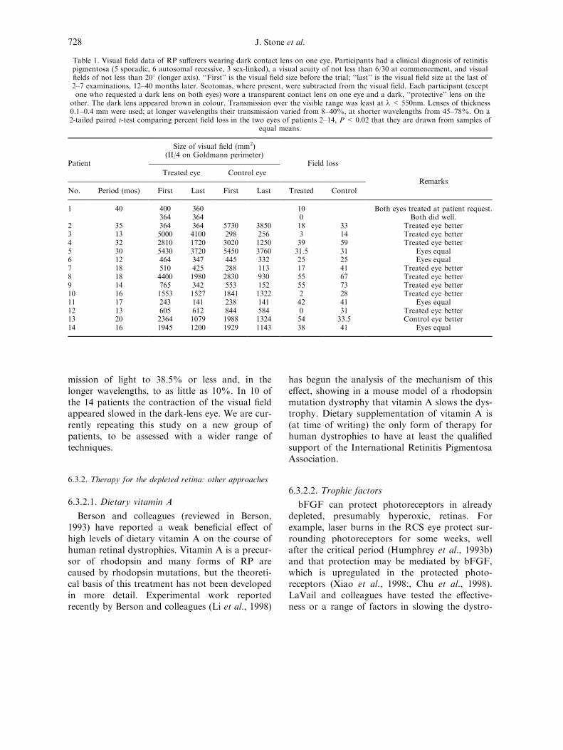
mission of light to 38.5% or less and, in thelonger wavelengths, to as little as 10%. In 10 ofthe 14 patients the contraction of the visual ®eldappeared slowed in the dark-lens eye. We are cur-rently repeating this study on a new group ofpatients, to be assessed with a wider range oftechniques.
6.3.2. Therapy for the depleted retina: other approaches
6.3.2.1. Dietary vitamin A
Berson and colleagues (reviewed in Berson,1993) have reported a weak bene®cial e�ect ofhigh levels of dietary vitamin A on the course ofhuman retinal dystrophies. Vitamin A is a precur-sor of rhodopsin and many forms of RP arecaused by rhodopsin mutations, but the theoreti-cal basis of this treatment has not been developedin more detail. Experimental work reportedrecently by Berson and colleagues (Li et al., 1998)
has begun the analysis of the mechanism of thise�ect, showing in a mouse model of a rhodopsinmutation dystrophy that vitamin A slows the dys-trophy. Dietary supplementation of vitamin A is(at time of writing) the only form of therapy forhuman dystrophies to have at least the quali®edsupport of the International Retinitis PigmentosaAssociation.
6.3.2.2. Trophic factors
bFGF can protect photoreceptors in alreadydepleted, presumably hyperoxic, retinas. Forexample, laser burns in the RCS eye protect sur-rounding photoreceptors for some weeks, wellafter the critical period (Humphrey et al., 1993b)and that protection may be mediated by bFGF,which is upregulated in the protected photo-receptors (Xiao et al., 1998:, Chu et al., 1998).LaVail and colleagues have tested the e�ective-ness or a range of factors in slowing the dystro-
Table 1. Visual ®eld data of RP su�erers wearing dark contact lens on one eye. Participants had a clinical diagnosis of retinitispigmentosa (5 sporadic, 6 autosomal recessive, 3 sex-linked), a visual acuity of not less than 6/30 at commencement, and visual®elds of not less than 208 (longer axis). ``First'' is the visual ®eld size before the trial; ``last'' is the visual ®eld size at the last of2±7 examinations, 12±40 months later. Scotomas, where present, were subtracted from the visual ®eld. Each participant (exceptone who requested a dark lens on both eyes) wore a transparent contact lens on one eye and a dark, ``protective'' lens on the
other. The dark lens appeared brown in colour. Transmission over the visible range was least at l< 550nm. Lenses of thickness0.1±0.4 mm were used; at longer wavelengths their transmission varied from 8±40%, at shorter wavelengths from 45±78%. On a2-tailed paired t-test comparing percent ®eld loss in the two eyes of patients 2±14, P < 0.02 that they are drawn from samples of
equal means.
Size of visual ®eld (mm2)(II/4 on Goldmann perimeter)
Patient
Treated eye Control eye
Field loss
No. Period (mos) First Last First Last Treated Control
Remarks
1 40 400 360 10 Both eyes treated at patient request.364 364 0 Both did well.
2 35 364 364 5730 3850 18 33 Treated eye better3 13 5000 4100 298 256 3 14 Treated eye better4 32 2810 1720 3020 1250 39 59 Treated eye better5 30 5430 3720 5450 3760 31.5 31 Eyes equal6 12 464 347 445 332 25 25 Eyes equal7 18 510 425 288 113 17 41 Treated eye better8 18 4400 1980 2830 930 55 67 Treated eye better9 14 765 342 553 152 55 73 Treated eye better10 16 1553 1527 1841 1322 2 28 Treated eye better11 17 243 141 238 141 42 41 Eyes equal12 13 605 612 844 584 0 31 Treated eye better13 20 2364 1079 1988 1324 54 33.5 Control eye better14 16 1945 1200 1929 1143 38 41 Eyes equal
J. Stone et al.728

phies which occur in the RCS rat (LaVail et al.,1992a), in several mouse mutants (LaVail et al.,1998) and in transgenic rats made dystrophic bymutant opsin transgenes (Steinberg et al., 1997).They demonstrated that bFGF can slow thesedystrophies over periods extending from the criti-cal period in these strains to considerably olderages. The present analysis suggests that the latestage of dystrophy is driven by a factor (oxygentoxicity) which is chronic. This would seem torequire that trophic factors be delivered chroni-cally and to raise two problems, the e�ect of longterm exposure to such factors on other elementsof the eye, such as the lens (Chamberlain andMcAvoy, 1997), and their e�ect on the functionof the surviving retina (Section 3.4).
6.3.2.3. Hyperbaric hyperoxia
Several groups with access to hyperbaric facili-ties have tested the e�cacy of hyperbaric oxygenexposure on the course of human retinal dystro-phies. The most systematic study is that ofVingolo and colleagues (Vingolo et al., 1997) whoreport on 24 RP patients subjected to standardpatterns of this treatment. Patients were exposedto 2.2 atmospheres of pressure for 90 min per dayonce a day for 5 days per week. During each ses-sion they breathed 100% oxygen through a mask.This treatment was given for a period of 4 weeks,then at decreasing intervals for up to three years.The authors reported that the ERG declined sig-ni®cantly less in treated patients than in a controlgroup with comparable disease forms.
Hyperbaric hyperoxia delivers high levels ofoxygen to all body tissues by increasing theamount of oxygen dissolved in serum. Theincrease is achieved partly by the hyperbaria,partly by the hyperoxia. It is the treatment ofchoice for gas gangrene and carbon monoxidepoisoning and appears to have bene®cial e�ectson, for example, the peripheral circulatory pro-blems of diabetes (Tibbles and Edelsberg, 1996).
Hyperbaric hyperoxia is a complex treatment,involving very high levels of oxygen tension pre-sented in daily episodes, with major changes inoxygen levels both at onset and o�set of each ex-posure (exposure must be limited because of thetoxicity of oxygen). At present these results are
intriguing and invite further testing but, to thepresent writers, remain di�cult to explain.
6.3.3. Treatment of late stage RP will provide a limited
improvement in vision
It seems possible that the blindness of the laterstages of RP stems from two sources, the loss ofsome photoreceptors and the loss of sensitivity inthe survivors. The loss of sensitivity could resultfrom two causes, the upregulation of factorswhich both protect photoreceptors and reduce theERG in the presence of normal outer segmentmorphology (Section 3.4); and/or from a re-duction in the photopigment content of outer seg-ments, as described in the response of the normalretina to bright circadian light (Penn et al., 1989).Therapy which relieves stress on photoreceptorssurviving in RP patients will presumably downre-gulate their self-protective mechanisms and mayallow regrowth of the outer segments. Penn et al.(1989) described an impressive instance of suchregrowth. In rats raised in bright circadian light,many photoreceptors are lost and the outer seg-ments of the survivors are greatly shortened andtheir rhodopsin content is correspondinglyreduced. Nevertheless, the retina is intact andfunctional. If these rats are returned to dim circa-dian light then within 3 weeks the length of theirouter segments and their pigment content tripleand the ERG increases markedly in amplitude(the b-wave by 60%, the a-wave by 250%).
Therapy which successfully reduced stress onphotoreceptors surviving in late stage dystrophiesmight similarly increase their sensitivity, to a limitset by the actual loss of photoreceptors. The pre-sent analysis also suggests a cautionary con-clusion. Therapy which relieves the stress onphotoreceptors may also downregulate their self-protection, making it important to maintain thecontinuity of the treatment, lest the cells re-ex-perience stress while unprotected.
6.3.4. The senescence of the retina can be slowed
The mammalian retina degenerates with age, soreliably that progressive loss of sensitivity and
Mechanisms of photoreceptor death and survival in mammalian retina 729

acuity in advanced age is considered normal. Theedge of the human retina undergoes an age-re-lated ``cystic'' degeneration and systematic countsof retinal neurones (Gao and Holly®eld, 1992)show a generalised age-related loss of neurones inall layers. The rate of loss was greatest in theyoungest ages studied (the teen years) but contin-ued throughout life, slowly depleting the retina offunctional neurones. Correspondingly, visionslowly fails with advanced age (Section 1.2).
The normal senescence of the retina can thusbe regarded as a very slow retinal dystrophy. Wehave argued above that hyperoxia of the outerretina is a major factor driving the late stages ofall retinal dystrophies. A logical extension of thisargument is that oxygen toxicity of outer retinamay be a factor in the senescence of the retina,and that treatments which slow late stage dystro-phies will slow senescent changes. We are cur-rently testing this idea in models of retinalsenescence in the rat (Shinowara et al. (1982); Laiet al., 1978; DiLoreto et al., 1996).
These predictions extend logical argument wellbeyond present data. They are made to be tested,con®rmed, disproved. If only partially con®rmedthey could provide a basis for the treatment oflongÐuntreatable dystrophies caused by geneticmutation, by environmental insult, by age.
REFERENCES
Alder, V. A. and Cringle, S. A. (1985) The e�ect of the retinalcirculation on vitreal oxygen tension. Curr. Eye Res. 4,121±129.
Alder, V. A., Ben-Nun, J. and Cringle, S. J. (1990) PO2 pro-®les and oxygen consumption in cat retina with anoccluded retinal circulation. InvestigativeOphthalmology and Visual Science 31, 1029±1034.
Alm, A. and Bill, A. (1972) The oxygen supply to the retina,II. E�ects of high intra-ocular pressure and ofincreased arterial carbon dioxide tension on uveal andretinal blood ¯ow in cats. Acta Physiol. Scand. 84, 306±319.
Ames, A., Li, Y.-Y., Heher, E. C. and Kimble, C. R. (1992)Energy metabolism of rabbit retina as related to func-tion: high cost of Na+ transport. Journal ofNeuroscience 12, 840±853.
Anderson, D. H., Fisher, S. K. and Steinberg, R. H. (1978)Mammalian cones: disc shedding, phagocytosis, andrenewal. Invest. Ophthal. Vis. Sci. 17, 117±133.
Anderson, D. H., Gue rin, C. J., Erickson, P. A., Stern, W. H.and Fisher, S. K. (1986) Morphological recovery in thereattached retina. Invest. Ophthal. Vis. Sci. 27, 168±183.
Anderson, D. H., Stern, W. H., Fisher, S. K., Erickson, P. A.and Borgula, G. A. (1983) Retinal detachment in thecat: the pigment epithelial-photoreceptor interface.Invest. Ophthal. Vis. Sci. 24, 906±926.
Barbe, M. F., Tytell, M., Gower, D. J. and Welch, W. J.(1988) Hyperthermia protects against light damage inthe rat retina. Science 241, 1817±1820.
Barker, D. J., Gluckman, P. D. and Robinson, J. S. (1995)Conference report: fetal origins of adult disease-reportof the First International Study Group, Sydney, 29±30October 1994. Placenta 16, 317±320.
Behbehani, M. M., Bowyer, D. W., Ru�olo, J. J. andKranias, G. (1984) Preservation of retinal function inthe RCS rat by laser treatment. Retina 4, 257±263.
Bennett, J., Tanabe, T., Sun, D., Zeng, Y., Kjeldbye, H.,Gouras, P. and Maguire, A. M. (1996) Photoreceptorcell rescue in retinal degeneration (rd) mice by in vivogene therapy. Nature Medicine 2, 649±654.
Berglin, L., Algvere, P. V. and Seregard, S. (1997)Photoreceptor decay over time and apoptosis in exper-imental retinal detachment. Graefes Archive for Clinicaland Experimental Ophthalmology 235, 306±312.
Berson, E. L. (1971) Light deprivation for early retinitis pig-mentosa. Arch. Ophthal. 85, 521±529.
Berson, E. L. (1973) Experimental and therapeutic aspects ofphotic damage to the retina. Invest. Ophthal. Vis. Sci.12, 35±44.
Berson, E. L. (1993) Retinitis Pigmentosa. Invest. Ophthal.Vis. Sci. 34, 1659±1676.
Bill, A. and Sperber, G. O. (1990) Control of retinal andchoroidal blood ¯ow. Eye 4, 319±325.
Bill, A., ToÈ rnquist, P. and Alm, A. (1980) Permeability of theintraocular blood vessels. Trans. ophthal. Soc. U.K.100, 332±336.
Bill, A., Sperber, G. and Ujiie, K. (1983) Physiology of thechoroidal vascular bed. International Ophthalmology 6,101±107.
BjoÈ rklund, H., Bignami, A. and Dahl, D. (1985)Immunohistochemical demonstration of glial ®brillaryacidic protein in normal rat MuÈ ller glia and retinalastrocytes. Neurosci. Lett. 54, 363±368.
Bowers, F. L. and Stone, J. (1998) The expression of cyto-chrome oxidase in the photoreceptors of the developingand adult rat. Proc. Aust. Neuroscience Soc. 9, 96.
Bowers, F., Valter, K. and Stone, J. (1998) Dual rescue e�ectof oxygen on light induced photoreceptor death in therat. Invest. Ophthal. Vis. Sci. 39, S127.
Braun, R. D., Linsenmeier, R. A. and Goldstick, T. K. (1995)Oxygen consumption in the inner and outer retina ofthe cat. Invest. Ophthal. Vis. Sci. 36, 542±554.
Brown, R. D., Linsenmeier, R. A. and Goldstick, T. K.(1996) Light-induced changes in retinal oxygen con-sumption. Invest. Ophthal. Vis. Sci. 37, 473±475.
Burns, M. S. and Robles, M. (1990) Muller cell GFAP ex-pression exhibits gradient from focus of photoreceptorlight damage. Curr. Eye Res. 9, 479±486.
Bush, R. A. and Williams, T. P. (1991) The e�ect of unilateraloptic nerve section on retinal light damage in rats. Exp.Eye Res. 52, 139±153.
Bush, R. A., Sugawara, T., Iuvone, P. M. and Sieving, P. A.(1997) The melatonin receptor antagonist luzindoleprotects retinal photoreceptors from light damage inrat. Invest. Ophthal. Vis. Sci. 38, S127.
J. Stone et al.730

Butler, F. K., Harris, D. J. and Reynolds, R. D. (1992)Altitude retinopathy on Mount Everest, 1989.Ophthalmology 99, 739±746.
Campochiaro, P. A., Chang, M., Ohsato, M., Vinores, S. A.,Nie, Z., Hjelmeland, L., Mansukhani, A., Basilico, C.and Zack, D. J. (1996) Retinal degeneration in trans-genic mice with photoreceptor speci®c expression of adominant-negative ®broblast growth factor receptor. J.Neurosci. 16, 1679±1688.
Cao, W., Li, F., LaVail, M. M. and Steinberg, R. H. (1997a)Development of injury-induced gene expression ofbFGF, FGFR-1, CNTF and GFAP in rat retina.Invest. Ophthal. Vis. Sci. 38, S604.
Cao, W., Wen, R., Li, F., LaVail, M. M. and Steinberg, R.H. (1997b) Mechanical injury increases bFGF andCNTF mRNA expression in the mouse retina. Exp.Eye Res. 65, 241±248.
Chang, C. J., Lai, W. W., Edward, D. P. and Tso, M. O.(1996) Apoptotic photoreceptor cell death after trau-matic retinal detachment in humans. Arch. Ophthal.114, 1158±1159.
Chang, G-Q., Hao, Y. and Wong, F. (1993) Apoptosis: ®nalcommon pathway of photoreceptor death in rd, rds,and rhodopsin mutant mice. Neuron 11, 595±605.
Chan-Ling, T. and Stone, J. (1993) Retinopathy of prematur-ity: Origins in the architecture of the retina. Prog. Ret.Res. 12, 155±176.
Chu, Y., Humphrey, M. F., Alder, V. A. and Constable, I. J.(1998) Immunocytochemical localisation of basic ®bro-blast growth factor and glial ®brillary acidic proteinafter laser photocoagulation in the Royal College ofSurgeons rat. Aust. N.Z. J. Ophthal. 26, 87±96.
Clerch, L. B. and Massaro, D. (1993) Tolerance of rats tohyperoxia. Lung antioxidant enzyme gene expression.J. Clin. Invest. 91, 499±508.
Clyde, B. L., Chang, L. Y., Auten, R. L., Ho, Y. S. andCrapo, J. D. (1993) Distribution of manganese super-oxide dismutase mRNA in normal and hyperoxic ratlung. Am. J. Resp. Cell Mol. Biol. 8, 530±537.
Cohen, L. H. and Noell, W. K. (1965) Relationships betweenvisual function and metabolism. In Biochemistry of theRetina (ed. C. Graymore), pp. 36±50. Academic Press,London.
Cook, B., Lewis, G. P., Fisher, S. K. and Adler, R. (1995)Apoptotic photoreceptor degeneration in experimentalretinal detachment. Invest. Ophthal. Vis. Sci. 36, 990±996.
Cowan, M. (1973) Neuronal death as a regulative mechanismin the control of cell number in the nervous system. InDevelopment and Aging in the Nervous System, pp. 267±280. Academic Press, New York.
Cunningham, T. J., Mohler, I. M. and Giordano, D. L.(1981) Naturally occurring neuron death in theganglion cell layer of the neonatal rat: morphology andevidence for regional correspondence with neurondeath in superior colliculus. Dev. Brain Res. 2, 203±215.
Demontis, G. C., Longoni, B., Gargini, C. and Cervetto, L.(1997) The energetic cost of photoreception in retinalrods of mammals. Arch. Ital. Biol. 135, 95±109.
Diaz-Araya, C. and Provis, J. M. (1992) Evidence of photo-receptor migration during early foveal development: Aquantitative analysis of human fetal retinae. VisualNeuroscience 8, 505±514.
DiLoreto, D., Cerro, M. D., Reddy, S. V., Janardhan, S.,Cox, C., Wyatt, J. and Balkema, G. (1996) Waterescape performance of adult RCS dystrophic and con-genic rats: a functional and histomorphometric study.Brain Res. 717, 165±172.
Dorey, C. K., Wu, G., Ebenstein, D., Garsd, A. and Weiter,J. J. (1989) Cell loss in the aging retina: relationship tolipofuscin accumulation and macular degeneration.Invest. Ophthal. Vis. Sci. 30, 1691±1699.
Dowling, J. E. and Sidman, R. L. (1962) Inherited retinal dys-trophy in the rat. J. Cell Biol. 14, 73±109.
Dreher, B. and Robinson, S. R. (1988) Development of theretinofugal pathway in birds and mammals: evidencefor a common `timetable'. Brain Behav. Evol. 31, 369±390.
Dreher, B., Barker, D. A., Bath, M. R. and Keay, K. A.(1996) Spatiotemporal pattern of ontogenetic ex-pression of calbindin-28/kD in the retinorecipient layersof rat superior colliculus. J. Comp. Neurol. 376, 223±240.
Dryja, T. and Berson, E. L. (1995) Retinitis pigmentosa andallied diseases. Implications of genetic heterogeneity.Invest. Ophthal. Vis. Sci. 36, 1197±1200.
Duncan, T. E. and O'Steen, W. K. (1985) The diurnal suscep-tibility of rat retinal photoreceptors to light-induceddamage. Exp. Eye Res. 41, 497±507.
Eisenfeld, A. J., Bunt-Milam, A. H. and Vijay Sarthy, P.(1984) Muller cell expression of glial ®brillary acidicprotein after genetic and experimental photoreceptordegeneration in the rat retina. Invest. Ophthal. Vis. Sci.25, 1321±1328.
Ekstrom, P., Sanyal, S., Narfstrom, K., Chader, G. J. andvan Veen, T. (1988) Accumulation of glial ®brillaryacidic protein in Muller radial glia during retinal de-generation. Invest. Ophthal. Vis. Sci. 29, 1363±1371.
Erickson, P. A., Fisher, S. K., Anderson, D. H., Stern, W. H.and Borgula, G. A. (1983) Retinal detachment in thecat: the outer nuclear and outer plexiform layers.Invest. Ophthal. Vis. Sci. 24, 927±942.
Erickson, P. A., Fisher, S. K., Gue rin, C. J., Anderson, D. H.and Kaska, D. D. (1987) Glial ®brillary acidic proteinincreases in MuÈ ller cells after retinal detachment. Exp.Eye Res. 44, 37±48.
Fain, G. L. and Lisman, J. E. (1993) Photoreceptor degener-ation in vitamin A deprivation and retinitis pigmen-tosa: the equivalent light hypothesis. Exp. Eye Res.57(3), 335±340.
Faktorovich, E. G., Steinberg, R. H., Yasumura, D.,Matthes, M. T. and LaVail, M. M. (1990)Photoreceptor degeneration in inherited retinal dystro-phy delayed by basic ®broblast growth factor. Nature347, 83±86.
Faktorovich, E. G., Steinberg, R. H., Yasumura, D.,Matthes, M. T. and LaVail, M. M. (1992) Basic ®bro-blast growth factor and local injury protect photo-receptors from light damage in the rat. J. Neurosci. 12,3554±3567.
Farber, D. B. (1995) From mice to men: the cyclic GMPphosphodiesterase gene in vision and disease. Invest.Ophthal. Vis. Sci. 36, 263±275.
Farber, D. B., Flannery, J. G. and Bowes-Rickman, C. (1994)The rd mouse story: seventy years of research on ananimal model of inherited retinal degeneration.Progress in Retinal and Eye Research 13, 31±64.
Mechanisms of photoreceptor death and survival in mammalian retina 731

Ferrer, I., Bernet, E., Soriano, E., Del Rio, T. and Fonseca,M. (1990) Naturally occurring cell death in the cerebralcortex of the rat and removal of dead cells by transi-tory phagocytes. Neuroscience 39, 451±458.
Ferrer, I., Tortosa, A., Blanco, R., Martin, F., Serrano, T.,Planas, A. and Macaya, A. (1994) Naturally occurringcell death in the developing cerebral cortex of the rat.Evidence of apoptosis-associated internucleosomalDNA fragmentation. Neurosci. Lett. 182, 77±79.
Fisher, S. K., Erickson, P. A., Lewis, G. P. and Anderson, D.H. (1991) Intraretinal proliferation induced by retinaldetachment. Invest. Ophthal. Vis. Sci. 32, 1739±1748.
Galli-Resta, L. and Ensini, M. (1996) An intrinsic time limitbetween genesis and death of individual neurons in thedeveloping retinal ganglion cell layer. J. Neurosci. 16,2318±2324.
Galli-Resta, L. and Resta, G. (1992) A quantitative model forthe regulation of naturally occurring cell death in thedeveloping vertebrate nervous system. J. Neurosci. 12,4586±4594.
Gao, H. and Holly®eld, J. G. (1992) Aging of the human ret-ina: di�erential loss of neurons and retinal pigment epi-thelial cells. Invest. Ophthal. Vis. Sci. 33, 1±17.
Gao, H. and Holly®eld, J. G. (1995) Basic ®broblast growthfactor in retinal development: Di�erential levels ofbFGF expression and content in normal and retinal de-generation (rd) mutant mice. Devl. Biol. 169, 168±184.
Gavrieli, Y., Sherman, Y. and Ben-Sasson, S. A. (1992)Identi®cation of programmed cell death in situ viaspeci®c labeling of nuclear DNA fragmentation. J.Cell. Biol. 119, 493±501.
Geller, S. F., Lewis, G. P., Anderson, D. H. and Fisher, S. K.(1995) Use of the MIB-1 antibody for detecting prolif-erating cells in the retina. Invest. Ophthal. Vis. Sci. 36,737±744.
Glaser, B. M. and Michels, R. G. (1989) Retina Volume IIISurgical Retina. C.V. Mosby, St. Louis.
Graymore, C. (1959) Metabolism of the developing retina.Brit. J. Ophthal. 43, 34±39.
Graymore, C. (1960) Metabolism of the developing retina:respiration in the developing normal rat retina and thee�ect of an inherited degeneration of the retinal neuro-epithelium. Brit. J. Ophthal. 44, 363±369.
Graymore, C. and Tansley, K. (1959) Iodoacetate poisoningof the rat retina. Brit. J. Ophthal. 43, 486±493.
Hamburger, V. and Oppenheim, R. W. (1982) Naturallyoccurring neuronal death in vertebrates. Neurosci.Comment. 1, 39±55.
Harman, A. M., Snell, L. L. and Beazley, L. D. (1989) Celldeath in the inner and outer nuclear layers of the devel-oping retina in the wallaby Setonix brachyurus(Quokka). J. Comp. Neurol. 289, 1±10.
Heckenlively, J. (1988) Retinitis Pigmentosa. Lippincott,Philadelphia.
Hochachka, P. W. (1986) Defense strategies against hypoxiaand hypothermia. Science 231, 234±241.
Humphrey, M. F., Constable, I. J., Chu, Y. and Wi�en, S.(1993a) A quantitative study of the lateral spread ofMuÈ ller cell responses to retinal lesions in the rabbit. J.Comp. Neurol. 334, 545±558.
Humphrey, M. F., Parker, C., Chu, Y. and Constable, I. J.(1993b) Transient preservation of photoreceptors onthe ¯anks of argon laser lesions in the RCS rat. Curr.Eye Res. 12, 367.
Kageyama, G. H. and Wong-Riley, M. T.T. (1984) The histo-chemical localization of cytochrome oxidase in the ret-ina and lateral geniculate nucleus of the ferret, cat, andmonkey, with particular reference to retinal mosaicsand On/O� center visual channels. J. Neurosci. 4,2445±2459.
Kaitz, M. and Auerbach, E. (1978) Action spectrum for light-induced retinal degeneration in dystrophic rats. VisionResearch 19, 1041±1044.
Kalloniatis, M., Marc, R. E. and Murry, R. F. (1996) Aminoacid signatures in the primate retina. The Journal ofNeuroscience 16, 6807±6829.
Kang, J. J. and Linsenmeier, R. A. (1998) E�ects of hypoxe-mia on the a- and b-waves of the electroretinogram(ERG) in dark-adapted cat retina. Invest. Ophthal. Vis.Sci. 39, S977.
Knoblauch, K., Saunders, F., Kusuda, M., Hynes, R.,Podgor, M., Higgins, K. E. and Monasterio, F. M.(1987) Age and illuminance e�ects in the Farnsworth-Munsell 100-hue test. Applied Optics 26, 1441±1448.
Kostyk, S. K., D'Amore, P. A., Herman, I. M. and Wagner,J. A. (1994) Optic nerve injury alters basic ®broblastgrowth factor localisation in the retina and optic tract.J. Neurosci. 14, 1441±1449.
Kretzer, F. L., Mehta, R. S., Johnson, A. T., Hunter, D. G.,Brown, E. S. and Hittner, H. M. (1984) Vitamin E pro-tects against retinopathy of prematurity through actionon spindle cells. Nature 309, 793±795.
Lai, Y-L., Jacoby, R. O. and Jonas, A. M. (1978) Age-relatedand light-associated retinal changes in Fischer rats.Invest. Ophthal. Vis. Sci. 17, 634±638.
LaVail, M. M. (1983) Outer segment disc shedding and pha-gocytosis in the outer retina. Trans opthal Soc UK 103,397±404.
LaVail, M. M., Rapaport, D. H. and Rakic, P. (1991)Cytogenesis in the monkey retina. J. Comp. Neurol.309, 86±114.
LaVail, M. M., Li, L., Turner, J. E. and Yasumura, D.(1992a) Retinal pigment epithelial cell transplantationin rcs rats: normal metabolism in rescued photo-receptors. Exp. Eye Res. 55, 555±562.
LaVail, M. M., Unoki, K., Yasumura, D., Matthes, M. T.,Yancopoulos, G. D. and Steinberg, R. H. (1992b)Multiple growth factors, cytokines, and neurotrophinsrescue photoreceptors from the damaging e�ects ofconstant light. Proc. Nat. Acad. Sci. USA 89, 11249±11253.
LaVail, M. M. and Battelle, B. A. (1975) In¯uence of eye pig-mentation and light deprivation on inherited retinaldystrophy on the rat. Exp. Eye Res. 21, 167±192.
LaVail, M. M., Yasumura, D., Matthes, M. T., Lau-Villacorta, C., Unoki, K., Sung, C. and Steinberg, R.H. (1998) Protection of mouse photoreceptors by survi-val factors in retinal degenerations. Invest. Ophthal.Vis. Sci. 39, 592±602.
Lewin, A. S., Hauswirth, W. W., Drenser, K., Flannery, J.G., Yasamura, D., Matthes, M. T. and LaVail, M. M.(1998) Ribozyme rescue of photoreceptor cells in atransgenic rat model of autosomal dominant retinitispigmentosa. Invest. Ophthal. Vis. Sci. 39, S1117.
Lewis, G., Fisher, S. K. and Anderson, D. H. (1996) Fate ofbiotinylated ®broblast growth factor in the retina fol-lowing intravitreal injection. Exp. Eye Res. 62, 309±324.
J. Stone et al.732
Lewis, G. P., Linberg, K. A. and Fisher, S. K. (1998) Neuriteoutgrowth from bipolar and horizontal cells after ex-perimental retinal detachment. Invest. Ophthalmol. Vis.Sci. 39, 424±434.
Li, L. and Turner, J. E. (1988) Inherited retinal dystrophy inthe RCS rat: prevention of photoreceptor degenerationby pigment epithelial cell transplantation. Exp Eye Res47, 911±917.
Li, T., Sandberg, M. A., Pawlyk, B. S., Hayes, K. C., Dryja,T. P. and Berson, E. L. (1998) Vitamen A supplemen-tation slows photoreceptor degeneration in mice carry-ing the T17M rhodopsin mutation. Invest. Ophthal. Vis.Sci. 39, S880.
Li, Y. Z., Chang, J. H. and Milam, A. H. (1997) Distributionof basic ®broblast growth factor in human retinas withretinitis pigmentosa. Exp. Eye Res. 65, 855±859.
Linsenmeier, R. A. (1986) E�ects of light and dark on oxygendistribution and consumption in the cat retina. J. Gen.Physiol. 88, 521±542.
Linsenmeier, R. A. and Yancey, C. M. (1989) E�ects ofhyperoxia on the oxygen distribution in the intact catretina. Invest. Ophthal. Vis. Sci. 30, 612±618.
Lisman, J. and Fain, G. (1995) Support for the equivalentlight hypothesis for RP. Nature Medicine 1, 1254±1255.
Liu, C., Peng, M., Laties, A. M. and Wen, R. (1998)Preconditioning with bright light evokes a protectiveresponse against light damage in the rat retina. The J.Neurosci. 18, 1337±1344.
Lolley, R. N., Rong, H. and Craft, C. M. (1994) Linkage ofphotoreceptor degeneration by apoptosis with inheriteddefect in phototransduction. Invest. Ophthal. Vis. Sci.35, 358±362.
Maslim, J., Egensperger, R., Hollander, H., Humphrey, M.and Stone, J. (1995) Receptor degeneration is a normalpart of retinal development, In Degenerative Diseasesof the Retina (eds. R. E. Anderson et al.), pp. 197±193.Plenum Press, New York.
Maslim, J., Valter, K., Egensperger, R., Hollander, H. andStone, J. (1997) Tissue oxygen during a critical devel-opmental period controls the death and survival ofphotoreceptors. Invest. Ophthal. Vis. Sci. 38, 1667±1677.
McFarland, R. A. and Forbes, W. H. (1940) The e�ects ofvariations in the concentration of oxygen and of glu-cose on dark adaption. J. Gen. Physiol. 24, 69±98.
McFarland, R. A., Halperin, M. H. and Niven, J. I. (1945)Visual thresholds as an index of the modi®cation of thee�ects of anoxia by glucose. Am. J. Physiol. 44, 378±388.
Merin, S. and Pe'er, J. (1981) Light restriction in therapy ofretinitis pigmentosa. Proceedings of the InternationalSymposium on Genetics in Ophthalmology, September1981, Jerusalem, Israel.
Mervin, K. and Stone, J. (1997) Photoreceptors in the retinaof the C57 black mouse are sensitive to hypoxia. Invest.Ophthal. Vis. Sci. 38, S102.
Mervin, K. A. and Stone, J. (1998)Developmentalphotoreceptor death in the C57 mouseresponse to an enriched oxygen environment. Invest.Ophthal. Vis. Sci. 39, S571.
Michels, R. G., Wilkinson, C. P. and Rice, T. A. (1990)Retinal Detachment. C.V. Mosby, St. Louis.
Mittag, T., Bayer, A. U., LaVail, M. M. and Gordon, J.(1997) Retinal degeneration in SOD 1 transgenic mice.Invest. Ophthal. Vis. Sci. 38, S718.
Morgan, I. G. and Boelen, M. K. (1996a) A retinal light-darkswitch: a review of the evidence. Visual Neuroscience13, 399±409.
Morgan, I. G. and Boelen, M. K. (1996b) Complexity ofdopaminergic function in the retinal dark-light switch.Aust. N.Z. J. Ophthal. 24, 56±58.
Mutti, D. O., Zadnik, K. and Adams, A. J. (1996) Myopia.The nature versus nurture debate goes on. Invest.Ophthal. Vis. Sci. 37, 2533±2534.
Naash, M. I., Ripps, H., Li, S., Goto, Y. and Peachey, N. S.(1996) Polygenic disease and retinitis pigmentosa: albin-ism exacerbates photoreceptor degeneration induced bythe expression of a mutant opsin in transgenic mice. J.Neurosci. 16, 1±7.
Nishikawa, S., Cao, W., Yasumura, D., Matthes, M. T., Lau-Villacorta, C., Steinberg, R. H. and LaVail, M. M.(1997) Comparing the ERG to retinal morphology intransgenic rats with inherited degenerations caused bymutant opsin. Invest. Ophthal. Vis. Sci. 38, S33.
Noell, W., Walker, V. S., Kang, B. S. and Berman, S. (1966)Retinal damage by light in rats. Invest. Ophthal. Vis.Sci. 5, 451±473.
O'Brodovich, H. M. and Mellins, R. B. (1985)Brochopulmonary Dysplasia. Am. Rev. Respir. Dis.132, 694±709.
Organisciak, D. T. and Winkler, B. S. (1994) Retinal lightdamage: Practical and theoretical considerations. Prog.Ret. Eye Res. 13, 1±29.
Organisciak, D. T., Darrow, R. M., Barsalou, L., Darrow, R.A., Kutty, R. K., Kutty, G. and Wiggert, B. (1998a)Light history and age-related changes in retinal lightdamage. Invest. Ophthal. Vis. Sci 39, 1107±1116.
Organisciak, D. T., Darrow, R. M., Barsalou, L., Miller, C-Y. and Fliesler, S. J. (1998b) Retinal light damage andthe circadian clock. Invest. Ophthal. Vis. Sci 39, S989.
Organisciak, D. T., Darrow, R. M., Noell, W. K. and Blanks,J. C. (1995) Hyperthermia accelerates retinal lightdamage in rats. Invest. Ophthal. Vis. Sci. 36, 997±1008.
Penn, J. S. and Anderson, R. E. (1987) E�ect of light historyon rod outer-segment membrane composition in therat. Exp. Eye Res. 44, 767±778.
Penn, J. S. and Anderson, R. E. (1991) E�ects of light historyon the rat retina. Prog. Ret. Res. 11, 75±98.
Penn, J. S., Naash, M. I. and Anderson, R. E. (1987) E�ectof light history on retinal antioxidants and lightdamage susceptibility in the rat. Eye Research 44, 779±788.
Penn, J. S., Thum, L. A. and Naash, M. I. (1989)Photoreceptor physiology in the rat is governed by thelight environment. Exp. Eye Res. 49, 205±215.
Peterson, W. M., Flannery, J. G., Hauswirth, W. W., Kleing,R. L., Meyer, E. M., Muzyczka, N., Yasumura, D.,Matthes, M. T. and LaVail, M. M. (1998) Enhancedsurvival of photoreceptors in P23H mutant rhodopsintransgenic rats by adeno-associated virus (AAV)-mediated delivery of neurotrophic genes. Invest.Ophthal. Vis. Sci. 39, S1117.
Phelps, D. L. (1986) Vitamin E and retinopathy of prematur-ity. In Retinopathy of Prematurity (eds. W. A.Silverman and J. T. Flynn), pp. 181±205. Blackwell,Boston.
Provis, J. M., Van Driel, D., Billson, F. A. and Russell, P.(1985) Development of the human retina: patterns ofcell distribution and redistribution in the ganglion celllayer. Journal of Comparative Neurology 233, 429±451.
Mechanisms of photoreceptor death and survival in mammalian retina 733

Raad, S., Szczesny, P. J., Munz, K. and Reme, C. E. (1996)Light damage in the rat retina: glial ®brillary acidicprotein accumulates in Muller cells in correlation withphotoreceptor damage. Ophthal. Res. 28, 99±107.
Ra�, M. C., Barres, B. A., Burne, J. F., Coles, H. S.,Ishizaki, Y. and Jacobson, M. D. (1993) Programmedcell death and the control of cell survival: lessons fromthe nervous system. Science 262, 695±700.
Ra�, M. C. (1992) Social controls on cell survival and celldeath. Nature 356, 397±400.
Rapaport, D. H. and Vietri, A. J. (1991) Identity of cells pro-duced by two stages of cytogenesis in the postnatal catretina. J. Comp. Neurol. 312, 341±352.
Reme, C., Reinbloth, J., Clausen, M. and Hafezi, F. (1996)Light damage revisited: converging evidence, divergingviews. Graefe's Arch. Clin. Exp. Ophthal. 234, 2±11.
Robinson, S. (1991) Development of the mammalian retina,In Neuroanatomy Of The Visual Pathways And TheirDevelopment (eds. B. Dreher and S. R. Robinson).Vision and Visual Dysfunction. Vol 3. (J. R. Cronly-Dillon, Series Ed.), pp. 69±128. Macmillan, UK.
Saunders, J. W. (1966) Death in embryonic systems. Science154, 604±612.
Shinowara, N. L., London, E. D. and Rapoport, S. I. (1982)Changes in retinal morphology and glucose utilizationin aging albino rats. Exp. Eye Res. 34, 517±530.
Shweiki, D., Itin, A., So�er, D. and Keshet, E. (1992)Vascular endothelial growth factor induced by hypoxiamay mediate hypoxia-initiated angiogenesis. Nature359, 843±848.
Sloane, M. E., Owsley, C. and Alvarez, S. A. (1988) Aging,senile miosis and spatial contrast sensitivity at lowluminance. Vision Res. 28, 1235±1246.
Stein, I., Neeman, M., Shweiki, D., Itin, A. and Keshet, E.(1995) Stabilization of vascular endothelial growth fac-tor mRNA by hypoxia and hypoglycemia and coregu-lation with other ischemia-induced genes. Mol. Cell.Biol. 15, 5363±5368.
Steinberg, R. H., Wood, I. and Hogan, M. J. (1976) Pigmentepithelial ensheathment and phagocytosis of extrafovealcones in human retina. Phil. Trans. R. Soc. Lond. B277, 460±469.
Steinberg, R. H. (1987) Monitoring communications betweenphotoreceptors and pigment epithelial cells: e�ects of``Mild'' systemic hypoxia. Invest. Ophthal. Vis. Sci. 28,1888±1903.
Steinberg, R. H., Matthes, M. T., Yasumura, D., Lau-Villacorta, C., Nishikawa, S., Cao, W., Flannery, J. G.,Naash, M., Chen, J. and LaVail, M. M. (1997a)Slowing by survival factors of inherited retinal degener-ations in transgenic rats with mutant opsin genes.Invest. Ophthal. Vis. Sci 38, S226.
Stone, J. Cervetto, L., Gargini, C. and Bisti, S. (1997) Opticnerve section suppresses the b-wave of the electroreti-nogram. Invest. Ophthal. Vis. Sci. 38: S1122.
Stone, J. (1983) Parallel Processing in fhe Visual System.Plenum, New York.
Stone, J. (1987) The origins of the cells of vertebrate retina.Prog. Ret. Res. 7, 1±19.
Stone, J. and Maslim, J. (1997) Mechanisms of retinal angio-genesis. Prog. Ret. Eye Res. 16, 157±181.
Stone, J., Itin, A., Alon, T., Pe'er, J., Gnessin, H., Chan-Ling, T. and Keshet, E. (1995) Development of retinalvasculature is mediated by hypoxia-induced vascular
endothelial growth factor (VEGF) expression by neuro-glia. J. Neurosci. 15, 4738±4747.
Tibbles, P. M. and Edelsberg, J. S. (1996) Hyperbaric oxygentherapy. New Engl. J. Med. 00, 1642±1648.
Travis, G. H., Groshan, K. R., Lloyd, M. and Bok, D. (1992)Complete rescue of photoreceptor dysplasia and de-generation in transgenic retinal degeneration slow (rds)mice. Neuron 9, 113±119.
Valter, K., Maslim, J., Bowers, F. and Stone, J. (1998)Photoreceptor dystrophy in the RCS rat: roles of oxy-gen, debris and bFGF. Invest. Ophthal. Vis. Sci. 39,242±245.
van der Hagen, A. M., Yolton, D. P., Kaminski, M. S. andYolton, R. L. (1993) Free radicals and antioxidant sup-plementation: a review of their roles in age-relatedmacular degeneration. J. Am. Optom. Ass. 64, 871±878.
Vingolo, E. M., Pelaia, P., Forte, R., Rocco, M., Giusti, C.and Pannarale, M. R. (1997) Does hyperbaric oxygen(HBO) rescue retinal photoreceptors in Retinitis pig-mentosa. Invest. Ophthal. Vis. Sci. 38, S713.
Voyvodic, J. T. (1996) Cell death in cortical development:How much?Why? So what?. Neuron 16, 693±696.
Wallman, J. (1994) Nature and nurture of myopia. Nature371, 201±202.
Wang, L., Tornquist, P. and Bill, A. (1997) Glucose metab-olism in pig outer retina in light and darkness. Acta.Physiol. Scand. 160, 75±81.
Wen, R., Cheng, T., Li, Y., Cao, W. and Steinberg, R. H.(1996) Adrenergic agonists induce basic ®broblastgrowth factor expression in photoreceptors in vivo andameliorate light damage. J. Neurosci. 16, 5986±5992.
Wen, R., Song, Y., Cheng, T., Matthes, M., Yasamura, D.,LaVail, M. and Steinberg, R. H. (1995) Injury-inducedupregulation of bFGF and CNTF mRNAS in the ratretina. J. Neurosci. 15, 7377±7385.
Wiechmann, A. F. and O'Steen, W. K. (1992) Melatoninincreases photoreceptor susceptibility to light-induceddamage. Invest. Ophthal. Vis. Sci. 33, 1894±1902.
Williams, R. W., Cavada, C. and Reinoso-Sua rez, ?. (1993)Rapid evolution of the visual system: a cellular assayof the retina and dorsal lateral geniculate nucleus ofthe spanish wildcat and the domestic cat. J. Neurosci.3, 208±228.
Wilson, D. J. and Green, W. R. (1987) Histopatholgic studyof the e�ect of retinal detachment surgery on 49 eyesobtained post mortem. Am. J. Opthathalmol. 103, 167±179.
Winkler, B. (1995) A quantitative assessment of glucosemetabolism in the isolated rat retina. In Les SeÂminairesOphtalmologiques d'IPSEN, vol. 6 Vision et Adaptation(eds. Y. Christen, M. Doly, M. T. Droy-Lefaix), pp.78±96. Elsevier, Paris.
Winkler, B. (1983) The intermediary metabolism of the retina:biochemical and functional aspects. In Biochemistry ofthe Eye (ed. R. E. Anderson), pp. 227±242. AmericanAcademy of Ophthalmology, San Francisco.
Wolbarsht, M. L., Stefansson, E. and Landers, M. B. (1987)Retinal oxygenation from the choroid in hyperoxia.Experimental Biology 47, 49±52.
Xiao, M., Sastry, S. M., Li, Z.-Y., Possin, D. E., Chang, J.H., Klock, I. G. and Milam, A. H. (1998) E�ects ofretinal laser photocoagulation on photoreceptor basic®broblast growth factor and survival. Invest. Ophthal.Vis. Sci 39, 618±000.
J. Stone et al.734

Young, J. Z. (1989) The Bayliss-Starling lecture some specialsenses in the sea. Journal of Physiology 411, 1±25.
Young, R. W. (1984) Cell death during di�erentiation of theretina in the mouse. J. Comp. Neurol. 229, 362±373.
Young, R. W. (1974) Biogenesis and renewal of visualcell outer segment membranes. Exp. Eye Res. 18,215±223.
Zawilska, J. B. (1994) The role of dopamine in the regulationof melatonin biosynthesis in vertebrate retina. ActaNeurobiologiae Experimentalis 54, 47±56.
Zimmer, C., Sampaolo, S., Sharma, H. S. and Cervo s-Navarro, J. (1991) Altered glial ®brillary acidic proteinimmunoreactivity in rat brain following chronic hy-poxia. Neuroscience 40, 353±361.
Mechanisms of photoreceptor death and survival in mammalian retina 735