Embed Size (px)
Citation preview
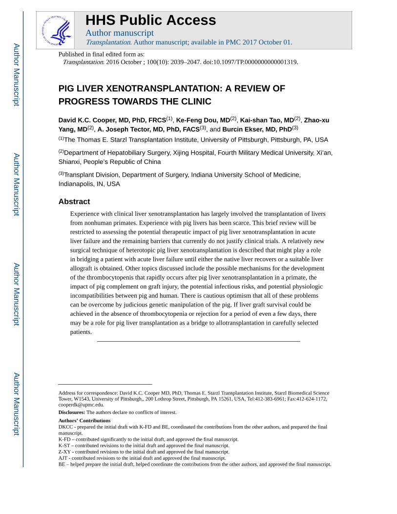
PIG LIVER XENOTRANSPLANTATION: A REVIEW OF PROGRESS TOWARDS THE CLINIC
David K.C. Cooper, MD, PhD, FRCS(1), Ke-Feng Dou, MD(2), Kai-shan Tao, MD(2), Zhao-xu Yang, MD(2), A. Joseph Tector, MD, PhD, FACS(3), and Burcin Ekser, MD, PhD(3)
(1)The Thomas E. Starzl Transplantation Institute, University of Pittsburgh, Pittsburgh, PA, USA
(2)Department of Hepatobiliary Surgery, Xijing Hospital, Fourth Military Medical University, Xi’an, Shianxi, People’s Republic of China
(3)Transplant Division, Department of Surgery, Indiana University School of Medicine, Indianapolis, IN, USA
Abstract
Experience with clinical liver xenotransplantation has largely involved the transplantation of livers
from nonhuman primates. Experience with pig livers has been scarce. This brief review will be
restricted to assessing the potential therapeutic impact of pig liver xenotransplantation in acute
liver failure and the remaining barriers that currently do not justify clinical trials. A relatively new
surgical technique of heterotopic pig liver xenotransplantation is described that might play a role
in bridging a patient with acute liver failure until either the native liver recovers or a suitable liver
allograft is obtained. Other topics discussed include the possible mechanisms for the development
of the thrombocytopenis that rapidly occurs after pig liver xenotransplantation in a primate, the
impact of pig complement on graft injury, the potential infectious risks, and potential physiologic
incompatibilities between pig and human. There is cautious optimism that all of these problems
can be overcome by judicious genetic manipulation of the pig. If liver graft survival could be
achieved in the absence of thrombocytopenia or rejection for a period of even a few days, there
may be a role for pig liver transplantation as a bridge to allotransplantation in carefully selected
patients.
Address for correspondence: David K.C. Cooper MD, PhD, Thomas E. Starzl Transplantation Institute, Starzl Biomedical Science Tower, W1543, University of Pittsburgh,, 200 Lothrop Street, Pittsburgh, PA 15261, USA, Tel:412-383-6961; Fax:412-624-1172, [email protected].
Disclosures: The authors declare no conflicts of interest.
Authors’ ContributionsDKCC - prepared the initial draft with K-FD and BE, coordinated the contributions from the other authors, and prepared the final manuscript.K-FD – contributed significantly to the initial draft, and approved the final manuscript.K-ST – contributed revisions to the initial draft and approved the final manuscript.Z-XY - contributed revisions to the initial draft and approved the final manuscript.AJT - contributed revisions to the initial draft and approved the final manuscript.BE – helped prepare the initial draft, helped coordinate the contributions from the other authors, and approved the final manuscript.
HHS Public AccessAuthor manuscriptTransplantation. Author manuscript; available in PMC 2017 October 01.
Published in final edited form as:Transplantation. 2016 October ; 100(10): 2039–2047. doi:10.1097/TP.0000000000001319.
Author M
anuscriptA
uthor Manuscript
Author M
anuscriptA
uthor Manuscript
Introduction
Liver transplantation offers several advantages for the treatment of patients with acute or
fulminant liver failure or end-stage chronic liver disease, but is limited by the shortage of
deceased human donor organs. In patients with acute liver failure, usually induced by
chemical or viral hepatitis, the onset of disease is sudden and identification of a suitable
donor organ is frequently not possible before permanent neurologic injury and/or death
occurs.
In the USA, data from the United Network for Organ Sharing (UNOS)/Organ Procurement
and Transplantation Network indicate that in 2014 the number of patients on the waiting list
for a human donor liver was close to 16,000. Of these, only 6,729 received a transplant.1 Of
close to 10,000 who did not receive a transplant, 3,178 died or were removed from the
waiting list because they were too sick to undergo a major operative procedure, which is
approximately 20% of those on the waiting list and 30% of those who did not receive a
transplant.
Support during the critical period when the patient is in acute liver failure may be aimed at
(i) ‘bridging’ the patient to liver allotransplantation in order to prevent irreversible cerebral
injury, or (ii) gaining time for regeneration of a damaged native liver, if this is considered
likely.
Potential solutions to the problem include (i) an artificial liver device, (ii) the transplantation
of hepatocytes or (iii) hepatocyte-like expanded human stem cells, (iv) ex vivo pig or
nonhuman primate (NHP) liver perfusion, or (v) the transplantation of a genetically-
engineered pig liver. Regenerative medicine techniques whereby a human or pig liver is
decellularized and recelluarized with cells from the potential recipient would not be
applicable to patients with acute liver failure. This brief review will be restricted to assessing
the potential therapeutic impact of pig liver xenotransplantation in acute liver failure and the
remaining barriers that currently do not yet justify clinical trials.
Clinical experience with ex vivo pig liver perfusion
Early experience has been reviewed by Hara et al.2 In the late 1960s and early 1970s, at least
141 ex vivo pig liver perfusions were performed to treat 87 patients with liver failure, but
then this therapeutic option was largely superseded for several years by orthotopic liver
allotransplantation. Neurologic improvement to at least hepatic coma grade III or II has been
documented in most patients. These clinical trials have provided valuable immunologic
information. The data suggested that unmodified (wild-type) pig livers may be rejected less
vigorously than other pig organs, possibly because hepatic failure is accompanied by
diminished complement levels, although there may be additional reasons.
In a small clinical trial by Levy et al3 livers from pigs transgenic for the human complement-
regulatory proteins (regulators of complement activation, RCA), CD55 (human decay-
accelarating factor [hDAF]) and CD59, were extracorporeally perfused in 2 patients with
acute hepatic failure for 6.5h and 10h, respectively, as bridging to successful
allotransplantation. The histopathological findings in these cases were similar to those
Cooper et al. Page 2
Transplantation. Author manuscript; available in PMC 2017 October 01.
Author M
anuscriptA
uthor Manuscript
Author M
anuscriptA
uthor Manuscript

described with nontransgenic pig livers. Of interest, the authors made no mention of whether
thrombocytopenia developed.
Clinical experience with pig liver xenotransplantation
Following the development of techniques of vascular anastomosis at the beginning of the
20th century, organ xenotransplantation became possible. Most of the early attempts at
clinical organ xenotransplantation used NHP species as sources of the organ (reviewed in 2),
although there were a few attempts using the pig and other nonprimate mammals, but
without significant success.4–6
Only 1 of these experiences related to the transplantation of a pig liver. In 1993, Makowka
and his colleagues performed the only heterotopic pig liver xenotransplant, with the aim of
performing orthotopic allotransplantation when a human donor became available, at which
time the auxiliary pig liver would have been removed.7,8 The patient was a 26-year-old
woman with a 14-year history of autoimmune hepatitis who was admitted to hospital with
grade III encephalopathy. Hepatitis C was detected serologically. She was listed with the
UNOS at the highest priority. Despite aggressive medical therapy, the patient continued to
deteriorate, with increasing encephalopathy and coagulopathy.
A wild-type (genetically-unmodified) pig liver was transplanted heterotopically. Before
transplantation, circulating natural anti-pig antibodies were removed by plasmapheresis and
ex vivo perfusion of the donor pig kidneys. After transplantation, the liver xenograft clearly
functioned, as documented by bile production, stabilization of prothrombin levels, and
reduction in the levels of lactic acid and the enzymes aspartate aminotransferase and alanine
aminotransferase. Unfortunately, this did not result in any improvement in the neurologic
status of the patient, who died after 34 hours from irreversible brain damage.
Despite the removal of >90% of the recipient’s natural xenoantibodies prior to
transplantation, antibody rapidly returned and was associated with complement-mediated
injury of the graft. A liver biopsy obtained 3 hours posttransplantation showed deposition of
antibody and complement components, and endothelial swelling, suggesting early graft
rejection. At the time of death, the pig liver showed thrombosis and ischemic necrosis.
Importantly, however, no mention was made as to whether thrombocytopenia was
documented, as seen in pig-to-NHP models (see below). Nevertheless, this experience
demonstrated the ability of a pig liver to function, at least temporarily, in a human recipient
and to provide some metabolic support during acute liver failure.
Experimental pig liver xenotransplantation in nonhuman primates
There have been several reports of liver xenotransplantation between species of NHPs
[reviewed in 2–42], but, for a number of reasons,9 it is very unlikely that NHPs will be
acceptable as sources of livers for clinical transplantation in the future. The pig-to-NHP
model is increasingly being investigated to assess strategies aimed at advancing towards
clinical xenotransplantation (Figure 1).
In untreated NHPs, wild-type pig livers have generally undergone early antibody-mediated
rejection (intravascular thrombosis, hemorrhagic necrosis, endothelial cell injury, deposition
Cooper et al. Page 3
Transplantation. Author manuscript; available in PMC 2017 October 01.
Author M
anuscriptA
uthor Manuscript
Author M
anuscriptA
uthor Manuscript

of IgM, IgG, C3), though no intravascular fibrin aggregation is seen (Figure 2A). For
example, Ramirez et al reported 3 baboons that survived for <12 hours and showed features
of hyperacute rejection (Table 1).10,11 Subsequently, Ekser et al provided sequential data on
the development of hyperacute rejection in this model.12
When genetically-engineered pigs have been the source of the liver, the results have
significantly improved. In the same series of experiments by Ramirez et al10,11 2 baboons
transplanted with livers from pigs transgenic for the human RCA, CD55 (human decay-
accelerating factor, hDAF) survived for 4 and 8 days, respectively (Table 1). Neither liver
xenograft demonstrated histopathologic features of hyperacute rejection. This study
indicated that, when hyperacute rejection is abrogated by the expression of hCD55, the
porcine liver can maintain reasonable levels of coagulation factors and protein in the baboon
for up to 8 days, although factors II, VII, and X decreased significantly. The authors
confirmed studies in rodents that established that the recipient progressively acquires the
protein profile of the donor species.13–15 In a subsequent brief report by this group, livers
from pigs transgenic for 2 hRCAs (CD55, CD59) and H-transferase survived only 13–24
hours in baboons.16
Ekser et al reported their experience of orthotopic pig liver xenotransplantation in baboons
with grafts from α1,3-galactosyltransferase gene-knockout (GTKO) pigs or GTKO pigs
transgenic for the human RCA, CD46 (GTKO/hCD46) and a clinically applicable
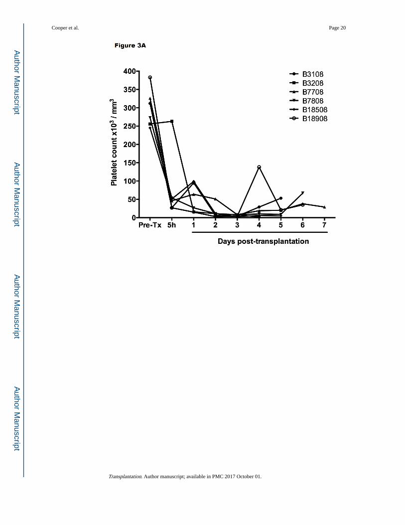
immunosuppressive regimen (Table 1).12,17 Profound thrombocytopenia developed within
one hour posttransplantation (Figure 3A), which resulted in spontaneous hemorrhages in
various native organs and in the liver graft, limiting recipient survival to a maximum of 7
days. However, throughout much of this time, hepatic function was documented near-normal
to normal (as assessed by liver enzymes, coagulation factors, coagulation assays, and
production of porcine-specific proteins).18 There was no definitive humoral and cellular
rejection (as documented on biopsies taken at 2 hours or at the time of euthanasia or death
(at 4–7 days posttransplant) (Figure 2B).
Kim et al reported GTKO Massachusetts General Hospital (MGH) miniature swine
orthotopic liver xenotransplantation in baboons with an intensive immunosuppressive
regimen (Table 1).19 Their initial 3 cases survived 6, 8, and 9 days, respectively. The 6-day
survivor died of spontaneous hemorrhage associated with profound thrombocytopenia, as
observed by Ekser et al.12 In the 8-and 9-day survivors, loss of platelets was ameliorated to
some extent by blocking fibrinolysis by the administration of aminocaproic acid. Although
thrombocytopenia was marginal (platelet counts remained between 40–50,000/mm3), the
baboons suffered severe blood loss and sepsis.
The mechanisms by which platelet aggregation and phagocytosis occurred were investigated
in vitro.20 Pig hepatocytes, liver sinusoidal endothelial cells, and aortic endothelial cells all
induced moderate aggregation of baboon platelets (but not of pig platelets), and sinusoidal
endothelial cells were shown to phagocytose baboon platelets (as demonstrated previously
with human platelets in an ex vivo perfusion model by Tector and his colleagues.21–24
Phagocytosis could be significantly reduced by certain agents, e.g., aurintricarboxylic acid
(which blocks von Willebrand Factor [vWF]).
Cooper et al. Page 4
Transplantation. Author manuscript; available in PMC 2017 October 01.
Author M
anuscriptA
uthor Manuscript
Author M
anuscriptA
uthor Manuscript
Yeh et al went on to assess the effect of maintaining the recipient liver in situ and
transplanting the pig liver heterotopically.25 Although thrombocytopenia developed as
before, the presence of baboon coagulation factors prevented severe spontaneous
hemorrhage from occurring. However, features of thrombotic microangiopathy and ischemia
developed in the graft (as seen in pig hearts and kidneys), resulting in graft failure in 2 cases.
Survival of the 3 grafts or recipients was limited to 6, 9, and 15 days, respectively, with
sepsis being a major cause of death.
Recently, Navarro-Alvarez et al reported 7 further cases (6 new, 1 historical) of orthotopic
GTKO pig-to-baboon liver xenotransplantation,26 in which they sought to determine the
effects of the administration of human coagulation factors (Table 1). Graft and recipient
survival was 1 and 3 days (with bolus administration) and 5–7 days (with continuous
administration), which was not different from survival of a historical control baboon (6
days). Platelet counts were maintained, but the baboons quickly developed large vessel
thrombosis and thrombotic microangiopathy. Several deaths were from infection.
The most recent report 27 documented 25-day survival of a GTKO pig liver graft in a baboon
treated with a human prothrombinase concentrate complex. Immunosuppressive therapy
included induction with antithymocyte globulin and cobra venom factor, with maintenance
with belatacept, tacrolimus, and methylprednisolone. Abnormalities of liver function tests on
day 7 were presumed to be associated with rejection, but were reversed by a course of
steroid pulses. Early thrombocytopenia began to recover by day 11 and was maximal on day
21 (614,000/mm3) without the need for platelet transfusions. Euthanasia was necessary on
day 25 from the development of plantar ulcerations (associated with peripheral edema),
progressive cholestasis, hemolysis, and a rising direct bilirubin. The liver showed no
macroscopic features of necrosis with all vessels free of thrombus. This report provides
encouragement that a pig liver graft might maintain life in a patient with fulminant hepatic
failure until either recovery of the native liver occurs (if the pig liver had been transplanted
heterotopically) or until an allograft becomes available.
Another recent experience has been gained by Dou’s group from Xi’an, China, who inserted
a left liver lobe from GTKO Wu Zhishan miniature swine as auxiliary grafts into Tibetan
monkeys (Table 1).28 Their innovative surgical technique is illustrated in Figure 4. Although
it requires native splenectomy, the Xi’an technique has the advantage that none of the native
liver needs to be excised as the graft fits comfortably into the splenic fossa. (Ekser et al17,
Navarro-Alvarez et al26, Yeh et al25, and Shah et al27 all performed splenectomy in order to
reduce thrombocytopenia. However, whether splenectomy is immunologically beneficial to
the outcome is uncertain, but there was no definitive evidence for this in the early studies of
pig heart or kidney xenotransplantation [reviewed in 29.)
Of interest, the surgical technique introduced by the Xi’an group was first used successfully
in clinical liver allotransplantation in 2007 in a patient with Wilson’s disease, splenomegaly,
and hypersplenism, and has since been employed in a further 14 patients, using both living
donors (excising segments 2 and 3) and split livers from deceased (donation after cardiac
death) donors (Dou K-F, et al, unpublished). Following the work of Olausson et al,30 in 1
patient with >90% panel-reactive antibodies (PRA) in need of kidney transplantation, the
Cooper et al. Page 5
Transplantation. Author manuscript; available in PMC 2017 October 01.
Author M
anuscriptA
uthor Manuscript
Author M
anuscriptA
uthor Manuscript

donor liver graft was inserted heterotopically before the donor kidney with the intention of
preventing antibody-mediated hyperacute rejection of the kidney, which it did successfully.
In their xenotransplantation studies, graft survival, as defined by various parameters of
hepatic function, extended between 2 and almost 14 days, although the presence of the
native recipient liver would have been beneficial. In none of the 3 monkeys was the fall in
platelet count during the first 24 hours as dramatic or as profound (Figure 3B) as in the
experience of others (Figure 3A).17,19,25 In the longest-surviving monkey, although the
platelet count remained at approximately 50,000/mm3 by the end of the experiment (day 14)
(Figure 3C), whether this difference can be accounted for by the fact that the recipient was a
Tibetan monkey rather than a baboon, or that the organ-source pig was a Wu Zhishan
miniature swine remains uncertain, but needs to be explored.
The study provided further valuable data on the coagulation disorders that develop following
pig liver transplantation in NHPs. The authors demonstrated that early activation of recipient
tissue factor is largely responsible for coagulation dysregulation (supporting the studies by
Lin et al,31,32 in part related to incompatibility between pig tissue factor pathway inhibitor
(TFPI) and primate tissue factor, which results in the inefficient inhibition of recipient tissue
factor.33 Accompanying in vitro studies indicated that the overexpression of human TFPI by
pig bone marrow-derived mesenchymal stromal cells inhibited clotting, which is an
observation that may be of potential therapeutic importance and warrants further
investigation.34,35
From the above combined studies, we can tentatively conclude that early after
transplantation of a liver from a genetically-engineered pig, microscopic and
immunohistochemical examination of a liver biopsy has been almost normal, with minimal
(patchy) or no deposition of IgM, IgG, C3, C4d, and C5b-9. At necropsy some days later,
some livers showed macroscopic changes consistent with cholestasis, except for some
patchy dark areas which microscopically showed hemorrhagic necrosis, platelet-fibrin
thrombi, monocyte/macrophage margination, and vascular endothelial cell hypertrophy
(Figure 2C).12,28,36–38 No cell infiltration was seen. Confocal microscopy has confirmed
tissue factor expression on platelets, and platelet and platelet/leukocyte aggregates in liver
sinusoids. The minimal or absent deposition of immunoglobulin or complement fractions
and absence of a cellular infiltrate suggest that neither antibody- nor cell-mediated rejection
plays a major role in injury to genetically-engineered pig liver grafts. Platelet loss and
platelet/leukocyte aggregation would appear to be a major problem.21,39 However, if the
native liver remains in situ, the production of primate coagulation factors leads to the
development of thrombotic microangiopathy in the graft, resulting in ischemic injury and
graft destruction (just as in pig heart and kidney xenografts).
Considerable progress in pig organ xenotransplantation into NHPs has been made in the past
few years, largely through the availability of pigs with an increasing number of genetic
manipulations.40 However, these advances have largely been confined to heart and kidney
xenotransplantation,41–48 as the barriers associated with pig lung 41 and liver 2,42,43
xenotransplantation are rather more complex.
Cooper et al. Page 6
Transplantation. Author manuscript; available in PMC 2017 October 01.
Author M
anuscriptA
uthor Manuscript
Author M
anuscriptA
uthor Manuscript
Investigation of the mechanisms of platelet loss
The studies summarized above have identified certain barriers specific to the liver that need
to be overcome. The major outstanding problem preventing successful long-term pig liver
xenotransplantation is the rapid loss of platelets from the recipient within minutes or hours
of the transplant. Evidence has been presented to indicate that (i) primate platelets are
phagocytosed by pig liver macrophages (Kupffer cells) and sinusoidal endothelial
cells,20–23,44 or (ii) may be lost in platelet-leukocyte aggregates in the graft and in certain
recipient organs.20,39
Potential factors that may contribute towards the phagocytosis include (i) the activity of the
asialoglycoprotein receptor-1 (ASGR1, present on sinusoidal endothelial cells) (Figure 5)
and the macrophage antigen complex-1 (CD11b/CD18; Mac-1, a surface integrin receptor
on Kupffer cells),22,44 (ii) interspecies incompatibility and phagocytic dysregulation in the
porcine signal regulatory protein-alpha (SIRPα)/human CD47 pathway (Figure 6),41,45–47
and (iii) upregulation of tissue factor expression on activated donor liver sinusoidal
endothelial cells activated by the immune response, as well as on recipient platelets and
mononuclear cells. Furthermore, ASGR1 is expressed on aortic and systemic arterial
vascular endothelium in pigs and this may play a role in binding and phagocytosis of human
platelets.48 There may therefore be multiple factors involved, but specific genetic
engineering of the organ source pig may negate this problem.
Although pigs naturally deficient in vWF have been available for some time,49–53 they are
extremely difficult to work with in surgical research and, furthermore, do not resolve the
problems faced in xenotransplantation. As pig vWF activates recipient primate platelets,54,55
an alternative approach might be to generate pigs in which pig vWF has been knocked out
and replaced with human vWF,41 but such pigs are not yet available.
In addition to the reported loss of platelets in NHPs, the ex vivo perfusion of a pig liver with
human blood demonstrated that pig macrophages continuously phagocytosed human
erythrocytes;56,57 this was unrelated to antibody binding and complement activation, but was
associated with direct recognition of erythrocytes by pig Kupffer cells.58,59 A pig liver
removed approximately 1 unit of human erythrocytes from the circulation every 24 hours.
Phagocytosis of recipient erythrocytes has not been documented in most studies in NHPs,
suggesting that the expression of N-glycolylneuraminic acid (NeuGc) on pig erythrocytes
and the production of anti-NeuGc antibodies in humans (but not in NHPs which, like pigs,
express NeuGc) may be playing a role 60 (reviewed in 61). Pigs in which the gene for the
enzyme that adds NeuGc to underlying carbohydrate chains has been deleted have recently
become available,62 and Butler et al63 showed that a GTKO/NeuGc-knockout pig liver
significantly reduced xenogeneic consumption of human platelets in an ex vivo perfusion
study.
The impact of pig complement on the liver graft
Despite these negative aspects of pig liver xenotransplantation, there may be some
advantages in comparison with xenotransplantation of the pig heart or kidney. As the liver is
the major site of synthesis of complement proteins, except for C1q, factor D, properdin, and
Cooper et al. Page 7
Transplantation. Author manuscript; available in PMC 2017 October 01.
Author M
anuscriptA
uthor Manuscript
Author M
anuscriptA
uthor Manuscript
C7, impaired hepatic synthetic function contributes to complement deficiency in patients
with hepatic disease.64–69 The level of membrane attack complex activity may be much
lower than that seen in healthy subjects, reducing the possibility of early graft injury.
Furthermore, after pig liver transplantation in humans (or NHPs), the graft will produce pig
complement. In vitro studies have indicated that (i) in the presence of human antibodies, pig complement is associated with less lysis of pig cells than human complement (and thus may
be associated with less graft injury), (ii) lysis by pig complement of GTKO pig cells is
significantly less than of wild-type pig cells, and (iii) the expression of a human RCA on the
pig cell can reduce pig complement-induced lysis of the cells.43 Rees and his colleagues also
provided evidence suggesting that human RCAs will successfully inhibit porcine
complement.59 In ex vivo liver perfusion with human blood, survival of hCD55 (hDAF)
transgenic pig livers for up to 72 hours indicated that a pig liver expressing both pig and
human RCAs may provide some protection from injury by pig complement.56,57 Although
human complement components will continue to be produced by various recipient cells, the
transgenic expression of a hRCA in the graft should be protective.
Independent of the low complement levels seen in patients with hepatic failure, numerous
studies have shown that the liver is more resistant than other solid organs to injury from
preformed graft antibodies.70–73 In some studies, human serum caused significantly less
cytotoxicity when incubated with porcine hepatic sinusoidal endothelial cells than with
porcine aortic endothelial cells (25% vs 72%).69 This relative resistance to injury could
possibly be related to the liver’s ability to clear soluble immune complexes,69 or to less
binding of human IgM and IgG to porcine hepatic sinusoidal endothelial cells when
compared to aortic endothelial cells (on flow cytometry), suggesting lower expression of
antigens.74
The liver complement levels in patients with liver failure and the possible resistance of the
sinusoidal endothelium to injury combine to contribute to protection of the liver from
antibody-mediated rejection. Serum from patients with hepatic failure caused significantly
less lysis of both porcine aortic (38% versus 72%) and hepatic sinusoidal (9% versus 25%)
endothelial cells.69
The potential of sensitization to pig antigens
If a pig liver is employed to bridge the patient to allotransplantation, will exposure to pig
tissues result in the production of anti-pig antibodies that might cross-react with
alloantigens, thus preventing or being detrimental to allotransplantation? Studies of cross-
reactivity between anti-species antibodies and/or sensitized T cells are few (reviewed in 75),
but there has been little evidence for the development of cross-reactive cytotoxic antibodies
or activated T cells that might mediate humoral or accelerated cellular rejection of a
subsequent allograft.76–78
In this respect, the 2 patients who underwent successful liver allotransplantation following
relatively transient bridging by ex vivo hemoperfusion of porcine livers (see above) did not
develop anti-HLA antibodies.3 Bridging with a bioartificial liver, which incorporated porcine
hepatocytes, has also been followed by successful liver allotransplantation.79 A liver
Cooper et al. Page 8
Transplantation. Author manuscript; available in PMC 2017 October 01.
Author M
anuscriptA
uthor Manuscript
Author M
anuscriptA
uthor Manuscript

allograft transplanted after a bridging pig xenograft would therefore not appear to be at
increased risk of either humoral or cellular rejection.
Potential infectious risks
The risks of the transfer of microorganisms with the pig organ to the human recipient and,
more importantly, of the transfer of a porcine infectious agent to the community are now
considered to be small.80–84 Indeed, the use of animal organs might have some advantages
with regard to recurrence of disease, such as viral hepatitis, as some viruses are species-
specific. For example, following pig-to-baboon organ transplantation, porcine
cytomegalovirus has not been documented to infect the host baboon, and baboon
cytomegalovirus has not been seen to infect the transplanted pig organ.85,86 Nevertheless,
porcine endogenous retroviruses (PERVs) are present in the genome of every pig cell, and
would therefore be transplanted with the organ. Although the risk of PERVs causing any
disease process in the recipient or in the community is considered low,83,84,87 using the new
CRISPR/Cas9 technology, it is now possible to render pig cells PERV-negative 88 and so it is
likely that PERV-negative pigs will become available in the future, if this is believed to be
essential.
Most aspects of physiologic compatibility between pig and human have been sparsely
investigated or not investigated at all, in part because it is difficult to accurately assess true
hepatic function in the presence of an ongoing immune response.89–92 Nevertheless,
significant incompatibilities between pig and human hepatic function are almost certain to
be present, though some of these may be relative rather than absolute hurdles. If a function,
e.g., the synthesis of a key protein, is found to be essential, and that function cannot be
achieved by the pig liver, genetic modification of the source pig can be carried out to
produce its human protein counterpart.
Conclusions
Attempts at liver xenotransplantation can best be justified if the patient will die rapidly
without transplantation. Although prolonged function (i.e., weeks or months) of a
transplanted pig liver in a primate cannot yet be guaranteed, in rapidly-deteriorating patients
with acute liver failure, liver xenotransplantation (particularly if a GTKO/hRCA pig liver is
implanted), could probably be considered as a short-term (days) bridge to allotransplantation
as long as thrombocytopenia and coagulopathy can be prevented.93,94 Auxiliary liver
xenotransplantation might allow recovery of the native organ. Orthotopic pig liver
xenotransplantation would be preferable if the state of the native liver was increasing the risk
of mortality to the patient, or if no recovery of the native liver is anticipated. 95
Acknowledgments
Funding
Work on xenotransplantation in the Thomas E. Starzl Transplantation Institute at the University of Pittsburgh has been supported in part by National Institutes of Health grants U01 AI068642, R21 AI074844, and U19 AI090959, and by sponsored research agreements between the University of Pittsburgh and Revivicor, Inc., Blacksburg, VA. Work on xenotransplantation in Xijing Hospital and the Fourth Military Medical University has been supported by the National Basic Research Program of China (973 Program; 2015CB554100), the National Natural Science
Cooper et al. Page 9
Transplantation. Author manuscript; available in PMC 2017 October 01.
Author M
anuscriptA
uthor Manuscript
Author M
anuscriptA
uthor Manuscript

Foundation (81270549 and 81300361), the Xijing Hospital disciplines boosting projects (XJZT12M09 and XJZT13Z01), the Science and Technology Research and Development Program of Shaanxi province (2013K12-18-02), and the National High Technology Research and Development Program (863 Program; 2012AA021005).
Abbreviations
GTKO α1,3-galactosyltransferase gene-knockout
NeuGc N-glycolylneuraminic acid
NHP nonhuman primate
PERV porcine endogenous retrovirus
RCA regulator of complement
TFPI tissue factor pathway inhibitor
UNOS United Network for Organ Sharing
vWF von Willebrand Factor
References
1. UNOS. [Accessed October 20, 2015] United Network for Organ Sharing. Http://www.unos.org. Updated 2016
2. Hara H, Gridelli B, Lin YJ, Marcos A, Cooper DK. Liver xenografts for the treatment of acute liver failure: clinical and experimental experience and remaining immunologic barriers. Liver Transpl. 2008; 14:425–434. [PubMed: 18383106]
3. Levy MF, Crippin J, Sutton S, et al. Liver allotransplantation after extracorporeal hepatic support with transgenic (hCD55/hCD59) porcine livers: clinical results and lack of pig-to-human transmission of the porcine endogenous retrovirus. Transplantation. 2000; 69:272–280. [PubMed: 10670638]
4. Cooper DK. A brief history of cross-species organ transplantation. Proc (Bayl Univ Med Cent). 2012; 25:49–57. [PubMed: 22275786]
5. Deschamps JY, Roux FA, Sai P, Gouin E. History of xenotransplantation. Xenotransplantation. 2005; 12:91–109. [PubMed: 15693840]
6. Taniguchi S, Cooper DK. Clinical xenotransplantation: past, present and future. Ann R Coll Surg Engl. 1997; 79:13–19. [PubMed: 9038490]
7. Makowa L, Cramer DV, Hoffman A, et al. The use of a pig liver xenograft for temporary support of a patient with fulminant hepatic failure. Transplantation. 1995; 59:1654–1659. [PubMed: 7604434]
8. Makowka L, Wu GD, Hoffman A, et al. Immunohistopathologic lesions associated with the rejection of a pig-to-human liver xenograft. Transplant Proc. 1994; 26:1074–1075. [PubMed: 8029835]
9. Cooper DK, Bottino R. Recent advances in understanding xenotransplantation: implications for the clinic. Expert Rev Clin Immunol. 2015; 11:1379–1390. [PubMed: 26548357]
10. Ramirez P, Chavez R, Majado M, et al. Life-supporting human complement regulator decay accelerating factor transgenic pig liver xenograft maintains the metabolic function and coagulation in the nonhuman primate for up to 8 days. Transplantation. 2000; 70:989–998. [PubMed: 11045632]
11. Ramirez P, Yelamos J, Parrilla P, Chavez R. Hepatic xenotransplantation will benefit from strategies aimed to reduce complement activation. Liver Transpl. 2001; 7:562–563. [PubMed: 11443590]
Cooper et al. Page 10
Transplantation. Author manuscript; available in PMC 2017 October 01.
Author M
anuscriptA
uthor Manuscript
Author M
anuscriptA
uthor Manuscript
12. Ekser B, Klein E, He J, et al. Genetically-engineered pig-to-baboon liver xenotransplantation: histopathology of xenografts and native organs. PLoS One. 2012; 7:e29720. [PubMed: 22247784]
13. Valdivia LA, Fung JJ, Demetris AJ, et al. Donor species complement after liver xenotransplantation. The mechanism of protection from hyperacute rejection. Transplantation. 1994; 57:918–922. [PubMed: 8154040]
14. Celli S, Valdivia LA, Fung JJ, Kelly RH. Early recipient-donor switch of the complement type after liver xenotransplantation. Immunol Invest. 1997; 26:589–600. [PubMed: 9399102]
15. Valdivia LA, Lewis JH, Celli S, et al. Hamster coagulation and serum proteins in rat recipients of hamster xenografts. Transplantation. 1993; 56:489–490. [PubMed: 8356608]
16. Ramirez P, Montoya MJ, Rios A, et al. Prevention of hyperacute rejection in a model of orthotopic liver xenotransplantation from pig to baboon using polytransgenic pig livers (CD55, CD59, and H-transferase). Transplant Proc. 2005; 37:4103–4106. [PubMed: 16386637]
17. Ekser B, Long C, Echeverri GJ, et al. Impact of thrombocytopenia on survival of baboons with genetically modified pig liver transplants: clinical relevance. Am J Transplant. 2010; 10:273–285. [PubMed: 20041862]
18. Ekser B, Echeverri GJ, Hassett AC, et al. Hepatic function after genetically engineered pig liver transplantation in baboons. Transplantation. 2010; 90:483–493. [PubMed: 20606605]
19. Kim K, Schuetz C, Elias N, et al. Up to 9-day survival and control of thrombocytopenia following alpha1,3-galactosyl transferase knockout swine liver xenotransplantation in baboons. Xenotransplantation. 2012; 19:256–264. [PubMed: 22909139]
20. Peng Q, Yeh H, Wei L, et al. Mechanisms of xenogeneic baboon platelet aggregation and phagocytosis by porcine liver sinusoidal endothelial cells. PLoS One. 2012; 7:e47273. [PubMed: 23118867]
21. Burlak C, Paris LL, Chihara RK, et al. The fate of human platelets perfused through the pig liver: implications for xenotransplantation. Xenotransplantation. 2010; 17:350–361. [PubMed: 20955292]
22. Paris LL, Chihara RK, Reyes LM, et al. ASGR1 expressed by porcine enriched liver sinusoidal endothelial cells mediates human platelet phagocytosis in vitro. Xenotransplantation. 2011; 18:245–251. [PubMed: 21848542]
23. Paris LL, Chihara RK, Sidner RA, Tector AJ, Burlak C. Differences in human and porcine platelet oligosaccharides may influence phagocytosis by liver sinusoidal cells in vitro. Xenotransplantation. 2012; 19:31–39. [PubMed: 22360751]
24. Paris LL, Estrada JL, Li P, et al. Reduced human platelet uptake by pig livers deficient in the asialoglycoprotein receptor 1 protein. Xenotransplantation. 2015; 22:203–210. [PubMed: 25728617]
25. Yeh H, Machaidze Z, Wamala I, et al. Increased transfusion-free survival following auxiliary pig liver xenotransplantation. Xenotransplantation. 2014; 21:454–464. [PubMed: 25130043]
26. Navarro-Alvarez N, Shah JA, Zhu A, et al. The effects of exogenous administration of human coagulation factors following pig-to-baboon liver xenotransplantation. Am J Transplant. [published online ahead of print November 27th, 2015].
27. Shah JA, Navarro-Alvarez N, DeFazio M, et al. A bridge to somewhere: 25-day survival after pig-to-baboon liver xenotransplantation. Ann Surg. [published online ahead of print January 28th, 2016].
28. Ji H, Li X, Yue S, et al. Pig BMSCs transfected with human TFPI combat species incompatibility and regulate the human TF pathway in vitro and in a rodent model. Cell Physiol Biochem. 2015; 36:233–249. [PubMed: 25967963]
29. Lambrigts D, Sachs DH, Cooper DK. Discordant organ xenotransplantation in primates: world experience and current status. Transplantation. 1998; 66:547–561. [PubMed: 9753331]
30. Olausson M, Mjornstedt L, Norden G, et al. Successful combined partial auxiliary liver and kidney transplantation in highly sensitized cross-match positive recipients. Am J Transplant. 2007; 7:130–136. [PubMed: 17227562]
31. Lin CC, Chen D, McVey JH, Cooper DK, Dorling A. Expression of tissue factor and initiation of clotting by human platelets and monocytes after incubation with porcine endothelial cells. Transplantation. 2008; 86:702–709. [PubMed: 18791452]
Cooper et al. Page 11
Transplantation. Author manuscript; available in PMC 2017 October 01.
Author M
anuscriptA
uthor Manuscript
Author M
anuscriptA
uthor Manuscript
32. Lin CC, Cooper DK, Dorling A. Coagulation dysregulation as a barrier to xenotransplantation in the primate. Transpl Immunol. 2009; 21:75–80. [PubMed: 19000927]
33. Cowan PJ, Robson SC, d’Apice AJ. Controlling coagulation dysregulation in xenotransplantation. Curr Opin Organ Transplant. 2011; 16:214–221. [PubMed: 21415824]
34. Ezzelarab M, Ezzelarab C, Wilhite T, et al. Genetically-modified pig mesenchymal stromal cells: xenoantigenicity and effect on human T-cell xenoresponses. Xenotransplantation. 2011; 18:183–195. [PubMed: 21696448]
35. Li J, Ezzelarab MB, Ayares D, Cooper DK. The potential role of genetically-modified pig mesenchymal stromal cells in xenotransplantation. Stem Cell Rev. 2014; 10:79–85. [PubMed: 24142483]
36. Ekser B, Burlak C, Waldman JP, et al. Immunobiology of liver xenotransplantation. Expert Rev Clin Immunol. 2012; 8:621–634. [PubMed: 23078060]
37. Ekser B, Lin CC, Long C, et al. Potential factors influencing the development of thrombocytopenia and consumptive coagulopathy after genetically modified pig liver xenotransplantation. Transpl Int. 2012; 25:882–896. [PubMed: 22642260]
38. Luo Y, Kosanke S, Mieles L, et al. Comparative histopathology of hepatic allografts and xenografts in the nonhuman primate. Xenotransplantation. 1998; 5:197–206. [PubMed: 9741458]
39. Ezzelarab M, Ekser B, Gridelli B, Iwase H, Ayares D, Cooper DK. Thrombocytopenia after pig-to-baboon liver xenotransplantation: where do platelets go? Xenotransplantation. 2011; 18:320–327. [PubMed: 22168139]
40. Cooper DK, Ekser B, Ramsoondar J, Phelps C, Ayares D. The role of genetically-engineered pigs in xenotransplantation research. J Pathol. 2016; 238:288–299. [PubMed: 26365762]
41. Cooper DK, Ekser B, Burlak C, et al. Clinical lung xenotransplantation--what donor genetic modifications may be necessary? Xenotransplantation. 2012; 19:144–158. [PubMed: 22702466]
42. Ekser B, Gridelli B, Veroux M, Cooper DK. Clinical pig liver xenotransplantation: how far do we have to go? Xenotransplantation. 2011; 18:158–167. [PubMed: 21696445]
43. Hara H, Campanile N, Tai HC, et al. An in vitro model of pig liver xenotransplantation--pig complement is associated with reduced lysis of wild-type and genetically modified pig cells. Xenotransplantation. 2010; 17:370–378. [PubMed: 20955293]
44. Chihara RK, Paris LL, Reyes LM, et al. Primary porcine Kupffer cell phagocytosis of human platelets involves the CD18 receptor. Transplantation. 2011; 92:739–744. [PubMed: 21836538]
45. Ide K, Wang H, Tahara H, et al. Role for CD47-SIRPalpha signaling in xenograft rejection by macrophages. Proc Natl Acad Sci U S A. 2007; 104:5062–5066. [PubMed: 17360380]
46. Navarro-Alvarez N, Yang YG. CD47: a new player in phagocytosis and xenograft rejection. Cell Mol Immunol. 2011; 8:285–288. [PubMed: 21258362]
47. Yang YG. CD47 in xenograft rejection and tolerance induction. Xenotransplantation. 2010; 17:267–273. [PubMed: 20723199]
48. Bongoni AK, Kiermeir D, Denoyelle J, et al. Porcine extrahepatic vascular endothelial asialoglycoprotein receptor 1 mediates xenogeneic platelet phagocytosis in vitro and in human-to-pig ex vivo xenoperfusion. Transplantation. 2015; 99:693–701. [PubMed: 25675194]
49. Badimon L, Badimon JJ, Rand J, Turitto VT, Fuster V. Platelet deposition on von Willebrand factor-deficient vessels. Extracorporeal perfusion studies in swine with von Willebrand’s disease using native and heparinized blood. J Lab Clin Med. 1987; 110:634–647. [PubMed: 3312444]
50. Brouland JP, Egan T, Roussi J, et al. In vivo regulation of von Willebrand factor synthesis: von Willebrand factor production in endothelial cells after lung transplantation between normal pigs and von Willebrand factor-deficient pigs. Arterioscler Thromb Vasc Biol. 1999; 19:3055–3062. [PubMed: 10591687]
51. Cantu E, Balsara KR, Li B, et al. Prolonged function of macrophage, von Willebrand factor-deficient porcine pulmonary xenografts. Am J Transplant. 2007; 7:66–75. [PubMed: 17109734]
52. Lau CL, Cantu E 3rd, Gonzalez-Stawinski GV, et al. The role of antibodies and von Willebrand factor in discordant pulmonary xenotransplantation. Am J Transplant. 2003; 3:1065–1075. [PubMed: 12919085]
53. Meyer C, Wolf P, Romain N, et al. Use of von Willebrand diseased kidney as donor in a pig-to-primate model of xenotransplantation. Transplantation. 1999; 67:38–45. [PubMed: 9921793]
Cooper et al. Page 12
Transplantation. Author manuscript; available in PMC 2017 October 01.
Author M
anuscriptA
uthor Manuscript
Author M
anuscriptA
uthor Manuscript

54. Schulte am Esch J 2nd, Cruz MA, Siegel JB, Anrather J, Robson SC. Activation of human platelets by the membrane-expressed A1 domain of von Willebrand factor. Blood. 1997; 90:4425–4437. [PubMed: 9373253]
55. Schulte Am Esch J 2nd, Robson SC, Knoefel WT, Hosch SB, Rogiers X. O-linked glycosylation and functional incompatibility of porcine von Willebrand factor for human platelet GPIb receptors. Xenotransplantation. 2005; 12:30–37. [PubMed: 15598271]
56. Luo Y, Levy G, Ding J, et al. HDAF transgenic pig livers are protected from hyperacute rejection during ex vivo perfusion with human blood. Xenotransplantation. 2002; 9:36–44. [PubMed: 12005103]
57. Rees MA, Butler AJ, Chavez-Cartaya G, et al. Prolonged function of extracorporeal hDAF transgenic pig livers perfused with human blood. Transplantation. 2002; 73:1194–1202. [PubMed: 11981409]
58. Ide K, Ohdan H, Kobayashi T, Hara H, Ishiyama K, Asahara T. Antibody- and complement-independent phagocytotic and cytolytic activities of human macrophages toward porcine cells. Xenotransplantation. 2005; 12:181–188. [PubMed: 15807768]
59. Rees MA, Butler AJ, Negus MC, Davies HF, Friend PJ. Classical pathway complement destruction is not responsible for the loss of human erythrocytes during porcine liver perfusion. Transplantation. 2004; 77:1416–1423. [PubMed: 15167601]
60. Bouhours D, Pourcel C, Bouhours JE. Simultaneous expression by porcine aorta endothelial cells of glycosphingolipids bearing the major epitope for human xenoreactive antibodies (Gal alpha 1–3Gal), blood group H determinant and N-glycolylneuraminic acid. Glycoconj J. 1996; 13:947–953. [PubMed: 8981086]
61. Padler-Karavani V, Varki A. Potential impact of the non-human sialic acid N-glycolylneuraminic acid on transplant rejection risk. Xenotransplantation. 2011; 18:1–5. [PubMed: 21342282]
62. Lutz AJ, Li P, Estrada JL, et al. Double knockout pigs deficient in N-glycolylneuraminic acid and galactose alpha-1,3-galactose reduce the humoral barrier to xenotransplantation. Xenotransplantation. 2013; 20:27–35. [PubMed: 23384142]
63. Butler JR, Paris LL, Blankenship RL, et al. Silencing porcine CMAH and GGTA1 genes significantly reduces xenogeneic consumption of human platelets by porcine livers. Transplantation. 2016; 100:571–576. [PubMed: 26906939]
64. Tector AJ, Fridell JA, Ruiz P, et al. Experimental discordant hepatic xenotransplantation in the recipient with liver failure: implications for clinical bridging trials. J Am Coll Surg. 2000; 191:54–64. [PubMed: 10898184]
65. Colten HR. Biosynthesis of complement. Adv Immunol. 1976; 22:67–118. [PubMed: 769502]
66. Ellison RT 3rd, Horsburgh CR Jr, Curd J. Complement levels in patients with hepatic dysfunction. Dig Dis Sci. 1990; 35:231–235. [PubMed: 2302981]
67. Morgan BP, Gasque P. Extrahepatic complement biosynthesis: where, when and why? Clin Exp Immunol. 1997; 107:1–7. [PubMed: 9010248]
68. Tector AJ, Berho M, Fridell JA, et al. Rejection of pig liver xenografts in patients with liver failure: implications for xenotransplantation. Liver Transpl. 2001; 7:82–89. [PubMed: 11172389]
69. Tector AJ, Elias N, Rosenberg L, et al. Mechanisms of resistance to injury in pig livers perfused with blood from patients in liver failure. Transplant Proc. 1997; 29:966–969. [PubMed: 9123611]
70. Tector AJ, Chen X, Soderland C, Tchervenkov JI. Complement activation in discordant hepatic xenotransplantation. Xenotransplantation. 1998; 5:257–261. [PubMed: 9915253]
71. Platt JL. Xenotransplantation of the liver: is more complement control needed? Liver Transpl. 2001; 7:933–934. [PubMed: 11679996]
72. Nakamura K, Murase N, Becich MJ, et al. Liver allograft rejection in sensitized recipients. Observations in a clinically relevant small animal model. Am J Pathol. 1993; 142:1383–1391. [PubMed: 8494042]
73. Manez R, Kelly RH, Kobayashi M, et al. Immunoglobulin G lymphocytotoxic antibodies in clinical liver transplantation: studies toward further defining their significance. Hepatology. 1995; 21:1345–1352. [PubMed: 7737641]
Cooper et al. Page 13
Transplantation. Author manuscript; available in PMC 2017 October 01.
Author M
anuscriptA
uthor Manuscript
Author M
anuscriptA
uthor Manuscript

74. Cattan P, Zhang B, Braet F, et al. Comparison between aortic and sinusoidal liver endothelial cells as targets of hyperacute xenogeneic rejection in the pig to human combination. Transplantation. 1996; 62:803–810. [PubMed: 8824481]
75. Cooper DK, Tseng YL, Saidman SL. Alloantibody and xenoantibody cross-reactivity in transplantation. Transplantation. 2004; 77:1–5. [PubMed: 14724427]
76. Baertschiger RM, Dor FJ, Prabharasuth D, Kuwaki K, Cooper DK. Absence of humoral and cellular alloreactivity in baboons sensitized to pig antigens. Xenotransplantation. 2004; 11:27–32. [PubMed: 14962290]
77. Key T, Schuurman HJ, Taylor CJ. Does exposure to swine leukocyte antigens after pig-to-nonhuman primate xenotransplantation provoke antibodies that cross-react with human leukocyte antigens? Xenotransplantation. 2004; 11:452–456. [PubMed: 15303982]
78. Ye Y, Luo Y, Kobayashi T, et al. Secondary organ allografting after a primary “bridging” xenotransplant. Transplantation. 1995; 60:19–22. [PubMed: 7624938]
79. Baquerizo A, Mhoyan A, Kearns-Jonker M, et al. Characterization of human xenoreactive antibodies in liver failure patients exposed to pig hepatocytes after bioartificial liver treatment: an ex vivo model of pig to human xenotransplantation. Transplantation. 1999; 67:5–18. [PubMed: 9921790]
80. Paradis K, Langford G, Long Z, et al. Search for cross-species transmission of porcine endogenous retrovirus in patients treated with living pig tissue. The XEN 111 Study Group. Science. 1999; 285:1236–1241. [PubMed: 10455044]
81. Onions D, Cooper DK, Alexander TJ, et al. An approach to the control of disease transmission in pig-to-human xenotransplantation. Xenotransplantation. 2000; 7:143–155. [PubMed: 10961299]
82. Fishman JA. Screening of source animals and clinical monitoring for xenotransplantation. Xenotransplantation. 2007; 14:349–352.
83. Wynyard S, Nathu D, Garkavenko O, Denner J, Elliott R. Microbiological safety of the first clinical pig islet xenotransplantation trial in New Zealand. Xenotransplantation. 2014; 21:309–323. [PubMed: 24801820]
84. Denner J, Mueller NJ. Preventing transfer of infectious agents. Int J Surg. 2015; 23:306–311. [PubMed: 26316157]
85. Mueller NJ, Kuwaki K, Dor FJ, et al. Reduction of consumptive coagulopathy using porcine cytomegalovirus-free cardiac porcine grafts in pig-to-primate xenotransplantation. Transplantation. 2004; 78:1449–1453. [PubMed: 15599308]
86. Mueller NJ, Livingston C, Knosalla C, et al. Activation of porcine cytomegalovirus, but not porcine lymphotropic herpesvirus, in pig-to-baboon xenotransplantation. J Infect Dis. 2004; 189:1628–1633. [PubMed: 15116299]
87. Denner J, Tonjes RR. Infection barriers to successful xenotransplantation focusing on porcine endogenous retroviruses. Clin Microbiol Rev. 2012; 25:318–343. [PubMed: 22491774]
88. Yang L, Guell M, Niu D, et al. Genome-wide inactivation of porcine endogenous retroviruses (PERVs). Science. 2015; 350:1101–1104. [PubMed: 26456528]
89. Hammer, C. Evolutionary obstacles to xenotransplantation. In: Cooper, DKCKE.; Platt, JL.; White, DJG., editors. Xenotransplantation. 2. Heidelberg: Springer; 1997. p. 716-735.
90. Hammer C. Physiological obstacles after xenotransplantation. Ann N Y Acad Sci. 1998; 862:19–27. [PubMed: 9928202]
91. Ibrahim Z, Busch J, Awwad M, Wagner R, Wells K, Cooper DK. Selected physiologic compatibilities and incompatibilities between human and porcine organ systems. Xenotransplantation. 2006; 13:488–499. [PubMed: 17059572]
92. Kanazawa A, Platt JL. Prospects for xenotransplantation of the liver. Semin Liver Dis. 2000; 20:511–522. [PubMed: 11200419]
93. Ekser B, Gridelli B, Tector AJ, Cooper DK. Pig liver xenotransplantation as a bridge to allotransplantation: which patients might benefit? Transplantation. 2009; 88:1041–1049. [PubMed: 19898198]
94. Horton PJ, Chaudhury P, Rochon C, Metrakos P, Tchervenkov J. Should trials of liver xenotransplantation proceed in toxic fulminant hepatic failure? Xenotransplantation. 2006; 13:483. [PubMed: 17059571]
Cooper et al. Page 14
Transplantation. Author manuscript; available in PMC 2017 October 01.
Author M
anuscriptA
uthor Manuscript
Author M
anuscriptA
uthor Manuscript
95. Tector J. New Hope for Liver Xenotransplantation. Ann Surg. 2016
96. Calne RY. Organ transplantation between widely disparate species. Transplant Proc. 1970; 2:550–556. [PubMed: 5000236]
97. Calne RY, Davis DR, Pena JR, et al. Hepatic Allografts and xenografts in primates. Lancet. 1970; 7638:103–106. [PubMed: 4188721]
98. Calne RY, White HJ, Herbertson BM, et al. Pig to baboon liver xenografts. Lancet. 1968; 7553:1176–1178. [PubMed: 4172293]
99. Powelson J, Cosimi AB, Austen W Jr, et al. Porcine-to-primate orthotopic liver transplantation. Transplant Proc. 1994; 26:1353–1354. [PubMed: 8029937]
100. Mieles L, Ye Y, Luo Y, et al. Auxiliary liver allografting and xenografting in the nonhuman primate. Transplantation. 1995; 59:1670–1676. [PubMed: 7604437]
Cooper et al. Page 15
Transplantation. Author manuscript; available in PMC 2017 October 01.
Author M
anuscriptA
uthor Manuscript
Author M
anuscriptA
uthor Manuscript

Figure 1. Time-line in experimental pig liver xenotransplantation in NHPs.
Abbreviations: a-CD154 = anti-CD154 monoclonal antibodies; ALG = antilymphocyte
globulin; ATG = antithymocyte globulin; Aza = azathioprine; Bela = belatacept; BM = bone
marrow; Cs = corticosteroids; CsA = cyclosporine; CVF = cobra venom factor; CyP =
cyclophosphamide; FK = tacrolimus; Gal abs = extracorporeal anti-Gal antibody adsorption;
GTKO = 1,3-galactosyltransferase gene-knockout; hCD46 = expression of the human
regulator of complement, hCD46; hCD55 = expression of the human regulator of
complement, CD55; HLT = heterotopic liver transplantation; HT = H-transferase; LoCD2b =
anti-CD2 monoclonal antibody; MMF = mycophenolate mofetil; OLT = orthotopic liver
transplantation; WBI = whole body irradiation; WT = wild-type.
Cooper et al. Page 16
Transplantation. Author manuscript; available in PMC 2017 October 01.
Author M
anuscriptA
uthor Manuscript
Author M
anuscriptA
uthor Manuscript

Cooper et al. Page 17
Transplantation. Author manuscript; available in PMC 2017 October 01.
Author M
anuscriptA
uthor Manuscript
Author M
anuscriptA
uthor Manuscript

Figure 2.
Cooper et al. Page 18
Transplantation. Author manuscript; available in PMC 2017 October 01.
Author M
anuscriptA
uthor Manuscript
Author M
anuscriptA
uthor Manuscript

Histopathology of (A) hyperacute rejection (<24 hours) in a wild-type pig liver transplanted
orthotopically into a baboon, (B) a GTKO/hCD46 orthotopic pig liver graft in a baboon that
survived for 6 days, and (C) a pig left liver lobe graft in a Tibetan monkey that survived for
14 days
(A) WT pig-to-baboon liver xenotransplantation at 1 h (x200). Severe hepatocellular
vacuolar change, focal hepatocyte necrosis, and few thrombi.
(B) Vacuolar hepatocellular cytoplasmic change with minimal hepatocellular necrosis on
postoperative day 6 (x200).
(C) The graft shows some lymphocyte infiltration in the portal area, but no major features of
antibody-mediated or cellular rejection (x100).
Cooper et al. Page 19
Transplantation. Author manuscript; available in PMC 2017 October 01.
Author M
anuscriptA
uthor Manuscript
Author M
anuscriptA
uthor Manuscript
Cooper et al. Page 20
Transplantation. Author manuscript; available in PMC 2017 October 01.
Author M
anuscriptA
uthor Manuscript
Author M
anuscriptA
uthor Manuscript

Figure 3. Platelet counts (A) after GTKO/hCD46 orthotopic pig liver transplantation in baboons (n=6)
that survived from 4–7 days, and (B) after a GTKO Wu Zhishan miniature swine heterotopic
left liver lobe transplant in Tibetan monkeys (n=3, mean ±SD) within the first 48 hours, and
(C) in the Tibetan monkey that survived for 14 days. (A is reproduced with permission from
Ekser et al. Transplantation 2010;90:483–493; B is reproduced with permission from Ji H, et
al.28
Cooper et al. Page 21
Transplantation. Author manuscript; available in PMC 2017 October 01.
Author M
anuscriptA
uthor Manuscript
Author M
anuscriptA
uthor Manuscript

Figure 4. Surgical technique of pig left liver lobe transplantation in Tibetan monkeys. After native
splenectomy, the pig liver graft was placed in the splenic recess. The recipient’s left renal
vein was divided, the distal end being anastomosed to the graft portal vein, and the proximal
end to the graft hepatic vein. The graft hepatic artery was anastomosed end-to-end to the
recipient splenic artery (using a microvascular technique with an operating microscope).
After reperfusion, the bile duct was drained through the abdominal wall to allow
measurement of bile drainage. (Using the same technique in clinical cases of
allotransplantation, the bile duct is drained into a Roux-en-Y jejunal loop.)
Cooper et al. Page 22
Transplantation. Author manuscript; available in PMC 2017 October 01.
Author M
anuscriptA
uthor Manuscript
Author M
anuscriptA
uthor Manuscript
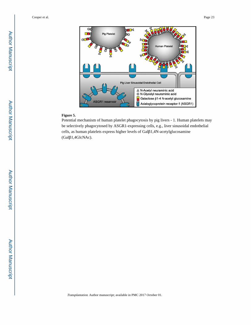
Figure 5. Potential mechanism of human platelet phagocytosis by pig livers - 1. Human platelets may
be selectively phagocytosed by ASGR1-expressing cells, e.g., liver sinusoidal endothelial
cells, as human platelets express higher levels of Galβ1,4N-acetylglucosamine
(Galβ1,4GlcNAc).
Cooper et al. Page 23
Transplantation. Author manuscript; available in PMC 2017 October 01.
Author M
anuscriptA
uthor Manuscript
Author M
anuscriptA
uthor Manuscript
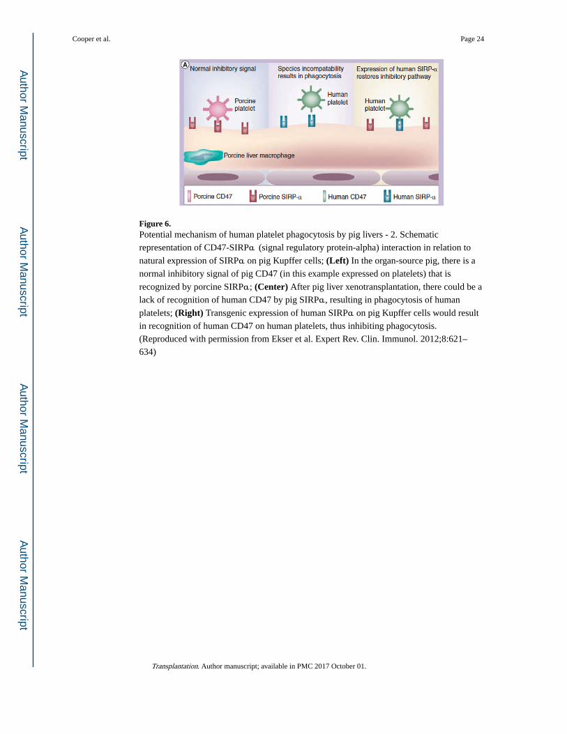
Figure 6. Potential mechanism of human platelet phagocytosis by pig livers - 2. Schematic
representation of CD47-SIRPα (signal regulatory protein-alpha) interaction in relation to
natural expression of SIRPα on pig Kupffer cells; (Left) In the organ-source pig, there is a
normal inhibitory signal of pig CD47 (in this example expressed on platelets) that is
recognized by porcine SIRPα; (Center) After pig liver xenotransplantation, there could be a
lack of recognition of human CD47 by pig SIRPα, resulting in phagocytosis of human
platelets; (Right) Transgenic expression of human SIRPα on pig Kupffer cells would result
in recognition of human CD47 on human platelets, thus inhibiting phagocytosis.
(Reproduced with permission from Ekser et al. Expert Rev. Clin. Immunol. 2012;8:621–
634)
Cooper et al. Page 24
Transplantation. Author manuscript; available in PMC 2017 October 01.
Author M
anuscriptA
uthor Manuscript
Author M
anuscriptA
uthor Manuscript

Author M
anuscriptA
uthor Manuscript
Author M
anuscriptA
uthor Manuscript
Cooper et al. Page 25
Tab
le 1
Exp
erie
nce
in e
xper
imen
tal l
iver
xen
otra
nspl
anta
tion
betw
een
pigs
and
non
hum
an p
rim
ates
Don
or p
igR
ecip
ient
Typ
e of
tra
nspl
ant
nSu
rviv
al (
days
)(R
ef)
WT
Bab
oon
OLT
7<
1–3
}96
–98
WT
Rhe
sus
mon
key
OLT
3<
1}
WT
Chi
mpa
nzee
OLT
1<
1}
WT
Cyn
omol
gus
mon
key
OLT
4<
1–3
}99
WT
Bab
oon
OLT
2
WT
Rhe
sus
mon
key
OLT
6<
1}
38,1
00
WT
Bab
oon
HLT
2<
1}
WT
Bab
oon
OLT
4<
1}
10,1
1
hCD
55B
aboo
nO
LT2
4, 8
}
WT
Bab
oon
OLT
4<
1}
16
hCD
55, h
CD
59, H
TB
aboo
nO
LT5
<1
WT
Bab
oon
OLT
1<
112
,17,
18
GT
KO
, CD
46B
aboo
nO
LT10
<1–
7
MG
H M
S, G
TK
OB
aboo
nO
LT3
6 8,
919
MG
H M
S, G
TK
OB
aboo
nH
LT3
6,9,
1525
WZ
MS,
GT
KO
Tib
etan
mon
key
HLT
32,
5,14
28
MG
H M
S, G
TK
OB
aboo
nO
LT6
1,3,
5,5,
6,7
26
MG
H M
S G
TK
OB
aboo
nO
LT1
2527
CD
46, C
D55
, CD
59 =
hum
an c
ompl
emen
t-re
gula
tory
pro
tein
s
GT
KO
= α
1,3-
gala
ctos
yltr
ansf
eras
e ge
ne-k
nock
out
HLT
= h
eter
otop
ic (
auxi
liary
) liv
er tr
ansp
lant
atio
n
HT
= H
-tra
nsfe
rase
MG
H M
S =
Mas
sach
uset
ts G
ener
al H
ospi
tal m
inia
ture
sw
ine
OLT
= o
rtho
topi
c liv
er tr
ansp
lant
atio
n
WZ
MS
= W
u Z
hans
hen
min
iatu
re s
win
e
WT
= w
ild-t
ype
(gen
etic
ally
unm
odif
ied)
Transplantation. Author manuscript; available in PMC 2017 October 01.