Embed Size (px)
Citation preview
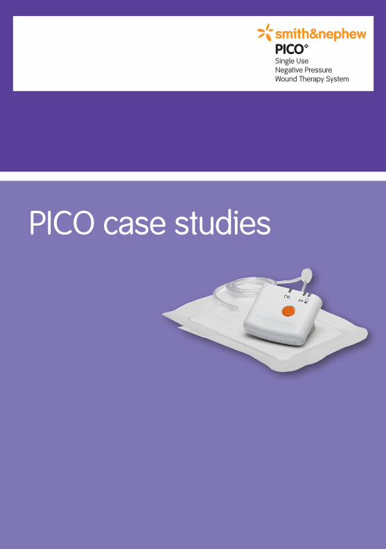
PICO case studies
Leg ulcers
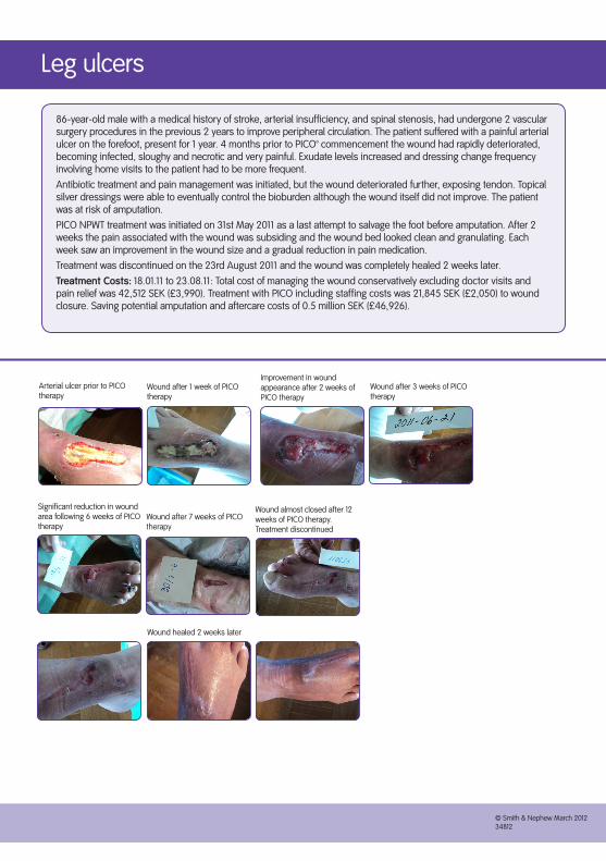
86-year-old male with a medical history of stroke, arterial insufficiency, and spinal stenosis, had undergone 2 vascular surgery procedures in the previous 2 years to improve peripheral circulation. The patient suffered with a painful arterial ulcer on the forefoot, present for 1 year. 4 months prior to PICO™ commencement the wound had rapidly deteriorated, becoming infected, sloughy and necrotic and very painful. Exudate levels increased and dressing change frequency involving home visits to the patient had to be more frequent. Antibiotic treatment and pain management was initiated, but the wound deteriorated further, exposing tendon. Topical silver dressings were able to eventually control the bioburden although the wound itself did not improve. The patient was at risk of amputation. PICO NPWT treatment was initiated on 31st May 2011 as a last attempt to salvage the foot before amputation. After 2 weeks the pain associated with the wound was subsiding and the wound bed looked clean and granulating. Each week saw an improvement in the wound size and a gradual reduction in pain medication. Treatment was discontinued on the 23rd August 2011 and the wound was completely healed 2 weeks later. Treatment Costs: 18.01.11 to 23.08.11: Total cost of managing the wound conservatively excluding doctor visits and pain relief was 42,512 SEK (£3,990). Treatment with PICO including staffing costs was 21,845 SEK (£2,050) to wound closure. Saving potential amputation and aftercare costs of 0.5 million SEK (£46,926).
Arterial ulcer prior to PICO therapy
Wound after 7 weeks of PICO therapy
Wound after 1 week of PICO therapy
Improvement in wound appearance after 2 weeks of PICO therapy
Wound after 3 weeks of PICO therapy
Significant reduction in wound area following 6 weeks of PICO therapy
Wound almost closed after 12 weeks of PICO therapy. Treatment discontinued
Wound healed 2 weeks later
© Smith & Nephew March 201234812

Leg ulcers
Non-healing leg ulcer (4.5 years) prior to PICO therapy
Wound after 2 weeks PICO therapy showing reduction in wound area and improved appearance
78-year-old female with the autoimmune disease Sjögren’s syndrome. A 10cm x 5cm wound on the lower limb failed to respond to conventional wound management therapy that the patient had been receiving up to 3 times a week for 4.5 years. The painful non-healing wound was significantly affecting the patients quality of life and not surprisingly, the patient had given up hope of ever healing her wound.The patient’s care provider was changed in April 2011 and on initial assessment, the presence of a biofilm was suspected. The wound was treated with ACTICOAT™ and ALLEVYN™ in conjunction with compression therapy (PROFORE™ Lite). Whilst the level of bioburden appeared to decline, no obvious progression in healing was noted. In July 2011, PICO™ treatment was considered to try and progress the wound further.Initially the patient was reluctant to try a new therapy but agreed to use PICO NPWT for 2 weeks. Following a notable improvement in the wound, the patient was happy to continue with PICO further. Compression therapy was continued in conjunction with PICO for a total of 18 weeks, after which time the wound dimensions had reduced to an area of 2.5cm2 and pain had subsided. Treatment Costs: Prior to treating with PICO, managing the wound for 4.5 years was estimated to cost 235,872 SEK (£22,137). The total cost of healing the wound with PICO for 18 weeks including staff was 25,500 SEK (£2,393). Although the cost per week of using PICO was greater than conventional treatment, the wound healed over a significantly shorter time period using PICO and the total cost to heal the wound was a fraction of the cost previously expended whilst managing the wound conservatively.
Wound after 16 weeks PICO therapy
Wound almost closed following 18 weeks PICO therapy
© Smith & Nephew March 201234812
© Smith & Nephew June 201236443
Pressure ulcers
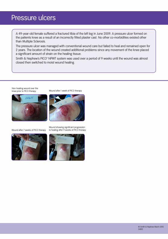
A 49-year-old female suffered a fractured tibia of the left leg in June 2009. A pressure ulcer formed on the patients knee as a result of an incorrectly fitted plaster cast. No other co-morbidities existed other than Multiple Sclerosis.The pressure ulcer was managed with conventional wound care but failed to heal and remained open for 2 years. The location of the wound created additional problems since any movement of the knee placed a significant amount of strain on the healing tissue.Smith & Nephew’s PICO™ NPWT system was used over a period of 9 weeks until the wound was almost closed then switched to moist wound healing.
Non-healing wound over the knee prior to PICO therapy
Wound after 7 weeks of PICO therapy
Wound after 1 week of PICO therapy
Wound showing significant progression to healing after 9 weeks of PICO therapy
© Smith & Nephew March 201234812

Pressure ulcers
92-year-old male with a history of hypertension, hip reconstruction and spinal stenosis was suffering from a painful wound under his right heal which had failed to heal with conventional wound therapy for 2 years.The foot was hard to move and mobility was restricted due to the painful ulcer, causing the patient to be wheelchair bound. As a result, the patient was taking significant pain relief medication (morphine, codeine, paracetamol and LyricaTM) to relieve the peripheral and neurological pain.The wound was treated with PICO™ NPWT for 8 weeks in total, after which time the wound had almost completley healed and it was deemed appropriate to switch treatment to conventional therapy. Treatment Costs: Prior to treating with PICO, costs of managing the wound for 2 years were estimated to total 91,360 SEK (£8,574), the total costs of healing the wound with PICO were 10,640 SEK (£998). Due to the simplicity of dressing application with PICO, wound care was delegated to nursing assistants which helped reduce staffing costs further.
Non-healing pressure ulcer prior to PICO therapy
Wound almost completely healed after 8 weeks of PICO therapy
Wound after 6 weeks of PICO therapy
© Smith & Nephew March 201234812
© Smith & Nephew March 201234812
Diabetic foot ulcers
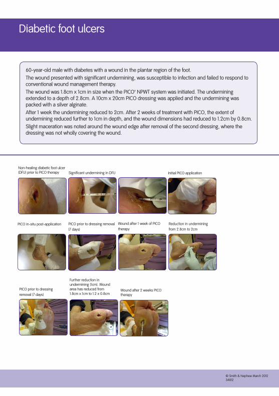
60-year-old male with diabetes with a wound in the plantar region of the foot.The wound presented with significant undermining, was susceptible to infection and failed to respond to conventional wound management therapy. The wound was 1.8cm x 1cm in size when the PICO™ NPWT system was initiated. The undermining extended to a depth of 2.8cm. A 10cm x 20cm PICO dressing was applied and the undermining was packed with a silver alginate. After 1 week the undermining reduced to 2cm. After 2 weeks of treatment with PICO, the extent of undermining reduced further to 1cm in depth, and the wound dimensions had reduced to 1.2cm by 0.8cm. Slight maceration was noted around the wound edge after removal of the second dressing, where the dressing was not wholly covering the wound.
Non-healing diabetic foot ulcer (DFU) prior to PICO therapy
PICO prior to dressing removal (7 days)
PICO prior to dressing removal (7 days)
Significant undermining in DFU Initial PICO application
PICO in-situ post-application Wound after 1 week of PICO therapy
Reduction in undermining from 2.8cm to 2cm
Further reduction in undermining (1cm). Wound area has reduced from 1.8cm x 1cm to 1.2 x 0.8cm
Wound after 2 weeks PICO therapy
© Smith & Nephew March 201234812
Single use negative pressure therapy following surgical debridement of a diabetic foot ulcer
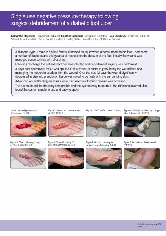
A diabetic (Type 1) male in his late forties sustained an injury when a horse stood on his foot. There were a number of fractures and a large area of necrosis on the dorsum of the foot. Initially the wound was managed conservatively with dressings.Following discharge the patient’s foot became infected and debridement surgery was performed.8 days post operatively, PICO™ was applied 13th July 2011 to assist in granulating the wound bed and managing the moderate exudate from the wound. Over the next 12 days the wound significantly decreased in size and granulation tissue was noted to be flush with the surrounding skin. Advanced wound healing dressings were then used until wound closure was achieved. The patient found the dressing comfortable and the system easy to operate. The clinicians involved also found the system simple to use and easy to apply.
Samantha Haycocks - Advanced Podiatrist, Heather Schofield - Advanced Podiatrist, Paul Chadwick - Principal Podiatrist, Salford Royal Foundation Trust, Podiatry and Foot Health, Salford Royal Hospital, Stott Lane, Salford
Figure 1. Wound post-surgical debridement (5/7/11)
Figure 2. Wound at commencement of PICO (13/7/11)
Figure 3. PICO in-situ post-application Figure 4. PICO prior to dressing change after 2 days in-situ (15/7/11)
Figure 5. Wound following 5 days of PICO therapy (18/7/11)
Figure 6. Wound following 12 days of PICO therapy (25/7/11)
Figure 7. Wound continuing to progress towards healing (3/8/11)
Figure 8. Wound completely healed (28/9/11)
© Smith & Nephew March 201234812
© Smith & Nephew June 201236274

Managing the wound of a patient with vascular disease and social isolation using PICO™
An 84-year-old lady had reperfusion surgery on her left leg following an ischaemic episode. The wound subsequently dehisced, possibly as a result of the patient’s co-morbidities and possibly due to infection. The patient was keen to be discharged home, and it was felt that negative pressure would be the best course of treatment for her wound. The patient lived alone in a rural area and it was felt that standard NPWT would not be suitable. On first review on the 19/8/11 the wound was mostly sloughy and around 1cm deep in places, measuring 8 cm x 4 cm. It was decided that the patient would be suitable for PICO therapy, which would optimize wound care whilst allowing the patient to mobilise fully, maintaining a degree of independence.Following one week of therapy the wound measured 6cm x 3cm and one week later the wound measure 5.5cm x 2.5cm. The wound tissue was mostly granulation with a thin layer of slough and importantly the tissue was level with the surrounding skin.PICO was discontinued on the 2/9/11. The patient found the system lightweight portable and discreet. The clinicians felt that PICO allows patients in the community to have access to treatments normally offered in acute care.
Sue Atkinson - Tissue Viability Nurse, Val Henderson - Lead Tissue Viability Nurse, Northumberland Care Trust, Nursery Park Primary Care Centre, Nursery Road, Ashington, Northumberland
Figure 1. First Review 19/8/11 Figure 2. Following PICO therapy
© Smith & Nephew June 201236274

An 82-year-old female patient presented with long-standing chronic ulceration on the anterior aspect of her lower leg, just distal to the knee. The wound had been present for several years and although relatively small in size measuring 1.5cm x 1.2cm the ulcer was characterised by consistently high levels of exudate which had proven difficult to manage effectively throughout the ulcers long history.
Although the patient had undergone bilateral knee replacement in 1999 it was unclear whether the ulcer and procedure had any association other than anatomical proximity. Radiological review by an orthopaedic surgeon had excluded underlying osteomyelitis so the precise aetiology of the wound remained unclear.
Although infection had been excluded the wound continued to generate a high volume of haemoserous exudate and treatment was focused on a managing this via a variety of absorbent wound management products. Despite several years of treatment no reduction in the level of wound exudate or progression towards healing had been achieved. This regimen was very resource intensive involving daily dressing changes performed largely in the patient’s home.
PICO single use Negative Pressure Wound Therapy (NPWT) was initiated with the twin goals of more effective exudate management and kick-starting wound healing by altering perfusion patterns within the wound bed, and in so doing restoring the wound to a trajectory for timely healing (Figure 1).
Following two weeks PICO therapy the wound showed considerable improvement. The level of wound exudate had decreased, the wound had reduced in size and the wound bed had improved so that it was now 100% healthy granulation tissue. After a further 4 weeks of PICO therapy the wound had almost healed (Figure 2).
The frequency of home visits was reduced since the PICO dressing needed changing just twice weekly rather than daily as was the case with conventional dressings.
The patient’s wellbeing improved with the new regimen which helped the patient to regain her independence through a reduction in the dressing change frequency and via the discreet nature of the PICO device.
Winnie Furlong - Clinical Nurse Specialist, Leg Ulcer Services The Princess Alexandra Hospital NHS Trust
PICO™ Single use Negative Pressure Wound Therapy on chronic ulceration
Figure 1. PICO NPWT in-situ Figure 2. Progression of the wound following the initiation of PICO NPWT
© Smith & Nephew June 201236274
© Smith & Nephew March 201341648
Non-healing lower extremity wounds
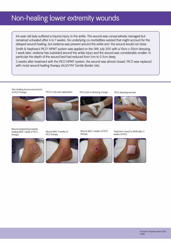
64-year-old lady suffered a trauma injury to the ankle. The wound was conservatively managed but remained unhealed after 6 to 7 weeks. No underlying co-morbidities existed that might account for the delayed wound healing, but oedema was present around the ankle and the wound would not close.Smith & Nephew’s PICO™ NPWT system was applied on the 14th July 2011 with a 10cm x 20cm dressing. 1 week later, oedema has subsided around the ankle injury and the wound was considerably smaller. In particular the depth of the wound bed had reduced from 1cm to 0.5cm deep. 3 weeks after treatment with the PICO NPWT system, the wound was almost closed. PICO was replaced with moist wound healing therapy (ALLEVYN™ Gentle Border Lite).
Non-healing trauma wound prior to PICO therapy
Wound after 2 weeks of PICO therapy
Wound after 3 weeks of PICO therapy
Wound progressing towards healing after 1 week of PICO therapy
Treatment moved to MHW after 3 weeks of PICO
PICO in situ post-application PICO prior to dressing change PICO dressing removal
© Smith & Nephew March 201234812
Non-healing lower extremity wounds
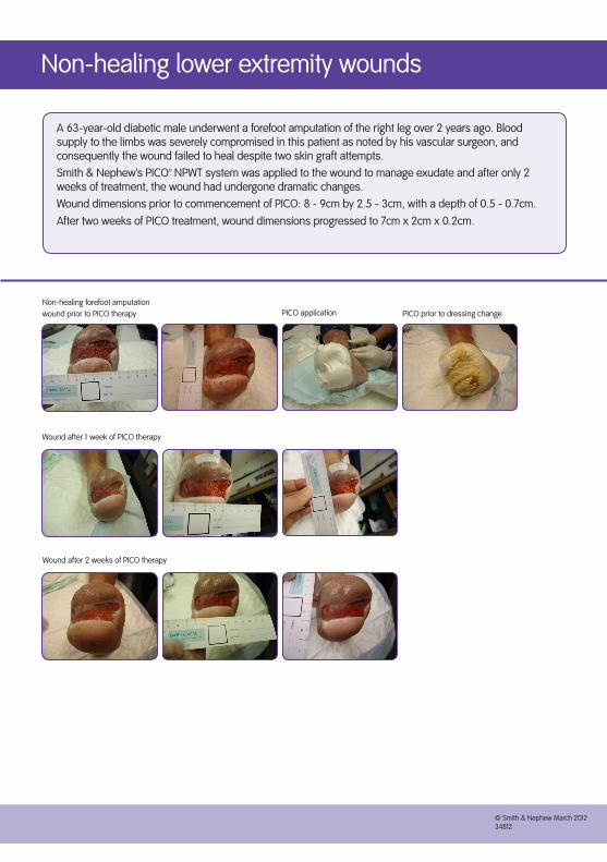
A 63-year-old diabetic male underwent a forefoot amputation of the right leg over 2 years ago. Blood supply to the limbs was severely compromised in this patient as noted by his vascular surgeon, and consequently the wound failed to heal despite two skin graft attempts. Smith & Nephew’s PICO™ NPWT system was applied to the wound to manage exudate and after only 2 weeks of treatment, the wound had undergone dramatic changes.Wound dimensions prior to commencement of PICO: 8 - 9cm by 2.5 - 3cm, with a depth of 0.5 - 0.7cm. After two weeks of PICO treatment, wound dimensions progressed to 7cm x 2cm x 0.2cm.
Non-healing forefoot amputation wound prior to PICO therapy
Wound after 1 week of PICO therapy
Wound after 2 weeks of PICO therapy
PICO application PICO prior to dressing change
© Smith & Nephew March 201234812
© Smith & Nephew March 201234812
Non-healing lower extremity wounds
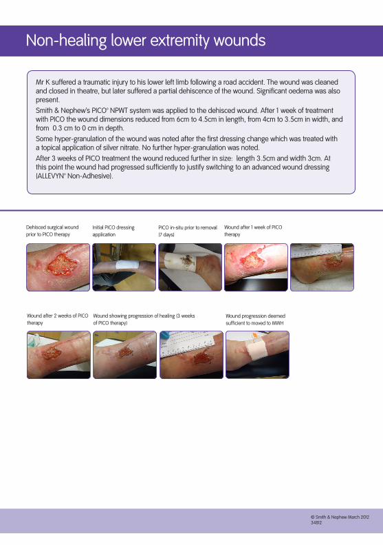
Mr K suffered a traumatic injury to his lower left limb following a road accident. The wound was cleaned and closed in theatre, but later suffered a partial dehiscence of the wound. Significant oedema was also present. Smith & Nephew’s PICO™ NPWT system was applied to the dehisced wound. After 1 week of treatment with PICO the wound dimensions reduced from 6cm to 4.5cm in length, from 4cm to 3.5cm in width, and from 0.3 cm to 0 cm in depth. Some hyper-granulation of the wound was noted after the first dressing change which was treated with a topical application of silver nitrate. No further hyper-granulation was noted.After 3 weeks of PICO treatment the wound reduced further in size: length 3.5cm and width 3cm. At this point the wound had progressed sufficiently to justify switching to an advanced wound dressing (ALLEVYN™ Non-Adhesive).
Dehisced surgical wound prior to PICO therapy
Wound after 1 week of PICO therapy
Wound showing progression of healing (3 weeks of PICO therapy)
Wound after 2 weeks of PICO therapy
Initial PICO dressing application
PICO in-situ prior to removal (7 days)
Wound progression deemed sufficient to moved to MWH
© Smith & Nephew March 201234812

Non-healing lower extremity wounds
An 82-year-old male with a history of diabetes, MI and poor peripheral circulation underwent a partial foot amputation 6 weeks earlier due to tissue necrosis, infection and undermining.Following surgery, the wound was treated with a combination of dressings prescribed by the orthopaedic team of MepitelTM in the bottom of the wound, then MELOLIN™ and sealed with TegadermTM. This resulted in the wound becoming macerated, and an increase in odour and pain caused the patient to worry that the wound was going to break down again and require further amputation. The patient changed to a different care provider and initially the patient was treated with ACTICOAT™ and ALLEVYN™ to manage infection prior to treating with PICO™. After 1 week the patient was completely pain free, presumably due to the reduction in bioburden and able to sleep at night again.The wound was then treated with PICO NPWT over the course of 9 weeks, by which time the wound was closed sufficiently to switch to conventional wound management therapy, and the patient was able to walk again.
Non-healing amputation wound prior to PICO treatment
Wound after 3 weeks of PICO therapy
Wound after 1 week of PICO therapy
Wound after 2 weeks of PICO therapy
Wound after 7 weeks of PICO therapy
Wound almost healed and PICO stopped after 9 weeks of therapy
© Smith & Nephew March 201234812
© Smith & Nephew March 201234812
Non-healing lower extremity wounds
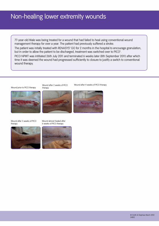
77-year-old Male was being treated for a wound that had failed to heal using conventional wound management therapy for over a year. The patient had previously suffered a stroke. The patient was initally treated with RENASYS™ GO for 2 months in the hospital to encourage granulation, but in order to allow the patient to be discharged, treatment was switched over to PICO™. PICO NPWT was intitiated 26th July 2011 and terminated 6 weeks later (8th September 2011) after which time it was deemed the wound had progressed sufficiently to closure to justify a switch to conventional wound therapy.
Wound prior to PICO therapy
Wound after 5 weeks of PICO therapy
Wound almost healed after 6 weeks of PICO therapy
Wound after 4 weeks of PICO therapy
Wound after 2 weeks of PICO therapy
© Smith & Nephew March 201234812
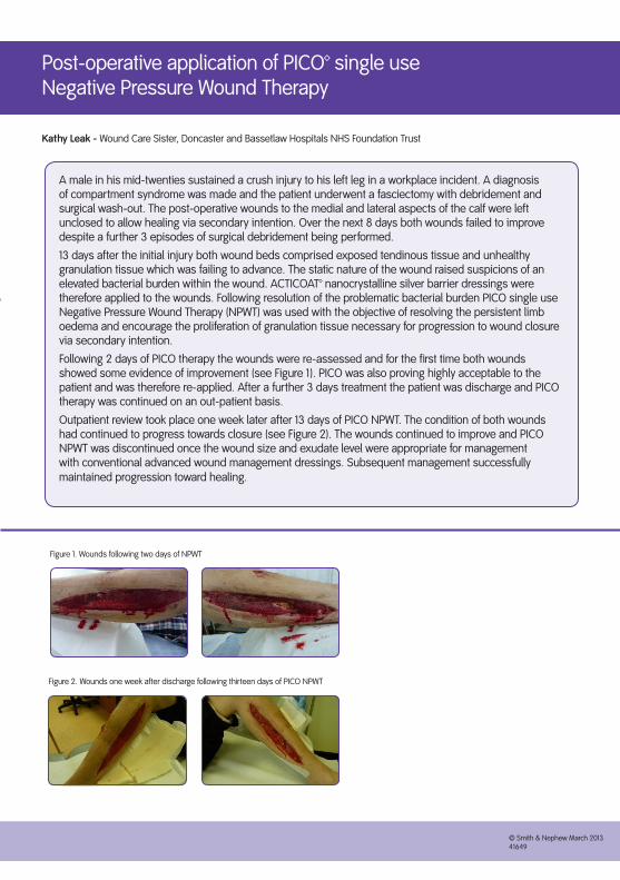
A male in his mid-twenties sustained a crush injury to his left leg in a workplace incident. A diagnosis of compartment syndrome was made and the patient underwent a fasciectomy with debridement and surgical wash-out. The post-operative wounds to the medial and lateral aspects of the calf were left unclosed to allow healing via secondary intention. Over the next 8 days both wounds failed to improve despite a further 3 episodes of surgical debridement being performed.
13 days after the initial injury both wound beds comprised exposed tendinous tissue and unhealthy granulation tissue which was failing to advance. The static nature of the wound raised suspicions of an elevated bacterial burden within the wound. ACTICOAT™ nanocrystalline silver barrier dressings were therefore applied to the wounds. Following resolution of the problematic bacterial burden PICO single use Negative Pressure Wound Therapy (NPWT) was used with the objective of resolving the persistent limb oedema and encourage the proliferation of granulation tissue necessary for progression to wound closure via secondary intention.
Following 2 days of PICO therapy the wounds were re-assessed and for the first time both wounds showed some evidence of improvement (see Figure 1). PICO was also proving highly acceptable to the patient and was therefore re-applied. After a further 3 days treatment the patient was discharge and PICO therapy was continued on an out-patient basis.
Outpatient review took place one week later after 13 days of PICO NPWT. The condition of both wounds had continued to progress towards closure (see Figure 2). The wounds continued to improve and PICO NPWT was discontinued once the wound size and exudate level were appropriate for management with conventional advanced wound management dressings. Subsequent management successfully maintained progression toward healing.
Figure 1. Wounds following two days of NPWT
Kathy Leak - Wound Care Sister, Doncaster and Bassetlaw Hospitals NHS Foundation Trust
Post-operative application of PICO™ single use Negative Pressure Wound Therapy
Figure 2. Wounds one week after discharge following thirteen days of PICO NPWT
© Smith & Nephew March 201234812
© Smith & Nephew March 201341649

A 73-year-old female was admitted to orthopaedics with a fractured right distal tibia. The patient also had chronic venous leg ulcers. Surgical procedure carried out was an above knee amputation due to poor peripheral circulation and existing cardiac disease.
Approximately four weeks after surgery, the stump wound started leaking and continued to deteriorate over a few weeks. CT scan revealed osteomyelitis of the distal femur at the tip of the stump. A further four weeks later, the wound measured 12cm x 2cm and was necrotic, sloughy and leaking large quantities of haemoserous fluid and pus.
Patient was referred to Limb Reconstruction Nurse Specialist for PICO Negative Pressure dressing. IV antibiotics were commenced.
Patient received 28 days of PICO negative pressure dressings. During which time the wound fully healed and patient was discharged to nursing home for full time nursing care.
Figure 1. Stump wound on first review note heavy exudate and sloughy wound bed
Figure 2. Day 35 The wound has almost completely healed. PICO discontinued
Emma Sharp - Limb Reconstruction Nurse Specialist, Gatehouse Building, Glasgow Royal Infirmary
A prospective evaluation of PICO™ single use Negative Pressure Wound Therapy for the treatment of patients with complex orthopaedic surgical and trauma wounds
© Smith & Nephew April 201341651
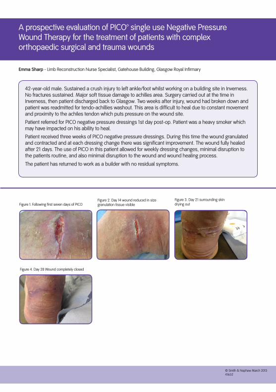
42-year-old male. Sustained a crush injury to left ankle/foot whilst working on a building site in Inverness. No fractures sustained. Major soft tissue damage to achilles area. Surgery carried out at the time in Inverness, then patient discharged back to Glasgow. Two weeks after injury, wound had broken down and patient was readmitted for tendo-achillies washout. This area is difficult to heal due to constant movement and proximity to the achiles tendon which puts pressure on the wound site.
Patient referred for PICO negative pressure dressings 1st day post-op. Patient was a heavy smoker which may have impacted on his ability to heal.
Patient received three weeks of PICO negative pressure dressings. During this time the wound granulated and contracted and at each dressing change there was significant improvement. The wound fully healed after 21 days. The use of PICO in this patient allowed for weekly dressing changes, minimal disruption to the patients routine, and also minimal disruption to the wound and wound healing process.
The patient has returned to work as a builder with no residual symptoms.
Figure 1. Following first seven days of PICOFigure 2. Day 14 wound reduced in size granulation tissue visible
Figure 3. Day 21 surrounding skin drying out
Emma Sharp - Limb Reconstruction Nurse Specialist, Gatehouse Building, Glasgow Royal Infirmary
A prospective evaluation of PICO™ single use Negative Pressure Wound Therapy for the treatment of patients with complex orthopaedic surgical and trauma wounds
Figure 4. Day 28 Wound completely closed
© Smith & Nephew April 201341651
© Smith & Nephew March 201341652

The use of PICO™ on a high risk abdominal incision
A 40-year-old patient with a history of diabetes, acute pancreatitis, peritoneal sepsis and an ischiorectal abscess. Following drainage of the abscess and excision of necrotising fasciitis of the abdominal wall the RENASYS™ Abdominal Dressing was used as temporary abdominal closure. Further surgery was necessary to repair a ruptured diverticulum and a Hartmann’s procedure performed.Once granulation had formed over the bowel, gauze-based negative pressure was used until the patient was ready for primary closure. PICO was applied in theatre and despite the wound appearance being irregular prior to application, following one week of therapy the wound edges were apposed and the wound healed without incident.
Fiona Russell - Dip Hen, PG cert., Tissue Viability Nurse, Grampian NHS Trust, Cornhill Hospital, Cornhill Road, Aberdeen
© Smith & Nephew June 201236274

A prospective case study of the PICO™ Negative Pressure Wound Therapy system in a patient with an abdominal wound
A 58-year-old male with a history of Crohn’s disease was admitted for an emergency Hartmann’s procedure following perforation of his sigmoid colon. His previous history included long term steroids, psoriatic arthritis and ischaemic heart disease.Following surgery, possibly relating to steroid therapy the wound dehisced when the sutures were removed on day 10. Initially the wound measured 15cm x 6cm x 3.5cm, this was packed with an alginate dressing and covered with foam.The wound deteriorated further and became sloughy, it was decided to use PICO NPWT to encourage healing and manage wound exudate. When PICO was commenced the wound measured 18cm x 8cm x 2cm deep.PICO was used for four weeks following which the wound reduced in size measuring 8cm x 4cm x 0.5cm. The wound had contracted significantly and the granulation tissue was almost level with the surrounding skin. The sloughy area was much reduced in size and there was no longer any necrotic tissue in the wound. The patient also reported much less pain in the wound and in the surrounding tissue, in addition he was very satisfied with the portability of the system which allowed him to remain mobile.
Robert Oldfield - RN, DipHE, Bsc (Hons), PG diploma (Nursing Salford), Tissue Viability Nurse Specialist. Trafford General Hospital, Manchester
Figure 1. Wound following suture removal 6th July 2011
Figure 3. Wound with PICO in situ
Figure 2. Following deterioration of the wound 8th September
Figure 4. Wound on 6th October 2011 PICO discontinued
© Smith & Nephew June 201236274
© Smith & Nephew June 201236274

Quality and Innovation: improving patient experience and clinical outcomes with new technology
A male in his mid-sixties was admitted for a colonic resection following which his abdomen was left open with RENASYS™ Abdominal dressing in situ for 4 weeks to prevent compartment syndrome. Once granulation tissue was established over the bowel, PICO™ NPWT was initiated to try and facilitate improved mobility and comfort for the patient. With PICO in situ the patient was able to take ward leave. The wound continued to granulate and reduce in size, following 7 days with PICO a split thickness skin graft was applied. PICO was shown to be a viable alternative to more traditional negative pressure wound products and in this case there was a significant cost saving also. Importantly, the patient found PICO an acceptable system both in relation to its portability and through the reduction in pain at dressing changes.
Sian Fumarola - MSc Tissue Viability Clinical Specialist, Stephanie Rylands - RGN, Tissue Viability Support Nurse, William Kisku - FRCSE Specialist Plastic Surgeon, University Hospital of North Staffordshire, Royal Infirmary, Princes Road, Stoke on Trent, Staffordshire
i. ii.
Figure 1. 4 weeks post-op (10.6.11) i. RENASYS in-situ
Figure 2. PICO single-use NPWT system applied pre-graft
Figure 3. i. PICO system having been in-situ for 4 days
i ii
Figure 4. Wound at the second PICO dressing change (7 days total PICO treatment)
Figure 5. Wound following split skin graft and 5 days management with PICO. Approx. 97% graft viability is evident
ii. PICO system removed after 4 days. Reduction in abdominal distension noted following improved mobility
ii. Healthy capsule covering bowel on removal of RENASYS
© Smith & Nephew June 201236274
The use of PICO™ in the management of a complex dehisced abdominal wound
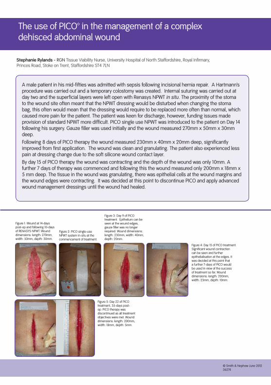
A male patient in his mid-fifties was admitted with sepsis following incisional hernia repair. A Hartmann’s procedure was carried out and a temporary colostomy was created. Internal suturing was carried out at day two and the superficial layers were left open with Renasys NPWT in situ. The proximity of the stoma to the wound site often meant that the NPWT dressing would be disturbed when changing the stoma bag, this often would mean that the dressing would require to be replaced more often than normal, which caused more pain for the patient. The patient was keen for discharge, however, funding issues made provision of standard NPWT more difficult. PICO single use NPWT was introduced to the patient on Day 14 following his surgery. Gauze filler was used initially and the wound measured 270mm x 50mm x 30mm deep.Following 8 days of PICO therapy the wound measured 230mm x 40mm x 20mm deep, significantly improved from first application. The wound was clean and granulating. The patient also experienced less pain at dressing change due to the soft silicone wound contact layer.By day 15 of PICO therapy the wound was contracting and the depth of the wound was only 10mm. A further 7 days of therapy was commenced and following this the wound measured only 200mm x 18mm x 5 mm deep. The tissue in the wound was granulating, there was epithelial cells at the wound margins and the wound edges were contracting. It was decided at this point to discontinue PICO and apply advanced wound management dressings until the wound had healed.
Stephanie Rylands - RGN Tissue Viability Nurse, University Hospital of North Staffordshire, Royal Infirmary, Princes Road, Stoke on Trent, Staffordshire ST4 7LN
Figure 1: Wound at 14-days post-op and following 10-days of RENASYS NPWT. Wound dimensions: length: 270mm, width: 50mm, depth: 30mm.
Figure 3: Day 9 of PICO treatment. Epithelium can be seen at the wound edges, gauze filler was no longer required. Wound dimensions: length: 230mm, width: 40mm, depth: 20mm.
Figure 2: PICO single-use NPWT system in-situ at the commencement of treatment.
Figure 4: Day 15 of PICO treatment. Significant wound contraction can be seen and further epithelialisation at the edges. It was decided at this point that a further 7-days of PICO would be used in view of the success of treatment so far. Wound dimensions: length: 200mm, width: 23mm, depth: 10mm.
Figure 5: Day 22 of PICO treatment, 35-days post-op. PICO therapy was discontinued as all treatment objectives were met. Wound dimensions: length: 200mm, width: 18mm, depth: 5mm.
© Smith & Nephew June 201236274
© Smith & Nephew June 201236274

Use of a single use Negative Pressure Wound Therapy system to treat a patient following digital amputation for Deupuytren’s contracture
A 58-year-old male was admitted for removal of the fifth digit on his right hand due to loss of function caused by excessive contracture. After 7 days the patient presented to the clinic with the wound edges poorly apposed and the subcutaneous tissues visible through a partial dehiscence. The wound was in an awkward area and was at risk of breaking down.PICO™ was applied to the wound on the 23/09/11 to help manage oedema and to help keep the wound edges apposed. A piece of thin hydrocolloid was used to help achieve a seal on the inter-digital areas. At final review on 30/09/11, following 8 days of therapy, the wound edges were apposed, there was no further leakage from the wound and PICO was discontinued. PICO NPWT assisted in managing wound exudate, reducing local oedema and improving approximation of the wound edges.
© Smith & Nephew June 201236274
Kathy Leak, Doncaster and Bassetlaw Hospitals NHS Foundation Trust, Bassetlaw Hospital, Blyth Road
Image 1. Appearance of the wound at initial presentation (23.9.11) Image 2. PICO in-situ prior to removal (30.9.11)
Image 3. Appearance of the wound at discontinuation of PICO therapy (30.9.11)
© Smith & Nephew June 201236274
PICO™ to support a gluteal flap
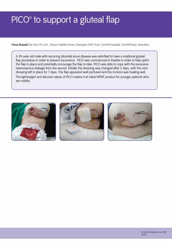
A 39-year-old male with recurring pilonidal sinus disease was admitted to have a rotational gluteal flap procedure in order to prevent recurrence. PICO was commenced in theatre in order to help splint the flap in place and potentially encourage the flap to take. PICO was able to cope with the excessive haemoserous leakage from the wound. Initially the dressing was changed after 2 days, with the next dressing left in place for 7 days. The flap appeared well perfused and the incision was healing well. The lightweight and discreet nature of PICO makes it an ideal NPWT product for younger patients who are mobile.
© Smith & Nephew June 201236274
Fiona Russell Dip Hen, PG cert., Tissue Viability Nurse, Grampian NHS Trust, Cornhill Hospital, Cornhill Road, Aberdeen
Wound Management www.smith-nephew.com/ukSmith & NephewHealthcare Ltd ™Trademark of Smith & NephewHealthcare House TMAll trademarks acknowledged101 Hessle Road © Smith & Nephew September 2013Hull HU3 2BN
T 01482 222200F 01482 222211
Customers in Ireland please callT 1890 224226 for productenquires and wound care advice
24hr Clinical SupportFreephone Number
UK: 0800 9155394Ireland: 1800 30 36 22