Embed Size (px)
Citation preview

Phytochemical and biological investigation of the
bark of Garcinia fusca Pierre.
A dissertation submitted to the
Faculty of Chemistry and Pharmacy, University of Regensburg
for the degree of
Doctor of Natural Sciences (Dr. rer. nat.)
Presented by
TRI HIEU NGUYEN
from Ho Chi Minh City University of Science, Vietnam
2015

The present work was carried out from 07/2011 until 01/2015 under the supervision of
Prof. Dr. Jörg Heilmann at the institute of Pharmacy, Faculty of Chemistry and
Pharmacy, University of Regensburg.
This dissertation was submitted on 1st June 2015
The Ph.D. defense was on 23rd June 2015
Board of examiners: PD Dr. Sabine Amslinger (Chairwoman)
Prof. Dr. Jörg Heilmann (First-examniner)
Prof. Dr. Thomas Schmidt (Second-examiner)
Prof. Dr. Gerhard Franz (Third-examiner)

ACKNOWLEDGEMENTS
I would like to express my sincere gratitude to my supervisor Prof. Dr. Jörg
Heilmann, for accepting me as a member of his group, for providing me helpful
working facilities, for his interest in my work as well as his willingness to proofread
my thesis.
I offer my deepest gratitude to Assoc. Prof. Dr. Nguyen Dieu Lien Hoa for showing
me patience and enthusiasm, for guiding me throughout many years in the field of
natural products, as well as being a great source of knowledge.
I would like to thank Assoc. Prof. Dr. Pham Dinh Hung for his willingness to help
me when necessary.
I am greatly indebted to my parish-priest Joseph Nguyen Hien Thanh, Dr. Vu
Quang Tuyen and M.Ed. Le Minh Ha for supporting and giving me an opportunity to
apply the Ph.D. scholarship of KAAD (Bonn, Germany).
My sincere thanks also go to:
- Dr. Guido Jürgenliemk for his kind help in CPC separation.
- Dr. Birgit Kraus for her useful advice in cell culture.
- Dr. Daniel Bücherl for helping me enthusiastically in use of HPLC and
connection with the network of the university.
- Dr. Marcel Flemming for his instruction in anti-oxidant assay on RAW cells.
- Dr. Sebastian Schmidt for showing me how to carry out proliferation assay on
HMEC-1 cells, as well as for his fruitful discussions in isolation and structural
elucidation of natural compounds.
- Gabi Brunner for instructing me in detail about cytotoxicity assay on HeLa cells.
She might be very patient with me.
- My labmate, Stefan Wiesneth for his help in rescue of my user account at the
university every time I forgot my passwords, as well as for his tolerance to my
clumsiness.

- My colleague, Edna Makule for her help in use of polarimeter and for honest
confidences which we exchanged.
I am grateful to Annette Schramm, Georgine Stühler, Fritz Kastner, Josef Kiermaier
and Wolfgang Söllner for measurement of NMR and MS spectra.
I am thankful to the secretary Hedwig Ohli for her kindness and enthusiasm. I wish
her all the best in her life.
Thanks go to all my colleagues, staff and students in Dr. Heilamnn’s group for their
helps and for the pleasant working atmosphere and enjoyable time we had together. I
will never forget our nice memories.
I am very grateful to Katholischer Akademisher Ausländer-Dienst (KAAD,
Germany) for providing me a scholarship to complete my Ph.D. project in Regensburg
University. I would especially like to thank Dr. Heinrich Geiger and Mrs. Karin Bialas
for their kind help to Vietnamese candidates.
I would like to convey my deepest thanks to my family, relatives and friends for
understanding, encouraging and supporting me spiritually throughout my life.
Finally, I would like to say with all my faith that I did not obtain all precious helps
and supports as mentioned above accidentally but they must have been the blessings of
Lord. I could not have completed my Ph.D. project without those valuable helps and
supports. With all my heart and soul, I would like to express my greatest gratitude to
the Lord for all the blessings I got from Him.


For my father and my late mother,
my sisters and brothers


TABLE OF CONTENTS
ACKNOWLEDGMENTS
ABBREVIATIONS .....................................................................................................i
SUMMARY ............................................................................................................... iv
1. INTRODUCTION ........................................................................................... 1
2. THE GENUS GARCINIA ............................................................................... 3
2.1. General characters ..................................................................................... 3
2.2. Chemical constituents of Garcinia ............................................................ 3
2.2.1. Xanthones ....................................................................................... 12
2.2.1.1. Simple oxygenated xanthones ................................................ 12
2.2.1.2. Prenylated xanthones ............................................................. 12
2.2.1.3. Caged xanthones .................................................................... 16
2.2.1.4. Bisxanthones .......................................................................... 16
2.2.2. Biflavonoids .................................................................................... 18
2.2.3. Benzophenones ............................................................................... 19
2.2.4. Phloroglucinols ............................................................................... 22
2.2.5. Depsidones ...................................................................................... 23
2.2.6. Tocotrienols .................................................................................... 24
2.2.7. Biphenyls ........................................................................................ 25
2.2.8. Triterpenoids ................................................................................... 25
2.2.9. Other compounds ............................................................................ 26
2.3. Pharmacological and biological properties of Garcinia ......................... 30
2.3.1. Anti-oxidant activity ....................................................................... 31
2.3.2. Antifungal activity .......................................................................... 31
2.3.3. Antimicrobial activity ..................................................................... 33
2.3.4. Anti-inflammatory activity .............................................................. 35
2.3.5. Anticancer activity .......................................................................... 36
2.3.6. Antiviral activity ............................................................................. 36

2.3.7. Other properties .............................................................................. 37
2.4. Study of the genus Garcinia in Vietnam ................................................. 40
2.5. Garcinia fusca Pierre .............................................................................. 45
2.5.1. Botanical features and uses in traditional medicine ......................... 45
2.5.2. Previous chemical and biological investigations .............................. 45
3. BIOSYNTHESYS OF XANTHONES AND BIFLAVONOIDS .................. 47
3.1. Biosynthetic pathway of xanthones ....................................................... 47
3.2. Biosynthetic pathway of bioflavonoids ................................................. 59
4. MATERIAL AND METHODS .................................................................... 68
4.1. General experimental procedures .......................................................... 68
4.2. Extraction, fractionation and isolation................................................... 69
4.2.1. Material of plant .............................................................................. 69
4.2.2. Extraction of plant material ............................................................. 69
4.2.3. Fractionation of crude extracts ........................................................ 70
4.2.4. Isolation of substances from fractions ............................................. 70
4.2.4.1. Thin layer chromatography (TLC) ........................................ 71
4.2.4.2. Preparative TLC ..................................................................... 73
4.2.4.3. Open column chromatography ............................................... 73
4.2.4.4. Flash chromatography ............................................................ 74
4.2.4.5. Size-exclusion chromatography.............................................. 74
4.2.4.6. Centrifugal partition chromatography ..................................... 75
4.2.4.7. Semi-preparative HPLC ......................................................... 76
4.3. Structural elucidation of isolated compounds .......................................... 78
4.3.1. Ultraviolet-visible spectroscopy ...................................................... 78
4.3.2. Mass spectrometry .......................................................................... 79
4.3.3. Nuclear magnetic resonance spectroscopy (NMR) .......................... 81
4.3.3.1. One dimensional (1D) NMR spectra ...................................... 82
4.3.3.1.1. 1H NMR spectra .............................................................. 82
4.3.3.1.2. Proton-decoupled 13C NMR spectra ................................ 83
4.3.3.2. Two dimensional (2D) NMR spectra ...................................... 83
4.3.3.2.1. COSY spectra ................................................................ 83

4.3.3.2.2. HSQC spectra ................................................................. 84
4.3.3.2.3. HMBC spectra ................................................................ 84
4.3.3.2.4. NOESY and ROESY spectra ........................................... 84
4.3.4. Optical rotation ............................................................................... 85
4.4. Biological assays .................................................................................. 86
4.4.1. Preparation of samples .................................................................... 86
4.4.2. Cytotoxicity assay on HeLa cells..................................................... 86
4.4.2.1. Cell culture ............................................................................ 86
4.4.2.2. Cell counting .......................................................................... 87
4.4.2.3. MTT assay ............................................................................. 87
4.4.3. Proliferation assay on HMEC-1 ....................................................... 88
4.4.3.1. Cell culture ............................................................................ 88
4.4.3.2. Proliferation assay .................................................................. 89
5. RESULTS AND DISCUSSION .................................................................... 90
5.1. Experiments ............................................................................................ 90
5.1.1. Fractionation and isolation of the n-hexane extract (GFH) ............. 90
5.1.1.1. Isolation from fraction GFH-2 ................................................ 94
5.1.1.2. Isolation of xanthones from fraction GFH-4 ........................... 94
5.1.1.3. Isolation of compounds from fraction GFH-6 ......................... 95
5.1.1.4. Isolation of xanthones from fraction GFH-7 ........................... 97
5.1.2. Fractionation of the ethyl acetate extract and further isolation ........ 99
5.1.2.1. Isolation of xanthones from fraction GFE-4 ......................... 103
5.1.2.2. Isolation of compounds from fraction GFE-6 ....................... 104
5.2. Structural elucidation of isolated substances ......................................... 105
5.2.1. Structural identification of substances from the n-hexane extract .. 105
5.2.1.1. Compound 21 (fuscaxanthone J) .......................................... 111
5.2.1.2. Compound 20 (fuscaxanthone K) ......................................... 114
5.2.1.3. Compound 19 ((+)-fuscaxanthone I) .................................... 118
5.2.1.4. Compound 18 (fuscaxanthone L) ......................................... 123
5.2.1.5. Compound 22 (fuscaxanthone M) ........................................ 128
5.2.2. Structural identification of substances from the EtOAc extract ...... 133

5.2.2.1. Compound 24 (fuscaxanthone N) ......................................... 136
5.2.2.2. Compound 25 (fuscaxanthone O) ......................................... 140
5.2.2.3. Compound 26 (fuscaxanthone P) .......................................... 144
5.3. Cytotoxicity of new substances ............................................................. 148
5.4. Proposed biosynthesis of the new xanthones in G. fusca Pierre ............. 150
6. CONCLUSION............................................................................................ 153
REFERENCES ................................................................................................. 154
LIST OF SCHEMES ........................................................................................ 194
LIST OF TABLES ............................................................................................ 195
APPENDICES .................................................................................................. 196

Abbreviations i
ABBREVIATIONS
AflO
AflY
[]D
Gene encoding the SAM-dependent O-methyltransferase in the
Aspergillus species
Gene encoding an enzyme catalyzing the Baeyer-Villiger oxidation
in the Aspergillus species
Specific optical rotation
brs
BHT
Broad singlet
Butylated hydroxytoluene
CC Column chromatography
CDCl3 Deuterated chloroform
C2D6CO
CHS
Deuterated acetone
Chalcone synthase
cm Centimetre
COSY Correlated spectroscopy
CPC Centrifugal partition chromatography
Chemical shift
d Doublet
1D One-dimensional
2D Two-dimensional
DCM Dichloromethane
dd Doublet of doublet
DMAPP Dimethylallylpyrophosphate
DMSO
DPPH
Dimethylsulphoxide
2,2-Dipheny-1-(2,4,6-trinitrophenyl)hydrazyl
EDTA Ethylenediaminetetraacetic acid
EtOAc Ethyl acetate
EtOH Ethanol
FC Flash chromatography
fr. Fraction
FCS
FAD
Fetal calf serum
Flavin adenine dinucleotide

ii Abbreviations
GPC Gel permeation chromatography
GPP Geranyldiphosphate
HMBC Heteronuclear multiple bond correlation
HMEC-1 Human microvascular endothelial cell line-1
HPLC High performance liquid chromatography
HRESI High resolution electrospray ionization
HSQC Heteronuclear single quantum coherence
IC50 50% Inhibition concentration
J Coupling constant
m
MdpB
MdpC
MdpD
MdpJ
MdpK
MdpL
Multiplet
Gene encoding dehydratase in Aspergillus spp.
Gene encoding phenol reductase in Aspergillus spp.
Gene encoding mono-oxygenase in Aspergillus spp.
Gene encoding glutathione-S-transferase in Aspergillus spp.
Gene encoding oxidoreductase in Aspergillus spp.
Gene encoding Baeyer-Villiger oxidase in Aspergillus spp.
MeCN Acetonitrile
MeOH Methanol
mg Milligram
min Minute
mm Millimetre
µM Micromolar
µl Microliter
MEM Minimum essential medium
MHz
MIC
Megahertz
Minimum inhibitory concentration
MS Mass spectrometry
MTT 3-(4,5-dimethylthiazol-2-yl)-2,5-diphenyl tetrazolium bromide
m/z Mass-to-charge ratio
n Number
NADPH Nicotinamide adenine dinucleotide phosphate
NMR Nuclear magnetic resonance
NOESY Nuclear overhauser enhancement spectroscopy

Abbreviations iii
p.a.
PAL
Pro analysis
Phenylalanine ammonia lyase
PE Petroleum ether
ppm Part per million
PBS Phosphate buffered saline
Rf Retardation factor
ROESY Rotating-frame nuclear overhauser effect correlation spectroscopy
RP18 Reversed phase C-18
s Singlet
SAM S-Adenosyl methionine
SDS Sodium dodecyl sulphate
t Triplet
TLC Thin layer chromatography
UV
Ver-A
Ver-1
XtpA
XtpB
XtpC
Ultraviolet
A cytochrome P450 monooxygenase in Aspergillus species
An NADH-dependent deoxygenase in Aspergillus species
Gene encoding prenyl transferase in Aspergillus spp.
Gene encoding prenyl transferase in Aspergillus spp.
Gene encoding oxidoreductase in Aspergillus spp.

iv Summary
SUMMARY
The Garcinia is a large genus of the family Guttiferae or Clusiaceae, nearly
consisting of 450 species. This genus has been of interest due to its therapeutic value
in the folk medicine for the treatment of various kinds of diseases such as vomiting,
swelling, tapeworms, dysentery, chronic diarrhoea, piles, pains and heart complaints,
as well as due to the successive finding of both chemically and biologically interesting
compounds (Vo et al., 2012). In Vietnam, around thirty one Garcinia species were
found, some of which were phytochemically and biologically investigated (Pham,
1999). For further phytochemical and biological study of the genus Garcinia in
Vietnam, Garcinia fusca Pierre was chosen for the investigation.
In this work, the air-dried and powdered bark of G. fusca Pierre (1.95 kg) was
successively extracted with n-hexane and ethyl acetate. The NMR-guided fractionation
of both the n-hexane and ethyl acetate extracts led to the isolation of thirty one
phenolic compounds by means of various chromatographic methods such as TLC, FC,
GPC, CPC and semi-preparative HPLC. The structural elucidation was carried out
based on data of UV, MS, 1D and 2D NMR spectra together with the optical rotation.
The compounds isolated from the n-hexane extract comprise a tocotrienol, a
benzaldehyde derivative and twenty xanthones, five of which (18 – 22) are new and
given trivial names fuscaxanthones (+) I, J – M. Meanwhile four biflavonoids and five
xanthones were obtained from the ethyl acetate extract, three of which (24 – 26) are
new and given trivial names fuscaxanthones N – P, respectively. All the xanthones are
prenylated 1,3,6,7-tetrahydroxyxanthone derivatives. Eight new xanthones possess the
basic structure of cowanol, which was previously reported from G. cowa (Na Pattalung
et al., 1994). However the different features were observed. Fuscaxanthone J (21)
might have been O-methylated at 7-OH to form cowanol. In fuscaxanthone (+) I (19),
the double-bond at C-17/C-18 of the geranyl group is hydroxylated, whereas the
double-bond of the 4-hydroxy-3-methylbut-2-enyl group is epoxylated in
fuscaxanthone K (20). In fuscaxanthones N and O (24 and 25), the double-bond at C-
22/C-23 of the geranyl group is hydroxylated and relocated to C-21/C-22 or C-23/C-
24. Meanwhile in fuscaxanthone L and P (18 and 26), the geranyl group is cyclized to
give a monoterpenoid substitute with a C6 ring. To the best of our knowledge,

Summary v
parvixanthone I, which was reported from G. parvifolia, was the only xanthone with
such a substitute (Xu et al., 2001). Fuscaxanthone M (22) is a dimer of cowanol, which
is combined by an unusual C-O-C linkage.
The new xanthones and cowanol were tested for cytotoxicity on HeLa cells to
establish a preliminary basis of a structure – activity relationship. The results showed
that these xanthones had cytotoxic effect with the IC50 values in the range of 19.1 –
45.9 µM, except that compounds 20 and 22 were inactive. The modification of the
geranyl group or the 4-hydroxy-3-methylbut-2-enyl group brought about a decrease or
loss of activity compared with cowanol. Moreover griffipavixanthone, which was
previously isolated from G. griffithii, G. pavifolia and G.oblongifolia (Xu et al., 1998;
Shi et al., 2014), exhibited a high cytotoxicity against HeLa cells with an IC50 value of
7.9 0.7 µM. This bixanthone was further evaluated for anti-angiogenic activity by in
vitro proliferation assay on HMEC-1 cell line and it showed a greatly strong anti-
proliferative effect with an IC50 value of 0.15 0.0085 µM after 72 h incubation,
which indicated that griffipavixanthone is a potential anti-angiogenic agent which
should be further demonstrated in vivo and in further molecular assays.
The results of this work together with those previously reported (Ito et al., 2003;
Nontakham et al., 2014) revealed that Garcinia fusca Pierre is a rich source of
tetraoxygenated xanthones and biflavonoids. The cytotoxicity of those xanthones
suggested that the bark of this plant may have an anti-tumor effect, which should be
further studied for use in medicine.


Introduction 1
1. INTRODUCTION
Vietnam is situated along the eastern coast of the Indochina Peninsula in Southeast
Asia and covers an area of 329,500 square kilometres. The country is bordered to the
north by China, to the west by Laos and Cambodia and to the east by South China Sea
(known in Vietnam as the East Sea). As a result of the warm and humid tropical
climate, approximately 39% of Vietnam territory is under forest cover with around
13,000 species of vascular plants, over 8,000 of which have been identified to date
(Sterling and Hurley, 2005; Queiroz et al., 2013). Early on Vietnamese people
discovered pharmaceutical properties of some plants and used them as medical
remedies to prevent or treat some diseases such as eating ginger to treat cold and flu,
drinking the water extract of Artemisia as an anti-malaria agent and chewing betel to
prevent tooth decay. These formed the basis of Vietnamese traditional medicine.
During the thousand years of Chinese domination, Vietnamese traditional medicine
was deeply influenced by Chinese traditional medicine. In the nineteenth century,
Western medical practices were introduced to Vietnam with the arrival of the French
(Ladinsky et al., 1987). Nowadays Vietnamese medicine is a harmonious merging of
Chinese, Vietnamese and Western medicinal system, in which traditional medicine has
an important role in promoting the health of Vietnamese people, especially in primary
health care at the commune level and the treatment of chronic illnesses such as AIDS
and cancer which seem to be increasing. The statistics of the Vietnam Ministry of
Health showed that about 30% of patients annually received the treatment with
traditional medicines (WHO, 2005). Vietnamese traditional medical remedies are
mainly based on medicinal plants. According to a report of the Vietnam Ministry of
Health in 2014, 3,948 of 10,386 identified species have been known as medicinal
plants, a large number of which have not been phytochemically investigated to
improve use value. Moreover pharmaceutical properties of the remaining species have
not determined yet. The report also showed that many valuable and rare medicinal
plants are in the risk of extinction because of deforestation and uncontrolled
exploitation of medicinal sources (The Vietnam Ministry of Health, 2014).

2 Introduction
The Guttiferae or Clusiaceae is a family of plants including about 37 genera and
1610 species of trees and shrubs. This family is mainly tropical (Gustafsson et al.,
2002). In Vietnam, there are around sixty five species of six genera comprising
Calophyllum, Cratoxylum, Garcinia, Mammea, Mesua and Ochrocapus. Some species
are used in folk medicine to treat various diseases such as dysentery, diarrhoea,
coughs, fever, scabies, burns, menstrual disturbances and ulcer; whilst pharmaceutical
properties of the remaining species have not been known (Vo, 1997; Pham, 1999).
Though many species of the family Guttiferae have been phytochemically and
biologically investigated all over the world, approximately twenty species collected in
Vietnam have been reported so far. In order to contribute to phytochemical and
biological study of the family Guttiferae in Vietnam, we have investigated the species
Garcinia fusca Pierre collected in south Vietnam. The aim of this work was the
isolation of natural compounds to characterize the phytochemical profile in detail and
to provide possible new structures for drug discovery whereby that helps to improve
use value of this species in the traditional medicine. Moreover, that also supplies
phytochemical database of this species for classification and conservation of medicinal
plants in Vietnam. The results of the phytochemical and biological investigation on the
n-hexane and ethyl acetate extracts of G. fusca Pierre are presented in this thesis.

The genus Garcinia 3
2. THE GENUS GARCINIA
2.1. General characters
The Garcinia is a large genus of the family Guttiferae (Clusiaceae), comprising
around 450 species of tropical trees and shrubs with yellow resin or latex, mostly
native to the Old World (tropical and South Africa, Madagascar, south-eastern Asia,
north-eastern Australia and western Polynesia), with a few species in the tropical
regions of the Americas. Various Asiatic species are used as a cathartic or stimulant,
dye or artist’s pigment. Many species are important timber resources used for
construction and furniture. In Vietnam there are about thirty one species (Pham, 1999).
The members of the genus are various considerably in size and form but generally
have opposite leaves (occasionally whorled), entire (untoothed or lobed), which are
often evergreen, often thick and leathery but occasionally papery, with prominent
secondary veins. The flowers, often fragrant, commonly have four or five parts, and
occur in singly or in clusters of up to 5, which may be terminal (at branch tips) or
axillary (where leaf meets stem). The fruit is a berry with a thin to leathery skin and
one to five seeds (or more) embedded in a fleshy or pulpy, often edible, aril. The
delicious mangosteen which is produced by G. mangostana, has been called “Queen of
fruits” (Bailey and Bailey, 1976; Xiwen et al., 2007).
2.2. Chemical constituents of Garcinia
The genus Garcinia consists of many medicinal plants containing potential
therapeutic agents which have been globally used in folk medicine. Those have
attracted researchers to pharmaceutical and phytochemical investigations of species
from the genus. To the best of our knowledge, approximately one hundred and seven
species of the genus have been phytochemically investigated so far. A variety of plant
parts were studied such as latex, resin, leaves, bark, root, stems, twigs, heartwood,
branches, fruits, pericarps, seeds and flowers, see Table 1. These investigations
revealed that the genus Garcinia produces different types of secondary metabolites
including xanthones, biflavonoids, benzophenones, acylphloroglucinols, triterpenoids,
depsidones, tocotrienols and biphenyls.

4 The genus Garcinia
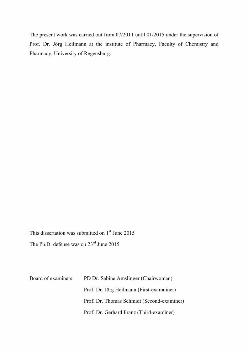
Table 1. Garcinia species which have been phytochemically investigated to date
Species Parts
G. achachairu Seeds Dal Molin et al., 2012
G. afzelii Stem bark
Seeds
Kamdem et al., 2006
Lannang et al., 2010
G. amplexicaulis Stem bark Lavaud et al., 2013 and 2015
G. aristata Fruits Cuesta-Rubio et al., 2001
G. assigu Stem bark Ito et al., 1997 and 2003
G. atroviridis Stem bark
Root
Fruits
Kosin et al., 1998; Tan et al., 2014
Permana et al., 2001 and 2005
Mackeen et al., 2002; Tan et al., 2013
G. bakeriana Leaves Al-Shagdari et al., 2013
G. bancana Twigs
Leaves
Rukachaisirikul et al., 2005
G. benthami Stem bark
Bark
Leaves
Elya et al., 2006
Nguyen et al., 2011
G. bracteata Leaves
Twigs
Stem bark
Stems
Fruits
Thoison et al., 2000 and 2005
Na et al., 2010; Li et al., 2015
Niu et al., 2012
Hu et al., 2013 and 2014
Na et al., 2013
G. brasiliensis Seeds
Pericarps
Martins et al., 2007; Naldoni et al., 2009
Martins et al., 2008; Gontijo et al., 2012
G. brevipedicellata Stem bark Ngoupayo et al., 2008
G. buchananii Heartwood
Stem bark
Jackson et al., 1971
Stark et al., 2012 and 2013
G. cambogia Latex
Root
Fruits
Rama et al., 1980
Iinuma et al., 1998
Masullo et al., 2010

The genus Garcinia 5
Table 1. (continued)
G. cantleyana Trunk bark
Twigs
Shadid et al., 2007
Jantan et al., 2012
G. celebica Leaves Elfita et al., 2009
G. chapelieri Bark Rambeloson et al., 2014
G. cochinchinensis Bark Nguyen et al., 2011; Trinh et al., 2013
G. conrauana Bark
Heartwood
Seeds
Leaves
Hussain and Waterman, 1982
G. cornea Stem bark Elfita et al., 2009
G. cowa Latex
Fruits
Stems
Stem bark
Na Pattalung et al., 1994; Mahabusarakam et al., 2005
Panthong et al., 2006; Sriyatep et al., 2014
Shen et al., 2007; Siridechakorn et al., 2012
G. cuneifolia Stem bark Ea et al., 2003
G. cymosa Stem bark Elfita et al., 2009
G. densivenia Stem bark Waterman and Crichton, 1980
G. dioica Bark Iinuma et al., 1996
G. dulcis Branches
Root
Bark
Leaves
Fruits
Flowers
Seeds
Harrison et al., 1994
Iinuma et al., 1996
Ito et al., 1997
Kosela et al., 2000
Deachathai et al., 2005
Deachathai et al., 2006
Deachathai et al., 2008
G. echinocarpa Bark Bandaranayake et al., 1975
G. edulis
G. esculenta
Root bark
Twigs
Magadula, 2010
Zhu, et al., 2014
G. eugeniifolia Heartwood Jackson et al., 1971

6 The genus Garcinia
Table 1. (continued)
G. ferrea
Stem bark
Bark
Hartati et al., 2008
Bui et al., 2014
G. forbesii Branches
Twigs
Harrison et al., 1992
G. fusca Stem bark
Root
Ito et al., 2003
Nontakham et al., 2014
G. gardneriana Leaves
Bark
Seeds
Castardo et al., 2008
Otuki et al., 2011
G. gaudichaudii Leaves
Bark
Cao et al., 1998
Xu et al., 2000
G. gerrardii Root bark Sordat-Diserens et al., 1989
G. griffithii Stem bark
Leaves
Bark
Elfita et al., 2009; Nilar et al., 2005
Alkadi et al., 2013
Xu et al., 1998
G. hanburyi Latex
Fruits
Leaves
Twigs
Asano et al., 1996; Tao et al., 2009
Reutrakul et al., 2007
Reutrakul, 2010
G. hombroniana Pericarps
Leaves
Twigs
Bark
Rukachaisirikul et al., 2000
Rukachaisirikul et al., 2005
Klaiklay et al., 2013
Jamila et al., 2014
G. huillensis Stem bark Bakana et al., 1987; Dibwe et al., 2012
G. humilis Bark Herath et al., 2005; Haase, 2011
G. indica Seeds
Leaves
Badami and Desai, 1968
Badami and Razdan, 1972
Heartwood
Fruits
Cotterill et al., 1977
Krishnamurthy et al., 1981 and 1982

The genus Garcinia 7
Table 1. (continued)
G. intermedia
Stem bark
Leaves
Lakshmi et al., 2002
Abe et al., 2004
G. kola Fruits
Seeds
Bark
Root
Stems
Mesocarp
Hussain et al., 1982
Iwu, 1985
Kabangu et al., 1987
Iwu et al., 1990; Niwa et al., 1994
Terashima et al., 1999a and 1999b
Morabandza et al., 2013
G. lateriflora Stem bark Kosela et al., 1999; Ren et al., 2010
G. lancilimba Stem bark Yang et al., 2007; Han et al., 2008
G. latissima Bark Ito et al., 1997
G. linii Root Chen et al., 2004 and 2006
G. livingstonei Heartwood
Root bark
Leaves
Fruits
Pelter et al., 1971
Sordat-Diserens et al., 1990 and 1992
Kaikabo et al., 2009
Yang et al., 2010
G. lucida Bark
Stem bark
Nyemba et al., 1990
Fotie et al., 2007
G. macrophylla Leaves
Flowers
Twigs
Andrade et al., 2007
Williams et al., 2003
G. madruno Aerial Osorio et al., 2013
G. maingayii Stem bark Hartati et al., 2007
G. malaccensis Stem bark Taher et al., 2012; Alkadi et al., 2013
G. mangostana Fruits
Leaves
Pericarps
Heartwood
Root bark
Govindachari et al. 1971; Suksamrarn et al., 2006
Parveen et al., 1990 and 1991
Sen et al., 1982; Suksamrarn et al., 2002
Nilar and Harrison, 2002
Ea et al., 2006

8 The genus Garcinia
Table 1. (continued)
G. mannii
Stem bark
Bark
Heartwood
Seeds
Leaves
Ea et al, 2008; Han et al., 2009
Crichton and Water, 1979
Hussain and Waterman, 1982
G. merguensis Bark
Wood
Twigs
Nguyen et al., 2003
Kijjoa et al., 2008
Trisuwan et al., 2013
G. multiflora Heartwood
Stems
Fruits
Twigs
Stem bark
Leaves
Chen et al., 1975
Chiang et al., 2003
Chen et al., 2008
Liu et al., 2010
Jing et al., 2013
Jiang et al., 2014
G. myrtifolia Bark Spino et al., 1995
G. neglecta Leaves Ito et al., 2001
G. nervosa Stem bark
Leaves
Ampofo and Waterman, 1986
Babu et al., 1988; Ilyas et al., 1994
G. nigrolineata Bark
Leaves
Twigs
Rukachaisirikul et al., 2003
Rukachaisirikul et al., 2003
Rukachaisirikul et al., 2005
G. nobilis
G. nujiangensis
Stem bark
Twigs
Twigs
Fouotsa et al., 2012
Fouotsa et al., 2014
Tang et al., 2015
G. oblongifolia Bark
Stems
Leaves
Bark
Hamed et al., 2006
Wu et al., 2008
Zhang et al., 2014
Feng et al., 2014
G. oligantha Stems Gao et al., 2013; Wu et al., 2013

The genus Garcinia 9
Table 1. (continued)
G. oliveri
G. opaca
Bark
Leaves
Bark
Ha et al., 2009 and 2012
Goh et al., 1992
Mori et al., 2014
G. ovalifolia Stem bark
Leaves
Waterman and Crichton, 1980; Lannang et al., 2013
Gustafson et al., 1992
G. parvifolia Leaves
Bark
Stem bark
Twigs
Xu et al., 2000
Xu et al., 2001
Kardono et al., 2006; Syamsudin et al., 2013
Rukachaisirikul et al., 2006
G. paucinervis Leaves
Stem bark
Stems
Gao et al., 2010; Wu et al., 2013
Fan et al., 2012
Hu et al., 2014
G. penangiana Leaves Jabit et al., 2007
G. penduculata Heartwood
Pericarps
Bark
Rao et al., 1974
Sahu et al., 1989
Vo et al., 2012
G. polyantha Stem bark
Wood
Root bark
Leaves
Lannang et al., 2005
Louh et al., 2008
Lannang et al., 2008
Lannang et al., 2014
G. porrecta Stem bark Kardono et al., 2006
G. prainiana Leaves
Twigs
Klaiklay et al., 2011; Mawa and Said, 2012
Susanti et al., 2013
G. preussii Leaves Messi et al., 2012
G. propinqua Twigs Tantapakul et al., 2012
G. pseudoguttifera Heartwood Ali et al., 2000
G. puat Leaves Ito et al., 2001
G. punctata Stem bark Ngameni et al., 2014
G. purpurea Pericarp Iinuma et al., 1996
10 The genus Garcinia
Table 1. (continued)
G. pyrifera Stem bark
Fruits
Ampofo and Waterman, 1986
Roux et al., 2000
G. quadrifaria Stem bark Waterman and Hussain, 1982
G. quaesita Bark Gunatilaka et al., 1984
G. rigida Leaves Elya et al., 2006 and 2011
G. scortechinii Twigs
Latex
Stem bark
Fruits
Rukachaisirikul et al., 2000
Rukachaisirikul et al., 2003
Rukachaisirikul et al., 2005
Sukpondma et al., 2005
G. semseii Stem bark
Pericarps
Magadula et al., 2008
Magadula et al., 2012
G. shomburgkiana Bark
Wood
Stems
Vo et al., 2012
Mungmee et al., 2013
Ito et al., 2013
G. smeathmannii Stem bark
Root bark
Komguem et al., 2005; Kuete et al., 2007
Lannang et al., 2006
G. solomonensis Stem bark Carroll et al., 2009
G. speciosa Bark
Trunk bark
Vieira et al., 2004
Rukachaisirikul et al., 2003
G. spicata Bark
Fruit
Leaves
Konoshima et al., 1970
Lyles et al., 2014
Gunatilaka et al., 1984
G. subelliptica Heartwood
Root bark
Wood
Seeds
Stem bark
Fruits
Leaves
Fukuyama et al., 1991
Iinuma et al., 1995
Fukuayama et al., 1998
Weng et al., 2003 and 2004
Abe et al., 2003
Zhang et al., 2010
Ito et al., 2013

The genus Garcinia 11
Table 1. (continued)
G. succifolia Wood Duangsrisai et al., 2014
G. staudtii Stem bark Waterman and Hussain, 1982
G. talboti Root Joshi et al., 1970
G. terpnophylla Bark
Wood
Bandaranayake et al., 1975
G. tetralata Stem bark
Branches
Leaves
Wang et al., 2008; Guo et al., 2011
Na and Xu, 2010
G. tetranda Wood Purwaningsih and Ersam, 2007
G. thwaitesii Bark
Timber
Gunatilaka et al., 1983
G. vieillardii Stem bark Hay et al., 2004 and 2008
G. vilersiana Bark Nguyen et al., 1999; Bui et al., 2012
G. virgata Stem bark Merza et al., 2004 and 2006
G. volkensii Heartwood
Bark
Herbin et al., 1970
Magadula et al., 2014
G. xanthochymus Fruits
Bark
Stem bark
Twigs
Karanjgoakar et al., 1973; Baggett et al., 2005
Chen et al., 2008 and 2010
Ji et al., 2012
Trisuwan et al., 2014
G. xipshuanbannaensis Fruits
Leaves
Twigs
Shen et al., 2006
Na and Xu, 2010
Han et al., 2008; Na and Xu, 2009

12 The genus Garcinia
2.2.1. Xanthones
The latex or resin of Garcinia species is commonly yellow because of the presence
of xanthone derivatives. Xanthones have been widely found in all species of this genus
including simple oxygenated xanthones, prenylated xanthones, caged xanthones and
bisxanthones.
2.2.1.1. Simple oxygenated xanthones
These compounds can be di-, tri-, tetra- or penta-oxygenated xanthones with simple
substituents such as hydroxyl, methoxyl and methyl groups (see Figure 1). Two
xanthones, 1,5- and 1,7-dihydroxyxanthones are simple di-oxygenated xanthones
found extensively in many species (Bennett and Lee, 1989). Simple tri-oxygenated
compounds as 1,3,5-trihydroxyxanthone and 1,5-dihydroxy-2-methoxyxanthone were
isolated from G. xanthochymus (Baslas, 1979). Three xanthones, 1,3,5,7-, 1,3,6,7-
tetrahydroxyxanthones and 1,2-dihyroxy-5,6-dimethoxyxanthone, which were reported
from G. penduculata and G. subelliptica, are examples for simple tetra-oxygenated
xanthones (Rao et al., 1974; Minami et al., 1995). Simple penta-oxygenated xanthones
are rarely found in the genus, whereas some were isolated from the genus Calophyllum
(Guttiferae). Three xanthones, 1,3,4,5,8-pentahydroxyxanthone, 1,3,8-trihydroxy-4,6-
dimethoxyxanthone and 1,3,7-trihydroxy-4,6-dimethoxyxanthone, are simple penta-
hydroxyxanthones identified from two species G. tetranda and G. hombroniana
(Purwaningsih and Ersam, 2007; Klaiklay et al., 2013).
2.2.1.2. Prenylated xanthones
Prenylated xanthones are di-, tri-, tetra- and penta-oxigenated xanthones, whose the
aromatic ring system is substituted by prenyl groups such as isoprenyl, 1,1-
dimethylprop-2-enyl, geranyl and farnesyl (see Figure 2). These groups can be
cyclized with adjacent hydroxyl groups at the ortho-position to give tetra- or penta-
cyclic xanthones with furano- or pyrano rings, respectively. A xanthone can be
substituted by one, two or three prenyl groups. Natural xanthones with more than three
prenyl groups have not yet been found up to date. A large number of prenylated tri-
and tetra-oxygenated xanthones have been reported from the genus. Prenylated penta-

The genus Garcinia 13
oxygenated xanthones are rare. 1,7-Dihydroxy-3-methoxy-2-(3-methylbut-2-
enyl)xanthone, isocowanol and dulciol C, three xanthones from G. mangostana, G.
pyrifera and G. dulcis, are three xanthones substituted by one, two and three prenyl
groups (Mahabusarakam et al., 1987; Ampofo and Waterman, 1986; Iinuma et al.,
1996). Garcigerrin A, a tri-oxygenated xanthone with a pyrano ring from G. gerrardii
and rheediaxanthone B, a tetra-oxygenated xanthone with a furano ring from G.
polyantha, are two examples of tetracyclic xanthones (Ampofo and Waterman, 1986;
Sordat-Diserens et al., 1989). Subelliptenone C from G. subelliptica is a tetra-cyclic
xanthone with a furo ring (Iinuma et al., 1995). BR-xanthone A, a xanthone from G.
mangostana and 1,5-dihydroxy-6,6-dimethylpyrano(2,3:6,7)-4,4,5-
trimethylfurano(2,3:3,4)-2-(3-methylbut-2-enyl)xanthone, a xanthone from G.
opaca, are representative of penta-cyclic xanthones (Balasubramanian and
Rajagopalan, 1988; Goh et al., 1992). 5-Farnesyltoxyloxanthone, a tetraoxygenated
xanthone isolated from G. merguensis, is a rare xanthone with a farnesyl group (Kijjoa
et al., 2008).

14 The genus Garcinia
O
O OH
OH
O
O OH
HO
O
O OH
OH
OH1,5-Dihydroxyxanthone 1,7-Dihydroxyxanthone 1,3,5-Trihydroxyxanthone
O
O OH
OH
OH
HO
O
O OH
OHHO
HO
O
O OH
OH
OHOH
OH
1,3,6,7-Tetrahydroxyxanthone 1,3,5,7-Tetrahydroxyxanthone 1,3,4,5,8-Pentahydroxyxanthone
O
O OH
OMe
OH
O
O OH
OH
MeO
OMe
1,5-Dihydroxy-2-methoxyxanthone1,2-Dihydroxy-5,6-dimethoxyxanthone
O
O OH
OH
OMe
MeO
OH
O
O OH
OH
OMe
HO
MeO
1,3,7-Trihydroxy-4,6-dimethoxyxanthone 1,3,8-Trihydroxy-4,6-dimethoxyxanthone
Figure 1. Simple xanthones from some Garcinia species

The genus Garcinia 15
O
OHO
OMe
HO
1,7-Dihydroxy-3-methoxy-2-(3-methylbut-2-enyl)xanthone O
O OH
OHHO
OH
MeO
O
O OH
OH
HO
OH
HO
Isocowanol
Dulciol C
O
OHO
OHO
HO
HO
O
OHO
OH
OHO
Garcigerrin A
Rheediaxanthone B
BR-xanthone A
O
OHO
OH OH
O
Subelliptenone C
O
OHO
OH
O O
1,5-Dihydroxy-6,6-dimethylpyrano(2,3:6,7)-4'',4'',5''-trimethylfurano(2'',3'':3,4)-2-(3-methylbut-2-enyl)xanthone
O
O
O OH
OHHO
5-Farnesyltoxyloxanthone
O
OHO
OHO
O
Figure 2. Prenylated xanthones from some Garcinia species

16 The genus Garcinia
2.2.1.3. Caged xanthones
These compounds are polyprenylated xanthones, in which the ring B is converted
into an unusual ring, 4-oxa-tricyclo[4.3.1.03,7]dec-2-one (caged) scaffold (Ollis et al.,
1965). This motif can be further modified with various substituents on the aromatic
ring A and can also be oxidized to form a wide range of compounds with interesting
structures, see Figure 3. Caged xanthones have been restrictively found in some
species of the genus Garcinia, especially G. hanburyi. Over one hundred compounds
have been identified from the genus. Morellin, a caged xanthone from G. morella, was
first reported in 1937 (Rao, 1937). Gambogellic acid, one of caged xanthones isolated
from G. hanburyi, has a polycyclic structure (Asano et al., 1996). Although the
majority of caged xanthones possess the general motif, a few compounds have
alternative structures. For example, 6-O-methylneobractatin identified from G.
bracteata, contains the neo-motif (Thoison et al., 2000). Lateriflorone isolated from G.
lateriflora, has a spiroxalactone core by oxidation of the ring C (Kosela et al., 1999),
whilst the ring B in the structure of scortechinone K, one of caged xanthones from G.
scortechinii, undergoes ring-opening oxidation (Rukachaisirikul et al., 2003).
2.2.1.4. Bisxanthones
Bisxanthones are dimeric xanthones, which have been found in some species of the
family Guttiferae. They are formed from prenylated monomeric xanthones by ring-
closure coupling through side-chains, see Figure 4. Garcilivins A-C, which was
isolated from the bark of the of roots G. livingstonei, have been reported for the first
time from the genus. These compounds are dimerics linked via a 6-membered ring,
which is formally derived from a Diels-Alder-type reaction between two isoprenyl
units. (Sordat-Diserens et al., 1992). Bigarcinenone A from the bark of G.
xanthochymus is a dimer linked via a 5-membered ring like cratoxyxanthone. A
biosynthetic pathway of the latter one was proposed to involve the coupling of two
xanthone radicals, see Scheme 11 (Zhong et al., 2008). Bigarcinenone B, which was
also obtained from G. xanthochymus, is a bisxanthone that is connected via a 6- and 6-
membered ring system (Chen et al., 2011).

The genus Garcinia 17
O O
O
O
A
B
C
O O
OH O
O
O
HOC
Morellin6-O-Methylneobractatin
O
OH O
O
O
O
O O
OH O
O
O
HOOC
Gabogellic acidGeneral motif of caged xanthones
O
O
O
O
O
O
O
O
OH
Lateriflorone
O
OH O
O
HOOC
O
OCH3
O
O
Scortechinone K
Figure 3. Caged xanthones from some Garcinia species

18 The genus Garcinia
O
O OH
OH OH
O
OOH
R
OH
OH
Garcilivin A (R = )H
Garcilivin C (R = )H
O
O OH
OH OH O
O
O
OH
HO
Garcilivin B
O
O OH
OH OH
O
O
O
HO
OH
OH
OH
H
H
O
O OH
OH
O
O
OH
HO
HO
OHHO
Bigarcinenone B
Bigarcinenone A
Figure 4. Bisxanthones from some Garcinia species
2.2.2. Biflavonoids
The genus Garcinia is shown to be a source of biflavonoids. To the best of our
knowledge, about one hundred biflavonoids have been identified from this genus to
date. The majority of these compounds are dimers of two flavanones, two flavones,
between a flavanone and a flavone or a flavanonol, which are linked at the positions C-
3 and C-8 or C-3 and C-8. There are four main groups of Garcinia biflavonoids:
GB-1 type with a flavanone-(3-8)-flavanonol system, GB-1a type with a flavanone-

The genus Garcinia 19
(3-8)-flavanone system, morelloflavone type with a flavanone-(3-8)-flavone system
and amentoflavone type with a flavone-(3-8)-flavone system (Ferreira et al., 2012;
Ito et al., 2013). Beside these four types, some biflavonoids have alternative structures.
For instance, lateriflavone with the 6-8 linkage has been isolated from G. lateriflora,
whereas (22)-biflavonol from G. nervosa has a rare linkage between C-2 and C-
2, see Figure 5 (Parveen et al., 1994; Ren et al., 2010).
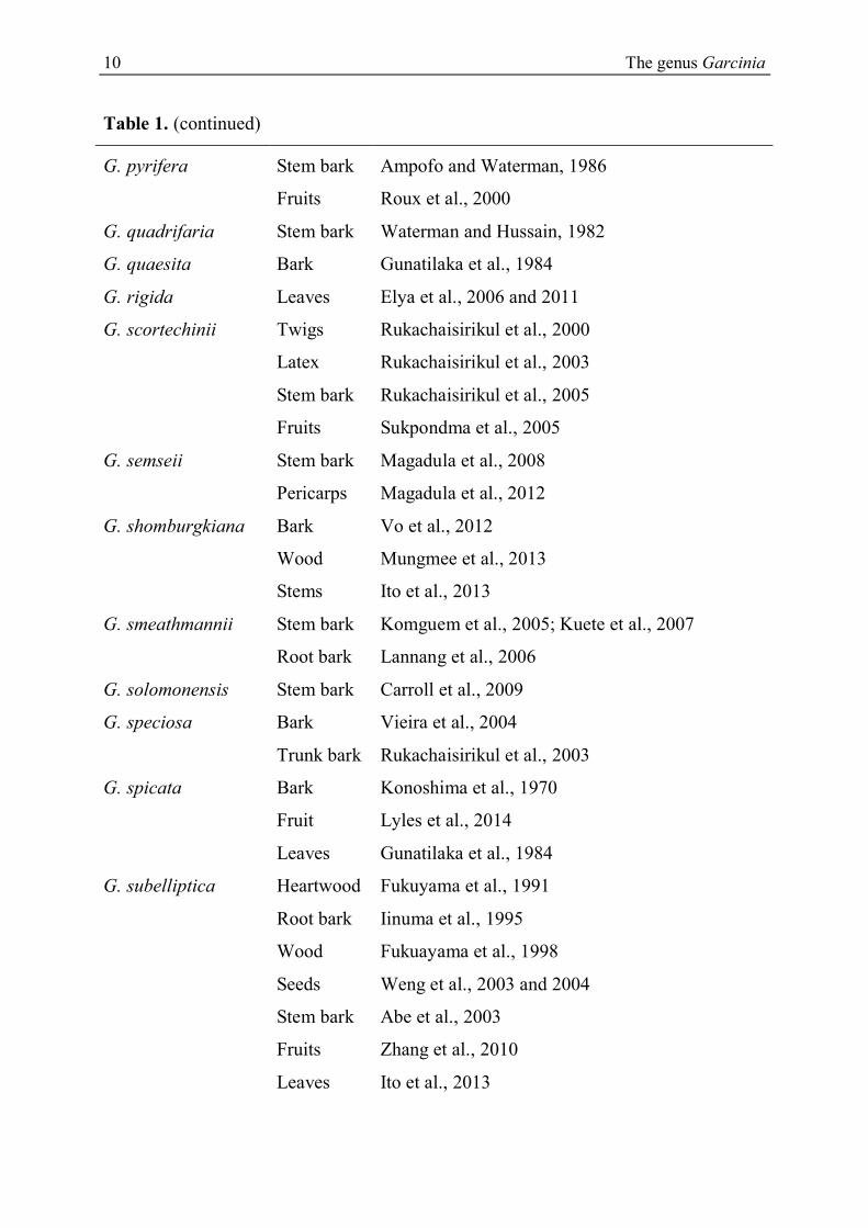
2.2.3. Benzophenones
Benzophenones from Garcinia comprise simple benzophenones and polyprenylated
benzophenones, in which the aromatic ring A can undergo oxidation, prenylation and
cyclization to form polycyclic benzophenones with a bicyclo[3.3.1]nonane-2,4,9-
trione skeleton (Zhang et al., 2010). These polycyclic benzophenones are divided into
three types corresponding to the position of the benzoyl group. Type A includes
compounds with the benzoyl group at C-1, for example garcimultiflorone A from G.
multiflora (Chen et al., 2009). Type B, with the benzoyl group at C-3 such as
pedunculol from G. pendunculata, was widely found in the genus (Sahu et al., 1989).
Garcinielliptone K, which was obtained from G. subelliptica, is representative of type
C with the benzoyl group at C-5 (Weng et al., 2004). Cyclization involving the β-
diketone and olefinic groups in polyprenylated compounds led to formation of
interesting adamantanyl benzophenones such as garciniagifolone A from G.
oblongifolia (Shan et al., 2012).. Additionally in some cases, the ring B can be
modified into a bicyclo[3.3.2]decane-2,4,10-trione skeleton such as gambogenone
reported from G. xanthochymus or can undergo intramolecular oxidative coupling
between the enol and the aromatic ring B to form corresponding polycyclic xanthone
derivatives, for examples garcinialone obtained from G. multiflora (Baggett et al.,
2005; Chien et al., 2008). Doitunggarcinone B, which was isolated from G. propinqua,
is an unusually rearranged benzophenone, see Figure 6 (Tantapakul et al., 2012).

20 The genus Garcinia
OHO
OH O
OH
OHO
OH O
OH
OH
OHO
OH O
OH
OHO
OH O
OH
OHO
OH O
OH
OHO
OH O
OH
OHO
OH O
OH
OHO
OH O
OH
OHO
OH O
OH
OHO
OH O
OH
OHO
OH O
OH
OH
HO
O
OH
OH
HO
O
3
8''
3'
8''
6
8''
2'
2'''
GB-1 GB-1a
Morelloflavone
Amentoflavone
Lateriflavone
(I-2', II-2''')-Biapigenin
Figure 5. Biflavonoids from some Garcinia species
The genus Garcinia 21
O
OH
O1
2
34
5
6
7
8
9
O
O
O
O
Bicyclo[3.3.1]nonane-2,4,9-trioneGarcimultiflorone A
O
OO
OH
HO O
Pedunculol
O
O
O
O
Garcinielliptone K
B
A
OO
H
HO
OH
O
O
O
OO
OH
HO HO
Garciniagifolone A
Gambogenone
O
HO OO
HO
HOHO
Garcinialone
Doitunggarcinone A
O
O
OH
HO
Figure 6. Benzophenones from some Garcinia species

22 The genus Garcinia
2.2.4. Phloroglucinols
Phloroglucinol derivatives reported in the genus include simple phloroglucinols and
complex compounds with an oxidized and polyprenylated nucleus or a
bicyclo[3.3.1]nonane-1,3,9-trione skeleton. Parvifoliol A is one of simple
phloroglucinols isolated from G. parvifolia (Rukachaisirikul et al., 2006).
Garcinielliptone HB is one of seven polyprenylated phloroglucinols identified from G.
subelliptica, whilst garcicowin A from G. cowa is a phloroglucinol derivative with the
bicycle[3.3.1]nonane-1,3,9-trione core (Lu et al., 2008; Xu et al., 2010).
Garcinielliptone HF, which was also obtained from G. subelliptica, is a phloroglucinol
with an unprecedented skeleton, see Figure 7 (Wu et al., 2008).
OH
MeO OMe
HO
O
Parvifoliol A
O
OMe
OO
HO
O
O
HO
OH
OH
HO
OH
O
Garcinielliptone HF
Garcicowin A
Garcinielliptone HB
O
O
O
Figure 7. Phloroglucinol derivatives from some Garcinia species

The genus Garcinia 23
2.2.5. Depsidones
Depsidones are polyphenolic compounds containing the 11H-dibenzo[b,e][1,4]-
dioxepin-11-one system. These compounds were mainly found in lichens, which
usually provide polyketide-derived natural products. However a substantial number of
depsidones were reported from the genus Garcinia, which is well known as a rich
source of shikimate-derived aromatic compounds. Depsidones from the genus are
commonly substituted with hydroxyl, methoxyl, isoprenyl and geranyl groups. For
instance, garcidepsidone A, one of four prenylated depsidones, was isolated from G.
parvifolia (Xu et al., 2000). Prenyl side-chains can be cyclized with hydroxyl groups at
the position ortho to give tetra- and penta-cyclic compounds such as garcinisidone B
and C, a tetracyclic depsidone and a penta-cyclic depsidone, respectively, were
identified from G. neglecta, see Figure 8 (Ito et al., 2001).
O
O
O
O
O
OHO
HO
OH
OH
O
O
OHO
OMe
OH
O
O
OHO
O
O
11H-dibenzo[b,e][1,4]-dioxepin-11-oneGarcidepsidone A
Garcinisidone C Garcinisidone B
Figure 8. Depsidones from some Garcinia species

24 The genus Garcinia
2.2.6. Tocotrienols
Tocotrienol derivatives were isolated from some Garcinia species. They have a 6-
chromanol skeleton substituted by a farnesyl group at the position C-2, which can be
oxidized at two terminal methyl groups. These compounds can be mono- or dimeric
derivatives. For instance, 5-formyl--tocotrienol is a mono derivative identified from
G. virgate (Merza et al., 2004), whilst δ,γ-bi-O-amplexichromanol, δ,γ-
biamplexichromanol and ,-biamplexichromanoate are dimeric tocotrienols reported
from G. amplexicaulis, see Figure 9 (Lavaud et al., 2015).
O
HO
CHOO
HO OH
OH
O
O
OH
OH
5-Formyl--tocotrienol
O
HO OH
OH
O
OH
OH
HO
O
HO
O
HO
OH
O
O
,-Bi-O-amplexichromanol
,-Biamplexichromanol
,-Biamplexichromanolate
Figure 9. Tocotrienols from some Garcinia species

The genus Garcinia 25
2.2.7. Biphenyls
A series of biphenyl derivatives were identified from the genus. These compounds
possess a biphenyl core substituted by hydroxyl, methoxyl and isoprenyl groups such
as garcibiphenyls B and C from the root of G. linii (Chen et al., 2006).
Oblongifoliagarcinines A and C, which were reported from G. oblongifolia, are tri-
and tetracyclic biphenyls, see Figure 10 (Wu et al., 2008).
OH
OMe
HO
HO
OH
OH
OMe
HO
Garcibiphenyl CGarcibiphenyl B
O
OH
OH
O
OH
O
Oblongifoliagarcinine A Oblongifoliagarcinine B
Figure 10. Biphenyl derivatives from some Garcinia species
2.2.8. Triterpenoids
Triterpenoids from this genus consist of lanostanes, friedolanostanes,
abeolanostanes, cycloartanes, friedocycloartanes, friedelanes, protostanes, triterpenes
with lup-20(29)-en-type and oleanolic acid skeletons. Garcihombronane A and D are a
friedolanostane and a lanostane of ten triterpenes isolated from the leaves of G.
hombroniana (Rukachaisirikul et al., 2000 and 2005). 14β,15β-Epoxy-3β-hydroxy-9-
oxo-11[10-8]-abeolanostan-22-cis,24-trans-dien-28-oic acid is one of four 11(10-8)-
abeolanostanes reported from G. speciosa (Vieira et al., 2004), whereas
garciosaterpene A is one of three protostanes also isolated from this species
(Rukachaisirikul et al., 2003). 2α-Hydroxy-3β-O-acetyllup-20(29)-en-28-oic acid, a
lup-20(29)-en-type triterpene and an oleane, 3-O-(4′-O-acetyl)-α-L-

26 The genus Garcinia
arabinopyranosyloleanolic acid, have been reported from the resin of G. hanburyi
(Wang et al., 2008). (22Z,24E)-3-Hydroxy-17,13-friedocycloarta-12,22,24-trien-26-
oic acid is a friedocycloartane obtained from the bark of G. benthami, whilst
ovalifolone A is a friedelane derivative from G. ovalifolia, see Figure 11 (Nguyen et
al., 2011; Lannang et al., 2013).
2.2.9. Other compounds
Hydroxycitric acid and its derivatives were isolated from the fruits of three species,
G. cambogia, G. indica and G. atroviridis (Lewis, 1969). (-)-Hydroxycitric acid has
drawn all worldwide attention because of its anti-obesity property (Krishnamurthy and
Sapna, 2008). Some sesquiterpenes were identified from the genus, for instance,
scortechterpenes A and B from the fruit of G. scortechinii (Sukpondma et al., 2005).
Garcinielliptones N and O are two novel terpenoids isolated from the seed of G.
subelliptica (Weng et al., 2004). Some unusual compounds were reported from the
genus such as three benzophenone-xanthone dimers from the root of G. dulcis,
garciduols A-C have been first identified in nature (Iinuma et al., 1996). Two
flavanone-chromone dimers, preussianone and I-4,I-5,II-5,I-7,II-7-
pentahydroxyflavanone[I-3,II-8]-chromone were isolated from the leaves of G.
preussii and G. dulcis (Messi et al., 2012; Ansari and Rahman, 1975). The
unsubstituted chromone moiety is supposed to be derived by elimination of a phenyl
ring from a biflavone. Garcinianins A and B, two new proanthocyanidins from the
leaves of G. multiflora, have been first reported from the genus (Jiang et al., 2014). In
addition, the essential oil of some species contains volatile compounds, see Figure 12
and 13 (Macleod and Pieris, 1982).

The genus Garcinia 27
HHO
COOH
H
COOH
O
HO
Garcihombronane A Garcihombronane D
HO
O
COOH
OH
CH3COO
COOHH
H810
Garciosaterpene A
14,15-Epoxy-3-Hydroxy-9-oxo-11[10-8]-abeolanostan-22-cis,24-trans-dien-28-oic acid
HO
H
HOOC
COOH
CH3COO
HO
COOH
OOH
OH
OCH3CO
2-Hydroxy-3-O-acetyllup-20(29)-en-28-oic acid
3-O-(4'-O-acetyl)--L-arabinopyranosyloleanolic acid
OH
HO
O
Ovalifolone A (22Z,24E)-3-Hydroxy-17,13-friedocycloarta-12,22,24-trien-26-oic acid
Figure 11. Triterpenoids from some Garcinia species

28 The genus Garcinia
O
O OR1
OR1OR1
OR1
OMe
OR1 O
R2O
O OH
OH
OH
O
HO OMe
OH
Garciduol A (R1=R2= H)Garciduol B (R1= H, R2= OH)
Garciduol C
O
OO
O
OH
HO
OHHO
OH
H
H
O
O
OH
HO
OH
H
OHH
OH
O
O OH
OH
O
OH
HO
OH
OHO
O
OHO O
OH
OH
HO
OH
OH
O
OH
HO
OH
OHO
HO O
OHO
HO
HO
OH
OH
Preussianone AI-4 ,I-5,II-5,I-7,II-7-pentahydroxyflavanone[I-3,II-8]-chromone
Garcinianin A
Garcinianin B
Figure 12. Some rare phenolic compounds in the genus Garcinia

The genus Garcinia 29
OMeH
H
O
MeO
H
O
H
COOH
H
HHO
COOHHO
COOHH
(-)-Hydroxycitric acid Scortechterpene A Scortechterpene B
Garcinielliptone N Garciniellipone O
O O O O
CH3OOC
Figure 13. (-)-Hydroxycitric acid and terpenoids from some Garcinia species

30 The genus Garcinia
2.3. Pharmacological and biological properties of Garcinia
Many species of the genus are used in traditional medicine around the world,
particularly in Asia and Africa. The pericarp of G. mangostana is used in Southeast
Asia for the treatment of skin infections, wounds, dysentery, diarrhoea, fever, arthritis
and inflammation (Pedraza-Chaverri, 2008). The leaves and seeds of G. dulcis are
used in Indonesian folk medicine to treat lymphatitis, parotitis and struma, whereas its
stem bark is used in Thailand as an antiseptic and the fruit juice as an anti-scurvy and
expectorant. In addition, its root extract is also used as an antipyretic and antitoxin
(Kalahari and Hemi, 1986; Wuttidhammavej, 1997). The bark of G. cowa is used in
Thai folk medicine as an antipyretic and antimicrobial agent. Its latex is also used as
anti-fever agent (Na Pattalung et al., 1994). In India, the fruit of G. indica is
anthelmintic and useful for piles, dysentery, tumours, pain and heart complaints (Jena
et al., 2002). G. cambogia extract has been used in Indian traditional medicine to treat
tumours, ulcers, haemorrhoids, diarrhoea, dysentery, fever, open sores and parasites
(Duke, 2002). The gum of G. hanburyii is used internally in Thailand as a purgative,
vermifuge and for treatment of infected wounds. It is also applied for treatment of
chronic dermatitis, haemorrhoids and bedsore. In China, it was developed as an
antitumor medicine (Saralamp et al., 1996; Han et al., 2006). G. xanthochymus is
widely used in Chinese traditional medicine for dispelling worms and removing food
toxin (Lin et al., 2003). G. hombroniana, a seashore mangosteen in Malaysia, is used
as protective medicine after child birth and to cure skin allergies (Jamila, 2014). In
Africa, G. preussii is traditionally used to treat stomach aches and its leaves are
prepared as a decoction to relieve toothache (Bouquet, 1969; Visser, 1975). Extracts of
G. kola are used in Nigerian ethnic medicine against laryngitis, cough and liver
diseases. Its seeds are used in Africa as an antidote (Iwu et al., 1985 and 1987). The
leaves and flowers of G. afzelii are used in Cameroon and Ghana for antibacterial
properties (Waffo et al., 2006). In Fiji, an extract of the leaves of G. pseudoguttifera is
mixed with coconut oil and used to relieve pain in the limbs (Cambie and Ash, 1994).
Pharmacological and biological investigations of natural products from the genus
showed that some of them possess a wide range of various properties such as anti-

The genus Garcinia 31
oxidant, antifungal, antimicrobial, anti-inflammatory, anticancer and antiviral
activities (Hemshekhar et al, 2011).
2.3.1. Anti-oxidant activity
1,8-Dihydroxy-6-methoxyxanthone, a tri-oxygenated xanthone from the wood G.
subelliptica exhibited inhibitory activities in three in vitro assays viz., anti-
lipidperoxidation in rat brain homogenates, DPPH free radical scavenging and
superoxide anion scavenging assays at 5 µg/ml (Minami et al., 1994). -Mangostin
from the pericarp of G. mangostana inhibited 7,12-dimethylbenz[]anthracene-
induced preneoplastic lesions in a mouse mammary organ culture assay with an IC50 of
1.0 µg/ml (Jung et al., 2006). Garcidepsidone B, a depsidone from the twigs of G.
parvifolia, gave an IC50 of 0.13 µM equal to that of BHT in the DPPH free radical
scavenging assay (Rukachaisirikul et al., 2006). The antioxidant activity of
bigarcinenone A, a bisxanthone from the bark of G. xanthochymus, is even stronger
than that of BHT in a DPPH radical scavenging test. Bigarcinenone A gave an IC50 of
9.2 µM, compared to the positive control, BHT with an IC50 of 20 µM, see Figure 14
(Zhong et al., 2008).
2.3.2. Antifungal activity
Beside the anti-oxidant activity, -mangostin also exhibited the inhibition towards
the fungi Alternaria solani, Cunninghamella echinulata and Candida albicans that
causes candidiasis with a MIC of 1 mg/ml. It was shown to be more efficient than the
existing antifungal drugs such as clotrimazole and nystatin (Sundaram et al., 1983;
Kaomonkolgit et al., 2009). Two isoprenylated tri-oxygenated xanthones, 1,4,5-
trihydroxy-3-(3-methylbut-2-enyl)xanthone and 4-(3,7-dimethylocta-2,6-dienyl)-
1,3,5-trihydroxyxanthone of G. livingstonei showed an activity against the plant
pathogenic fungus Cladosporium cucumerinum at 0.5 and 0.2 µg, respectively, see
Figure 14 (Sordat-Diserens et al., 1992).

32 The genus Garcinia
O
O OH
MeO
OH
O
O OH
HO OH
MeO
O
O
HO
HO OH
OH
OO
O OH
OH
O
O
OH
HO
HO
OHHO
Bigarcinenone A
-Mangostin
1,8-Dihydroxy-6-methoxyxanthone
Garcidepsidone B
O
O OH
HO OH
O
O OH
HO
OH
1,4,5-Trihydroxy-3-(3-methylbut-2-enyl)xanthone
4-(3',7'-Dimethylocta-2',6'-dienyl)-1,3,5-trihydroxyxanthone
Figure 14. Some anti-oxidant and antifungal compounds from the genus Garcinia

The genus Garcinia 33
2.3.3. Antimicrobial activity
Rubraxanthone isolated from G. dioica displayed higher activity against
Staphylococcal strains (MIC = 0.31-1.25 µg/ml) than that of the antibiotic,
vancomycin with MIC values of 3.13-6.25 µg/ml (Iinuma et al., 1996). Garcilivin A, a
bisxanthone from G. livingstonei, showed a high anti-parasitic activity against two
trypanosomes, T. brucei brucei and T. cruzi that cause the fata human diseases,
sleeping sickness and chagas disease with IC50 values of 0.4 µM and 4.0 µM,
respectively. Moreover, this compound also exhibited antiplasmodial property against
Plasmodium falciparum with an IC50 of 6.7 µM (Mbwambo et al., 2006).
Xanthochymol, a polyprenylated benzophenone from G. xanthochymus and G.
subelliptica, was evaluated for the antibacterial property against methicillin-resistant
Staphylococcus aureus with the lowest minimum inhibitory concentration at 3.1-12.5
µg/ml, nearly equal to that of vancomycin (Iinuma et al., 1995). Guttiferone A, a
polyisoprenylated benzophenone from the fresh fruits of G. aristata, showed a potent
antiplasmodial effect against Plasmodium falciparum with an IC50 of 0.5 µM, nearly
similar to that of chloroquine (IC50 = 0.3 µM), a 4-aminoquinoline drug used in the
treatment or prevention of malaria (Monzote et al., 2011). Amentoflavone, a biflavone
from some Garcinia species, was reported to be more active against Mycobacterium
smegmatis than the drug isoniazid used in the clinical treatment of tuberculosis. This
compound gave a MIC of 0.6 µg/ml compared to isoniazid with a MIC of 1.3 µg/ml
(Kaikabo and Eloff et al., 2011). Kolaviron, a biflavonoid complex from the seeds of
G. kola containing GB-1, GB-2 and kolaflavanone, exhibited potent antiplasmodial
activity against Plasmodium berghei infection in Swiss albino mice, see Figure 15
(Oluwatosin et al., 2014).

34 The genus Garcinia
O
O OH
HO
MeO
OH
O
O OH
HO OMe
MeO
-Mangostin
Rubraxanthone
O
O OH
OH OH
O
OOH
OH
OH
H
Garcilivin A
OHO
HO OO
OH
Xanthochymol
OHO
HO OO
OH
Guttiferone A
O
OH
O
HO
OH
O
O
OH
OH
HO
O
OH
O
HO
OHO
OOH
HO
R1
OH
R2
Amentoflavone
GB1 (R1= OH, R2= H)GB2 (R1= OH, R2= OH)Kolaflavanone (R1= OMe, R2= OH)
Figure 15. Some anti-microbial compounds from the genus Garcinia

The genus Garcinia 35
2.3.4. Anti-inflammatory activity
Garcinielliptones L and M, two polyisoprenylated phloroglucinols from the seeds of
G. subelliptica, showed potent inhibitory effects on the release of β-glucuronidase and
on histamine from peritoneal mast cell stimulated with p-methoxy-N-
methyphenethylamine in a concentration-dependent manner. They also exhibited
potent activities on NO production in culture media of RAW 264.7 cells in response to
lipopolysaccharide (LPS) and in culture media of N9 cells in response to
LPS/interferon- (IFN-) (Weng et al., 2004). Two xanthones, - and -mangostin
from the pericarps of G. mangostana, showed significant properties in the expression
decrease of TNF-, IL-1β, IL-6, IL-8, MCP-1 (monocyte chemoattractant protein) and
TLR-2 (toll-like receptor). They also potently inhibited the LPS induced NO and PEG2
activity in RAW264.7 macrophages with IC50 concentration of 3.1 and 6.0 µM
(Bumrungpert et al., 2009; Chen et al., 2008). In the study of the effects on neutrophil
pro-inflammatory responses of benzophenones from G. multiflora, garcimultiflorone D
potently inhibited fMLP/CB-induced superoxide anion generation and elastase release
with IC50 values of 7.21 and 6.0 µg/ml, respectively, see Figure 16 (Ting et al., 2012).
O
O
O
HO
O
R
Garcinielliptone L (R= )Garcinielliptone M (R= )
HH
O
OO
O
OH
H
Garcimultiflorone D
O
O OH
OHHO
HO
-Mangostin
Figure 16. Some anti-inflammatory compounds from the genus Garcinia

36 The genus Garcinia
2.3.5. Anticancer activity
Gambogic acid and epigambogic acid, two caged xanthones from the gamboges of
G. hanburyi, were examined for their cytotoxicity against human leukaemia K562/S
and doxorubicin-resistant K562/R cell lines. They were shown to be potent agents
against both cell lines with IC50 values of 1.32 and 0.89 µM for gambogic acid, 1.11
and 0.86 µM for epigambogic acid, respectively (Han et al., 2005). 7-
Hydroxyforbesione, a caged xanthone from the leaves of G. cantleyana, exhibited
significant cytotoxicity against MDA-MB-231, CaOV-3, MCF-7 and HeLa cancer cell
lines with IC50 values ranging from 0.22 - 2.17 µg/ml (Shadid et al., 2007). For 3-O-
(4′-O-acetyl)-α-L-arabinopyranosyloleanolic acid, a triterpene from the resin of G.
hanburyi, the anti-proliferative effects and the apoptosis induction abilities in four
human leukaemia cell lines consisting of HL-60, NB4, U937 and K562 were
determined with IC50 values of 2.45, 2.69, 2.42 and 4.15 µM, respectively (Wang et
al., 2008). Guttiferone A, an anti-oxidant benzophenone from some Garcinia species,
displayed strong activity against HTC-116 and HT29 cell lines with the same IC50
values of 5.0 µM (Yang et al. 2010). GB1, a biflavone reported from some Garcinia
species, inhibited -glucosidase and aromatase with IC50 values of 0.9 and 11.3 µM,
respectively. It was discussed to be a potential dietary supplement or phytomedicine
for the prevention of breast cancer and type II diabetes mellitus. Morelloflavone,
another Garcinia biflavone, was found to inhibit proteasome at an IC50 concentration
of 1.3 µM, see Figure 17 (Antia et al., 2010; Ren et al., 2010).
2.3.6. Antiviral activity
Morelloflavone demonstrated potent activity against HIV-1 (strain LAV-1) in
phytohemagglutinin-stimulated primary human peripheral blood mononuclear cells at
an EC50 value of 6.9 µM and a selectivity index value of approximately 10, whilst
amentoflavone exhibited significant antiviral activity against two strains of influenza
A, H1N1 and H3N2 with EC50 values of 3.1 and 4.3 µg/ml, respectively (Lin et al.,
1999). Morellic acid, gambogic acid and dihydroisomorellin, three caged xanthones
from G. hanburyi, showed potent HIV-1 RT inhibitory property with IC50 values < 50
µg/ml (Reutrakul et al., 2007). Garciosaterpenes A and C, two pronostanes from the

The genus Garcinia 37
bark of G. speciosa, were determined to have strong inhibitory activities against HIV-1
RT with IC50 values of 15.5 and 12.2 µg/ml, respectively, see Figure 17 and 18
(Rukachaisirikul et al., 2003).
2.3.7. Other properties
(-)-Hydroxycitric acid, which was found in the fruits of three species G. cambogia,
G. indica and G. atroviridis, exhibited the in vitro inhibitory effect on the conversion
of lactate, acetate and glucose to fatty acids in bovine and rat adipose tissues (Hood et
al., 1985). Furthermore, anti-inflammatory, anti-oxidative stress and insulin resistance
properties of this acid were evaluated using obese male Zucker rats with type II
diabetes associated with inflammation of the IL-6 and plasma C-reactive protein and
oxidative stress makers of malondialdehyde, protein carbonyl and protein tyrosine
nitration. The results showed that (-)-hydroxycitric acid reduced food-intake, body
weight gain as well as decreased the inflammation, oxidative stress and insulin
resistance (Asgar et al., 2007). Two biflavonoids of G. kola seeds (GB1 and GB2)
were tested for anti-hepatotoxic activities using four experimental toxins, namely
carbon tetrachloride, galactosamine, -amanitin and phalloidin. These compounds
significantly modified the action of all these hepatoxins. At 100 mg/kg orally, they
reduced thiopental-induced sleep in CCl4-poisoned rats, see Figure 19 (Iwu et al.,
1987).

38 The genus Garcinia
O O O
COOH
O
OOH
O O O
COOH
O
OOH
O OH
OHO
O
HO
O
Gambogic acid
Epigambogic acid
Hydroxyforbesione
COOH
OOH
OH
OCH3CO
3-O-(4'-O-acetyl)--L-arabinopyranosyloleanolic acid
O O
O OH
OH
HO
Guttiferone A
OHO
OH O
OH
OHO
OH O
OH
MorelloflavoneOHO
OH O
OHOHO
OH O
OH
OH
GB1
Figure 17. Some compounds from the genus Garcinia with cytotoxic activity

The genus Garcinia 39
O O
OHO
O
O
CHO
Dihydroisomorellin
O O
OHO
O
O
COOH
Morellic acid
HCH3COO
COOHH
H
Garciosaterpene A
H
COOHH
H
O
Garciosaterpene C
Figure 18. Some antiviral compounds from the genus Garcinia
COOH
H
HHO
COOHHO
COOHH
(-)-Hydroxycitric acid
O
OOH
HO
OH
OH
OH
O
OH
O
HO
OH
GB2
Figure 19. Compounds with other biological activities from the genus Garcinia

40 The genus Garcinia
2.4. Study of the genus Garcinia in Vietnam
There are around thirty one species of the genus in Vietnam, see Table 2. They are
distributed in different areas of the country, particularly in the forests. Fruits of some
species are edible such as G. harmandii, G. merguensis, G. multiflora, G. fusca and
especially G. mangostana, which is cultivated as a fruit-tree. Young leaves of some
species are used as vegetables, for example, G. cowa and G. oliveri. Some species are
used in traditional medicine for the treatment of various diseases. The bark of G.
cochincinensis is used to cure allergy, itches and skin diseases, whereas the buds are
useful for threatened abortion. The ground bark of G. oliveri is mixed with that of G.
vilersiana to make a powder used as medicinal agent against sprains. G. pendunculata
is used to treat constipation and digestive problems. G. schomburgkiana is used for the
treatment of coughs and menstrual disturbances. The pericarp of G. mangostana is
used as antibacterial and antibiotic agent as well as for the treatment of dysentery,
fever and inflammation (Vo, 1997; Pham, 1999).
Twelve species, which were collected in Vietnam, have phytochemically been
investigated so far, as shown in Table 2 with bold types. New compounds including
prenylated xanthones, caged xanthones, prenylated benzophenones, phloroglucinol
derivatives, triterpenoids and depsidones, were reported from these species. Some of
them possess biological properties such as cytotoxic, antitumor and anti-oxidant
activities. Neoisobractatins A and B, two caged xanthones from the leaves of G.
bracteata, exhibited a significant cytotoxic activity on KB cells with IC50 values of
0.14 and 0.16 µg/ml, respectively (Thoison, 2005). (+)-Guttiferone G, which was
identified from the bark of G. cochinchinensis, was tested as an inhibitor of human
sirtuins SIRT1 and SIRT2 (Gey et al., 2007). Guttiferone Q, a polyisoprenylated
benzophenone from the pericarp of G. cochinchinensis, showed potent cytotoxicity
against cell lines, MCF-7, HeLa and NCI-H460 with IC50 values in the range of 2.74-
4.04 µg/ml (Nguyen et al., 2011). Oliveridepsidones A-D, four depsidones from the
bark of G. oliveri, exhibited DPPH radical scavenging activity, see Figure 20 (Ha et
al., 2011).
Some of the remaning species collected in other countries have been
phytochemically and biologically studied such as G. cowa (Na Pattalung et al., 1994;
The genus Garcinia 41
Panthong et al., 2006; Mahabusarakam et al., 2005), G. gaudichaudii (Xu et al., 2000;
Cao et al., 1998a and 1998b), G. multiflora (Chen et al., 1975; Chiang et al., 2003;
Chen et al., 2009, Liu et al., 2010), G. nigrolineata (Rukachaisirikul et al., 2003a,
2003b and 2005), G. oligantha (Gao et al., 2013; Wu et al., 2013; Yang et al., 2014)
and G. xanthochymus (Karanjoakar et al, 1973; Baggett et al., 2005; Chen et al., 2010;
Trisuwan et al., 2014). However these species have not yet been collected in Vietnam
to date. Nowadays collection and identification of Garcinia species in Vietnam are
indeed very difficult because the majority of species was not easily found in nature any
more. They are merely distributed in the thick forests throughout the country. Some
species are seemingly extinct such as G. fagraeoides (Vietnam’s Red Data Book).

42 The genus Garcinia
Table 2. Garcinia species distributed in Vietnam
Species name Plant part New compounds
G. bassacensis Pierre
G. benthami Pierre Bark
Leaves
Two triterpenoids, a friedolanostane and a
friedocycloartane, along with a benzophenone,
benthaphenone
Two friedolanostanes (Nguyen et al., 2011)
G. bracteata Wu ex Li Bark
Leaves
Two prenylated xanthones, garcibracteatone
and 5-O-methylxanthone V1, together with a
benzophenone, xerophenone C
Ten prenylated xanthones including eight
caged xanthones, bractatin, isobractatin, 1-O-
methylbractatin, 1-O-methylisobractatin, 1-O-
methyl-8-methoxy-8,8a-dihydrobractatin, 1-O-
methylneobractatin, neoisobractatins A and B,
bracteaxanthone I and II.
(Thoison et al., 2000 and 2005)
G. cochinchinensis Lour Pericarp
Bark
Three polyisoprenylated benzophenones,
guttiferones Q-S (Nguyen et al., 2011)
Two prenylated benzophenones, guttiferone T
and (+)-guttiferone G (Trinh et al., 2013; Gey
et al., 2007)
G. cowa Roxb
G. delpyana Pierre Bark In study
G. ferrea Pierre Bark Three triterpenoids, a protostane and two
lanostanes (Bui et al., 2014)
G. fagraeoides
G. fusca Pierre
G. gaudichaudii Planch
G. gracilis Pierre
G. hainanensis Merrill

The genus Garcinia 43
Table 2. (continued)
G. hanburyii Hook. f.
G. harmandii Pierre
G. lanessanii Pierre
G. mackeaniana Craib
G. mangostana L. Pericarp (Nguyen et al., 2009; Ha et al., 2009)
G. multiflora Champ. Ex
G. merguensis Wight Bark A xanthone, merguenone (Nguyen et al., 2003;
Vo et al., 2008)
G. nigrolineata Pl. ex T.
G. oblongifolia Champ. ex Bark Four polyprenylated benzophenones,
oblongifolins A-D (Hamed et al., 2006)
G. oligantha Merr
G. oliveri Pierre Bark Four depsidones, oliverixanthones A-D,
together with two prenylated xanthones 6-O-
methylcowanin and oliverixanthone (Ha, 2009
and 2012)
G. pendunculata Roxb Bark Three isoprenylated xanthones,
pendunxanthones A-C (Vo et al., 2012)
G. planchonii Pierre
G. poilanei Gagn
G. schefferi Pierre
G. schomburgkiana Pierre Bark Two tetraoxygenated xanthones, 6-O-
demethyloliverixanthone and
schomburgxanthone (Vo et al., 2012)
G. tinctoria (DC.)
G. vilersiana Pierre Bark A xanthone, 1-O-methylglobuxanthone
(Nguyen and Harrison, 2000; Nguyen et al.,
2009; Bui et al., 2012)
G. xanthochymus Hook. f.

44 The genus Garcinia
O
O
O
O
R
OH
Neoisobractatin A (R= )Neoisobractatin B (R= )
CH3 CH3
O O
O OH
OH
HO
(+) Guttiferone G
Guttiferone Q
O
O O
HO
O
O
HO
HO OH
OH
O
O
O
HO
HO O
OH
O
HO
O
O
HO
HO O
OH
O
O
O
HO
HO O
O
O
Oliveridepsidone A
Oliveridepsidone BOliveridepsidone C
Oliveridepsidone D
Figure 20. Compounds with different bioactivities from some Garcinia species
collected in Vietnam

The genus Garcinia 45
2.5. Garcinia fusca Pierre
2.5.1. Botanical features and uses in traditional medicine
Garcinia fusca Pierre is belonged to the genus Garcinia of the family Guttiferae
(Clusiaceae). The trivial name of this plant in Vietnamese is Bứa lửa. It is a ca. 8 m
high tree with reddish wood and dark brown bark. The leaf blade is relatively narrow
and elliptic-lanceolate. Leaves are blackish on dry. Male flowers are three in cymules
with four petals. Fruits are pointed and edible with segments. Seeds are 12-15 cm long.
This species is distributed in table forests from the middle to the South East of
Vietnam (Pham, 1999). It can be also found in some Asian countries with the tropical
climate such as Thailand, Malaysia, Indonesia and India. Green fruits and leaves are
commonly used in food preparation as a spice or favouring. The root, stem, leaf and
fruit of this plant are ethno-medically used for improvement of blood circulation,
expectorant, treatment of coughs and indigestion, laxative, and the relief of fever
(Poomipamorn and Kumkong, 1997).
2.5.2. Previous chemical and biological investigations
In 2003, Ito et al. investigated the stem bark of this species collected in Thailand.
The investigation resulted in the identification of eight new prenylated xanthones,
fuscaxanthones A-H, along with eight known xanthones, which exhibited the
inhibitory effects on 12-O-tetradecanoylphorbol-13-acetate induced Espstein-Barr
virus early antigen activation in Raji cells, see Figure 21 (Ito et al., 2003). In 2014,
Nontakham et al. carried out a chemical investigation on the roots of this genus as part
of the on-going project to search for new bioactive compounds from Garcinia species
in Thailand. A new oxygenated xanthone, fuscaxanthone I, along with thirteen known
compounds consisting of nine xanthone, a biphenyl and three biflavonoids were
isolated. Anti-Helicobacter pylori activity of these compounds was evaluated
(Nontakham et al., 2014).

46 The genus Garcinia
O O
O OH
MeO
HO O O
O OH
MeO
HO
OH
O OMe
O OH
MeO
MeO O OMe
O OH
MeO
MeO
OH
O OH
O OH
HO
O OH
O OH
HO
O OH
O O
MeO
HO
O OH
O OH
MeO
HO
OH
O OH
O OH
MeO
HO
OH
OH
Fuscaxanthone A Fuscaxanthone B
Fuscaxanthone C Fuscaxanthone D
Fuscaxanthone E
Fuscaxanthone F
Fuscaxanthone G
Fuscaxanthone H
Fuscaxanthone I
Figure 21. New xanthones, fuscaxanthones A-I, from G. fusca Pierre

Biosynthesis of xanthones and biflavonoids 47
3. BIOSYNTHESIS OF XANTHONES AND BIFLAVONOIDS
3.1. Biosynthetic pathway of xanthones
Xanthones are natural polyphenolic compounds commonly occurring in higher
plants, fungi and lichens. These compounds have the symmetrical basic skeleton, 9H-
xanthen-9-one as shown in Figure 22 (Cardona et al., 1990; Peres and Nagem, 1996).
The families Gentianaceae, Guttiferae and Polygalaceae are rich sources of xanthones
(Negi et al., 2013). Natural xanthones can be subdivided based on the nature of
substituents into six main groups: simple oxygenated xanthones, prenylated xanthones,
xanthone glycosides, bisxanthone, xanthonolignoids and miscellaneous xanthones
(Mandal et al., 1992).
O
O1
2
3
45
6
7
8
9
10a
9a8a
4a
Figure 22. Basic skeleton of xanthones.
The biosynthesis of xanthones has been thoroughly investigated in vivo and in vitro
for fourty years via radiolabeled experiments (El-Seedi et al., 2010). It has been known
that compounds may be formed from polyhydroxybenzophenone intermediates
through two different processes involved:
- The acetate polymalonic pathway in lower plants and fungi.
- The mixed shikimate – acetate pathway in higher plants.
In the latter one, L-phenylalanine, which is formed from shikimate, is oxidized to
form m-hydroxybenzoic acid by losing two carbon atoms from the side chain. This
acid combines with three units of malonyl coenzyme A via a series of Claisen
condensations to yield the intermediate as shown in Scheme 1. The shikimate-acetate
intermediate undergoes subsequent folding and ring-closure of a Dieckmann
condensation and enolization to give a substituted benzophenone, which generates the
central ring of the xanthone moiety via an oxidative phenol coupling catalysed by

48 Bisosynthesis of xanthones and biflavonoids
xanthone synthase. This reaction can take place in two ways depending on the folding
of the bezophenone either in the ortho or the para position to the 3-hydroxyl group to
give 1,3,5- or 1,3,7-trihydroxyxanthone. These two xanthones are precursors for most
xanthones in higher plants. This pathway has been proved by experiments, in which
Gentiana lutea plant was supplied with 14C-labeled phenylalanine or 14C-labeled
acetate (Gupta and Lewis, 1971; Fujita and Inoue, 1980; Beerhues et al., 1999).
OH
HO OH
COOH COOH
NH2
OH
O SCoA
SCoA
O
OOC3 x
HO
O O
ACoS OO
Shikimic acid L-Phenylalanine2-Hydroxybenzoic acid
HO
OH
HO OH
O
Dieckmann reaction,enolization
2,3',4,6-Tetrahydroxybenzophenone
O
O OH
HO
OH
O
O OH
OH
OH
1,3,7-Trihydroxyxanthone1,3,5,-Trihydroxyxanthone
Oxidation,cyclization
or
3'
1'
2'
4'
5'
6'
1
2
3
4
56
Claisen condensation
- CO2
Scheme 1. Formation of trihydroxyxanthone from shikimic acid.
The reaction mechanism of the regioselective intermolecular cyclization of the
2,3,4,6-tetrahydroxybenzophenone is likely to involve two one-electron oxidation
steps. The first one-electron transfer and a deprotonation yield a phenoxy radical in the
B-ring, which cyclizes the benzophenone by an electrophilic attack to give a hydroxyl-
CoAS

Biosynthesis of xanthones and biflavonoids 49
cyclohexadienyl radical. This radical loses an electron and a proton to form a
xanthone, as shown in Scheme 2 (Peters et al., 1998).
O
O OH
HO
OHH
O
O OH
OH
OHH
1,3,7-Trihydroxyxanthone1,3,5,-Trihydroxyxanthone
HO
O OH
OH
HO1
2
3
4
56
1'
2'3'
4'
5'
6'
-e- -H
12
3
4
56
1'
2'
3'
4'
5'
6'
12
3
4
56
1'
2'3'
4'
5'
6'
HO
O
O OH
OH
OH
O
O OH
OH
-e--e-
O
O OH
OH
OHH
O
O OH
HO
OHH
-H+-H+
O
O OH
HO
OHO
O OH
OH
OH
Scheme 2. The reaction mechanism of the regioselective intermolecular cyclization
of the 2,3,4,6-tetrahydroxybenzophenone.

50 Bisosynthesis of xanthones and biflavonoids
An alternative biosynthesis of xanthone in cell cultures of Hypericum androsaemum
has been determined. The condensation of benzoyl-CoA with three molecules of
malonyl-CoA gives 2,4,6-trihydroxybenzophenone, which is hydroxylated at C-3 by
the presence of benzophenone-3-hydroxylase to yield 2,3,4,6-
tetrahydroxybenzophenone. The 3-hydroxy group is essential for the oxidative
phenolic coupling reaction catalysed by xanthone synthase, as shown in Scheme 3
(Schmidt and Beerhues, 1997).
HO
O OH
HO
OH
HO
O OH
OH
12
3
4
56
1'
2'3'
4'
5'
6'
COOH
Benzoic acid
Coenzyme CoA
SACoO
3x Malonyl CoA
Benzoyl-CoA2,4,6-Trihydroxybenzophenone
Benzophenone-3'-hydroxylase
Xanthone synthase
O
O OH
HO
OH
1,3,7-Trihydroxyxanthone
Scheme 3. Biosynthesis of 1,3,7-trihydroxyxanthone in cell cultures of H. androsaemum.
Another demonstrated biosynthetic pathway in the formation of plant xanthones
involves intermediate glucosidation at an early stage of benzophenone biosynthesis
before cyclization of the two rings. This pathway has been found in the herb
Hypericum annulatum, in which 2,3,4,6-tetrahydroxybenzophenone-2-O-glucoside
has been confirmed to be a precursor of 1,3,7-trihydroxyxanthone. The glucoside
undergoes acidic or enzymatic hydrolysis and subsequent dehydration of the hydroxyl
groups from phloroglucinol rings A and B in the ortho position to the carbonyl group
to give the xanthone. This would confirm the hypothesis that some xanthones are
formed in plants by dehydration of 2,2-dihydroxybenzophenone, see Scheme 4
(Kitanov and Nedialkov, 2001).
SCoA

Biosynthesis of xanthones and biflavonoids 51
HO
O OH
OH
OH
OH
HO
O OH
OH
O
OH
Glc
12
3
4
56
1'
2'
3'
4'
5'
6'
2,3',4,6-Trihydroxybenzophenone-6'-O-glucoside
O
O OH
HO
OH
1,3,7-Trihydroxyxanthone
D-Glc
-H2O
Scheme 4. Formation of plant xanthones involves intermediate glucosidation.
Xanthones occurring in fungi, such as Aspergillus strains, have a complex
biosynthesis. They are proposed to be formed via an anthrone or anthraquinone
intermediate derived from eight acetate units as shown in Scheme 5 (Holker et al.,
1974). In 1975, Birch et al. has proved that polyketides are the biosynthetic precursors
of xanthones for fungi by feeding experiments with radiolabeled acetate.
OR
O
O O O
O O O O
Ox8
Octaketide
O
O
OH OH
Anthraquinone
Bayer-Villiger oxidation
OOH OH
COOHHO
Benzophenone
O
OOH OH
1,8-Dihydroxyxanthone
Scheme 5. Formation of xanthones from fungi.

52 Bisosynthesis of xanthones and biflavonoids
In 2005, Henry and Townsend reported the generation of sterigmatocystin through
an enzyme-catalysed sequence of epoxidation, rearrangement, deoxygenation, Bayer-
Villiger oxidation and further decarboxylation of versicolorin A, an anthraquinone
derivative from Aspergillus strains, see Scheme 6 (Henry and Townsend, 2005). In
2012, Simpson has described the biosynthetic pathway of shamixanthone via key steps
of oxidative ring-scission, ring-closure and reduction of emodin, which are catalysed
by synthase enzymes in Aspergillus nidulans, see Scheme 7 (Simpson, 2012).
O
O
O OHOH
HO
O
O
O
O OHOH
HO
O
O[O]
O
O
O OHOH
O
O
OHO
O
O OHO
O
OH
Bayer Villiger oxidation
Deoxygenation
O
O
O OHO
OH
CO2H OH
-CO2
-H2O
+H2O
O
O OHOH
O
O
Ver-A
Ver-1
Methylation
Vesicolorin A
O
OOH
O
O
OMe
Sterigmatocystin
AflY
AflO
Scheme 6. Biosynthetic pathway of sterigmatocystin in Aspergillus strains.

Biosynthesis of xanthones and biflavonoids 53
Hydroxylation
Emodin
OH
O OHOH
O
-H2O
Prenylation
O OHCO2H
OHHO
[O]
Deoxygenation
O
O OHHO
O
O OH
HO
O
OH
O
O OHHO
HO
-H
O
O OH
HO
O
OH
Shamixanthone
MdpBCJKL
MdpD
XptA+B
XtpC
Scheme 7. Biosynthetic pathway of shamixanthone in Aspergillus nidulans.
As displayed above, xanthones in both plants and fungi are formed from
benzophenone intermediates, which are generated from two distinct pathways.
Therefore, the pattern of oxidation between them also differs. Fungal xanthones almost
have hydroxylation at C-1 and C-8, whereas those in plants frequently display 1,3,5- or
1,3,7-hydroxylation (Wezeman et al, 2014). The aromatic ring system of xanthones
may be substituted with a variety of prenyl, hydroxyl and methoxyl groups that form a
large number of possible structures. Prenyl groups can be modified by hydroxylation,
oxidation or cyclization with adjacent hydroxyl groups via nucleophilic attack to give
tetra- or penta-cyclic xanthones, see Scheme 8 (Dewick, 2009).

54 Bisosynthesis of xanthones and biflavonoids
[O]
NADPH
OH
OH
OOH
H
OH
O
-H2O
OH
OH
O
OH
O
-H2O
6-Membered pyran ring
5-Membered furan ring
Fe Enz
OO2
NADPH
OOH
Fe Enz
OH
- Acetone
O
OH O
OHO2, NADPH
OH
OH
O
O
NAD(P)H
OH
O[O]
H
-H2O
NADH
-H2O
NADH
O
Nucleophilic attack
[O]
O
OH
OH
-H
O
Scheme 8. Cyclization of prenyl groups with adjacent hydroxyl groups.
FAD
pyrano ring
furano ring
furo ring

Biosynthesis of xanthones and biflavonoids 55
Different hypotheses have been proposed for the biosynthetic conversation of
simpler xanthones into the more complex caged structures (Dakanali and Theosorakis,
2011). In the first one, a xanthone is prenylated at an early stage at C-5 and C-7
positions. An oxidation-reduction-oxidation sequence of the prenylated xanthone gives
an intermediate, which can be cyclized by a nucleophilic attack of the hydroxyl group
at C-5 on the prenyl group at C-7 to form the caged structure (Kartha et al, 1963) as
showed in Scheme 9 (A). The second one has been proposed for the generation of the
caged motif via a Claisen rearrangement followed by a Diel-Alder reaction on the
intermediate dienone as illustrated in Scheme 9 (B, Quillinan and Scheinmann, 1971).
O
O
OH5
7
PrenylationO
O
OH5
7Oxidation
Reduction 5
7
O
O
O
OH
O
O
O
OH75
O
O
O
O
O
O
O
O
O
OH
OH
OH
O
O-prenylation
O
OO
OH O
Claisen rearrangement
OO
OH O
O
Diels-Alder reaction
O
O
OH
O
O
A
B
Scheme 9. Two hypotheses regarding the formation of caged xanthones.

56 Bisosynthesis of xanthones and biflavonoids
Mono xanthones can be linked to generate dimeric or trimeric types by a variety of
ways: a rotatable or atropisomeric biaryl C-C bond, a biaryl ether C-O-C linkage, aryl-
O-alkyl linkages and prenyl derivative-prenyl derivative linkages. Although the
biosynthesis of dimeric xanthones has been studied for some compounds, until now it
is still unclear, particularly regarding the involvement of enzymes. The common way
of xanthone dimerization found in plants, bacteria, lichens and fungi is the biaryl
linkage as proposed in Scheme 10. First, a hydroxy group deprotonates to give an
electron-negative intermediate. This can be easily oxidized by single-electron-transfer
to a xanthonyl radical, which then forms aryl radicals by resonant effects. Radicals can
couple to electron-donors at the ortho and para positions to give dimers (Wezeman et
al, 2014).
O
O OH
O
O O
B -e-
O
O O
O
OH O
-H
-e-
O
O
O
O
O
O
-H
-e-
-H
-e-
O
O OHO
OH O
O
O O
O
O OH
O
O OH
- H
Scheme 10. Common formation of dimeric xanthones.

Biosynthesis of xanthones and biflavonoids 57
Bisxanthones which have been found in some plants of the family Gutttiferae
(Clusiaceae) are mostly generated by aryl-O-alkyl or prenyl derivative-prenyl
derivative linkages, for example cratoxyxanthone from Cratoxylum cochinchinese and
griffipavixanthone from Garcinia griffithii (Sia et al., 1995; Xu et al., 1998). A
possible biosynthetic pathway of cratoxyxanthone was proposed via the coupling of
two mangostin-derived radicals, see Scheme 11. Meanwhile the biosynthesis of
griffipavixanthone was suggested to involve an initial Diels-Alder reaction of two
prenyl groups of the two xanthones to provide a cyclohexene derivative and this one is
followed by another ionic or radical cyclization to give a fused 5-membered ring as
shown in Scheme 12.
O
OOH
HO
OMe
OH O
OOH
HO
OMe
OH
O
O
MeO
HO OOH
-H2O-e-
O
O OH
HO
MeO
OO
O
OMe
OH
H
HO
OHOH
-e-
Mangostin
[O]
OH
Scheme 11. Proposed biosynthesis of cratoxyxanthone.

58 Bisosynthesis of xanthones and biflavonoids
O
OOH
HO OH
OH
O
O OH
HO OH
OH
+ Diels-Alder
-e-
O OH
OH
O
O
OH
OH
HO OH
OOH
HO
O
HO OH
O
O
OH
O
O
H
O
HO O
O
O
OH
O
O
-H2O
O
OH
O
OH
O
HO OH
O
H O
HO OH
O
O
OH
O
OH
HO
-H
Scheme 12. Proposed biosynthesis of griffipavixanthone.

Biosynthesis of xanthones and biflavonoids 59
3.2. Biosynthetic pathway of biflavonoids
Biflavonoids are dimers of flavonoids linked by a C-C or C-O-C bond at different
positions in flavonoid skeletons to give a large number of possible structures.
Monomers can be flavans, flavones, flavonols, flavanones, flavanonols, chalcones,
aurones, isoflavonoids or even neoflavonoids. Biflavonoids can be formed by two
identical or non-identical units of flavonoids in a symmetrical or asymmetrical type
(Mercader and Pomilio, 2012). Ginkgetin was the first biflavonoid found in nature.
This compound was isolated from the leaves of Ginkgo biloba by Furukawa in 1929
(Baker and Simmonds, 1940). Nowadays, the number of biflavonoids continually
increases. Though the biosynthetic pathway of flavonoids has been well established,
the formation of natural biflavonoids has not been thoroughly identified. It was
proposed to concern a radical dimerization (Geiger and Quinn, 1975). Up to date the
enzymatic dimerization of the monomeric units has not been observed yet. Chalcones
derived from the shikimate pathway are showed to be precursors of different classes of
flavonoids, see Scheme 13. L-Phenylalanine, which is generated from shikimate, is
deaminated by phenylalanine deaminase to yield (E)-cinnamic acid. This acid is
subsequently transformed into 4-coumaroyl-CoA by enzyme-catalysed hydroxylation
and reduction of HSCoA. The chain extension of 4-coumaroyl-CoA with three
malonyl-CoA via Claisen condensation forms a polyketide intermediate which can be
folded to give naringenin chalcone (chalcones). This chalcone can undergo oxidation
and nucleophilic attack to afford aureusidin (aurones). An internal Michael-type
nucleophilic attack of naringenin chalcone also produces naringenin (flavanones). This
key flavanone may undergo radical oxidation, rearrangement and dehydration to give
daidzein (isoflavonoids). Moreover naringenin may be transformed into apigenin
(flavones) by oxidation (hydroxylation) and dehydration at C-2/C-3 or hydroxylated at
C-3 to yield aromadendrin (flavanonols) and further oxidized to form kaempferol
(flavonols) as shown in Scheme 14. Leucopelargonidin (flavandiols) can be generated
by ketone-elimination of aromadendrin. This flavandiol may be transformed into
afzelechin (flavanols) by dehydroxylation at C-4 or undergo a series of oxidation and
dehydration to yield pelargonidin (anthocyanidins) (Harborne, 1998; Velisek et al.,
2008; Dewick, 2009).

60 Bisosynthesis of xanthones and biflavonoids
HO
OH
OH
COOHCO2
OH
NH2
CO2
OH
HSCoAPAL
OH
CoAS
O
OH
O
SCoA
O
O
O
CHSOH
O
OH
HO
OH
3x Malonyl-CoAClaisen condensation
OH
O
O
HO
OH
H
L-phenylamine
Naringenin chalcone
Nucleophylicattack
HO
OH
OH
O
O
O[O]
O
O
H
HO
OH
O
HO
O
O
HO
OH
HO
OH
Aureusidin
Naringenin
O
Fe Enz
O
O
HO
OH
OH
O
O
HO
OHOH
Rearrangement
Fe Enz
OH
O
O
HO
OHOH
OHO
O
HO
OHOH
-H2O
Daidzein
Scheme 13. Formation of chalcones, flavanones, aurones and isoflavonoids.
L-phenylalanine

Biosynthesis of xanthones and biflavonoids 61
OH
O
O
HO
OH
Naringenin
OH
O
O
HO
OH
OH
OH
OHO
OH OH
OH
OH
OHO
OH
OH
Pelargonidin
OH
O
O
HO
OH
OH
O
O
HO
OH
OH
Aromadendrin Kaempferol
Apigenin Leucopelargonidin
O2, NADPH
-H2ONADPH
Hydroxylation
[O]
-H2O
[O]
-H2OOH
OHO
OH
OH
Dehydroxylation
Afzelechin Scheme 14. Formation of flavanonols, flavones, flavonols, flavandiols, flavanols and
anthocyanins.
Biflavonoids was proposed to be formed from the radical dimerization of
monomeric flavonoids. Because flavonoids are phenolic compounds, they are
susceptible to one-electron oxidation to generate radicals. Theoretically, one-electron
oxidation can occur in any type of flavonoids. Geiger and Quinn suggested that
chalcones, the precursors of flavonoids, undergo one-electron oxidation to afford a
series of appropriate radicals which may couple to form biflavonoids as illustrated in
Scheme 15 (Geiger and Quinn, 1975). The removal of a phenol proton brings about an
oxygen free radical, which can be stabilized by delocalization. This key radical in turn
generates radicals (a-e). In principle, those radicals can freely couple to produce
biflavonoids. However not all dimers have been found to exist in nature so far. There
are around twenty four types of simple biflavonoids identified to date, see Table 3 and
Figures 23-26 (Mercader and Pomilio, 2012).

62 Bisosynthesis of xanthones and biflavonoids
OOH
HO OH OH
OOH
O OH OH
OOH
O OH OH
OOH
HO OH O
Naringenin chalcone a
bc
OOH
HO OH O
d e
OOH
HO OH O
OOH
HO OOH
OH
Eriodyctiol
OOH
HO OO
OH
OOH
HO OO
OHOH
OOH
HO OO
gh
f
Scheme 15. Formation of flavonoid radicals.
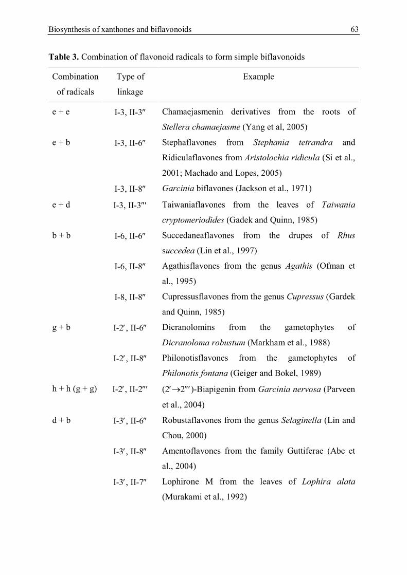
Biosynthesis of xanthones and biflavonoids 63
Table 3. Combination of flavonoid radicals to form simple biflavonoids
Combination
of radicals
Type of
linkage
Example
e + e I-3, II-3 Chamaejasmenin derivatives from the roots of
Stellera chamaejasme (Yang et al, 2005)
e + b I-3, II-6 Stephaflavones from Stephania tetrandra and
Ridiculaflavones from Aristolochia ridicula (Si et al.,
2001; Machado and Lopes, 2005)
I-3, II-8 Garcinia biflavones (Jackson et al., 1971)
e + d I-3, II-3 Taiwaniaflavones from the leaves of Taiwania
cryptomeriodides (Gadek and Quinn, 1985)
b + b I-6, II-6 Succedaneaflavones from the drupes of Rhus
succedea (Lin et al., 1997)
I-6, II-8 Agathisflavones from the genus Agathis (Ofman et
al., 1995)
I-8, II-8 Cupressusflavones from the genus Cupressus (Gardek
and Quinn, 1985)
g + b I-2, II-6 Dicranolomins from the gametophytes of
Dicranoloma robustum (Markham et al., 1988)
I-2, II-8 Philonotisflavones from the gametophytes of
Philonotis fontana (Geiger and Bokel, 1989)
h + h (g + g) I-2, II-2 (22)-Biapigenin from Garcinia nervosa (Parveen
et al., 2004)
d + b I-3, II-6 Robustaflavones from the genus Selaginella (Lin and
Chou, 2000)
I-3, II-8 Amentoflavones from the family Guttiferae (Abe et
al., 2004)
I-3, II-7 Lophirone M from the leaves of Lophira alata
(Murakami et al., 1992)

64 Bisosynthesis of xanthones and biflavonoids
Table 3. (continued)
h + b I-6, II-6 Hegoflavones from the fronds of Alsophila spinulosa
(Wada et al., 1985)
h + h I-5, II-5 (I-5,II-5)-Bisdihydroquercetin from the bark of
Pseudotsuga menziesii (Lai et al., 1992)
e + c I-3 ,O, II-4 Delicaflavone from the aerial parts of the Selaginella
delicatula (Lin and Chou, 2000)
b + a I-6 ,O, II-7 Masazinoflavanone from the leaves of Rhus tripartitum
(Mahjoub et al., 2005)
I-6 ,O, II-8 (I-6,O,II-8)-Biapigenin from the leaves of Viburnum
cotinifolium (Muhaisen et al., 2002)
d + c I-3, O, II-4 Ochnaflavones from the genus Ochna (Reddy et al.,
2008)
d + a I-3, O, II-7 Lophirone L from the leaves of Lophira alata (Tih et
al., 1992)
c + b I-4 ,O, II-6 Hinokiflavone from Thuja javanica (Roy et al., 1985)
I-4, O, II-8 Lanaroflavone from the whole plant of Lanaria lanata
(Dora and Edwards, 1991)
I-4, O, II-4 Loniflavone from Lonicera japonica (Kang, 2005)
I-8,CH2, II-8 Pentagrammetin from Pentagramma triangularis
(Roitman et al., 1993)
O
OH
HO
OH OHO
OH
O
OH
O
3
8''
Volkensiflavone
I
II
O
OH
HO
OH O
O
O OH
OH
HO
H
H
H
H
3
3''
Chamaejasmin
I
II
Figure 23. Examples of different types of simple biflavonoids.

Biosynthesis of xanthones and biflavonoids 65
O
OH
MeO
OH OOMeO
HOO
OMe
36''
Stephaflavone A
I
O
OH
HO
OH O
O
O OH
OH
HO
3I
3''
Taiwaniaflavone
O
OH
HO
OH OOHO
O
OH
OH
I
II
68''
Agathisflavone
I
II
OHO
OH OO
O
OH
HO
OH
OH
Succedaneaflavone
66''
OHO
OH O
OHII
OHOOH
OH O
I
8
8''
Cupressuflavone
OHO
OH O
OH
O
HO
O OH
OH
I II
(I-2',II-2''')-Biapigenin
2'
2'''
O
O
OH
OH
OH
O
OH
OH
OOH
HO
OHI
II
2'
6''
Dicranolomin
O
OH
OH
OOH
HO
O
OH
OH
OH
HO
O
I
II
2'
8''
Philonotisflavone
Figure 24. Examples of different types of simple biflavonoids (continued)

66 Bisosynthesis of xanthones and biflavonoids
O
OMe
HO
O
38''O O
OH
O4'-Methoxypyranoamentoflavone
3'
I
II
O
OH
OOH
HO
OH
O
OH
OH
O
OHHO
OH
OH
(I-5',II-5')-bis-I-2,3,II-2,3-Dihydroquercetin
5'5'''
I
II
O
OH
HO
OH O
I
O
O
HO OH
OH
II
3'
6''
Robustaflavone
O
OH
OOH
HO
OHOHO
O
OHLophirone M
3'7''
I
II
O
OH
OOH
HO
O
O
O
OHHO
I
II
3
4'''
Delicaflavone
O
OOH
HO
OH
O
O
O
HO
OH
I
II
Masazinoflavone
6
7''
O
O
OH
OH
O
HO
OH
OHOH
O
HO
I
II
Hegoflavone A
5'6''
Figure 25. Examples of different types of simple biflavonoids (continued)

Biosynthesis of xanthones and biflavonoids 67
O
O
HO
OH
OH
O
O
HO
HO
O
OH
I
II
6
8''
(I-6,O,II-8'')-Biapigenin
O
OH
O
OOH
HO
OO
HO
HO
3'
7''
Lophirone L
I
II
O
OH
O
OOH
HO
O
O
OH
HO
3'
4'''I
II
Ochnaflavone
O
O
OOH
HO
O
O
OHHO
OH
Lanaroflavone
4' 8''
I
II
O
O
OOH
HO
O
O
OHHO
OH
I
II
4' 4'''
Loniflavone
O
O
O
O
OMe
HO
OH
OH
OH
MeO
OH
HO
Pentagrametin
8
8''
I
II
Figure 26. Examples of different types of simple biflavonoids (continued)

68 Material and methods
4. MATERIAL AND METHODS
4.1. General experimental procedures
Extraction was carried out using a sohxlet system of 1000 ml (Ducan). Thin layer
chromatography (TLC) was performed using silica gel 60 F254 and RP18 F254 pre-
coated aluminium sheets (Merck, 0.2 and 2.0 mm). TLC plates were visualized by UV
light (CAMAG) at 254 nm and 366 nm after sprayed with anisaldehyde/sulphuric acid
reagent (anisaldehyde, 2 ml; H2SO4 98%, 10 ml, AcOH, 16 ml and MeOH, 170 ml)
and heated on a thermoplate (Desaga). The solution of FeCl3 in methanol was used for
detecting phenolic compounds. Open column chromatography (CC) was carried out on
silica gel (Merck, 63 – 200 µm) using glass columns of various dimensions. Gel
permeation chromatography (GPC) was performed on Sephadex LH-20 (GE
Healthcare) with MeOH or MeOH – CHCl3 3:7 as eluent. Flash chromatography (FC)
was performed on an Armen Instrument Spot System with columns: SVF D26 (26 x
110 mm) and SVP D40 (4- x 160 mm) on silica gel 60 (Merck, 63 – 200 µm).
Centrifugal partition chromatography (CPC) was carried out using an Armen Spot
Centrifugal Partition Chromatography SCPC-250 with the solvent system HEMWat
(n-hexane/ethyl acetate/methanol/water) as eluent. All solvents used for TLC, CC,
GPC, FC and CPC were p.a. (pro analysis, Merck). Semi-preparative HPLC
separations were performed on a Varian ProStar 210 solvent delivery module equipped
with a Varian Prostar 335 photodiode array detector. Two prepacked columns, Varian
Dynamax Pursuit XRs C18 of 21.4 x 250 mm and Eclipse XDB-C18 of 9.4 x 250 mm
(HP), were used for RP18 HPLC. All solvents used were HPLC grade (Merck).
1D and 2D NMR spectra were measured using a Bruker ADVANCE 600
spectrometer (600 MHz for 1H and 150 MHz for 13C) or 400 spectrometer (400 MHz
for 1H and 115 MHz for 13C) with tetramethylsilane (TMS) as an internal standard and
CDCl3 or acetone-d6 (Deutero GmbH) as the solvent. The spectra were processed
using the NMR softwares: MestReNOva 6.0.2-5475 (Mestrelab Research) and
Topspin 3.1 (Brucker). High-resolution mass spectra were measured on a Finnigan
MAT SSQ 710 A spectrometer at 70 eV (HRESIMS, positive mode) or recorded on an
Agilent 6540 UHD (HRESIMS, positive and negative mode). UV spectra were

Material and methods 69
obtained by the Varian Prostar 335 photodiode array detector of the HPLC system.
Optical rotation was recorded using an UniPol L1000 polarimeter (Schmidt &
Haensch).
The MEM-Earle (0.85 g/l NaHCO3, Biochrom) supplemented with 1% of L-
glutamine (200 mM), 10% of fetal bovine serum and 1 % of non-essential amino
(Biochrom) were used for culture of HeLa cells (ATCC CCL17). The medium ECGM
(Porvitro GmbH) with supplement mix and antibiotics (Porvitro GmbH) were used for
HMEC-1 cells (CDC). The incubation was carried out in a NU-5500E incubator
(NUAIRE). Cell-counting was performed using a neubauer improved haemocytometer
(Brand) and a CK X 41 SF microscope (Olympus). A Megafuge 1.0 centrifugal
(Heraeus Sepatech) was used for centrifugation. Autoclaving was carried out using an
Autoklav 23 (Melag) or a Varioklav 500 (H+P). All the pipettes used were from
Eppendorf. Tris(4-dimethylaminophenyl)methyliumchlorid (crystal violet) was
obtained from Merck. 3-(4,5-Dimethylthiazol-2-yl)-2,5-diphenyltetrazoliumbromide
(MTT) and 3,3′-Dimethyl-4,4′-bis(5-amino-4-hydroxy-2,7-disulfonaphtyl-3-azo)-[1,1′-
biphenyl] dye were from Sigma-Aldrich. The optical density was measured at 560 nm
using a SpectraFlour Plus microplate reader (Tecan). Cytotoxicity and proliferation
assays were performed in 96 well plates (TPP).
4.2. Extraction, fractionation and isolation
4.2.1. Material of plant
The bark of Garcinia fusca Pierre was collected in Binh Phuoc Province, south
Vietnam in January 2011 and was identified by Mr Dang Van Son, Institute of
Tropical Biology, Ho Chi Minh City. A voucher specimen (Hieu-GF-01/2011) is
deposited in the Natural Product and Medicinal Chemistry Lab, Ho Chi Minh City
University of Sciences. The bark was air-dried and ground into powder.
4.2.2. Extraction of plant material
The powder of the bark of G. fusca Pierre (1.95 kg) was first exhaustively extracted
with 3.0 L of n-hexane by using a soxhlet system (1000 ml), a form of solid-liquid

70 Material and methods
extraction. The solvent was heated to reflux and successively percolated the material.
The extraction of n-hexane was done until the liquid passing through the material was
colourless. The residue of material was subsequently extracted with the same volume
of ethyl acetate. After extraction the solvents were removed by a rotary evaporator to
yield the crude extracts of n-hexane and ethyl acetate, which were shortly named as
GFH (G. fusca n-hexane extract) and GFE (G. fusca ethyl acetate extract),
respectively.
4.2.3. Fractionation of crude extracts
The n-hexane extract (GFH, 90.5 g) was divided into two portions. Each portion
was subjected to an open CC using a column of 10 x 40 cm dimension, 800 g of silica
gel as stationary phase and a solvent system of acetone and petroleum ether (0-100%)
as mobile phase to give 30 fractions (500 ml of each). On the basis of TLC, identical
fractions were combined to afford 8 main fractions which were shortly named GFH-1 -
8. The ethyl acetate extract (GFE, 112.3 g) was also split into two portions which were
in turn applied to CC with the same column and amount of silica gel using a gradient
elution of acetone and petroleum ether (0-100%) to produce 35 fractions (500 ml of
each). The combination of similar fractions was also based on TLC to give 8 main
fractions (GFE-1 - 8).
4.2.4. Isolation of substances from fractions
Chromatography is one of the powerful methods for the separation and purification
of natural products. It is based on the distribution of a component between two phases
- a moving one (mobile phase) that is passed over an immobile phase (stationary
phase). Components will be released by the mobile phase at various rates because of
their different affinities with the stationary phase. A variety of chromatography
methods are widely used for the isolation of natural products such as adsorption,
partition, ion-exchange and permeation chromatography. Mobile phase can be either
gas or liquid. For the isolation of compounds from G. fusca Pierre, the adsorption,
partition and permeation chromatography were used. The general strategy of the
isolation and structural elucidation of substances is shown in Scheme 16.

Material and methods 71
Scheme 16. The general strategy of the isolation and structural elucidation of
substances from G. fusca Pierre
4.2.4.1. Thin layer chromatography (TLC)
TLC was used to select appropriate solvent systems for the CC of fractions based on
their Rf (retardation factor) values. The Rf value refers to the distance that a compound
moves in chromatography relative to the solvent front. It can be calculated by the
following formula (Braithwaite and Smith, 1996).
Formula 1: Calculation of retardation factor (Rf)
Rf = Distance travelled by compound
Distance travelled by solvent front
Sub-fractions
Flash CC on silica gel or GPC on Sephadex LH-20 or CPC
GPC Sephadex LH-20 or CPC
Sub-fractions
Pure compounds
Semi-preparative HPLC on RP18 silica gel or preparative TLC on silica gel
Sub-fractions
Pure compounds
Semi-preparative HPLC on RP18 silica gel or preparative TLC on silica gel
Structural elucidation using spectroscopic methods
Cytotoxic assays
Crude extract
Fractionation using open CC on silica gel
Fractions ˃ 5 g Fractions < 5 g
Open CC on silica gel
Sub-fractions
Flash CC on silica gel

72 Material and methods
A suitable solvent system should give useful Rf values of between 0.3 and 0.7, i.e., a
compound can be separated from other components without staying at or so close to
the original point or with the solvent front. The separation between two compounds is
possible with an Rf value (the difference between two Rf values) of at least 0.2-0.3
(Braithwaite and Smith, 1996). TLC is also used to determine two identical
compounds relying on their Rf values in the same solvent system or for the
combination of similar fractions. It is even used to show the relative number of
components existing in a given fraction or if an isolated substance was pure. Moreover
TLC is also applied to identify types of compounds in extracts or fractions based on
their physical and chemical properties with ultraviolet (UV) and typical reagents
(Gibbons, 2006).
In the present work, aluminium sheets coated by silica gel or reverse phase silica
C18 of 0.2 mm thickness with fluorescent indicator (Merck) were used for TLC.
Samples were applied on the plate via a micropipette with a volume of 0.5 µl and a
concentration of 5 mg/ml. For the selection of suitable eluent of CC, the plates were
developed with different solvent systems. After the development, the location of
compounds was detected using UV or chemical reagents. Coloured substances are
easily visible to naked-eyes as separate spots, whilst colourless ones require chemical
or physical methods. Substances having an enone or a benzene ring are detectable with
UV. Therefore they can be visualized by UV when irradiated at 254 nm. The plates
contain a fluorescent indicator which absorbs light at 254 nm and re-emits at the green
end of the spectrum, thus if a substance absorbs at 254 nm, it will be detectable as a
dark spot against the green background. Methanolic ferric chloride (FeCl3/MeOH)
reagent was used to detect phenolic compounds as dark green spots. Compounds
which are not detectable with UV could be visualized by spraying the plate with
anisaldehyde-H2SO4 reagent or sulphuric acid 10%. The plate was heated after
spraying at 100-105ºC until the colours have totally occurred. Depending on the
structure of compounds, the colours of spots changed from pink to dark brown. After
heated, substances were also detected by UV at 366 nm as light spots against the dark
violet background.

Material and methods 73
4.2.4.2. Preparative TLC
The practice of this technique is similar to that of qualitative analysis but differs in
the thickness of plates. They are principally coated by 1-5 mm of adsorbent for using
in the preparation of large sample quantities (10-100 mg). Pre-coated glass plates of 2
mm thickness (Merck) and 20 x 20 cm dimension with fluorescence indicator were
used for the present work. The adsorbent was silica gel 60 (63-200 µm). The sample
was dissolved in acetone (5 mg/ml) and applied onto the plate as a streak by a pasteur
pipette. The chromatography was carried out in a glass tank of 22 x 22 cm with 100 ml
of a suitable solvent system. A filter paper of 20 x 20 cm was soaked with the solvent
and put into the tank to keep the internal atmosphere saturated with solvent vapour.
After the development was complete, the plate was dried and the bands of substances
were detected by UV at 254 nm. The bands were then scraped off with a spatula and
the substance was washed out of the adsorbent by acetone. The pure substance
obtained after the removal of acetone was structurally determined by spectroscopic
techniques.
4.2.4.3. Open column chromatography
Open CC using silica gel as an adsorbent is one of traditional forms of adsorption
chromatography. This is a simple and low cost CC technique. Fractions were subjected
to open CC by using simple glass columns of various dimensions depending on the
amount of sample. The stationary phase was silica gel 60 (Merck, 63-200 µm) and the
mobile phase was the appropriate solvent systems selected by TLC. The column was
packed by wet-column method, in which silica gel was soaked in the solvent with the
ratio of 0.5 mg/ml before poured into the column. The quantity of silica gel used was
fifty times of the sample amount. The packed column was equilibrated by at least two
bed volumes of the solvent before the sample adsorbed on silica gel was applied onto
the top of the packed column. Afterwards the ready column was eluted by a gradient
elution. The solvent runs through the column under the influence of gravity. The
chromatography was carried out within 3-7 hours and the eluent containing separated
components was collected in test tubes or conical flasks. Collected fractions were
subsequently applied to TLC for combination.

74 Material and methods
4.2.4.4. Flash chromatography
The standard method of flash chromatography (FC) is based on the report of Still in
1978 (Still et al., 1978). This is a rapid column chromatography technique using a
narrower packing than classical CC and a slightly increased over-pressure to achieve
adequate flow-rates. The technique is much efficient for the separation of components
with Rf values of less than 0.2. It is suitable for sample loads ranging from 10 mg to
10 g depending on the difference of Rf values (Braithwaite and Smith, 1996). The flash
chromatography system used in the present work is Spot Liquid Chromatography
Flash (Armen Instrument). Plastic columns of 26 x 110 mm (SVF D26-Si60) or 40 x
160 mm (SVP D40-Si60) were tightly packed with silica gel 60 (63-200 µm) without
solvent. The packed column was subsequently eluted with solvent by a mini pressure
of maximum 7 bar until it was homogeneous without air-bubbles. The sample
adsorbed on silica gel was then applied onto the top of the column. The column SVF
D26-Si60 was used for the sample quantity of 0.3-1.5 g, whilst the column SVP D40-
Si60 was suitable for 0.9-4.5 g of the sample. The elution was a gradient with the
flow-rate of 10-30 ml/min and the eluent was automatically collected in test tubes. The
chromatography was controlled by the built-in UV detector at 290 nm. The
combination of collected fractions was based on the result of TLC.
4.2.4.5. Size-exclusion chromatography
Size-exclusion chromatography or gel filtration is one of the permeation
chromatographic methods, in which gels or polymers are used as stationary phase and
the separation of uncharged compounds is based on the relative size or mass of
molecules, i.e., large components leave the column first followed by smaller ones in
order of their decreasing size. This technique was first performed in 1954 by Mould
and Synge and the systematic use of the principle was introduced in 1959 by Porath
and Flodin (Braithwaite and Smith, 1996). In this work, Sephadex LH-20 (GE
Healthcare) was used as the stationary phase and the mobile phase was methanol or a
mixture of methanol and chloroform of 3:7 by volume. Sephadex LH-20 is a
hydroxypropylated polysaccharide developed for gel filtration and CC of natural
products in the range of 100-5000 molecular weight. In the experiment, the gel was

Material and methods 75
suspended in the solvent of 4.0 ml/g before poured into an appropriate glass column
depending on the amount of sample. The packed column was stationary in at least
three hours for full swelling of the gel and afterwards equilibrated with three bed
volumes of the solvent. The sample was dissolved in the solvent before applied onto
the packed column in such a way that the sample volume was 5% of the total bed
volume. The ready column was then eluted with a flow rate of 0.5 ml/min. The solvent
flows through the column under the influence of gravity. The eluent was manually
collected in test tubes. The fractions were then combined into sub-fractions based on
TLC.
4.2.4.6. Centrifugal partition chromatography
Centrifugal partition chromatography (CPC) is a liquid-liquid chromatographic
technique originally pioneered by Ito in 1964 (Ito, 1970). The principle of this
technique is based on the different partitions of components between two immiscible
liquid phases. Each one has a specific partition (distribution) coefficient (Kd) defined
by the following formula (Kumar et al., 2014).
Formula 2: Calculation of partition coefficient (Kd)
Kd = [X]upper
[X]lower
For a given compound X, at the equilibrium of two liquid phases and for a given
temperature:
[X]upper = the concentration of X in the upper phase
[X]lower = the concentration of X in the lower phase
CPC functions with one of two liquid phases used as stationary phase and the other
one as mobile phase. The column system consists of a series of channels linked in
cascade by ducts and aligned in cartridges or disks in a circle around a rotor. The
liquid stationary phase is fed into the column and held in channels by the centrifugal
force generated by the spinning rotor. The liquid mobile phase containing the

76 Material and methods
components of the sample is then pumped through the column. The separation is
achieved as a function of the specific partition coefficient (Kd) of each component. For
the ideal separation of the components, the solvent system should result in a partition
coefficient of 1. However based on practical experiments, 0.2 < Kd < 5 can be used
without excessive elution and broadening band (Wilson, 2012). Many strategies for
selection of solvent system have been reported in literature. Foucault suggested that
there should be a solvent that is soluble in the other solvents of the solvent system and
this solvent should completely dissolve the sample (Foucault, 1995)
For this research, the CPC system used is Spot Centrifugal Partition
Chromatography SCPC-250 (Armen). The solvent system of HEMWat (n-
hexane/ethyl acetate/methanol/water) was selected with an appropriate ratio so that the
partition coefficient is between 0.2 and 5. Two modes of the CPC used for the
separation are descending mode in which the lower phase is the mobile phase and
ascending mode in which the upper phase is the mobile phase. For the
chromatography, the sample amount of 1 g was dissolved in 10 ml of a mixture
containing 50% of mobile phase and stationary phase. The speed of the rotor was 1000
U min-1 and the flow rate of elution was 5 ml/m. The eluent containing separated
components was automatically collected in test tubes. At first, the column was fed
with the lower phase. The upper phase was subsequently pumped through the column
until the equilibration was established. The sample volume of 10 ml was then ejected
into the equilibrated column. The upper phase passes through the column and elutes
separated components. After 60 minutes of the chromatography, the ascending mode
was switched into descending mode, i.e., the lower phase was at that moment used as
the mobile phase. The chromatography was carried out within at least two hours.
Collected fractions were subsequently applied to TLC for combination.
4.2.4.7. Semi-preparative high performance liquid chromatography
High performance liquid chromatography (HPLC) is a column chromatographic
technique using a small particle size packing and a large pressure elution in order to
get a high performance of separation. In 1967, Horvath et al. set up one of the first
practical HPLC systems for their research on nucleotides (Braithwaite and Smith,

Material and methods 77
1996). Nowadays HPLC is widely used for qualitative and quantitative analysis as
well as preparation of natural or synthetic compounds. In the present work, semi-
preparative HPLC was used for the isolation and purification of substances is a ProStar
system (Varian) consisting of an auto-sampler, two pumps with maximum pressure of
400 bar, a diode array detector (DAD) and a fraction collector. Two pre-packed
columns were used for the preparation are Dynamax Pursuit XRs C18 of 21.4 x 250
mm and Eclipse XDB-C18 of 9.4 x 250 mm. Both columns were packed with RP18
silica of 5 µm. The former was used for the sample amount of 10-20 mg and the flow
rate of maximum 20 ml/m, whilst the later was used for that less than 10 mg of the
sample and the flow rate of maximum 6 ml/min. The solvent system of water and
acetonitrile was selected for the elution. The sample was dissolved in acetonitrile with
the ratio of 10 mg/ml before injected into the column. The preparation time was
around 30-40 minutes and the separation was monitored by UV. The purified
substances were then structurally identified using spectroscopic techniques such as 1D
and 2D NMR, UV and mass spectrometry.
78 Material and methods
4.3. Structural elucidation of isolated compounds
Spectroscopic and spectrometric methods are considered as effective and useful
tools for the structural identification of natural products. These include ultraviolet -
visible (UV-vis), infrared (IR), nuclear magnetic resonance (NMR), X-ray, circular
dichroism (CD) and mass spectrometry. A molecular structure including the relative
and the absolute configurations can be completely determined by the combination of
these methods. In this work, the structural determination of the new natural
compounds was mainly performed by one (1D) and two dimensional (2D) NMR
spectroscopy associated with UV and mass spectroscopy as well as optical rotation.
The steps of the structural elucidation was executed in succession as follows
- Identification of the class of substance based on 1H NMR and UV spectra.
- Calculation of unsaturation degree or hydrogen deficiency index and
establishment of the molecular formula from data of 13C NMR and mass spectra.
- Analysis of skeletal connectivity to accomplish the covalent structure relying on
2D NMR spectra such as HMQC, HMBC and COSY.
- Determination of the relative stereochemistry using NOESY or ROESY
spectroscopy and optical rotation.
4.3.1. Ultraviolet-visible spectroscopy
As the electromagnetic radiation in the ultraviolet and visible region (190-800 nm)
passes through a transparent substance, a portion of the radiation may be absorbed in a
form of energy which can make electrons in molecules move from a state of low
energy (ground state) to a state of higher energy (excited state). The absorbed energy
is exactly equal to the energy difference between these two states. Once that happens,
the residual radiation is recorded by a detector to give a spectrum which is called an
absorption spectrum. The possible transitions of electrons are from the or
nonbonding (n) orbitals to unoccupied or anti-bonding orbitals (* and *). The UV-
vis spectrum is basically recorded as a plot of absorbance (A, mAU) versus
wavelength (, nm). The absorbance obeying the Beer-Lambert Law can be calculated
for a given wavelength by the following formula (Pavia et al., 2009)

Material and methods 79
Formula 3: Calculation of absorbance (A)
A = absorbance
I0 = intensity of light incident upon sample cell
I = intensity of light leaving sample cell
c = molar concentration of solute
l = length of sample cell (cm)
= molar absorptivity
The appearance of several peaks of absorbed maximum wavelengths (max) in the
UV-vis spectrum is caused by conjugated systems and electronic transitions between
the different vibrational and rotational energy levels for each electronic state. The max
values are useful for the structural elucidation and those of conjugated systems such as
diens or enones can be predicted by Woodward’s rules for the * transition. Each
type of compound gives specific absorption bands. For examples, carotenoids
commonly show three distinct peaks between 400 and 500 nm in methanol, whilst
biflavonoids perform two peaks at 260-290 nm and 320-340 nm. Xanthones give four
different peaks at 230-245, 250-265, 305-330 and 340-400 nm (Harborne, 1998). In
the present work, the UV spectra of the isolated compounds were measured in the 200-
420 nm region by the diode array spectrophotometer of Prostar HPLC system.
4.3.2. Mass spectrometry (MS)
A substance can be transformed into gas phase ions by various ionization methods.
These ions are then accelerated by an electromagnetic field, separated by their mass-
to-charge (m/z) ratio and counted by a detector. The signal is recorded and output as a
graph of the number of ions detected versus their m/z ratio, called a mass spectrum.
Common ionization methods can be divided into three main groups, viz gas-phase,
desorption and evaporative ionization. The gas-phase ionization including electron
impact (EI) and chemical ionization (CI) methods is suitable for volatile and small
organic molecules. The desorption ionization consists of three methods: secondary ion
A = log(I0/I ) = .c.l

80 Material and methods
mass spectrometry (SIMS), fast atom bombardment (FAB) and matrix-assisted laser
desorption ionization (MALDI). The evaporative ionization comprising thermo-spray
(TS) and electro-spray (ES) methods, along with desorption, are appropriate for non-
volatile and large molecules, especially peptides and proteins (Pavia et al., 2009).
There are two significant categories of mass spectrometers, low resolution and high
resolution. High resolution mass spectrum (HRMS) can determine an extremely
precise m/z value up to four or five decimal numbers, i.e., only one rational molecular
formula will fit the data especially in combination with 1H and 13C NMR data. In the
present work, the molecular weight and the molecular formula of new compounds
were obtained from electrospray ionization- high resolution spectra (ESI-HRMS)
which were measured by the instrument Q-TOF 6540 UHD (Agilent). In ESI, the
sample in solution is sprayed out from a capillary into a heated chamber at nearly
atmospheric pressure. The tip of the capillary is maintained a high voltage potential of
2-5 keV to expel the sample molecules in charged droplets into the ionization
chamber. The charged droplets are subjected to a counter-flow of a nebulizing gas that
evaporates solvent molecules from the droplets until solvent-free sample ions are
released in the vapour phase. These ions will then pass through the high resolution
mass spectrometer to yield a HRESI-MS. With this ionization method [M+H]+,
[M+Na]+ or [M+K]+ ions are produced but fragmentation is rare (Silverstein et al.,
2005). For a compound of general formula CaHbNcOdXe in which X is a halogen atom,
the degree of unsaturation can be calculated by the following formula
Formula 4: Calculation of degree of unsaturation
Degree of unsaturation =(2 + 2a - b + c - e)
2
a = number of carbon atoms b = number of hydrogen atoms
c = number of nitrogen atoms e = number of halogen atoms

Material and methods 81
4.3.3. Nuclear magnetic resonance spectroscopy
Nuclear magnetic resonance (NMR) has been shown to be a powerful spectroscopic
method for the structural determination of natural products, especially novel
compounds sometimes possessing unusual or unprecedented structures. It has become
the main choice for the establishment of chemical structures, particularly when X-ray
crystallography is inapplicable. Nowadays modern NMR spectroscopy can be
performed by high magnetic spectrometers of up to 900 MHz which allow to
accomplish multi-pulsed and multidimensional experiments with a small sample
amount of under 0.5 mg. Nuclei have an angular momentum described by a spin
quantum number I, which gives 2I+1 of allowed spin states with integral differences
ranging from +I to –I. Each spin state possesses a corresponding energy level when a
nucleus with I 0 is immersed in a magnetic field of strength B0. A spectroscopic
transition may occur between these levels by the absorption of electromagnetic
radiation of appropriate frequency in accordance with the following equation (Pavia et
al., 2009)
Formula 5: The equation of energy difference of nuclei in a magnetic field
E = the energy difference between the possible spin states
= the frequency of the absorbed electromagnetic radiation which is usually in the
radiofrequency region of ca. 1-1000MHz
= magnetogyric ratio, a constant for each nucleus
Natural products commonly contain nuclei having spin number I of ½ such as 1H,
13C, 15N and 31P. Among these, 1H and 13C are the most important nuclei because they
constitute the skeleton of substances. The proton 1H is quite sensitive with NMR
because of its high natural abundance (99.98 %) and therefore it is first investigated
for the structural elucidation of substances (Neri and Tringali, 2001). In the present
work, the 1D and 2D NMR spectra of isolated compounds were obtained using NMR
E = (h/2)B0 = h
= B0/2

82 Material and methods
spectrometers of AVANCE 300, 400 and 600 Kryo (Bruker). The sample was
dissolved in 0.6 ml of deuterated chloroform (CDCl3) or deuterated acetone (C2D6CO)
and the solution was then dispensed into a 5 mm diameter NMR tube. The spectra
were measured at the room temperature or 393ºK using the Fourier transform (FT)
technique. The NMR-active nuclei are simultaneously excited by an irradiation pulse.
When the pulse is switched off, the excited nuclei slowly return to their original spin
state and emit electromagnetic radiation of different frequencies. This is so-called
relaxation process. The signal is recorded in the form of the free induction decay (FID)
(Mitchell and Costisella, 2007). Tetramethylsilane (TMS), (CH3)4Si, was used as the
internal reference compound.
4.3.3.1. One dimensional (1D) NMR spectra
1D NMR spectra are typically displayed as an absorption spectra, the axes of which
are the frequency (chemical shift) and the intensity. Many functional groups or types
of hydrogens or carbons contained in a molecule can be identified by the characteristic
chemical shift values in 1H and 13C NMR spectra.
4.3.3.1.1. 1H NMR spectra
Protons in a molecule have resonances at various frequencies because of their
different chemical environment. Therefore, each type of proton gives a resonance peak
in a limited range of chemical shifts. The peak of a nucleus can be split into a multiplet
with (n + 1) components as a result of the spin-spin coupling between this nucleus and
adjacent equivalent nuclei, where n is the number of neighbouring equivalent protons
with the same coupling constant. If the nuclei are not equivalent, the peak will appear
as a multiple-multiplet of 2n components (Neri and Tringali, 2001). The relative
intensity ratios of components in multiplets follow entries in Pascal’s triangle. 1H
NMR spectra reveal information about types of hydrogens and the number of each in a
molecule based on their chemical shifts, integration values and coupling constants. For
instance, aromatic protons have resonance in the chemical shift range of 6-8 ppm,
whereas protons bonded with sp3 carbons have resonance in between 0 to 3 ppm. The
proton of aldehyde group has resonance near 9 to 10 ppm and that of acid group is in

Material and methods 83
the 10-12 ppm region. In alkenes, protons that are cis to each other have 3J coupling
constants of 6-12 Hz, whilst those of trans protons are between 11 and 18 Hz. In
aromatic compounds, ortho protons (3J = 7-10 Hz) couple stronger than meta ones (3J
= 2-3 Hz) and para ones (3J = 0-1 Hz) (Pavia et al., 2009).
4.3.3.1.2. Proton-decoupled 13C NMR spectra
Spin-spin coupling between 13C atoms are rarely observed but the spin-spin
interaction of protons bonded directly to 13C atoms can split the carbon signal
responding to the n + 1 Rule. However proton-coupled 13C NMR spectra of large
molecules are too complicated to interpret because of overlap of the multiplets from
different carbons. This is caused by the 13C-H coupling constants being often larger
than the chemical shift differences of the carbons. In proton-decoupled 13C NMR
spectra, overlapping multiplets are transformed into singlets and therefore, the spectra
are easier to interpret. 13C NMR spectra show information about the number and types
of carbons and functional groups. For example, the carbonyl carbons of ketones often
have resonance at low field of 180-220 ppm, whereas those of aldehydes and
carboxylic acids have resonance near 170 to 190 ppm. The chemical shifts of aromatic
and unsaturated carbons are in the 90-170 ppm region and those of saturated carbons
are between 10 and 90 ppm (Pavia et al., 2009).
4.3.3.2. Two dimensional (2D) NMR spectra
2D NMR spectra are obtained by recording resonance signals as a function of two
time variables and carrying out two Fourier transformations on a matrix of data.
Therefore both of the horizontal and vertical axes in 2D NMR spectra are two
chemical shift (frequency) axes. The 2D spectra give cross peaks showing correlations
between the two axes and the data are displayed as a series of contours (Mitchell and
Costisella, 2007).
4.3.3.2.1. 1H-1H COrrelation SpectroscopY (COSY)
A COSY spectrum shows homonuclear correlations between coupling protons in a
molecule. The 1H chemical shifts are thus plotted on both axes. The off-diagonal peaks

84 Material and methods
in the spectrum are the important peaks, whilst the diagonal peaks are only considered
as reference points. The spectrum provides information on which proton couples with
which one. It also indicates H-H connectivities, gemical, vicinal or long range
couplings. Correlations can be observed for all protons except the hydroxylic proton,
which is often rapidly exchanged in protonic solvents (Breitmaier, 2002).
4.3.3.2.2. Heteronuclear Single Quantum Correlation (HSQC) spectra
A HSQC spectrum displays the heteronuclear correlations of protons with 13C atoms
to which they are directly attached. Therefore the 1H chemical shifts are plotted on an
axe and the 13C chemical shifts on the other one. All C-H single bonds of the molecule
can be determined by the HSQC spectrum and hence it provides information on which
protons are bonded to which carbons. Multiplets of protons in large molecules
frequently overlap in 1H NMR spectra and are difficult to be identified. HSQC spectra
can help to determine these protons (Breitmaier, 2002).
4.3.3.2.3. Heteronuclear Multiple Bond Coherence (HMBC) spectra
The HSQC spectrum shows the 1H-13C correlations through a single bond (1JCH) and
thereby is only applied to 13C atoms which are attached by protons. The HMBC
technique is the modification of HSQC technique. A HMBC spectrum is applicable to
non-protonated or quaternary 13C atoms to provide 2JCH, 3JCH and 4JCH correlations.
The HMBC spectrum is very useful to identify quaternary carbons which can not be
observed in the 13C NMR spectrum in some cases because of their low signal intensity
(Breitmaier, 2002). The combination of data of COSY, HSQC and HMBC spectra
produces essential information of skeletal connectivity on which structural fragments
can be determined and then reassembled to accomplish the overall covalent structure
of the substance.
4.3.3.2.4. Nuclear Overhauser Effect SpectroscopY (NOESY) and Rotating-frame
Overhauser Effect SpectroscopY (ROESY) spectra
NOESY and ROESY spectra display the correlations of protons that are close to
each other in space with the common distance of 4.5 Å or less. Spatial interactions

Material and methods 85
between protons can be obtained via many bonds. In these spectra, the 1H chemical
shifts are plotted on both axes like a COSY spectrum. NOESY and ROESY spectra are
extremely helpful for the determination of relative stereochemistry in molecular
structures (Silverstein et al., 2005).
4.3.4. Optical rotation
Optical activity is the ability of chiral substances to rotate the plane of polarisation
of polarised light. Optical rotation is considered to be positive (+) for dextrorotatory
substances, i.e. those that rotate the plane of polarisation in a clockwise direction and
negative (-) for laevorotatory substances. The specific rotation of a chiral compound
can be calculated from the observed angle of rotation according to the expression
(Carey, 2001).
Formula 6: The calculation of specific rotation
[
c.l
t
D
[] = specific rotation
= angle of rotation observed by a polarimeter
c = concentration of the substance in g/100 ml
l = length of the polarimeter tube in decimetres
t = temperature of measurement (ºC)
D = D-line of sodium ( = 589.3 nm)
In the present work, the optical rotations of new substances were obtained using a
polarimeter of UniPol L1000 (Schmidt and Bonaduz). The samples were prepared for
a concentration of 1 mg/ml in methanol and then dispensed in a polarimeter tube of 5
cm. The angle of rotation was observed at the wavelength of the D-line of sodium at
22 0.5 ºC. Measurements were repeated at least five times and the final angle of
rotations was obtained as an average value.

86 Material and methods
4.4. Biological assays
The cytotoxicity of new compounds was tested on HeLa cell line using MTT assay.
Moreover anti-angiogenic activity was carried out with a bixanthone using
proliferation assay on human microvascular endothelial cell line-1 (HMEC-1)
4.4.1. Preparation of samples
The samples were prepared as stock solutions with a concentration of 100 mM in
dimethyl sulfoxide (DMSO) and stored at -20 ºC. For the assays, the final
concentration of DMSO per well was not more than 1%.
4.4.2. Cytotoxicity assay on HeLa cells
The HeLa cell line was originated by Gey et al. in 1952 from cervical cells taken
from Henrietta Lacks, a young cancer patient. These cells were the first human cells
continuously grown in culture and widely used for the research of anti-cancer activity
of natural or synthesis substances (Heneen, 1976). The cytotoxicity of new compounds
was evaluated by the MTT assay on HeLa cells as described by Mosman (1983) and
modified according to Heilmann et. al (2001).
4.4.2.1. Cell culture
HeLa cells were cultured in Minimum Essential Medium (MEM-Earle with 0.85 g/l
NaHCO3 of Biochrom) supplemented with 1% of L-glutamine (200 mM), 10% of fetal
bovine serum and 1 % of non-essential amino acids at 37 ºC in a humidified 5 % CO2
environment. For the passage of the cells, the medium was removed through a pasteur-
pipette and the cells were washed with 5 ml of phosphate buffered saline (PBS). The
cells were then incubated with 2 ml of trypsin/EDTA for 5 minutes to release from the
bottom of flask. The cell suspension was centrifuged with 8 ml of the medium in a 15
ml centrifuge tube at a speed of 700 U/min at 20 ºC for 3 minutes. The upper solution
was removed and the cells were homogenised with 7 ml of the medium. Afterwards
350 µl of the cell suspension was dispersed into a 50 ml culture flask containing 20 ml
of the medium for growth. After 2-3 days, the old medium was changed by the same
amount of fresh medium. The cells were split after 4-5 days.

Material and methods 87
4.4.2.2. Cell counting
The counting of cells was carried out using a haemocytometer of nine 1 x 1 mm
squares. The cell suspension (10 µl) was mixed with 10 µl of Trypan Blue solution and
then a volume of 10 µl of the mixture was applied to the chamber of haemocytometer.
Trypan Blue is used to staining dead cells. The number of unstained cells in the four
large corner squares was counted using a microscope. The number of cells per ml can
be calculated as the following equation.
Formula 7: The calculation of the number of cells per ml
In this study, the dilution factor is 2. Because the volume of each large square is 0.1
µl, the average of cells per large square must be multiplied with 104 to give the number
of cells per millilitre.
4.4.2.3. MTT assay
The assays were carried out in 96-well plates and the number of cells per well was
9000 cells. Nine different concentrations of the samples were prepared in the medium
with a volume of 75 µl per well and 75 µl of the cell suspension was then added to
each well so that the total volume per well was 150 µl. The plates were incubated at 37
ºC in a humidified 5 % CO2 environment for 72 hours. After incubation, 15 µl of an
aqueous solution of methylthiazoyl tetrazolium bromide (MTT) was added into each
well containing the cells and incubated for next 4 hours. MTT is reduced to a purple
formazan by NADH of living cells. After incubation, the solution was removed and
150 µl of sodium dodecylsulfate (SDS) solution (10%) was added to each well. The
plates were subsequently placed in the dark for 24 hours at the room temperature. The
optical density was measured at 560 nm using a microplate reader (Tecan SpectraFlour
Plus). For the determination of the IC50 values, the optical density was plotted against
the log scale of the concentration. Each concentration was performed in hexaplicates
and the assays have been repeated at least three times. Maximal observed standard
deviation was about 15% (absolute).
Cells per ml = The average of cells per large square x Dilution factor x 104

88 Material and methods
4.4.3. Proliferation assay on HMEC-1
Vascular endothelial cells line the inside of blood vessel walls throughout the
circulatory system to compose an interface between flowing blood and the vessel wall.
They are involved in the formation of new blood vessels called angiogenesis.
Angiogenesis has been shown as a target for the fight against diseases characterized by
either poor vascularisation or abnormal vasculature. Compounds that inhibit or induce
the formation of new blood vessels (angiogenesis inhibitors) can help to combat such
diseases (Birbrair et al., 2014). The proliferation assay on human microvascular
endothelial cell line-1 (HMEC-1) is (among others) an in vitro method to identify the
anti-angiogenic activity of a substance. The HMEC-1 is derived from human foreskin
and transfected by a gene of simian virus 40. These transfected cells have been created
by Dr. Thomas Lawley and his team including members from the University of Emory
and the Centers for Disease Control and Prevention (CDC), USA. They possess all of
the characteristic makers of endothelial cells and can last from 25 to 27 generations
without losing their genetic characteristics (Ades et al., 1992). The proliferation assay
was carried out as described by Schmidt et al. (2012).
4.4.3.1. Cell culture
HMEC-1 was cultured in Endothelial Cell Growth Medium (ECGM, Provitro)
supplemented with 10% of FCS and antibiotics at 37 ºC in a humidified 5 % CO2
environment. For the passage of the cells, the medium was removed through a pasteur-
pipette and the cells were washed twice with 10 ml of phosphate buffered saline
(PBS). The cells were then released by incubation with 4 ml of trypsin/EDTA for 5
min. The cell suspension was centrifuged with 6 ml of the medium in a 15 ml
centrifuge tube at a speed of 1000 U/min at 20 ºC for 5 minutes. The upper solution
was removed and the cells were homogenised with 10 ml of the medium. Afterwards 1
ml of the cell suspension was dispersed into a 50 ml culture flask containing 20 ml of
the medium (ECGM) for growth.

Material and methods 89
4.4.3.2. Proliferation assay
The assays were carried out in 96-well plates. The HMEC-1 cells were seeded in
the plates coated with collagen (100 µl of the cell suspension per well) and the number
of cells in each well was 1500 cells. The plates were incubated at 37 ºC in a 5% CO2
atmosphere with constant humidity. After 24 hours, the medium in a reference plate
was removed and the cells in this plate were stained with a crystal violet solution for
10 minutes in the dark at the room temperature to serve for the baseline. The cells in
other plates were treated with 100 µl of the samples with various concentrations in the
culture medium. After 72 h incubation, the solution was removed and the cells were
stained with the crystal violet solution for 10 minutes in the dark at the room
temperature. The plates were then washed with distilled water and 100 µl of 0.1 M
sodium nitrate solution was added in each well. The optical density was measured at
560 nm using a microplate reader (Tecan SpectraFlour Plus). For the determination of
the IC50 values, the optical density was plotted against the log concentration. Every
test was performed at least in hexaplicates and the assays have been repeated at least
three times. The maximal observed standard deviation was about 15% (absolute).
Xanthohumol was used as positive control and pure solvent (0.1 % DMSO) was used
as negative control.

90 Results and discussion
5. RESULTS AND DISCUSSION
5.1. Experiments
5.1.1. Fractionation and isolation of the n-hexane extract
The n-hexane extract of the bark of Garcinia fusca (GFH, 90.5 g) was divided into
two portions which were in turn applied to open column chromatography (CC) using a
column of 10 x 40 cm dimension, 800 g of silica gel as stationary phase and a solvent
system of acetone and petroleum ether (0-100%) as mobile phase to give 30 sub-
fractions (500 ml of each). Identical fractions were combined on the basis of TLC to
afford 8 main fractions which were shortly named GFH-1 - 8 as shown in Scheme 17.
Scheme 17. Fractionation of the n-hexane extract (GFH)
The 1H NMR spectra of the fractions (see Figure 27 - 29) revealed that the fractions
from GFH-3 to GFH-7 contained prenylated xanthone derivatives based on typical
signals at 13 - 14 ppm (hydrogen-bonded hydroxyl groups), 6 – 7.5 ppm (aromatic
protons), 5 – 5.5 ppm (olefinic protons of prenyl groups), 4 – 4.2 ppm (methylene
protons of prenyl substitutes at the peri position, C-8), 3.7 – 4 ppm (methoxyl groups
and protons of oxygenated sp3 carbons) and 1 – 2 ppm (vinyl methyls in prenyl
chains). The fractions GFH-4 and GFH-5 comprised xanthones with peri prenyl
groups because their spectra showed resonance peaks at 4 – 4.2 ppm. On the contrary,
the absence of this signal in the spectrum of the fraction GFH-6 indicated that this
fraction did not contain xanthones with prenyl groups at the peri position (C-8).
- CC, silica gel (800 g) - Acetone-PE (0 – 100 %) - 30 fractions (500 ml/fr.)
Crude extract GFH (90.5 g)
GFH-1 (3.8 g)
GFH-2 (3.6 g)
GFH-3 (2.9 g)
GFH-5 (6.7 g)
GFH-6 (37.1)
GFH-4 (31.4 g)
GFH-7 (3.4 g)
GFH-8 (0.72 g)

Result and discussion 91
Moreover in the spectrum of the fraction GFH-7, there was a signal at 4.3 ppm due to
the resonance of a deshielded proton of an oxygenated sp3 carbon. On the basis of
those spectral data and TLC, we chose fractions GFH-2, GFH-4, GFH-6 and GFH-7
for phytochemical investigation.
GFH-1
GFH-2
Figure 27. 1H NMR spectra of fractions GFH-1 and GFH-2

92 Results and discussion
GFH-3
GFH-4
GFH-5
Figure 28. 1H NMR spectra of fractions GFH-3 – 5

Result and discussion 93
GFH-6
GFH-7
GFH-8
Figure 29. 1H NMR spectra of fractions GFH-6 - 8

94 Results and discussion
5.1.1.1. Isolation from fraction GFH-2
The fraction GFH-2 (3.6 g) was applied to FC (40 x 160 mm, 100 g of silica gel, 0 –
100 % EtOAc-PE, 30 ml/min) to yield 82 fractions (20 ml/fraction) which were
combined on the basis of TLC to give 5 fractions (GFH-2.1 – 5). The fraction GFH-2.2
(145.7 mg) was then purified by GPC (2 x 70 cm, 23 g of Sephadex LH-20,
MeOH/CHCl3 6 : 4) to give 1 (120.0 mg), see Scheme 18.
Scheme 18. Isolation of compound 1
5.1.1.2. Isolation of xanthones from fraction GFH-4
The half of the fraction GFH-4 (15.3 g) was subjected to CC (5 x 50 cm, 400 g of
silica gel, 0-100 % EtOAc-PE) to yield 28 fractions (250 ml/fraction) combined on the
basis of TLC to give 4 fractions (GFH-4.1 - 5), see Scheme 19. An amount of 1.0 g of
the fraction GFH-4.2 was further applied to CPC (n-hexane/EtOAc/MeOH/H2O
8:2:8:2, 5 ml/min) to afford 330 fractions (2.5 ml/fraction) combined by relying on
TLC to give 7 fractions (GFH-4.2.1 - 7). The fraction GFH-4.2.3 (57.3 mg) was then
separated by preparative TLC (2 mm, silica gel, acetone/CHCl3 3 : 7) to yield 2 (15.0
mg) and 3 (14.3 mg). The fraction GFH-4.2.5 (46.9 mg) was similarly applied to semi-
preparative HPLC to produce 4 (12.4 mg) and 5 (5.7 mg). The fraction GFH-4.4 (3.3
g) was further chromatographed by FC (40 x 160 mm, 100 g of silica gel, 0 – 100 %
EtOAc-PE, 30 ml/min) to afford 86 fractions (20 ml/fraction) combined on the basis of
TLC to give 4 fractions (GFH-4.3.1 - 4). Semi-preparative HPLC (21.4 x 250 mm, RP-
- GPC, Sephadex LH-20 (56 g) - MeOH/CHCl3 (6 : 4)
- FC, silica gel (100 g) - EtOAc-PE (0 – 100%) - 82 fractions (20 ml/fr.)
GFH-2 (3.6 g)
GFH-2.2 (145.7 mg)
1 (120.0 mg)

Result and discussion 95
18 silica, 50 - 100% MeCN-H2O, 10 ml/min) of GFH-4.3.2 (41.8 mg) furnished 6 (6.8
mg) and 7 (18.0 mg).
Scheme 19. Isolation of xanthones from fraction GFH-4
5.1.1.3. Isolation from fraction GFH-6
An amount of 10.7 g of GFH-6 was fractionated by CC (5 x 50 cm, 300 g of silica
gel, 0-100 % acetone-PE) to produce 36 fractions (150 ml/fraction) combined on the
basis of TLC similarities to give 6 fractions (GFH-6.1 – 6). Fraction GFH-6.2 (1.2 g)
was purified by GPC (5 x 70 cm, 56 g of Sephadex LH-20, MeOH/CHCl3 6 : 4) and
further subjected to semi-preparative HPLC (21.4 x 250 mm, RP18 silica, 50 - 100%
MeCN-H2O, 10 ml/min) to give 8 (8.2 mg) and 9 (6.4 mg). Besides the fraction GFH-
6.4 (1.4 g) was chromatographed by CPC (n-hexane/EtOAc/MeOH/H2O 8:2:8:2, 5
GFH-4 (15.3 g)
- CC, silica gel (400 g) - EtOAc-PE (0 – 100%) - 28 fractions (200 ml/fr.)
GFH-4.2 (1.0 g) GFH-4.4 (3.3 g)
- CPC, 5 ml/min - n-Hexane/EtOAc/MeOH/H2O (8:2:8:2) - 330 fractions (2.5 ml/fr.)
- FC, silica gel, 30 ml/min - EtOAc-PE (0 – 100%) - 86 fractions (20 ml/fr.)
GFH-4.2.3 (57.3 mg) GFH-4.2.5 (46.9 mg)
- RP18 HPLC, 5 ml/min - MeCN-H2O (50 – 100%)
2 (15.0 mg)
3 (14.3 mg)
4 (12.4 mg)
5 (5.7 mg)
GFH-4.3.3 (41.8 mg)
- RP18 HPLC, 5 ml/min - MeCN-H2O (50 – 100%)
6 (6.8 mg) 7 (18.0 mg)
- Preparative TLC - Silica gel - Acetone-CHCl3 (3 :7)

96 Results and discussion
- Preparative TLC - Silica gel - Aceton-CHCl3 (-
4:6)
ml/min) to afford 330 fractions (2.5 ml/fraction) combined on the basis of TLC
similarities to give 5 fractions (GFH-6.4.1 – 5). The separation of fraction GFH-6.4.3
(12.4 mg) using preparative TLC (2 mm, silica gel, acetone/CHCl3 4:6) obtained 10
(4.1 mg). The fraction GFH-6.4.2 (30.7 mg) was purified by semi-preparative HPLC
(21.4 x 250 mm, RP18 silica, 50 - 100% MeCN-H2O, 10 ml/min) to furnish 11 (7.2
mg) and 12 (2.7 mg). Similarly, semi-preparative HPLC of GFH-6.4.4 (52.2 mg)
yielded 13 (20.0 mg) and 14 (4.4 mg), see Scheme 20.
Scheme 20. Isolation of compounds from fraction GFH-6
GFH-6.4.3 (12.4 mg)
- Preparative TLC - Silica gel - Acetone-CHCl3 (4:6)
12 (4.1 mg)
GFH-6.4.4 (41.8
- CPC, 5 ml/min - n-Hexane/EtOAc/MeOH/H2O (8:2:8:2)
- RP18 HPLC - MeCN-H2O
GFH-6 (10.7 g)
- CC, silica gel (300 g) - PE-EtOAc (0 – 100 %) - 30 fractions (150
GFH-6.2 (1.2 g) GFH-6.4 (1.4 g)
- GPC, Sephadex LH-20 (56 g) - MeOH
GFH-6.2.3 (25.7
8 (8.2 mg)
- RP18 HPLC, 5 ml/min - MeCN-H2O (50 – 100%)
GFH-6.4.2 (30.7 GFH-6.4.4 (52.2 mg)
13 (20.0 mg)
14 (4.4 mg)
- CPC, 5 ml/min - n-Hexane/EtOAc/MeOH/H2O (8:2:8:2) - 330 fractions (2.5 ml/fr.)
- RP18 HPLC - MeCN-H2O
GFH-6 (10.7 g)
- CC, silica gel (300 g) - PE-EtOAc (0 – 100%) - 30 fractions (150 ml/fr.)
GFH-6.2 (1.2 g) GFH-6.4 (1.4 g)
- GPC, Sephadex LH-20 (56 g) - MeOH - 60 fractions (10 ml/fr.)
GFH-6.2.3 (25.7 mg)
9 (6.4 mg)
10 (7.2 mg)
11 (2.7 mg)
GFH-6.4.2 (30.7 mg)

Result and discussion 97
5.1.1.4. Isolation of xanthones from fraction GFH-7
FC (40 x 160 mm, 100 g of silica gel, 0 – 100% acetone-CHCl3, 30 ml/min) of the
fraction GFH-7 (3.4 g) produced 90 fractions (20 ml/fraction) combined by relying on
TLC to give 5 fractions (GFH-7.1 - 5). The fraction GFH-7.2 (345.0 mg) was purified
by semi-preparative HPLC (21.4 x 250 mm, RP18 silica, 60 - 100% MeCN-H2O, 10
ml/min) to yield 15 (10.5 mg) and 16 (20.3 mg). The fraction GFH-7.4 (1.3 g) was
further chromatographed by CPC (n-hexane/EtOAc/MeOH/H2O 8:2:8:2, 5 ml/min) to
afford 330 fractions (2.5 ml/fraction) combined on the basis of TLC to give 6 fractions
(GFH-7.4.1 – 6). GPC (2 x 70 cm, 23 g of Sephadex LH-20, MeOH) of GFH-7.4.3
(328.0 mg) produced 86 fractions (5 ml/fraction) combined by relying on TLC to give
3 fractions (GFH-7.4.3.1 – 3). Semi-preparative HPLC (21.4 x 250 mm, RP18 silica, 60
- 100% MeCN-H2O, 10 ml/min) of GFH-7.4.3.2 (79.6 mg) to furnish 17 (2.5 mg), 18
(3.1 mg), 19 (6.5 mg) and 20 (7.3 mg). The fraction GFH-7.4.5 (146.0 mg) was
separated by GPC (2 x 70 cm, 23 g of Sephadex LH-20, MeOH) to produce 74
fractions (5 ml/fraction) combined on the basis of TLC to give 3 fractions (GFH-
7.4.5.1 - 3). The purification of GFH-7.4.5.1 (56.1 mg) using semi-preparative HPLC
(21.4 x 250 mm, RP18 silica, 60 - 100% MeCN-H2O, 10 ml/min) yielded 21 (7.0 mg),
whereas GFH-7.4.5.3 (17.4 mg) was similarly purified by semi-preparative HPLC to
obtain 22 (10.7 mg), see Scheme 21.

98 Results and discussion
Scheme 21. Isolation of xanthones from fraction GFH-7
GFH-7 (3.4 g)
GFH-7.2 (345.0 mg) GFH-7.4 (1.3 g)
- FC, silica gel, 30 ml/min - Acetone-CHCl3 (0 – 100%) - 90 fractions (20 ml/fr.)
GFH-7.4.3 (328.0 mg)
- RP18 HPLC, 5 ml/min - MeCN-H2O (60 – 100%)
15 (10.5 mg)
16 (20.3 mg)
GFH-7.4.3.2 (79.6 mg)
18 (3.1 mg)
GFH-7.4.5 (146.0 mg)
- RP18 HPLC, 10 ml/min - MeCN-H2O (60 – 100%)
21 (7.0 mg) 22 (10.7 mg)
- CPC, 5 ml/min - n-Hexane/EtOAc/MeOH/H2O (8:2:8:2) - 330 fractions (2.5 ml/fr.)
- GPC, MeOH - Sephadex LH-20 (23 g) - 86 fractions (5 ml/fr.)
17 (2.5 mg)
GFH-7.4.5.1 (56.1 mg) GFH-7.4.5.3 (17.4 mg)
- RP18 HPLC, 10 ml/min - MeCN-H2O (60 – 100%)
19 (6.5 mg)
20 (7.3 mg)
- GPC, Sephadex LH-20 (23 g) - MeOH - 74 fractions (5 ml/fr.)

Result and discussion 99
5.1.2. Fractionation of the ethyl acetate extract and further isolation
The ethyl acetate extract of Garcinia fusca (GFE, 112.3 g) was split into two
portions which was in turn applied to CC (10 x 40 cm, 800 g of silica gel, 0 – 100%
acetone-PE) to produce 35 sub-fractions (500 ml of each). The combination of
identical fractions was based on TLC to give 8 main fractions (GFE1 - 8) (Scheme
22).
Scheme 22. Fractionation of the ethyl acetate extract (GFE)
The 1H NMR spectra of fractions (see Figure 30 - 31) indicated that the fractions
GFE-3 and GFE-4 contained prenylated xanthone derivatives based on typical signals
at 13.9 ppm (hydrogen-bonded hydroxyl groups), 6 – 7.5 ppm (aromatic protons), 5 –
5.5 ppm (olefinic protons of prenyl groups), 4.1 ppm (methylene protons of prenyl
substitutes at the peri position, C-8), 3.8 ppm (methoxyl groups and protons of
oxygenated sp3 carbons) and 1 – 2 ppm (tertiary methyls in prenyl chains). On the
other hand, the spectra of fraction GFE-4 also showed a signal at 4.3 ppm due to the
resonance of deshielded proton of an oxygenated sp3 carbon. The spectra of the
fractions from GFE-5 to GFE-7 showed the characteristic signals of biflavonoids at
11.5 – 12.0 ppm and 12.5 – 14.0 ppm (hydroxyl groups in hydrogen bonds), 6 - 8 ppm
(aromatic and olefinic protons), 5.5 – 6. ppm (deshielded protons of oxygenated sp3
carbons) and 4.5 -5.2 (deshielded protons of tertiary carbons). On the basis of those
spectral data and TLC, we chose fractions GFE-4 and GFE-6 for phytochemical
investigation.
- CC, silica gel (800 g) - Acetone-PE (0 – 100 %) - 35 fractions (500 ml/fr.)
Crude extract GFE (112.3 g)
GFE-1 (2.7 g)
GFE-2 (11.5 g)
GFE-3 (6.5 g)
GFE-5 (24.2 g)
GFE-6 (14.2 g)
GFE-4 (14.1 g)
GFE-7 (10.4 g)
GFE-8 (12.4 g)
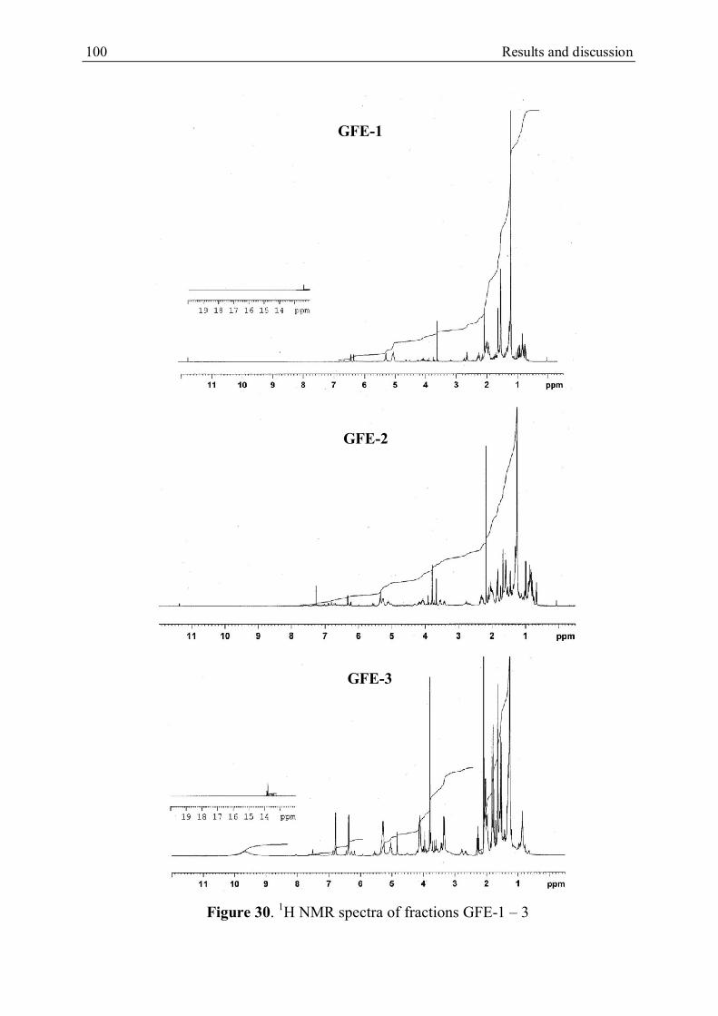
100 Results and discussion
Figure 30. 1H NMR spectra of fractions GFE-1 – 3
GFE-1
GFE-2
GFE-3
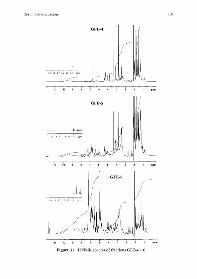
Result and discussion 101
Figure 31. 1H NMR spectra of fractions GFE-4 – 6
GFE-4
GFE-5
GFE-6

102 Results and discussion
Figure 32. 1H NMR spectra of fractions GFE-7 and GFE-8
GFE-7
GFE-8

Result and discussion 103
5.1.2.1. Isolation of xanthones from fraction GFE-4
CC (5 x 50 cm, 400 g of silica gel, 0-100% acetone-PE) of the fraction GFE-4 (14.1
g) produced 26 fractions (250 ml/fraction) combined by relying on TLC to give 6
fractions (GFE-4.1 – 6). The fraction GFE-4.2 (1.7 g) was further applied to FC (40 x
160 mm, 80 g of silica gel, 0 – 100% acetone-CHCl3, 20 ml/min) to afford 86 fractions
(15 ml/fraction) combined on the basis of TLC to give 5 fractions (GFE-4.3.1 - 5).
GPC (2 x 70 cm, 23 g of Sephadex LH-20, MeOH) of GFE-4.3.2 (398.7 mg) yielded
92 fractions (5 ml/fraction) combined by relying on TLC to give 3 fractions (GFE-
4.3.2.1 – 3). The repeated purification of fraction GFE-4.3.2.2 (60.7 mg) using semi-
preparative HPLC (9.4 x 250 mm, RP18 silica, 60 - 100% MeCN-H2O, 3 ml/min)
furnished 23 (2.9 mg), 24 (5.2 mg)) and 25 (2.7 mg). Moreover 26.8 mg of the fraction
GFE-4.3.2.3 was purified by semi-preparative HPLC (9.4 x 250 mm, RP18 silica, 60 -
100% MeCN-H2O, 3 ml/min) to obtain 26 (3.1 mg), see Scheme 23.
Scheme 23. Isolation of xanthones from fraction GFE-4
- CC, silica gel (400 g) - EtOAc-PE (0 – 100%) - 26 fractions (250 ml/fr.)
GFE-4.3 (1.7 g)
- FC, silica gel, 20 ml/min - Acetone-CHCl3 (0 – 100%) - 86 fractions (15 ml/fr.)
GFE-4.3.2.2 (60.7 mg)
GFE-4.3.2 (398.7 mg)
- RP18 HPLC, 3 ml/min - MeCN-H2O (60 – 100%)
23 (2.9 mg) 24 (5.2 mg)
- GPC, Sephadex LH-20 - MeOH - 92 fractions (5 ml/fr.)
GFE-4.3.2.3 (26.8 mg)
26 (3.1 mg)
25 (2.7 mg)
- RP18 HPLC, 3 ml/min - MeCN-H2O (60 – 100%)
GFE-4 (14.1 g)

104 Results and discussion
5.1.2.2. Isolation from fraction GFE-6
The fraction GFE-6 (14.2) was applied to CC (5 x 50 cm, 400 g of silica gel, 0-
100% acetone-CH2Cl2) of the fraction GFE-4 (14.1 g) to afford 28 fractions (250
ml/fraction) combined on the basis TLC to give 5 fractions (GFE-6.1 – 5). GPC (5 x
70 cm, 56 g of Sephadex LH-20, MeOH) of the fraction GFE-6.3 (2.8 g) produced 96
fractions (10 ml/fraction) combined by relying on TLC to give 6 fractions (GFE-6.3.1
– 6). The fraction GFE-6.3.4 was 27 (35.7 mg). the fraction GFE-6.3.3 (841.7 mg) was
further chromatographed by FC (40 x 160 mm, 35 g silica gel, 10 ml/min) to yield 60
fractions (10 ml/fraction) combined on the basis of TLC to give 3 fractions (GFE-
6.3.3.1 – 3). GFE-6.3.3.1 (126.5 mg) was purified using semi-preparative HPLC to
obtain 28 (15.8 mg). The repeated purification of the fraction GFE-6.3.3.3 (356.4 mg)
using semi-preparative HPLC (9.4 x 250 mm, RP18 silica, 60 - 100% MeCN-H2O, 3
ml/min) furnished 29 (20.5), 30 (15.6) and 31 (22.0), see Scheme 24.
Scheme 24. Isolation of compounds from fraction GFE-6
GFE-6 (14.2 g)
- CC, silica gel (400 g) - Acetone-CH2Cl2 (0 – 100%) - 28 fractions (250 ml/fr.)
GFE-6.3 (2.8 g)
- GPC, Sephadex LH-20 - MeOH - 96 fractions (10 ml/fr.)
27 (35.7 mg)
GFE-6.3.3.3 (356.4 mg)
- FC, silica gel, 20 ml/min - Acetone-CHCl3 (0 – 100%) - 86 fractions (15 ml/fr.)
GFE-6.3.3 (841.7 mg)
- RP18 HPLC, 3 ml/min - MeCN-H2O (60 – 100%)
29 (20.5 mg) 30 (15.6 mg) 31 (22.0 mg)
GFE-6.3.3.1 (126.5 mg)
- RP18 HPLC, 3 ml/min - MeCN-H2O (60 – 100%)
28 (15.8 mg)

Result and discussion 105
5.2. Structural elucidation of isolated substances
5.2.1. Structural identification of substances from the n-hexane extract
Thin layer and column chromatography as well as semi-preparative HPLC of the n-
hexane extract resulted in the isolation of twenty two substances. On the basis of NMR
spectra data (see Appendices 1 – 17) and comparison with those of literatures, the
structures of seventeen known compounds were determined as shown in Table 4.
Compound 8 was previously reported as a semi-synthetic xanthone. Five other
substances (18 – 22) have been first isolated and their structures were elucidated by
relying on the data of UV, 1D and 2D NMR, mass spectrometry and optical rotation.
In general, all the new compounds (18 - 22) gave positive test with FeCl3 in
methanol, which revealed their phenolic nature. The UV spectra of these compounds
displayed four maxima at 213 - 220, 242 - 244, 314 - 318 and 356 - 360 nm, which are
similar to those of 1,3,6,7-tetraoxygenated xanthones (Iinuma et al., 1996; Nilar and
Harrison, 2002). The 1H and 13C NMR spectra of these compounds indicated that they
are prenylated xanthone derivatives containing a hydrogen-bonded hydroxyl group at
H 13.72 – 14.08 with the appropriate interacting carbonyl carbon at C 181.7 – 183.0,
two isolated aromatic protons at H 6.79 – 6.83 and 6.23 – 6.36 and a methoxyl group
at H 3.78 – 3.83 (except for 21). Besides the 1H and 13C NMR spectra also showed the
presence of geranyl and isoprenyl side chains, whose benzylic methylene protons are
deshielded by the adjacent carbonyl group. This allowed the placement of these side
chains to the peri position (C-8) (Xu et al., 2001; Nilar and Harrison, 2002).

106 Results and discussion
Table 4. The structure of substances isolated from the n-hexane extract
Isolated
compounds
Structure
1
O
HO
-Tocotrienol (Ohnmacht et al., 2007)
2
O
H3CO
H3CO
O OH
OH
1,3-Dihydroxy-6,7-dimethoxy-2,8-diprenylxanthone
(Pattanaprateeb et al., 2004)
3
O
H3CO
HO
O OH
OCH3
-Mangostin (Mahaburarakam et al., 1987)

Result and discussion 107
4
O
H3CO
HO
O OH
OH
Cowanin (Na Pattalung et al., 1994)
5
OO
OH OH
OH
O
Garbogiol (Iinuma et al., 1997)
6
O
H3CO
HO
O OH
OH
-Mangostin (Mahaburarakam et al., 1987)

108 Results and discussion
7
O
H3CO
HO
O OH
OH
Rubraxanthone (Ampofo and Waterman, 1986)
8
O
H3CO
H3CO
O OH
OCH3
1-Hydroxy-3,6,7-trimethoxy-2-(3-methyl-
but-2-enyl)-xanthone (Nilar and Harrison, 2002)
9
O
H3CO
HO
O OH
OCH3
1,6-Dihydroxy-3,7-dimethoxy-2-(3-methyl-
but-2-enyl)-xanthone (Nilar and Harrison, 2002)
10
O
O
CHO
Piperaldehyde

Result and discussion 109
11
OHO
O OH
OCH3
OH
Nigrolineaxanthone E (Rukachaisirikul et al., 2003)
12
O
O OH
HO
OH
Fuscaxanthone E (Ito et al., 2003)
13
O
OHO
OH
H3CO
HO
Cowaxanthone (Na Pattalung et al., 1994)
14
O
OHO
OH
OH
Mangostinone (Asai et al., 1995)
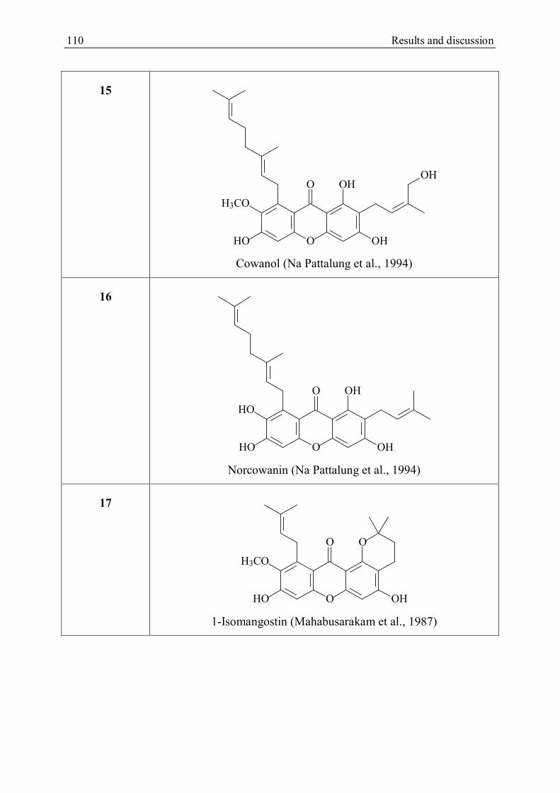
110 Results and discussion
15
O
H3CO
HO
O OH
OH
OH
Cowanol (Na Pattalung et al., 1994)
16
O
HO
HO
O OH
OH
Norcowanin (Na Pattalung et al., 1994)
17
O
H3CO
HO
O O
OH
1-Isomangostin (Mahabusarakam et al., 1987)

Result and discussion 111
5.2.1.1. Compound 21 (fuscaxanthone J)
Figure 33. Structure of fuscaxanthone J (21)
Compound 21 was isolated as a yellow oil. The molecular formula was established
as C28H32O7 by HRESIMS (m/z 481.2217 [M+H]+). The 1H and 13C NMR spectra of
21 (see Figure 34; Table 5 and 6) contained signals for a hydrogen-bonded hydroxyl
group [H 14.08 (1H, s, 1-OH)] with the interacting carbonyl carbon [C 183.0 (C-9)],
two isolated aromatic protons [H 6.81 (1H, s, H-5) and 6.35 (1H, s, H-4); C 101.0 (C-
5) and 93.3 (C-4)], a geranyl group [H 5.34 (1H, m like t, J = 8.0 Hz, H-22), 5.06
(1H, m like t, J = 6.8 Hz, H-17), 4.21 (2H, d, J = 6.8 Hz, H2-16), 2.05 (2H, m, H2-21),
1.97 (2H, m, H2-20), 1.86 (3H, d, J = 0.8 Hz, H3-19), 1.57 (3H, d, J = 0.8 Hz, H3-24)
and 1.53 (3H, s, H3-25); C 135.0 (C-18), 131.4 (C-23), 125.2 (C-22), 124.4 (C-17),
40.6 (C-20), 27.4 (C-21), 26.2 (C-16), 25.7 (C-24), 17.6 (C-25) and 16.6 (C-19)] and
an isoprenyl group [H 5.37 (1H, m like t, J = 8.0 Hz, H-12), 4.30 (2H, s, H2-14), 3.45
(2H, d, J = 8.0 Hz, H2-11) and 1.73 (3H, d, J = 1.2 Hz, H3-15); C 135.7 (C-13), 125.4
(C-12), 61.7 (C-14), 22.0 (C-15) and 21.7 (C-11)], in which one of two vinyl methyl
groups is hydroxylated to form a 4-hydroxy-3-methylbut-2-enyl group. This was
confirmed by the HMBC correlations of the protons of the oxygenated vinyl
methylene group at H 4.30 with two olefinic carbons (H 135.7, C-13 and 125.4, C-
14) and the vinyl methyl carbon (C 22.0, C-15). The 13C NMR spectra also showed
resonances due to ten substituted aromatic carbons, six of which are oxygenated (C
152.6 – 162.7). The 1H and 13C NMR data of 21 are the same as those of cowanol,
O
OHO
OH
HO
HO
OH
1
2
345
6
78
9
11
12
13
14
15
1617
18
1920
21
23
22
2425
4a
9a8a
10a

112 Results and discussion
which has previously reported from G. cowa (Na Pattalung et al., 1994). However
there is a difference regarding the absence of the signal for a methoxyl group.
In the HMBC spectrum, the chelated hydroxyl proton (H 14.08, 1-OH) correlated
with an oxygenated aromatic carbon (C 161.2) and two substituted aromatic carbons
(C 109.7 and 103.7). The isolated aromatic proton (H 6.35) gave cross-peaks with
two latter carbons and two oxygenated aromatic carbons (C 162.7 and 155.9). On the
other hand, the benzylic methylene protons (H 3.45) had correlations with a
substituted aromatic carbon at C 109.7 and two oxygenated aromatic carbons (C
161.2 and 162.7). These HMBC data revealed that the oxygenated aromatic carbon at
C 161.2 is C-1, C-3 is oxygenated (C 162.7), C-4a is the carbon at C 155.9, C-4 is
protonated (C 93.3) and the 4-hydroxy-3-methylbut-2-enyl group is attached to C-2
(C 109.7). In the xanthone B ring, two benzylic methylene protons at H 4.21 (C
26.2) exhibited a less shielding by the adjacent carbonyl group, i.e., the geranyl group
is placed at C-8. Furthermore these protons gave HMBC correlations with an
oxygenated aromatic carbon (C 141.7) and two substituted aromatic carbons (C 128.9
and 111.9). Meanwhile, the isolated aromatic proton at H 6.81 (C 101.0) showed
cross-peaks to a substituted aromatic carbon at C 111.9 and three oxygenated
aromatic carbons (C 153.6, 152.6 and 141.7). Thus, the substituted aromatic carbon at
C 128.9 is C-8, bearing the geranyl group and the other one is C-8a. Three oxygenated
aromatic carbons are C-6, C-10a and C-7, respectively. The isolated aromatic proton is
bonded to C-5. The NOESY correlations between H-14/H-11 and H-15/H-12 indicated
that the double bond of the isoprenyl group has the Z-configuration. The compound 21
was identified as 1,2,6,7-tetrahydroxy-2-(Z)-(4-hydroxy-3-methylbut-2-enyl)-8-(3,7-
dimethylocta-2,6-dienyl)xanthen-9-one and was given the trivial name fuscaxanthone
J.

Result and discussion 113
Figure 34. 1H and 13C NMR spectra of fuscaxanthone J (21)
O
OHO
OH
HO
HO
OH14.08
6.81 6.35
5.34
5.064.21
2.05
1.97 1.86
1.571.53
5.37
4.30
3.45
1.73
O
OHO
OH
HO
HO
OH
183.0
101.0 93.3
135.0
131.4
125.2
124.4
40.6
27.4
26.2
25.717.6
16.6
135.7
125.4
61.7
22.0
21.7
109.7
103.0
162.7
161.2
155.9
111.9
153.6 152.6
128.9
141.7

114 Results and discussion
5.2.1.2. Compound 20 (fuscaxanthone K)
Figure 35. Structure of fuscaxanthone K (20)
Compound 20 was obtained as a yellow oil and given the molecular formula
C29H34O8 by HRESIMS (m/z 511.2334 [M + H]+). In the 1H NMR spectrum (see
Figure 38; Table 5 and 6), the presence of a chelated hydroxyl group [H 13.72 (1H, s,
1-OH)], two isolated aromatic protons [H 6.83 (1H, s, H-5) and 6.23 (1H, s, H-4)], a
methoxyl group [H 3.79 (3H, s, 7-OCH3)] and a geranyl group [H 5.28 (1H, m like t,
J = 6.6 Hz, H-17), 5.04 (1H, m, H-22), 4.12 (2H, d, J = 6.6 Hz, H-16), 2.05 (2H, m,
H2-21), 1.98 (2H, m, H2-20), 1.82 (3H, s, H3-19), 1.55 (3H, s, H3-24) and 1.51 (3H, s,
H3-25)] were observed together with a benzylic methylene group [H 3.22 and 3.12
(2H, dd, J = 8.1 and 15.6 Hz, H2-11)], a deshielded methine proton [H 5.03 (1H, m,
H-12)], an oxygenated methylene group [H 3.69 and 3.57 (2H, d, J = 9.6 Hz, H2-14)]
and a vinyl methyl group [H 1.19 (3H, s, H3-15)]. The 13C NMR spectrum (see Figure
38; Table 5 and 6) contained signals of twenty nine carbons including a conjugated
carbonyl carbon (C 182.2), twelve aromatic carbons and four oxygenated sp3 carbons
(C 60.8 – 89.1). Of twelve aromatic carbons, six are oxygenated (C 157.5 – 167.4).
These spectral data supported 20 to be a tetra-oxygenated xanthone derivative carrying
a hydrogen-bonded hydroxyl group, a geranyl group, a methoxyl group and a C5 unit.
O
OHO
OH
H3CO
HO
O
OH1
2
345
6
78
9
11
12
13
14
1516
17
18
1920
21
23
22
2425
4a
9a8a
10a

Result and discussion 115
In the HMBC spectrum, two protons of the oxygenated methylene group (H 3.69
and 3.57; C 67.2) correlated with an oxygenated tertiary carbon (C 73.6), an
oxygenated secondary carbon (H 5.03; C 89.1) and the secondary carbon (H 1.19; C
20.2). The benzylic methylene protons (H 3.22 and 3.12, C 26.1) also showed cross-
peaks to the oxygenated tertiary carbon and the oxygenated secondary carbon.
Moreover these protons gave the COSY correlation with the deshielded methine
proton (H 5.03). These data suggested that the C5 unit is an isoprenyl group, in which
the double bond is oxidized and a vinyl methyl group is hydroxylated to form the 2,3-
epoxy-4-hydroxy-3-methylbutyl group. Besides the chelated hydroxyl group (H
13.72) correlated with C-1 (C 158.0) and C-9a (C 103.7) and a substituted aromatic
carbon (C 108.5), whereas the benzylic methylene protons of the 2,3-epoxy-4-
hydroxy-3-methylbutyl side chain also exhibited correlations with C-1, the substituted
aromatic carbon and another oxygenated aromatic carbon (C 167.4). The side chain is
therefore bonded to C-2 (C 108.5) and C-3 is oxygenated (C 167.4). The isolated
aromatic proton at H 6.23 is attached to C-4 because it gave cross-peaks to C-2, C-3
and another oxygenated aromatic carbon (C 157.4), which is C-4a. In the xanthone B
ring, the downfield shift (H 4.12) of the benzylic methylene protons of the geranyl
group revealed that this group is located at C-8 as in the case of compound 21. These
methylene protons (H2-16) and the methoxyl protons (H 3.79) showed correlations to
an oxygenated aromatic carbon (C 144.2), indicating that this carbon is C-7, which
carries the methoxyl group. This was confirmed by the HMBC correlation between
H2-16 with C-7, C-8a (C 111.2) and a substituted aromatic carbon (C 137.4) which
has to be C-8. Meanwhile the remaining isolated aromatic proton (H 6.83) gave cross-
peaks to C-7, C-8a and two oxygenated aromatic carbons (H 157.4 and 155.9). This
proton is therefore bonded to C-5 and two oxygenated aromatic carbons are C-6 and
C-10a.
In the ROESY spectrum (Figure 36), the protons H2-14 gave an interaction with the
proton H-12, whereas they did not show any interaction with H2-11. This indicated
that the oxygenated methyl group is trans to the benzylic methylene group in the 2,3-
epoxy-4-hydroxy-3-methylbutyl side chain. Therefore, the configuration of two chiral

116 Results and discussion
carbons C-12 and C-13 was suggested as R by comparison of the optical rotation of 20
with that of (2S-trans)-2-methyl-3-phenyloxiranemethanol, see Figure 37 (Gao et al.,
1987). Two chiral carbons of this compound possess S configuration corresponding to
the optical rotation []25D – 16.9º, whereas that of 20 is + 76.3º (measured at 22 ºC).
Hence 20 was determined as 12R,13R-1,3,5-trihydroxy-2-(2,3-epoxy-4-hydroxy-3-
methylbutyl)-7-methoxyxanthen-9-one and given the trivial name fuscaxanthone K.
Figure 36. The ROESY interaction of H2-14
Figure 37. (2S-trans)-2-methyl-3-phenyloxiranemethanol
OH
O

Result and discussion 117
Figure 38. 1H and 13C NMR spectra of fuscaxanthone K (20)
O
OHO
OH
H3CO
HO
O
OH182.2
102.0 88.2
134.5
131.0
124.6
124.4
40.0
26.8
26.3
25.317.2
16.1
73.6
67.2
20.2
26.1
108.5
103.7
167.4
158.0
157.4
111.2
157.4 155.6
137.4
144.289.1
60.8
O
OHO
OH
H3CO
HO
O
OH
6.83 6.23
13.72
3.79
5.28
5.04
4.12
2.05
1.98
1.551.51
1.82
3.22 & 3.12
5.03 3.69 & 3.57
1.19

118 Results and discussion
5.2.1.3. Compound 19 ((+)-fuscaxanthone I)
Figure 39. Structure of (+)-fuscaxanthone I (19)
Compound 19 was isolated as a yellow amorphous powder and showed a [M+H]+
ion peak at m/z 513.2478 in HRESIMS, corresponding to the molecular formula
C29H36O8. The 1H and 13C NMR spectrum of 19 (see Figure 40; Table 5 and 6)
showed resonances for a hydrogen-bonded hydroxyl group [H 13.89 (1H, s, 1-OH)]
corresponding to the chelated carbonyl carbon [C 181.7 (C-9)], two isolated aromatic
protons [H 6.79 (1H, s, H-5) and 6.34 (1H, s, H-4); C 101.6 (C-5) and 92.5 (C-4)], a
methoxyl group [H 3.83 (3H, s, 7-OCH3), C 60.5 (CH3O-7)], a 4-hydroxy-3-
methylbut-2-enyl side chain [H 5.35 (1H, t, J = 7.4 Hz, H-12), 4.28 (2H, s, H2-14),
3.42 (2H, d, J = 7.4 Hz, H2-11), 1.71 (3H, s, H-15); C 134.7 (C-13), 124.4 (C-12),
60.7 (C-14), 21.0 (C-15) and 20.7 (C-11)], an isoprenyl group [H 5.17 (1H, t, J = 7.2
Hz, H-22), 2.18 (2H, m, H2-21), 1.66 (3H, s, H3-24) and 1.63 (3H, s, H3-25); C 130.3
(C-23), 125.2 (C-22), 24.8 (C-24), 22.4 (C-21) and 16.7 (C-25)], a benzylic methylene
group [H 3.42 (2H, m, H2-16), C 21.9 (C-16)], two methylene groups [H 1.75 (2H,
m, H2-17) and 1.57 (2H, m, H2-20); C 42.5 (C-17) and 41.6 (C-20)] and a methyl
group [H 1.28 (3H, s, H3-19); C 26.4 (C-19)]. The 1H and 13C NMR data of 19 quite
resembled to those of fuscaxanthone I, which was previously reported from G. fusca
collected in Thailand (Nontakham et al., 2014). In this xanthone, the double bond at C-
17/C-18 of the geranyl group is hydroxylated to produce the 3-hydroxy-3,7-dimethyl-
oct-6-enyl group. This group was confirmed by the HMBC and COSY correlations.
O
OHO
OH
H3CO
HO
OH
OH
1
2
345
6
78
9
11
12
13
14
15
1617
181920
21
23
22
2425
4a
9a8a
10a

Result and discussion 119
The HMBC correlations between the shielded benzylic methylene protons (H 3.22)
with the secondary carbon (C 42.5) and an oxygenated quaternary carbon (C 71.3), in
addition to the COSY interaction with the methylene protons (H 1.75) showed that the
secondary carbon is C-17 and C-18 is the oxygenated quaternary carbon. The HMBC
cross-peaks of a methyl group (H 1.28) with C-17, C-18 and a secondary carbon (C
41.6) indicated that this methyl group is located at C-18 and the secondary carbon is
C-20. The COSY interaction between H-20 and the vinyl methylene protons of the
isoprenyl group pointed out the connectivity of C-20 with the isoprenyl group.
Moreover the HMBC correlations of the benzyl methylene protons H2-16 with two
aromatic substituted carbons at C 138.9 (C-8) and 110.8 (C-8a) and an oxygenated
aromatic carbon at C 143.5 (C-7) as well as the ROESY interaction with the methoxyl
protons allowed the placement of the hydroxylated geranyl group at C-8. On the other
hand, the benzylic methylene protons of the 4-hydroxy-3-methylbut-2-enyl side chain
gave the HMBC correlations with C-1 (C 160.6), the substituted aromatic carbon at C
109.7 (C-2) and C-3 (C 162.3), i.e. this side chain is obviously attached to C-2. Thus,
19 was identified as 1,3,6-trihydroxy-2-(4-hydroxy-3-methylbut-2-enyl)-8-(3-hydroxy-
3,7-dimethyl-oct-6-enyl)-7-methoxyxanthen-9-one. However the optical rotation of 19
([]22D + 36.0º) is different from that of fuscaxanthone I ([]26
D – 9.5º), in which the
configuration of the chiral carbon C-18 could not be resolved. Comparison of the
optical rotation of 19 with that of (3’S)-4-hydroxy-3-(3’-hydroxy-3’,7’-dimethyloct-6’-
enoyl)benzaldehyde ([]25D – 12.0º) (see Figure 41) (Chakor et al., 2011) suggested
that the chiral carbon C-18 of 19 has R configuration and the compound was given
trivial name (+)-fuscaxanthone I.
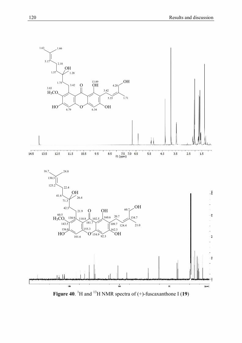
120 Results and discussion
Figure 40. 1H and 13H NMR spectra of (+)-fuscaxanthone I (19)
O
OHO
OH
H3CO
HO
OH
OH
6.79 6.34
13.89
3.83
1.75
5.17
3.42
2.18
1.57
1.661.63
1.28
3.42
5.35
4.28
1.71
O
OHO
OH
H3CO
HO
OH
OH
181.7
101.6 92.5
71.3
130.3
125.2
42.5
41.6
22.4
21.9
24.816.7
26.4
134.7
60.7
21.0
20.7
109.7
102.5
162.3
160.6
154.9
110.8
156.8 155.3
138.9
143.5124.4
60.5

Result and discussion 121
Figure 41. (3’S)-4-Hydroxy-3-(3’-hydroxy-3’,7’-dimethyloct-6’-enoyl)benzaldehyde
Table 5. 1H NMR data of 21, 19 and 20 in acetone-d6
Position 21 19 20
1-OH 14.08 s 13.89 s 13.72 s
4 6.35 s 6.34 s 6.23 s
5 6.81 s 6.79 s 6.83 s
11 3.45 d (8.0 Hz) 3.42 d (7.4 Hz) 3.22 & 3.12 dd (8.1 & 15.6 Hz)
12 5.37 t (8.0 Hz) 5.35 t (7.4 Hz) 5.03 m
14 4.20 s 4.28 s 3.96 & 3.57 d (9.6 Hz)
15 1.73 s 1.71 s 1.19 s
16 4.21 d (6.8 Hz) 3.42 overlap with H-11 4.12 d (6.6 Hz)
17 5.06 t (6.8 Hz) 1.75 m 5.28 t (6.6 Hz)
19 1.86 d (0.8 Hz) 1.28 s 1.82 s
20 1.97 m 1.57 m 1.98 m
21 2.05 m 2.18 m 2.05 m
22 5.34 t (8.0 Hz) 5.17 t (7.2 Hz) 5.04 m
24 1.57 s 1.66 s 1.55 s
25 1.53 s 1.63 s 1.51 s
7-OCH3 3.83 s 3.79 s
600 MHz (J in Hz and in ppm) at 298ºK.
OH
CHO
O OH

122 Results and discussion
Table 6. 13C NMR data of 21, 19 and 20 in acetone-d6
Position 21 19 20
1 161.2 160.6 158.0
2 109.7 109.7 108.5
3 162.7 162.3 164.7
4 93.3 92.5 88.2
4a 155.9 154.9 157.4
5 101.0 101.6 102.0
6 153.6a 156.8a 157.4a
7 141.7 143.5 144.2
8 128.9 138.9 137.4
8a 111.9 110.8 111.2
9 183.0 181.7 182.2
9a 103.0 102.5 103.7
10a 152.6a 155.3a 155.6a
11 21.7 20.7 26.1
12 125.4 124.4 89.1
13 135.7 134.7 73.6
14 61.7 60.7 67.2
15 22.0 21.0 20.2
16 26.2 21.9 26.3
17 124.4 42.5 124.4
18 135.0 71.3 134.5
19 16.6 26.4 16.1
20 40.6 41.6 40.0
21 27.4 22.4 26.8
22 125.2 125.2 124.6
23 131.4 130.3 131.0
24 25.7 24.8 25.3
25 17.6 16.7 17.2
7-OCH3 60.5 60.8
a Interchangeable in the same column, 150 MHz ( in ppm) at 298ºK
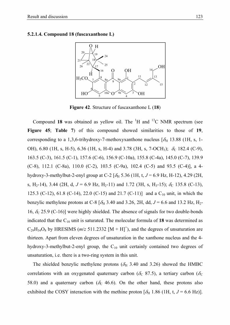
Result and discussion 123
5.2.1.4. Compound 18 (fuscaxanthone L)
Figure 42. Structure of fuscaxanthone L (18)
Compound 18 was obtained as yellow oil. The 1H and 13C NMR spectrum (see
Figure 45; Table 7) of this compound showed similarities to those of 19,
corresponding to a 1,3,6-trihydroxy-7-methoxyxanthone nucleus [H 13.88 (1H, s, 1-
OH), 6.80 (1H, s, H-5), 6.36 (1H, s, H-4) and 3.78 (3H, s, 7-OCH3); C 182.4 (C-9),
163.5 (C-3), 161.5 (C-1), 157.6 (C-6), 156.9 (C-10a), 155.8 (C-4a), 145.0 (C-7), 139.9
(C-8), 112.1 (C-8a), 110.0 (C-2), 103.5 (C-9a), 102.4 (C-5) and 93.5 (C-4)], a 4-
hydroxy-3-methylbut-2-enyl group at C-2 [H 5.36 (1H, t, J = 6.9 Hz, H-12), 4.29 (2H,
s, H2-14), 3.44 (2H, d, J = 6.9 Hz, H2-11) and 1.72 (3H, s, H3-15); C 135.8 (C-13),
125.3 (C-12), 61.8 (C-14), 22.0 (C-15) and 21.7 (C-11)] and a C10 unit, in which the
benzylic methylene protons at C-8 [H 3.40 and 3.26, 2H, dd, J = 6.6 and 13.2 Hz, H2-
16, C 25.9 (C-16)] were highly shielded. The absence of signals for two double-bonds
indicated that the C10 unit is saturated. The molecular formula of 18 was determined as
C29H34O8 by HRESIMS (m/z 511.2332 [M + H]+), and the degrees of unsaturation are
thirteen. Apart from eleven degrees of unsaturation in the xanthone nucleus and the 4-
hydroxy-3-methylbut-2-enyl group, the C10 unit certainly contained two degrees of
unsaturation, i.e. there is a two-ring system in this unit.
The shielded benzylic methylene protons (H 3.40 and 3.26) showed the HMBC
correlations with an oxygenated quaternary carbon (C 87.5), a tertiary carbon (C
58.0) and a quaternary carbon (C 46.6). On the other hand, these protons also
exhibited the COSY interaction with the methine proton [H 1.86 (1H, t, J = 6.6 Hz)].
1
2
345
6
78
9
11
12
13
14
15
16
17
18
1920
21
252226
24
4a
9a8a
10a O
OHO
OH
H3CO
HO
OH
O H
H

124 Results and discussion
Thus, the tertiary carbon is C-17, the quaternary carbon is C-18 and C-22 is the
oxygenated quaternary carbon. The HMBC correlations of the protons of two methyl
carbons (H 1.09 and 0.89; C 25.7 and 24.5) with C-17, C-18 and an oxygenated
tertiary carbon [H 3.63 (1H, d, J = 5.4 Hz); C 86.4] revealed that these two methyl
groups are bonded to C-18 and the oxygenated tertiary carbon is C-19. The COSY
interactions between the methylene protons [H 1.82 and 1.51 (2H, m); C 26.5] with
H-19 and the other methylene protons [H 1.42 and 1.24 (2H, m); C 40.1] showed that
the secondary carbon at H 26.5 is C-20 and C-21 is the other carbon (C 40.1). The
HMBC cross-peaks of the methyl protons [H 1.27 (3H, s); C 18.9] with C-17, C-21
and C-22 confirmed the connectivity of C-21 with C-22 as well as the position of this
methyl group at C-22. Hence the C10 unit was established as the 1,3,3-trimethyl-7-
oxabicyclo[2.2.1]hept-2-ylmethyl group comprising three chiral carbons at C-17, C-19
and C-22. This was supported by comparison of the spectral data of 18 with those of
parvixanthone I, which was previously isolated from G. parvifolia (Xu et al., 2001).
This compound also contains a 1,3,3-trimethyl-7-oxabicyclo[2.2.1]hept-2-ylmethyl
group at C-8 but the carbon C-2 is bonded with a proton instead of the 4-hydroxy-3-
methylbut-2-enyl side chain as in the case of 18. Due to the boat conformation of the
7-oxabicyclo[2.2.1] system with the oxo-bridge, the direction of the proton H-19 and
the methyl group at C-22 is required to be endo. In the NOESY spectrum, H-19 gave
the interaction with the protons of the methyl group (H 1.09, H3-24) and a proton (H
1.82) of H2-20, i.e. the direction of this methyl group and this proton is consequently
exo (Figure 43). Meanwhile the direction of H-17 (H 1.86) and a proton (H 1.42) of
H2-21 were determined as endo based on the NOESY interaction with the methyl
protons H3-25 (see Figure 44) Furthermore, the HMBC cross-peaks of the benzylic
methylene protons (H 3.40 and 3.26, C-16) with C-7 (C 145.0), C-8 (C 139.9) and
C-8a (C 112.1) pointed out the location of the 1,3,3-trimethyl-7-
oxabicyclo[2.2.1]hept-2-ylmethyl group at C-8. Thus, compound 18 was identified as
1,3,6-trihydroxy-2-(4-hydroxy-3-methylbut-2-enyl)-7-methoxy-8-(1,3,3-trimethyl-7-
oxabicyclo[2.2.1]hept-2-ylmethyl)xanthen-9-one and was given a trivial name

Result and discussion 125
fuscaxanthone L. However the absolute configuration of the chiral carbons could not
be determined.
Figure 43. The NOESY interaction between H3-24, H-20 and H-19
Figure 44. The NOESY interaction between H-21, H-17 and H3-25

126 Results and discussion
Figure 45. 1H and 13C NMR spectra of fuscaxanthone L (18)
O
OHO
OH
H3CO
HO
OH
O H
H
13.88
6.80 6.36
3.78
5.36
4.29
3.44
1.72
3.40 &3.261.86
0.89
1.091.82 & 1.51
3.63
1.27
1.42 &1.24
O
OHO
OH
H3CO
HO
OH
O H
H
182.4
163.5
161.5
157.6
155.8
145.0
139.9 112.1
110.0
103.5
102.4 93.5
156.9
135.8
125.3
61.8
22.0
21.7
87.5 58.0
25.9
46.6
25.7
24.586.4
26.5
40.1
18.9
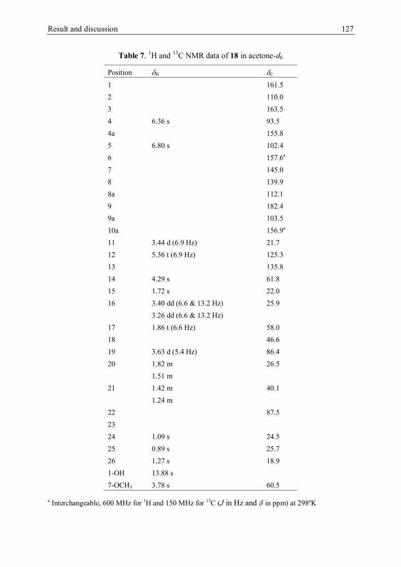
Result and discussion 127
Table 7. 1H and 13C NMR data of 18 in acetone-d6
Position H C
1 161.5
2 110.0
3 163.5
4 6.36 s 93.5
4a 155.8
5 6.80 s 102.4
6 157.6a
7 145.0
8 139.9
8a 112.1
9 182.4
9a 103.5
10a 156.9a
11 3.44 d (6.9 Hz) 21.7
12 5.36 t (6.9 Hz) 125.3
13 135.8
14 4.29 s 61.8
15 1.72 s 22.0
16 3.40 dd (6.6 & 13.2 Hz) 25.9
3.26 dd (6.6 & 13.2 Hz)
17 1.86 t (6.6 Hz) 58.0
18 46.6
19 3.63 d (5.4 Hz) 86.4
20 1.82 m
1.51 m
26.5
21 1.42 m
1.24 m
40.1
22 87.5
23
24 1.09 s 24.5
25 0.89 s 25.7
26 1.27 s 18.9
1-OH 13.88 s
7-OCH3 3.78 s 60.5
a Interchangeable, 600 MHz for 1H and 150 MHz for 13C (J in Hz and in ppm) at 298ºK

128 Results and discussion
5.2.1.5. Compound 22 (fuscaxanthone M)
Figure 46. Structure of fuscaxanthone M (22)
Compound 22 was isolated as yellow oil. The 1H and 13C NMR spectra (see Figure
49 and Table 8) indicated that this compound contains two prenylated xanthone
fragments. The fragment B is cowanol (Na Pattalung et al., 1994), comprising a
hydrogen-bonded hydroxyl group [H 13.80 (1H, s, H-1’)] corresponding with a
chelated carbonyl carbon [C 181.8 (C-8’)], two isolated aromatic protons [H 6.82
(1H, s, H-5’) and 6.29 (1H, s, H-4’); C 101.6 (C-5’) and 93.7 (C-4’)], a methoxyl
group [H 3.82 (3H, s, 7’-OCH3); C 62.1 (CH3O-7’)], a 4-hydroxy-3-methyl-but-2-
enyl group [H 5.36 (1H, t, J = 7.7 Hz, H-12’), 4.23 (1H, d, J = 10.8 Hz, HA-14’), 4.16
(1H, d, J = 10.8 Hz, HB-14’), 3.66 (2H, d, J = 7.7 Hz, H2-11’) and 1.82 (3H, s, H-15’);
C 131.9 (C-13’), 126.6 (C-12’), 61.8 (C-14’), 23.6 (C-15’) and 21.4 (C-11’)] and a
geranyl group [H 5.27 (1H, t, J = 6.0 Hz, H-17’), 5.16 (1H, t, J = 7.2 Hz, H-22’), 4.10
(2H, d, J = 6.0 Hz, H2-16’), 2.04 (2H, m, H2-21’), 1.99 (2H, m, H2-20’), 1.82 (3H, s,
H3-19), 1.60 (3H, s, H3-24’) and 1.54 (3H, s, H3-25’); C 135.5 (C-18’), 131.3 (C-23’),
124.3 (C-22’), 123.4 (C-17’), 39.8 (C-20’), 26.6 (C-16’), 26.5 (C-21’), 25.5 (C-24’),
17.7 (C-25’) and 16.5 (C-19’)]. The fragment A resembled 19, including a hydroxyl
group [H 13.92 (1H, s, H-1)] chelated with a conjugated carbonyl carbon [C 181.9
(C-8)], two isolated aromatic protons [H 6.81 (1H, s, H-5) and 6.28 (1H, s, H-4); C
101.8 (C-5) and 93.9 (C-4)], a methoxyl group [H 3.80 (3H, s, 7-OCH3); C 62.0
1
2
345
6
78
9
11
12
13
14
15
16
17
1819
20
21
23
22
2425
4a
9a8a
10a
1'
2'
3'4'5'
6'
7'
8'
9'
17'
11'
12'13'
14'
15'
16'
18'19'
20'
21'22'
23'24'25'
O
O OH
OHHO
H3CO
O
OHO
OH
H3CO
HO
OHO

Result and discussion 129
(CH3O-7)], a 4-hydroxy-3-methyl-but-2-enyl group [H 5.47 (1H, t, J = 6.8 Hz, H-12),
4.36 (2H, s, H2-14), 3.52 (2H, d, J = 6.8 Hz, H-11) and 1.78 (3H, s, H-15); C 133.1
(C-13), 127.0 (C-12), 63.0 (C-14), 21.6 (C-11) and 23.0 (C-15)] and a hydroxylated
geranyl [H 5.02 (1H, t, J = 6.9 Hz, H-22), 3.42 (2H, m, H2-16), 2.15 (2H, m, H2-21),
1.92 (1H, m, HA-20), 1.82 (1H, m, HA-17), 1.76 (1H, m, HB-17), 1.70 (3H, s, H3-24),
1.67 (1H, m, HB-20), 1.64 (3H, s, H3-25) and 1.45 (3H, s, H3-19); C 131.6 (C-23),
124.4 (C-22), 79.3 (C-18), 37.9 (C-20), 37.8 (C-17), 25.7 (C-24), 22.9 (C-19), 22.4 (C-
21), 21.9 (C-16) and 17.7 (C-25)].
In the HMBC spectrum, the benzyl methylene protons H-16 (H 3.42) correlated
with the secondary carbon C-17 and the oxygenated quaternary carbon C-18. The
methylene protons H-17 gave cross-peaks with C-16 and C-18. These data together
with the COSY interaction between H-16 and H-17 indicated that the double bond at
C-17/C-18 of the geranyl group is hydroxylated. Furthermore the HMBC correlations
of the proton H-16 with C-8, C-7 and C-8a, in addition to the NOESY interaction with
the methoxyl protons 7-OCH3 allowed the placement of the hydroxylated geranyl
group at C-8. The molecular formula of 22 was calculated to be C58H68O14 with 25
degrees of unsaturation based on HRESIMS (m/z 1011.4503 [M + Na]+). Because the
cowanol fragment has seven oxygen atoms, the fragment A therefore contains seven
oxygen atoms. Due to six oxygenated aromatic carbons of the xanthone nucleus and an
oxygenated methyl carbon of the 4-hydroxy-3-methyl-but-2-enyl group, the
oxygenated quaternary carbon C-18 of the hydroxylated geranyl group has to be
combined with the cowanol fragment by a C-O-C linkage. This was confirmed by the
HMBC correlation of H-14’ with C-18 (Figure 47), in addition to the NOESY
interaction between the vinyl methyl protons H3-15’ and the methylene protons H2-17,
see Figure 48. Hence compound 22 was determined as 8-{3-[1’,3’,6’,-trihydroxy-2’-
(4-hydroxy-3-methylbut-2-enyl)-7’-methoxy-8’-(3,7-dimethylocta-2,6-dienyl)xanthen-
9-one-14’-oxyl]-3,7-dimethyloct-2,6-dienyl}-1,3,6-trihydroxy-2-(4-hydroxy-3-
methylbut-2-enyl)-7-methoxy-8-(3,7-dimethylocta-6-enyl)bixanthone and was given
trivial name fuscaxanthone M. The configuration of the chiral carbon C-18 was
suggested to be R by comparison of the optical rotation of 22 ([]22D + 54.6º ) with

130 Results and discussion
that of (3’S)-4-hydroxy-3-(3’-hydroxy-3’,7’-dimethyloct-6’-enoyl)benzaldehyde
([]25D – 12.0º) (Chakor et al., 2011).
Figure 47. The HMBC correlation of H-14' with C-18
Figure 48. The NOESY interaction between H-15' and H-17

Result and discussion 131
Figure 49. 1H and 13C NMR spectra of fuscaxanthone M (22)
O
O OH
OHHO
H3CO
O
OHO
OH
H3CO
HO
OHO13.80
5.36
6.82 6.29
4.23 & 4.16
3.66
1.82
5.27
5.16
4.10
2.04
1.991.82
1.601.54
3.82
13.92
6.81 6.28
3.80
5.47
4.36
3.52
1.45
1.78
5.02
3.42
2.15
1.92 & 1.67
1.82 & 1.76
1.671.64
6.326.36
O
O OH
OHHO
H3CO
O
OHO
OH
H3CO
HO
OHO
126.6
101.6 93.7
61.8
21.4
23.6
123.4
124.3
26.6
26.5
39.816.5
25.517.7
62.1
101.8 93.9
62.0
127.0
63.0
21.6
22.9
23.0
124.4
21.9
22.4
37.9
37.8
25.717.7
181.8181.9
131.9
135.5
131.3
160.4
107.6
103.0
163.0
155.1
154.3 155.8
142.6
112.2137.0 160.7
108.0
161.9
155.3
133.1103.2
154.5 155.8
131.6
79.3
142.4
138.1112.3

132 Results and discussion
Table 8. 1H and 13C NMR data of 22 in acetone-d6
Fragment A Fragment B
H C H C
1 160.7 1' 160.4
2 108.0 2' 107.6
3 161.9 3' 163.0
4 6.28 s 93.9 4' 6.29 s 93.7
4a 155.3 4'a 155.1
5 6.81 s 101.8 5' 6.82 s 101.6
6 154.5a 6' 154.3a
7 142.4 7' 142.6
8 138.1 8' 137.0
8a 112.3 8'a 112.2
9 181.9 9' 181.8
9a 103.2 9'a 103.0
10a 155.8a 10'a 155.8a
11 3.52 d (6.8 Hz) 21.6 11' 3.66 d (7.7 Hz) 21.4
12 5.47 t (6.8 Hz) 127.0 12' 5.36 t (7.7 Hz) 126.6
13 133.1 13' 131.9
14 4.36 s 63.0 14' 4.23 d (10.8 Hz)
4.16 d (10.8 Hz)
61.8
15 1.78 s 23.0 15' 1.82 s 23.5
16 3.42 m 21.9 16' 4.10 d (6.0 Hz) 26.6
17 1.82 m
1.76 m
37.8 17' 5.27 t (6.0 Hz) 123.4
18 79.3 18' 135.5
19 1.45 s 22.9 19' 1.82 s 16.5
20 1.92 m
1.67 m
37.9 20' 1.99 m 39.8
21 2.15 m 22.4 21' 2.04 m 26.5
22 5.05 t (6.9 Hz) 124.4 22' 5.16 t (7.2 Hz) 124.3
23 131.6 23' 131.3
24 1.70 s 25.7 24' 1.60 s 25.5
25 1.64 s 17.7 25' 1.54 s 17.7
1-OH 13.92 s 1'-OH 13.88 s
7-OCH3 3.80 s 62.0 7'-OCH3 3.78 s 62.1
a Interexchangeable, 600 MHz for 1H and 150 MHz for 13C (J in Hz and in ppm) at 298ºK

Result and discussion 133
5.2.2. Structure identification of substances from the ethyl acetate extract
Thin layer and column chromatography together with semi-preparative HPLC of the
ethyl acetate extract led to the isolation of nine substances. Analysis of NMR spectra
(see Appendix 18 – 23) and comparison with those of previous reports, the structures
of six known compounds were elucidated as shown in Table 9. Meanwhile the
structures of three new substances were identified by relying on data of UV, 1D and
2D NMR, MS spectra and optical rotation.
Generally, all three new compounds (24 - 26) reacted positively with FeCl3 in
methanol, which indicated their phenolic feature. The UV spectra of these compounds
are similar to those of 1,3,6,7-tetraoxygenated xanthones, comprising four maxima at
208 - 213, 243 - 245, 315 - 317 and 355 - 360 nm (Iinuma et al., 1996; Nilar and
Harrison, 2002). The 1H and 13C NMR spectra of these compounds resembled those of
the new compounds from the n-hexane extract, which revealed that they were
prenylated xanthone derivatives containing a hydrogen-bonded hydroxyl group at H
13.88 – 13.99, a conjugated carbonyl carbon at C 182.8 – 183.1, two isolated aromatic
protons at H 6.81 – 6.83 and 6.34 – 6.36 and a methoxyl group at H 3.77 – 3.79.
Moreover the 1H and 13C NMR spectra also showed the presence of hydroxylated
geranyl and isoprenyl groups.

134 Results and discussion
Table 9. The structure of substances isolated from the ethyl acetate extract
Isolated
compounds
Structure
23
O
H3CO
HO
O O
OH
OH
11-Hydroxy-1-isomangostin (Sia et al., 1995)
27
O
O OH
OH
OH
HO
O
O
HO
HO
OHHO
Griffipavixanthone (Xu et al., 1998)
28
O
OO
OOH
HO
HO
OH
OH
OH
OH
Morelloflavone (Waterman and Crichton, 1980)

Result and discussion 135
29
O
OO
OOH
HO
HO
OH
OH
OH
Volkinflavone (Chen et al., 1975)
30
O
OO
OOH
HO
HO
OH
OH
OH
GB-1a (Jackson et al., 1971)
31
O
OO
OOH
HO
HO
OH
OH
OH
OH
OH
GB-2 (Jackson et al., 1971)

136 Results and discussion
5.2.2.1. Compound 24 (fuscaxanthone N)
Figure 50. Structure of fuscaxanthone N (24)
Compound 24 was obtained as a yellow oil and the molecular formula was deduced
to be C29H34O8 from the HRESIMS (m/z 509.2169 [M – H]-). In the 1H and 13C NMR
spectrum (see Figure 53 and Table 10), the presence of a hydrogen-bonded hydroxyl
group [H 13.88 (1H, s, 1-OH)] corresponding with a chelated carbonyl carbon [C
182.8 (C-9)], two separated aromatic protons [H 6.83 (1H, s, H-5) and 6.36 (1H, s, H-
4); C 102.8 (C-5) and 93.5 (C-4)], a methoxyl group [H 3.79 (3H, s, 7-OCH3); C
61.4 (CH3O-7)] and a 4-hydroxy-3-methylbut-2-enyl group [H 5.34 (1H, t, J = 7.8 Hz,
H-12), 4.29 (1H, s, H-14), 3.44 (2H, d, J = 7.8 Hz, H2-11) and 1.72 (3H, s, H3-15); C
135.7 (C-13), 125.3 (C-12), 61.8 (C-14), 22.0 (C-15) and 21.7 (C-11)] were observed
together with three vinyl protons [H 5.58 (1H, m, H-22), 5.55 (1H, m, H-21) and 5.35
(1H, t, J = 6.9 Hz, H-17); C 136.9 (C-22), 128.6 (C-21) and 125.5 (C-17)], a benzylic
methylene group [H 4.13 (2H, t, J = 6.9 Hz, H2-16); C 26.8 (C-16)], an vinyl
methylene group [H 2.67 (2H, d, J = 6.0 Hz, H2-20), C 43.5 (C-20)] and three methyl
groups [H 1.81 (3H, s, H3-19), 1.29 (3H, s, H3-24) and 1.21 (3H, s, H3-25); C 24.9
(C-24 and C-25) and 16.5 (C-19)]. The 13C NMR spectrum also showed signals for six
oxygenated aromatic carbons (C 144.5 – 163.2) and an oxygenated quaternary
carbon [C 81.4 (C-23)]. These spectral data indicated that 24 is a prenylated 1,3,6,7-
tetrahydroxyxanthone like the new xanthones isolated from the n-hexane extract,
O
OHO
OH
H3CO
HO
OH
OH
1
2
345
6
78
9
11
12
13
14
15
1617
181920
21
23
22
2425
4a
9a8a
10a
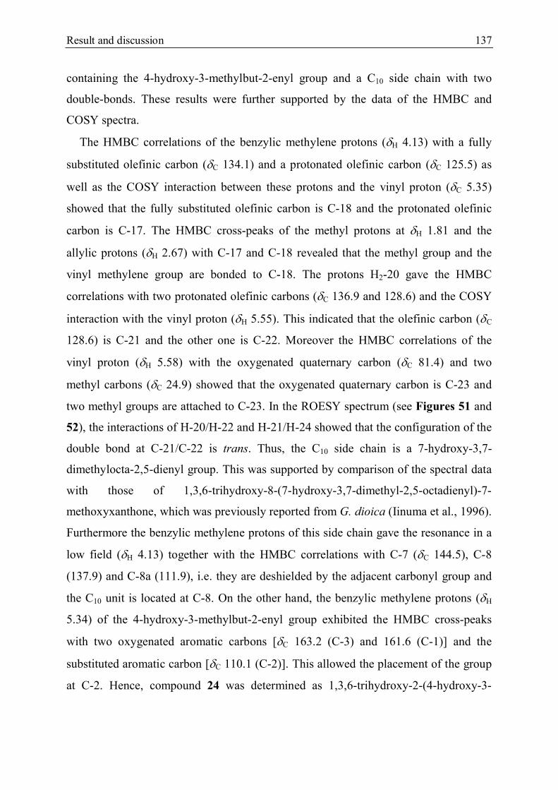
Result and discussion 137
containing the 4-hydroxy-3-methylbut-2-enyl group and a C10 side chain with two
double-bonds. These results were further supported by the data of the HMBC and
COSY spectra.
The HMBC correlations of the benzylic methylene protons (H 4.13) with a fully
substituted olefinic carbon (C 134.1) and a protonated olefinic carbon (C 125.5) as
well as the COSY interaction between these protons and the vinyl proton (C 5.35)
showed that the fully substituted olefinic carbon is C-18 and the protonated olefinic
carbon is C-17. The HMBC cross-peaks of the methyl protons at H 1.81 and the
allylic protons (H 2.67) with C-17 and C-18 revealed that the methyl group and the
vinyl methylene group are bonded to C-18. The protons H2-20 gave the HMBC
correlations with two protonated olefinic carbons (C 136.9 and 128.6) and the COSY
interaction with the vinyl proton (H 5.55). This indicated that the olefinic carbon (C
128.6) is C-21 and the other one is C-22. Moreover the HMBC correlations of the
vinyl proton (H 5.58) with the oxygenated quaternary carbon (C 81.4) and two
methyl carbons (C 24.9) showed that the oxygenated quaternary carbon is C-23 and
two methyl groups are attached to C-23. In the ROESY spectrum (see Figures 51 and
52), the interactions of H-20/H-22 and H-21/H-24 showed that the configuration of the
double bond at C-21/C-22 is trans. Thus, the C10 side chain is a 7-hydroxy-3,7-
dimethylocta-2,5-dienyl group. This was supported by comparison of the spectral data
with those of 1,3,6-trihydroxy-8-(7-hydroxy-3,7-dimethyl-2,5-octadienyl)-7-
methoxyxanthone, which was previously reported from G. dioica (Iinuma et al., 1996).
Furthermore the benzylic methylene protons of this side chain gave the resonance in a
low field (H 4.13) together with the HMBC correlations with C-7 (C 144.5), C-8
(137.9) and C-8a (111.9), i.e. they are deshielded by the adjacent carbonyl group and
the C10 unit is located at C-8. On the other hand, the benzylic methylene protons (H
5.34) of the 4-hydroxy-3-methylbut-2-enyl group exhibited the HMBC cross-peaks
with two oxygenated aromatic carbons [C 163.2 (C-3) and 161.6 (C-1)] and the
substituted aromatic carbon [C 110.1 (C-2)]. This allowed the placement of the group
at C-2. Hence, compound 24 was determined as 1,3,6-trihydroxy-2-(4-hydroxy-3-

138 Results and discussion
methylbut-2-enyl)-8-(7-hydroxy-3,7-dimethylocta-2,5-dienyl)-7-methoxyxanthone and
was given a trivial name fuscaxanthone N.
Figure 51. The NOESY interaction between H-22 and H2-20
Figure 52. The NOESY interaction between H-21 and H3-24
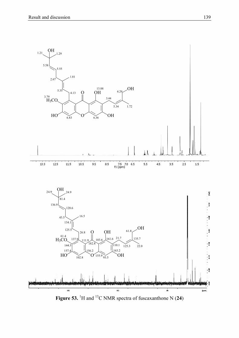
Result and discussion 139
Figure 53. 1H and 13C NMR spectra of fuscaxanthone N (24)
O
OHO
OH
H3CO
HO
OH
OH
13.88
6.83 6.36
3.79
5.34
4.29
3.44
1.72
5.585.55
5.354.13
2.671.81
1.291.21
O
OHO
OH
H3CO
HO
OH
OH
102.8 93.5
125.3
61.8
21.7
22.0
136.9128.6
125.526.8
43.5 16.5
24.924.9
182.8
61.4135.7
81.4
134.1
163.2
161.6
110.1
103.6
155.9
144.5
111.9137.9
156.2157.4

140 Results and discussion
5.2.2.2. Compound 25 (fuscaxanthone O)
Figure 54. Structure of fuscaxanthone O (25)
Compound 25 was isolated as a yellow oil and the molecular formula was
established as C29H34O8 based on the HRESIMS (m/z 511.2322 [M + H]+). The 1H and
13C NMR spectra (Figure 56 and Table 10) were similar to those of compound 24,
comprising the signals for a 1,3,6,7-tetrahydroxyxanthone nucleus [H 13.91 (1H, s, 1-
OH), 6.81 (1H, s, H-5), 6.35 (1H, s, H-4); C 181.8 (C-9), 163.3 (C-3), 161.6 (C-1),
157.6 (C6/C-10a), 156.3 (C10a/C-6), 155.9 (C-4a), 144.6 (C-7), 138.1 (C-8), 111.8 (C-
8a), 110.1 (C-2), 103.5 (C-9a), 102.7 (C-5) and 93.5 (C-4)], a methoxyl group [H 3.79
(3H, s, 7-OCH3); C 61.4 (CH3O-7)], a 4-hydroxy-3-methylbut-2-enyl group [H 5.35
(1H, t, J = 7.8 Hz, H-12), 4.29 (2H, s, H2-14), 3.43 (2H, d, J = 7.8 Hz, H2-11) and 1.72
(3H, s, H3-15); C 135.7 (C-13), 125.3 (C-12), 61.8 (C-14), 22.0 (C-15) and 21.7 (C-
11)] and a modified geranyl group [C 5.30 (1H, t, J = 6.6 Hz, H-17), 4.82 (1H, br s,
HZ-24), 4.67 (1H, br s, HE-24), 4.13 (2H, d, J = 6.6 Hz, H2-16), 3.94 (1H, t, J = 6.6 Hz,
H-22), 2.05 (1H, m, HA-20), 1.98 (1H, m, HB-20), 1.84 (3H, s, H3-19), 1.63 (3H, s, H3-
25) and 1.58 (2H, m, H2-21); C 149.2 (C-23), 135.2 (C-18), 124.7 (C-17), 110.3 (C-
24), 75.2 (C-22), 36.6 (C-20), 34.5 (C-21), 26.8 (C-16), 17.8 (C-25) and 16.7 (C-19)].
The benzylic methylene protons of the geranyl group gave the less shielded signal
at H 4.13 and the HMBC correlations with C-7, C-8 and C-8a, i.e. this group is
bonded to C-8. The COSY interaction between two allylic protons (H 2.05 and 1.98)
and two methylene protons at H 1.58 indicated the connectivity of C-20 and C-21.
O
OHO
OH
H3CO
HO
OH
HO
1
2
345
6
78
9
11
12
13
14
15
1617
181920
21
2322
2425
4a
9a8a
10a

Result and discussion 141
The protons H2-21 exhibited the HMBC cross-peaks with a fully substituted olefinic
carbon (C 149.2) and an oxygenated tertiary carbon (C 75.2), in addition to the
COSY interaction with the proton of the oxygenated tertiary carbon (H 3.94), which
revealed that the oxygenated quaternary carbon is C-22 and C-23 is the fully
substituted olefinic carbon. The proton H-22 gave the HMBC correlations to an
unsubstituted olefinic carbon (C 110.3) and a vinyl methyl group (C 17.8), i.e. the
unsubstituted olefinic carbon is C-24 and the methyl group is C-25. The vinyl proton
(H 4.82) is HZ due to the NOESY interaction with the protons of the vinyl methyl
group and the other one (H 4.67) is HE, corresponding to the NOESY interaction with
the proton H-22 (see Figure 55). Hence the geranyl group is modified into the 6-
hydroxy-3,7-dimethyloct-2,7-dienyl group. This was further confirmed by comparison
of the spectral data of this group with those of parvixanthone C, which was previously
isolated from G. parvifolia (Xu et al., 2001). Compound 25 was therefore identified as
1,3,6-trihydroxy-2-(4-hydroxy-3-methylbut-2-enyl)-8-(6-hydroxy-3,7-dimethyloct-
2,7-dienyl)-7-methoxyxanthen-9-one and was given a trivial name fuscaxanthone O.
However the configuration of the chiral carbon C-22 could not be determined.
Figure 55. The NOESY interaction between H2-24 and H-22 and H3-25
HE HZ

142 Results and discussion
Figure 56. 1H and 13C NMR spectra of fuscaxanthone O (25)
O
OHO
OH
H3CO
HO
OH
HO
HE
HZ
161.6
110.1
163.3
93.5102.7
103.5
155.9
181.8
157.6 156.3
144.6
138.1111.8
61.4135.721.7
125.3
61.8
22.0
149.2
135.2
124.7
110.3
75.2
36.6
34.5
17.8
16.7
26.8
O
OHO
OH
H3CO
HO
OH
HO
HE
HZ
6.356.81
3.793.43
5.35
4.29
1.72
5.30
3.94
2.05 & 1.98
1.58
1.63
1.84
4.1313.91
4.82
4.67

Result and discussion 143
Table 10. 1H and 13C NMR data of 24 and 25 in acetone-d6
Position 24 25
H C H C
1 161.6 161.6
2 108.0 110.1
3 163.2 163.3
4 6.36 s 93.5 6.35 s 93.5
4a 155.9 155.9
5 6.83 s 102.8 6.81 s 102.7
6 157.4a 157.6a
7 144.5 144.6
8 137.8 138.1
8a 111.9 111.8
9 182.8 181.8
9a 103.6 103.5
10a 156.2a 156.3a
11 3.44 d (7.8 Hz) 21.7 3.43 d (7.8 Hz) 21.7
12 5.34 t (7.8 Hz) 125.3 5.35 t (7.8 Hz) 125.3
13 135.7 135.7
14 4.29 s 61.8 4.29 s 61.8
15 1.72 s 22.0 1.72 s 22.0
16 4.13 t (6.9 Hz) 26.8 4.13 d (6.6 Hz) 26.8
17 5.35 t (6.9 Hz) 125.5 5.30 t (6.6 Hz) 124.7
18 134.1 135.2
19 1.81 s 16.5 1.84 s 16.7
20 2.67 d (6.0 Hz) 43.5 2.05 m
1.98 m
36.6
21 5.55 m 128.6 1.58 m 34.5
22 5.58 m 136.9 3.94 t (6.6 Hz) 75.2
23 81.4 149.2
24 1.29 s 24.9 4.82 brs HZ
4.67 brs HE
110.3
25 1.21 s 24.9 1.63 s 17.8
1-OH 13.88 s 13.91 s
7-OCH3 3.79 s 61.4 3.79 s 62.1
a Interexchangeable, 600 MHz for 1H and 150 MHz for 13C (J in Hz and in ppm) at 298ºK
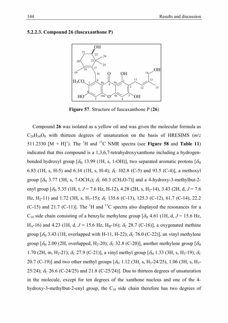
144 Results and discussion
5.2.2.3. Compound 26 (fuscaxanthone P)
Figure 57. Structure of fuscaxanthone P (26)
Compound 26 was isolated as a yellow oil and was given the molecular formula as
C29H34O8 with thirteen degrees of unsaturation on the basis of HRESIMS (m/z
511.2330 [M + H]+). The 1H and 13C NMR spectra (see Figure 58 and Table 11)
indicated that this compound is a 1,3,6,7-tetrahydroxyxanthone including a hydrogen-
bonded hydroxyl group [H 13.99 (1H, s, 1-OH)], two separated aromatic protons [H
6.83 (1H, s, H-5) and 6.34 (1H, s, H-4); C 102.8 (C-5) and 93.5 (C-4)], a methoxyl
group [H 3.77 (3H, s, 7-OCH3); C 60.3 (CH3O-7)] and a 4-hydroxy-3-methylbut-2-
enyl group [H 5.35 (1H, t, J = 7.6 Hz, H-12), 4.28 (2H, s, H2-14), 3.43 (2H, d, J = 7.6
Hz, H2-11) and 1.72 (3H, s, H3-15); C 135.6 (C-13), 125.3 (C-12), 61.7 (C-14), 22.2
(C-15) and 21.7 (C-11)]. The 1H and 13C spectra also displayed the resonances for a
C10 side chain consisting of a benzylic methylene group [H 4.61 (1H, d, J = 15.6 Hz,
HA-16) and 4.23 (1H, d, J = 15.6 Hz, HB-16); C 28.7 (C-16)], a oxygenated methine
group [H 3.43 (1H, overlapped with H-11, H-22); C 76.0 (C-22)], an vinyl methylene
group [H 2.00 (2H, overlapped, H2-20); C 32.8 (C-20)], another methylene group [H
1.70 (2H, m, H2-21); C 27.9 (C-21)], a vinyl methyl group [H 1.33 (3H, s, H3-19); C
20.7 (C-19)] and two other methyl groups [H 1.12 (3H, s, H3-24/25), 1.06 (3H, s, H3-
25/24); C 26.6 (C-24/25) and 21.8 (C-25/24)]. Due to thirteen degrees of unsaturation
in the molecule, except for ten degrees of the xanthone nucleus and one of the 4-
hydroxy-3-methylbut-2-enyl group, the C10 side chain therefore has two degrees of
O
OHO
OH
H3CO
HO
OH
OH
1
2
345
6
78
9
11
12
13
14
15
16
171819
20
21
23
2224
25
4a
9a8a
10a

Result and discussion 145
unsaturation, i.e. this group contains a double-bond and a ring, which was further
confirmed by the HMBC and COSY spectra.
The benzylic methylene protons (H 4.61) showed the HMBC correlations with two
fully substituted olefinic carbons (C 135.8 and 125.6) and a quaternary carbon (C
41.7). Besides the protons of two methyl groups (H 1.12 and 1.06) gave the HMBC
cross-peaks to the fully substituted carbon (C 135.8) and the quaternary carbon as
well as the ROESY interactions with the benzylic methylene protons. This revealed
that the fully substituted carbon at C 135.8 is C-17, the other one is C-18, C-23 is the
quaternary carbon and two methyl groups are attached to C-23. The HMBC
correlations of the proton of the oxygenated methine group (H 3.43; C 76.0) with C-
23 and the two methyl groups pointed out the connectivity of this oxygenated methine
group with C-23 and the oxygenated tertiary carbon is C-22. The COSY interaction
between the methylene proton (H 1.70) and H-22 indicated that this methylene group
is linked with C-22. The allylic protons (H 2.00) gave the HMBC correlations to C-
17, C-18, C-21 and C-22, which revealed that these protons are located at C-20.
Moreover the vinyl methyl protons (H 1.33) exhibited the HMBC cross-peaks with C-
17, C-18 and C-20, i.e. this methyl group is bonded to C-18. Hence the C10 side chain
was determined as the 5-hydroxy-2,6,6-trimethylcyclohexenylmethyl group. The
HMBC correlation of H-16 with C-7 (C 145.7), C-8 (C 139.4) and C-8a (112.5), in
addition to the ROESY interaction with the methoxyl protons (7-OCH3) allowed the
placement of the 5-hydroxy-2,6,6-trimethylcyclohexenylmethyl group at C-8.
Compound 26 was identified as 1,3,6-trihydroxy-2-(4-hydroxy-3-methylbut-2-enyl)-8-
(5-hydroxy-2,6,6-trimethylcyclohexenylmethyl)-7-methoxyxanthone and was given a
trivial name fuscaxanthone P. However the configuration of the chiral carbon C-23
could not be resolved.

146 Results and discussion
Figure 58. 1H and 13C NMR spectra of fuscaxanthone P (26)
O
OHO
OH
H3CO
HO
OH
OH
6.346.83
3.42
5.35
4.28
1.72
4.61 & 4.23
1.33
2.00
1.703.43
1.12
1.06
13.99
3.77
O
OHO
OH
H3CO
HO
OH
OH
93.5102.8
21.7
125.3
61.7
22.2
20.7
32.8
27.976.0
26.6
21.8
135.6
41.7
135.8125.6
28.7
161.4
163.7
155.7
103.4
110.0183.1
157.9 156.2
145.7
112.5139.4

Result and discussion 147
Table 11. 1H and 13C NMR data of 26 in acetone-d6
Position H C
1 161.4
2 110.0
3 163.7
4 6.34 s 93.5
4a 155.7
5 6.83 s 102.8
6 157.9a
7 145.7
8 139.4
8a 112.5
9 183.1
9a 103.4
10a 156.2a
11 3.43 d (7.6 Hz) 21.7
12 5.35 t (7.6 Hz) 125.3
13 135.6
14 4.28 s 61.7
15 1.72 s 22.2
16 4.61 d (15.6 Hz)
4.23 d (15.6 Hz)
28.7
17 135.8
18 125.6
19 1.33 d (5.4 Hz) 20.7
20 2.00 overlap with H-20 32.8
21 1.70 m 27.9
22 3.43 overlap with H-11 76.0
23 41.7
24 1.12a s 26.6
25 1.06a s 21.8
1-OH 13.99 s
7-OCH3 3.77 s 60.3
a Interchangeable, 600 MHz for 1H and 150 MHz for 13C (J in Hz and in ppm) at 298ºK

148 Results and discussion
5.3. Cytotoxicity of new substances
Eight new xanthones, cowanol and griffpavixanthone were tested for the
cytotoxicity on HeLa cells using a modified MTT assay according to Mosman (1983).
As shown in Table 12, griffpavixanthone possessed relatively high activity with IC50
value of 7.9 0.7 µM, whereas cowanol and (+)-fuscaxanthone I (19) showed
moderate cytotoxicity with IC50 values of 19.1 1.0 and 19.9 0.9 µM, respectively.
Five compounds fuscaxanthone L (18), fuscaxanthone J (21), fuscaxanthone N (24),
fuscaxanthone O (25) and fuscaxanthone P (26) exhibited a weak inhibitory effect with
the range of IC50 values of 32.5 – 45.9 µM. Meanwhile the remaining compounds
fuscaxanthone K (20) and fuscaxanthone M (22) were not cytotoxic. The new
xanthones possess the basic structure of cowanol with modification of the geranyl or
4-hydroxy-3-methylbut-2-enyl group. Demethylation of the methoxyl group in 21 or
modification of the double-bond at C-22/C-23 as in fuscaxanthone N and O (24 and
25) or ring-closure of the geranyl group in fuscaxanthone L and P (18 and 26) resultes
in a decrease of the activity compared to that of cowanol. The hydroxylation of the
double-bond at C-17/C-18 in (+)-fuscaxanthone I (19) has no effect on the
cytotoxicity, whereas the epoxylation of the double-bond at C-12/C-13 in
fuscaxanthone K (20) brings about the losing of activity. Compound 22 is derived
from two cowanol units through a C-O-C linkage. Though cowanol showed moderate
activity, the C-O-C linkage in fuscaxanthone M (22) resultes in the loss of
cytotoxicity.
Griffipavixanthone is a dimeric xanthone which was previously isolated from three
species of the Garcinia genus, G. griffithii, G. pavifolia and G.oblongifolia (Xu et al.,
1998; Shi et al., 2014). This xanthone was reported to exhibit a high in vitro
cytotoxicity against three cancer cell lines P388, LL/2 and Wehil64 with ED50 values
of 3.4, 6.8 and 4.6 µg/ml, respectively (Xu et al., 1998). In 2014, Shi et al. investigated
the effect of griffipavixanthone on cell proliferation of several human non-small cell
lung cancer cell lines (H520, H549, H1299 and A-549), human breast cancer cells
(MCF-7 and MDA-231), human prostate cells (DU145, PC3 and LNcaP) and human
colon cancer cells (HCT-116, HT-29 and SW-480). The investigation showed
significant anti-proliferation activity of griffipavixanthone on all of these cells with the

Result and discussion 149
IC50 values in the range of 3.03 – 29.33 µM. Furthermore this xanthone showed low
cytotoxic effects on non-tumorigenic LO2 cells indicating that it may be selectively
cytotoxic for human cancer cells (Shi et al., 2014). Because of such interesting
biological properties, in the present work, griffipavixanthone was additionally tested
for anti-angiogenic activity by in vitro proliferation assay on HMEC-1 cell line as
described by Schmidt et al. (2012). It exhibited a very potent anti-proliferative effect
on HMEC-1 cells with an IC50 value of 0.15 0.0085 µM after 72 h incubation. The
result revealed that griffipavixanthone is a promising anti-angiogenic agent which
should be further demonstrated in vivo.
Table 12. Cytotoxicity of 18 – 22, 24 – 26, cowanol and griffipavixanthone against
HeLa cells.
Compound IC50 SD (µM)a
Cowanol 19.1 1.0
Fuscaxanthone L (18) 41.5 0.4
(+)-Fuscaxanthone I (19) 19.9 0.9
Fuscaxanthone K (20) inactive
Fuscaxanthone J (21) 32.5 2.8
Fuscaxanthone M (22) inactive
Fuscaxanthone N (24) 44.3 2.5
Fuscaxanthone O (25) 35.4 1.7
Fuscaxanthone P (26) 45.9 1.9
Griffipavixanthone 7.9 0.4
a Mean of six measurements

150 Results and discussion
5.4. Proposed biosynthesis of the new xanthones in Garcinia fusca Pierre
On the basis of the structure of eight new xanthones as well as the presence of
rubraxanthone, norcowanin, cowanin and cowanol, a biosynthetic route (Scheme 25
and 26) is suggested for the new xanthones from 1,3,7-trihydroxyxanthone, which
together with 1,3,5-trihydroxyxanthone were shown as the precursors for most of
xanthones in plants (see section 3.1, page 48). This xanthone is hydroxylated at C-6 by
O2/NADPH to form 1,3,6,7-tetrahydroxyxanthone, which will be subsequently
alkylated at C-8 by a geranyl diphosphate (GPP) to give 1,3,6,7-tetrahydroxy-8-
geranylxanthone. The hydroxyl group at C-7 of this xanthone can be methylated by
SAM to give rubraxanthone. Meanwhile prenylation of this xanthone at C-2 by a
dimethylallyl diphosphate (DMAPP) yielded norcowanin, which is further O-
methylated at 7-OH by SAM to produce cowanin. On the other hand, a vinyl methyl of
isoprenyl group in norcowanin is hydroxylated by O2/NADPH to form fuscaxanthone
J (21), which can be subsequently O-methylated at 7-OH by SAM to give cowanol.
Epoxylation of the double-bond of the isoprenyl group in cowanol yield fuscaxanthone
K (20), whilst hydroxylation of the double-bond at C-17/C-18 of the geranyl group
will produce (+)-fuscaxanthone I (19). Moreover the double-bond at C-22/C-23 of the
geranyl group in cowanol can be epoxylated by O2/NADPH and then the epoxy ring
can be protonated and opened by a nucleophilic attack of water to generate a diol
derivative which is dehydrated to form fuscaxanthone N and O (24 and 25). Besides
the epoxy ring can be opened by the nucleophilic attack of the double-bond at C-17/C-
18 to yield the 5-hydroxy-2,6,6-trimethylcyclohexenylmethyl group as in
fuscaxanthone P (26). The double-bond of the 5-hydroxy-2,6,6-
trimethylcyclohexenylmethyl group could further undergo the nucleophilic attack of
the hydroxyl group to generate an oxo-bridge as in fuscaxanthone L (18). Similarly,
compound 22 (fuscaxanthone M) is derived from two cowanol units via the C-O-C
linkage, which can be formed either by the nucleophilic attack of the hydroxyl group
of the 4-hydroxy-3-methylbut-2-enyl side chain to the double-bond at C-17/C-18 of
the geranyl group or by condensation between cowanol and (+)-fuscaxanthone I (19).

Result and discussion 151
Scheme 25. Proposed biosynthesis of the new xanthones in G. fusca
O
O OH
OH
HO
O
O OH
OH
HO
HO
O2
NADPH
PPO
Alkylation
1,3,7-Trihydroxyxanthone 1,3,6,7-Tetrahydroxyxanthone
O
O OH
OH
HO
HO
1,3,6,7-Tetrahydroxy-8-geranylxanthone
O
O OH
OH
HO
HO
O-Methylation
SAM
OPP
Prenylation
O
O OH
OH
H3CO
HO
Rubraxanthone Norcowanin
O-Methylation
O
O OH
OH
H3CO
HO
Cowanin
SAM
Hydroxylation
O2/NADPH
O
O OH
OH
HO
HO
OH
O-Methylation
SAM
O
O OH
OH
H3CO
HO
OH
Cowanol
Epoxylation
[O]O
O OH
OH
H3CO
HO
O
OH
Fuscaxanthone K (20)
Fuscaxanthone J (21)
O
O OH
OH
H3CO
HO
OHOH
O
O OH
OH
H3CO
HO
OHO
H Protonation
(+)-Fuscaxanthone I (19)
HydroxylationEpoxylation

152 Results and discussion
Scheme 26. Proposed biosynthesis of the new xanthones in G. fusca (continued)
Scheme 26. Proposed biosynthesis of the new xanthones in G. fusca (continued)
H2O
Epoxy-ring opening Dehydration
O
O OH
OH
H3CO
HO
OH
OH
O
HOH
OH
O
O OH
OH
H3CO
HO
OH
OH
Dehydration
a
b
c
a and b
c
O
O OH
OH
H3CO
HO
OH
H
OH
- H
O
O OH
OH
H3CO
HO
OH
OH
O
O OH
OH
H3CO
HO
OH
O
Fuscaxanthone O (25)
Fuscaxanthone N (24)
Fuscaxanthone P (26) Fuscaxanthone L (18)
OHOH
OH
2 H3CO
HO
8
H3CO
HO O
O OH
OH
OH
O
O
H3CO
HO OH
OHO
Fuscaxanthone M (22)
OH
OH
O
H
H
2 H3CO
HO
HO
8
- H2O
or

Conclusion 153
6. CONCLUSION
The phytochemical investigation of the n-hexane extract (GFH) and the ethyl
acetate extract (GFE) of the bark of Garcinia fusca collected in south Vietnam resulted
in the isolation of thirty one natural compounds using various chromatographic
methods such as TLC, CC, FC, CPC, GPC and semi-preparative HPLC on silica gel,
RP18 and Sephadex LH-20. The structures of isolated compounds were determined on
the basis of spectral data such as UV, 1D and 2D NMR and MS spectra, as well as the
optical rotation. These compounds include a tocotrienol, a benzaldehyde derivative,
four biflavonoids and twenty five prenylated xanthones, nine of which have been first
found in nature. The cytotoxicity of the new xanthones was tested on HeLa cells using
MTT assay. The results indicated that five of them exhibited cytotoxic effect with the
IC50 values in the range of 19.9 – 45.9 µM. Furthermore, the anti-angiogenic activity
of a bixanthone, griffipavixanthone was evaluated in a proliferation assay using
HMEC-1 cell line. This bixanthone showed strong anti-proliferation activity with the
IC50 of 0.15 0.0085 µM, which revealed that it is a potential anti-angiogenic agent
and should be further investigated in vivo.
Because of the limited time, some fractions have not been studied yet. However the
present work showed the difference of the chemical composition of the bark between
this species and the species collected in Thailand (Ito et al., 2003; Nontakham et al.,
2014). For further work, the remaining fractions will be investigated to isolate more
compounds and new compounds will be biologically studied for various properties
such as anti-oxidant, antibacterial and anticancer activities.

154 References
REFERENCES
A
Abe, F., Nagafuji, S., Okabe, H., Higo, H. And Akahane, H. (2003): Trypanocidal
constituents in plants 2. Xanthones from the stem bark of Garcnia subelliptica. In:
Biol. Pharm. Bull. 26, 1730-1733.
Abe, F., Nagafuji, S., Okabe, H., Akahane, H., Estrada-Muniz, E., Huerta-Reyes, M.
And Reyes-Chilpa, R. (2004): Trypanocidal constituents in plants 3. Leaves of
Garcinia intermedia and heartwood of Calophyllum brasiliense. In: Bio. Pharm.
Bull. 27, 141-143.
Ades, E. W., Candal, F. J., Swerlick, R. A., George, V. G., Summers, S., Bosse, D. C.
and Lawley, T. J. (1992): HMEC-1: establishment of an immortalized human
microvascular endothelial cell line. In: J. Invest. Dermatol. 99, 684-690.
Ali, S., Goundar, R., Sotheeswaran, S., Beaulieu, C. and Spino, C. (2000):
Benzophenones of heartwood of Garcinia pseudoguttifera (Clusiaceae). In:
Phytochemistry 53, 281-284.
Alkadi, K. A. A., Adam, A., Taha, M., Hasan, M. H. and Shah, S. A. A. (2013):
Antiplatelet aggregation activity of 5-hydroxyflavone, 2'-hydroxyflavone, paenol
and bergenin isolated from stem bark of Garcinia maclaccensis in human whole
blood. In: Orient. J. Chem. 29, 871-875.
Alkadi, K. A. A., Adam, A., Taha, M., Hasan, M. H., Mizaton, H. and Shah, S. A. A.
(2013): Prenylated xanthone and rubraxanthone with antiplatelet aggregation
activity in human whole blood isolated from Garcinia griffithii. In: Orient. J.
Chem. 29, 1291-1295.
Al-Shagdari, A., Alarcon, A. B., Cuesta-Rubio, O., Piccinelli, A. L. and Rastrelli, L.
(2013): Biflavonoids, main constituents from Garcinia bakeriana leaves. In: Nat.
Prod. Commun. 8, 1237-1240.
Ampofo, S. A. and Waterman, P. G. (1986): Xanthones from three Garcinia nervosa,
pyrifera and polyantha. In: Phytochemistry 25, 2351-2355.

References 155
Andrade, M. S., Sampaio, T. S., Nogueira, P. C. L., Ribeiro, A. S., Bittrich, V. and
Amaral, M. C. E. (2007): Volatile compounds from the leaves and flowers of
Garcinia macrophylla. In: Chem. Nat. Comp. 43, 221-224.
Asano, J., Chiba, K., Tada, M. and Yoshii, T. (1996): Cytotoxic xanthones from
Garcinia hanburyi. In: Phytochemistry 41, 815-820.
Asgar, M., Monjok, E., Kouamou, G., Ohia, S. E., Bagchi, D. and Lokhandwala, M. F.
(2007): Super citrimax (HCA-SX) attenuates increases in oxidative stress,
inflammation, insulin resistance and body weigh in developing obese Zucker rats.
In: Mol. Cell. Biochem. 4, 93-99.
Ansari, W. H. and Rahman, W. (1975): Biflavanoids and a flavanone-chromone from
the leaves of Garcinia dulcis (Roxb.) Kurz. In: J. C. S. Perkin I, 1458-1463.
Antia, B. S., Pansanit, A., Ekpa, O. D., Ekpe, U. J., Mahidol, C. and Kittakoop, P.
(2010): Alpha-glucosidase inhibitory, aromatase inhibitoryand antiplasmodial
activities of a biflavonoid GB1 from Garcinia kola stem bark. In: Planta Med. 76,
276-277.
B
Babu, V., Ali, S. M., Sultana, S. and Ilyas, M. (1988): A biflavonoid from Garcinia
nervosa. In: Phytochemistry 27, 3332-3335.
Badami, R. C. and Desai, V. A. (1968): Component acids of Garcinia indica seed oil.
In: J. Karnatak University 13, 29-31.
Badami, R. C. and Razdan, M. K. (1972): Isolation and identification of L-leucine as
DNP [dinitrophenol]-L-leucine hydrochloride in the leaves of Garcinia indica. In:
J. Indian Chem. Soc. 49, 583.
Baggett, S., Protvita, P., Mazzola, E. P., Yang, H., Ressler, E. T., Basile, M. J.,
Weinstein, I. B. and Kennelly, E. J. (2005): Bioactive benzophenones from
Garcinia xanthochymus fruits. In: J. Nat. Prod. 68, 354-360.
Bailey, L.H. and Bailey, E.Z. (1976): Hortus Third: A concise dictionary of plants
cultivated in the United States and Canada, New York: Macmillan, p. 494.

156 References
Bakana, P., Claeys, M., Totte, J., Pieters, L. A., Van Hoof, L., Tamba-Vemba, Van,
den B. D. A. and Vlientinck, A. J. (1987): Structure and chemotherapeutical
activity of a polyisoprenylated benzophenone from the stem bark of Garcinia
huillensis. In: J. Ethnopharmacol 21, 75-84.
Baker, W. and Simmonds, W. H. C. (1940): Derivatives of 5:6:4'- and 5:8:4'-
trihydroxyflavones and a note on the structure of ginkgetin. In: J. Chem. Soc.,
1370-1970.
Balasubramanian, K. and Rajagopalan, K. (1988): Novel xanthones from Garcinia
mangostana, structures of BR-xanthone-A and BR-xanthone-B. In:
Phytochemistry 27, 1552-1554.
Bandaranayake, W. M., Selliah, S. S., Sultanbawa, M. U. S. and Ollis, W. D. (1975):
Chemical investigations of Ceylonese Plants. 18. Biflavonoids and xanthones of
Garcinia terpnophylla and G. echinocarpa. In: Phytochemistry 14, 1878-1880.
Baslas, R. K. and Kumar, P. (1979): Chemical examination of the fruits of Garcinia
xanthochymus. In: India. Curr. Sci. 48, 814-815.
Beerhues, L., Barillas, W., Peters, S. and Schmidt, W. (1999): Biosynthesis of plant
xanthones. In: Bioorganic chemistry. Highlights and New Aspects. Weinheim,
Germany: Wiley-VCH, p. 322-328.
Bennett, G. J. and Lee, H.-H. (1989): Xanthones from Guttiferae. In: Phytochemistry
28, 967-998.
Birbrair, A., Zhang, T., Wang, Z.-M., Messi, M. L., Mintz, A. and Delbono, O. (2014):
Type-2 pericytes participate in normal and tumoral angiogenesis. In: AJP: Cell
Physiology 307, C25.
Birch, A. J., Baldas, J., Hlubucek, J. R., Simpson, T. J. and Westerman, P. W. (1975):
Biosynthesis of the fungal xanthone ravenelin. In: J. C. S. Perkin I, 898-904.
Bouquet, A. (1969): Féticheurs et Médecines Traditionnelles du Congo (Brazzaville),
Office de la Recherche Scientifique et Technique Outre-Mer O.R.S.T.O.M.: Paris,
France, p. 132–133.

References 157
Braithwaite, B. and Smith, F. J. (1996): Chromatographic Methods, Chapman & Hall.
Breitmaier, E. (2002): Structure elucidation by NMR in organic chemistry: a practical
guide, 3rd edition. John Willey & Sons, Ltd.
Bui, Q. T., Trinh, T. T. T., Nguyen, N. C., Le, V. T. and Nguyen, D. L.-H. (2012):
Chemical constituents of the bark of Garcinia vilersiana. In: Vietnamese J. Sci.
Tech. 50, 145-150.
Bui, A. D., Vu, M. K., Nguyen, D. H., Nguyen, T. L.-T., Dang, V. S. and Nguyen, D.
L.-H. (2014): A protostane and two lanostanes from the bark of Garcinia ferrea.
In: Phytochem. Lett. 10, 123-126.
Bumrungpert, A., Kalpravidh, R. W., Chitchumroonchokchai, C., Chuang C. C., West,
T., Kennedy, A. and McIntosh, M. (2009): Xanthones from mangosteen prevent
lipopolysaccharide-mediated inflammation and insulin resistance in primary
cultures of human adipocytes. In: J. Nutr. 139, 1185-1191.
C
Cambie, R. C. and Ash, J. (1994): Fijian Medicinal Plants, Australia: CSIRO, p. 121.
Cao, S.-G., Sng, V. H. L., Wu, X.-H., Sim, K.-Y., Tan, B. H. K., Pereira, J. T. and
Goh, S. H. (1998): Novel cytotoxic polyprenylated xanthonoids from Garcinia
gaudichaudii (Guttiferae). In: Tetrahedron 54, 10915-10924.
Cardona, M. L., Fernández, I., Pedro, J. R. and Serrano, A. (1990): Xanthones from
Hypericum reflexum. In: Phytochemistry 29, 3003-3006.
Carroll, A. R., Suraweera, L., King, G., Rali, T. and Quinn, R. J. (2009): Guttiferones
O and P, prenylated benzophenone MAPKAPK-2 inhibitors from Garcinia
solomonensis. In: J. Nat. Prod. 72, 1699-1701.
Carey, F. A. (2001): Organic chemistry. The McGraw Companies, p. 265.
Castardo, J. C., Prudente, A. S., Ferreira, J., Guimaraes, C. L., Monache, F. D., Filho,
V. C., Otuki, M. F. and Cabrini, D. A. (2008): Anti-inflammatory effects of
hydroalcoholic extract and two biflavonoids from Garcinia gardneriana leaves in
mouse paw oedema. In: J. Ethnopharmacol. 118, 405-411.

158 References
Chen, F.-C., Lin, Y.-M. and Hung, J.-C. (1975): Phenolic compounds from the
heartwood of Garcinia multiflora. In: Phytochemistry 14, 300-303.
Chen, J.-J., Chen, I.-S. and Duh, C.-Y. (2004): Cytotoxic xanthones and biphenyls
from the root of Garcinia linii. In: Planta Med. 70, 1195-1200.
Chen, J.-J., Peng, C.-F., Huang, H.-Y. and Chen, I.-S. (2006): Benzopyrans, biphenyls
and xanthones from the root of Garcinia linii and their activity against
Mycobacterium tuberculosis. In: Planta Med. 71, 473-477.
Chen, Y., Zhong, F.-F., He, H.-W., Hu, Y., Zhu, D. and Yang, G.-Z. (2008): Structure
elucidation and NMR spectral assignment of five new xanthones from the bark of
Garcinia xanthochymus. In: Magn. Reson. Chem. 46, 1180-1184.
Chen, L. G., Yang, L. L. and Wang, C. C. (2008): Anti-inflammatory activity of
mangostins from Garcinia mangostana. In: Food Chem. Toxicol. 46, 688-693.
Chen, J.-J., Ting, C.-W., Hwang, T.-L. And Chen, I.-S. (2009): Benzophenone
derivatives from the fruits of Garcinia multiflora and their anti-inflammatory
activity. In: J. Nat. Prod. 72, 253-258.
Chen, Y., Fan, H., Yang, G.-Z., Jiang, Y., Zhong, F.-F. and He, H.-W. (2010):
Prenylated xanthones from the bark of Garcinia xanthochymus and their 1,1-
diphenyl-2-picrylhydrazyl radical scavenging activitives. In: Molecules 15, 7438-
7449.
Chen, Y., Fan, H., Yang, G.-Z., Jiang, Y., Zhong, F.-F. and He, H.-W. (2011): Two
unusual xanthones from the bark of Garcinia xanthochymus. In: Helv. Chim. Acta
94, 662-668.
Chiang, Y.-M., Kuo, Y.-H., Oota, S. and Fukuyama, Y. (2003): Xanthones and
benzophenones from the stems of Garcinia multiflora. In: J. Nat. Prod. 66, 1070-
1073.
Chien, S.-C., Cyu, C.-F., Chang, I-S., Chiu, H.-L. und Kuo, Y.-H. (2008): A novel
polyprenylated phloroglucinol, garcinialone, from the roots of Garcinia multiflora.
In: Tetrahedron lett. 49, 5276-5278.

References 159
Cotterill, P. J., Scheinmann, F. and Puranik, G. S. (1977): Extractives from Guttiferae.
Part 32. Phenolic compounds from the heartwood of Garcinia indica. In:
Phytochemistry 16, 148-149.
Crichton, E. G. and Waterman, P. G. (1979): Manniflavanone, a new 3,8-linked
flavanone dimer from the stem bark of Garcinia mannii. In: Phytochemistry 18,
1553-1557.
Cuesta-Rubio, O., Padron, A., Castro, H. V., Pizza, C., Rastrelli, L. (2001):
Aristophenones A and B. A new tautomeric pair of polyisoprenylated
benzophenones from Garcinia aristata. In: J. Nat. Prod. 64, 973-975.
D
Dakanali, M. and Theodorakis, E. A. (2013): Polyprenylated phloroglucinols and
xanthones. In: Biomimetic organic synthesis. Wiley-VCH Verlag GmbH & Co. p.
433-467.
Dal Molin, M. M., Silva, S., Alves, D. R., Quintao, N. L. M., Nara, L. M., Delle
Monache, F., Cechinel Filho V. and Niero, R. (2012): Phytochemical analysis and
antinociceptive properties of seeds of Garcinia achachairu. In: Arch. Pharmacal
Res. 35, 623-631.
Deachathai, S., Mahabusarakam, W., Phongpaichit, S. and Taylor, W. C. (2005):
Phenolic compounds from the fruits of Garcinia dulcis. In: Phytochemistry 66,
2368-2375.
Deachathai, S., Mahabusarakam, W., Phongpaichit, S., Taylor, W. C., Zhang, Y.-J.
and Yang, C.-R. (2006): Phenolic compounds from the flowers of Garcinia dulcis.
In: Phytochemistry 67, 464-469.
Deachathai, S., Phongpaichit, S. and Mahabusarakham, W. (2008): Phenolic
compounds from the seeds of Garcinia dulcis. In: Nat. Prod. Res. 22, 1327-1332.
Dewick, P. M. (2009): Medicinal natural products: a biosynthesis approach, 3rd
edition. John Wiley & Son, Inc., Hoboken, New Jersey.

160 References
Dibwe, D. F., Awale, S., Kadota, S. and Tezuka, Y. (2012): Damnacanthal from the
Congolese medicinal plant Garcinia huillensis has a potent preferential
cytotoxicity against human pancreatic cancer PANC-1 cells. In: Phytother. Res.
26, 1920-1926.
Dora, G. and Edward, J. M. (1991): Taxonomic status of Lanaria lanata and isolation
of a novel biflavone. In: J. Nat. Prod. 54, 796-801.
Duangsrisai, S., Choowongkomon, K., Bessa, L. J., Costa, P. M., Amat, N. and Kijjoa,
A. (2014): Antibacteria and EGFR-tyrosine kinase inhibitory activities of
polyhydroxylated xanthones from Garcinia succifolia. In: Molecules 19, 19923-
19934.
Duke, J. A., Bogenschutz-Godwin, M. J., duCellier, J. and Duke, P.-A. K. (2002):
Handbook of Medicinal Herbs, 2nd ed. CRC Press, Boca Raton, Fl, p. 481.
E
Ea, G. C. L., Phong, K. H., Mong, X. J., Shaari, K. and Sukari, M. A. (2003):
Cuneifolin, a new xanthone from Garcinia cuneifolia. In: Nat. Prod. Sci. 9, 174-
176.
Ea, G. C. L., Daud, S., Taufiq-Yap, Y. H., Ismail, N. H. and Rahmani, M. (2006):
Xanthones from Garcinia mangostana. In: Nat. Prod. Res. 20, 1067-1073.
Ea, G. C. L., Daud, S., Izzaddin, S. A. and Rahmani, M. (2008): Garcinia
mangostana: a source of potential anti-cancer lead compounds against CEM-SS
cell line. In: J. Asian Nat. Prod. Res. 10, 481-485.
Elfita, E., Muharni, M., Latief, M., Darwati, D., Widiyantoro, A., Supriyatna, S.,
Bahti, H. H., Dachriyanus, D., Cos, P., Maes, L., et al. (2009): Antiplasmodial and
other constituents from four Indonesian Garcinia species. In: Phytochemistry 70,
907-912.
El-Seedi, H. R., El-Barbary, M. A., El-Ghorab, D. M. H., Bohlin, L., Borg-Karlson,
A.-K., Goeransson, U. and Verpoorte, R. (2010): Recent insights into the

References 161
biosynthesis and biological activities of natural xanthones. In: Curr. Med. Chem.
17, 854-901.
Elya, B., He, H. P., Kosela, S., Hanafi, M. and Hao, X. J. (2006): A new
benzophenone from the stem bark of Garcinia benthami. In: Nat. Prod. Res. 20,
1059-1062.
Elya, B., He, H. P., Kosela, S., Hanafi, M. and Hao, X. J. (2006): Two new xanthones
from Garcinia rigida leaves. In: Nat. Prod. Res. 20, 788-791.
Elya, B., He, H. P., Kosela, S., Hanafi, M. and Hao, X. J. (2011): Triterpenoids from
Garcinia rigida. In: Rec. Nat. Prod. 5, 56-59.
F
Fan, Q., Na, Z., Hu, H., Xu, Y. and Tang, T. (2012): Chemical constituents from stem
barks of Garcinia paucinervis. In: Zhongcaoyao 43, 436-439.
Feng, C., Huang, S.-X., Gao, X.-M., Xu, H.-X. and Luo, K. Q. (2014):
Characterization of proapoptixic compounds from the bark of Garcinia
oblongifolia. In: J. Nat. Prod. 77, 1111-1116.
Ferreira, O. R., Carvalho, de M. G. and Silva, da T. M. S. (2012): Occurrence of
biflavonoids in Clusiaceae: chemical and pharmacological aspects. In: Quim.
Nova. 35, 2271-2277.
Fotie, J., Bohle, D. S., Oliver, M., Gomez, M. A. and Nzimiro, S. (2007):
Trypanocidal and antileishmanial dihydrochelerythrine derivatives from Garcinia
lucida. In: J. Nat. Prod. 70, 1650-1653.
Foucault, A. P. (1995): Solvent systems in centrifugal partition chromatography. In:
Centrifugal partition chromatography, Chromatographic Sciences Series 68, 71-
97, Marcel Dekker, New York.
Fouotsa, H., Lannang, A. M., Mbazoa, C. D., Rasheed, S., Marasini, B. P., Ali, Z.,
Devkota, A. P., Kengfack, A. E., Shaheen, F., Choudahary, M. I., et al (2012):
Xanthones inhibitors of -glucosidase and glycation from Garcinia nobilis. In:
Phytochem. Lett. 5, 236-239.

162 References
Fouotsa, H., Tatsimo, S. J. N., Neumann, B., Michalek, C., Mbazoa, C. D., Nkengfack,
A. E., Sewald, N. and Lannang, A. M. (2014): A new xanthone derivative from the
twigs of Garcinia nobilis. In: Nat. Prod. Res. 28, 1030-1036.
Fujita, M. and Inoue, T. (1980): Biosynthesis of mangiferin in Anemarrhena
asphodeloides bunge. I. The origin of the xanthone nucleus. In: Chem. Pharm.
Bull. 28, 2476-2481.
Fukuyama, Y., Kamiyama, A., Mima, Y. and Kodama, M. (1991): Prenylated
xanthones from Garcinia subelliptica. In: Phytochemistry 30, 3433-3436.
G
Gadek, P. A. and Quinn, C. J. (1985): Biflavones of the subfamily Cupressoideae,
Cupressaceae. In: Phytochemistry 24, 267-272.
Gao, X.-M., Yu, T., Lai, F. S. F., Pu, J.-X., Qiao, C.-F., Zhou, Y., Liu, X., Song, J.-Z.,
Luo, K. Q. and Xu, H.-X. (2010): Novel polyisoprenylated benzophenone
derivatives from Garcinia paucinervis. In: Tetrahedron Lett. 51, 1441-1446.
Gao, X.-M., Cui, M.-Z., Yu, T., Hu, Q.-F., Pu, J.-X., Du, X., Liu, T.-C. and Luo, K. Q.
(2013): A novel xanthone from Garcinia oligantha. In: Helv. Chim. Acta 96, 494-
498.
Geiger, H. and Quinn, C. (1975): Biflavonoids. In: The flavonoids. Academic Press,
New York, p. 692-742.
Geiger, H. and Bokel, M. (1989): The biflavonoid pattern of Philonotis fontana
(Hedw.) Brid (Bartramiaceae). In: Z. Naturforsch. C: J. Biosci. 44, 559-562.
Gey, C., Kyrylenko, S., Hennig, L., Nguyen, D. L.-H., Buettner, A., Pham, D. H. and
Giannis, A. (2007): Phloroglucinol derivatives guttiferone G, aristoforin and
hyperforin: inhibitors of human sirtuins SIRT1 and SIRT2. In: Angew. Chem. Int.
Ed. 46, 5219-5222.
Gibbons, S. (2006): An introduction to planar chromatography. In: Natural product
isolation, edited by Sarker, S. D., Latif Z. and Gray, A. I., Humana Press Inc., p.
77-117.

References 163
Goh, S. H., Jantan, I., Gray, A. I. and Waterman, P. G. (1992): Prenylated xanthones
from Garcinia opaca. In: Phytochemistry 31, 1383-1386.
Gontijo, V. S., Judice, W. A. S., Codonho, B., Pereira, I. O., Assis, D. M., Januario, J.
P., Caroselli, E. E., Maria, A., Dosatti, A. de C. and Marques, M. J. (2012):
Leishmanicidal, antiproteolytic and antioxidant evaluation of natural biflavonoids
isolated from Garcinia brasiliensis and their semisynthetic derivatives. In: Eur. J.
Med. Chem. 58, 613-623.
Govindachari, T. R., Kalyanaaman, P. S., Muthukumaraswamy, N. and Pai, B. R.
(1971): Isolation of three new xanthones from Garcinia mangostana. In: Indian J.
Chem. 9, 505-506.
Gunatilaka, A. A. L., Sriyani, H. T. B., Sotheeswaran, S. and Waight, E. S. (1983):
2,5-Dihyroxy-1,6-dimethoxyxanthone and biflavonoids of Garcinia thwaitesii. In:
Phytochemistry 22, 233-235.
Gunatilaka, A. A. L., De Silva, A. M. Y. J., Sotheeswaran, S., Balasubramaniam, S.
and Wazeer, M. I. M. (1984): Studies on medicinal and related plants of Sri Lanka.
Part 11. Terpenoid and biflavonoid constituents of Calophyllum calaba and
Garcinia spicata from Sri Lanka. In: Phytochemistry 23, 323-328.
Gunatilaka, A. A. L., Sriyani, H. T. B. and Sotheeswaran, S. (1984): Quaesitol, a
phenol from Garcinia quaesita. In: Phytochemistry 23, 2679-2681.
Gupta, P. and Lewis, J. R. (1971): Biogenesis of xanthones in Gentiana lutea. In: J.
Chem. Soc. C. 4, 629-631.
Gustafson, K. R., Blunt, J. W., Munro, M. H. G., Fuller, R. W., Mckee, T. C.,
Cardellina, J. H., McMahon, J. B., Cragg, G. M. and Boyd, M. R. (1992): The
guttiferones, HIV-inhibitory benzophenones from Symphonia globulifera,
Garcinia livingstonei, Garcinia ovalifolia and Clusia rosea. In: Tetrahedron 48,
10093-10102.
Guo, Y.-E., Wang, L.-N., Li, Z.-L., Niu, S.-L., Liu, X.-Q., Hua, H.-M., Chen, H.-C.
and Zhang, T.-C. (2011): Triterpenes and xanthones from the stem bark of
Garcinia tetralata. In: J. Asian Nat. Prod. Res. 13, 440-443.

164 References
H
Ha, D. L., Hansen, P. E., Vang, O., Duus, F., Pham, D. H. and Nguyen, D. L.-H.
(2009): Cytotoxic geranylated xanthones and O-alkylated derivatives of -
mangostin. In: Chem. Pharm. Bull. 57, 830-834.
Ha, D. L., Hansen, P. E., Duus, F., Pham, D. H. and Nguyen, D. L.-H. (2011):
Oliverdepsidones A-D, antioxidant depsidones from Garcinia oliveri. In: Magn.
Reson. Chem. 50, 242-245.
Haase, D. N. (2011): Potential chemopreventive xanthones and a new
polyisoprenylated benzophenone derivative from Garcinia humilis. 242nd ACS
National Meeting & Exposition, Dever, CO, USA.
Hamed, W., Brajeul, S., Mahuteau-Betzer, F., Thoison, O., Mons, S., Delpech, B.,
Nguyen, V. H., Sevenet, T. and Marazano, C. (2006): Oblongifolins A-D,
polyprenylated benzoylphloroglucinol derivatives from Garcinia oblongifolia. In:
J. Nat. Prod. 69, 774-777.
Han, Q.-B., Wang, Y.-L., Yang, L., Tso, T.-F., Qiao, C.-F., Song, J.-Z., Xu, L.-J.,
Chen, S.-L., Yang, D.-J. and Xu, H.-X. (2006): Cytotoxic polyprenylated
xanthones from the resin of Garcinia hanburyi. In: Chem. Pharm. Bull. 54, 265-
267.
Han, Q.-B., Yang, N.-T., Tian, H.-L., Qiao, C.-F., Song, J.-Z., Chang, D. C., Chen, S.-
L., Luo, K. Q. and Xu, H.-Xi. (2008): Xanthones with growth inhibition against
HeLa cells from Garcinia xipshuanbannaensis. In: Phytochemistry 69, 2187-2192.
Han, Q.-B., Yang, N.-T., Tian, H.-L., Qiao, C.-F., Song, J.-Z., Chang, D. C., Chen, S.-
L., Luo, K. Q. and Xu, H.-Xi. (2008): Polyprenylated xanthones from Garcinia
lancilimba showing apoptotic effects against Hela-C3 cells. In: Chem. Bio. 5,
2710-2717.
Han, A.-R., Kim, J.-A., Lantvit, D., Kardono, L. B. S., Riswan, S., Chai, H., carcache,
de B. E. J., Farnsworth, N. R., Swanson, S. M. and Kinghorn, A. D. (2009):
Cytotoxic xanthone constituents of the stem bark of Garcinia mangostana. In: J.
Nat. Prod. 72, 2028-2031.

References 165
Harrison, L. J., Leong, L. S., Sia, G. L., Sim, Y. and Tan, H. T. W. (1993): Xanthones
from Garcinia forbesii. In: Phytochemistry 33, 727-728.
Harborne, J. B. (1994): The flavonoids advanced in research since 1986. Chapman &
Hall, London, New York, Tokyo, p. 316-319.
Harborne, J. B. (1998): Phytochemical methods: a guide to modern techniques of plant
analysis, 3rd edition. Chapman & Hall, p. 40-96.
Harrison, L. J., Leong, L.-S., Leong, Y.-W., Sia, G.-L., Sim, K.-Y. and Tan, H. T.-W.
(1994): Xanthone and flavonoid constituents of Garcinia dulcis (Guttiferae). In:
Nat. Prod. Lett. 5, 111-116.
Hartati, S., Wang, H.-B., Kardono, L. B. S., Kosela, S. and Qin, G.-W. (2007):
Chemical constituents of Garcinia maingayii. In: Zhongguo Tianran Yaowu 5,
272-276.
Hartati, S., Soemiati, A., Wang, H.-B., Kardono, L. B. S., Hanafi, M., Kosela, S. and
Qin, G.-W. (2008): A novel polyisoprenyl benzophenone derivative from Garcinia
eugeniaefolia. In: J. Asian Nat. Prod. Res. 10, 509-513.
Hay, A.-E., Aumond, M.-C., Mallet, S., Dumontet, V., Litaudon, M., Rondeau, D. and
Richomme, P. (2004): Antioxidant xanthones from Garcinia vieillardii. In: J. Nat.
Prod. 67, 707-709.
Heilmann, J., Wasescha, M. R., Schmidt, T. J. (2001): The influence of glutathione
and cysteine levels on the cytotoxicity of helenanolide type sesquiterpene lactones
against KB cells. In: Bioorg. Med. Chem. 9, 2189–2194.
Hemshekhar, M., Sunitha, K., Santhosh, M. S., Devaraja, S., Kemparaju, K.,
Vishwanath, B. S., Niranjana, S. R. and Girish, K. S. (2011): An overview on
genus Garcinia: phytochemical and therapeutical aspects. In: Phytochem. Rev. 10,
325-351.
Heneen, W. K. (1976): HeLa cells and their possible contamination of other cell lines:
karyotype studies. In: Hereditas 82, 217-248.

166 References
Henry, K. M. and Townsend, C. A. (2005): Ordering the reductive and cytochrome
P450 oxidative steps in demethylsterigmatocystin formation yields general insights
into the biosynthesis of aflatoxin and related fungal metabolites. In: J. Am. Chem.
Soc. 127, 3724-3733.
Herath, K., Jayasuriya, H., Ondeyka, J. G., Guan, Z., Borris, R. P., Stifhoorn, E.,
Stevenson, D., Wang, J., Sharma, N., MacNaul, K., Menke, J. G., Ali, A.,
Schulman, M. J. and Singh, S. B. (2005): Guttiferone I, a new prenylated
benzophenone from Garcinia humulis as a liver X receptor ligand. In: J. Nat.
Prod. 68, 617-619.
Herbin, G. A., Jackson, B., Locksley, H. D., Scheinmann, F. and Wolstenholme, W. A.
(1970): Extractives from Guttiferae. XV. Biflavonoids of Garcinia volkensii. In:
Phytochemistry 9, 221-226.
Holker, J. S. E., Lapper, R. D. and Simpson T. J. (1974): The biosynthesis of fungal
metabolites. Part IV. Tajixanthone: 13C-nuclear magnetic resonance spectrum and
feedings with [1-13C]- and [2-13C]-acetate. In: J. Chem. Soc. Perkin I, 2135-2140.
Hood, R. L., Beitz, D. C. and Johnson, D. C. (1985): Inhibition by potential metabolic
inhibitors of in vitro adipose tissue lipogenesis. In: Comp. Biochem. Physiol. 81B,
667-670.
Hu, Q., Niu, D., Wang, S., Qin, Y., Yang, Z., Zhao, G., Yang, Z., Gao, X. and Chen,
Z. (2013): New xanthones from Garcinia bracteata and their cytotoxicities. In:
Heterocycles 87, 1127-1132.
Hu, Q., Niu, D., Wang, S., Qin, Y., Yang, Z., Zhao, G., Yang, Z., Gao, X. and Chen,
Z. (2014): New flavones from Garcinia bracteata and their biological activities.
In: Chem. Nat. Comp. 50, 985-988.
Hu, Q., Meng, Y., Yao, J., Qin, Y., Yang, Z., Zhao, G., Yang, Z., Gao, X. and Li, T.
(2014): Flavonoids from Garcinia paucinervis and their biological activities. In:
Chem. Nat. Comp. 50, 994-997.
Hussain, R. A. and Waterman, P. G. (1982): Lactones, flavonoids and benzophenones
from Garcinia conrauana and G. mannii. In: Phytochemistry 21, 1393-1396.

References 167
Hussain R. A., Owegby, A. G., Parimoo, P. and Waterman P. G. (1982): Kolanone, a
novel polyisoprenylated benzophenone with antimicrobial properties from the fruit
of Garcinia kola. In: Planta Med. 44, 78-81.
I
Iinuma, H., Kinoshita, M., Fukuyama, Y., Kodama, M., Yoshizawa, T., Sugiura, M.,
Nakagawa, K. and Tago, H. (1994): Antioxidant xanthones from Garcinia
subelliptica. In: Phytochemistry 36, 501-506.
Iinuma, M., Tosa, H., Tanaka, T., Asai, F. and Shimano, R. (1995): Three xanthones
from the root bark of Garcinia subelliptica. In: Phytochemistry 38, 247-249.
Iinuma, M., Ito, T., Tosa, H., Tanaka, T. and Riswan, S. (1996): Five new xanthones
from Garcinia dulcis. In: J. Nat. Prod. 59, 472-475.
Iinuma, M., Ito, T., Tosa, H., Tanaka, T. and Riswan, S. (1996): Three new
benzophenone-xanthone dimers from the root of Garcinia dulcis. In: Chem.
Pharm. Bull. 44, 1744-1747.
Iinuma, M., Tosa, H., Tanaka, T. and Riswan, S. (1996): Three new xanthones from
the bark of Garcinia dioica. In: Chem. Pharm. Bull. 44, 232-234.
Iinuma, M., Tosa, H., Tanaka, T., Kanamaru, S., Asai, F., Kobayashi, Y., Miyauchi,
K.-I., and Shimano, R. (1996): Antibacterial activity of some Garcinia
benzophenone derivatives against methicillin-resistant Staphylococcus aureus. In:
Japan. Biol. Pharm. Bull. 19, 311-314.
Iinuma, M., Ito, T., Miyake, R., Tosa, H., Tanaka, T. and Chelladurai, V. (1998): A
xanthones from Garcinia cambogia. In: Phytochemistry 47, 1169-1170.
Ilyas, M., Kamil, M., Parveen, M. And Kahn, M. S. (1994): Isoflavones from Garcinia
nervosa. In: Phytochemistry 36, 807-809.
Ito, Y. and Bowman, R. L. (1970): Countercurrent chromatography: liquid-liquid
partition chromatography without solid support. In: J. Chrom. Sci. 8, 315-323.

168 References
Ito, C., Miyamoto, Y., Nakayama, M., Kawai, Y., Rao, K. S. and Furukawa, H.
(1997): A novel depsidone and some new xanthones from Garcinia species. In:
Chem. Pharm. Bull. 45, 1403-1413.
Ito, C., Itoigawa, M., Mishina, Y., Tomiyasu, H., Litaudon, M., Cosson, J.-P.,
Mukainaka, T., Tokuda, H., Nishino, H. and Furukawa, H. (2001): Cancer
chemopreventive agents. New depsidones from Garcinia neglecta and G. puat. In:
J. Nat. Prod. 64, 147-150.
Ito, C., Itoigawa, M., Takakura, T., Ruangrungsi, N., Enjo, F., Tokuda, H., Nishino, H.
and Furukawa, H. (2003): Chemical constituents of Garcinia fusca: structure
elucidation of eight new xanthones and their cancer chemopreventive activity. In:
J. Nat. Prod. 66, 200-205.
Ito, C., Itoigawa, M., Miyamoto, Y., Onoda, S., Rao, K. S., Mukainaka, T., Tokuda,
H., Nishino, H. and Furukawa, H. (2003): Polyprenylated benzophenones from
Garcinia assigu and their potential cancer chemopreventive activities. In: J. Nat.
Prod. 66, 206-209.
Ito, T., Yokota, R., Watarai, T., Mori, K., Oyama, M., Nagasawa, H., Matsuda, H. and
Iinuma, M. (2013): Isolation of six isoprenylated biflavonoids from the leaves of
Garcinia subelliptica. In: Chem. Pharm. Bull. 61, 551-558.
Ito, C., Matsui, T., Noda, E., Ruangrungsi, N. And Itoigawa, M. (2013): Biphenyl
derivatives from Garcinia schomburgkiana and the cytotoxicity of the isolated
compounds. In: Nat. Prod. Commun. 8, 1265-1267.
Iwu, M. M. (1985): Antithepatoxic constituents of Garcinia kola seeds. In: Experientia
41, 699-700.
Iwu, M., Igboko, O. A., Onwuchekwa, U. A. and Okunji, C. O. (1987): Evaluation of
the antiheptotoxic activity of the biflavonoids of Garcinia kola seeds. In: J.
Ethnopharmacol. 21, 127-138.
Iwu, M. M., Igboko, O. A. and Tempesta, M. S. (1990): Biflavonoid constituents of
Garcinia kola roots. In: Fitoterapia 61, 178-181.

References 169
J
Jackson, B., Locksley, H. D., Scheinmann, F. and Wolstenholme, W. A. (1971):
Extractives from Guttiferae. XXII. Isolation and structure of four novel
biflavanones from the heartwoods of Garcinia buchananii and G. eugeniifolia. In:
J. Chem. Soc. C, 3791-3804.
Jabit, M. L., Khalid, R., Abas, F., Shaari, K., Hui, L. S., Stanslas, J. and Lajis, N. H.
(2007): Cytotoxic xanthones from Garcinia penangiana. In: Z. Naturforsch., C: J.
Biosci. 62, 786-792.
Jamila, N., Khairuddean, M., Yaacob, N. S., Kamal, N. N. S. N. M., Osman, H., Khan,
S. N. and Khan, N. (2014): Cytotoxic benzophenone and triterpene from Garcinia
hombroniana. In: Bio. Chem. 54, 60-67.
Jantan, I. and Saputri, F. C. (2012): Benzophenones and xanthones from Garcinia
cantleyana var. cantleyana and their inhibitory activitives on human low-density
lipoprotein oxidation and platelet aggregation. In: Phytochemistry 80, 58-63.
Jena, B. S., Jayaprakasha, G. K., Singh, R. P. And Sakariah, K. K. (2002): Chemistry
and biochemistry of (-)-hydroxycitric acid from Garcinia. In: J. Agric. Food.
Chem. 50, 10-22.
Ji, F., Li, Z.-L., Liu, G.-F., Niu, S.-L., Zhao, N., Liu, X.-Q. and Hua, H. (2012):
Xanthones with antiproliferation effects on prostate cancer cells from the stem
bark of Garcinia xanthochymus. In: Nat. Prod. Commun. 7, 53-56.
Jiang, G., Du, F. and Fang, G. (2014): Two new proanthocyanidins from the leaves of
Garcinia multiflora. In: Nat. Prod. Res. 28, 449-453.
Jing, W.-Y., Jiang, C., Ji, F., Hua, H.-M. and Li, Z.-L. (2013): Chemical constituents
from the stem barks of Garcinia multiflora. In: J. Asian Nat. Prod. Res. 15, 1152-
1157.
Joshi, B. S., Kamat, V. N. and Viswanathan, N. (1970): Isolation and structure of two
biflavones from Garcinia talboti. In: Phytochemistry 9, 881-888.

170 References
Jung, H. A., Su, B. N., Keller, W. J., Mehta, R. G. and kinghorn, D. (2006):
Antioxidant xanthones from pericarp of Garcinia mangostana (Mangosteen). In: J.
Agric. Food. Chem. 54, 2077-2082.
K
Kabangu, K., Galeffi, C., Aonzo, E., Nicoletti, M. and Messana, L. (1987): Research
on African medicinal plants. Part XII. A new biflavanone from the bark of
Garcinia kola. In: Planta Med. 53, 275-277.
Kaikabo, A. A., Samuel, B. B. and Eloff, J. N. (2009): Isolation and activity of two
antibacterial biflavonoids from the leaf extracts of Garcinia livingstonei. In: Nat.
Prod. Commun. 4, 1363-1366.
Kaikabo, A. A., Eloff, J. N. (2011): Antibacterial activity of two biflavonoids from
Garcinia livingstonei leaves against Mycobacterium smegmatis. In: J.
Ethnopharmacol. 138, 253-255.
Kamdem, W., Alain, F., Mulholland, D., Wansi, J. D., Mbaze, L. M., Powo, R.,
Mpondo, T. N., Fomum, Z. T., Konig, W., Nkengfack, A. E. (2006):
Afzeliixanthones A and B, two new prenylated xanthones from Garcinia afzelii
ENGL. (Guttiferae) In: Chem. Pharm. Bull. 54, 448-451.
Kang, S. S., Koh, Y. M., Kim, J. S., Lee, M. W. and Lee, D. S. (1995): Phytochemical
analysis of Ginkgo biloba yellow leaves. In: Kor. J. Pharmacogn. 26, 23-26.
Kaomongkolgit, R., Jamdee, K. and Chaisomboon, N. (2009): Antifungal activity of
alpha-mangostin against Candida albicans. In: J. Oral. Sci. 51, 401-406.
Karanjgoakar, C. G., Rao, A. V. R., Venkataraman, K., Yemul, S. S. and Palmer, K. J.
(1973): Constitution of xanthochymol and isoxanthochymol. In: Tetrahedron Lett.
(50), 4977-80.
Kardono, L. B. S., Hanafi, M., Sherley, G., Kosela, S. and Harrison, L. J. (2006):
Bioactive constituents of Garcinia porrecta and G. parvifolia grown in Indonesia.
In: Pak. J. Biol. Sci. 9, 483-486.

References 171
Kartha, G., Ramachandran, H. B., Bhat, H. B., Nair, P. M., Raghavan, V. K. V. and
Venkataraman, K. (1963): The constitution of morellin. In: Tetrahedron Lett. 4,
459-472.
Kasahara, S. and Henmi, S. (1986): Medicinal Herb Index in Indonesia, P.T. Eisai
Indonesia: Jakarta, p. 92.
Kijjoa, A., Gonzalez, M. J., Pinto, M. M., Nascimento, M. S. J., Campos, N.,
Mondranondra, I.-O., Silva, A. M. S., Eaton, G. and Herz, W. (2008): Cytotoxicity
of prenylated xanthones and other constituents from the wood of Garcinia
merguensis. In: Planta Med. 74, 864-866.
Kitanov, G. M. and Nedialkov, P. T. (2001): Benzophenone O-glucoside, a biogenic
precursor of 1,3,7-trioxygenated xanthones in Hypericum annulatum. In:
Phytochemistry 57, 1237-1243.
Krishnamurthy, N., Lewis, Y. S. and Ravindranath, B. (1981): On the structure of
garcinol, isogarcinol and camboginol. In: Tetrahedron Lett. 22, 793-796.
Krishnamurthy, N., Lewis, Y. S. and Ravindranath, B. (1982): Chemical constituents
of kokam fruit rind. In: J. Food Sci. Tech. 19, 97-100.
Krishnamurthy, K. S. and Sapna, V. P. (2008): Garcinia. In: Chemistry of spices, CAB
International, p. 342-355.
Klaiklay, S., Sukpondma, Y., Rukachaisirikul, V., Hutadilok-Towatana, N. and
Chareonrat, K. (2011): Flavanone glucuronides from the leaves of Garcinia
prainiana. In: Canadian J. Chem. 89, 461-464.
Klaiklay, S., Sukpondma, Y., Rukachaisirikul, V. and Phongpaichit, S. (2013):
Friedolanostanes and xanthones from the twigs of Garcinia hombroniana. In:
Phytochemistry 85, 161-166.
Koisin, J., Ruangrungsi, N., Ito, C. And Furukawa, H. (1998): A xanthone from
Garcinia atroviridis. In: Phytochemistry 47, 1167-1168.
Komguem, J., Meli, A. L., Manfouo, R. N., Lontsi, D., Ngounou, F. N., Kuete, V.,
Kamdem, H. W., Tane, P., Ngadjui, B. T., Sondengam, B. L., et al. (2005):

172 References
Xanthones from Garcinia smeathmannii (Oliver) and their antimicrobial activity.
In: Phytochemistry 66, 1713-1717.
Konoshima, M., Ikeshiro, Y. and Miyahara, S. (1970): Constitution of biflavonoids
from Garcinia plants G. spicata. In: Tetrahedron lett. (48), 4203-4206.
Kosela, S., Cao, S.-G., Wu, X.-H., Vittal, J., Sukri, T., Masdianto, Goh, S.-H. and Sim,
K.-Y. (1999): Lateriflorone, a cytotoxic spiroxalactone with a novel skeleton, from
Garcinia lateriflora BI. In: Tetrahedron Lett. 40, 157-160.
Kosela, S., Hu, L.-H., Rachmatia, T., Hanafi, M. and Sim, K.-Y. (2000): Dulxanthones
F-H, three new pyranoxanthones from Garcinia dulcis. In: J. Nat. Prod. 63, 406-
407.
Kuete, V., Komguem, J., Beng, V. P., Meli, A. L., Tangmouo, J. G., Etoa, F.-X. and
Lontsi, D. (2007): Antimicrobial components of the methanolic extract from the
stem bark of Garcinia smeathmannii. In: S. Afr. J. Bot.73, 347-354.
Kumar, G. M., Ajitha, N. A. and Rao, V. U. M. (2014): Centrifugal partition
chromatography: an overview. In: IJPRA 4, 353-360.
L
Ladinsky J. L., Volk, N. D. and Robinson, M. (1987): The influence of traditional
medicine in shaping medical care practices in Vietnam today. In: Soc. Sci. Med.
25, 1105-1110.
Lai, Y. F., Helm, R. And Karchesy, J. (1992): A B-ring linked biflavonoid from
Pseudotsuga menziesii. In: Phytochemistry 31, 1444-1445.
Lakshmi, C., Kumar, K. A., and Dennis, T. J. (2002): Polyprenylated benzophenones
from Garcinia indica. In: J. Indian Chem. Soc. 79, 868-969.
Lannang, A. M., Komguem, J., Ngninzeko, F. N., Tangmouo, J. G., Lontsi, D., Ajaz,
A., Choudhary, M. I., Ranjit, R., Devkota, K. P. and Sondengam, B. L. (2005):
Bangangxanthone A and B, two xanthones from the stem bark of Garcinia
polyantha Oliv. In: Phytochemistry 66, 2351-2355.

References 173
Lannang, A. M., Komguem, J., Ngninzeko, N., Tangmouo, J. G., Lontsi, D., Ajaz, A.,
Choudhary, M. I., Sondengam, B. L. and Atta-ur-Rahman (2006): Antioxidant
benzophenones and xanthones from the root bark of Garcinia smeathmannii. In:
Bull. Chem. Soc. Ethiopia 20, 247-252.
Lannang, A. M., Louh, G. N., Lonsti, D., Specht, S., Sarite, S. R., Florke, U., Hussain,
H., Hoerauf, A. and Krohn, K. (2008): Antimalarial compounds from the root bark
of Garcinia polyantha Oliv. In: J. Antibiot. 61, 518-523.
Lannang, A. M., Louh, G. N., Biloa, B. M., Komguem, J., Mbazoa, C. D., Sondengam,
B. L., Naesens, L., Pannecouque, C., De Clercq, E., El, A. and El, S. H. (2010):
Cytotoxicity of natural compounds isolated from the seeds of Garcinia afzelii. In:
Planta Med. 76, 708-712.
Lannang, A. M., Noudou, B. S. and Sewald, N. (2013): Ovalifolone A and B: new
friedelane derivatives from Garcinia ovalifolia. In: Phytochem. Lett. 6, 157-161.
Lannang, A. M., Tatsimo, S. J. N., Fouotsa, H., Dzoyem, J. P., Saxena, A. K. and
Sewald, N. (2014): Cytotoxic compounds from the leaves of Garcinia polyantha.
In: Chem. Biodivers. 11, 975-981.
Lavaud, A., Richomme, P., Litaudon, M., Andriantsitohaina, R. and Guilet, D. (2013):
Antiangiogenic tocotrienol derivatives from Garcinia amplexicaulis. In: J. Nat.
Prod. 76, 2246-2252.
Lavaud, A., Richomme, P., Gatto, J., Aumond, M.-C., Poullain, C., Litaudon, M.,
Andriantsitohaina, R. and Guilet, D. (2015): A tocotrienol series with an oxidative
terminal prenyl unit from Garcinia amplexicaulis. In: Phytochemistry 109, 103-
110.
Lewis, Y. S. (1969): Isolation and properties of hydroxycitric acid. In: Methods
Enzymol. 13, 613-619.
Li, Y., Wang, Z., Wu, X., Yang, Y., Qin, Y., Xia, C., Meng, Y., Li, M., Gao, X.-M.
and Hu, Q. (2015): Biphenyl derivatives from the twigs of Garcinia bracteata and
their biological activities. In: Phytochem. Lett. 11, 24-27.

174 References
Lin, Y. M., Anderson, H., Flavin, M. T., Pai, Y. H. S., Mata-Greenwood, E.,
Pengsuparp, T., Pezzuto, J. M., Schinazi, R. F. and Hughes, S. H. (1997): In vitro
anti-HIV activity of biflavonoids isolated from Rhus succedanea and Garcinia
multiflora. In: J. Nat. Prod. 60, 884-888.
Lin, Y.-M., Flavin, M. T., Schure, R., Chen, F.-C., Sidwell, R., Barnard, D. L.,
Huffman, J. H. and Kern, E. R. (1999): Antiviral activities of biflavonoids. In:
Planta Med. 65, 120-125.
Lin, L.-C. and Chou, C.-J. (2000): Three new biflavonoids from Selaginella delicatula.
In: Chin. Pharm. J. 52, 211-218.
Lin, Y. F., Zhuan, Y. and Zhao, Y. H. (2003): Chinese Dai Medicine Colorfull
Illustration, Yunnan National Publishing House, p. 6.
Liu, X., Yu, T., Gao, X.-M., Zhou, Y., Qiao, C.-F., Peng, Y., Chen, S.-L., Luo, K. Q.
and Xu, H.-X. (2010): Apoptoxic effects of polyprenylated benzoylphloroglucinol
derivatives from the twigs of Garcinia multiflora. In: J. Nat. Prod. 73, 1355-1359.
Louh, G. N., Lannang, A. M., Mbazoa, C. D., Tangmouo, J. G., Komguem, J.,
Castilho, P., Ngninzenko, F. N., Qamar, N., Lontsi, D., Choudhary, M. I., et al
(2008): Polyanxanthone A, B and C, three xanthones from the wood trunk of
Garcinia polyantha Oliv. In: Phytochemistry 69, 1013-1017.
Lu, Y.-H., Wei, B.-L., Ko, H.-H. and Lin, C.-N. (2008): DNA strand-scission by
phloroglucinols and lignans from heartwood of Garcinia subelliptica Merr. and
Justicia plants. In: Phytochemistry 69, 225-233.
Lyles, J. T., Negrin, A., Khan, S. I., He, K. and Kennelly, E. J. (2014): In vitro
antiplasmodial activity of benzophenones and xanthones from edible fruits of
Garcinia species. In: Planta Med. 80, 676-681.
M
Machado, M. B. and Lopes, L. M. X. (2005): Chalcone-flavone tetramer and
biflavones from Aristolochia ridicula. In: Phytochemistry 66, 669-674.

References 175
Mackeen, M. M., Ali, B. M., Lajis, N. H., Kawazu, K., Kikuzaki, H. and Nakatani, N.
(2002): Antifungal garcinia acid esters from the fruits of Garcinia atroviridis. In:
Z. Naturforsch. C: J. Biosci. 57, 291-295.
MacLeod, A. J. and Pieris, N. M. (1982): Volatiles flavour components of
mangosteen, Garcinia mangostana. In: Phytochemistry 21, 117-119.
Magadula, J. J., Kapingu, M. C., Bezabih, M. And Abegaz, B. M. (2008):
Polyisoprenylated benzophenones from Garcinia semseii. In: Phytochem. Lett. 1,
215-218.
Magadula, J. J. (2010): A bioactive isoprenylated xanthone and other constituents of
Garcinia edulis. In: Fitoterapia 81, 420-423.
Magadula, J. J. (2012): Bioactive benzophenones from the stem bark and fruit hulls of
Garcinia semseii Verdc. In: Int. J. Res. Phyto. Pharm. 2, 16-20.
Magadula, J. J., Mbwambo, Z. H., Gatto, J., Debre, S., Guilet, D. and Richomme, P.
(2014): Polyphenolic compounds with anti-ages activity from Garcinia semsii and
G. volkensii. In: European J. Med. Plants 4, 1336-1344.
Mahabusarakam, W., Wiriyachitra, P. and Taylor, W. C. (1987): Chemical
constituents of Garcinia mangostana. In: J. Nat. Prod. 50, 474-478.
Mahabusarakam, W., Chairerk, P. and Taylor, W. C. (2005): Xanthones from Garcinia
cowa Roxb. latex. In: Phytochemistry 66, 1148-1153.
Mahjoub, M. A., Ammar, S. and Mighri, Z. (2005): A new biflavonoid and an
isobiflavonoid from Rhus tripartitum. In: Nat. Prod. Res. 19, 723-729.
Mandal, S., Das, P. C. and Joshi, P. C. (1992): Naturally occuring xanthones from
terrestrial flora. In: J. Indian Chem. Soc. 69, 611-636.
Martin, F. T., Cruz, J. W. Jr., Derogis, P. B. M. C., Dos Santos, M. H., Veloso M. P.,
Ellena, J. and Doriguetto, A. C. (2007): Natural polyprenylated benzophenones:
keto-enol tautomerism and stereochemistry. In: J. Braz. Chem. Soc. 18, 1515-
1523.

176 References
Martin, F. T., Doriguetto, A. C., Souza de, T. C., Souza de, K. R. D., Dos Santos, M.
H., Moreira, M. E. C. and Narbosa, L. C. A. (2008): Composition and anti-
inflammatory and antioxidant activities of the volatile oil from the fruit peel of
Garcinia brasiliensis. In: Chem. Biodivers. 5, 251-258.
Masullo, M., Bassarello, C., Bifulco, G. and Piacente, S. (2010): Polyisoprenylated
benzophenone derivatives from the fruits of Garcinia cambogia and their absolute
configuration by quantum chemical circular dichroism calculations. In:
Tetrahedron 66, 139-145.
Mawa, S. and Said, I. M. (2012): Chemical constituents of Garcinia prainiana. In:
Sains Malaysiana 41, 585-590.
Mbwambo, Z. H., Kapingu, M. C., Moshi, M. J., Machumi, F., Apers, S., Cos, P.,
Ferreira, D., Marais, J. P. J., Berghe, D. V., Maes, L., Vlietinck, A. and Pieters, L.
(2006): Antiparasitic activity of some xanthones and biflavonoids from the root
bark of Gracina livingstonei. In: J. Nat. Prod. 69, 369-372.
Mercader, A. G. and Pomilio, A. B. (2012): Biflavonoids: occurrence, structural
features and bioactivity. Nova Science Publishers, Inc.
Merza, J., Aumond, M.-C., Rondeau, D., Dumontet, V., Le Ray, A.-M., Seraphin, D.
and Richomme, P. (2004): Prenylated xanthones and tocotrienols from Garcinia
virgata. In: Phytochemistry 65, 291502820.
Merza, J., Mallet, S., Litaudon, M., Dumontet, V., Seraphin, D. and Richomme, P.
(2006): New cytotoxic guttiferone analogues from Garcinia virgata from New
Caledonia. In: Planta Med. 72, 87-89.
Messi, B. B., Ndjoko-Ioset, K., Hertlein-Amslinger, B., Lannang, A. M., Mkengfack,
A. E., Wolfender, J.-L., Hostettmann, K. and Bringmann, G. (2012): Preussianone,
a new flavanone-chromone biflavonoid from Garcinia preussii Engl. In:
Molecules 17, 6114-6125.
Mitchell, T. N. and Costisella, B. (2007): NMR - from spectra to structure: an
experimental approach, 2nd edition. Springer.

References 177
Monzote, L., Cuesta-Rubio, O., Matheeussen, A., Assche, T. V., Maes, L. and Cos, P.
(2011): Antimicrobial evaluation of the polyisoprenylated benzophenones
nemorosone and guttiferone A. In: Phytother. Res. Wiley Online Library.
Morabandza, C. J., Ongoka, R. P., Matini, L., Epa, C., Nkounkou, L. C. and Abena, A.
A. (2013): Chemical composition of the mesocarp of Garcinia kola Heckel
(Clusiaceae) fruit. In: Res. J. Recent Sci. 2, 53-58.
Mori, R., Nugroho, A. E., Hirasawa, Y., Wong, C. P., Kaneda, T., Shirota, O., Hadi,
A. H. A. and Morita, H. (2014): Opaciniols A-C, new terpenoids from Garcinia
opaca. In: J. Nat. Med. 68, 186-191.
Mosman, T. (1983): Rapid colorimetric assay for cellular growth and survival:
application to proliferation and cytotoxicity assays. In: J. Immunol. Methods 65,
55–63.
Muhaisen, H. M. H., Ilyas, M., Mushfig, M., Parveen, M. and Basudan, O. A. (2002):
Flavonoid from Viburnum continifolium. In: J. Chem. Res. (S), 480-481.
Mungmee, C., Sitthigool, S., Buakeaw, A. and Suttisri, R. (2013): A new biphenyl and
other consituents from the wood of Garcinia schomburgkiana. In: Nat. Prod. Res.
27, 1949-1955.
N
Na, Z. and Xu, Y.-K. (2009): Chemical constituents from twigs of Garcinia
xipshuanbannanensis. In: Zhongguo Zhongyao Zazhi 34, 2338-2342.
Na, Z. and Xu, Y.-K. (2010): A new prenylated xanthone from Garcinia
xipshuanbannanensis. In: Nat. Prod. Res. 24, 1648-1653.
Na, Z. and Xu, Y. (2010): Chemical constituents of Garcinia tetralata. In:
Zhongcaoyao 41, 367-370.
Na, Z., Hu, H.-B. and Fan, Q.-F. (2010): Three new caged prenylxanthones from
Garcinia bracteata. In: Helv. Chim. Acta 93, 958-963.
Na, Z., Hu, H.-B. and Xu, Y.-K. (2013): Cytotoxic caged xanthones from the fruits of
Garcinia bracteata. In: Chem. Nat. Comp. 49, 505-506.

178 References
Na Pattalung P., Thongtheeraparp W., Wiriyachitra, P. and Taylor, W. C. (1994):
Xanthones of Garcinia cowa. In: Planta Med. 60, 365-368.
Naldoni, F. J., Claudino, A. L. R., Cruz, J. W., Chavasco, J. K., Faria e Silva, P. M.,
Veloso, M. P. and Dos Santos, M. H. (2009): Antimicrobial activity of
benzophenones and extracts from the fruits of Garcinia brasiliensis. In: J. Med.
Food 12, 403-407.
Negi, J. S., Bisht, V. K., Singh, P., Rawat, M. S. M. and Joshi, G. P. (2013): Naturally
occuring xanthones: chemistry and biology. In: Journal of Applied Chemistry.
Hindawi Publishing Corporation online.
Neri, P. and Tringali, C. (2001): Applications of modern NMR techniques in the
structural elucidation of bioactive natural products. In: Bioactive compounds from
natural sources, edited by Corrado Tringali. Taylor & Francis, London and New
York.
Ngameni, B., Fotso, G. W., Ngachussi, E., Poumale, H. M., Ngadjui, B. T., Shiono, Y.
and Murayama, T. (2014): Hemisynthesis and spectroscopic characterization of
two novel O-allylated benzophenones from Garcinia punctata Oliv. (Clusiaceae).
In: Asian J. Chem. 26, 6943-6949.
Ngoupayo, J., Tabopda, T. K. Ali, M. S. and Tsamo, E. (2008): α-Glucosidase
inhibitors from Garcinia brevipedicellata (Clusiaceae). In: Chem. Pharm. Bull. 56,
1466-1469.
Nguyen, D. L.-H. and Harrison, L. J. (2000): Xanthones and triterpenoids from the
bark Garcinia vilersiana. In: Phytochemistry 53, 111-114.
Nguyen, D. L.-H., Vo, T. H., Pham, D. H., Connolly, J. D. and Harrison, L. J. (2003):
Xanthones from the bark of Garcinia merguensis. In: Phytochemistry 63, 467-470.
Nguyen, D. H., Trinh, T. D. B. and Nguyen, D. L.-H. (2011): Guttiferones Q-S,
cytotoxic polyisoprenylated benzophenones from the pericarp of Garcinia
cochinchinensis. In: Phytochem. Lett. 4, 129-133.

References 179
Nguyen, D. H., Trinh, T. D. B., Tran, N. Q., Nguyen, D. H., Pham, D. H., Hansen, P.
E., Duus, F., Connolly, J. D. and Nguyen, D. L.-H. (2011): Friedolanostane,
friedocycloartane and benzophenone constituents of the bark and leaves of
Garcinia benthami. In: Phytochemistry 72, 290-295.
Nilar and Harrison L. J. (2002): Xanthones from the heartwood of Garcinia
mangostana. In: Phytochemistry 60, 541-548.
Nilar, Nguyen, D. L.-H., Venkatraman, G., Sim, K.-Y. and Harrison, L. J. (2005):
Xanthones and benzophenones from Garcinia griffithii and G. mangostana. In:
Phytochemistry 66, 1718-1723.
Niu, S.-L., Li, Z.-L., Ji, F., Liu, G.-Y., Zhao, N., Liu, X.-Q., Jing, Y.-K. and Hua, H.-
M. (2012): Xanthones from the stem bark of Garcinia bracteata with growth
inhibitory effects against HL-60 cells. In: Phytochemistry 77, 280-286.
Niwa, M., Terashima, K., Ito, J. and Aqil, M. (1994): Two novel arylbenzofurans,
garcifuran A and B from Garcinia kola. In: Heterocycles 38, 1071-1076.
Nontakham, J., Charoenram, N., Upamai, W., Taweechotipatr, M. and Suksamrarn, S.
(2014): Anti-Helicobacter pylori xanthones of Garcinia fusca. In: Arch. Pharm.
Res. 37, 972-977.
Nyemna, A. M., Mpondo, T. N., Connolly, J. D. and Rycroft, D. S. (1990):
Cycloartane derivatives from Garcinia lucida. In: Phytochemistry 29, 994-997.
O
Ofman, D. J., Markham, K. R., Vilain, C. and Molloy, B. P. J. (1995): Flavonoid
profiles of New Zealand kauri and other species of Agathis. In: Phytochemistry 38,
1223-1228.
Ohnmacht, S., West, R., Simionescu, R. and Atkinson, J. (2007): Assignments of the
1H and 13C NMR of tocotrienols. In: Magn. Reson. Chem. 46, 287-294.
Ollis, W. D., Ramsay, M. V. J., Sutherland, I. O. and Mongkolsuk, S. (1965): The
constitution of gambogic acid. In: Tetrahedron 21, 1453-1470.

180 References
Oluwatosin, A., Tolulope, A., Ayokulehin, K., Patricia, O., Aderemi, K., Catherine, F.
and Olusegun, A. (2014): Antimalarial potential of kolaviron, a biflavonoid from
Garcinia kola seeds, against Plasmodium berghei infection in Swiss albino mice.
In: Asian Pac. J. Trop. Med., 97-104.
Osorio, E., Londono J. and Bastida, J. (2013): Low-density lipoprotein (LDL)-
antioxidant biflavonoids from aerial parts of Garcinia madruno. In: Molecules 18,
6092-6100.
Otuki, M. F., Camila A., Prudente, A. S., Laskoski, K., Gomig, F., Horinouchi, C. D.
S., Guimaraes, C. L., Ferreira, J., Delle-Monache, F., Cechinel-Filho, V. et al
(2011): Garcinia gardneriana (Planchon & Triana) Zappi. (Clusiaceae) as a
tropical anti-inflammatory alternative for cutaneous inflammation. In: Basic Clin.
Pharmacol. 109, 56-62.
P
Panthong, K., Pongcharoen, W., Phongpaichit, S. and Taylor, W. C. (2006):
Tetraoxygenated xanthones from the fruits of Garcinia cowa. In: Phytochemistry
67, 999-1004.
Parveen, M., Khan, N. U. D., Achari, B. and Dutta, P. K. (1990): Triterpenoids from
Garcinia mangostana. In: Fitoterapia 61, 86-87.
Parveen, M., Khan, N. U. D., Achari, B. and Dutta, P. K. (1991): A triterpene from
Garcinia mangostana. In: Phytochemistry 30, 361-362.
Parveen, M., Ilyas, M, Mushfiq, M., Busudan, O. A. and Muhaisen, H. M. H. (2004):
A new biflavonoid from the leaves of Garcinia nervosa. In: Nat. Prod. Res. 18,
269-275.
Pavia, D. L., Lampman, G. M., Kris, G. S. and Vyvyan, J. R. (2009): Introduction to
spectroscopy, 4th edition. Brooks/Cole, Cengage Learning.
Pedraza-Chaverri, J., Cárdenas-Rodríguez, N., Orozco-Ibarra, M. and Pérez-Rojas, J.
(2008): Medicinal properties of mangosteen (Garcinia mangostana). In: Food
Chem. Toxicol. 46, 3227-3239.

References 181
Pelter, A., Warren, R., Chexal, K. K., Handa, B. K. and Rahman, W. (1971):
Biflavonyls from Guttiferae, Garcinia livingstonei. In: Tetrahedron 27, 1625-
1634.
Peres, V. and Nagem, T. J. (1996): Trioxygenated naturally occuring xanthones. In:
Phytochemistry 44, 191-214.
Permana, D., Lajis, N. H., Mackeen, M. M., Ali, A. M., Aimi, N., Kitajima, M. and
Takayama, H. (2001): Isolation and bioactivities of constituents of the roots of
Garcinia atroviridis. In: J. Nat. Prod. 64, 976-979.
Permana, D., Abas, F., Maulidiani, Shaari, K., Stanslas, J., Ali, A. M. and Lajis, N. H.
(2005): Antrovirisidone B, a new prenylated depsidone with cytotoxic property
from the roots of Garcinia atroviridis. In: Z. Naturforsch. C: J. Biosci. 60, 523-
526.
Peters, S., Schmidt, W. and Beerhues, L. (1998): Regioselective oxidative phenol
couplings of 2,3',4,6-tetrahydroxybenzophenone in cell cultures of Centaurium
erythraea Rafn. and Hypericum androsaemum L. In: Planta Med. 204, 64-69.
Pham, H. H. (1999): The flora of Vietnam, Band I, Youth Publishing House,
HoChiMinh City, p. 449-465.
Poomipamorn, S. and Kumkong, A. (1997): Edible multipurpose tree species.
Bangkok: Faung Fa Printing.
Purwaningsih, Y. and Ersam, T. (2007): Senyawa santon sebagai antioksidan dari kayu
batang Garcinia tetranda Pierre. In: Akta Kimindo 2, 103-108.
Q
Queiroz, de J. S., Griswold, D., Nguyen, D. T. and Hall, P. (2013): Vietnam tropical
forest and biodiversity assessment. Sun Mountain International and The Cadmus
Group, Inc.
Quillinan, A. J. and Scheinmann, F. (1971): Application of Claisen rearrangement to
the synthesis of heterocyclic bicycle-2,2,2-octenone: an approach to the morellins
based on new biogenetic suggestions. In: J. Chem. Soc. Chem. Commun., 966-967.

182 References
R
Rama Rao, A. V., Venkatswamy, G. and Pendse, A. D. (1980): Camboginol and
cambogin. In: Tetrahedron Lett. 21, 1975-1978.
Rambeloson, V. H. V., Rasoanaivo, L. H., Wadouachi, A., Randrianasolo, R., Krebs,
H. C. and Raharisololalao, A. (2014): Two new xanthones from Garcinia
chapelieri: chapexanthone A; chapexanthone B. In: J. Pharmacogn. Phytochem. 2,
98-105.
Rao, A. V. R., Sarma, M. R., Venkataraman, K and Yemul, S. S. (1974):
Benzophenone and xanthone with unusual hydroxylation patterns from the
heartwood of Garcinia penduculata. In: Phytochemistry 13, 1241-1244.
Reddy, B. A. K., Reddy, N. P., Gunasekar, D., Blond, A. and Bodo, B. (2008):
Biflavonoids from Ochna lanceolata. In: Phytochem. Lett. 1, 27-30.
Ren, Y., Lantvit, D. D., Carcache, de B., Esperanza, J., Kardono, L. B. S., Riswan, S.,
Chai, H., Cottrell, C. E., Farnsworth, N. R., Swanson, S. M., Ding Y., et al.
(2010): Proteasome-inhibitory and cytotoxic constituents of Garcinia lateriflora:
absolute configuration of caged xanthones. In: Tetrahedron 66, 5311-5320.
Reutrakul, V., Anantachoke, N., Pohmakotr, M., Jaipetch, T., Sophasan, S., Yoosook,
C., Kasisit, J., Napaswat, C., Santisuk, T. and Tuchinda, P. (2007): Cytotoxic and
anti-HIV-1 caged xanthones from the resin and fruits of Garcinia hanburyi. In:
Planta Med. 73, 33-40.
Reutrakul, V., Anantachoke, N., Pohmakotr, M., Jaipetch, T., Sophasan, S., Yoosook,
C., Kasisit, J., Napaswat, C., Panthong, A., Santisuk, T., Prabpai, S. et al (2010):
Anti-HIV-1 and anti-inflammatory lupanes from the leaves, twigs and resin of
Garcinia hanburyi. In: Planta Med. 76, 368-371.
Roitman, J. N., Wong, R. Y. and Wollenweber, E. (1993): Methylene bisflavonoids
from frond exudate of Pentagramma triangularis ssp. triangularis. In:
Phytochemistry 34, 297-301.

References 183
Roux, D., Hadi, H. A., Thoret, S., Guenard, D., Thoison, O., Paies, M. and Sevenet, T.
(2000): Structure-activity relationship of polyisoprenyl benzophenones from
Garcinia pyrifera on the tubulin/microtubule system. In: J. Nat. Prod. 63, 1070-
1076.
Roy, S. K., Qasim, M. A., Kamil, M. and Ilyas, M. (1987): Biflavones from Thuja
javanica and Thuja gigantea. In: J. Indian Chem. Soc. 64, 514-515.
Rukachaisirikul, V., Kaewnok, W., Koysomboon, S., Phongpaichit, S. and Taylor, W.
C. (2000): Caged-tetraprenylated xanthones from Garcinia scortechinii. In:
Tetrahedron 56, 8539-8543.
Rukachaisirikul, V., Adair, A., Dampawan, P., Taylor, W. C. and Turner, P. C. (2000):
Lanostanes and friedolanostanes from the pericarp of Garcinia hombroniana. In:
Phytochemistry 55, 183-188.
Rukachaisirikul, V., Kamkaew, M., Sukavisit, D., Phongpaichit, S. Sawangchote, P.
and Taylor, W. C. (2003): Antibacterial xanthones from the leaves of Garcinia
nigrolineata. In: J. Nat. Prod. 66, 1531-1535.
Rukachaisirikul, V., Ritthiwigrom, T., Pinsa, A., Sawangchote, P. and Taylor, W. C.
(2003): Xanthones from the stem bark of Garcinia nigrolineata. In:
Phytochemistry 64, 1149-1156.
Rukachaisirikul, V., Painuphong, P., Sukpondma, Y., Koysomboon, S., Sawangchote,
P. and Taylor, W. C. (2003): Caged-triprenylated and -tetraprenylated xanthones
from the latex of Garcinia scortechinii. In: J. Nat. Prod. 66, 933-938.
Rukachaisirikul, V., Pailee, P., Hiranrat, A., Tuchinda, P., Yoosook, C., Kasisit, J.,
Taylor, W. C. and Reutrakul, V. (2003): Anti-HIV-1 protostane triterpenes and
digeranylbenzophenone from trunk bark and stems of Garcinia speciosa. In:
Planta Med. 69, 1141-1146.
Rukachaisirikul, V., Naklue, W., Sukpondma, Y. and Phongpaichit, S. (2005): An
antibacterial biphenyl derivative from Garcinia bancana MIQ. In: Chem. Pharm.
Bull. 53, 342-343.

184 References
Rukachaisirikul, V., Tadpetch, K., Watthanaphanit, A., Saengsanae, N. and
Phongpaichit, S. (2005): Benzopyran, biphenyl and tetraoxygenated xanthone
derivatives from the twigs of Garcinia nigrolineata. In: J. Nat. Prod. 68, 1218-
1221.
Rukachaisirikul, V., Phainuphong, P., Sukpondma, Y., Phongpaichit, S. and Taylor,
W. C. (2005): Antibacterial caged-tetraprenylated xanthones from the stem bark of
Garcinia scortechinii. In: Planta Med. 71, 165-170.
Rukachaisirikul, V., Saelim, S., Karnsomchoke, P. and Phongpaichit, S. (2005):
Friedalanostanes and lanostanes from the leaves of Garcinia hombroniana. In: J.
Nat. Prod. 68, 1222-1225.
Rukachaisirikul, V., Naklue, W., Phongpaichit, S., Towatana, N. H. and Maneenoon,
K. (2006): Phloroglucinols, depsidones and xanthones from the twigs of Garcinia
parvifolia. In: Tetrahedron 62, 8578-8585.
S
Sahu, A., Das, B. and Chatterjee, A. (1989): Polyisoprenylated benzophenones from
Garcinia penduculata. In: Phytochemistry 28, 1233-1235.
Saralmp, P., Chuakul, W., Temsiririrkkul, R. and Clayton, T. (1996): Medicinal Plants
in Thailand, Vol I, Amarin Printing and Publishing Public Co., Ltd, Bangkok, p.
97.
Schmidt, W. and Beerhues, L. (1997): Alternative pathways of xanthone biosynthesis
in cell cultures of Hypericum androsaemum L. In: FEBS Lett. 420, 143-146.
Shadid, K. A., Shaari, K., Abas, F., Israf, D. A., Hamzah, A. S., Syakroni, N., Saha, K.
and Lajis, N. H. (2007): Cytotoxic caged-poprenylated xanthonoids and a
xanthone from Garcinia cantleyana. In: Phytochemistry 68, 2537-2544.
Shan, W.-G., Lin, T.-S., Yu, H.-N., Chen, Y. and Zhan, Z.-J. (2012): Polyprenylated
xanthones and benzophenones from the bark of Garcinia oblongifolia. In: Helv.
Chim. Acta 95, 1442-1448.

References 185
Shen, J., Yang, J.-S. and Zhou, S.-X. (2006): Chemical constituents of fruit of
Garcinia xipshuanbannanensis. In: Zhongguo Tianran Yaowu 4, 440-443.
Shen, J., Tian, Z. and Yang, J.-S. (2007): The constituents from the stems of Garcinia
cowa Roxb. and their cytotoxic activities. In: Pharmazie 62, 549 and 551.
Shi, J.-M., Huang, H.-J., Qiu, S.-X., Feng, S.-X. and Li, Xu.-E (2014):
Griffipavixanthone from Garcinia oblongifolia Champ induces cell apoptosis in
human non-small-cell-lung cancer H520 cells in vitro. In: Molecules 19, 1422-
1431.
Si, D., Zhong, D., Sha, Y. and Li, W. (2001): Biflavonoids from the aerial part of
Stephania tetranda. In: Phytochemistry 58, 563-566.
Sia, G.-L., Bennett, G. J., Harrison, L. J. and Sim, K.-Y. (1995): Minor xanthones
from the bark of Cratoxylum cochinchinense. In: Phytochemistry 38, 1521-1528.
Silverstein, R. M., Webster, F. X. and Kiemle, D. J. (2005): Spectrometric
identification of organic compounds, 7th edition. John Willey & Sons, Inc.
Simpson, T. J. (2012): Genetic and biosynthetic studies of the fungal prenylated
xanthone shamixanthone and related metabolites in Aspergillus spp. revisited. In:
ChemBioChem 13, 1680-1688.
Siridechakon, I., Phakhodee, W., Ritthiwigrom, T., Promgool, T., Deachathai, S.,
Cheenpracha, S., Prawat, U. and Laphookhieo, S. (2012): Antibacterial
dihydrobenzopyran and xanthone derivatives from Garcinia cowa stem barks. In:
Fitoterapia 83, 1430-1434.
Schmidt, S., Jürgenliemk, G., Schmidt, T. J., Skaltsa, H. and Heilmann, J. (2012): Bi-,
tri-, and polycyclic acylphloroglucinols from Hypericum empetrifolium. In: J. Nat.
Prod. 75, 1679-1705.
Sordat-Diserens, I., Marston, A., Hamburger, M., Rogers, C. and Hostettmann, K.
(1989): Novel prenylated xanthones from Garcinia gerrardii Harvey. In: Helv.
Chim. Acta 72, 1001-1007.

186 References
Sordat-Diserens, I., Hamburger, M., Roger, C. and Hostettmann, K. (1992): Dimeric
xanthones from Garcinia livingstonei. In: Phytochemistry 31, 3589-3893.
Sordat-Diserens, I., Toyota, M., Hamburger, M., Sordat, B. and Hostettmann, K.
(1990): Novel biologically active xanthanes from Garcinia livingstonei
(Guttiferae). In: Bulletin de Liaison – Group Polyphenols 15, 175-178.
Spino, C., Lal, J., Sotheeswaran, S. and Aalbersberg, W. (1995): Three prenylated
phenolic benzophenones from Garcinia myrtifolia. In: Phytochemistry 38, 233-
236.
Sriyatep, T., Maneerat, W., Sripisut, T., Cheenpracha, S., Machan, T., Phakhodee, W.
and Laphookhieo, S. (2014): Cowabenzophenones A and B, two new
tetracyclo[7.3.3.33,11.03,7]tetradecane-2,12,14-trione derivatives, from ripe fruits
of Garcinia cowa. In: Fitoterapia 92, 285-289.
Stark, T. D., Matsutomo, T., Loesch, S., Boakye, P. A., Balemba, O. B., Pasilis, S. P.
and Hofmann, T. (2012): Isolation and structure elucidation of highly
antioxidative 3,8''-linked biflavanones and flavanone C-glycosides from Garcinia
buchananii bark. In: J. Agr. Food Chem. 60, 2053-2062.
Sterling, E. J. and Hurley, M. M. (2005): Conserving biodiversity in Vietnam:
applying biogeography to conservation research. In: Prod. Calif. Acad. Sci. 56, 98-
118.
Still, W. C., Kahn, M. and Mitra, A. (1978): Rapid chromatographic technique for
preparative separation with moderate resolution. In: J. Org. Chem. 43, 2923-2925.
Sukpondma, Y., Ruchaisirikul, V. and Phongpaichit, S. (2005): Xanthone and
sesquiterpene derivatives from the fruits of Garcinia scortechinii. In: J. Nat. Prod.
68, 1010-1017.
Suksamrarn, S., Komutiban, O., Ratananukul, P., Chimnoi, N., Lartpornmatulee, N
and Suksamrarn, A. (2006): Cytotoxic prenylated xanthones from the young fruit
of Garcinia mangostana. In: Chem. Pharm. Bull. 54, 301-305.

References 187
Suksamrarn, S., Suwannapoch, N., Ratananukul, P., Aroonlerk, N. and Suksamrarn, A.
(2002): Xanthones from the green fruit hulls of Garcinia mangostana. In: J. Nat.
Prod. 65, 761-763.
Sundaram, B. M., Gopalakrishnan, C., Subramanian, S., Shankaranarayanan, D. and
Kameswaran, L. (1983): Antimicrobial activities of Garcinia mangostana. In:
Planta Med. 48, 59-60.
Susanti, D., Amiroudine, M. Z. A. M., Rezali, M. F. and Taher, M. (2013): Friedelin
and lanosterol from Garcinia prainiana stimulated glucose uptake and adipocytes
differentiation in 3T3-L1 adipocytes. In: Nat. Prod. Res. 27, 417-424.
Syamsudin, S., Wahyuono, S., Simanjuntak, P. and Mustofa (2013): Heme
polymerization inhibitory activities of xanthone from G. parvifolia (Miq) stem
bark as an antimalarial agent. In: Asian J. Chem. 25, 1311-1314.
T
Tan, W.-N., Wong, H.-C., Khairuddean, M., Eldeen, I. M., Asmawi, M. Z. and
Sulaiman, B. (2013): Volatile constituents of the fruit of Garcinia atroviridis and
their antibacterial and anti-inflammatory activities. In: Flavour Frag. J. 28, 2-9.
Tan, W.-N., Khairuddean, M., Wong, K.-C., Khaw, K.-Y. and Vikneswaran, M.
(2014): New cholinesterase inhibitors from Garcinia atroviridis. In: Fitoterapia
97, 261-267.
Tang, Z.-Y., Xia, Z.-X., Qiao, S.-P., Jiang, C., Shen, G.-R., Cai, M.-X. and Tang, X.-
Y. (2015): Four new cytotoxic xanthones from Garcinia nujiangensis. In:
Fitoterapia 102, 109-114.
Tao, S.-J., Guan, S.-H., Wang, W., Lu, Z.-Q., Chen, G.-T., Sha, N., Yue, Q.-X., Liu,
X. and Guo D.-A. (2009): cytotoxic polyprenylated xanthones from the resin of
Garcinia hanburyi. In: J. Nat. Prod. 72, 117-124.
Taher, M., Susanti, D., Rezali, M. F., Zohri, F. S. A., Ichwan, S. J. A., Alkhamaiseh,
S. I. and Ahmad, F. (2012): Apoptosis, antimicrobial and antioxidant activities of

188 References
phytochemicals from Garcinia malaccensis Hk.f. In: Asian Pac. J. Trop. Med. 5,
136-141.
Tantapakul, C., Phakhodee, W., Ritthiwigrom, T, Cheenpracha, S., Prawat, U.,
Deachathai, S. and Laphookhieo, S. (2012): Rearranged benzophenones and
prenylated xanthones from Garcinia propinqua twigs. In: J. Nat. Prod. 75, 1660-
1664.
Terashima, K., Kondo, Y., Aqil, M. and Niwa, M. (1999): A new xanthones from the
stems of Garcinia kola. In: Nat. Prod. Letters 14, 91-97.
Terashima, K., Kondo, Y., Aqil, M., Waziri, M. and Niwa, M. (1999): A study of
biflavanones from the stems of Garcinia kola (Guttiferae). In: Heterocycles 50,
283-290.
The Vietnam Ministry of Health (2014): Conference: Conservation of gene and seeds
of medicinal plants in Vietnam, Hanoi 01/2014.
Thoison, O., Fahy, J., Dumontet, V., Chiaroni, A., Riche, C., Tri, M. V. and Sevenet,
T. (2000): Cytotoxic prenylxanthones from Garcinia bracteata. In: J. Nat. Prod.
63, 441-446.
Thoison, O., Dao, D. C., Gramain, A., Chiaroni, A., Nguyen, V. H. and Sevenet, T.
(2005): Further rearranged prenylxanthones and benzophenones from Garcinia
bracteata. In: Tetrahedron 61, 8529-8535.
Tih A. E., Ghogomu, R. T., Sondengam, B. L., Caux, C. and Bodo, B. (2006): Minor
biflavonoids from Lophira alata leaves. In: J. Nat. Prod. 69, 1206-1208.
Ting, C.-W., Hwang, T.-L., Chen, I.-S., Yen, M.-H. and Chen, J.-J. (2012): A new
benzoylphloroglucinol derivative with an adamantyl skeleton and other
constituents from Garcinia multiflora: effects on neutrophil pro-inflammatory
responses. In: Chem. Biodivers. 9, 99-105.
Trinh, T. D. B., Nguyen, N.-T. T., Ngo, T. N. N., Tran, T. P., Nguyen, T. L.-T. and
Nguyen, D. L.-H. (2013): Polyisoprenylated benzophenone and xanthone

References 189
constituents of the bark of Garcinia cochichinensis. In: Phytochem. Lett. 6, 224-
227.
Trisuwan, K., Rukachaisirikul, V., Phongpaichit, S., and Hutadilok-Towatana, N.
(2013): Tetraoxygenated xanthones and biflavanoids from the twigs of Garcinia
merguensis. In: Phytochem. Lett. 6, 511-513.
Trisuwan, K., Boonyaketgoson, S., Rukachaisirikul, V. and Phongpaichit S. (2014):
Oxygenated xanthones and biflavanoids from the twigs of Garcinia
xanthochymus. In: Tetrahedron Lett. 55, 3600-3602.
V
Velisek, J., Davidek, J. and Cejpek, K. (2008): Biosynthesis of food constituents:
natural pigments. Part 2 – a review. In: Czech J. Food Sci. 26, 73-98.
Vieira, L. M. M., Kijjoa, A., Wilairat, R., Nascimento, M. S. J., Gales, L., Damas, Ana
A. M., Silva, A. M. S., Mondranondra, I.-O. and Herz, W. (2004): Bioactive
Friedolanostanes and 11(10→8)-Abeolanostanes from the Bark of Garcinia
speciosa. In: J. Nat. Prod. 67, 2043-2047.
Visser, L.E. (1975) Plantes Médicinales de la Côte d'Ivoire. Mededelingen
Landbouwhogeschool Wageningen: Wageningen, The Netherlands, p. 54.
Vo, V. C. (1997): Dictionary of Medicinal Plants in Vietnam. Medicinal Publishing
House, Ho Chi Minh City.
Vo, T. H., Nguyen, T. N.-T., Maas, G., Werz, U. R., Pham, H. D. and Nguyen, D. L.-
H. (2012): Xanthones from the bark of Garcinia pedunculata. In: Phytochem. Lett.
5, 766-769.
Vo, H. T., Nguyen, T. N.-T., Nguyen, T. H., Do, Q. K., Connolly, J. D., Maas, G.,
Heilmann, J., Werz, U. R., Pham, H. D. and Nguyen, D. L.-H. (2012): Cytotoxic
tetraoxygenated xanthones from the bark of Garcinia schomburgkiana. In:
Phytochem. Lett. 5, 553-557.

190 References
Volasoa H. V. R., Lea, H. R., Anne, W., Rivoarison, R., Hans, C. K. and Amelie, R.
(2014): Two new xanthones from Garcinia chapelieri: chapexanthone A;
chapexanthone B. In: J. Pharmacogn. Phytochem. 2, 164-171.
W
Wada, H., Satake, T., Murakami, T., Kojima, T., Saiki, Y. and Chen, C. M. (1985):
Chemical and chemotaxonomic study of pteridophytes. LIX. Studies of the
chemical constituents of Alsophila spinulosa Tryon. In: Chem. Pharm. Bull. 33,
4182-4187.
Waffo, A. F. K., Mulholland, D., Wansi, J. D., Mbaze, L. M., Powa, R., Mpondo, T.
N., Fomum, Z. T., Koenig, W. and Nkengfack, A. E. (2006): Afzeliixanthones A
and B, two new prenylated xanthones from Garcinia afzelii. In: Chem. Pharm.
Bull. 54, 448-451.
Wang, L.-L., Li, Z., Hua, H. and Liu, X. (2008): Chemical constituents from barks of
Garcinia tetralata. In: Zhongguo Zhongyao Zazhi 33, 2350-2352.
Wang, L.-L., Li, Z.-L., Song, D.-D., Sun, L., Pei, Y.-H., Jing, Y.-K. and Hua, H.-M.
(2008): Two novel triterpenoids with antiproliferative and apoptotic activities in
human Leukemia cells isolated from the resin of Garcinia hanburyi. In: Planta
Med. 74, 1735-1740.
Waterman, P. G. and Crichton, E. G. (1980): Xanthones, benzophenones and
triterpenes from the stem bark of Garcinia ovalifolia. In: Planta Med. 40, 351-355.
Waterman, P. G. and Crichton, E. G. (1980): Xanthones and biflavonoids from
Garcinia densivenia stem bark. In: Phytochemistry 19, 2723-2726.
Waterman, P. G. and Hussain, R. A. (1982): Major xanthones from Garcinia
quadrifaria and G. staudtii stem barks. In: Phytochemistry 21, 2099-2101.
WHO (2005): Global atlas of traditional, complementary and alternative medicine, p.
210.
References 191
Weng, J.-R., Lin, C.-N., Tsao, L.-T. and Wang, J.-P. (2003): Terpenoids with a new
skeleton and novel triterpenoids with anti-inflammatory effects from Garcinia
subelliptica. In: Chemistry – A European Journal 9, 5520-5527.
Weng, J.=R., Tsao, L.-T., Wang, J.-P., Wu, R.-R. and Lin, C.-N. (2004): Anti-
inflammatory phloroglucinols and terpenoids from Garcinia subelliptica. In: J.
Nat. Prod. 67, 1796-1799.
Wezeman, T., Braese, S. and Masters, K.-S. (2014): Xanthone dimers: a compound
family which is both common and privileged. In: Nat. Prod. Rep. The Royal
Society of Chemistry.
Williams, R. B., Hoch, J., Glass, T. E., Evans, R., Miller, J. S., Wisse, J. H. and
Kingston, D. G. I. (2003): A novel cytotoxic guttiferone analogue from Garcinia
macrophylla from the Surinam rainforest. In: Planta Med. 69, 864-866.
Wilson, E. G. (2012): Contributions to the quality control of two crops of economic
importance: hops and yerba mate. Dissertation of Leiden University, p. 68-106.
Wu, X., Ke, C.-Q., Yang, Y.-P. and Ye, Y. (2008): New biphenyl constituents from
Garcinia oblongifolia. In: Helv. Chim. Acta 91, 938-943.
Wu, C.-C., Lu, Y.-H., Wei, B.-L., Yang, S.-C., Won, S.-J. and Lin, C.-N. (2008):
Phloroglucinols with prooxidant activity from Garcinia subelliptica. In: J. Nat.
Prod. 71, 246-250.
Wu, Y.-P., Zhao, W., Xia, Z.-Y., Kong, G.-H., Lu, X.-P., Hu, Q.-F. and Gao, X.-M.
(2013): Three new xanthones from the stems pf Garcinia oligantha and their anti-
TMV activity. In: Phytochem. Lett. 6, 629-632.
Wu, Y.-P., Zhao, W., Xia, Z.-Y., Kong, G.-H., Lu, X.-P., Hu, Q.-F. and Gao, X.-M.
(2013): Three novel xanthones from Garcinia paucinervis and their anti-TMV
activity. In: Molecules 18, 9663-9669.
Wuttidhammavej, W. (1997): Thai Traditional Medicine, 2nd edn. Odean Store:
Bangkok.

192 References
X
Xiwen, L., Jie, L. and Stevens, P. F. (2007): Garcinia Linnaeus, Sp. Pl. 1:333. 1751.
In: Flora of China 13, 40-47.
Xu, Y.-J., Cao, S.-G., Wu, X.-H., Lai, Y.-H., Tan, B. H. K., Pereira, J. T., Goh, S. H.,
Venkatraman, G., Harrison, L. J. and Sim, K.-Y. (1998): Griffipavixanthone, a
novel cytotoxic bixanthone from Garcinia griffithii and G. pavifolia. In:
Tetrahedron Lett. 39, 9103-9106.
Xu, Y. J., Yip, S. C., Kosela, S., Fitri, E., Hana, M., Goh, S. H. and Sim, K. Y. (2000):
Novel cytotoxic polyprenylated heptacyclic xanthonoids from Indonesian
Garcinia gaudichaudii. In: Org. Lett. 2, 3945-3948.
Xu, Y.-J., Chiang, P.-Y., Lai, Y.-H., Vittal, J. J., Wu, X.-H., Tan, B. K. H., Imiyabir,
Z. and Goh, S.-H. (2000): Cytotoxic prenylated depsidones from Garcinia
parvifolia. In: J. Nat. Prod. 63, 1361-1363.
Xu, Y.-J., Lai, Y.-H., Imiyabir, Z. and Goh, S.-H. (2001): Xanthones from Garcinia
parvifolia. In: J. Nat. Prod. 64, 1191-1195.
Xu, G., Kan, W. L. T., Zhou, Y., Song, J.-Z., Han, Q.-B., Qiao, C.-F., Cho, C.-H.,
Rudd, J. A., Lin, G. and Xu, H.-X. (2010): Cytotoxic acylphloroglucinol
derivatives from the twigs of Garcinia cowa. In: J. Nat. Prod. 73, 104-108.
Y
Yang, G, Liao, Z., Xu, Z., Zhang, H. and Chen, D. (2005): Antimitotic and antifungal
C-3/C3''-biflavanones from Stellera chamaejasme. In: Chem. Pharm. Bull. 53,
776-779.
Yang, N.-Y., Han, Q.-B., Cao, X.-W., Qiao, C.-F., Song, J.-Z., Chen, S.-L., Yang, D.-
J., Yiu, H. and Xu, H.-X. (2007): Two new xanthones isolated from the stem bark
of Garcinia lancilimba. In; Chem. Pharm. Bull. 55, 950-952.
Yang, H., Figueroa, M., To, S., Baggett, S., Jiang, B., Basile, M. J., Weinstein, I. B.
and Kennelly, E. J. (2010): Benzophenones and biflavonoids from Garcinia
livingstonei fruits. In: J. Agric. Food Chem. 58, 4749-4755.

References 193
Z
Zhang, L.-J., Chiou, C.-T., Cheng, J.-J., Huang, H.-C., Kuo, L.-M. Y., Liao, C.-C.,
Bastow, K. F., Lee, K.-H. and Kuo, Y.-H. (2010): Cytotoxic polyisoprenyl
benzophenonoids from Garcinia subelliptica. In: J. Nat. Prod. 73, 557-562.
Zhang, H., Tao, L., Fu, W.-W., Liang, S., Yang, Y.-F., Yuan, Q.-H., Yang, D.-J., Lu,
A.-P. and Xu, H.-X. (2014): Prenylated benzoylphloroglucinols and xanthones
from the leaves of Garcinia oblongifolia with antienteroviral activity. In: J. Nat.
Prod. 77, 1037-1046.
Zhong, F.-F., Chen, Y. and Yang, G.-Z. (2008): Chemical constituents from the bark
of Garcinia xanthochymus and their 1,1-diphenyl-2-picrylhydrazyl (DPPH)
radical-scavenging activities. In: Helv. Chim. Acta 91, 1695-1703.
Zhu, L.-L., Fu, W.-W., Watanabe, S., Shao, Y.-N., Tan, H.-S., Zhang, H., Tan, C.-H.,
Xiu, Y.-F., Norimoto, H. and Xu, H.-X. (2014): Xanthine oxidase inhibitors from
Garcinia esculenta. In: Planta Med. 80, 1721-1726.

194 List of schemes
LIST OF SCHEMES
Scheme 1. Formation of trihydroxyxanthone from shikimic acid.
Scheme 2. The reaction mechanism of the regioselective intermolecular cyclization of
the 2,3,4,6-tetrahydroxybenzophenone.
Scheme 3. Biosynthesis of 1,3,7-trihydroxyxanthone in cell cultures of Hypericum
androsaemum.
Scheme 4. Formation of plant xanthones involves intermediate glucosidation.
Scheme 5. Formation of xanthones from fungi.
Scheme 6. Biosynthetic pathway of versicolorin A in Aspergillus strains.
Scheme 7. Biosynthetic pathway of shamixanthone in Aspergillus nidulans.
Scheme 8. Cyclization of prenyl groups with adjacent hydroxyl groups.
Scheme 9. Two hypotheses regarding formation of caged xanthones.
Scheme 10. Common formation of dimeric xanthones.
Scheme 11. Proposed biosynthesis of cratoxyxanthone.
Scheme 12. Proposed biosynthesis of griffipavixanthone.
Scheme 13. Formation of chalcones, flavanones, aurones and isoflavonoids.
Scheme 14. Formation of flavanonols, flavones, flavonols, flavandiols, flavanols and
anthocyanins.
Scheme 15. Formation of flavonoid radicals.
Scheme 16. The general strategy of the isolation and structural elucidation of substances
from G. fusca Pierre
Scheme 17. Fractionation of the n-hexane extract (GFH)
Scheme 18. Isolation of compound 1
Scheme 19. Isolation of xanthones from fraction GFH-4
Scheme 20. Isolation of compounds from fraction GFH-6
Scheme 21. Isolation of xanthones from fraction GFH-7
Scheme 22. Fractionation of the ethyl acetate extract (GFE)
Scheme 23. Isolation of xanthones from fraction GFE-4
Scheme 24. Isolation of compounds from fraction GFE-6
Scheme 25. Proposed biosynthesis of the new xanthones in G. fusca Pierre
Scheme 26. Proposed biosynthesis of the new xanthones in G. fusca Pierre (continued)

List of tables 195
LIST OF TABLES
Table 1. Garcinia species which have been phytochemically investigated to date
Table 2. Garcinia species distributed in Vietnam
Table 3. Combination of flavonoid radicals to form biflavonoids
Table 4. The structure of substances isolated from the n-hexane extract
Table 5. 1H NMR data of 21, 19 and 20 in acetone-d6
Table 6. 13C NMR data of 21, 19 and 20 in acetone-d6
Table 7. 1H and 13C NMR data of 18 in acetone-d6
Table 8. 1H and 13C NMR data of 22 in acetone-d6
Table 9. The structure of substances isolated from the ethyl acetate extract
Table 10. 1H and 13C NMR data of 24 and 25 in acetone-d6
Table 11. 1H and 13C NMR data of 26 in acetone-d6
Table 12. Cytotoxicity of 18 – 22, 24 – 26, cowanol and griffipavixanthone against
HeLa cells.

196 Appendices
Appendix 1. 1H and 13C NMR spectra of δ-tocotrienol (1)
O
HO

Appendices 197
Appendix 2. 1H and 13C NMR spectra of 1,3-dihydroxy-6,7-dimethoxy-2,8-
diprenylxanthone (2)
O
H3CO
H3CO
O OH
OH

198 Appendices
Appendix 3. 1H and 13C NMR spectra of β-mangostin (3)
O
H3CO
HO
O OH
OCH3

Appendices 199
Appendix 4. 1H and 13C NMR spectra of cowanin (4)
O
CH3O
HO
O OH
OH

200 Appendices
Appendix 5. 1H and 13C NMR spectra of garbogiol (5)
OO
OH OH
OH
O

Appendices 201
Appendix 6. 1H and 13C NMR spectra of α-mangostin (6)
O
H3CO
HO
O OH
OH

202 Appendices
Appendix 7. 1H and 13C NMR spectra of rubraxanthone (7)
O
H3CO
HO
O OH
OH

Appendices 203
Appendix 8. 1H and 13C NMR spectra of 1-hydroxy-3,6,7-trimethoxy-2-(3-methyl-
but-2-enyl)-xanthone (8)
O
H3CO
H3CO
O OH
OCH3

204 Appendices
Appendix 9. 1H and 13C NMR spectra 1,6-dihydroxy-3,7-dimethoxy-2-(3-methyl-
but-2-enyl)-xanthone (9)
O
H3CO
HO
O OH
OCH3

Appendices 205
Appendix 10. 1H and 13C NMR spectra of piperaldehyde (10)
O
O
CHO

206 Appendices
Appendix 11. 1H and 13C NMR spectra of nigrolineaxanthone (11)
OHO
O OH
OCH3
OH

Appendices 207
Appendix 12. 1H and 13C NMR spectra of fuscaxanthone F (12)
O
O OH
HO
OH

208 Appendices
Appendix 13. 1H and 13C NMR spectra of cowaxanthone (13)
O
OHO
OH
H3CO
HO

Appendices 209
Appendix 14. 1H and 13C NMR spectra of mangostinone (14)
O
OHO
OH
OH

210 Appendices
Appendix 15. 1H and 13C NMR spectra of cowanol (15)
O
H3CO
HO
O OH
OH
OH

Appendices 211
Appendix 16. 1H and 13C NMR spectra of norcowanin (16)
O
HO
HO
O OH
OH

212 Appendices
Appendix 17. 1H and 13C NMR spectra of 1-isomangostin (17)
O
H3CO
HO
O O
OH

Appendices 213
Appendix 18. 1H and 13C NMR spectra of 11-hydroxy-1-isomangostin (23)
O
H3CO
HO
O O
OH
OH

214 Appendices
Appendix 19. 1H and 13C NMR spectra of griffipavixanthone (27)
O
O OH
OH
OH
HO
O
O
HO
HO
OHHO

Appendices 215
Appendix 20. 1H and 13C NMR spectra of morelloflavone (28)
O
OO
OOH
HO
HO
OH
OH
OH
OH

216 Appendices
Appendix 21. 1H and 13C NMR spectra of volkinflavone (29)
O
OO
OOH
HO
HO
OH
OH
OH

Appendices 217
Appendix 22. 1H and 13C NMR spectra of GB-1a (30)
O
OO
OOH
HO
HO
OH
OH
OH

218 Appendices
Appendix 23. 1H and 13C NMR spectra of GB-2 (31)
O
OO
OOH
HO
HO
OH
OH
OH
OH
OH

CURRICULUM VITAE
Name Tri Hieu Nguyen
Date of birth 21st, Dec 1980
Place of birth Ho Chi Minh City, Vietnam
Nationality Vietnamese
Marital status Single
Email address [email protected]
Education:
1986 - 1990 Primary School Thai Van Lung, Ho Chi Minh City
1991 - 1994 Secondary School Thai Van Lung, Ho Chi Minh City
1995 - 1997 High School Thu Duc, Ho Chi Minh City
1998 - 2002 Bachelor student, Faculty of Chemistry, Ho Chi Minh City
University of Science (HCMUS)
2003 - 2006 Teaching assistant and MSc. student, Department of Organic
Chemistry, Faculty of Chemistry, HCMUS
2006 - 2011 Lecturer, Department of Organic Chemistry, Faculty of
Chemistry, HCMUS
July 2011 - June 2015 Ph.D. student under the supervision of Prof. Dr. Jörg
Heilmann, Institute of Pharmacy, Faculty of Chemistry and
Pharmacy, University of Regensburg
Research interest - Isolation and structural elucidation of natural products
from medicinal plants using modern spectroscopic methods
as well as chemical methods
- Biologically active testing of natural products and semi-
synthesis or total synthesis of bioactive natural compounds
Hobbies Drawing, cooking, photography, badminton, ping-pong,
camping and traveling