Embed Size (px)
Citation preview
Journal of Diseases and Medicinal Plants 2016; 2(3): 14-25
http://www.sciencepublishinggroup.com/j/jdmp
doi: 10.11648/j.jdmp.20160203.11
ISSN: 2469-8202 (Print); ISSN: 2469-8210 (Online)
Phytochemical Analysis and Antimicrobial Activity of Methanolic, Ethanolic and Acetonic Extracts of Stem Bark and Leaf of Neem Plant (Azadirachta indica)
Effiong Edet Bassey1, Gwana Adamu Mohammed
2, *, Halima Mohammed Bala
3,
Umeh Sophina Ogonna1, Bagudu Buhari Yawuri
4, Okoli C. Maduchi1
1Department of Applied Microbiology and Brewing, Nnamdi Azikiwe University, Awka, Nigeria 2Laboratory Unit, A. H. P Department, Mohamet Lawan College of Agriculture, Maiduguri, Nigeria 3Depatment of Basic Science and Technology, Mohamet Lawan College of Agriculture, Maiduguri, Nigeria 4Department of Science Laboratory Technology, Waziri Umaru Federal Polytechnic, Birnin Kebbi, Nigeria
Email address: [email protected] (Effiong Edet Bassey), [email protected] (Gwana Adamu Mohammed),
[email protected] (Halima Mohammed Bala), [email protected] (Umeh Sophia Ogonna),
[email protected] (Bagudu Buhari Yawuri)
*Corresponding author
To cite this article: Effiong Edet Bassey, Gwana Adamu Mohammed, Halima Mohammed Bala, Umeh Sophina Ogonna, Bagudu Buhari Yawuri, Okoli C.
Maduchi. Phytochemical Analysis and Antimicrobial Activity of Methanolic, Ethanolic and Acetonic Extracts of Stem Bark and Leaf of
Neem Plant (Azadirachta indica). Journal of Diseases and Medicinal Plants. Vol. 2, No. 3, 2016, pp. 14-25.
doi: 10.11648/j.jdmp.20160203.11
Received: December 30, 2015; Accepted: February 28, 2016; Published: June 7, 2016
Abstract: This study was carried out on phytochemicals and in vitro screening of antibacterial potentials of ethanolic,
methanolic and acetonic extracts of stem bark and leaves of Neem plant (Azadirachta indica) by using the methods of AOAC;
and agar diffusion technique. The extracts of the leaves and the stem bark were prepared and screened for the presence of
different phytochemicals. The results obtained showed that both the leaf and stem bark extracts contain alkaloid, flavonoid,
reducing sugar, tannin, saponin and polyphenol. The extracts were tested against selected pathogens; Staphylococcus aureus,
Pseudomonas aeruginosa, Escherichia coli, Aspergillus niger, Aspergillus fumigatus and Candida albicans by using agar well
diffusion technique. In this present research work, the acetonic, ethanolic and methanolic leaves and bark extracts of Neem
plant were investigated for antimicrobial activity against these selected pathogens. The Minimum inhibitory concentration
(MIC) and minimum bactericidal concentration (MBC) were determined. The MIC for the bacterial isolates was 25 mg / ml of
the leaf extracts and that for stem bark was 6.25 mg / ml. The MBC was 25 mg / ml. Results showed that the bark extract
exhibited strongest antimicrobial activity against bacteria and fungi at different concentrations when compared with the
activity of the leaf extract. The acetonic stem bark extract had the highest antibacterial activity with a zone of inhibition of 22
mm, and then followed closely by the stem bark's ethanol extract with a zone of inhibition of 21 mm. More so, the methanolic
stem bark extract had the highest antifungal activities with a zone of inhibition of 22.50 mm. Thus, this work showed that both
leaf and stem bark extracts had some phytochemicals and antimicrobial activity.
Keywords: Antimicrobial Activity, Bactericidal, Concentration, Alcoholic Extracts, Inhibitory, Minimum,
Azadirachta indica, Phytochemical
1. Introduction
In recent years, secondary plant metabolites
(photochemical), with unknown pharmacological activities
have been extensively investigated as a source of medicinal
agents been produced [4,5,6]. According to World Health
Organization (WHO) medicinal plants would be the best
source to obtain a variety of drugs [22]. Many parts of this
plant (leaves, stem bark, and latex) have reported to exhibit

15 Effiong Edet Bassey et al.: Phytochemical Analysis and Antimicrobial Activity of Methanolic, Ethanolic and
Acetonic Extracts of Stem Bark and Leaf of Neem Plant (Azadirachta indica)
antibacterial activity [21]. There are more than 35,000 plants
species with various phytochemicals in them being used in
various human cultures and veterinary around the world for
medicinal purposes. About 80 % of individuals from
developed and underdeveloped countries use traditional
medicine, which has compound derived from medicinal
plants in various form of therapies [13,11,10]. There are more
than a thousand known preventive chemicals in plants that
ward off diseases; these are known as phytochemicals [24].
Phytochemical is a word derived from Greek. Phyto means
plant. Any plant derived chemical is called a Phytochemical.
Phytonutrient is synonym to Phytochemical. These
phytonutrients differ from traditional nutrients, because they
are not essential for life. They are primarily called
Phytochemical for clarity [4,25,23].
The plant Neem (Azadirachta indica) Meliaceae,
commonly known as Neem is native of India and naturalized
in most tropical and sub-tropical countries is of great
medicinal value and distributed wide spread in the world.
Neem is an omnipotent tree and as acrid gift of nature. The A.
indica is a very useful traditional medicinal plant in the
African sub-continent and each part of the tree has some
medicinal properties [19,1,8]. Neem tree is a tree in the
mahogany family Meliaceae, is evergreen tree found in most
tropical countries. Neem has been used extensively by human
kind to treat various ailments before the availability of
written records which recorded the beginning of history
[14,17,23]. Since pre-historic times, Neem has been used by
human kind. Neem trees (leaf, stem, bark and seed) are
known to antibacterial, antifungal activities against different
pathogenic microorganisms and antiviral activity against
Vaccinia, Chikungunya, measles and Coxsakie B viruses.
Neem also contain biologically active principles isolated
from different parts of the plant include: azadirachtin,
meliacin; gedunin, salanin, nimbin, valassin, and many other
derivatives of these principles. Meliacin forms the bitter
principles of Neem seed oil; the seed also contain tignic acid
(5-methyl-2-butanicacid) responsible for the distinctive
odour of the oil [17]. These compounds belong to natural
products called triterpenoids (limonoids). The active
principles are slightly hydrophilic, but freely lipophilic and
highly soluble in organic solvent like hydrocarbon, alcohols,
ketones and esters. Also, Neem twigs are used as tooth
brushes in some tropics [8].
However, the presence of the phytochemical constituents
such as alkaloids, flavonoids, tannins, and phenolic
compounds has been reported to be important compounds in
many other medicinal plants [14]. These secondary
metabolites are organic compounds that are not directly
involved in the normal growth, development and
reproduction of organisms. Absence of secondary metabolites
does not result in immediate death, but long term impairment
of organisms. They play an important role in plant defense.
The secondary metabolites are used for medicine, flavourings
and recreational drugs. These compounds after possible
chemical manipulation provide and improved drugs to treat
the infectious diseases [19].
With the increasing failure of chemotherapeutics and
antibiotic resistance exhibited by pathogenic microbial agents
has led to the screening of several medicinal plants for their
potential antimicrobial activity. Plants produce a diverse
range of bioactive molecules, making them rich sources of
different types of medicines; therefore, such plants with
possible antimicrobial activities need to be tested. This study
is aimed at screening for phytochemical properties and
investigating the antimicrobial activity of methanolic,
ethanolic and acetonic extracts of stem bark and leaf of Neem
against some pathogenic bacteria and fungi.
2. Materials and Methods
The following standard materials were required and used
in the cause of this scientific research study and Standard
Operation Procedures (SOP) are absolutely been observed.
Materials and reagents (Biotec, H&B Warners, Merck and
Pfizer product) for this study were of analytical grade and
were obtained commercially.
2.1. Sampling and Samples Collection
The samples of plant parts (leaves and stem bark) of Neem
plant (Azadirachta indica) were collected in the month of
January, 2015 from the tree growing in side environs of
Nnamdi Azikiwe University, Awka. They were identified by a
Botanist, Maxwell Nwata from the Department of Botany,
(the plant is locally called Mbritem or native name given by
Igbo language) the voucher specimen were deposited in
herbarium (ASC Number 221) within the same department
and University, and finally transported to the Research
Laboratory, Department of Applied Microbiology and
Brewing, in the same University.
2.2. Preparation of the Samples
The plant parts materials obtained were prepared and
standard operation procedures (SOP) are absolutely being
observed and as described by Gwana et al, (2014).
2.2.1. Pulverization of the Samples
After the collection and authentication of the samples, the
leaves were destalked carefully. The leaves and the stems
bark were separately washed under running tap water. Each
sample was washed with distilled water and finally with
deionised water in order to eliminate dust and other foreign
particles. But the stems bark was chopped in to pieces with a
sharp knife before washing them. They were shade dried for
12 days at room temperature. After which the barks were
grinded in to powder by using a blender (homogenizer) and
the leaves were grinded in to fine powder by using a mortar
and pestle, and subsequently by a blender. Each sample was
transferred and packed in to clean, grease free and sterilized
plastic bottle. They were labeled, B-leaf and C-bark for the
leaves powder and the stems bark powder respectively. The
labeled plastic bottles containing the plant powders samples
were airtight screwed and capped, stored at dry and cool
condition, kept away from light and under temperature of

Journal of Diseases and Medicinal Plants 2016; 2(3): 14-25 16
18°C to 25°C ready for extraction.
2.2.2. Preparation of Crude Extracts of the Samples
About 10 grams of the each sample (B-leaf and C-bark)
were electronically weighed in to six 250 ml conical flasks (3
flasks for each sample). One hundred ml of each solvent
(absolute acetone, ethanol and methanol) were added in to
each flask, and shaken with a vibrator - shaker (that can
house six conical flasks) for 4 hours at room temperature.
The movement of the vibrator – shaker serves to disrupt the
plant tissue so that the solvents were allowed in to the tissue
resulting in adequate extraction.
On completion of homogenization, the mixture was
filtered using Whatman filter paper No1 at room temperature
(30°C). The extracts were labeled appropriately as followed;
B acetone, BA; B ethanol, BE; B methanol, BM for the
sample B – leaf and C acetone CA, C ethanol CE and C
methanol, CM for sample C – bark respectively. Then each
sample was dispensed in to a sterile beaker and placed in the
water bath for evaporation at the boiling point of each solvent.
After evaporation, the residual masses obtained individually
were measured and dimethyl-sulfoxide (DMSO) was used to
prepare a starting concentration of 100 mg / ml for all the
extracts. All the extracts were stored in refrigerator at 4°C
until when needed. Another 10 grams of each sample were
weighed individually in to six 250 ml conical flask and 100
ml of each solvent were added and shaken with a vibrator –
shaker for 4 hours at room temperature. On the completion of
homogenization, filtration, the extracts were labeled and kept
in a refrigerator at 4°C for phytochemical screening on each
of the solvent extract.
2.3. Phytochemical Analysis
The extracts were analyzed to test for the presence of the
active chemical constituents such as alkaloid, tannin,
saponins, steroid, flavonoids, anthraquinones, hydroxyl
methyl anthraquinones, reducing sugar, polyphenol,
terpenoid and cardiac glycoside. The phytochemical
analysis was done on the two samples, leaves (B) and bark
(C) using the following solvent extract - acetone, methanol,
and ethanol by using the methods of AOAC, (1990); Egan
et al, (1981).
2.3.1. Mayer’s Test for Alkaloids
The following procedures were performed.
i. Procedure:-
2ml of acetonic leaf extract was added in to a test tube
and the mixture was heated for 20 minutes using water
bath. The heated mixture was filtered and 1ml of the
filtrate was measured in to a test tube and 0.5 ml of
Wagner's reagent was added to it. A reddish brown
coloration was observed.
ii. Frothing test
Procedure:-
3 ml of the acetonic leaf extract was pipette in to a test tube;
2 ml of distilled water was added to it. Then it was shaken
vigorously. A persistence frothing movement was observed.
iii. Emulsion test
Procedure:-3 ml of the acetonic leaf extract was pipette
out in to a test tube and 5 drops of olive oil was also
incorporated in to it and then it was shaken vigorously
emulsification was observed (tiny droplets incorporated in to
the body of the extract).
2.3.2. Lieberman – Buchard’s Test for Steroids
Procedure:-1 ml of the extract was treated with 0.5 ml of
acetic acid, 0.5 ml of chloroform and 1 ml of concentrated
H2SO4 was also added to it. A reddish brown ring was formed
at the separating level of the two liquids indicating the
presence of steroids.
2.3.3. Sodium Hydroxide’s Test for Flavonoids
Procedure:-
3 ml of the acetonic leaf extract was pipette out and 10 ml
of distilled water was added to it and it was shaken and 1 ml
of 10% NaOH was also added in to the mixture. A yellow
coloration was observed showing the presence of flavonoid.
2.3.4. Ferric chloride’s Test for Tannins
Procedure:-1 ml of the extract was measured in to a test
tube and it was heated. One drop of 10% ferric chloride was
added to it. The mixture showed a green coloration.
2.3.5. Free / Combined Anthraquinones Test
Procedure:-2 ml of leaf extract was shaken with 5 ml of 10%
ammonia solution. The mixture was shaken and the presence
of a pink - red to violet colour in the ammoniacal (lower)
phase indicated by the presence of anthraquinones.
2.3.6. Bourn stranger’s Test for Hydroxyl Methyl
Anthraquinones
Procedure:-
2 ml of acetonic extract was treated with 5 ml of 10%
ammonia solution. The formation of a red coloration or
precipitate indicates the presence of hydroxyl methyl
anthraquinones.
2.3.7. Free Reducing Sugar’s Test for Reducing Sugar
Procedure:-
2 ml of acetonic extract in a test tube was added to 5 ml of
Fehling solutions and heated in a water bath at 80°C for 10
minutes. The formation of a brick red precipitate or solution
was taken; as an evidence for the presence of reducing
compounds.
2.3.8. Test for Polyphenol
Procedure:-To 2 ml of extract was added 5 ml of distilled
water and heated in a water bath for 10 minutes. 1 ml of
ferric chloride was added to the mixture followed by 1 ml of
1% potassium ferricyanide. The formation of a green – blue
coloration indicated the presence for polyphenol.
2.3.9. Salkowski’s Test for Terpenoid
Procedure:-
5 ml of each extract was mixed with 2 ml of chloroform
(CHC13) in a test tube. 3 ml of concentrated H2SO4 was
carefully added to the mixture to form a layer. An interface
17 Effiong Edet Bassey et al.: Phytochemical Analysis and Antimicrobial Activity of Methanolic, Ethanolic and
Acetonic Extracts of Stem Bark and Leaf of Neem Plant (Azadirachta indica)
with reddish brown coloration was formed, if terpenoid
constituent is present.
2.3.10. General Test for Cardiac Glycoside
Procedure:-
0.5 g of each extract was dissolved in 2 ml of chloroform.
Concentrated (2 ml) sulphuric acid was carefully added to it
to form a lower layer; a reddish – brown colour at the
interface indicated the presence of a steroidal ring (a
glycogen portion of the cardiac glycoside).
2.4. Preparation of Culture Media
The experiments were conducted under sterile condition
and hygienic environment. The media used were Muller
Hinton Agar (Biotec product), Sabouraud Dextrose Agar
(Merck product), Nutrient Broth (Biotec), Sabouraud
Dextrose Broth (Merck) and Nutrient agar (Biotec). The
antimicrobial drugs used were Fluconazole (Pfizer) and
ciprofloxacin (Pfizer product) for fungi and bacteria isolates
respectively. The media were prepared according to the
manufacturer's instructions. The Muller Hinton agar (MHA)
was used for the bacteria, while the Sabouraud Dextrose Agar
was used for the fungi. After preparing the two media, they
were allowed to cool to 45°C before dispensing aseptically in
to fourteen (14) plastic Petri dishes each. The plates were
allowed to solidify before inversion.
2.4.1. Source of Microorganisms
The organisms used were Escherichia coli, Pseudomonas
aeruginosa, Staphylococcus aureus, Aspergillus niger,
Aspergillus fumigatus and Candida albicans. The organisms
were obtained from Glanson Medical centre, Awka and
Department of Applied Microbiology and Brewing
Laboratory stock culture, Azikiwe University, Awka, Nigeria.
2.4.2. Maintenance of Organisms (Bacterial and Fungal)
Isolates
The organisms were maintained in agar slant wrapped with
aluminium foil and kept in the refrigerator at 4°C.
2.4.3. Fungal Isolate
They were maintained in agar slants containing Sabouraud
Dextrose agar. They were carefully wrapped aluminium foil
and kept in the refrigerator at 4°C.
2.4.4. Inoculums Preparation
The Bacterial and fungal inoculums were prepared by
inoculating a loopful of test organisms in 10 ml of nutrient
broth in to three separate Bijou bottles (for bacterial isolates)
and 10 ml of Sabouraud dextrose broth (SDB) in to three
separate Bijou bottles (for fungal isolates). They were
incubated at 37°C and 25°C for 4 – 6 hours for bacteria and
fungi respectively till a moderate turbidity were developed.
2.4.5. Standard Antibiotic
The standard drug of quality; Ciprofloxacin and
Fluconazole (Pfizer product) were obtained commercially as
a standard for the working concentration and antimicrobial
activity test.
2.5. Determination of Antimicrobial Activity
The antimicrobial activity of the leaf and bark extracts
(acetonic, ethanolic and methanolic) were determined using
agar well diffusion method by the following procedure as
described by Joanne et al., (2011). A 2 – fold serial dilution
of these extracts using Dimethyl sulfoxide (DMSO) as the
diluents were prepared separately to obtain l00 mg / ml, 50
mg / ml, 25 mg / ml, 2.50 mg / ml and 6.25 mg / ml. Muller
Hinton agar plates prepared earlier were inoculated with test
organisms with the aid of sterile syringes and the bacterial
inoculums (0.l ml) were spread on the surfaces of each media
using sterile swab sticks. The plates were allowed to dry
before the holes were made. Agar surfaces were cut with the
help of a sterile cork borer having a diameter of 5 mm size to
make appropriate wells.
Each leaf extract (acetonic, ethanolic and methanolic) had
four plates and two holes each for the first two plates were
made. Also 3 holes each in the other two plates were made
(this represent l00 mg / ml and 50 mg / ml for the first two
plates while the other two plates represent 2.5 mg / ml, 12.50
mg / ml and 6.25 mg / ml).
Different concentrations – 100 mg / ml, 500 mg / ml, 25
mg / ml, 12.50 mg / ml and 6.25 mg / ml respectively of the
leaf extracts were added to the holes of Escherichia coli,
Pseudomonas aeruginosa and Staphylococcus aureus plates
respectively.
Also, a working concentration of the ciprofloxacin (as a
standard) was obtained after reconstitution and it was poured
in to the wells of E. coli, Pseudomonas aeruginosa and
Staphylococcus aureus plate respectively. This was done after
boring two holes on each different plate for the organisms.
Sabouraud Dextrose Agar (SDA) plates were used for the
fungal isolates. The SDA was dispensed in to fourteen (14)
plastic Petri dishes for each extracts (acetonic, ethanolic and
methanolic) and this is a total of forty – two (42) plates. 2 –
fold serial dilutions were also prepared using DMSO as the
diluents.
However, the concentration obtained were l00 mg / ml, 50
mg / ml, 25 mg / ml, 12.50 mg / ml, 6.25 mg / ml, 3.13 mg /
ml and 1.56 mg / ml. The SDA plates were inoculated with
the organisms with the aid of sterile syringe and the fungi
inoculums (0.l ml in each plate) were spread on the surfaces
of each media using sterile swab sticks. The plates were
allowed to dry before the holes were bored. Agar surface
were cut with the help of a sterile cork borer having a
diameter of 5 mm size to make appropriate wells. Each leaf
extracts (i.e. acetonic, ethanolic and methanolic) had four
plates and four (4) holes on each plate were made. The SDA
plates for the standard drug (Fluconazole) were six – two for
each organism. Two holes were bored on the six (6) plates (a
total of 14 plates for each organism were used).
Different concentrations of the serially diluted plant
extracts were added in to the holes of Aspergillus niger,
Aspergillus fumigatus and Candida albicans plates
respectively. Also, the working concentration of the
reconstituted Fluconazole was added in to the wells of
Journal of Diseases and Medicinal Plants 2016; 2(3): 14-25 18
Aspergillus niger, Aspergillus fumigatus and Candida
albicans plates respectively.
In addition, the same procedures were used for the bark
extracts (acetonic, methanolic and ethanolic) for fungi and
bacteria culture. The plates were left for some time to allow for
the diffusion of extracts and drugs before incubation. Bacterial
cultures were incubated at 37°C for 24 hours and fungal
cultures at 25°C for 48 hours. Antimicrobial activities were
determined by measuring the zone of inhibition surrounding
the well in millimeter (mm) using a pair of divider and a ruler.
Each concentration included duplicates and also the drugs
(standard). The results are average of the two independent
experiments. The results were recorded on tables.
2.6. Cultural Characteristics
The bacterial isolates were cultured on different selective
media. For E. coli on EMB agar was used, Staphylococcus
aureus on Mannitol Salt agar and Pseudomonas on
Centrimide agar were used for Pseudomonas aeruginosa and
the following tests were used to confirm the bacterial isolates;
Gram staining reaction, Biochemical tests: Catalase,
Coagulate, Indole, Methyl red, Voges proskaeur, Urease tests,
and Motility, while fungi isolates were confirmed by germ
tube and slide culture tests.
2.7. Determination of Minimum Inhibitory Concentration
MIC
The minimum inhibitory concentration values were
determined by broth dilution assay. 2 – fold serial dilutions
for MIC of each extracts (leaf and bark acetonic, ethanolic
and methanolic extracts) were prepared. To perform MIC
experiment, four (4) test tubes were taken, washed and dried.
0.5 ml of nutrient broth was dispensed to each test tube,
plugged the mouths and sterilized at 121°C for 15 minutes.
After cooling, 0.5 ml plant extract from the stock (l00 mg /
ml) test tube was added to the first test tube was mixed
properly and 0.5 ml mixture of this test tube was transferred
to the next (second) test tube. 0.5 ml was taken from this
second test tube and dispensed it to the third test tube, and
then the procedure was repeated until the fourth (4th
) test tube.
0.5 ml from the last test tube was removed and discarded.
Then 0.5 ml bacterial culture (4 – 6 hours old) were added to
each test tube and incubated at 37°C for 18 – 24 hours.
Controls were done simultaneously. The first control test tube
contains 0.5 ml of nutrient broth and 0.5 ml of the test
organism, while the other control test tube contains 0.5 ml of
nutrient broth and 0.5 ml of the extract. They were also
incubated under the same condition.
However, for the fungal isolates, six (6) test tubes and two
control test tubes. The six test tubes and the control contained
Sabouraud Dextrose Broth (SDB). After washing of test
tubes, drying and sterilization (at 121°C for 15 minutes), a 2
– fold serial dilutions were also prepared. From the stock
(crude extract also l00 mg / ml), 0.5 ml plant extract was
added to the first test tube, was mixed properly and 0.5 ml
mixture of this test tube was taken and added to the next
(second) test tube. 0.5 ml from this second test tube was
taken and dispensed in to the next tube; same thing was done
to the last tube subsequently. 0.5 ml from the last test tube
was discarded. Then 0.5 ml fungal culture (4 – 6 hours old)
were added to each test tube and incubated for 25°C for 48
hours. The controls were done simultaneously. The first
control contain: SDB (0.5 ml) and test organism (0.5 ml)
while the other control had SDB (0.5 ml) and plant extract
(0.5 ml). This procedure was done for all six (6) organisms (3
bacteria and 3 fungi) using the acetonic, ethanolic and
methanolic extracts of leaves and bark respectively.
3. Results
Plants produce a diverse range of bioactive molecules,
making them rich sources of different types of medicines and
foods. Plant that produces such product, antimicrobial
activities should be tested against appropriate microbes to
confirm the activity and to ascertain the parameters associate
with it, and this leads to the results obtained from this
research work on phytochemicals and in vitro screening of
antibacterial potentials of acetonic, ethanolic, and methanolic
extracts of stem bark and leaves of Neem plant (Azadirachta
indica), procedures of analysis were done on six organisms
(3 bacteria and 3 fungi) were that:
Table 1 showed the serial dilution, the concentrations
obtained after the 2 – fold serial dilutions before incubation
were for quality control, the Neem extracts (acetonic,
ethanolic and methanolic leaf and bark extracts) were
separately cultured on Muller Hinton Agar (MHA) and
Sabouraud Dextrose Agar (SDA) to determine their purity.
After overnight incubation, no growth of any colonies of
bacteria was observed. Also for the SDA after 48 hours of
incubation, no growth of fungi was observed.
Table 2 and 3 showed the results of the phytochemical test
which was done to find the presence of active chemical
constituents such as terpenoid, polyphenol, cardiac glycoside,
anthraquinones, flavonoids, saponins; steroids, tannins,
alkaloids, reducing sugar and hydroxyl methyl
anthraquinones.
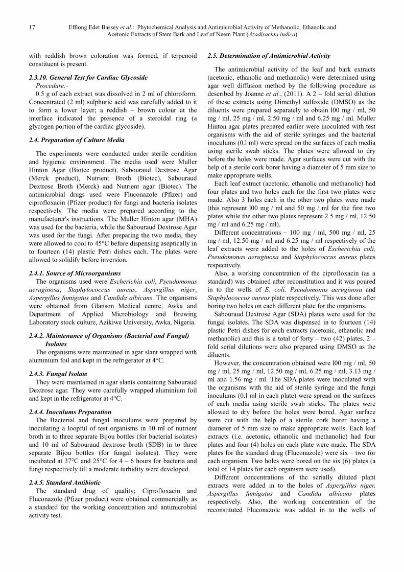
Table 4 showed the characteristic and identification
features of the fungal isolate being observed and examined.
Table 5 showed the physical characteristics of the bacterial
isolates being examined.
Table 6 showed the biochemical test characteristics of the
bacterial isolates being examined.
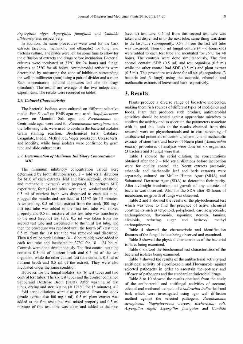
Table 7 showed the results of the antibacterial activity and
antifungal activity of ciprofloxacin and Fluconazole against
selected pathogens in order to ascertain the potency and
efficacy of pathogens and the standard antimicrobial drugs.
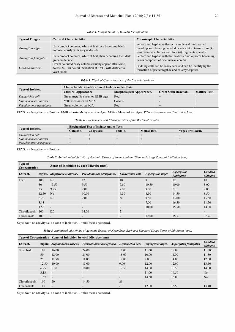
Table 8 to 10 showed the results obtained from the study
of the antibacterial and antifungal activities of acetone,
ethanol and methanol extracts of Azadirachta indica leaf and
bark which were investigated using agar well diffusion
method against the selected pathogens; Pseudomonas
aeruginosa, Staphylococcus aureus, Escherichia coli,
Aspergillus niger, Aspergillus fumigatus and Candida

19 Effiong Edet Bassey et al.: Phytochemical Analysis and Antimicrobial Activity of Methanolic, Ethanolic and
Acetonic Extracts of Stem Bark and Leaf of Neem Plant (Azadirachta indica)
albicans. All the examined extracts showed varying degrees
of antimicrobial activities against the pathogens.
Table 11, 12 and 13 showed the results that were obtained
from the analysis and it revealed the minimum inhibitory
concentration (MIC) values of the acetonic (leaf; 12.5 – 25
and bark; 3.13 – 12.5), ethanolic (leaf; 6.25 – 25 and bark;
6.25) and methanolic (leaf; 6.25 – 25 and bark; 6.25 - 25) of
Neem Plant Parts Extracts in mg / ml respectively.
Table 14 showed the results obtained from this study that,
the minimum bacterial concentration (MBC) values of
acetone (0 – 25 mm), ethanol (12.5 – 25 mm) and methanol
(0 – 25 mm) extracts of Neem leaf.
Table 15 showed the results that were obtained from the
analysis and it revealed the minimum bacterial concentration
(MBC) values of acetone (12.5 – 25 mm), ethanol (0 – 25
mm) and methanol (12.5 – 25 mm) extracts of Neem stem
bark.
Figure 1, 2, 3, and 4 showed the antibacterial activity of
acetonic, ethanolic and methanolic extract of Neem plant
parts.
Table 1. Concentrations of Fungi and Bacteria obtained after the 2 – fold
serial dilutions.
Type of Microbe. Tube. Dilution in mg / ml.
Fungi 1st 25
2nd 12.50
3rd 6.25
4th 3.13
5th 1.56
6th 0.78
Bacteria 1st 25
2nd 12.50
3rd 6.25
4th 3.13
NB: The concentrations obtained after the 2 – fold serial dilutions before
incubation.
Table 2. Qualitative Phytochemical Analysis of Acetone, Ethanol, Methanol Leaf Extract.
Type of Phytochemical. Test Type. Type of Leaf Extract.
Acetone. Ethanol. Methanol
Alkaloids Mayer’s - + -
Fronthing - - +
Emulsification ++ - ++
Tannin cchloride’s + - +
Glycoside cardiac General - + -
Cyanogenic - + -
Flavonoid Sodium hydroxide’s - + -
Anthraquinones Free / Combined - + -
Reducin gsugar Free Reducing Sugar’s + + +++
Polyphenol - +++ -
Terpenoid Salkowski’s - + -
Steroid Sodium hydroxide’s - + -
Saponins - - -
KEYS: - = Absent, + = Scanty, ++ = Moderate, +++ = Abundance.
Table 3. Qualitative Phytochemical Analysis of Acetone, Ethanol, Methanol Stem Bark Extract.
Type of Phytochemical. Test Type. Type of Leaf Extract.
Acetone. Ethanol. Methanol
Alkaloids Mayer’s - + +
Fronthing - - -
Emulsification - + +
Tannin Ferric chloride’s +++ - -
Glycoside cardiac General - - -
Cyanogenic - - -
Flavonoid Sodium hydroxide’s + - -
Anthraquinones Free / Combined - - -
Reducing sugar Free Reducing Sugar’s +++ ++ +
Polyphenol ++ - ++
Terpenoid Salkowski’s - - -
Steroid
-
Sodium hydroxide’s
-
-
Saponins - - -
KEYS: - = Absent, + = Scanty present, ++ = Moderate, +++ = Abundance.
Journal of Diseases and Medicinal Plants 2016; 2(3): 14-25 20
Table 4. Fungal Isolates (Moulds) Identification.
Type of Fungus. Cultural Characteristics. Microscopic Characteristics.
Aspergillus niger. Flat compact colonies, white at first then becoming black
homogeneously with grey underside.
Septate and hyphae with erect, simple and thick walled
conidiophores bearing conidial heads split in to over four (4)
loose conidia columns with four (4) fragments apically.
Aspergillus fumigatus. Flat compact colonies, white at first, then becoming then dark
green underside.
Septate and hyphae with thin walled conidiophores becoming
heads composed of catenucleac conidial.
Candida albicans.
Cream coloured pasty colonies usually appear after some
hours (24 – 48 hours) incubation at 37°C, with distinctive
yeast smell.
Budding cells can be easily seen and can be identify by the
formation of pseudohyphae and chlamydospores.
Table 5. Physical Characteristics of the Bacterial Isolates.
Type of Isolates. Characteristic identification of Isolates under Tests.
Cultural Appearance Morphological Appearance. Gram Stain Reaction. Motility Test.
Escherichia coli Green metallic sheen on EMB agar Rod + -
Staphylococcus aureus Yellow colonies on MSA Coccus - +
Pseudomonas aeruginosa Green colonies on PCA Rod + -
KEYS: - = Negative, + = Positive, EMB = Eosin Methylene Blue Agar, MSA = Mannitol Salt Agar, PCA = Pseudomonas Centrimide Agar.
Table 6. Biochemical Test Characteristics of the Bacterial Isolates.
Type of Isolates. Biochemical Test of Isolates under Tests.
Catalase. Coagulase. Indole. Methyl Red. Voges Proskaeur.
Escherichia coli - + + + -
Staphylococcus aureus + + + + +
Pseudomonas aeruginosa + - - - -
KEYS: - = Negative, + = Positive.
Table 7. Antimicrobial Activity of Acetonic Extract of Neem Leaf and Standard Drugs Zones of Inhibition (mm).
Type of
Concentration Zones of Inhibition by each Microbe (mm).
Extract. mg/ml. Staphyloccus aureus. Pseudomonas aeruginosa. Escherichia coli. Aspergillus niger. Aspergillus
fumigatus.
Candida
albicans
Leaf 100 No 12 10 8 12 10
50 13.50 9.50 9.50 10.50 10.00 8.00
25 9.75 9.00 7.00 9.00 No 9.00
12.50 No 10.00 6.50 8.50 14.50 8.50
6.25 No 9.00 No 8.50 13.00 15.50
3.13 - - - 7.00 16.50 11.50
1.56 - - - 10.00 15.50 14.00
Ciprofloxacin 100 l20 14.50 21. - - -
Fluconazole 100 - - - 12.00 15.5. 13.40
Keys: No = no activity i.e. no zone of inhibition, - = this means not tested.
Table 8. Antimicrobial Activity of Acetonic Extract of Neem Stem Bark and Standard Drugs Zones of Inhibition (mm).
Type of Concentration Zones of Inhibition by each Microbe (mm).
Extract. mg/ml. Staphyloccus aureus. Pseudomonas aeruginosa. Escherichia coli. Aspergillus niger. Aspergillus fumigatus. Candida
albicans
Stem bark. 100 16.00 24.00 12.00 11.00 19.00 11.000
50 12.00 21.00 18.00 10.00 11.00 11.50
25 11.50 11.00 12.00 7.00 14.00 12.00
12.50 10.00 13.00 9.00 12.00 12.00 13.50
6.25 6.00 10.00 17.50 14.00 10.50 14.00
3.13 - - - 11.00 16.50 No
1.57 - - - 14.50 16.00 No
Ciprofloxacin 100 20 14.50 21. - - -
Fluconazole 100 - - - 12.00 15.5. 13.40
Keys: No = no activity i.e. no zone of inhibition, - = this means not tested.
21 Effiong Edet Bassey et al.: Phytochemical Analysis and Antimicrobial Activity of Methanolic, Ethanolic and
Acetonic Extracts of Stem Bark and Leaf of Neem Plant (Azadirachta indica)
Table 9. Antimicrobial Activity of Methanolic Extract of Neem Leaf and Standard Drugs Zones of Inhibition (mm).
Type of Concentration Zones of Inhibition by each Microbe (mm).
Extract. mg/ml. Staphyloccus aureus. Pseudomonas aeruginosa. Escherichia coli. Aspergillus niger. Aspergillus fumigatus. Candida
albicans
Leaf 100 14.50 18.00 18.00 11.00 12.00 11.50
50 11.00 16.00 10.00 9.00 15.00 7.50
25 9.00 11.00 12.00 10.00 20.00 No
12.50 9.40 10.50 9.50 No 10.00 13.00
6.25 7.00 10.00 8.50 No 14.50 13.50
3.13 - - - 10.00 13.00 11.00
1.56 - - - 12.00 12.00 14.00
Ciprofloxacin 100 20 14.50 21. - - -
Fluconazole 100 - - - 12.00 15.5. 13.40
Keys: No = no activity i.e. no zone of inhibition, - = this means not tested.
Table 10. Antimicrobial Activity of Methanolic Extract of Neem Stem Bark and Standard Drugs Zones of Inhibition (mm).
Type of Concentration Zones of Inhibition by each Microbe (mm).
Extract. mg/l Staphyloccus
aureus. Pseudomona aeruginosa. Escherichia coli. Aspergillus niger. Aspergillus fumigatus.
Candida
albicans
StemBark 100 17.50 17.00 15.00 22.50 No 16.00
50 13.00 14.00 14.00 11.50 14.50 12.00
25 11.50 13.00 9.00 15.50 No No
12.5 9.00 12.00 14.50 7.50 10.50 No
6.25 10.50 No 8.50 19.50 10.00 14.00
3.13 - - - 14.50 14.00 No
1.56 - - - 14.00 20.00 No
Ciprofloxacin 100 20 14.5 21. - - -
Fluconazole 100 - - - 12.00 15.5. 13.40
Keys: No = no activity i.e. no zone of inhibition, - = this means not tested.
Table 11. Minimum Inhibitory Concentration Values of the Acetonic Neem
Plant Parts Extracts in mg / ml.
Name of Microbe. Neem Plant Parts mg / ml.
Leaf Extract. Stem Bark Extract.
Staphylococcus aureus 25 No
Pseudomonas aeruginosa 25 6.25
Escherichia coli 25 6.25
Aspergillus niger 25 12.50
Aspergillus fumigatus 25 12.50
Candida albicans 12.50 3.13
Key: No = value for MIC was obtained.
Table 12. Minimum Inhibitory Concentration Values of the Ethanolic Neem
Plant Parts Extracts in mg / ml.
Name of Microbe. Neem Plant Parts mg / ml.
Leaf Extract. Stem Bark Extract.
Staphylococcus aureus No 6.25
Pseudomonas aeruginosa 25 6.25
Escherichia coli No No
Aspergillus niger 12.50 6.25
Aspergillus fumigatus 12.50 6.25
Candida albicans 6.25 6.25
Key: No = value for MIC was obtained
Table 13. Minimum Inhibitory Concentration Values of the Methanolic Neem
Plant Parts Extracts in mg / ml.
Name of Microbe. Neem Plant Parts mg / ml.
Leaf Extract. Stem Bark Extract.
Staphylococcus aureus 25 6.25
Pseudomonas aeruginosa 6.25 12.50
Escherichia coli No 6.25
Name of Microbe. Neem Plant Parts mg / ml.
Leaf Extract. Stem Bark Extract.
Aspergillus niger 25 25
Aspergillus fumigatus 12.50 6.25
Candida albicans 6.25 6.25
Key: No = value for MIC was obtained
Table 14. MBC values of Acetone, Ethanol and Methanol Extracts of Neem Leaf.
Type of Microbe. Neem Leaf Extract.
Acetone. Ethanol. Methanol.
Staphylococcus aureus 25 No 25
Pseudomonas aeruginosa 25 25 25
Escherichia coli 25 No No
Aspergillus niger 25 12.50 25
Aspergillus fumigatus 25 25 25
Candida albicans No 25 25
Key: No = value for MBC was obtained.
Table 15. MBC values of Acetone, Ethanol and Methanol Extracts of Neem
Stem Bark.
Type of Microbe. Neem Leaf Extract.
Acetone. Ethanol. Methanol.
Staphylococcus aureus No 25 12.50
Pseudomonas aeruginosa 12.50 25 25
Escherichia coli 25 No 12.50
Aspergillus niger 25 25 25
Aspergillus fumigatus 25 25 25
Candida albicans No No No
Key: No = value for MBC was obtained.
Journal of Diseases and Medicinal Plants 2016; 2(3): 14-25 22
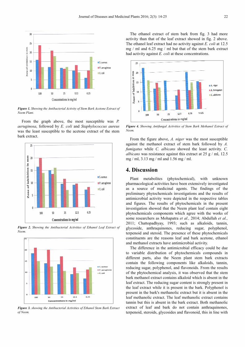
Figure 1. Showing the Antibacterial Activity of Stem Bark Acetone Extract of
Neem Plant.
From the graph above, the most susceptible was P.
aeruginosa, followed by E. coli and Staphylococcus aureus
was the least susceptible to the acetone extract of the stem
bark extract.
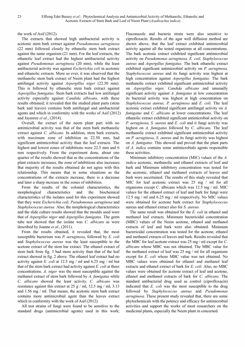
Figure 2. Showing the Antibacterial Activities of Ethanol Leaf Extract of
Neem.
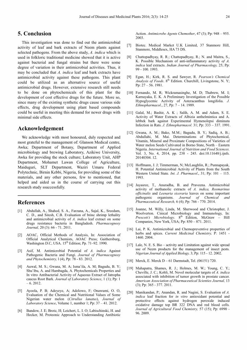
Figure 3. showing the Antibacterial Activities of Ethanol Stem Bark Extract
of Neem.
The ethanol extract of stem bark from fig. 3 had more
activity than that of the leaf extract showed in fig. 2 above.
The ethanol leaf extract had no activity against E. coli at 12.5
mg / ml and 6.25 mg / ml but that of the stem bark extract
had activity against E. coli at these concentrations.
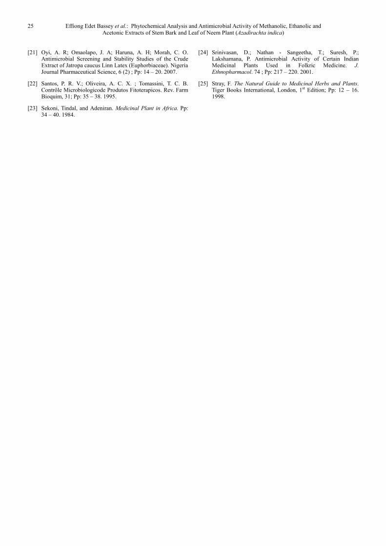
Figure 4. Showing Antifungal Activities of Stem Bark Methanol Extract of
Neem.
From the figure above, A. niger was the most susceptible
against the methanol extract of stem bark followed by A.
fumigatus while C. albicans showed the least activity. C.
albicans was resistance against this extract at 25 g / ml, 12.5
mg / ml, 3.13 mg / ml and 1.56 mg / ml.
4. Discussion
Plant metabolites (phytochemical), with unknown
pharmacological activities have been extensively investigated
as a source of medicinal agents. The findings of the
preliminary phytochemicals investigations and the results of
antimicrobial activity were depicted in the respective tables
and figures. The results of phytochemicals in the present
investigation showed that the Neem plant leaf contain eight
phytochemicals components which agree with the works of
some researchers as Mohapatra et al., 2014; Abdullah et al.,
2011; Chattopadhyay, 1993; such as alkaloids, tannin,
glycoside, anthraquinones, reducing sugar, polyphenol,
terpenoid and steroid. The presence of these phytochemicals
constituents are the reasons leaf and bark acetone, ethanol
and methanol extracts have antimicrobial activity.
The difference in the antimicrobial efficacy could be due
to variable distribution of phytochemicals compounds in
different parts, also the Neem plant stem bark extracts
contain the following components like alkaloids, tannin,
reducing sugar, polyphenol, and flavonoids. From the results
of the phytochemical analysis, it was observed that the stem
bark methanol extract contains alkaloid which is absent in the
leaf extract. The reducing sugar content is strongly present in
the leaf extract while it is present in the bark. Polyphenol is
present in the bark's methanolic extract but it is absent in the
leaf methanolic extract. The leaf methanolic extract contains
tannin but this is absent in the bark extract. Both methanolic
extract of leaf and bark do not contain anthraquinones,
terpenoid, steroids, glycosides and flavonoid, this in line with
23 Effiong Edet Bassey et al.: Phytochemical Analysis and Antimicrobial Activity of Methanolic, Ethanolic and
Acetonic Extracts of Stem Bark and Leaf of Neem Plant (Azadirachta indica)
the work of Asif (2012).
The extracts that showed high antibacterial activity is
acetonic stem bark extract against Pseudomonas aeruginosa
(22 mm) followed closely by ethanolic stem bark extract
against the same organism (21 mm). For the leaf extracts, the
ethanolic leaf extract had the highest antibacterial activity
against Pseudomonas aeruginosa (20 mm), while the least
antibacterial activity was against Escherichia coli in acetonic
and ethanolic extracts. More so ever, it was observed that the
methanolic stem bark extract of Neem plant had the highest
antifungal activity against Aspergillus niger (22.50 mm).
This is followed by ethanolic stem bark extract against
Aspergillus fumigatus. Stem bark extracts had low antifungal
activity especially against Candida albicans, with these
results obtained; it revealed that the studied plant parts (stem
bark and leaves) contains both antifungal and antibacterial
agents and which in conformity with the works of Asif (2012)
and Jayasree et al., (2014).
Overall, the extract of the neem plant part with no
antimicrobial activity was that of the stem bark methanolic
extract against C. albicans. In addition, stem bark extracts,
with maximum zone of inhibition as 22.50 mm, had
significant antimicrobial activity than the leaf extracts. The
highest and lowest zones of inhibitions were 22.5 mm and 6
mm respectively. From the experiment done, about one
quarter of the results showed that as the concentrations of the
plant extracts increases, the zone of inhibitions also increases.
But majority of the results obtained do not agree with this
relationship. This means that in some situations as the
concentrations of the extracts increase, there is a decrease
and later a sharp increase in the zones of inhibition.
From the results of the colonial characteristics, the
morphological characteristics and the biochemical
characteristics of the isolates used for this experiment showed
that they were Escherichia coli, Pseudomonas aeruginosa and
Staphylococcus aureus. Also, the morphological characteristics
and the slide culture results showed that the moulds used were
that of Aspergillus niger and Aspergillus fumigatus. The germ
tube test showed that the isolate was C. albicans as were
described by Joanne et al., (2011).
From the results obtained, it revealed that, the most
susceptible bacterium was P. aeruginosa, followed by E. coli
and Staphylococcus aureus was the least susceptible to the
acetone extract of the stem bar extract. The ethanol extract of
stem bark from fig. 3 had more activity than that of the leaf
extract showed in fig. 2 above. The ethanol leaf extract had no
activity against E. coli at 12.5 mg / ml and 6.25 mg / ml but
that of the stem bark extract had activity against E. coli at these
concentrations. A. niger was the most susceptible against the
methanol extract of stem bark followed by A. fumigatus while
C. albicans showed the least activity. C. albicans was
resistance against this extract at 25 g / ml, 12.5 mg / ml, 3.13
and 1.56 mg / ml. That means, the acetonic stem bark extract
contains more antimicrobial agent than the leaves extract
which in conformity with the work of Asif (2012).
All test strains of fungi were found to be sensitive to the
standard drugs (antimicrobial agents) used in this work;
Fluconazole and bacteria strain were also sensitive to
ciprofloxacin. Results of the agar well diffusion method are
shown above, that the leaf extract exhibited antimicrobial
activity against all the tested organisms at all concentrations.
The bark acetonic extract exhibited significant antimicrobial
activity on Pseudomonas aeruginosa, E. coil, Staphylococcus
aureus and Aspergillus fumigatus. The bark ethanolic extract
exhibited significant antimicrobial activity on P. aeruginosa,
Staphylococcus aureus and its fungi activity was highest at
high concentration against Aspergillus fumigatus. The bark
methanolic extract exhibited significant antimicrobial activity
on Aspergillus niger, Candida albicans and unusually
significant activity against A. fumigatus at low concentration
its bacterial activity was highest at high concentration on
Staphylococcus aureus, P. aeruginosa and E. coli. The leaf
acetonic extract exhibited significant antifungal activity on A.
fumigatus and C. albicans at lower concentrations. The leaf
ethanolic extract exhibited significant antimicrobial activity on
P. aeruginosa, S. aureus and E. coli and it fungi activity was
highest on A. fumigatus followed by C. albicans. The leaf
methanolic extract exhibited significant antimicrobial activity
on P. aeruginosa, S. aureus and its fungi activity was highest
on A. fumigatus. This showed and proved that the plant parts
of A. indica contains some antimicrobials agents responsible
these activities.
Minimum inhibitory concentration (MIC) values of the A.
indica acetonic, methanolic and ethanol extracts of leaf and
bark and Minimum inhibitory concentration was tested for
the acetonic, ethanol and methanol extracts of leaves and
bark were ascertained. The results of this study revealed that
MIC for leaf acetonic extract was 25 mg / ml for all
organisms except C. albicans which was 12.5 mg / ml. MIC
values for the ethanol extract of leaf and bark for fungi were
12.5 mg / ml and 6.25 mg / ml respectively. No MIC values
were obtained for acetone bark extract for Staphylococcus
aureus and ethanol extract also for S. aureus.
The same result was obtained for the E. coli in ethanol and
methanol leaf extracts. Minimum bactericidal concentration
(MBC) values of the Neem acetone, ethanol and methanol
extracts of leaf and bark were also obtained. Minimum
bactericidal concentration was tested for the acetone, ethanol
and methanol extracts of leaves and bark. Results revealed that
the MBC for leaf acetone extract was 25 mg / ml except for C.
albicans whose MBC was not obtained. The MBC value for
the methanol extract of leaf was 25 mg / ml for all organisms
except for E. coli whose MBC value was not obtained. No
MBC values were obtained for ethanol and methanol leaf
extracts and ethanol extract of bark for E. coli. Also, no MBC
values were obtained for acetone extract of leaf and acetone,
ethanol and methanol extracts of bark for C. albicans. The
standard antibacterial drug used as control (ciprofloxacin)
indicated that E. coli was the most susceptible to the drug
followed by Staphylococcus aureus and Pseudomonas
aeruginosa. These present study revealed that, there are some
phytochemicals with the potency and efficacy for antimicrobial
activities and support the works of most researchers on the
medicinal plants, especially the Neem plant in concerned.
Journal of Diseases and Medicinal Plants 2016; 2(3): 14-25 24
5. Conclusion
This investigation was done to find out the antimicrobial
activity of leaf and bark extracts of Neem plants against
selected pathogens. From the above study, A. indica which is
used in folkloric traditional medicine showed that it is active
against bacterial and fungal strains but there were some
degree of variation in their antimicrobial activities. Thus, it
may be concluded that A. indica leaf and bark extracts have
antimicrobial activity against these pathogens. This plant
could be utilized as an alternative source of useful
antimicrobial drugs. However, extensive research still needs
to be done on phytochemicals of this plant for the
development of cost effective drugs for the future. More so,
since many of the existing synthetic drugs cause various side
effects, drug development using plant based compounds
could be useful in meeting this demand for newer drugs with
minimal side effects.
Acknowledgement
We acknowledge with most honoured, duly respected and
most grateful to the management of: Glanson Medical centre,
Awka; Department of Botany, Department of Applied
microbiology and brewing Laboratory, Azikiwe University,
Awka for providing the stock culture; Laboratory Unit, AHP
Department, Mohamet Lawan College of Agriculture,
Maiduguri, SLT Department, Waziri Umaru Federal
Polytechnic, Birnin Kebbi, Nigeria, for providing some of the
materials, and any other persons, few to mentioned, that
helped and aided us in the course of carrying out this
research study ssuccessfully.
References
[1] Abdullah, A., Shahed, S. A., Farzana, A., Sajal, K., Sreedam, C. D., and Sitesh, C.B. Evaluation of brine shrimp lethality and antimicrobial activity of A. indica leaf extract on some drugs resistance bacteria in Bangladesh. Pharmacognosy Journal, 20 (3): 66 - 71. 2011.
[2] AOAC, Official Methods of Analysis, In: Association of Official Analytical Chemists, AOAC Press; Gaithersburg, Washington D.C, USA. 15th Edition; Pp. 71–92. 1990.
[3] Asif, M. Antimicrobial Potential of A. indica Against Pathogenic Bacteria and Fungi. Journal of Pharmacognosy and Phytochemistry, l (4), Pp: 78 - 83. 2012.
[4] Auwal, M. S.; Gwana, M. A; Isma’ila, A. M; Bagudu, B. Y; Shu’ibu, A. and Hambagda, A. Phytochemicals Properties and In vitro Antibacterial Activity of Aqueous Extract of Jatropha caucus Root Bark. Journal of Laboratory Science, 1 (1); Pp: 1 – 6. 2012.
[5] Ayoola, P. B; Adeyeye, A; Adelowo, F; Onawumi, O. O, Evaluation of the Chemical and Nutritional Values of Some Nigerian water melon (Citrullus lanatus), Journal of Laboratory Science, Volume 1, number 1; Pp: 37 – 41. 2012.
[6] Bandow, J. E; Brotz, H; Leichert, L. I. O; Labischinski, H. and Hecker, M. Proteonic Approach to Understanding Antibiotic
Action. Antimicrobe Agents Chemother, 47 (3); Pp: 948 – 955. 2003.
[7] Biotec. Medical Market U.K Limited, 37 Stanmore Hill, Stanmore, Middlesex, HA 73 DS.
[8] Chattopadhyay, R. R.; Chattopadhyay, R.; N. and Maitra, S., K. Possible Mechanism of anti-inflammatory activity of A. indica leaf extracts. Indian Journal of Pharmacology, 25; Pp: 99 - 100. 1993.
[9] Egan, H.; Kirk, R. S. and Sawyer, R. Pearson’s Chemical Analysis of Foods. 8th Edition. Churchill, Livingstone, N. Y; Pp: 27 – 56. 1981.
[10] Fernando, M. R; Wickramasinghe, M. D; Thabrew, M. I; Karnayaka, E. K. A Preliminary Investigation of the Possible Hypoglycemic Activity of Asteracanthus longifolia. J. Ethnopharmacol., 27, Pp: 7 – 14. 1989.
[11] Galal, M.; Bashir, A. K ; Salih, A. M. and Adam, S. E. Activity of Water Extracts of Albizia anthelmintica and A. lebbek bark against Experimental Hymenolepsi diminuta Infection in Rats. J. Ethnopharmacol. 31; Pp: 333 – 337. 1991.
[12] Gwana, A. M.; Bako, M.M.; Bagudu, B. Y.; Sadiq, A. B.; Abdullahi, M. Mai. Determinations of Phytochemical, Vitamin, Mineral and Proximate Compositions of Varieties of Water melon Seeds Cultivated in Borno State, North – Eastern Nigeria. International Journal of Nutrition and Food Sciences. Vol. 3, No. 4, 2014, pp. 238 - 245. doi:10.11648/j.ijnfs. 20140304. 12.
[13] Hoffmann, J. J; Timmerman, N; McLaughlin, R.; Punnapayak, H. Potential Antimicrobial Activity of Plants from the South Western United State. Int. J. Pharmacol., 31; Pp: 101 – 115. 1993.
[14] Jayasree, T., Anuradha, B. and Praveena. Antimicrobial activity of methanolic extracts of A. indica, Rosmarinus officinalis and Leenaria siceraria leaves on some important pathogenic organisms. Journal of Chemical and Pharmaceutical Research, 6 (4); Pp: 766 - 770. 2014.
[15] Joanne, M. Willy, Linda, M. Sherwood and Christopher, J. Woolverton. Cinical Microbiology and Immunology, In: Prescott’s Microbiology. 8th Edition, McGraw – Hill Companies, New York, USA; Pp: 850 – 871. 2011.
[16] Lai, P. K. Antimicrobial and Chemopreventive properties of herbs and spices. Current Medicinal Chemistry, P: 1451 - 1460. 2004.
[17] Lale, N. E. S. Bio – activity and Limitation against wide spread use of Neem products for the management of insect pests. Nigerian Journal of Applied Biology, 3; Pp: 115 - 12. 2002.
[18] Merck, E. Merck D – 61 Darmstadt, Tel. (06151) 720.
[19] Mahapatra, Shames, R. J.; Holmes, M. W.; Young, C. Y.; Cheville, J. C.; Kohli, M. Novel molecular targets of A. indica associated with inhibition of tumor growth in prostate cancer. American Association of Pharmaceutical Scienties Journal, 13 (3); Pp: 365 - 377. 2011.
[20] Manikandan, P.; Anandan, R. and Nagini, S. Evaluation of A. indica leaf fraction for in vitro antioxidant potential and protective effects against hydrogen peroxide induced oxidative damage top BR 322 DNA and red blood cells. Journal of Agricultural Food Chemistry, 57 (15); Pp: 6990 - 96. 2009.
25 Effiong Edet Bassey et al.: Phytochemical Analysis and Antimicrobial Activity of Methanolic, Ethanolic and
Acetonic Extracts of Stem Bark and Leaf of Neem Plant (Azadirachta indica)
[21] Oyi, A. R; Omaolapo, J. A; Haruna, A. H; Morah, C. O. Antimicrobial Screening and Stability Studies of the Crude Extract of Jatropa caucus Linn Latex (Euphorbiaceae). Nigeria Journal Pharmaceutical Science, 6 (2) ; Pp: 14 – 20. 2007.
[22] Santos, P. R. V.; Oliveira, A. C. X. ; Tomassini, T. C. B. Contrôle Microbiologicode Produtos Fitoterapicos. Rev. Farm Bioquim, 31; Pp: 35 – 38. 1995.
[23] Sekoni, Tindal, and Adeniran. Medicinal Plant in Africa. Pp: 34 – 40. 1984.
[24] Srinivasan, D.; Nathan - Sangeetha, T.; Suresh, P.; Lakshamana, P. Antimicrobial Activity of Certain Indian Medicinal Plants Used in Folkric Medicine. J. Ethnopharmacol. 74 ; Pp: 217 – 220. 2001.
[25] Stray, F. The Natural Guide to Medicinal Herbs and Plants. Tiger Books International, London, 1st Edition; Pp: 12 – 16. 1998.