Embed Size (px)
Citation preview
Physiological Mini Reviews
Volume10
Vol. 10, November-December 2017ISSN 1669-5410 (Online)pmr.safisiol.org.ar
Physiological Mini-Reviews
[ISSN 1669-5410 (Online)]
Edited by the Argentinean Physiological Society
Journal address: Centro de Investigaciones Cardiovasculares y Cátedra de Fisiología y Física Biológica.
Facultad de Ciencias Médicas; Universidad Nacional de La Plata;
La Plata, Buenos Aires, Argentina. Tel.-Fax: +54-211-4834833
http://pmr.safisiol.org.ar
Physiological Mini-Reviews is a scientific journal, publishing brief reviews on "hot" topics in Physiology. The scope is quite broad, going from "Molecular Physiology" to "Integrated Physiological Systems". As indicated by our title it is not our intention to publish exhaustive and complete reviews. We ask to the authors concise and updated descriptions of the "state of the art" in a specific topic. Innovative and thought-provoking ideas are welcome.
Editorial Board:
Alejandro Aiello, La Plata, Argentina.
Bernardo Alvarez, La Plata, Argentina.
Eduardo Arzt, Buenos Aires, Argentina.
Oscar Candia, New York, United States
Claudia Capurro, Buenos Aires, Argentina
Daniel Cardinali, Buenos Aires, Argentina.
Marcelino Cereijido, México City, México.
Alberto Crottogini, Buenos Aires, Argentina.
Adolfo De Bold, Ottawa, Canada.
Osvaldo Delbono, Winston-Salem, United States.
Irene Ennis, La Plata, Argentina.
Ana Franchi, Buenos Aires, Argentina.
Carlos González, Valparaíso, Chile
Cecilia Hidalgo, Santiago, Chile.
Elena Lascano, Buenos Aires, Argentina.
Carlos Libertun, Buenos Aires, Argentina.
Gerhard Malnic, Sao Paulo, Brazil.
Raúl Marinelli, Rosario, Argentina.
Jorge Negroni, Buenos Aires, Argentina.
Patricia Rocco, Río de Janeiro, Brazil.
Juan Saavedra, Bethesda, United States.
David Sabatini, New York, United States.
Margarita Salas, La Plata, Argentina.
María Inés Vaccaro, Buenos Aires, Argentina.
Martín Vila-Petroff, La Plata, Argentina.
Editor in chief: Alicia Mattiazzi, La Plata, Argentina
Associate Editor: Leticia Vittone, La Plata, Argentina
Founding Editor: Mario Parisi, Buenos Aires, Argentina
Publishing Scientific
Committee: Carlos A. Valverde, La Plata, Argentina
Matilde Said, La Plata, Argentina
Cecilia Mundiña-Weilenmann, La Plata, Argentina
Editorial Assistant: María Inés Vera, La Plata, Argentina
Preparation and Submission of manuscripts:
"Physiological Mini-Reviews" will have a maximum of 3000 words, 50 references and 3 figures. Material will be
addressed to scientific people in general but not restricted to specialist of the field. For citations in the text please
refer to Instructions in our webpage. Final format will be given at the Editorial Office. Most contributions will be
invited ones, but spontaneous presentations are welcome. Send your manuscript in Word format (.doc or .docx)
Advertising:
For details, rates and specifications contact the Associate Editor at the Journal address e-mail: [email protected]
The “Sociedad Argentina de Fisiología” is a registered non-profit organization in Argentina. (Resol. IGJ 763-04)

Physiological Mini Reviews, Vol.10 Nº6, 2018
60
RNA SPLICING, AGEING, TDP 43 AGGREGATION AND
NEURODEGENERATION
Francisco Ernesto Baralle
International Centre for Genetic Engineering and Biotechnology (ICGEB); Padriciano 99, Trieste, Italy
*Correspondence to:
Dr. Francisco Ernesto Baralle ([email protected])
ABSTRACT
The role of splicing factors in pathological processes is increasingly documented. This is
particularly evident in the pathologies affecting the nervous system where either the mis-
folding or aggregation of RNA binding proteins is a key event in triggering the
neurodegeneration. The aggregation of TDP-43 is the major distinguishing feature of most
cases of Amyotrophic Lateral Sclerosis (ALS). There is still a significant uncertainty in regards
to many aspects concerning the nature of the aggregates and their functional consequences.
The biochemical and genetic aspects of TDP-43 aggregation need further definition, as new
therapies could be directed towards them. A better understanding of TDP-43 structure and
interactions is needed, from what we currently know it has been possible to construct cellular
and animal models of TDP- 43 ALS like aggregation. Using these models drugs that can revert
aggregation and recover function have been identified. Finally, ALS is a disease that occurs
mostly during the fifth to the seventh decade of life. In this respect, it has been observed that
the onset of the locomotion defect in an ALS fly model coincides with an age-related 4-fold
drop in TBPH levels (the Drosophila TDP43 orthologous), similar TDP 43 reduction with age
was observed in mice brain. Thus, understanding the relationship between aging and TDP-43
production and mis-folding in cell culture, animal models, and human tissues might provide
further clues to explain the time of disease onset.
Keywords: Molecular Biology, gene expression, RNA splicing, Amyotrophic Lateral
Sclerosis, Aging.
Original received: December 22, 2017; Accepted: December 29, 2017; Published: January 2018.

Physiological Mini Reviews, Vol.10 Nº6, 2018
61
Introduction
In this brief review I would like to describe how the research on basic molecular mechanisms
of gene expression ends up making an impact on an apparently unrelated field such as
neurobiology and ends up in translational work searching for effective therapeutic drugs to be
used in neurodegenerative disease. To evidence these points as clear as possible I will spend a
good part of the manuscript in describing the impact that the discovery of the RNA splicing
process has had in molecular and developmental biology and enter in the role of splicing factors
in neuro-degeneration.
RNA splicing, a fundamental and unexpected step in RNA metabolism, was observed for the
first time in the late 70s. At that time, a peculiar RNA processing phenomena was described
in the adenovirus 5’UTR; three pieces of noncoding sequences were joined together and the
sequences between them eliminated to form the viral mRNA [1, 2]
This was soon followed by the observation that the recently cloned globin mRNA did not
hybridize to a single band in a Southern blot but there clearly were other sequences, interrupting
the coding sequences [3]. These genomic nucleotide fragments were even longer than the
coding sequences. Finally, after the, at the time, laborious process of cloning higher eukaryotic
genes, the sequencing of these clones showed that the presence of noncoding sequences that
interrupted the coding sequences was the rule rather than the exception. The name of introns
and exons was adopted for the intervening sequences and the coding sequences respectively
and the prediction made that the protein coding content of genomes would comprise only a
fraction of the total DNA [4]. The process of cutting the introns and joining the exons was
called splicing. This discovery had immediate ramifications regarding biology, namely, how
are the exons identified?
The first gene sequences showed, surprisingly, poor sequence homology at the junctions of
introns and exons. The only universally conserved nucleotides were the GU at the 5’ site of the
intron and AG at its 3’site [5] obviously, these elements would be insufficient to direct the
splicing process which is usually error free. In the following years other features were
uncovered that increased the accuracy of exon/intron junction definition resulting in the so
called consensus sequences around the exon/intron junctions (splice sites) and the intronic
poly-pirimidine tract near the 3’ splice site [6].
In the early 1980s it was discovered that there were variations in exon selection and that several
mRNA isoforms could be produced from one pre-mRNA [7], this process was called
alternative splicing and was followed by the discovery both in vitro and in vivo that auxiliary
sequences overlapping with the coding sequences in the genomic context of an alternative
spliced exon [8, 9]. Subsequently, specific cis-acting elements within these fragment were
identified that according to their location and effect on splicing were called exon splicing
enhancers or silencers (ESE and ESS respectively) and intron splicing enhancers and silencers
(ISE and ISS respectively) whose effects are transduced through their interaction with RNA
binding proteins (RBPs). In addition to the cis-acting element, other processes that influence
splicing outcome exist. They include the effect on splicing of the RNA structure [10], the
transcription rate [11], chromatin remodelling and epigenetic modifications [12].
Alternative splicing in development and ageing
All organisms during development and reaching a mature reproductive stage undergo slight
changes and adjustments in the cellular and tissue molecular and physiological processes that
taken to the extreme can be called an aging process. One metabolic area where these alterations
can be clearly seen is in the RNA splicing process and in particular in the alternative splicing
variations. This is because RNA splicing is a ubiquitous regulatory mechanism that the cell
utilize to produce more than one mRNA transcript from a single gene. Alternatively spliced
transcripts can be translated into different protein isoforms with diverse functions and/or

Physiological Mini Reviews, Vol.10 Nº6, 2018
62
localizations, they can also occur in untranslated regions affecting mRNA stability,
localization, or translation. Indeed, alternative splicing is not a rare event: in humans more than
90% of the genes are estimated to undergo alternative splicing. From the ~20,000 human
protein-coding genes, high-resolution mass spectrometry analyses have shown that ~37% of
them generate multiple protein isoform [13] evidencing alternative splicing contribution to
protein diversification. These alternative splicing events are in part directed by a series of trans
acting factors that bind a variety of exonic and intronic sequences and in this way can heavily
influence splice site choice and exon inclusion. Indeed, the physiological importance of
alternative splicing is highlighted by the enormous number of human diseases caused by
mutations in cis-acting RNA-sequence elements, trans-acting splicing factors or spliceosome
components. Indeed alternative splicing is a main player in cell linage and tissue-identity
acquisition and maintenance, cell differentiation, and tissue/organ development. Molecular
understanding of developmental transitions has also revealed important bases of pathological
mechanisms in diseases where normal networks are mis-regulated. Multiple mechanisms
regulate splicing in nature, in particular during development [14].
The hnRNP TDP 43
There is a myriad of splicing factors that can act positively or negatively on exon inclusion.
Many of them belong to the RNA binding proteins family, the positive factors in splicing are
often the Serine-Arginine (SR) proteins and the negative factors are Heterogeneous Nuclear
Ribonucleoproteins (hnRNPs). These splicing factors are involved in several pathological
processes among these neurodegeneration.
The case of the hnRNP
TDP 43 (TAR DNA
Binding Protein) is a
paradigm of these phe
nomena. Our laboratory
characterized TDP 43
as an hnRNP with a role
in splicing events
leading to Cystic
Fibrosis [15]. In fact it
was known that a
polymorphism in the
3’splice site of CFTR
exon 9 composed by a
variable number of UG
repeat and U runs was
associated with milder
clinical presentation of
Cystic Fibrosis (Fig 1,
middle panel).
Figure 1: Schematic representation of the region of exon 8, 9 and 10 of the CFTR pre mRNA (Middle
panel). Exon 9 is included in variable proportions in different individuals. If there is no interference of
the TDP 43/A2 complex with the 3’ss U2AF complex is predominantly included (Upper panel). On the
other hand if the U2AF-pre mRNA complex formation is hampered by the TDP43/A2 complex, exon
9 is predominantly skipped (Lower panel).
Physiological Mini Reviews, Vol.10 Nº6, 2018
63
The UGm and Un may exist in different combinations in normal individuals, the approximate
range of UGm is from 5 to 13 while that of Un is from 3 to 9. They can exist in different
combinations but not all of them are compatible with normal CFTR RNA processing and
consequently function. For example UG9 U7 yields normal exon 9 splicing (Fig 1, upper
panel), UG13 U5 results in higher proportion of exon 9 skipping but the residual exon 9
inclusion prevents the development of classical CF and is associated with single defects such
as vas deferens obstruction or bronchiectasis. Finally the polymorphism configuration UG13
U3 produces total skipping of exon 9 (Fig 1, lower panel). Hence a non-functional CFTR
protein and full blown CF [16]. Investigating the molecular basis of this phenomena we found
that hnRNP TDP 43 bound to the UG sequence and interacted with other hnRNPS such as A2
that in turn inhibited the recognition of the 3’ss by the U2AF complex (Fig 1).
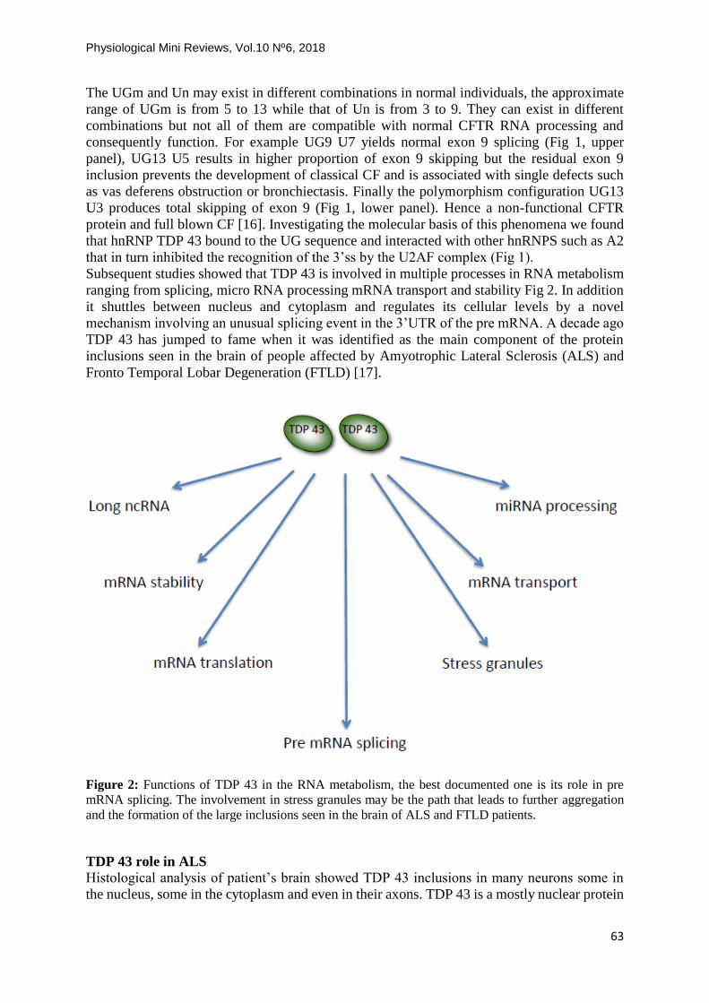
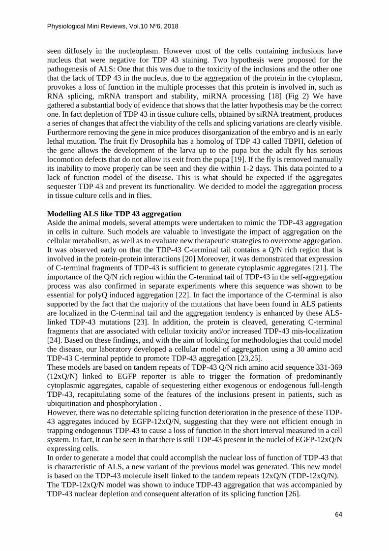
Subsequent studies showed that TDP 43 is involved in multiple processes in RNA metabolism
ranging from splicing, micro RNA processing mRNA transport and stability Fig 2. In addition
it shuttles between nucleus and cytoplasm and regulates its cellular levels by a novel
mechanism involving an unusual splicing event in the 3’UTR of the pre mRNA. A decade ago
TDP 43 has jumped to fame when it was identified as the main component of the protein
inclusions seen in the brain of people affected by Amyotrophic Lateral Sclerosis (ALS) and
Fronto Temporal Lobar Degeneration (FTLD) [17].
Figure 2: Functions of TDP 43 in the RNA metabolism, the best documented one is its role in pre
mRNA splicing. The involvement in stress granules may be the path that leads to further aggregation
and the formation of the large inclusions seen in the brain of ALS and FTLD patients.
TDP 43 role in ALS
Histological analysis of patient’s brain showed TDP 43 inclusions in many neurons some in
the nucleus, some in the cytoplasm and even in their axons. TDP 43 is a mostly nuclear protein
Physiological Mini Reviews, Vol.10 Nº6, 2018
64
seen diffusely in the nucleoplasm. However most of the cells containing inclusions have
nucleus that were negative for TDP 43 staining. Two hypothesis were proposed for the
pathogenesis of ALS: One that this was due to the toxicity of the inclusions and the other one
that the lack of TDP 43 in the nucleus, due to the aggregation of the protein in the cytoplasm,
provokes a loss of function in the multiple processes that this protein is involved in, such as
RNA splicing, mRNA transport and stability, miRNA processing [18] (Fig 2) We have
gathered a substantial body of evidence that shows that the latter hypothesis may be the correct
one. In fact depletion of TDP 43 in tissue culture cells, obtained by siRNA treatment, produces
a series of changes that affect the viability of the cells and splicing variations are clearly visible.
Furthermore removing the gene in mice produces disorganization of the embryo and is an early
lethal mutation. The fruit fly Drosophila has a homolog of TDP 43 called TBPH, deletion of
the gene allows the development of the larva up to the pupa but the adult fly has serious
locomotion defects that do not allow its exit from the pupa [19]. If the fly is removed manually
its inability to move properly can be seen and they die within 1-2 days. This data pointed to a
lack of function model of the disease. This is what should be expected if the aggregates
sequester TDP 43 and prevent its functionality. We decided to model the aggregation process
in tissue culture cells and in flies.
Modelling ALS like TDP 43 aggregation
Aside the animal models, several attempts were undertaken to mimic the TDP-43 aggregation
in cells in culture. Such models are valuable to investigate the impact of aggregation on the
cellular metabolism, as well as to evaluate new therapeutic strategies to overcome aggregation.
It was observed early on that the TDP-43 C-terminal tail contains a Q/N rich region that is
involved in the protein-protein interactions [20] Moreover, it was demonstrated that expression
of C-terminal fragments of TDP-43 is sufficient to generate cytoplasmic aggregates [21]. The
importance of the Q/N rich region within the C-terminal tail of TDP-43 in the self-aggregation
process was also confirmed in separate experiments where this sequence was shown to be
essential for polyQ induced aggregation [22]. In fact the importance of the C-terminal is also
supported by the fact that the majority of the mutations that have been found in ALS patients
are localized in the C-terminal tail and the aggregation tendency is enhanced by these ALS-
linked TDP-43 mutations [23]. In addition, the protein is cleaved, generating C-terminal
fragments that are associated with cellular toxicity and/or increased TDP-43 mis-localization
[24]. Based on these findings, and with the aim of looking for methodologies that could model
the disease, our laboratory developed a cellular model of aggregation using a 30 amino acid
TDP-43 C-terminal peptide to promote TDP-43 aggregation [23,25].
These models are based on tandem repeats of TDP-43 Q/N rich amino acid sequence 331-369
(12xQ/N) linked to EGFP reporter is able to trigger the formation of predominantly
cytoplasmic aggregates, capable of sequestering either exogenous or endogenous full-length
TDP-43, recapitulating some of the features of the inclusions present in patients, such as
ubiquitination and phosphorylation .
However, there was no detectable splicing function deterioration in the presence of these TDP-
43 aggregates induced by EGFP-12xQ/N, suggesting that they were not efficient enough in
trapping endogenous TDP-43 to cause a loss of function in the short interval measured in a cell
system. In fact, it can be seen in that there is still TDP-43 present in the nuclei of EGFP-12xQ/N
expressing cells.
In order to generate a model that could accomplish the nuclear loss of function of TDP-43 that
is characteristic of ALS, a new variant of the previous model was generated. This new model
is based on the TDP-43 molecule itself linked to the tandem repeats 12xQ/N (TDP-12xQ/N).
The TDP-12xQ/N model was shown to induce TDP-43 aggregation that was accompanied by
TDP-43 nuclear depletion and consequent alteration of its splicing function [26].

Physiological Mini Reviews, Vol.10 Nº6, 2018
65
Our cell-based models of ALS are useful tools for the identification of active agents capable of
reducing TDP-43 inclusions. In fact as a proof of principle we have tested a series of tricyclic
anti depressive drugs that showed a reasonable activity in eliminating aggregates by stimulation
of the cell clearing systems and restoring TDP 43 functionality. This model is now used in a
wide new molecule screening for drugs and is showing optimal results.
We have now also generated an animal model based on this 12xQ/N construct. We have used,
in the first instance Drosophila melanogaster a powerful model to study human
neurodegenerative diseases. Several characteristics make Drosophila the organism of choice.
Among them, the short generation time (approximately 10 days) and short life span (around 60
to 80 days). In particular these features make Drosophila amenable to study age-related
disorders. In addition, approximately 75% of human genes known to be associated with disease
have a Drosophila orthologue. In most neurodegenerative disease, specific neuronal regions
begin to degenerate late in life. In order to study this, several methods are available to express
genes in a spatially and temporally restricted manner. Moreover, synaptic activity can be
measured using electrophysiological and imaging techniques from the neuromuscular junction
and adult central nervous system, making Drosophila particularly amenable to study motor
neuron diseases, such as ALS.
The Drosophila 12xQ/N transgene showed that its locomotion was compromised with aging
and its lifespan was shorter. The animals with the severe locomotion phenotype present a sharp
atrophy and retraction of the Neuro-Muscular Junction. The onset of the locomotion phenotype
around day 15 happens in mature flies, a similarity with the rather early onset of ALS in
humans. The onset of the locomotion phenotype in Drosophila coincides with a four-fold
reduction in the levels of brain TDP43 (TBPH in flies) relative to the one day old fly [27].
The decrease in TDP 43 tissue levels is physiological and evolutionary conserved
The investigation of tissue levels of TDP 43 and other RNA binding proteins in function of age
was extended from Drosophila to zebrafish and mice. A similar decay was observed in these
species. In particular the mice showed also a programmed fourfold reduction of TDP 43 in the
brain between 10 and 90 day old animals, a compatible age range to the Drosophila equivalent.
The mice studies revealed in addition that the decay was tissue specific and have different rate
of progress in different tissues (Pacetti, De Conti et al, 2018, submitted). In fact while in liver
the TDP 43 levels were maintained through age, in brain there is an age related decay similar
to the one observed in Drosophila and in muscle there is an early sharp decrease of TDP 43
levels. This reduction is seen in both TDP 43 protein and mRNA.
The decrease of TDP 43 levels is due at least in part to a reduction of the transcription
rate
We are also investigating the mechanism(s) behind the reduction of TDP 43 levels that we
found to be tissue specific. In liver TDP 43 levels are maintained through the lifetime of the
animal, but there is a mild constant reduction in brain and a sharp reduction in muscle. The
modulation of TDP 43 expression seems to occur through an increase of methylation in the
promoter of the gene with age, observed specifically in those tissues where there is a decrease
of protein levels. As expected there is an inverse correlation with the time and size of the
reduction: With age TDP 43 levels are lower in muscle<brain<liver while the degree of
methylation goes the opposite way muscle>brain>liver.
In conclusion a possible pathogenic mechanism for the onset of ALS is schematically shown
in Fig 3. TDP 43 participates in several RNA metabolism steps in the cell (Fig 2), thus keeping
its cellular levels within a restricted range is extremely important and a self-regulation
mechanism is in place [28], as both excess and lack of TDP 43 are harmful to the cell [28].
During the lifetime in long lived cells such as the neurons a stress may occur that results in
Physiological Mini Reviews, Vol.10 Nº6, 2018
66
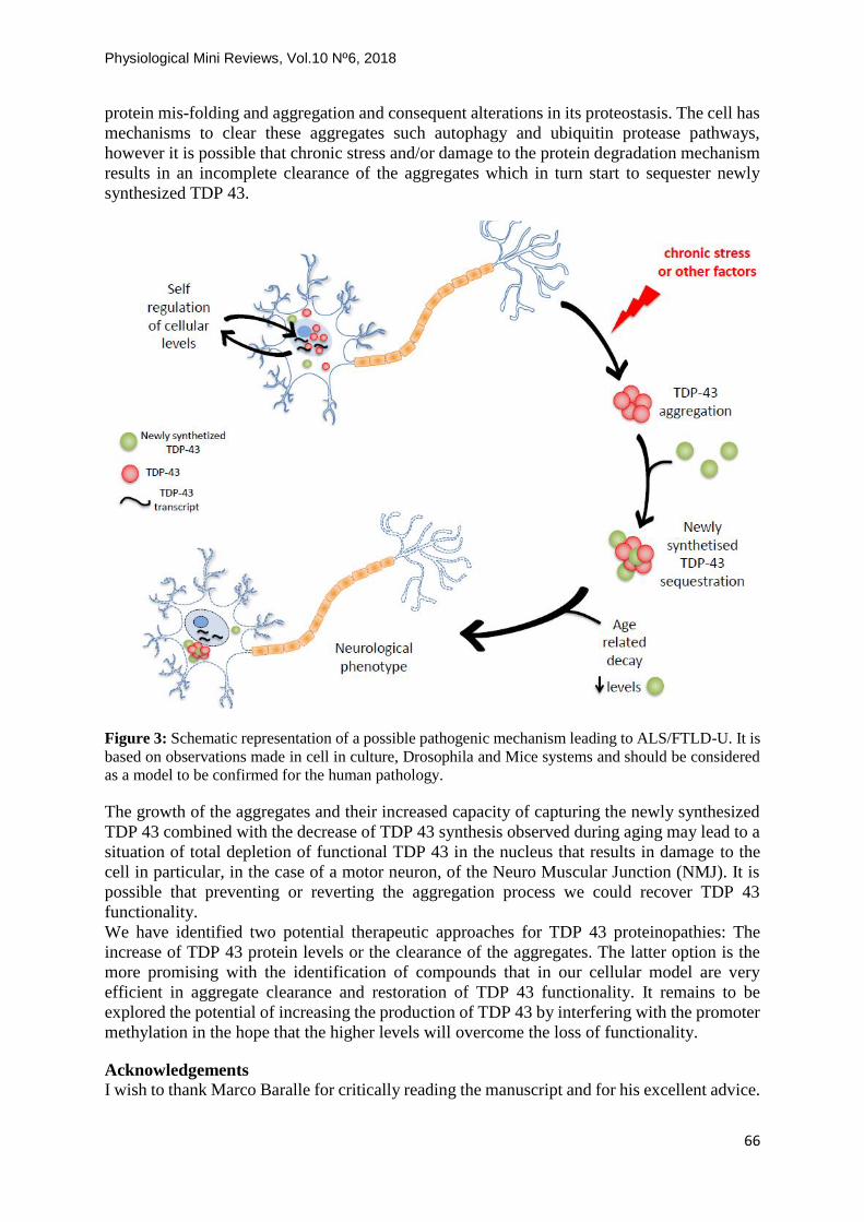
protein mis-folding and aggregation and consequent alterations in its proteostasis. The cell has
mechanisms to clear these aggregates such autophagy and ubiquitin protease pathways,
however it is possible that chronic stress and/or damage to the protein degradation mechanism
results in an incomplete clearance of the aggregates which in turn start to sequester newly
synthesized TDP 43.
Figure 3: Schematic representation of a possible pathogenic mechanism leading to ALS/FTLD-U. It is
based on observations made in cell in culture, Drosophila and Mice systems and should be considered
as a model to be confirmed for the human pathology.
The growth of the aggregates and their increased capacity of capturing the newly synthesized
TDP 43 combined with the decrease of TDP 43 synthesis observed during aging may lead to a
situation of total depletion of functional TDP 43 in the nucleus that results in damage to the
cell in particular, in the case of a motor neuron, of the Neuro Muscular Junction (NMJ). It is
possible that preventing or reverting the aggregation process we could recover TDP 43
functionality.
We have identified two potential therapeutic approaches for TDP 43 proteinopathies: The
increase of TDP 43 protein levels or the clearance of the aggregates. The latter option is the
more promising with the identification of compounds that in our cellular model are very
efficient in aggregate clearance and restoration of TDP 43 functionality. It remains to be
explored the potential of increasing the production of TDP 43 by interfering with the promoter
methylation in the hope that the higher levels will overcome the loss of functionality.
Acknowledgements
I wish to thank Marco Baralle for critically reading the manuscript and for his excellent advice.
Physiological Mini Reviews, Vol.10 Nº6, 2018
67
References
[1] Chow LT, Gelinas RE, Broker TR and Roberts RJ. (1977) An amazing sequence
arrangement at the 5' ends of adenovirus 2 messenger RNA. Cell, 12, 1-8.
[2] Berget SM, Moore C and Sharp PA. (1977) Spliced segments at the 5' terminus of
adenovirus 2 late mRNA. Proceedings of the National Academy of Sciences of the
United States of America, 74, 3171-3175.
[3] Jeffreys AJ and Flavell RA. (1977) The rabbit beta-globin gene contains a large large
insert in the coding sequence. Cell, 12, 1097-1108.
[4] Gilbert W. (1978) Why genes in pieces? Nature, 271, 501.
[5] Breathnach R, Benoist C, O'Hare K, Gannon F and Chambon P. (1978) Ovalbumin
gene: evidence for a leader sequence in mRNA and DNA sequences at the exon-intron
boundaries. Proceedings of the National Academy of Sciences of the United States of
America, 75, 4853-4857.
[6] Shapiro MB and Senapathy P. (1987) RNA splice junctions of different classes of
eukaryotes: sequence statistics and functional implications in gene expression. Nucleic
acids research, 15, 7155-7174.
[7] Kornblihtt AR, Vibe-Pedersen K and Baralle FE. (1984) Human fibronectin:
molecular cloning evidence for two mRNA species differing by an internal segment
coding for a structural domain. The EMBO journal, 3, 221-226.
[8] Reed R and Maniatis T. (1986) A role for exon sequences and splice-site proximity
in splice-site selection. Cell, 46, 681-690.
[9] Mardon HJ, Sebastio G and Baralle FE. (1987) A role for exon sequences in alternative
splicing of the human fibronectin gene. Nucleic acids research, 15, 7725-7733.
[10] Buratti E and Baralle FE. (2004) Influence of RNA secondary structure on the pre-
mRNA splicing process. Molecular and cellular biology, 24, 10505-10514.
[11] Kornblihtt AR. (2007) Coupling transcription and alternative splicing. Advances in
experimental medicine and biology, 623, 175-189.
[12] Luco RF, Allo M, Schor IE, Kornblihtt AR and Misteli T. (2011) Epigenetics in
alternative pre-mRNA splicing. Cell, 144, 16-26.
[13] Kim MS, Pinto SM, Getnet D, Nirujogi RS, Manda SS, Chaerkady R, Madugundu
AK, Kelkar DS, Isserlin R, Jain S et al. (2014) A draft map of the human proteome.
Nature, 509, 575-581.
[14] Baralle FE and Giudice J. (2017) Alternative splicing as a regulator of development
and tissue identity. Nature reviews. Molecular cell biology, 18, 437-451.
[15] Buratti E and Baralle FE (2001) Characterization and functional implications of the
RNA binding properties of nuclear factor TDP-43, a novel splicing regulator of CFTR
exon 9. The Journal of biological chemistry, 276, 36337-36343.
[16] Buratti E, Dork T, Zuccato E, Pagan, F, Romano M and Baralle FE. (2001) Nuclear
factor TDP-43 and SR proteins promote in vitro and in vivo CFTR exon 9 skipping.
The EMBO journal, 20, 1774-1784.
[17] Neumann M, Sampathu DM, Kwong LK, Truax AC, Micsenyi MC, Chou TT,
Bruce J, Schuck T, Grossman M, Clark CM et al. (2006) Ubiquitinated TDP-43 in

Physiological Mini Reviews, Vol.10 Nº6, 2018
68
frontotemporal lobar degeneration and amyotrophic lateral sclerosis. Science, 314, 130-
133.
[18] Buratti E and Baralle FE (2012) TDP-43: gumming up neurons through protein-
protein and protein-RNA interactions. Trends in biochemical sciences, 37, 237-247.
[19] Feiguin F, Godena VK, Romano G, D'Ambrogio A, Klima R and Baralle FE (2009)
Depletion of TDP-43 affects Drosophila motoneurons terminal synapsis and
locomotive behavior. FEBS letters, 583, 1586-1592.
[20] D'Ambrogio A, Buratti E, Stuani C, Guarnaccia C, Romano M, Ayala YM and
Baralle FE. (2009) Functional mapping of the interaction between TDP-43 and hnRNP
A2 in vivo. Nucleic acids research, 37, 4116-4126.
[21] Igaz LM, Kwong LK, Chen-Plotkin A, Winton MJ, Unger TL, Xu Y, Neumann
M, Trojanowski JQ and Lee VM. (2009) Expression of TDP-43 C-terminal
Fragments in Vitro Recapitulates Pathological Features of TDP-43 Proteinopathies. The
Journal of biological chemistry, 284, 8516-8524.
[22] Fuentealba RA, Udan M, Bell S, Wegorzewska I, Shao J, Diamond MI, Weihl CC
and Baloh RH (2010) Interaction with polyglutamine aggregates reveals a Q/N-rich
domain in TDP-43. The Journal of biological chemistry, 285, 26304-26314.
[23] Budini M, Romano V, Avendano-Vazquez SE, Bembich S, Buratti E and Baralle
FE. (2012) Role of selected mutations in the Q/N rich region of TDP-43 in EGFP-
12xQ/N-induced aggregate formation. Brain research, 1462, 139-150.
[24] Zhang YJ, Xu YF, Cook C, Gendron TF, Roettges P, Link CD, Lin WL, Tong J,
Castanedes-Casey M, Ash P et al. (2009) Aberrant cleavage of TDP-43 enhances
aggregation and cellular toxicity. Proceedings of the National Academy of Sciences of
the United States of America, 106, 7607-7612.
[25] Budini M, Buratti E, Stuani C, Guarnaccia C, Romano V, De Conti L and Baralle
FE. (2012) Cellular model of TAR DNA-binding protein 43 (TDP-43) aggregation
based on its C-terminal Gln/Asn-rich region. The Journal of biological chemistry, 287,
7512-7525.
[26] Budini M, Romano V, Quadri Z, Buratti E and Baralle FE. (2015) TDP-43 loss of
cellular function through aggregation requires additional structural determinants
beyond its C-terminal Q/N prion-like domain. Human molecular genetics, 24, 9-20.
[27] Cragnaz L, Klima R, De Conti L, Romano G, Feiguin F, Buratti E, Baralle M and
Baralle FE. (2015) An age-related reduction of brain TBPH/TDP-43 levels precedes
the onset of locomotion defects in a Drosophila ALS model. Neuroscience, 311, 415-
421.
[28] Avendano-Vazquez SE, Dhir A, Bembich S, Buratti E, Proudfoot N and Baralle
FE. (2012) Autoregulation of TDP-43 mRNA levels involves interplay between
transcription, splicing, and alternative polyA site selection. Genes & development, 26,
1679-1684.

Physiological Mini Reviews, Vol.10 Nº6, 2018
69
About author
Prof. Francisco E. Baralle es Dr. en Química (UBA) y Dr. en
Medicina (UBA/Nápoles). Hizo su trabajo post-doctoral en el MRC
Laboratory of Molecular Biology Cambridge UK (1974-1980) y de
1980 a 1990 fue Profesor de Patología en la Universidad de Oxford
(UK) y miembro del Magdalen College. De 1990 al 2014 fue Director
del International Centre for Genetic Engineering and Biotechnology
(ICGEB). Del 2014 al presente es PI del grupo de RNA Biology en
la misma institución.
Sus principales contribuciones a la Biología Molecular incluyen la
primer secuencia completa de un mRNA eucariote (beta- globina) y
el aislamiento y caracterización del gen humano de la globina embriónica epsilon. Al inicio de
la decada del 80 describió por la primera vez el procesamiento alternativo de un pre mRNA
celular (fibronectina) y reprodujo este fenómeno en sistemas celulares con el uso de mini genes.
Desde entonces su principal interés son los mecanismos moleculares del procesamiento del
pre-mRNA en el metabolismo normal y patológico. Ha estudiado extensivamente los defectos
moleculares a la base de enfermedades hereditarias causadas por mutaciones en un solo gen,
como talasemia, fibrosis quística, neurofibromatosis y en enfermedades poli factoriales como
dyslipoproteinemias, hipertensión y neurodegenerativas. En este último campo se focaliza su
investigación corriente en particular sobre la proteína TDP 43 caracterizada en su laboratorio
y luego identificada como el componente de las inclusiones neuronales típicas de los pacientes
con Sclerosis Lateral Amiotrofica y algunas formas de demencia incluso una fracción de
pacientes con Alzheimer. Recientemente ha coordinado el proyecto mis splicing and disease
dentro del European RNA Alternative Splicing Network of Excellence (EURASNET).
En su laboratorio del ICGEB además de llevar a cabo investigación básica en los temas citados
anteriormente, se llevan adelante proyectos aplicados a la producción de proteínas
recombinantes para uso terapéutico que se transfieren a instituciones y/o empresas de los países
miembros del ICGEB. Productos como Erythropoietina, Interferones, insulina producidos con
la tecnología ICGEB están ya en el mercado en varios países particularmente en países de
Latinoamérica y Asia.
En 1980 dicto en Argentina el primer curso de manipulación genética, que fue replicado
bianualmente hasta 1988. Como director del ICGEB desde 1990 ha tenido numerosas
colaboraciones extremadamente fructíferas con grupos científicos e industriales
Latinoamericanos.