Embed Size (px)
Citation preview
University of New EnglandDUNE: DigitalUNE
Case Report Papers Physical Therapy Student Papers
12-2016
Physical Therapy Intervention For A Patient WithTemporomandibular Joint Dysfunction Caused ByTwo Traumatic Events: A Case StudyElyse DetweilerUniversity of New England
Follow this and additional works at: http://dune.une.edu/pt_studcrpaper
Part of the Physical Therapy Commons
© 2016 Elyse Detweiler
This Course Paper is brought to you for free and open access by the Physical Therapy Student Papers at DUNE: DigitalUNE. It has been accepted forinclusion in Case Report Papers by an authorized administrator of DUNE: DigitalUNE. For more information, please contact [email protected].
Recommended CitationDetweiler, Elyse, "Physical Therapy Intervention For A Patient With Temporomandibular Joint Dysfunction Caused By TwoTraumatic Events: A Case Study" (2016). Case Report Papers. 66.http://dune.une.edu/pt_studcrpaper/66

Detweiler, Traumatic Onset TMD
1
1
2
3
4
Physical Therapy Intervention for a Patient with Temporomandibular Joint 5
Dysfunction caused by Two Traumatic Events: A Case Study 6
7
Elyse Detweiler, BS, is a Doctor of Physical Therapy student at the University of New England, 716 8
Stevens Ave, ME, 04103. 9
Address all correspondence to Elyse Detweiler: [email protected]. 10
The patient signed an informed consent allowing the use of medical information, video footage, and/ 11
photography for this report and received information on the institution’s policies regarding the Health 12
Insurance Portability and Accountability Act. 13
The author acknowledges Amy Litterini, PT, DPT, for assistance with case report conceptualization, 14
Gabe Redmond, PT, MS for supervision and assistance with video photography, and the patient for her 15
participation as the case study participant. 16
17

Detweiler, Traumatic Onset TMD
2
ABSTRACT 18
19 Background and Purpose: 20
Temporomandibular disorders (TMD) are pathoanatomical dysfunctions of the temporomandibular joint 21
(TMJ) associated with symptoms throughout the head and neck. Limited information exists regarding 22
conservative physical therapy (PT) and post-surgical management of TMD. The dental profession is the 23
main source of published literature specific to TMD. This paper describes a conservative and post-24
surgical PT plan of care (POC) for TMD. 25
Case Description: 26
A 32-year old female experienced two separate traumatic events at work resulting in TMD. She was 27
referred to PT after the second assault because of symptoms of severe pain, limited range of motion, and 28
jaw locking. She was unable to speak, eat, or return to work. The POC included manual therapy, 29
therapeutic exercise, and patient education. She attended 16 total visits and she underwent two 30
arthrocentesis procedures performed by an oral surgeon. 31
Outcomes: 32
The patient responded well to PT both pre- and post-arthrocentesis procedures in regards to ROM 33
(Depression: 17 to 31 mm, L Lateral Excursion: 4 to 8 mm, R Lateral Excursion: 4 to 9.5 mm), numeric 34
pain rating scale (7/10 to 1/10), and a reduction in locking symptoms. She met all her goals, which 35
correlated with the decreasing Mandibular Functional Impairment Questionnaire results, and met most of 36
the PT goals by discharge. She returned to a normal diet and full time work with minimal restrictions. 37
Discussion: 38
The patient had a positive outcome from her POC including conservative and surgical management of 39
TMD. More research is needed to identify consistent indicators for individuals who would benefit from an 40
interdisciplinary approach, and investigate the potential benefits of PT for TMD. 41
42
43
44

Detweiler, Traumatic Onset TMD
3
BACKGROUND and PURPOSE 45
Temporomandibular disorders (TMD) are a collection of pathoanatomical dysfunctions 46
of the temporomandibular joint (TMJ) associated with a variety of symptoms throughout the 47
head and neck, including jaw and cervical pain, headaches, postural changes, and various other 48
impairments.1-2 TMD is usually accompanied by postural abnormalities of the cervical spine; 49
research has highlighted the importance of evaluating the TMJ and cervical spine together.2 50
Moreover, a complete physical therapy (PT) initial evaluation includes a postural analysis.2 51
Scientific literature on TMD provides valuable information on the pathological condition, 52
signs and symptoms, and background information, but there is a severe lack of supportive 53
evidence for interventions currently used in the conservative and/or surgical treatment of TMD.1 54
The literature reviews by Shaffer et al1 and Dickerson et al3 highlighted the majority of 55
interventions used within physical therapy treatment of TMD where each focuses on the 56
available supportive evidence for each intervention. Furthermore, each highlighted the variation 57
in dosage regimes between studies, the wide variety of exercises utilized, and the inconsistent 58
results supporting or negating the use of one type of intervention over another.1,3 59
The dental profession has provided much of the current literature on TMD, and this, 60
again, is limited in both conservative and surgical interventions that improve symptoms.1 Current 61
dental literature pertaining specifically to the arthrocentesis procedure examined differs greatly 62
in outcomes. The randomized control trial by Vos et al4 showed arthrocentesis to be a beneficial 63
procedure to perform initially, but the long-term outcomes for pain and functional impairments 64
were comparable to conservative treatment. Conversely, the literature review by Monje-Gil et 65
al,5 showed the wide variation in variables studied and highlighted the importance for more 66
research to determine the homogenous indicators for an arthrocentesis procedure. 67
Dentists are among the most common health professionals who evaluate and treat TMD, 68

Detweiler, Traumatic Onset TMD
4
but TMJ mobility assessments, range of motion (ROM), muscle testing, and postural assessments 69
are most commonly performed by a physical therapists.2 Collaborative care of TMD between 70
dentists, oral surgeons, and physical therapists does not always occur, but should be considered 71
best practice. 72
The rationale for this paper is to describe a physical therapy plan of care for TMD. The 73
purpose is to provide information regarding conservative and post-surgical physical therapy 74
treatment of TMD due to a traumatic mechanism of injury. 75
76
CASE DESCRIPTION 77
The patient signed an informed consent allowing the use of medical information, video 78
footage, and/or photography for this case report. 79
KD was a 32-year-old female who worked as an education technician with adolescents 80
with mental and behavioral problems. She initially sustained multiple blows to the head and face 81
from one of her students at work; then approximately six months later, a different student 82
became violent and exacerbated the original injuries to her left mandible and head with a second 83
physical assault. 84
After the first incident, she received initial medical care from a physician through the 85
worker’s compensation contract with her employer and received a diagnosis of TMD. She also 86
received care through her PCP and her dentist. No imaging was performed after the first event 87
and she had a custom TMJ splint made by her dentist. After the second incident, the splint no 88
longer fit, and KD was advised to discontinue use. She was referred to PT after the second 89
assault with the chief complaint of pain of the TMJ, locking, and the inability to open her mouth 90
to speak or eat. The pain in her face and neck was reported as sharp during movement and achy 91
during rest. The physical therapist observed the jaw deviate to the left during mandibular 92

Detweiler, Traumatic Onset TMD
5
depression. KD was on no medications, except for the occasional acetaminophen when needed 93
for pain. She had no other comorbidities and an extremely supportive family. Self-care 94
techniques used at home included ice packs, hot packs, and rest from speaking or eating. She was 95
on a liquid diet for the four weeks, followed by soft foods only. She described herself as 96
frustrated with the loss of function of her jaw and the pain. Refer to Figure 1 for timeline of 97
events. 98
Locking of the jaw was described during the initial PT evaluation and episodes increased 99
during the first three weeks of PT treatment. The PCP ordered a magnetic resonance imaging 100
(MRI) study to help determine the cause of locking. The MRI showed an anterior dislocation of 101
the left TMJ disc and the left mandibular head did not move simultaneously with the right during 102
depression or elevation. KD was then referred to an oral surgeon, who performed an 103
arthrocentesis on the left TMJ and then the right TMJ two weeks later. Refer to Figure 1 for 104
timeline. 105
KD’s main goal was to improve ROM of the bilateral TMJ, in order to resume a normal 106
diet, communication, and return to work. Refer to Table 3 for goals. 107
108
Clinical Impression 1 109
Following the subjective history and systems review, KD’s problem was identified as 110
bilateral TMD, left > right (see Table 1 for systems review results). Further tests and measures to 111
confirm this hypothesis included: goniometry, palpation of TMJ mechanics and facial 112
musculature, and strength measurements of the jaw. Moreover, postural assessment, sensory 113
testing of the face, palpation of cervical spine, neck, and shoulders, and joint assessments of the 114
cervical spine were to be assessed. Differential diagnoses included dislocation of the TMJ disc 115
and/or fracture of the jaw; therefore, imaging was requested by the PCP after locking episodes 116

Detweiler, Traumatic Onset TMD
6
increased. 117
KD was a good candidate for a case report due to multiple traumatic injuries to the face. 118
After the second assault, pain and tightness increased severely. She was unable to open her 119
mouth or speak because of pain. Current literature typically describes episodes of gradual onset 120
of TMD.1-4 This case report examined how multiple traumatic events resulted in TMD. 121
122
Examination – Tests and Measures 123
Pain was assessed throughout the course of treatment using the Numeric Pain Rating 124
Scale (NPRS).6 Reliability and validity of the NPRS is not established for facial pain resulting 125
from TMD, but it is a helpful tool to determine subjective information about pain and has been 126
tested for validity and reliability for acute and chronic musculoskeletal pain.6 Jaw motions were 127
assessed using goniometry (Dynasplint Systems, Inc., MD), specifically in millimeters as described 128
by Norkin and White.9 Goniometry has been shown to be a reliable and valid form of 129
measurement for TMJ motions according to research by Walker et al10 that showed mandibular 130
depression is valid in discriminating between someone with or without TMD.10 Observation of 131
KD’s speech was used to assess whether the mandible deviated and it was observed that KD’s 132
mandible deviated to the left, but rested in a neutral position.1 133
Palpation revealed pain and clicking bilaterally. Excursion of the condyles was not equal; 134
the left did not move smoothly and lagged behind the right. Left TMJ clicking was less 135
pronounced but pain was reported to be more significant. Palpation assessed KD’s joint mobility, 136
which displayed hypomobility of bilateral TMJ. According to Shaffer et al,1 palpation is helpful 137
in providing information on symptom provocation, hypersensitivity of retrodiscal tissues, 138
abnormalities of mandibular head motions, popping, clicking, localized tenderness, and changes 139
in facial and cervical musculature. 140

Detweiler, Traumatic Onset TMD
7
The Mandibular Function Impairment Questionnaire (MFIQ) was used to calculate the 141
perceived difficulty of tasks in comparison to jaw complaints.9 The MFIQ score portrayed severe 142
difficulty with everyday tasks in relation to jaw complaint. The MFIQ outcome measure has 143
been tested and shown to be reliable by Kropmans et al9 and others for assessing impairments in 144
mandibular function.11,12 Inspection of dentition showed no impairments. Cervical ROM was 145
assessed as described by Norkin and White10 and was within normal limits, but tightness and 146
localized tenderness was found within the cervical and shoulder musculature. Strength of 147
cervical and upper extremity musculature was assessed as described by Kendall12 and found to be 148
normal bilaterally. Refer to Table 2 for initial evaluation results. 149
150
Clinical Impression 2 151
Pain, locking, and muscular tightness of the facial musculature were all consistent with 152
TMD and confirmed the initial impression. The MRI findings of anterior dislocation of the left 153
disc, and movement abnormalities of the left mandibular head during depression and elevation, 154
were also consistent with symptoms of locking and the pain described by KD. She remained 155
appropriate for this case report due to the unique mechanism of injury. 156
Due to documented impairments ICD 10 code: M26.96 (other specified disorders of 157
TMJ) was determined to be the most appropriate PT diagnosis. 158
The patient had no other comorbidities and an extremely supportive family. She was 159
highly motivated to improve her symptoms and return to work. All these factors were considered 160
positive prognostic indicators signifying a desirable outcome. There was no plan for referral to 161
other health professionals. The plan for interventions was for the patient to be seen twice weekly 162
for six weeks, focusing on manual soft tissue mobilization and therapeutic exercises. 163

Detweiler, Traumatic Onset TMD
8
Collaborative communication was performed with all medical personnel already working 164
with KD. The MFIQ functional outcome measure was used every eighth visit to determine 165
whether the patient had any subjective change in symptoms, function of the jaw, and reauthorize 166
additional visits.8 Testing of mandibular depression, and left and right mandibular excursions 167
were measured every other session to determine whether there were improvements. Pain rating 168
was assessed each session by the NPRS. Palpation was performed each session during soft tissue 169
mobilization to determine whether musculature tightness had changed. 170
KD would discharged upon achievement of established PT short-term goals for ROM and 171
pain. For short-term and long-term goals, see Table 3. 172
Intervention 173
Collaborative communication occurred regularly and documentation was provided to all 174
medical personnel working with KD. Her POC was coordinated by a team, including her case 175
manager, PCP, oral surgeon and physical therapist. Patient/client instruction included a home 176
exercise program (HEP) in the form of pictures and written instructions. 177
Procedural interventions initially consisted of manual therapy in the form of soft tissue 178
mobilization and manual cervical traction to improve circulation, elongate tissues, increase range 179
of motion, and decrease pain in mandibular and cervical musculature. According to Shaffer et al1 180
soft tissue mobilization is a commonly used intervention and important in the management of 181
TMD, even with limited support in the literature.1 Effleurage, petrissage, myofascial trigger point 182
therapy, and cross friction massage were performed on jaw, cervical, and shoulder musculature. 183
KD was taught to perform self-massage techniques at home for symptom management. Refer to 184
Appendices 2 and 3 for intervention protocols, descriptions, and images. As her pain and 185
muscular tightness decreased around session thirteen, the amount of soft tissue mobilization and 186

Detweiler, Traumatic Onset TMD
9
manual traction provided by the physical therapist was decreased around session thirteen, 187
ultimately changing the focus of the treatment session to other impairments. 188
A dry needling intervention was performed (Myotech US Dry Needling & Physio 189
Products, Kirkland,WA) to attempt to release the masseter muscles bilaterally. This was 190
performed on the fourth intervention day due to limited success of soft tissue mobilization on the 191
left masseter. KD was provided with written and verbal notification of the benefits and 192
contraindication of the dry needling, in addition to expectations of the treatment. The dry 193
needling intervention was performed by another physical therapist, who is certified in dry 194
needling technique, level 2. Shaffer et al1 supports the use of dry needling when pain can be 195
attributed to musculature, and in this case some of the patient’s pain was due to severely tight 196
bilateral masseters.11 The dry needling was only performed during the fourth intervention session 197
because the patient did not feel she could tolerate another session due to a fear of needles. 198
Because of this, soft tissue mobilization was the focus of therapy until her pain and stiffness 199
improved around the ninth intervention day. 200
Mandibular ROM stretches were performed to elongate and improve circulation to the 201
masseter, medial and lateral pterygoids, and cervical musculature each treatment session as 202
tolerated. According to Shaffer et al1 the use of gentle stretching is useful in reducing pain and 203
Lateral excursion was not tolerated on the fourth, seventh, and eighth intervention days, 204
specifically when moving to the right. 205
Postural exercises such as external rotation pull-outs with yellow or red resistance band 206
and chin tucks were performed to increase postural awareness and circulation to postural 207
muscles. Refer to Appendix 2 for sets and repetitions. These exercises were helpful to reduce the 208
head forward and rounded shoulders posture described in the initial evaluation and by Friedman 209
in patients with postural deviations.2 Cervical and upper extremity exercises were incorporated to 210

Detweiler, Traumatic Onset TMD
10
improve circulation and strength of upper extremity and postural muscles. A Paramount 211
Functional Trainer (Paramount Fitness Corp., St. Louis, MO) was utilized to perform resistance 212
training including low rows, bilateral pull downs, and triceps presses, each with a resistance 213
equal to 10 pounds initially. Progression of resistance exercises occurred by initially increasing 214
the number of repetitions, but progressed with increased weight when patient no longer found 215
them challenging. For ROM exercises, she performed a side bend stretch, upper trapezius 216
stretch, and a towel/foam roller stretch to decrease pain and stiffness of the cervical and shoulder 217
muscles for three repetitions holding for 30 seconds each time. Refer to Appendices 2 and 3 for 218
protocol, description, and images. Shaffer et al1 supports the use of interventions specific to the 219
cervical spine because failing to address impairments of the cervical spine may limit a patient’s 220
rehabilitation potential with TMD. 221
A home exercise program (HEP) incorporating ROM and stretching of the facial and 222
cervical musculature was given. The HEP focused on mandibular depression and lateral 223
excursion ROM exercises and self-massage techniques because decreasing pain and increasing 224
ROM were the focus of KD’s goals. Self-massage techniques were given to improve symptoms 225
of stiffness, fatigue, and to give the patient the ability to proactively manage her pain at home. 226
According to Shaffer et al1 a multimodal approach is the most beneficial for patients with 227
TMD. Incorporating soft tissue mobilization, gentle isometrics, guided ROM exercises, postural 228
corrections, and relaxation techniques is an effective strategy in reducing symptoms associated 229
with anterior disc displacement and myofascial pain dysfunction of the TMJ.1 230
KD attended two sessions each week for 8 weeks total. She reported she performed her 231
HEP at least twice per day to help with symptoms. Re-evaluation was performed during the 232
eighth and 16th visits according to the facility guidelines and for reauthorization for additional 233
visits. 234

Detweiler, Traumatic Onset TMD
11
OUTCOME 235
To evaluate significant changes throughout the physical therapy treatment, the same 236
equipment, such as the goniometer, was used at each round of testing and the MFIQ was 237
completed. At the eighth visit re-evaluation and the 16th visit when she was discharged, she 238
showed significant improvements in pain, ROM in all directions, and tolerance of exercises. She 239
met all her short-term goals, in addition to achieving the long-term goal for pain at the eighth 240
visit re-evaluation. At discharge, KD had met all her major goals. Refer to Figure 2 for specifics 241
on when KD met each goal. Her MFIQ score changed from a 0.70 to a 0.40 at the re-evaluation, 242
to a 0.06 at discharge, showing significant changes throughout her course of physical therapy. 243
She could consume a normal diet, with the exception of certain sized foods that tended to over-244
stress the jaw. Lastly, she had returned to work full-time with some restrictions, such as not 245
being responsible for restraining students when they became violent. Refer to Appendix 1 for test 246
and measures comparing initial evaluation, 8th visit re-evaluation, and 16th visit discharge 247
findings. 248
DISCUSSION 249
There is limited research to support the benefits of the current PT interventions for TMD 250
and none specific to traumatic jaw injury. Therefore, it was difficult to develop a PT plan of care 251
based on the current literature. The literature did, however, provide valuable information on the 252
pathological condition, signs and symptoms, and background information that improved the 253
physical therapist’s understanding of TMD. The available literature also assisted in providing a 254
basis for hypotheses for the patient’s underlying impairments, resulting in the focus of soft tissue 255
mobilization and a stretching program. 256
The patient made good progress throughout her POC with the most dramatic changes 257
after each of the arthrocentesis procedures. This progress allowed her to return to her normal diet 258

Detweiler, Traumatic Onset TMD
12
and work as an education technician. Without the pain, she was once again able to communicate 259
with her family and friends. Most importantly, KD was pleased with her progress and happy 260
about her ability to return to the many things she enjoyed. Positive factors that contributed to 261
KD’s outcome included the collaborative care provided by her PCP, oral surgeon, and physical 262
therapists. 263
More research is needed in conservative TMD treatments, specifically the efficacy for 264
soft tissue mobilization techniques in reducing tightness in the masseter and cervical 265
musculature. This would be helpful in determining whether these interventions could be a 266
primary focus of treatment for reducing pain and improving range of motion. Moreover, 267
additional research evaluating the efficacy of combined conservative and post-surgical 268
treatments, specific to the arthrocentesis procedure combined with physical therapy would enable 269
healthcare professionals to more successfully treat patients with TMD. 270
271 REFERENCES 272
273 1. Shaffer SM, Brismee JM, Sizer PS, Courtney CA. Temporomandibular disorders. Part 2: 274
conservative management. J Man Manip Ther. 2014;22(1):13-23. Doi: 275
10.1179/2042618613Y.0000000061 276
2. Friedman MH, Weisberg J. Application of orthopedic principles in evaluation of the 277
temporomandibular joint. Phys Ther. 1982; 62: 597-603. 278
3. Dickerson S, Weaver, J, Boyson A, Thacker J, Junak, A et al. The effectiveness of 279
exercise therapy for temporomandibular dysfunction: A systematic review of meta-280
analysis. Clin Rehabil. 2016; dio: 10.1177/0269215516672275. 281
4. Vos LM, Huddleston Slatter JJ, & Stengena B. Arthrocentesis as initial treatment for 282
temporomandibular joint arthropathy: a randomized controlled trial. J Craniomaxillofac 283
Detweiler, Traumatic Onset TMD
13
Surg. 2014; 42(5): 134-9. Doi: 10.1016/j.jcms.2013.07.0101. 284
5. Monje-Gil F, Nitzan D, Gonzalez-Garcia R. Temporomanidbular joint arthrocentesis. 285
Review of the literature. Med Oral Cir Bucal. 2012; 7(4): 575-81. Dio: 286
10.4317/medoral.17670. 287
6. Santiesteban AJ. Isometric exercises and a simple appliance for temporomandibular joint 288
dysfunction: a case report. Phys Ther. 1989;69:463-466. 289
7. Braun B, Treatment of an acute anterior disk displacement in the temporomandibular 290
joint: a case report. Phys Ther. 1987;67:1234-1236. 291
8. Rehab Measures: Numeric Pain Rating Scale. Rehab Measures Web Site. 292
http://www.rehabmeasures.org/Lists/RehabMeasures/PrintView.aspx?ID=891. Accessed 293
June 12, 2016. 294
9. Norkin CC & White DJ. Measurements of Joint Measurement: A Guide to Goniometry. 295
4th ed. Philadelphia, PA: F.A. Davis Company; 2009. 296
10. Walker N, Bohannon RW, and Cameron D. Discriminant validity of temporomandibular 297
joint range of motion measurements obtained with a ruler. J Orthop Sports Phys Ther. 298
2000; 30(8): 484-92. Doi: 10.2519/jospt.2000.30.8.484. 299
11. Kropmans TJ, Kijkstra PU, van Veen A, and de Bont LG. The smallest detectable 300
difference of mandibular function impairments in patients with a painfully restricted 301
temporomandibular joint. J Dent Res. 1999;78(8):1445-9. Doi: 302
10.1177/00220345990780081001. 303
12. Kendall FP, McCreary EK, Provance PG, Rodgers MM, Romani WA. Muscles Testing 304
and Function with Posture and Pain. 5th ed. Baltimore, MD: Lippincott Williams & 305
Wilcons; 2001. 306
13. Fernandez-Carnero J, La Touche R, Ortega-Santiago R, et al. Short-term effects of dry 307

Detweiler, Traumatic Onset TMD
14
needling of active myofascial trigger points in the masseter muscle in patients with 308
temporomandibular disorders. J Orofac Pain. 2010;24(1):106-12. 309
14. TMJ Exercise Images. Oral Facial Section. 310
http://www.hep2go.com/exercises.php?ex_type=25&ex_subtype=147&userRef=gciaake311
&order=default&group=all&position=-1. Accessed September 2016. 312
313
Detweiler, Traumatic Onset TMD
15
TABLES and FIGURES 314
315 316 317 318 319 320
321 322 Table 1
Systems Review
Cardiovascular/Pulmonary o Blood pressure: 138/86
o Heart rate: 84
o Respiration rate: 14
Unimpaired
Musculoskeletal o Severe tightness of the jaw, face, and cervical
muscles:
o Masseter
o Trapezius
o Semispinalis capitus
o Rectus capitus posterior major and minor
o Obliquus capitus superior and inferior
o Temporalis
o Left scalenes
o Left sternocleidomastoid
Impaired

Detweiler, Traumatic Onset TMD
16
o The head of the mandible on the left was not
moving simultaneously with the right, resulting in
locking, popping, and pain.
Neuromuscular o Numbness and tingling was described for the left
maxilla and mandible during the initial evaluation.
o Crude touch highlighted sensation differences on
the mandible.
Impaired
Integumentary o No signs of bruising or abrasions
o No redness or signs of infection
o No swelling
Unimpaired
Communication o Communication was impaired due to the locking,
popping, pain, and fatigue.
o The TMJ mechanics were impaired resulting in
communication limitations because speaking was
extremely uncomfortable and painful.
o She was able to communicate verbally for short
periods of time, gestures, and in written form.
Impaired
Affect, Cognition,
Language, Learning Style
o Affect, cognition, language, and learning style were
unimpaired.
o Demonstrations, pictures, and verbal instructions
were preferred.
Unimpaired
323 Table 2
Tests & Measures Initial Evaluation Results
Goniometric
Measurements
Depression: 17 mm with an increase in pain
Right Lateral Excursion: 4 mm with an increase in pain
Left Lateral Excursion: 4 mm with an increase in pain
Cervical: Within normal limits
Strength
Facial musculature not tested at initial evaluation because of pain.
Cervical and upper extremity strength normal (5/5).
Palpation of Joint
Mechanics
Positive bilaterally for clicking/popping; it was felt and heard.
Right and left TMJ did not move simultaneously; the left TMJ moved after
the right TMJ
Sensation
Crude touch:
Numbness and paraesthesia reported on left mandible; resolved within two
weeks of initial evaluation
Soft Tissue Integrity Tightness: masseter, temporalis, scalenes, SCM, trapezius, levator scapula,
rectus capitus major and minor, obliques capitus major and minor, splenius
capitus, longissimus capitis
Joint mobility assessment Right TMJ: 2/6 (hypomobile)
Left TMJ: 2/6 (hypomobile)
Restricted bilaterally with L>R
Detweiler, Traumatic Onset TMD
17
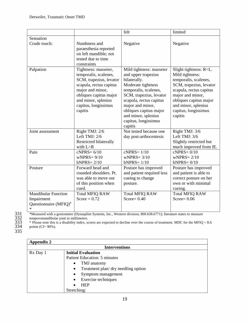
Pain cNPRS= 6/10
wNPRS= 9/10
bNPRS= 2/10
Observation in sitting and
standing
Forward head and rounded shoulders. Pt. was able to move out of this
position when cued.
Mandibular Function
Impairment Questionnaire
(MFIQ)
Total MFIQ RAW Score= 0.72; Q1: 1 / Q2: 2 / Q3: 4 / Q4: 4 / Q5: 1 / Q6: 1
/ Q7: 0 / Q8: 3 / Q9: 3 / Q10: 3 / Q11: 3 / Q12: 4 / Q13: 4 / Q14: 4 / Q 15: 4
/ Q16: 4 / Q17: 4
* Please note this is a disability index, scores are expected to decline over
the course of treatment.
MDC for the MFIQ = 8.6 points (CI= 90%).
Literature states to measure temporomandibular joint in millimeters; Measured in accordance to Kendall et al9; IE= initial evaluation; 324 cNPRS= current Numerical Pain Rating Scale; wNPRS= worst Numerical Pain Rating Scale; bNPRS= best Numerical Pain Rating Scale; L= left; 325 R= right; Q= question 326 327 Table 3
Goals
Short Term Goals
(4 weeks)
Goal
Achieved
Long Term Goals
(8 weeks)
Goal
Achieved
Mandibular
ROM Goals
Patient’s mandibular
depression will
improve to 25 mm to
improve ability to eat
and speak.
8th visit
Patient’s
mandibular
depression will
improve to 50 mm
week to improve
ability to eat and
speak.
Goal was not
met; 62% of
goal met at
d/c
Patient’s L lateral
excursion will
improve to 6 mm to
improve ability to eat
and speak.
8th visit
Patient’s L lateral
excursion will
improve to 10 mm
to improve ability
to eat and speak.
Goal not met;
95% of goal
met at d/c
Patient’s R lateral
excursion will
improve to 6 mm to
improve ability to eat
and speak.
8th visit
Patient’s R lateral
excursion will
improve to 10 mm
to improve ability
to eat and speak.
Goal not met;
80% of goal
met at d/c
Mandibular
Tightness
Goal
Jaw tightness will
decrease from
moderate to mild to
improve comfort and
mobility so she is able
to eat and speak
comfortably.
8th visit
Jaw tightness will
decrease from
moderate to trace to
improve comfort
and mobility so she
is able to eat and
speak comfortably.
16th visit
Mandibular
Locking Goal
Jaw locking will
decrease from mild to 8th visit
Jaw locking will
decrease from mild 16th visit

Detweiler, Traumatic Onset TMD
18
ROM= Range of Motion; IE= initial evaluation; RE= re-evaluation; D/C= Discharge; R=Right; L=Left; mm= millimeters 328 329 APPENDICES 330
Appendix 1
Tests & Measures Initial Evaluation
Results
Re-evaluation:
8th Visit
Discharge
16th Visit
Goniometry*
Depression: 17 mm
with an increase in pain
Right Lateral
Excursion: 4 mm with
an increase in pain
Left Lateral Excursion:
4 mm with an increase
in pain
Cervical: Within
normal limits
Depression: 23 mm with
an increase in pain at
end range
Right Lateral Excursion:
6 mm with an increase
in pain at end range
Left Lateral Excursion:
6 mm with an increase
in pain at end range
Cervical: Within normal
limits
Depression: 31 mm
Right Lateral Excursion:
9.5 mm soreness at end
range
Left Lateral Excursion:
8 mm
Strength Not tested at initial
evaluation because of
pain.
Depression: 4/5 with
pain
Right Lateral Excursion:
4/5
Left Lateral Excursion:
4/5 with soreness
Depression: 5/5 with
pain
Right Lateral Excursion:
5-/5 with some soreness
at end range
Left Lateral Excursion:
5/5
Retrodiscal Fad Pad
Sign
Positive bilaterally
with clicking/popping
being felt and heard.
Negative on left
Positive on right for
clicking/popping being
Negative on left
Positive on right for
clicking/popping but
minimal (<2x/week)
to improve comfort
and fear of eating.
to trace to improve
comfort and fear of
eating.
Pain Goal
Pain rating will
improve to range of 3-
5/10 during all
activities to improve
patient’s ability to
socialize,
communicate, and
perform daily tasks.
8th visit
Pain rating will
improve to range of
0-3/10 during all to
improve patient’s
ability to socialize,
communicate, and
perform daily tasks.
8th visit
Work Goal
Patient will be able to
return to part-time
employment with
some restrictions.
8th visit
Patient will be able
to return to full-
time employment
without restrictions.
Goal not met;
90% of goal
was met
(patient
returned to
full time with
some
restrictions)
at 15th visit.
Detweiler, Traumatic Onset TMD
19
felt limited
Sensation
Crude touch:
Numbness and
paraesthesia reported
on left mandible; not
tested due to time
constraints
Negative
Negative
Palpation Tightness: masseter,
temporalis, scalenes,
SCM, trapezius, levator
scapula, rectus capitus
major and minor,
obliques capitus major
and minor, splenius
capitus, longissimus
capitis
Mild tightness: masseter
and upper trapezius
bilaterally.
Moderate tightness
temporalis, scalenes,
SCM, trapezius, levator
scapula, rectus capitus
major and minor,
obliques capitus major
and minor, splenius
capitus, longissimus
capitis
Slight tightness: R<L.
Mild tightness:
temporalis, scalenes,
SCM, trapezius, levator
scapula, rectus capitus
major and minor,
obliques capitus major
and minor, splenius
capitus, longissimus
capitis
Joint assessment Right TMJ: 2/6
Left TMJ: 2/6
Restricted bilaterally
with L>R
Not tested because one
day post-arthocentesis
Right TMJ: 3/6
Left TMJ: 3/6
Slightly restricted but
much improved from IE.
Pain cNPRS= 6/10
wNPRS= 9/10
bNPRS= 2/10
cNPRS= 1/10
wNPRS= 3/10
bNPRS= 1/10
cNPRS= 0/10
wNPRS= 2/10
bNPRS= 0/10
Posture Forward head and
rounded shoulders. Pt.
was able to move out
of this position when
cued.
Posture has improved
and patient required less
cueing to change
posture.
Posture has improved
and patient is able to
correct posture on her
own or with minimal
cueing.
Mandibular Function
Impairment
Questionnaire (MFIQ)9
*
Total MFIQ RAW
Score = 0.72
Total MFIQ RAW
Score= 0.40
Total MFIQ RAW
Score= 0.06
*Measured with a goniometer (Dynasplint Systems, Inc., Western division, 800.638.6771); literature states to measure 331 temporomandibular joint in millimeters. 332 * Please note this is a disability index, scores are expected to decline over the course of treatment. MDC for the MFIQ = 8.6 333 points (CI= 90%). 334 335
Appendix 2
Interventions
Rx Day 1 Initial Evaluation
Patient Education: 5 minutes
TMJ anatomy
Treatment plan/ dry needling option
Symptom management
Exercise techniques
HEP
Stretching:
Detweiler, Traumatic Onset TMD
20
Face Jaw Depression: 3 sets x 10 repetitions
Face Jaw Lateral Excursion: 3 sets x 10 repetitions
Manual Therapy:
Cervical manual traction: 8 minutes
Rx Day 2 Warm up:
Moist heat: 8 minutes
o Supine with cervical heat positioned on each TMJ
Stretching:
Face Jaw Depression: 3 sets x 10 repetitions
Face Jaw Lateral Excursion: 3 sets x 10 repetitions
Jaw protrusion: 3 sets x 10 repetitions
Manual Therapy:
Cervical manual traction: 8 minutes
Soft tissue mobilization: 8 minutes
o Bilateral and multidirectional
o Targeted masseter, temporalis, and trapezius
Exercise Activities:
Chin tucks: 3 sets x 10 repetitions
Low rows: 3 sets x 10 repetitions
ER pullouts with yellow band: 3 sets x 10 repetitions
Rx Day 3 Warm up:
Moist heat: 8 minutes
o Supine with cervical heat positioned on each TMJ
Stretching:
Face Jaw Depression: 3 sets x 10 repetitions
Face Jaw Lateral Excursion: 3 sets x 10 repetitions
Side bend/rotation stretch: 3 sets x 30 second holds
Manual Therapy:
Cervical manual traction: 5 minutes
Soft tissue mobilization: 5 minutes
o Bilateral and multidirectional
o Targeted masseter, temporalis, and trapezius
Mobilization with movement: 5 minutes
o TMJ
Exercise Activities:
Chin tucks: 3 sets x 10 repetitions
Low rows: 3 sets x 10 repetitions
Rx Day 4 Patient Education: 5 minutes
Dry needling
Symptom management
Exercise techniques
Manual Therapy:
Cervical manual traction: 5 minutes
Soft tissue mobilization: 12 minutes
o Bilateral and multidirectional
o Targeted masseter, temporalis, and trapezius
Dry Needling Intervention: 12 minutes

Detweiler, Traumatic Onset TMD
21
o Education on intervention/ written and verbal consent
o BL masseter
Stretching:
Face Jaw Depression: 3 sets x 10 repetitions
Sidebend/rotation stretch: 3 sets x 30 second holds
Upper trapezius stretch: 3 sets x 60 second holds
Rx Day 5 Manual Therapy:
Soft tissue mobilization: 12 minutes
o Bilateral and multidirectional
o Targeted masseter, temporalis, and trapezius
Patient Education: 5 minutes
Dry needling option
Symptom management
Exercise techniques
Stretching:
Face Jaw Depression: 3 sets x 10 repetitions
Face Jaw Lateral Excursion: 3 sets x 10 repetitions
Side bend/rotation stretch: 3 sets x 30 second holds
Upper trapezius stretch: 3 sets x 60 second holds
Exercise Activities:
Low rows: 3 sets x 10 repetitions
Bilateral pull downs: 3 sets x 10 repetitions
Rx Day 6 Manual Therapy:
Cervical manual traction: 5 minutes
Soft tissue mobilization: 15 minutes
o Bilateral and multidirectional
o Targeted masseter, temporalis, and trapezius
Mobilization with movement: 5 minutes
o TMJ
Patient Education: 5 minutes
Dry needling option
Symptom management
Stretching:
Face Jaw Depression: 3 sets x 10 repetitions
Face Jaw Lateral Excursion: 3 sets x 10 repetitions
Side bend/rotation stretch: 3 sets x 30 second holds
Upper trapezius stretch: 3 sets x 60 second holds
Towel Stretch: 3 minute hold
Rx Day 7 Manual Therapy:
Cervical manual traction: 8 minutes
Soft tissue mobilization: 10 minutes
o Bilateral and multidirectional
o Targeted masseter, temporalis, and trapezius
Stretching:
Face Jaw Depression: 3 sets x 10 repetitions
Door stretch: 3 x 60 seconds
Exercise Activities:
Detweiler, Traumatic Onset TMD
22
Low rows: 3 sets x 10 repetitions
Bilateral pull downs: 3 sets x 10 repetitions
4- Way cervical isometrics: 1 x 10 each way
Chin tucks: 3 sets x 10 repetitions
ER pullouts with yellow band: 3 sets x 10 repetitions
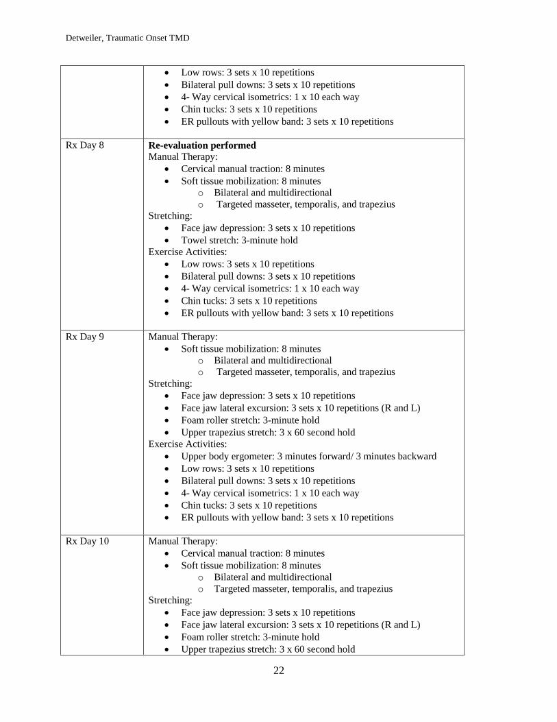
Rx Day 8 Re-evaluation performed
Manual Therapy:
Cervical manual traction: 8 minutes
Soft tissue mobilization: 8 minutes
o Bilateral and multidirectional
o Targeted masseter, temporalis, and trapezius
Stretching:
Face jaw depression: 3 sets x 10 repetitions
Towel stretch: 3-minute hold
Exercise Activities:
Low rows: 3 sets x 10 repetitions
Bilateral pull downs: 3 sets x 10 repetitions
4- Way cervical isometrics: 1 x 10 each way
Chin tucks: 3 sets x 10 repetitions
ER pullouts with yellow band: 3 sets x 10 repetitions
Rx Day 9 Manual Therapy:
Soft tissue mobilization: 8 minutes
o Bilateral and multidirectional
o Targeted masseter, temporalis, and trapezius
Stretching:
Face jaw depression: 3 sets x 10 repetitions
Face jaw lateral excursion: 3 sets x 10 repetitions (R and L)
Foam roller stretch: 3-minute hold
Upper trapezius stretch: 3 x 60 second hold
Exercise Activities:
Upper body ergometer: 3 minutes forward/ 3 minutes backward
Low rows: 3 sets x 10 repetitions
Bilateral pull downs: 3 sets x 10 repetitions
4- Way cervical isometrics: 1 x 10 each way
Chin tucks: 3 sets x 10 repetitions
ER pullouts with yellow band: 3 sets x 10 repetitions
Rx Day 10 Manual Therapy:
Cervical manual traction: 8 minutes
Soft tissue mobilization: 8 minutes
o Bilateral and multidirectional
o Targeted masseter, temporalis, and trapezius
Stretching:
Face jaw depression: 3 sets x 10 repetitions
Face jaw lateral excursion: 3 sets x 10 repetitions (R and L)
Foam roller stretch: 3-minute hold
Upper trapezius stretch: 3 x 60 second hold

Detweiler, Traumatic Onset TMD
23
Exercise Activities:
Upper body ergometer: 3 minutes forward/ 3 minutes backward
Low rows: 3 sets x 10 repetitions
Bilateral pull downs: 3 sets x 10 repetitions
ER pullouts with yellow band: 3 sets x 10 repetitions
Rx Day 11 Manual Therapy:
Cervical manual traction: 8 minutes
Soft tissue mobilization: 8 minutes
o Bilateral and multidirectional
o Targeted masseter, temporalis, and trapezius
Stretching:
Face jaw depression: 3 sets x 10 repetitions
Face jaw lateral excursion: 3 sets x 10 repetitions (R and L)
Foam roller stretch: 3-minute hold
Upper trapezius stretch: 3 x 60 second hold
Exercise Activities:
Upper body ergometer: 3 minutes forward/ 3 minutes backward
Low rows: 3 sets x 10 repetitions
Bilateral pull downs: 3 sets x 10 repetitions
ER pullouts with yellow band: 3 sets x 10 repetitions
Rx Day 12 Manual Therapy:
Cervical manual traction: 8 minutes
Soft tissue mobilization: 8 minutes
o Bilateral and multidirectional
o Targeted masseter, temporalis, and trapezius
Stretching:
Face jaw depression: 3 sets x 10 repetitions
Face jaw lateral excursion: 3 sets x 10 repetitions (R and L)
Foam roller stretch: 3-minute hold
Upper trapezius stretch: 3 x 60 second hold
Exercise Activities:
Upper body ergometer: 3 minutes forward/ 3 minutes backward
Low rows: 3 sets x 10 repetitions
Bilateral pull downs: 3 sets x 10 repetitions
Triceps press: 3 sets x 10 repetitions
ER pullouts with yellow band: 3 sets x 10 repetitions
Rx Day 13 Manual Therapy:
Cervical manual traction: 8 minutes
Soft tissue mobilization: 8 minutes
o Bilateral and multidirectional
o Targeted masseter, temporalis, and trapezius
Stretching:
Face jaw depression: 3 sets x 10 repetitions

Detweiler, Traumatic Onset TMD
24
Face jaw lateral excursion: 3 sets x 10 repetitions (R and L)
Foam roller stretch: 3-minute hold
Upper trapezius stretch: 3 x 60 second hold
Exercise Activities:
Upper body ergometer: 3 minutes forward/ 3 minutes backward
Low rows: 3 sets x 10 repetitions
Bilateral pull downs: 3 sets x 10 repetitions
Triceps press: 3 sets x 10 repetitions
ER pullouts with yellow band: 3 sets x 10 repetitions
Rx Day 14 Manual Therapy:
Soft tissue mobilization: 8 minutes
o Bilateral and multidirectional
o Targeted masseter, temporalis, and trapezius
Stretching:
Face jaw depression: 3 sets x 15 repetitions
Face jaw lateral excursion: 3 sets x 15 repetitions (Bilateral)
Foam roller stretch: 3-minute hold
Upper trapezius stretch: 3 x 60 second hold
Exercise Activities:
Upper body ergometer: 3 minutes forward/ 3 minutes backward
Low rows: 3 sets x 15 repetitions; 20 pound
Bilateral pull downs: 3 sets x 15 repetitions; 20 pounds
Triceps press: 3 sets x 10 repetitions; 15 pounds
ER pullouts with red band: 3 sets x 15 repetitions
Rx Day 15 Manual Therapy:
Soft tissue mobilization: 8 minutes
o Bilateral and multidirectional
o Targeted masseter, temporalis, and trapezius
Stretching:
Foam roller stretch: 3-minute hold
Upper trapezius stretch: 3 x 60 second hold
Side bend/ rotation stretch: 3 x 60 second hold
Exercise Activities:
Upper body ergometer: 3 minutes forward/ 3 minutes backward
Low rows: 3 sets x 15 repetitions; 20 pound
Bilateral pull downs: 3 sets x 15 repetitions; 20 pounds
Triceps press: 3 sets x 10 repetitions; 15 pounds
ER pullouts with red band: 3 sets x 15 repetitions
Sidelying Horizontal GH Abduction: 3 sets x 10 repetitions, 1 pound
Rx Day 16
(D/C)
Performed during HEP: (not performed during session)
Upper trapezius stretch: 3 x 60 second hold
Side bend/ rotation stretch: 3 x 60 second hold
Face jaw depression: 3 sets x 15 repetitions
Face jaw lateral excursion: 3 sets x 15 repetitions (Bilateral)
Patient Education: 8 minutes
Home Exercise Program
o Resisted Exercises

Detweiler, Traumatic Onset TMD
25
Self-mobilization technique
Manual Therapy:
Soft tissue mobilization: 8 minutes
o Bilateral and multidirectional
o Targeted masseter, temporalis, and trapezius
Stretching:
Foam roller stretch: 3-minute hold
Exercise Activities:
Upper body ergometer: 3 minutes forward/ 3 minutes backward
Low rows: 3 sets x 15 repetitions; 20 pound
Bilateral pull downs: 3 sets x 15 repetitions; 20 pounds
Triceps press: 3 sets x 10 repetitions; 15 pounds
ER pullouts with red band: 3 sets x 15 repetitions
Sidelying Horizontal GH Abduction: 3 sets x 10 repetitions, 1
pound; bilateral
336
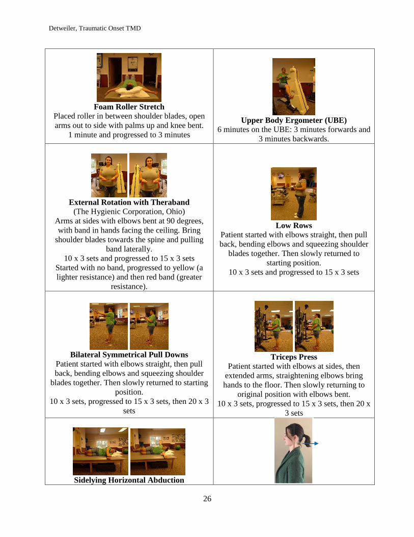
Appendix 3
Intervention Descriptions
Mandibular Depression Stretch
With tongue pushing into roof of mouth, patient
performs mandibular depression.
(10 x 3 sets and progressed to 15 x 3 sets)
Mandibular Lateral Excursion Stretch
With tongue pushing into roof of mouth,
patient performs mandibular left or right
lateral excursion.
(10 x 3 sets and progressed to 15 x 3 sets)
Upper Trapezius Stretch
Patient sat with neutral posture, tilted head to
one side until she felt a strong but gently stretch.
Held stretch for 60 seconds, 3x per each side.
Side Bend / Rotation Stretch
Patient sat with neutral posture with upper
extremities placed at sides with palms up, she
then look toward her opposite pocket until she
felt a strong, but gentle stretch. Held stretch
for 60 seconds, 3x per side.
Detweiler, Traumatic Onset TMD
26
Foam Roller Stretch
Placed roller in between shoulder blades, open
arms out to side with palms up and knee bent.
1 minute and progressed to 3 minutes
Upper Body Ergometer (UBE)
6 minutes on the UBE: 3 minutes forwards and
3 minutes backwards.
External Rotation with Theraband
(The Hygienic Corporation, Ohio)
Arms at sides with elbows bent at 90 degrees,
with band in hands facing the ceiling. Bring
shoulder blades towards the spine and pulling
band laterally.
10 x 3 sets and progressed to 15 x 3 sets
Started with no band, progressed to yellow (a
lighter resistance) and then red band (greater
resistance).
Low Rows
Patient started with elbows straight, then pull
back, bending elbows and squeezing shoulder
blades together. Then slowly returned to
starting position.
10 x 3 sets and progressed to 15 x 3 sets
Bilateral Symmetrical Pull Downs
Patient started with elbows straight, then pull
back, bending elbows and squeezing shoulder
blades together. Then slowly returned to starting
position.
10 x 3 sets, progressed to 15 x 3 sets, then 20 x 3
sets
Triceps Press
Patient started with elbows at sides, then
extended arms, straightening elbows bring
hands to the floor. Then slowly returning to
original position with elbows bent.
10 x 3 sets, progressed to 15 x 3 sets, then 20 x
3 sets
Sidelying Horizontal Abduction

Detweiler, Traumatic Onset TMD
27
(1-pound weight in photo)
Patient was positioned in sidelying with arms
extended, hands resting on top of one another.
Perform horizontal abduction with top arm
without allowing hips to move, allowing the
chest to open.
10 x 3 sets and progressed to 15 x 3 sets
Chin Tucks14
Patient was instructed to sit in neutral posture
and slowly draw her head backwards, as if
there was a string attached to the base of her
skull.
10 x 3 sets and progressed to 15 x 3 sets,
Resisted Depression14
Patient sits with jaw slightly open for correct
alignment. Depresses mandible while hand
provides mild resistance. Holds for 5-10 seconds.
Resisted Lateral Excursion14
Patient sit and opens jaw slightly for correct
alignment. Move mandible to side while using
hand to give mild resistance. Holds 5-10
seconds.
Soft Tissue Mobilization: Masseter14
Patient finds localized tenderness, applies
moderate pressure and then opens the jaw.
3-4 repetitions for discomfort or as needed
337