Embed Size (px)
Citation preview

Phosphorylation of TET Proteins Is Regulated viaO-GlcNAcylation by the O-Linked N-AcetylglucosamineTransferase (OGT)*□S
Received for publication, August 21, 2014, and in revised form, December 19, 2014 Published, JBC Papers in Press, January 7, 2015, DOI 10.1074/jbc.M114.605881
Christina Bauer‡1, Klaus Göbel‡, Nagarjuna Nagaraj§, Christian Colantuoni‡, Mengxi Wang‡, Udo Müller‡,Elisabeth Kremmer¶, Andrea Rottach‡2, and Heinrich Leonhardt‡�3
From the ‡Biocenter, Ludwig-Maximilians University Munich, 82152 Planegg-Martinsried, the §Max Planck Institute of Biochemistry,D-82152 Martinsried, the ¶Institute for Molecular Immunology, Helmholtz Center Munich, 81377 München-Gro�hadern, and the �Centerfor Integrated Protein Science Munich (CIPSM), 81377 München, Germany
Background: TET proteins oxidize 5-methylcytosine and contribute to active DNA demethylation.Results: O-Linked GlcNAc transferase modifies TET proteins with GlcNAc and thereby reduces TET phosphorylation.Conclusion: TET proteins are subjected to a dynamic interplay of post-translational modifications at low-complexity regions.Significance: This first map of TET phosphorylation and O-GlcNAcylation sites at amino acid resolution provides a valuableresource for future studies of TET regulation.
TET proteins oxidize 5-methylcytosine to 5-hydroxymethyl-cytosine, 5-formylcytosine, and 5-carboxylcytosine and thusprovide a possible means for active DNA demethylation inmammals. Although their catalytic mechanism is well charac-terized and the catalytic dioxygenase domain is highly con-served, the function of the regulatory regions (the N terminusand the low-complexity insert between the two parts of thedioxygenase domains) is only poorly understood. Here, we dem-onstrate that TET proteins are subject to a variety of post-trans-lational modifications that mostly occur at these regulatoryregions. We mapped TET modification sites at amino acid res-olution and show for the first time that TET1, TET2, and TET3are highly phosphorylated. The O-linked GlcNAc transferase,which we identified as a strong interactor with all three TETproteins, catalyzes the addition of a GlcNAc group to serine andthreonine residues of TET proteins and thereby decreases boththe number of phosphorylation sites and site occupancy. Inter-estingly, the different TET proteins display unique post-trans-lational modification patterns, and some modifications occur indistinct combinations. In summary, our results provide a novelpotential mechanism for TET protein regulation based on adynamic interplay of phosphorylation and O-GlcNAcylation atthe N terminus and the low-complexity insert region. Our datasuggest strong cross-talk between the modification sites thatcould allow rapid adaption of TET protein localization, activity,or targeting due to changing environmental conditions as wellas in response to external stimuli.
A major epigenetic mechanism of gene regulation in highereukaryotes is methylation of DNA at C5 of cytosines (1, 2).Recently, the family of TET (ten-eleven translocation) proteinshas been shown to successively oxidize 5-methylcytosine to5-hydroxymethylcytosine, 5-formylcytosine, and 5-carboxylcy-tosine (3– 6), providing novel insights into the dynamics ofDNA modifications. TET proteins are also active on genomicthymine residues, leading to the generation of 5-hydroxyuracil(7). In Gnathostomata, there are three TET proteins, TET1, TET2,and TET3 (8), which show distinct expression patterns and func-tions in different tissues or during development (9–13). TET1 andTET2 are highly expressed in mouse embryonic stem cells(mESCs)4 and are associated with oxidation of transcription startsites and gene bodies, respectively (14). TET3 is up-regulated inthe oocyte and oxidizes the silenced paternal pronuclear DNA (10,15). High levels of TET proteins and genomic 5-hydroxymethyl-cytosine are described for neuronal tissues (11, 16–18). In severalpatients with myeloid malignancies, mutations of TET2 correlatewith decreased 5-hydroxymethylcytosine levels and altered geneexpression patterns (19–22).
The activity of TET proteins directly depends on two cofac-tors: Fe(II) and 2-oxoglutarate (3, 8). Interestingly, gain-of-function mutations of the enzymes responsible for 2-oxo-glutarate synthesis, IDH1 and IDH2, have been associated withtumorigenesis, in particular glioblastomata and acute myeloidleukemia (20, 23, 24). These mutations lead to the synthesis of2-hydroxyglutarate, a potent inhibitor of 2-oxoglutarate-de-pendent dioxygenases such as TET proteins (24, 25). BecauseIDH1 and IDH2 are enzymes of the Krebs cycle, these findingsrepresent a direct link of TET protein activity to metabolism,especially because low 5-hydroxymethylcytosine levels arefound in acute myeloid leukemia patients not only with TET2loss-of-function mutations but also with IDH2 gain-of-func-tion mutations (20). Besides 2-hydroxyglutarate, ascorbate hasalso been shown to influence cytosine oxidation by TET pro-
* This work was supported in part by Deutsche Forschungsgemeinschaft (DFG)Collaborative Research Center Grants SFB 646/B10 and SFB 1064/A17.Author’s Choice—Final version full access.
□S This article contains supplemental Data S1, S2, and S3 and supplementalTable S4.
1 Supported by the International Max Planck Research School for Molecularand Cellular Life Sciences (IMPRS-LS).
2 To whom correspondence may be addressed. E-mail: [email protected] Member of the Nanosystems Initiative Munich (NIM). To whom correspond-
ence may be addressed: Dept. of Biology II, Ludwig-Maximilians UniversityMunich, Gro�hadernerstr. 2, 81925 Planegg-Martinsried, Germany. Tel.:49-89-2180-74229; Fax: 49-89-2180-74236; E-mail: [email protected].
4 The abbreviations used are: mESC, mouse embryonic stem cell; OGT,O-linked GlcNAc transferase; PTM, post-translational modification.
THE JOURNAL OF BIOLOGICAL CHEMISTRY VOL. 290, NO. 8, pp. 4801–4812, February 20, 2015Author’s Choice © 2015 by The American Society for Biochemistry and Molecular Biology, Inc. Published in the U.S.A.
FEBRUARY 20, 2015 • VOLUME 290 • NUMBER 8 JOURNAL OF BIOLOGICAL CHEMISTRY 4801
by guest on June 6, 2020http://w
ww
.jbc.org/D
ownloaded from

teins (26 –28). In summary, TET protein activity appears to bemodulated by several small molecules, either inhibitory such as2-hydroxyglutarate or stimulating such as ascorbate.
TET proteins are influenced not only by certain metabolites butalso by interacting proteins. TET1 forms complexes with hetero-chromatin-associated proteins such as HDAC1, HDAC2, SIN3A,and EZH2 (29). All three TET proteins interact with a variety offactors of the base-excision repair pathway, including PARP1,LIG3, and XRCC1, and also with several DNA glycosylases,including thymine-DNA glycosylase, NEIL1, and MDB4 (30).Another known interactor of TET proteins is the glycosyltrans-ferase OGT (31–36), which represents an additional interestingconnection with metabolism. OGT catalyzes the addition of aGlcNAc group to serine or threonine residues of target proteins(37). Its activity is dependent on the availability of a variety ofmetabolic molecules such as glucose, ATP, glutamine, andacetyl-CoA (38). The association of OGT with TET proteinshas been reported to influence histone modifications and geneexpression (31, 36), TET1 protein stability (33) and activity (34),and TET3 subcellular localization (35).
TET protein activity is widely studied in the context of devel-opment, tumorigenesis, and metabolic conditions. However,only very little is known about the structure and function of thenon-catalytic domains of TET proteins. In this study, we showthat TET proteins are subject to a large number of post-trans-lational modifications (PTMs), predominantly occurring at thetwo low-complexity regions, which display only little sequenceconservation: the N terminus and the insert region that sepa-rates the two parts of the catalytic dioxygenase domain and ispredicted to be unstructured (8). We demonstrate that TETproteins are phosphorylated and that this phosphorylation canbe suppressed via O-GlcNAcylation by the glycosyltransferaseOGT. Detailed mapping of modification sites to the proteinsequence shows that mostly the N terminus and insert region ofTET proteins are subjected to PTMs and that their regulationdepends on a dynamic interplay of different PTMs.
EXPERIMENTAL PROCEDURES
Antibody Generation—A His-tagged protein fragment fromthe insert region of each TET protein (see Fig. 1a) wasexpressed in Escherichia coli BL21(DE3) cells (Novagen, Darm-stadt, Germany) and purified with the TALON Superflow metalaffinity resin system (Clontech, Saint Germain, France) undernative conditions as described previously (39). Amino acids1682–1914 for TET1, amino acids 1332–1779 for TET2, andamino acids 976–1521 for TET3 were used as antigens. Approxi-mately 100 �g of each antigen was injected both intraperitoneallyand subcutaneously into Lou/C rats using CPG2006 (TIBMOLBIOL, Berlin, Germany) as adjuvant. After 8 weeks, theimmune response was boosted intraperitoneally and subcutane-ously 3 days before fusion. Fusion of the myeloma cell lineP3X63-Ag8.653 with rat immune spleen cells was performedusing PEG 1500 (Roche Diagnostics Deutschland GmbH,Mannheim, Germany). After fusion, the cells were cultured in96-well plates using RPMI 1640 medium with 20% fetal calfserum, penicillin/streptomycin, pyruvate, and nonessentialamino acids (PAA, Linz, Austria) supplemented with ami-nopterin (Sigma). Hybridoma supernatants were tested in a
solid-phase immunoassay. Microtiter plates were coated over-night with His-tagged TET antigens at a concentration of 3–5�g/ml in 0.1 M sodium carbonate buffer (pH 9.6). After blockingwith nonfat milk (Frema Reform, granoVita, Heimertingen,Germany), hybridoma supernatants were added. Bound ratmonoclonal antibodies were detected with a mixture of bioti-nylated mouse monoclonal antibodies against rat IgG heavychains, avoiding anti-IgM monoclonal antibodies (anti-IgG1,anti-IgG2a, and anti-IgG2b (American Type Culture Collec-tion, Manassas, VA) and anti-IgG2c (Ascenion GmbH,Munich, Germany)). The biotinylated monoclonal antibodieswere visualized with peroxidase-labeled avidin (Alexis, SanDiego, CA) and o-phenylenediamine as chromogen in the per-oxidase reaction. Anti-TET1 (clones 5D6, 5D8, 2H9, and 4H7;rat IgG2a), anti-TET2 (clone 9F7; rat IgG2a), and anti-TET3(clones 11B6 and 23B9; rat IgG2a) antibodies were stably sub-cloned and further characterized (see Fig. 1b).
mESC Culture, Co-immunoprecipitation, and MS/MS Analysis—mESCs (J1) were cultured as described previously (9). Endoge-nous TET1 and TET2 proteins were pulled out via monoclonalantibodies (clones 5D6, 5D8, and 9F7) coupled to proteinG-Sepharose beads as described (39). After co-immunoprecipi-tation, protein samples were digested on beads with trypsinaccording to standard protocols. Peptide mixtures were ana-lyzed by electrospray MS/MS spectrometry. Experiments wereperformed with an LTQ Orbitrap XL mass spectrometer(Thermo Scientific, Waltham, MA). Spectra were analyzedwith Mascot software (Matrix Science, Boston, MA).
Expression Constructs—Expression constructs for GFP-TET1,GFP-TET2, GFP-TET3, GFP, and Cherry were described previ-ously (40–42). To generate the Cherry-OGT construct, the cod-ing sequence was amplified using cDNA from E14 mESCs astemplate and subcloned into the pCAG-Cherry-IB vector.Expression constructs for Cherry-OGT(H508A) (hereafterreferred to as OGTmut) were generated by overlap extensionPCR. All constructs were verified by DNA sequencing (EurofinsGenomics, Ebersberg, Germany).
HEK293T Culture, Co-immunoprecipitation, and WesternBlot Analysis—Co-immunoprecipitation followed by Westernblotting with GFP- and Cherry-tagged proteins expressed inHEK293T cells was performed as described previously (30).O-GlcNAc was detected with a mouse monoclonal antibody(RL2, Abcam, Cambridge, United Kingdom) and an Alexa647N-conjugated secondary antibody (Sigma).
Sample Preparation for Mass Spectrometric Analysis—Allexperiments were performed in biological triplicates. GFP-tagged TET proteins and/or Cherry-tagged OGT and OGTmut
were expressed in HEK293T cells. Cell lysis with radioimmuneprecipitation assay buffer and immunoprecipitation with theGFP-Trap (ChromoTek GmbH, Martinsried, Germany) wereperformed as described previously (30). After immunoprecipi-tation, samples on beads were rinsed two times with washbuffer (20 mM Tris-HCl (pH 7.5), 300 mM NaCl, and 0.5 mM
EDTA) and two times with immunoprecipitation buffer (20 mM
Tris-HCl (pH 7.5), 150 mM NaCl, and 0.5 mM EDTA).100 �l of denaturation buffer (6 M guanidine hydrochloride,
10 mM tris(2-carboxyethyl)phosphine, and 40 mM chloroacet-amide in 100 M Tris (pH 8.5)) was added to the beads and heated
Phosphorylation and O-GlcNAcylation of TET Proteins
4802 JOURNAL OF BIOLOGICAL CHEMISTRY VOLUME 290 • NUMBER 8 • FEBRUARY 20, 2015
by guest on June 6, 2020http://w
ww
.jbc.org/D
ownloaded from

at 70 °C for 5 min. The samples were then subjected to sonica-tion in a Diagenode Bioruptor Plus system (UCD-300-TO) atmaximum power settings for 10 cycles consisting of a 30-s pulseand 30-s pause. Following sonication, the samples were diluted1:10 with digestion buffer (25 mM Tris (pH 8.5) containing 10%acetonitrile) and mixed by vortexing prior to enzyme digestion.Each sample was digested with 1 �g of endoproteinase Lys-C(Wako Chemicals, Neuss, Germany) for 4 h with subsequentdigestion using 1 �g of trypsin (Promega, Madison, WI) undergentle rotation at 37 °C. After digestion, the samples wereplaced in a SpeedVac concentrator for 10 min to remove ace-tonitrile from the sample before StageTip purification usingSDB-XC material (43). Peptides were then eluted from theStageTip and placed in the SpeedVac concentrator to reducethe sample volume to �6 �l, and 5 �l of the sample was injectedonto the column for MS/MS analysis.
LC-MS/MS and Data Analysis—Samples were loaded onto acolumn (15-cm length and 75-�m inner diameter; New Objec-tive, Woburn, MA) packed with 3-�m ReproSil C18 beads (Dr.Maisch GmbH, Ammerbuch-Entringen, Germany) using anEASY-nLC autosampler (Thermo Scientific) coupled via ananoelectrospray source to the LTQ Orbitrap XL mass spec-trometer. Each sample was analyzed using a 2-h reversed-phasegradient and a top 5 method for data-dependent acquisition.Full scans were acquired in the Orbitrap mass spectrometerafter accumulating up to 1 � 106 charges, and MS/MS with thefive most abundant precursors was performed using low-en-ergy ion-trap collision-induced dissociation. MS/MS spectrawere recorded using the ion trap by radial ejection.
All raw files were analyzed using the MaxQuant computa-tional proteomics platform (version 1.4.1.6) (44). Peak listswere searched with an initial mass deviation of 7 ppm and frag-ment ion deviation of 0.5 Thomson. Carbamidomethylationwas used as a fixed modification. Oxidation of methionine;phosphorylation of serine, threonine, and tyrosine; O-GlcNAcylation of serine and threonine; ubiquitination (digly-cine motif) of lysine; and acetylation of the protein N terminuswere used as variable modifications. All unmodified and oxi-dized methionine- and N-acetylation-containing peptides wereused for protein quantification. The MaxQuant software quan-tifies the different versions of modified peptides in a label-freefashion. Briefly, the occupancy reflects the extracted signaldifferences between modified and unmodified peptides andalso includes the protein ratios between samples. The differ-ent forms of modified peptides, e.g. peptides with single, dou-ble, and triple O-GlcNAc sites, are individually quantified andlisted separately in the output (supplemental Table S4). Details onlabel-free quantification of modification sites are provided else-where (45).
MaxQuant output data were further analyzed with Perseussoftware (version 1.5.0.15) (44). Only modifications that weredetected in at least two of three biological replicates in at leastone experimental setup were included in the analysis. PTMsthat were detected in non-unique peptides were also excluded.Significance was tested using a Student’s two-tailed paired ttest.
RESULTS
Characterization of Anti-TET Antibodies—The three TETproteins share a common domain architecture: the C-terminalcatalytic dioxygenase domain is split into two parts separatedby a low-complexity insert region and is preceded by an exten-sion enriched in cysteines (8). All three TET proteins have alarge N-terminal region that is mostly uncharacterized so far,except for a CXXC-type zinc finger at the N terminus of TET1and TET3 (8, 40, 47). Murine TET3 exists in two isoforms: onewith the zinc finger and one without (41). The cysteine-richregion and the split dioxygenase domain are conserved amongthe three murine TET proteins, whereas the N terminus andinsert region display only little sequence similarity (Fig. 1a).The three-dimensional structure of mammalian TET proteinsremains unresolved, with the exception of the cysteine-rich anddioxygenase domains of TET2 (48), leaving the structure and func-tion of the N terminus and low-complexity insert unknown.
We generated antibodies against murine TET1, TET2, andTET3 using protein fragments derived from the insert region ofthe catalytic domains as antigens. The rat monoclonal antibod-ies were tested for their applicability in Western blotting,immunoprecipitation, and immunofluorescence (Fig. 1b). Fig.1 (c–e) shows exemplary data from the antibody characteriza-tion process of selected clones. For antibody testing, GFP-tagged TET proteins were expressed in HEK293T cells anddetected in the cell lysate by Western blotting using anti-TETantibodies and an anti-GFP antibody as a positive control (Fig.1c and data not shown). For immunoprecipitation, antibodieswere coupled to protein G beads, incubated with the cell lysates,and analyzed by anti-GFP Western blotting for efficient pull-down of the respective TET protein (Fig. 1d and data notshown). mESCs were used to test the suitability of the obtainedantibodies for immunofluorescence. Antibodies preselected forspecificity in Western blot analyses that showed a clear nuclearstaining were judged as applicable in immunofluorescence (Fig.1e and data not shown).
TET Proteins Interact with and Are O-GlcNAcylated byOGT—As a first step toward understanding the regulation ofTET proteins, we screened for interaction partners in mESCs.Because TET1 and TET2 are constitutively expressed inmESCs, clones 5D6 (anti-TET1), 5D8 (anti-TET1), and 9F7(anti-TET2) were used to pull down endogenous TET1 andTET2. Subsequent LC-MS/MS analysis revealed that bothTET1 and TET2 interacted with the glycosyltransferase OGT.In accordance with this result, co-immunoprecipitationanalysis of GFP-tagged TET1, TET2, and TET3 expressed inHEK293T cells shows high enrichment of OGT in the pulldown(Fig. 2a).
Having observed the interaction between TET proteins andOGT, we examined whether TET proteins are modified byOGT and screened for O-GlcNAcylation, the modification thatis transferred to the OH group of serine or threonine residues oftarget proteins by OGT (38, 49). To this end, we specificallyenriched GFP-tagged TET proteins coexpressed with eitherOGT or its catalytically inactive point mutant OGTmut withGFP-Trap and probed the subsequent Western blot with ananti-GlcNAc antibody. All three TET proteins were found to
Phosphorylation and O-GlcNAcylation of TET Proteins
FEBRUARY 20, 2015 • VOLUME 290 • NUMBER 8 JOURNAL OF BIOLOGICAL CHEMISTRY 4803
by guest on June 6, 2020http://w
ww
.jbc.org/D
ownloaded from

be increasingly O-GlcNAcylated upon the coexpression ofcatalytically active OGT (Fig. 2b).
O-GlcNAcylation Reduces Phosphorylation of TET Proteins—To identify OGT-dependent O-GlcNAcylation sites on TET pro-teins, we performed mass spectrometric analysis of semipurifiedproteins. We therefore expressed GFP-tagged TET1, TET2, andTET3 in HEK293T cells with either OGT or OGTmut or withoutinteractor. After pulldown with GFP-Trap and stringent washingsteps, the samples were analyzed by LC-MS/MS. An overall
sequence coverage of �50% was achieved for TET1, �60% forTET2, and �65% for TET3 (supplemental Data S1 and TableS4). For data analysis, only sites were considered that weredetected in at least two of three biological replicates. Fig. 3ashows an exemplary MS/MS spectrum of an O-GlcNAcylatedTET1 peptide. Without coexpression of interactor, only a fewresidues on TET proteins were found to be O-GlcNAcylated atlow site occupancy. Coexpression of OGT led to a strongincrease in both the number of O-GlcNAcylation sites and site
FIGURE 1. Generation of anti-TET monoclonal antibodies. a, schematic representation of the domain architecture of the three murine TET proteins. Thecatalytic dioxygenase domain (D) is split in two parts, separated by a presumably unstructured low-complexity insert (8), and is N-terminally preceded by acysteine-rich region (Cys). The Fe(II)-binding residues are marked with green asterisks. The N terminus (NT) of TET1 contains a CXXC-type zinc finger (ZF). TET3exists in two isoforms, one with a zinc finger and one without (41). The mean percent identity of the single domains of TET1, TET2, and TET3 is represented bydifferent shades of gray and was calculated with Clustal 2.1 (59). aa, amino acids. b, overview of the generated anti-TET monoclonal antibodies (mAbs) and theirpossible applications. IP, immunoprecipitation; WB, Western blotting; IF, immunofluorescence; x, antibody not suited for the indicated application. c, exampleof Western blot analysis of two anti-TET antibodies with an anti-GFP antibody as a positive control. The antibodies detected only their target protein, but notthe other two TET proteins. The WT protein from mESC whole cell lysates was also detected specifically (black arrowhead). d, example of an immunoprecipi-tation experiment with the indicated anti-TET3 antibodies. Clone 23B9 efficiently precipitated TET3 compared with clone 11B6. Western blot analysis wasperformed with an anti-GFP antibody. I, input; FT, flow-through; B, bound). e, immunofluorescence staining of mESCs with anti-TET1 antibodies (clones 5D8 and4H7) and DAPI as a DNA counterstain. Whereas clone 5D8 showed a clear nuclear pattern, clone 4H7 displayed only a weak and diffuse signal. Confocal imagingwas performed with a Leica TCS SP5 confocal laser scanning microscope with a �63/1.4 numerical aperture Plan-Apochromat oil immersion objective. Scalebar � 5 �m.
Phosphorylation and O-GlcNAcylation of TET Proteins
4804 JOURNAL OF BIOLOGICAL CHEMISTRY VOLUME 290 • NUMBER 8 • FEBRUARY 20, 2015
by guest on June 6, 2020http://w
ww
.jbc.org/D
ownloaded from

occupancy for TET2 and TET3. The difference in the numberof O-GlcNAc sites was either due to de novo modification byOGT or because the site occupancy without OGT coexpressionwas below the detection limit. For TET1, however, theO-GlcNAc pattern was relatively heterogeneous, and only a fewO-GlcNAc sites could be detected. This heterogeneity is alsoillustrated by the fact that residues 1327 and 327, which wereO-GlcNAcylated in the TET1 samples, were modified only inone of three replicates in the TET1/OGT samples. AlthoughCherry-OGTmut is supposed to be catalytically inactive, coex-pression led to a small increase in O-GlcNAcylation and repre-sented a distinct state from basal levels (Fig. 4 and Tables 1–3).
Because O-GlcNAcylation occurs at serine or threonine res-idues of the target protein, we also screened for another PTMthat can occur at these amino acids, namely phosphorylation.Interestingly, high phosphorylation of TET1, TET2, and, to alesser extent, TET3 was observed. Phosphorylation of all TETproteins decreased significantly upon coexpression of OGTregarding both site occupancy and the number of detectedphosphorylation sites (Fig. 4 and Tables 1–3). An example of aMS/MS spectrum of a phosphorylated TET1 peptide is shownin Fig. 3b. The MS/MS spectra of all modified TET peptides areprovided in supplemental Data S2 and S3.
PTMs Occur Mostly at the N terminus and in the Low-com-plexity Insert of TET Proteins—To date, the domains of TETproteins are largely uncharacterized, except for the conservedcatalytic dioxygenase domain and the CXXC-type zinc finger atthe N terminus of TET1 (8, 40, 48). Mapping the detectedO-GlcNAc and phosphorylation sites to the TET proteinsequence revealed that mostly the N terminus and low-com-plexity insert, which separates the two parts of the dioxy-
genase domain, were subjected to PTMs (Fig. 5). Remark-ably, O-GlcNAcylation and phosphorylation rarely occurredat the exact same residue, although O-GlcNAcylation sup-pressed phosphorylation. Furthermore, the three TET pro-teins had different modification patterns: whereas TET1 was mod-ified mostly at the N terminus and the very C-terminal part andwas hardly glycosylated, TET2 and TET3 showed strongO-GlcNAcylation in the low-complexity insert region. The first350 amino acids of TET3 remained free of PTMs. The observedpattern is not due to differences in sequence coverage, as thedetected peptides are homogenously distributed over the wholeprotein sequence (supplemental Data S1).
Interestingly, some of the modifications were detected on thesame peptides, indicating that they occurred together at thesame molecule. For example, TET2 Ser-23 phosphorylationcould be found with Ser-15 phosphorylation, and Ser-376 phos-phorylation occurred only when Ser-374 was O-GlcNAcylated,but not when it was phosphorylated (Table 2). For TET3, avariety of PTM combinations could be observed for residues360 –368 and 1071–1077. Phosphorylation at Ser-362, forexample, existed either alone or in combination with Ser-360O-GlcNAcylation and Ser-368 phosphorylation. Phosphoryla-tion of Ser-362 also co-occurred with O-GlcNAcylation of Ser-361. If Ser-362 was O-GlcNAcylated, however, no further mod-ifications on this peptide were observed (Fig. 4 and Table 3).Apparently, some residues such as TET3 Ser-362 serve asO-GlcNAcylation/phosphorylation switches that can eitherpromote or suppress neighboring PTMs. These data indicate astrong cross-talk between O-GlcNAcylation and phosphoryla-tion at different residues. On the other hand, modifications onTET1 appeared more isolated, and no peptide bearing morethan one modification was detected (Table 1). In summary, wedetected many interdependent modification sites on TET pro-teins, suggesting that TET1, TET2, and TET3 are dynamicallyregulated by PTMs.
DISCUSSION
Because oxidation of 5-methylcytosine to 5-hydroxymethyl-cytosine, 5-formylcytosine, and 5-carboxylcytosine by TETproteins represents a potential mechanism for active DNAdemethylation in higher vertebrates (3–5), these proteins areintensively investigated. Here, we provide evidence that allthree TET proteins are subject to O-GlcNAcylation throughOGT. This finding is in accordance with previous studies show-ing that TET1 and TET2 interact with OGT in embryonic stemcells and are O-GlcNAcylated (33, 34). TET3 has also beendescribed to associate with OGT (32, 35) and to alter its subcel-lular localization dependent on glucose metabolism andO-GlcNAcylation (35). Not only does OGT directly modifyTET proteins, but the interaction also promotes histone mod-ifications such as H3K4me3 and H2BS112GlcNAc (31, 36).TET1 has been shown to associate with the repressive SIN3Acomplex (50), and TET2 and TET3 have been shown to associ-ate with the SET1/COMPASS complex (31).
We have shown that, by default, TET proteins are phosphory-lated. Basal O-GlcNAc levels are low but increase upon OGTexpression. Simultaneously, the phosphorylation levels decrease.This finding identifies regulation of the phosphorylation signal as a
FIGURE 2. TET proteins interact with OGT and are O-GlcNAcylated. a, num-ber of unique peptides detected in immunoprecipitation experiments fol-lowed by LC-MS/MS. Left, immunoprecipitation of endogenous TET1 or TET2with the indicated antibodies. Protein G beads without antibody wereused as a negative control (neg. ctrl). Right, immunoprecipitation of GFP-tagged TET1, TET2, or TET3 expressed in HEK293T cells. Pulldown of GFPserved as a negative control. b, Western blot analysis of TET1, TET2, andTET3 specifically enriched with GFP-Trap. Upon coexpression of activeOGT, the O-GlcNAcylation signal increased for TET1 and TET3 (black arrow-heads) compared with coexpression of catalytically inactive OGTmut. For TET2,protein levels in the OGTmut samples were higher (white arrowhead), whereasthe O-GlcNAc signal remained constant, suggesting a higher proportion ofO-GlcNAcylated TET2 in the OGT sample. Interaction between TET proteinsand OGT was independent of OGT activity. Anti-RED antibody (60) detectedthe coexpressed Cherry (Ch)-tagged OGT. IP, immunoprecipitation; I, input; B,bound.
Phosphorylation and O-GlcNAcylation of TET Proteins
FEBRUARY 20, 2015 • VOLUME 290 • NUMBER 8 JOURNAL OF BIOLOGICAL CHEMISTRY 4805
by guest on June 6, 2020http://w
ww
.jbc.org/D
ownloaded from

novel function for TET O-GlcNAcylation. Interestingly, theunderlying mechanism of this observation seems not to be directcompetition for the serine or threonine residue that is to be mod-ified, but rather proximal site competition as neighboring residuesare interdependent (51). O-GlcNAcylation and phosphorylationof TET proteins occur at distinct amino acids, and several mod-ifications of the same type often appear in close proximity in“modification islands,” e.g. O-GlcNAcylation at Ser-1252/Ser-1256/Ser-1263 of TET3 or phosphorylation at Ser-15/Ser-23/Ser-39 of TET2. It is important to note that only a few and moreisolated O-GlcNAcylation sites are detected on TET1 com-pared with TET2 and TET3 and that glycosylation of TET1 isless conserved within biological replicates. We also did notobserve O-GlcNAcylation of TET1 at Thr-535, which has beendescribed previously as a major TET1 glycosylation site (33, 52).O-GlcNAcylation of TET1 seems to be very dynamic. This
hypothesis is also supported by the fact that Myers et al. (52)detected TET1 Thr-535 O-GlcNAcylation in only one of threereplicates, similar to our observation of heterogeneous TET1glycosylation patterns.
To distinguish between mere interaction of TET proteinswith OGT and catalytic activity of OGT on TET proteins, weused a catalytically inactive point mutant of OGT as a control.Interestingly, O-GlcNAcylation of TET proteins was slightlyincreased by OGTmut. This might be due to residual activity ofthe mutant (53) or, more likely, to recruitment of endogenousactive OGT via trimerization of the tetratricopeptide repeatdomain (54). Nevertheless, this supposed heterotrimer seemsto target the same residues, as 91% of all detected O-GlcNAcsites in the OGTmut samples were also modified in the OGTsamples. Regarding phosphorylation, coexpression of OGTmut
also represents an intermediate state between the basal state,
FIGURE 3. Exemplary MS/MS spectra of modified TET1 peptides. a, MS/MS spectrum of a TET1 peptide modified with O-GlcNAc (o-) at the threonine residue.O-GlcNAcylation is characterized by a neutral loss of 203.8 Da as indicated. y ions are depicted in red, and b ions are depicted in blue. Labeling of neutral lossesof H2O or NH3 (orange peaks) has been removed for clarity. Fully annotated spectra are provided in supplemental Data S2 and S3. b, MS/MS spectrum of thesame TET1 peptide phosphorylated (-ph) at the serine residue. Phosphorylated ions show a neutral loss of 97.98 Da as indicated. y ions are depicted in red, b ionsare depicted in blue. Labeling of neutral losses of H2O or NH3 (orange peaks) has been removed for clarity. Fully annotated spectra are provided in supplementalData S2 and S3.
Phosphorylation and O-GlcNAcylation of TET Proteins
4806 JOURNAL OF BIOLOGICAL CHEMISTRY VOLUME 290 • NUMBER 8 • FEBRUARY 20, 2015
by guest on June 6, 2020http://w
ww
.jbc.org/D
ownloaded from
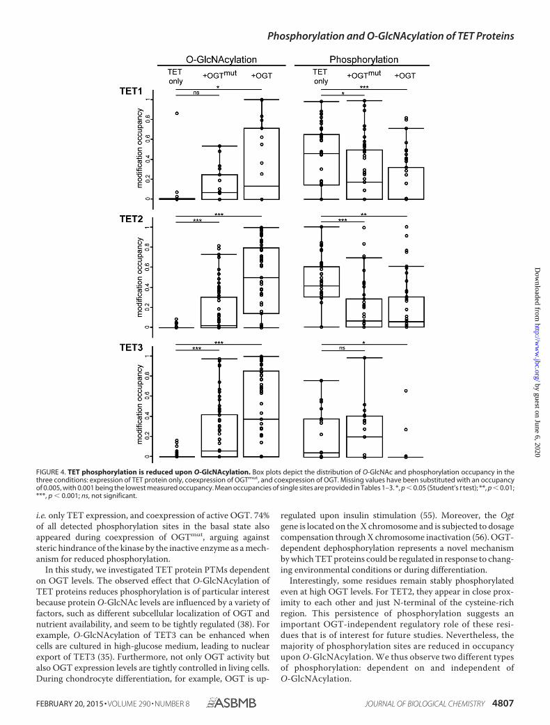
i.e. only TET expression, and coexpression of active OGT. 74%of all detected phosphorylation sites in the basal state alsoappeared during coexpression of OGTmut, arguing againststeric hindrance of the kinase by the inactive enzyme as a mech-anism for reduced phosphorylation.
In this study, we investigated TET protein PTMs dependenton OGT levels. The observed effect that O-GlcNAcylation ofTET proteins reduces phosphorylation is of particular interestbecause protein O-GlcNAc levels are influenced by a variety offactors, such as different subcellular localization of OGT andnutrient availability, and seem to be tightly regulated (38). Forexample, O-GlcNAcylation of TET3 can be enhanced whencells are cultured in high-glucose medium, leading to nuclearexport of TET3 (35). Furthermore, not only OGT activity butalso OGT expression levels are tightly controlled in living cells.During chondrocyte differentiation, for example, OGT is up-
regulated upon insulin stimulation (55). Moreover, the Ogtgene is located on the X chromosome and is subjected to dosagecompensation through X chromosome inactivation (56). OGT-dependent dephosphorylation represents a novel mechanismby which TET proteins could be regulated in response to chang-ing environmental conditions or during differentiation.
Interestingly, some residues remain stably phosphorylatedeven at high OGT levels. For TET2, they appear in close prox-imity to each other and just N-terminal of the cysteine-richregion. This persistence of phosphorylation suggests animportant OGT-independent regulatory role of these resi-dues that is of interest for future studies. Nevertheless, themajority of phosphorylation sites are reduced in occupancyupon O-GlcNAcylation. We thus observe two different typesof phosphorylation: dependent on and independent ofO-GlcNAcylation.
FIGURE 4. TET phosphorylation is reduced upon O-GlcNAcylation. Box plots depict the distribution of O-GlcNAc and phosphorylation occupancy in thethree conditions: expression of TET protein only, coexpression of OGTmut, and coexpression of OGT. Missing values have been substituted with an occupancyof 0.005, with 0.001 being the lowest measured occupancy. Mean occupancies of single sites are provided in Tables 1–3. *, p � 0.05 (Student’s t test); **, p � 0.01;***, p � 0.001; ns, not significant.
Phosphorylation and O-GlcNAcylation of TET Proteins
FEBRUARY 20, 2015 • VOLUME 290 • NUMBER 8 JOURNAL OF BIOLOGICAL CHEMISTRY 4807
by guest on June 6, 2020http://w
ww
.jbc.org/D
ownloaded from

FIGURE 5. N termini and insert regions of TET proteins are densely modified. Shown are a schematic and scaled mapping of all TET phosphorylation andO-GlcNAcylation sites in the protein sequence. Modifications are found mostly in the N terminus and insert region and rarely occur at the same residue. Residuenumbering refers to the murine protein sequences specified in supplemental Data S1. Green asterisks indicate catalytic Fe(II)-binding residues. Basal O-GlcNAcsites occur without any coexpression of OGT or OGTmut; persistent phosphorylation sites show high occupancy despite an increase in O-GlcNAcylation. Anexample of the PTM cross-talk on TET proteins is shown for TET3 Ser-360/Ser-361/Ser-362/Ser-368. White arrowheads, two co-occurring modifications; blackarrowheads, three co-occurring modifications; blunt arrows, mutual exclusivity.
TABLE 1Detected modified peptides of TET1(ph), phosphorylated; (o), O-GlcNAcylated; (ox), oxidized. Localization probability was calculated with MaxQuant software (44). Residue numbering refers to the murineprotein sequences specified in supplemental Data S1. The arithmetic mean � S.D. of the occupancy is depicted for each data set. ND, not detected.
Modifiedamino acid
Localizationprobability Modified sequence
MeanTET1
MeanTET1 � OGTmut
MeanTET1 � OGT
160 1.00 H . . . ATVS(ph)PGTENGEQNR 0.15 � ND 0.98 � ND 0.34 � 0.08177 1.00 CLVEGES(ph)QEITQSCPVFEER 0.63 � 0.01 0.32 � 0.15 0.48 � ND253 0.85 NT(o)SNQLADLSSQVESIK ND 0.07 � 0.01 0.37 � ND270 0.84 LS(o)DPSPNPTGSDHNGFPDSSFR ND 0.08 � 0.02 0.67 � 0.06320 1.00 FILAGS(ph)QPDVFDTKPQEK 0.60 � 0.16 0.44 � 0.03 0.30 � 0.20327 1.00 FILAGSQPDVFDT(o)KPQEKa 0.32 � 0.47 0.36 � 0.15 0.55 � ND556 0.81 A . . . STSS(ph)PPCNSTPPMVER 0.23 � 0.10 ND ND561 0.89 A . . . STSSPPCNS(ph)TPPM(ox)VER 0.20 � 0.09 0.88 � ND ND734 0.98 QQTNPS(ph)PTFAQTIR 0.44 � ND 0.46 � 0.33 0.32 � ND736 0.96 QQTNPSPT(ph)FAQTIR 0.67 � 0.06 ND ND794 0.77 DAM(ox)SVTTS(o)GGECDHLK ND 0.48 � ND 1.00 � 0.00854 1.00 DGS(ph)PVQPSLLSLMK 0.73 � 0.13 0.54 � 0.07 0.25 � 0.34892 0.70 L . . . SESSS(ph)PSKPEK 0.51 � 0.48 0.27 � 0.03 0.79 � ND950 1.00 S(ph)PDSFATNQALIKb 0.68 � 0.26 0.72 � 0.20 0.49 � 0.11969 0.74 SQGYPSS(ph)PT . . . 0.61 � 0.03 ND ND1327 0.66 REAQT(o)SSN . . . Ka 0.01 � 0.00 ND 0.79 � ND1964 0.89 ELHATTSLRS(ph)PK 0.33 � 0.21 0.17 � ND 0.47 � 0.332016 1.00 PADRECPDVS(ph)PEANLSHQIPSR 0.68 � 0.21 0.37 � 0.18 0.81 � ND2016 0.56 PADRECPDVS(o)PEANLSHQIPSR ND 0.26 � 0.10 0.55 � 0.412042 0.99 DNVVTVS(ph)PYSLTHVAGPYNR 0.73 � 0.12 ND 0.38 � ND
a Basal O-GlcNAc sites.b Persistent phosphorylation sites.
Phosphorylation and O-GlcNAcylation of TET Proteins
4808 JOURNAL OF BIOLOGICAL CHEMISTRY VOLUME 290 • NUMBER 8 • FEBRUARY 20, 2015
by guest on June 6, 2020http://w
ww
.jbc.org/D
ownloaded from

TABLE 2Detected modified peptides of TET2(ph), phosphorylated; (o), O-GlcNAcylated; (ox), oxidized. Multiple modifications occurirng on one peptide are shown in boldface. Localization probability was calculatedwith the MaxQuant software (44). Residue numbering refers to the murine protein sequences specified in supplemental Data S1. The arithmetic mean � S.D. of theoccupancy is depicted for each data set. ND, not detected.
Modifiedamino acid
Localizationprobability Modified sequence
MeanTET2
MeanTET2 � OGTmut
MeanTET2 � OGT
15 1.00 TTHAEGTRLS(ph)PFLIAPPS . . . K 0.57 � 0.06 0.20 � 0.01 0.01 � 0.0023 1.00 T . . . LS(ph)PFLIAPPS(ph)PIS . . . K 0.66 � 0.09 0.32 � 0.17 0.32 � 0.0439 0.98 LQNGS(ph)PLAERPHPEVNGDTK 0.45 � 0.10 ND ND95 0.98 RT(o)VS(o)EPSLSGLHPNK ND 0.06 � 0.00 0.26 � 0.1797 1.00 TVS(ph)EPSLSGLHPNK 0.53 � 0.26 0.49 � 0.30 0.29 � 0.0297 0.97 RT(o)VS(o)EPSLSGLHPNK ND 0.01 � ND 0.29 � 0.06165 1.00 S . . . TSTTQESSGADAFPT(o)R ND 0.74 � 0.06 0.98 � 0.02317 0.98 SALDIGPS(o)RAENK ND ND 0.48 � 0.03374 0.82 DS(ph)ISPTTVTPPSQSLLAPR ND ND 0.34 � 0.49374 0.99 DS(o)IS(ph)PTTVTPPSQSLLAPR ND 0.19 � 0.22 0.49 � 0.37376 0.99 DS(o)IS(ph)PTTVTPPSQSLLAPR 1.00 � ND 0.44 � 0.38 0.30 � 0.20464 1.00 T . . . LPEQHQNDCGS(ph)PS(ph)PEK 0.79 � 0.03 ND ND466 1.00 T . . . LPEQHQNDCGS(ph)PS(ph)PEK 0.79 � 0.03 ND ND514 0.89 QT(o)QGSVQAAPGWIELK ND 0.09 � 0.03 0.59 � 0.23545 0.94 DIS(o)LHSVLHSQT . . . M(ox)SSK ND 0.46 � 0.10 0.78 � 0.02552 0.87 DIS(o)LHSVLHS(o)QT . . . MSSK ND 0.13 � ND 0.76 � 0.20561 0.95 DIS . . . VNQMS(o)S(o)K ND 0.07 � 0.00 0.79 � 0.17562 0.97 DIS . . . VNQMS(o)S(o)Ka 0.01 � 0.01 0.42 � 0.06 0.80 � 0.17565 0.98 QS(o)TGNVNM(ox)PGGFQR ND ND 0.41 � 0.04603 1.00 AQMYQVQVNQGPS(ph)PG . . . K 0.41 � 0.17 0.06 � ND 0.14 � 0.05625 0.96 ALYQECIPRT(o)DPSS . . . Ra 0.05 � 0.01 0.73 � 0.13 0.98 � 0.02746 0.98 VEESFCVGNQYS(o)K ND 0.23 � 0.16 0.83 � 0.06778 0.92 ILT(o)PNSSNLQILPSNDTHPACER 0.09 � ND 0.31 � 0.01 0.64 � 0.05807 1.00 EQALHPVGS(o)K ND 0.01 � ND 0.58 � 0.12889 1.00 ALPVPEQGGSQTQT(ph)PPQKb 0.57 � 0.23 0.78 � 0.30 0.57 � 0.05944 1.00 YPLS(ph)PPQENMSSRb 0.43 � 0.14 0.46 � 0.22 0.67 � 0.11951 0.97 PSSYRYPLSPPQENMS(ph)SRb 0.24 � ND 0.32 � ND 0.51 � 0.681437 0.63 QM(ox)T(o)AQPQLS . . . R ND ND 0.67 � 0.061443 0.98 QMTAQPQLS(o)GPVIR ND 0.05 � 0.05 0.52 � 0.421613 0.87 D . . . PPIHT(o)LHQQTFGDSPSK ND ND 0.09 � 0.101622 0.74 Y . . . TLHQQTFGDS(ph)PSK 0.45 � 0.15 0.07 � 0.04 0.76 � ND1640 0.76 DAFT(o)TNSTLKPN . . . Ka 0.05 � 0.01 0.50 � 0.20 0.84 � 0.031672 1.00 M(ox)DSHFM(ox)GAAS(o)R ND ND 0.93 � 0.011749 1.00 TASAQELLYSLTGSS(ph)QEK 0.31 � 0.02 0.07 � 0.05 0.27 � 0.02
a Basal O-GlcNAc sites.b Persistent phosphorylation sites.
TABLE 3Detected modified peptides of TET3(ph), phosphorylated; (o), O-GlcNAcylated; (ox), oxidized. Multiple modifications occurring on one peptide are shown in boldface. Localization probability was calculatedwith MaxQuant software (44). Residue numbering refers to the murine protein sequences specified in supplemental Data S1. The arithmetic mean � S.D. of the occupancyis depicted for each data set. ND, not detected.
Modifiedamino acid
Localizationprobability Modified sequence
MeanTET3
MeanTET3 � OGTmut
MeanTET3 � OGT
360 0.93 VEAPS(o)SS(ph)PAPVPS(ph)PISQR ND 0.10 � 0.07 0.91 � 0.09361 0.79 VEAPSS(o)S(ph)PAPVPSPISQRa 0.02 � 0.01 0.52 � 0.13 ND362 1.00 VEAPSSS(ph)PAPVPSPISQR 0.03 � 0.03 0.38 � 0.03 0.01 � ND362 0.67 VEAPSSS(o)PAPVPSPISQR ND 0.96 � 0.02 ND368 1.00 VEAPS(o)SS(ph)PAPVPS(ph)PISQR 0.34 � ND 0.55 � 0.40 0.28 � ND478 1.00 S(ph)RDM(ox)QPLFLPVR 0.46 � 0.13 0.38 � 0.13 0.66 � ND557 0.83 S(ph)PSPM(ox)VALQSGST . . . R 0.23 � ND 0.22 � 0.27 ND557 0.76 S(o)PSPM(ox)VALQSGST . . . R ND ND 0.44 � 0.301008 0.83 VS(o)SGAIQVLTAFPR ND 0.91 � 0.01 0.36 � 0.511071 0.97 QEALELAGVT(o)T(o)DPGLSLK ND ND 0.96 � 0.011072 0.89 QEALELAGVT(o)T(o)DPGLSLK ND ND 0.96 � 0.011077 0.99 QEALELAGVTT(o)DPGLS(o)LK ND 0.50 � 0.40 0.53 � 0.311105 0.89 YS(o)GNAVVESYSVLGS . . . R ND 0.40 � 0.07 0.73 � 0.111252 0.94 VPQLHPAS(o)RDPSPFAQSSSCYNR ND 0.42 � ND 0.95 � 0.031256 0.62 VPQLHPASRDPS(o)PFAQSSSCYNRa 0.04 � 0.01 ND 0.98 � 0.011263 0.84 VPQLHPASRDPSPFAQSSS(o)CYNR ND 0.48 � ND 0.98 � 0.031282 0.88 QEPIDPLTQAES(o)IPR ND 0.30 � 0.06 0.91 � 0.091293 1.00 T(o)PLPEAS . . . SGGPSMSPKa 0.01 � 0.00 0.53 � 0.06 0.99 � 0.011318 1.00 TPLPEAS . . . SGGPSM(ox)S(ph)PK 0.43 � 0.07 0.00 � ND ND1351 0.61 LNSFGAS(ph)CLTPSHFPES . . . R 0.45 � 0.39 ND ND1404 0.76 FGNGTSALTGPSLT(o)EKa 0.02 � 0.02 ND 0.74 � 0.151412 1.00 PWGM(ox)GT(o)GDFNPALK ND 0.06 � 0.00 0.65 � 0.141651 0.72 Q . . . SAVT(o)VSSYAYTK ND ND 0.18 � 0.051653 0.71 Q . . . SAVTVS(o)SYAYTK ND 0.43 � 0.19 0.73 � 0.051654 0.77 Q . . . SAVTVS(o)S(o)YAYTK ND ND 0.24 � 0.051658 0.99 G . . . TDSAVTVSSYAYT(o)Ka 0.14 � 0.03 0.12 � 0.05 0.75 � 0.14
a Basal O-GlcNAc sites.
Phosphorylation and O-GlcNAcylation of TET Proteins
FEBRUARY 20, 2015 • VOLUME 290 • NUMBER 8 JOURNAL OF BIOLOGICAL CHEMISTRY 4809
by guest on June 6, 2020http://w
ww
.jbc.org/D
ownloaded from

The hypothesis of interdependence of PTMs on TET proteins isfurther strengthened by the fact that some modifications aredetected on the same peptides in stable combinations, whereasothers occur as stand-alone modifications. Certain residuesappear to be O-GlcNAcylation/phosphorylation switches thatinfluence the PTM pattern on the neighboring amino acids.The observed cross-talk of modifications enables a variety ofpotential regulatory mechanisms that could fine-tune TETactivity dependent on different environmental conditions suchas nutrient availability.
To date, the domain architecture and three-dimensionalstructure of TET proteins are only poorly understood. The cat-alytic domain is highly conserved and homologous to othertypes of Fe(II)- and 2-oxoglutarate-dependent dioxygenasesthat act on nucleic acids (8, 57). Recently, the crystal structureof the catalytic region of TET2 has provided insights into thereaction mechanism (48). However, the large N terminus andlow-complexity insert, which is characteristic for TET proteins,remain poorly understood in terms of both structure and func-tion. So far, no homologous domains have been described,except for the CXXC-type zinc finger at the N terminus, and theinsert region is predicted to be largely unstructured (8). In thisstudy, we have shown that these two regions are subject tomany dynamic PTMs. For TET1 and TET3, a few modificationsites are also found at the very C terminus of the proteins, butthe N terminus and insert region are the major targets ofO-GlcNAcylation and phosphorylation. In general, the lowerthe conservation of one region, the more modification sites aredetected. The selective modification of these regions mightcontribute to the regulation of TET protein activity, stability, ortargeting. TET1, TET2, and TET3 have been described to co-localize with OGT at transcription start sites and influencegene expression (31, 34). The different modifications describedin this study might alter binding of TET interaction partnersand thus provide a possible explanation for the observed dualrole in transcription activation and repression (58).
In summary, we have provided the first systematic mappingof O-GlcNAcylation and phosphorylation sites on TET pro-teins at amino acid resolution. The distribution of these PTMsand the described cross-talk provide new perspectives on the reg-ulatory role of the so far poorly characterized non-catalyticdomains: the N terminus and low-complexity insert region. Theobserved O-GlcNAcylation and phosphorylation are linked tometabolic conditions and thus provide a possible mechanism ofTET protein regulation in response to external stimuli.
REFERENCES1. Goll, M. G., and Bestor, T. H. (2005) Eukaryotic cytosine methyltrans-
ferases. Annu. Rev. Biochem. 74, 481–5142. Suzuki, M. M., and Bird, A. (2008) DNA methylation landscapes: provoc-
ative insights from epigenomics. Nat. Rev. Genet. 9, 465– 4763. Tahiliani, M., Koh, K. P., Shen, Y., Pastor, W. A., Bandukwala, H., Brudno,
Y., Agarwal, S., Iyer, L. M., Liu, D. R., Aravind, L., and Rao, A. (2009)Conversion of 5-methylcytosine to 5-hydroxymethylcytosine in mamma-lian DNA by MLL partner TET1. Science 324, 930 –935
4. Ito, S., Shen, L., Dai, Q., Wu, S. C., Collins, L. B., Swenberg, J. A., He, C., andZhang, Y. (2011) Tet proteins can convert 5-methylcytosine to 5-formyl-cytosine and 5-carboxylcytosine. Science 333, 1300 –1303
5. He, Y. F., Li, B. Z., Li, Z., Liu, P., Wang, Y., Tang, Q., Ding, J., Jia, Y., Chen,Z., Li, L., Sun, Y., Li, X., Dai, Q., Song, C. X., Zhang, K., He, C., and Xu, G. L.
(2011) Tet-mediated formation of 5-carboxylcytosine and its excision byTDG in mammalian DNA. Science 333, 1303–1307
6. Pfaffeneder, T., Hackner, B., Truss, M., Münzel, M., Müller, M., Deiml,C. A., Hagemeier, C., and Carell, T. (2011) The discovery of 5-formylcy-tosine in embryonic stem cell DNA. Angew. Chem. Int. Ed. Engl. 50,7008 –7012
7. Pfaffeneder, T., Spada, F., Wagner, M., Brandmayr, C., Laube, S. K., Eisen,D., Truss, M., Steinbacher, J., Hackner, B., Kotljarova, O., Schuermann, D.,Michalakis, S., Kosmatchev, O., Schiesser, S., Steigenberger, B., Raddaoui,N., Kashiwazaki, G., Müller, U., Spruijt, C. G., Vermeulen, M., Leonhardt,H., Schär, P., Müller, M., and Carell, T. (2014) Tet oxidizes thymine to5-hydroxymethyluracil in mouse embryonic stem cell DNA. Nat. Chem.Biol. 10, 574 –581
8. Iyer, L. M., Tahiliani, M., Rao, A., and Aravind, L. (2009) Prediction ofnovel families of enzymes involved in oxidative and other complex mod-ifications of bases in nucleic acids. Cell Cycle 8, 1698 –1710
9. Szwagierczak, A., Bultmann, S., Schmidt, C. S., Spada, F., and Leonhardt,H. (2010) Sensitive enzymatic quantification of 5-hydroxymethylcytosinein genomic DNA. Nucleic Acids Res. 38, e181
10. Gu, T. P., Guo, F., Yang, H., Wu, H. P., Xu, G. F., Liu, W., Xie, Z. G., Shi, L.,He, X., Jin, S. G., Iqbal, K., Shi, Y. G., Deng, Z., Szabó, P. E., Pfeifer, G. P., Li,J., and Xu, G. L. (2011) The role of Tet3 DNA dioxygenase in epigeneticreprogramming by oocytes. Nature 477, 606 – 610
11. Kim, M., Park, Y. K., Kang, T. W., Lee, S. H., Rhee, Y. H., Park, J. L., Kim,H. J., Lee, D., Lee, D., Kim, S. Y., and Kim, Y. S. (2014) Dynamic changes inDNA methylation and hydroxymethylation when hES cells undergo dif-ferentiation toward a neuronal lineage. Hum. Mol. Genet. 23, 657– 667
12. Langlois, T., da Costa Reis Monte-Mor, B., Lenglet, G., Droin, N., Marty,C., Le Couédic, J. P., Almire, C., Auger, N., Mercher, T., Delhommeau, F.,Christensen, J., Helin, K., Debili, N., Fuks, F., Bernard, O. A., Solary, E.,Vainchenker, W., and Plo, I. (2014) TET2 deficiency inhibits mesodermand hematopoietic differentiation in human embryonic stem cells. StemCells 32, 2084 –2097
13. Rudenko, A., Dawlaty, M. M., Seo, J., Cheng, A. W., Meng, J., Le, T., Faull,K. F., Jaenisch, R., and Tsai, L. H. (2013) Tet1 is critical for neuronalactivity-regulated gene expression and memory extinction. Neuron 79,1109 –1122
14. Huang, Y., Chavez, L., Chang, X., Wang, X., Pastor, W. A., Kang, J.,Zepeda-Martínez, J. A., Pape, U. J., Jacobsen, S. E., Peters, B., and Rao, A.(2014) Distinct roles of the methylcytosine oxidases Tet1 and Tet2 inmouse embryonic stem cells. Proc. Natl. Acad. Sci. U.S.A. 111, 1361–1366
15. Wossidlo, M., Nakamura, T., Lepikhov, K., Marques, C. J., Zakhartchenko,V., Boiani, M., Arand, J., Nakano, T., Reik, W., and Walter, J. (2011) 5-Hy-droxymethylcytosine in the mammalian zygote is linked with epigeneticreprogramming. Nat. Commun. 2, 241
16. Hahn, M. A., Qiu, R., Wu, X., Li, A. X., Zhang, H., Wang, J., Jui, J., Jin, S. G.,Jiang, Y., Pfeifer, G. P., and Lu, Q. (2013) Dynamics of 5-hydroxymethyl-cytosine and chromatin marks in mammalian neurogenesis. Cell Rep. 3,291–300
17. Kriaucionis, S., and Heintz, N. (2009) The nuclear DNA base 5-hydroxy-methylcytosine is present in Purkinje neurons and the brain. Science 324,929 –930
18. Xu, Y., Xu, C., Kato, A., Tempel, W., Abreu, J. G., Bian, C., Hu, Y., Hu, D.,Zhao, B., Cerovina, T., Diao, J., Wu, F., He, H. H., Cui, Q., Clark, E., Ma, C.,Barbara, A., Veenstra, G. J., Xu, G., Kaiser, U. B., Liu, X. S., Sugrue, S. P.,He, X., Min, J., Kato, Y., and Shi, Y. G. (2012) Tet3 CXXC domain anddioxygenase activity cooperatively regulate key genes for Xenopus eye andneural development. Cell 151, 1200 –1213
19. Abdel-Wahab, O., Mullally, A., Hedvat, C., Garcia-Manero, G., Patel, J.,Wadleigh, M., Malinge, S., Yao, J., Kilpivaara, O., Bhat, R., Huberman, K.,Thomas, S., Dolgalev, I., Heguy, A., Paietta, E., Le Beau, M. M., Beran, M.,Tallman, M. S., Ebert, B. L., Kantarjian, H. M., Stone, R. M., Gilliland,D. G., Crispino, J. D., and Levine, R. L. (2009) Genetic characterization ofTET1, TET2, and TET3 alterations in myeloid malignancies. Blood 114,144 –147
20. Konstandin, N., Bultmann, S., Szwagierczak, A., Dufour, A., Ksienzyk, B.,Schneider, F., Herold, T., Mulaw, M., Kakadia, P. M., Schneider, S., Spiek-ermann, K., Leonhardt, H., and Bohlander, S. K. (2011) Genomic 5-hy-
Phosphorylation and O-GlcNAcylation of TET Proteins
4810 JOURNAL OF BIOLOGICAL CHEMISTRY VOLUME 290 • NUMBER 8 • FEBRUARY 20, 2015
by guest on June 6, 2020http://w
ww
.jbc.org/D
ownloaded from

droxymethylcytosine levels correlate with TET2 mutations and a distinctglobal gene expression pattern in secondary acute myeloid leukemia. Leu-kemia 25, 1649 –1652
21. Delhommeau, F., Dupont, S., Della Valle, V., James, C., Trannoy, S., Massé,A., Kosmider, O., Le Couedic, J. P., Robert, F., Alberdi, A., Lécluse, Y., Plo,I., Dreyfus, F. J., Marzac, C., Casadevall, N., Lacombe, C., Romana, S. P.,Dessen, P., Soulier, J., Viguié, F., Fontenay, M., Vainchenker, W., and Ber-nard, O. A. (2009) Mutation in TET2 in myeloid cancers. N. Engl. J. Med.360, 2289 –2301
22. Ko, M., Huang, Y., Jankowska, A. M., Pape, U. J., Tahiliani, M., Banduk-wala, H. S., An, J., Lamperti, E. D., Koh, K. P., Ganetzky, R., Liu, X. S.,Aravind, L., Agarwal, S., Maciejewski, J. P., and Rao, A. (2010) Impairedhydroxylation of 5-methylcytosine in myeloid cancers with mutant TET2.Nature 468, 839 – 843
23. Dang, L., Jin, S., and Su, S. M. (2010) IDH mutations in glioma and acutemyeloid leukemia. Trends Mol. Med. 16, 387–397
24. Dang, L., White, D. W., Gross, S., Bennett, B. D., Bittinger, M. A.,Driggers, E. M., Fantin, V. R., Jang, H. G., Jin, S., Keenan, M. C., Marks,K. M., Prins, R. M., Ward, P. S., Yen, K. E., Liau, L. M., Rabinowitz, J. D.,Cantley, L. C., Thompson, C. B., Vander Heiden, M. G., and Su, S. M.(2009) Cancer-associated IDH1 mutations produce 2-hydroxygl-utarate. Nature 462, 739 –744
25. Xu, W., Yang, H., Liu, Y., Yang, Y., Wang, P., Kim, S. H., Ito, S., Yang, C.,Wang, P., Xiao, M. T., Liu, L. X., Jiang, W. Q., Liu, J., Zhang, J. Y., Wang, B.,Frye, S., Zhang, Y., Xu, Y. H., Lei, Q. Y., Guan, K. L., Zhao, S. M., and Xiong,Y. (2011) Oncometabolite 2-hydroxyglutarate is a competitive inhibitor of�-ketoglutarate-dependent dioxygenases. Cancer Cell 19, 17–30
26. Blaschke, K., Ebata, K. T., Karimi, M. M., Zepeda-Martínez, J. A.,Goyal, P., Mahapatra, S., Tam, A., Laird, D. J., Hirst, M., Rao, A.,Lorincz, M. C., and Ramalho-Santos, M. (2013) Vitamin C inducesTet-dependent DNA demethylation and a blastocyst-like state in EScells. Nature 500, 222–226
27. Chen, J., Guo, L., Zhang, L., Wu, H., Yang, J., Liu, H., Wang, X., Hu, X., Gu,T., Zhou, Z., Liu, J., Liu, J., Wu, H., Mao, S. Q., Mo, K., Li, Y., Lai, K., Qi, J.,Yao, H., Pan, G., Xu, G. L., and Pei, D. (2013) Vitamin C modulates TET1function during somatic cell reprogramming. Nat. Genet. 45, 1504 –1509
28. Minor, E. A., Court, B. L., Young, J. I., and Wang, G. (2013) Ascorbateinduces ten-eleven translocation (Tet) methylcytosine dioxygenase-mediated generation of 5-hydroxymethylcytosine. J. Biol. Chem. 288,13669 –13674
29. Cartron, P. F., Nadaradjane, A., Lepape, F., Lalier, L., Gardie, B., and Val-lette, F. M. (2013) Identification of TET1 partners that control its dna-demethylating function. Genes Cancer 4, 235–241
30. Müller, U., Bauer, C., Siegl, M., Rottach, A., and Leonhardt, H. (2014)TET-mediated oxidation of methylcytosine causes TDG or NEIL glycosylasedependent gene reactivation. Nucleic Acids Res. 42, 8592–8604
31. Deplus, R., Delatte, B., Schwinn, M. K., Defrance, M., Méndez, J., Murphy,N., Dawson, M. A., Volkmar, M., Putmans, P., Calonne, E., Shih, A. H.,Levine, R. L., Bernard, O., Mercher, T., Solary, E., Urh, M., Daniels, D. L.,and Fuks, F. (2013) TET2 and TET3 regulate GlcNAcylation and H3K4methylation through OGT and SET1/COMPASS. EMBO J. 32, 645– 655
32. Ito, R., Katsura, S., Shimada, H., Tsuchiya, H., Hada, M., Okumura, T.,Sugawara, A., and Yokoyama, A. (2014) TET3-OGT interaction increasesthe stability and the presence of OGT in chromatin. Genes Cells 19, 52– 65
33. Shi, F. T., Kim, H., Lu, W., He, Q., Liu, D., Goodell, M. A., Wan, M., andSongyang, Z. (2013) Ten-eleven translocation 1 (Tet1) is regulated byO-linked N-acetylglucosamine transferase (Ogt) for target gene repres-sion in mouse embryonic stem cells. J. Biol. Chem. 288, 20776 –20784
34. Vella, P., Scelfo, A., Jammula, S., Chiacchiera, F., Williams, K., Cuomo, A.,Roberto, A., Christensen, J., Bonaldi, T., Helin, K., and Pasini, D. (2013)Tet proteins connect the O-linked N-acetylglucosamine transferase Ogtto chromatin in embryonic stem cells. Mol. Cell 49, 645– 656
35. Zhang, Q., Liu, X., Gao, W., Li, P., Hou, J., Li, J., and Wong, J. (2014)Differential Regulation of the ten-eleven translocation (TET) family ofdioxygenases by O-linked �-N-acetylglucosamine transferase (OGT).J. Biol. Chem. 289, 5986 –5996
36. Chen, Q., Chen, Y., Bian, C., Fujiki, R., and Yu, X. (2013) TET2 promoteshistone O-GlcNAcylation during gene transcription. Nature 493, 561–564
37. Hanover, J. A., Yu, S., Lubas, W. B., Shin, S. H., Ragano-Caracciola, M.,Kochran, J., and Love, D. C. (2003) Mitochondrial and nucleocytoplasmicisoforms of O-linked GlcNAc transferase encoded by a single mammaliangene. Arch Biochem. Biophys. 409, 287–297
38. Harwood, K. R., and Hanover, J. A. (2014) Nutrient-driven O-GlcNAccycling–think globally but act locally. J. Cell Sci. 127, 1857–1867
39. Jost, K. L., Rottach, A., Milden, M., Bertulat, B., Becker, A., Wolf, P., San-doval, J., Petazzi, P., Huertas, D., Esteller, M., Kremmer, E., Leonhardt, H.,and Cardoso, M. C. (2011) Generation and characterization of rat andmouse monoclonal antibodies specific for MeCP2 and their use in X-in-activation studies. PLoS ONE 6, e26499
40. Frauer, C., Rottach, A., Meilinger, D., Bultmann, S., Fellinger, K., Ha-senöder, S., Wang, M., Qin, W., Söding, J., Spada, F., and Leonhardt, H.(2011) Different binding properties and function of CXXC zinc fingerdomains in Dnmt1 and Tet1. PLoS ONE 6, e16627
41. Liu, N., Wang, M., Deng, W., Schmidt, C. S., Qin, W., Leonhardt, H., andSpada, F. (2013) Intrinsic and extrinsic connections of Tet3 dioxygenasewith CXXC zinc finger modules. PLoS ONE 8, e62755
42. Spruijt, C. G., Gnerlich, F., Smits, A. H., Pfaffeneder, T., Jansen, P. W.,Bauer, C., Münzel, M., Wagner, M., Müller, M., Khan, F., Eberl, H. C.,Mensinga, A., Brinkman, A. B., Lephikov, K., Müller, U., Walter, J., Boel-ens, R., van Ingen, H., Leonhardt, H., Carell, T., and Vermeulen, M. (2013)Dynamic readers for 5-(hydroxy)methylcytosine and its oxidized deriva-tives. Cell 152, 1146 –1159
43. Rappsilber, J., Ishihama, Y., and Mann, M. (2003) Stop and go extractiontips for matrix-assisted laser desorption/ionization, nanoelectrospray, andLC/MS sample pretreatment in proteomics. Anal. Chem. 75, 663– 670
44. Cox, J., and Mann, M. (2008) MaxQuant enables high peptide identifica-tion rates, individualized p.p.b.-range mass accuracies and proteome-wideprotein quantification. Nat. Biotechnol. 26, 1367–1372
45. Sharma, K., D’Souza, R. C., Tyanova, S., Schaab, C., Wisniewski, J. R., Cox,J., and Mann, M. (2014) Ultradeep human phosphoproteome reveals adistinct regulatory nature of Tyr and Ser/Thr-based signaling. Cell Rep. 8,1583–1594
46. Deleted in proof47. Zhang, H., Zhang, X., Clark, E., Mulcahey, M., Huang, S., and Shi, Y. G.
(2010) TET1 is a DNA-binding protein that modulates DNA methylationand gene transcription via hydroxylation of 5-methylcytosine. Cell Res. 20,1390 –1393
48. Hu, L., Li, Z., Cheng, J., Rao, Q., Gong, W., Liu, M., Shi, Y. G., Zhu, J.,Wang, P., and Xu, Y. (2013) Crystal structure of TET2-DNA complex:insight into TET-mediated 5mC oxidation. Cell 155, 1545–1555
49. Hanover, J. A., Krause, M. W., and Love, D. C. (2012) Bittersweet memo-ries: linking metabolism to epigenetics through O-GlcNAcylation. Nat.Rev. Mol. Cell Biol. 13, 312–321
50. Williams, K., Christensen, J., Pedersen, M. T., Johansen, J. V., Cloos, P. A.,Rappsilber, J., and Helin, K. (2011) TET1 and hydroxymethylcytosine intranscription and DNA methylation fidelity. Nature 473, 343–348
51. Butkinaree, C., Park, K., and Hart, G. W. (2010) O-Linked �-N-acetylglu-cosamine (O-GlcNAc): extensive crosstalk with phosphorylation to regu-late signaling and transcription in response to nutrients and stress.Biochim. Biophys. Acta 1800, 96 –106
52. Myers, S. A., Panning, B., and Burlingame, A. L. (2011) Polycomb repres-sive complex 2 is necessary for the normal site-specific O-GlcNAc distri-bution in mouse embryonic stem cells. Proc. Natl. Acad. Sci. U.S.A. 108,9490 –9495
53. Lazarus, M. B., Nam, Y., Jiang, J., Sliz, P., and Walker, S. (2011) Structure ofhuman O-GlcNAc transferase and its complex with a peptide substrate.Nature 469, 564 –567
54. Jínek, M., Rehwinkel, J., Lazarus, B. D., Izaurralde, E., Hanover, J. A., andConti, E. (2004) The superhelical TPR-repeat domain of O-linked GlcNActransferase exhibits structural similarities to importin �. Nat. Struct. Mol.Biol. 11, 1001–1007
55. Andrés-Bergós, J., Tardio, L., Larranaga-Vera, A., Gómez, R., Herrero-Beaumont, G., and Largo, R. (2012) The increase in O-linked N-acetyl-glucosamine protein modification stimulates chondrogenic differentia-tion both in vitro and in vivo. J. Biol. Chem. 287, 33615–33628
56. Olivier-Van Stichelen, S., and Hanover, J. A. (2014) X-inactivation normalizes
Phosphorylation and O-GlcNAcylation of TET Proteins
FEBRUARY 20, 2015 • VOLUME 290 • NUMBER 8 JOURNAL OF BIOLOGICAL CHEMISTRY 4811
by guest on June 6, 2020http://w
ww
.jbc.org/D
ownloaded from

O-GlcNAc transferase levels and generates an O-GlcNAc-depleted Barrbody. Front. Genet. 5, 256
57. Loenarz, C., and Schofield, C. J. (2009) Oxygenase catalyzed 5-methylcy-tosine hydroxylation. Chem. Biol. 16, 580 –583
58. Wu, H., D’Alessio, A. C., Ito, S., Xia, K., Wang, Z., Cui, K., Zhao, K., Sun,Y. E., and Zhang, Y. (2011) Dual functions of Tet1 in transcriptional reg-
ulation in mouse embryonic stem cells. Nature 473, 389 –39359. Sievers, F., and Higgins, D. G. (2014) Clustal Omega, accurate alignment of
very large numbers of sequences. Methods Mol. Biol. 1079, 105–11660. Rottach, A., Kremmer, E., Nowak, D., Leonhardt, H., and Cardoso, M. C.
(2008) Generation and characterization of a rat monoclonal antibody spe-cific for multiple red fluorescent proteins. Hybridoma 27, 337–343
Phosphorylation and O-GlcNAcylation of TET Proteins
4812 JOURNAL OF BIOLOGICAL CHEMISTRY VOLUME 290 • NUMBER 8 • FEBRUARY 20, 2015
by guest on June 6, 2020http://w
ww
.jbc.org/D
ownloaded from

Udo Müller, Elisabeth Kremmer, Andrea Rottach and Heinrich LeonhardtChristina Bauer, Klaus Göbel, Nagarjuna Nagaraj, Christian Colantuoni, Mengxi Wang,
-Acetylglucosamine Transferase (OGT)N-Linked O-GlcNAcylation by the OPhosphorylation of TET Proteins Is Regulated via
doi: 10.1074/jbc.M114.605881 originally published online January 7, 20152015, 290:4801-4812.J. Biol. Chem.
10.1074/jbc.M114.605881Access the most updated version of this article at doi:
Alerts:
When a correction for this article is posted•
When this article is cited•
to choose from all of JBC's e-mail alertsClick here
Supplemental material:
http://www.jbc.org/content/suppl/2015/01/07/M114.605881.DC1
http://www.jbc.org/content/290/8/4801.full.html#ref-list-1
This article cites 59 references, 13 of which can be accessed free at
by guest on June 6, 2020http://w
ww
.jbc.org/D
ownloaded from


![Two Types of Tet-On Transgenic Lines for Doxycycline ......(Tet)- or doxycycline (Dox)-inducible Tet-On system [10,11] has been used in zebrafish to conditionally control Tet-responsive](https://img.dokumen.tips/doc/110x75/5f7b76c185c7f11b071fcfbc/two-types-of-tet-on-transgenic-lines-for-doxycycline-tet-or-doxycycline.jpg)


![[ TET Presentation ]](https://img.dokumen.tips/doc/110x75/557e7486d8b42a4d108b47f0/-tet-presentation-.jpg)