Embed Size (px)
Citation preview

The Journal of Neuroscience, March 1995, 15(3): 2385-2395
Phosphorylation of Rabphilin-3A by Ca2+/Calmodulin- and CAMP-Dependent Protein Kinases in vitro
Else Marie Fykse,” Cai Li, and Thomas C. Siidhof
Department of Molecular Genetics and Howard Hughes Medical Institute, The University of Texas Southwestern Medical Center, Dallas, Texas 75235
Regulation of neurotransmitter release is thought to in- volve modulation of the release probability by protein phosphorylation. In order to identify novel targets for such regulatory processes, we have studied the phosphorylation of rabphilin9A in vitro. Rabphilin-3A is a synaptic vesicle protein that interacts with rab3A in a GTP-dependent man- ner and binds Ca*+ in a phospholipid-dependent manner. Here we show that rabphilin-3A is an efficient substrate for Ca*+/calmodulin-dependent protein kinase II, which phos- phorylates rat rabphilin-3A at residue 234 and 274, and for CAMP-dependent protein kinase, which phosphorylates rat rabphilin-3A at residue 234. This identifies the middle re- gion of rabphilin4A situated between the N-terminal rab3A- binding sequences and the C-terminal &-domains involved in Ca2+/phospholipid binding as a regulatory domain. Thus, rabphilin9A is a second phosphoprotein on synaptic ves- icles that, similar to synapsin I, may integrate phosphory- lation signals from multiple protein kinase signaling path- ways in the cell.
[Key words: synaptic vesicles, synaptic plasticity, exo- cytosis, protein kinases, Ca2+, rab proteins, phosphoryla- tion]
In nerve terminals, neurotransmitters are stored in synaptic ves- icles and secreted by synaptic vesicle exocytosis. Neurotrans- mitter secretion is acutely regulated by Ca2+, which triggers exo- cytosis in a rapid (<l msec) and cooperative manner. On a lon- ger time scale, neurotransmitter release is modulated by a variety of regulatory processes, such as occur during the different forms of short-term synaptic plasticity (Zucker, 1989), modulation by presynaptic receptors (Takeuchi and Takeuchi, 1966; Silinsky, 1984; Scholz and Miller, 1992; Weisskopf et al., 1993), and pos- sibly long-term potentiation (LTP) and long-term depression (LTD) (Bekkers and Stevens, 1990; Malinow and Tsien, 1990; Bolshakov and Siegelbaum, 1994). These regulatory phenomena are thought to involve covalent modifications of proteins, in par- ticular, by protein phosphorylation. In support of this notion, mice carrying a mutant form of the gene for a-Caz+/calmodulin-
Received July 15, 1994; revised Oct. 3, 1994; accepted Oct. 5, 1994. We thank I. Leznicki, A. Roth, and E. Borowicz for excellent technical
assistance, Dr. H. Schulman (Stanford University), Dr. M. Kennedy (California Institute of Technology), and Dr. R. Jahn (Yale University) for supplying us with purified enzymes and with antibodies; and Drs. H. McMahon and B. Davletov for advice.
Correspondence should be addressed to Thomas C. Stidhof at the above address.
“Present address: Norwegian Defence Research Establishment, Division of Environmental Toxicology, P.B. 25, N-2007 Kjeller, Norway. Copyright 0 1995 Society for Neuroscience 0270-6474/95/152385-l 1$05,00/O
dependent protein kinase II (a-CaMK II) exhibit dramatic changes in short-term synaptic plasticity even in the heterozy- gous state, which presumably only lowers the levels of enzyme by half (Silva et al., 1992; A. Silva, personal communication). LTP in mossy fiber synapses, a known presynaptic form of syn- aptic plasticity (Zalutsky and Nicoll, 1990), is related to CAMP- dependent protein kinase (PKA) activation (Frey et al., 1993). Pharmacological experiments using a variety of protein kinase inhibitors and stimulators also suggest a role for protein kinases in synaptic plasticity (Zhuo et al., 1994; reviewed in Stevens, 1993). Thus, there is significant evidence for a major role of protein phosphorylation events in at least some forms of regu- lation of neurotransmitter release.
At least two possible mechanisms can be envisioned by which protein phosphorylation could regulate neurotransmitter release. First, protein kinases could phosphorylate ion channels and their associated proteins. As a regulatory endpoint, such phosphory- lation events could lead to an increase or decrease in the intra- cellular Ca2+ levels achieved after an action potential and there- by regulate release (for a review, see Nicoll et al., 1990). Sec- ond, protein kinases could phosphorylate proteins involved in the synaptic vesicle traffic. Thereby, they could modulate the efficiency of synaptic vesicle trafficking as a function of Ca2+. Although there is evidence in support of both mechanisms (Tak- euchi and Takeuchi, 1966; Man-Son-Hing et al., 1989; Dale and Kandel, 1990; Scholz and Miller, 1992), the molecular basis for these processes is largely obscure. In particular, for the synaptic release apparatus, the mechanisms underlying the modulation of release and the relevant phosphorylation targets are mostly un- known.
Several protein kinase substrates in the nerve terminal have been extensively studied. The synapsins constitute a family of phosphoproteins that are associated with synaptic vesicles (De Camilli et al., 1983a,b; Stidhof et al., 1989) and are substrates for at least four different protein kinases, CaMK I and II, PKA, and proline-directed kinase (reviewed in Jahn and Stidhof, 1994). The abundance and regulated phosphorylation of the syn- apsins suggest that they function in regulating neurotransmitter release, but their exact functions are unknown except for the C-terminal phosphorylation site for CaMK II in synapsin I, which appears to act as a negative regulator during paired-pulse facilitation (Rosahl et al., 1993). GAP-43 (also called B-50) rep- resents an abundant phosphoprotein of the nerve terminal plas- mamembrane, with a possible role in exocytosis but no currently known phosphorylation-dependent function (Dekker et al., 1989). Dynamin I is a synaptic phosphoprotein with the inter- esting property of quantitative dephosphorylation during nerve

2386 Fykse et al. l Phosphotylation of Rabphilin-3A
Anti body MS Anti-Rabphilin
Cytosol + - + + + +
Activator Ca - - cAMPcGMP Ca . .:i ./ ,... ;..;.
- 85 kDa
Figure 1. Phosphorylation of rabphilin-3A by CAMP- and Ca*+-dependent protein kinases. Immunoprecipitates of rat brain proteins with preim- mune serum (PZS, lej? lane) or with polyclonal antibodies to rabphilin3A (Anti-Rabphilin) were incubated with or without cytosol as a source of kinases and with Ca*+, CAMP, and cGMP as activators. Samples were analyzed by SDS-PAGE, blotted onto nitrocellulose membranes, and exposed to film for 3 hr at -70°C with a screen.
Antibody PIS Anti-Rabphilin
Kinase CaMKiI - pm CaMKII
Activator - Ca - - - Ca
- 85 kDa
- 85 kDa
I - IgG
Figure 2. Phosphorylation of rabphilin-3A by the catalytic subunit of CAMP-dependent protein kinase (PKA) and by Ca*+, calmodulin-dependent protein kinase II (C&X II). Rabphilin and control immunoprecipitates (see legend to Fig. 1) were incubated with the indicated protein kinases as described in Experimental Procedures. Samples were subjected to SDS-PAGE, blotted onto nitrocellulose membranes, and analyzed by autoradiog- raphy (iop panel; exposure: 5 hr at -70°C with screen) and by immunoblotting with rabphilin antibodies (bottom panel) to control for the amount of rabphilin-3A immunoprecipitated. The additional phosphoprotein bands observed in the samples incubated with CaMK II and Ca2+ are probably due to autophosphorylated CaMK II (see also Fig. 7).

The Journal of Neuroscience, March 1995, f5(3) 2387
PP Rabphilin-3A
NH2 COOH
domain structure rab3A binding C2domains
Residue Numbers
GST
GST-RaphA
B
C
D
E
F
G
H
I
J
PP
Legend: Point mutation *
Fhosphoryiafion site P
GST gggg
N-terrnlnal domain /
First C2 domain m
Second C2 domain
terminal depolarization that probably plays a role in triggering synaptic vesicle endocytosis (Robinson et al., 1993).
These and other synaptic phosphoproteins (e.g., AP180, syn- aptotagmin, and others) are candidates for mediating some reg- ulatory functions in the synaptic vesicle cycle. However, the currently known protein kinase targets at the synapse are clearly insufficient to explain most of these synaptic regulatory phe- nomena. For example, introduction of CaMK II into presynaptic nerve terminals causes major changes in synaptic vesicle exo- cytosis (Nichols et al., 1990; Llinas et al., 1991). However, the only currently characterized synaptic CaMK II substrate is syn- apsin I. Although it was originally hypothesized that CaMK II exerts its presynaptic functions in potentiating neurotransmitter release via synapsin I, the genetic experiments clearly showed that this is not the case (Rosahl et al., 1993). Another example is PKA, which appears to modulate neurotransmitter release and to have a major function in mossy fiber LTP in the hippocampus
0
1-181
67-226
199-361
250-361
380-684
380-531
524-684
1-361
1-361
1-361
400 amino acids
Figure 3. Schematic overview of the bacterial glutathione S-transferase (GS7’) fusion proteins used to map the phosphorylation sites in rabphilin3A. The diagram olt top depicts the domain structure of rabphilin-3A. The stmc- _ I__-_ tures of GST ana of 10 GST-rabphilin fusion proteins used in the current study are shown in the middle, and the legend is shown on the bottom. The lo- cations of the phosphorylation sites that were identified in the current study are indicated by “P.” GST-RaphH contains the entire N-terminal half of rabphilin-3A. Serine-to-alanine muta- tions were introduced into serine234 (the putative PK.4 phosphorylation site) in GST-RaphI, and into serine234 and ser- ine274 (the putative CaMK II phosphor- ylation site) in GST-RaphJ. A mutant in which only the putative CaMK II phosphorylation site (serine*“) was substituted was also made but the ex- pressed protein was unstable in bacteria and, therefore, not analyzed. However. the corresponding mutant was also ex- pressed as a holo-protein in COS cells (see below).
(Frey et al., 1993; Chavez-Noriega and Stevens, 1994). Again, the only currently known synaptic PKA substrates are synapsins I and II, which do not appear to account for all of the regulatory phenomena observed with PKA. Therefore, it would be of great interest to define additional protein kinase substrates in the nerve terminal that have properties compatible with a role in nerve terminal regulation.
Rabphilin-3A was identified because of its GTP-dependent binding to rab3A (Shirataki et al., 1992). Rabphilin-3A has a distinct domain structure consisting of a conserved N-terminal domain that binds rab3A, a variable middle domain, and a C-ter- minal region containing two C,-domains with homology to those of synaptotagmin (Shirataki et al., 1993; Li et al., 1994). In addition to rab3A, rabphilin-3A may also binds Ca2+ and phos- pholipids in a ternary complex (Shirataki et al., 1993; Yama- guchi et al., 1993). Recently we showed that rabphilin-3A is a synaptic vesicle protein, identifying it as a second potential Ca2+

2388 Fykse et al. l Phospholylation of Rabphilin-3A
GST-Raph
GST A B C D E F G
Control
CAMP
ca*+
Coomassie
. . . ._ . . ” . . . I . . - . . . . -.1x x_x(.x._.__ ~~_x_ ,,___ 1 ,__.__( _,_s
j
111..._ l.~
- 106 - 80
- 32
- 106
- 80
-50
- 106 - 80
-50
- 32
- 106 - 80
- 50
- 32
Figure 4. Phosphorylation of rabphilin-GST fusion-proteins by endogenous kinases present in rat brain cytosol. Identical amounts of recombinant GST proteins (proteins are identified on top and described in see Fig. 3; approximately 3 p,g protein were loaded per lane) were incubated with rat brain cytosol and BL~~~P-ATP without additions (top panel), in the presence of CAMP (second panel), or in the presence of Ca*+ (third panel).

The Journal of Neuroscience, March 1995, 75(3) 2389
sensor on synaptic vesicles in addition to synaptotagmin (Li et al., 1994). Furthermore, it was found that in mice lacking rab3A, neurons that do not contain rab3C show a defect in the synaptic targeting of rabphilin-3A, indicating that rabphilin-3A is targeted to synaptic vesicles via its N-terminal binding to rab3A (Geppert et al., 1994; Li et al., unpublished observations). Together, these findings implicate rabphilin-3A as a potential regulatory protein in the nerve terminal that functionally interacts with rab3A. This functional interaction was confirmed in experiments with rab3A- deficient mice in which rab3A deficiency leads to a 70% reduc- tion in the levels of rabphilin-3A (Geppert et al., 1994). In the current study, we show that in vitro, rabphilin-3A is an excellent substrate for at least two protein kinases that have been shown to regulate synaptic transmission, PKA and CaMK II. The phos- phorylation sites for the two kinases were identified in the mid- dle domain of rabphilin-3A, suggesting that this is a regulatory domain. The identification of rabphilin-3A as a substrate for multiple protein kinases suggests that rabphilin-3A, similar to the synapsins, may serve as a signal integration point for mul- tiple protein kinases in the nerve terminal.
Materials and Methods Construction of eukaryotic expression vectors and expression of pro- teins in COS cells by trunsfection. For the construction of the wild-type rabphilin-3A expression vector (pCMV854), PCR was performed on the full-length rat rabphilin cDNA (Li et al., 1994) using oligonucleo- tides A and B (seouences [redundant nucleotides in brackets]: GCCTC- TAGACGGTACCATGACTGACACTGTGGT and GCCGTCGACTA- GTCGGAGGAIC.GlACIA.GlTG~A.GlTT~~ClTC~A.GlTT~T.ClTG~. The 2.07 kb product- was digested with KpnI and Sal1 and subcloned into the same sites of pCMV5. For construction of the expression vector encoding rabphilin3A with a serinez24 to alanine substitution (pCMV85- 3), PCRs were performed on the full length rat rabphilin-3A cDNA clone with two pairs of oligonucleotides, A and C (sequence: TCC- GGGCCTCCGCGGCCCTGCGTGTGGGAG) and oligonucleotides B and D (sequence: GCAGGGCCGCGGAGGCCCGGATGAGTA). The products from the two amplifications were gel purified, cut with SacII, and ligated to each other. They were then reamplified with oligonu- cleotides A and B, subcloned into pCMV5 as above, and confirmed by restriction enzyme mapping. The expression vector encoding rabphilin- 3A with a single point-mutation in-the putative CaMK II phosphory- lation site (pCMV855) substituting serine 274 to alanine was constructed similarly with two internal oligonucleotides E and F (sequences: TGGT- CTAGAGGCCTGGACTGCGTTAGCTCGCCT and GCCTCTAGAC- CAGCTCCAGCCTCCATG). exceot that the first PCR nroducts were cut with XbaI before ligation and performing PCR again. The double mutant (pCMV85-6) (serine 234 to alanine, serine274 to alanine) was con- structed by introducing the CaMK II phosphorylation site mutation as described above into the vector encoding rabphilin-3A with the PKA- site mutation. Expression vectors were transfected into COS cells using the DEAE-Dextran method as described (Gorman, 1985).
Construction of bacterial vectors encoding GST-fusion proteins and expression and puriJcation of recombinant proteins. Eleven bacterial expression vectors were constructed for this study using standard re- combinant DNA techniques (Sambrook et al., 1989) by subcloning cDNA fragments into the vector pGEX-KG (Guan and Dixon, 1991). The following GST-rabphilin-3A fusion proteins were encoded by the following vectors (vector names and encoded residue numbers of rat rabphilin-3A shown in brackets): GST-RaphA (pGEX85-6, l-181), con- structed by PCR amplification of the corresponding coding region using oligonucleotides A (see above) and G (sequence: GGGGTCGAC- TACGGCTCACCAGCAGGC) and cloning the PCR product cut with XbaI-Sal1 into the same sites of pGEX-KG; GST-RaphB (pGEX85-3, 67-226), obtained by subcloning 0.48 kb NcoI fragment of rat rabphil-
t
in-3A cDNA into NcoI site of pGEX-KG; GST-RaphC (pGEX85-11; 199-361) constructed by subcloning 0.48 kb XhoI-Sal1 fragment of pGEX85-4 (see below) into Sal1 site of pGEX-KG; GST-RaphD (pGEX85-13; 250-361), constructed by subcloning 0.33 kb NcoI-Sal1 fragment of pGEX85-4 (see below) into NcoI-Sal1 sites of pGEX-KG; GST-RaphE (pGEX85-8, 38s684), constructed by PCR amplification of the corresponding coding region using oligonucleotides H (sequence: CGCGCTAGCAACCACACTGGGTG) and B (see above), and cloning the PCR product cut with NheI-Sal1 into the XbaI-Sal1 sites of pGEX- KG; GST-RaphF (pGEX85-7, 380-531), constructed by PCR amplifi- cation of the corresponding coding region using oligonucleotides H and I (sequence: GGGGTCGACTACTGCTCCTCCTCATAGAG), and cloning the PCR product cut with NheI-Sal1 into the XbaI-Sal1 sites of pGEX-KG; GST-RaphG (pGEX85-5, 524-684), constructed by PCR amplification of the corresponding coding region using oligonucleotides J (sequence: GCGTCTAGACATGGCTCTCTATGAGGA) and B (see above), and cloning the PCR product cut with XbaI-Sal1 into the same sites of pGEX-KG; GST-RaphH (pGEX85-4, l-361), constructed by PCR amplification of the corresponding coding region using oligonu- cleotides A and K (sequence: GGGGTCGACTACTGGCGGGCAGGG- GCCG), and cloning the PCR product cut with XbaI-Sal1 into the same sites of pGEX-KG; GST-RaphI (pGEX85-12, l-361 with a serinez34 to alanine point mutation), constructed by two-stage PCR analogously to pCMV85-3 and pGEX85-4, as described above; GST-RaphJ (pGEX85- 15, l-361 with two point mutations: serine234 to alanine, and serine274 to alanine), constructed by two-stage PCR with pGEX85-12 as template analogously to pCMV85-6 and pGEX85-4 as described above; GST- RaphK (pGEX85-14, l-361 with point mutations of serine2” to ala- nine), constructed by two-stage PCR with pGEX85-4 as template as described above for pCMV85-5 and pGEX85-4, the protein produced by this plasmid in different strains of bacteria was unstable and could not be obtained in sufficient quantities for experiments. Recombinant GST-proteins were expressed in bacteria and purified on glutathione- agarose (Sigma) as described (Smith and Johnson, 1988).
Protein assays, SDS-PAGE, and protein stains. Protein assays were carried out using a Coomassie-blue based assay (Bio-Rad; Bradford, 1976). SDS-PAGE was performed as described (Laemmli, 1970). Gels were either stained with Coomassie blue or transferred to nitrocellulose filter and stained with Ponceau S (Sigma).
Antibodies, immunoblotting, and immunoprecipitations. Previously described polyclonal antibodies against rabphilin (Li et al., 1994) were used in immunoblotting experiments as described (Johnston et al., 1989). For immunoprec~pita~ons, two to four rat brains were homoge- nized in 15-30 ml 10 mM HEPES-NaOH DH 7.4. 0.25 M NaCl. 2 mu EDTA in the presence of protease inhibitors (PMSE leupeptin, and pep- statin). Triton X-100 was added to 1% (v/v), the homogenate was ro- tated at 4°C for 20 min, and subjected to consecutive low and high speed centrifugations (10 min at 20,000 X g and 30 min at 100,000 X g) to obtain a protein fraction containing total rat brain solubilized pro- teins. Antisera (I734 rabphilin3A immune or preimmune sera) were added to this fraction at a final concentration of 1% (v/v), incubated overnight at 4°C and precipitated by incubation with protein G-Se- pharose (Pharmacia) for 90 min. Protein G-Sepharose with bound pro- teins were recovered by centrifugation (2 min at 1000 X g), washed twice in 5 mM HEPES-NaOH pH 7.4, 125 mrvr NaCl, 0.5% Triton X-100, and once in 10 mrvr HEPES-NaOH pH 7.4, 150 mrvr NaCl, and resuspended in the same buffer. Immunoprecipitations of rabphilins ex- pressed by transfection in COS cells were performed similarly except that the transfected COS cells were directly homogenized in Triton X-100 containing buffer and incubated with the antibody only for 2 hr to prevent degradation by the more abundant protease in COS cell ho- mogenates. The immunoprecipitates of rabphilin-3A bound to antibod- ies on protein G-Sepharose were directly used for phosphorylation ex- periments.
Phosphorylation experiments. Phosphorylation reactions were carried out with rat brain cytosol to first screen for Ca2+- and cyclic nucleotide- stimulated phosphorylation events, and purified kinases to positively identify the enzymes that phosphorylate rabphilin-3A. Crude rat brain cytosol was prepared fresh for each experiment by lysing crude syn-
Samples were analyzed by SDS-PAGE and autoradiography [exposure times (all at -70°C with screen): top and second panel, 90 min; third panel, 25 min]. The bottom panel depicts a protein stain of an SDS-gel of the samples used for the phosphorylations to demonstrate protein purity. Numbers on the right indicate positions of molecular weight markers.
2390 Fykse et al. l Phosphorylation of Rabphilin-3A
A Cytosol
Activator - CAMP Ca
GST-Raph H I J H I J H I J
- 80
-50
B
Kinase PKA CaMKll
Activator - Ca
GST-Raph H I J H I J H I J H I J
-80
-50
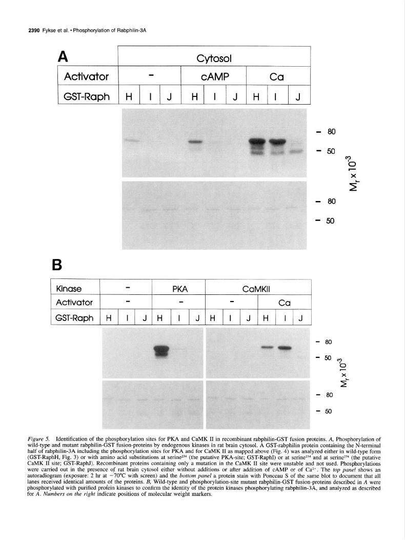
Figure 5. Identification of the phosphorylation sites for PKA and CaMK II in recombinant rabphilin-GST fusion proteins. A, Phosphorylation of wild-type and mutant rabphilin-GST fusion-proteins by endogenous kinases in rat brain cytosol. A GST-rabphilin protein containing the N-terminal half of rabphilin-3A including the phosphorylation sites for PKA and for CaMK II as mapped above (Fig. 4) was analyzed either in wild-type form (GST-RaphH, Fig. 3) or with amino acid substitutions at serine *B (the putative PKA-site; GST-RaphI) or at serine234 and at serine*74 (the putative CaMK II site; GST-RaphI). Recombinant proteins containing only a mutation in the CaMK II site were unstable and not used. Phosphorylations were carried out in the presence of rat brain cytosol either without additions or after addition of CAMP or of Ca*+. The top panel shows an autoradiogram (exposure: 2 hr at -70°C with screen) and the bottom panel a protein stain with Ponceau S of the same blot to document that all lanes received identical amounts of the proteins. & Wild-type and phosphorylation-site mutant rabphilin-GST fusion-proteins described in A were phosphorylated with purified proiein kinases to confirm the identity of the protein kinases phosphorylating rabphilin-3A, and analyzed as described for A. Numbers on the right indicate positions of molecular weight markers.

The Journal of Neuroscience, March 1995, 75(3) 2391
Pi
P-Ser
P-Thr
Activator : - CAMP cGMP Co
Kinase : - - - -
aptosomes by resuspension in 10 mu HEPES-NaOH pH 7.4 and re- taining 10 mu HEPES-NaOH pH 7.4, 2 mM MgCl,, 1 mM EGTA, 25 moval of the membranes by centrifugation at 12,000 X g. Purified cat- PM y3*P-ATP (4000-6000 cpm/pmol) and rat brain cytosol (25 mg/l) alytic subunit of PKA was purchased from Sigma, and purified CaMK with either no additions or with additions of the following activators: II was a generous gift from Dr. H. Schulman (Stanford University) and 50 PM CAMP, 50 ~,LM cGMP, or 0.4 mu Ca2+/0.1 mM EGTA (instead from Dr. M. Kennedy (CalTech, Pasadena). Immunoprecipitated rab- of 1 mu EGTA). Reactions were incubated at 37°C for 10 min followed philin-3A from rat brain or transfected COS cells (100-300 ng estimated by 3 min at room temperature. After the incubations, an equal volume by SDS-PAGE and Coomassie-blue staining) bound to protein G-Se- of 5 mu HEPES-NaOH pH 7.4, 125 mM NaCl, 0.5% Triton X-100, and pharose with antibodies was phosphorylated in a reaction (50 ~1) con- 2.5 mM NaF was added. Protein G-Sepharose beads were centrifuged
PKA
P-Tyr Figure 6. Phosphoamino acid analy- sis of the rabphilin-GST fusion protein phosphorylated by rat brain protein ki- nases or by PKA. GST-RaphH (Fig. 3)
t phosphorylated by endogenous protein kinases in rat brain cytosol in the pres- ence or absence of the indicated acti- vators and by PKA (right lane) was studied by acid hydrolysis and phos- phoamino acid analysis. The migration positions of inorganic phosphate (Pi) and of phosphoamino acid standards as
Origin well as the position of the origin are shown on the right. The arrow points to phosphopeptides. Similar analyses with identical results were performed with phosphorylated rabphilin-3A iso- lated by immunoprecipitation (data not shown). Autoradiogram was exposed for 30 hr with screen at -70°C.
Activator - Ca - - - ca - - - Ca - - - Ca - - - Ca
Plasmid Ctrl. pCMV&-4 pcwa5-3 pCMV&-5 pCMV85-6
Rabphilln + 32 P
Rabphilin +
lmmunoblot
Figure 7. Phosphorylation of wild-type rabphilin-3A and rabphilin-3A with point mutations in putative phosphorylation sites expressed by trans- fection in COS cells. COS cells were transfected with vectors encoding wild-type rat rabphilin-3A (pCMV85-4), rabphilin- 3A in which serine234 was substituted for alanine (pCMV85-3), rabphilin-3A in which serine274 was substituted for alanine (pCMV85-5), and rabphilin-3A with both point mutations (pCMV85-6). Rabphilin-3A was immunoprecipitated from the cytosol of the transfected COS cells and phosphorylated by adding y3*P- ATP, Ca2+, and purified protein kinases as indicated. As a control, COS cells transfected with wild-type rabphilin-3A expression vector were immunoprecipitated with preimmune serum but otherwise analyzed as the other immunoprecipitates (I& lanes labeled “Ctrl.“). Samples were analyzed by SDS-PAGE and autoradiography [top panel; exposure at -70°C with screen for 8 hr (left half) or for 13 hr (right half)] or immunoblot with a rubphilin-3A antibody (bottom panel) to ensure comparable levels of recombinant rabphilin-3A for the different mutants. All rabphilin-3A immunoblots of transfected material show a doublet except for pCMV85-5. This doublet does not correlate with phosphorylation and is currently unexplained.

2392 Fykse et al. - Phosphotylation of Rabphilin-3A
Rab3A-Binding 1 Phosphorylation 1 Ca*+/Phospholipid Binding
Rabphilin-3A N C Cysteine-rich p p a b
C2 Domains
Synaptotagmin
Figure 8. Comparative domain models of rabphilin-3A and synaptotagmin. Bar diagrams compare the domain structure of rabphilin-3A (rap panel) with that of synaptotagmin (Perin et al., 1991). Rabphilin domains are identified above the bar diagram. Positions of phosphorylation sites are indicated by Ps. The two &domains of rabphilin-3A and synaptotagmin are labeled n and b, and the single transmembrane region in synap- totagmin is identified by TMR.
and washed twice in this buffer and once in phosphate-buffered saline before analysis of bound proteins by SDS-PAGE followed by blotting onto nitrocellulose membrane and autoradiography. Phosphorylation of itnmunoprecipitated rabphilin-3A from rat brain or from transfected COS cells by the catalytic subunit of PKA was performed in an identical manner as described above except that the PKA catalytic subunit was added instead of the rat brain cytosol. For the phosphorylations by purified CaMK II, incubations contained 10 mM MgCl,, 10 mu Dn, 2.5 pg calmodulin, and 2.5 ng purified CaMK II in addition to or in- stead of the components listed above, and either 1 mM EGTA (control) or 1 mu Ca*+, 0.1 mM EGTA (test) to stimulate the kinase. All phos- phorylation experiments utilizing recombinant bacterial proteins ex- pressed as GST-fusion proteins were performed analogously to those described for immunoprecipitated rabphilin-3As except that fusion pro- teins bound to glutathione-agarose beads were used as substrates for the phosphorylation reactions instead of immunoprecipitated rabphilin-3A bound to protein G-Sepharose via antibodies. All phosphorylation re- actions were started by the addition of protein kinase.
Phosphoamino acid analysis was performed by blotting phosphory- lated proteins separated by SDS-PAGE onto polyvinylidene difluoride membranes, cutting out the appropriate band, and hydrolyzing the pro- tein in 5.7 M HCl at 110°C for 90 min. The hydrolysate was lyophilized, resuspended in pyridine:acetic acid:water (5:50:945), spotted onto cel- lulose membrane sheets (Kodak) together with phosphoamino acid stan- dards (Sigma), and analyzed by electrophoresis (1 hr at 950 V) followed by autoradiography. Location of standards was identified by ninhydrin treatment.
Results In order to study the phosphorylation of synaptic proteins im- plicated in regulating synaptic vesicle traffic, we isolated can- didate proteins by immunoprecipitation, mixed them with rat brain cytosol as a protein kinase source, and studied their phos- phorylation as a function of cyclic nucleotides and Ca*+. Using this approach, we observed a dramatic increase in the phos- phorylation of rabphilin-3A as a function of cyclic nucleotides and of Ca*+ (Fig. 1). Immunoprecipitates obtained with preim- mune serum as a control exhibited no phosphorylated band at the molecular weight of rabphilin-3A. CAMP, cGMP, and Ca2+ triggered phosphorylation of rabphilin-3A, suggesting that it is a substrate for CAMP- and cGMP-dependent protein kinases and for a Ca*+-stimulated kinase. Ca2+-stimulated phosphorylation of rabphilin-3A is approximately twice as effective as that trig- gered by cyclic nucleotides. Addition of Y~~P-ATP to immuno- precipitated rabphilin-3A without cytosol also caused rabphilin- 3A phosphorylation. However, this background phosphorylation was variable between experiments (compare the second lane in Fig. 1 with third lane in Fig. 2), and recombinant rabphilin-3A showed no phosphorylation in the absence of added kinases (data not shown, see below), suggesting that rabphilin-3A does
not autophosphorylate but that an unidentified protein kinase can coimmunoprecipitate with rabphilin-3A.
To determine which protein kinases are responsible for the CAMP- and Ca*+-stimulated phosphorylation of rabphilin-3A, we incubated immunoprecipitated rabphilin-3A with the catalyt- ic subunit of PKA and with purified CaMK II (Fig. 2). Both phosphorylated rabphilin-3A, with Ca’+-dependent CaMK II phosphorylation, again exceeding PKA phosphorylation. Thus, rabphilin-3A is a likely target for both PKA and CaMK II.
Rabphilin-3A has a distinct domain structure with three over- all regions: an N-terminal sequence that binds rab3A, an inter- mediate region of undetermined function, and a C-terminal re- gion with two C,-domains that probably mediate Ca*+/phospho- lipid binding (Yamaguchi et al., 1993; Li et al., 1994). To lo- calize the phosphorylation sites for PKA and for CaMK II in the structure of rabphilin-3A, we constructed a series of recom- binant glutathione S-transferase (GST) fusion proteins with rab- philin-3A that cover rabphilin-3A (Fig. 3). These proteins were expressed in bacteria, purified, and used in phosphorylation re- actions with rat brain cytosol in the presence and absence of CAMP and Ca*+ as activators (Fig. 4). Low levels of phosphor- ylation were observed with two of the seven GST-rabphilin fu- sion proteins (GST-RaphC and D) in the absence of CAMP or Ca2+. Upon addition of CAMP, only GST-RaphC was strongly phosphorylated, whereas addition of Ca2+ stimulated phosphor- ylation of GST-RaphC and GST-RaphD (Fig. 4). Ca*+ also stim- ulated weak phosphorylation of GST-RaphE.
The rabphilin-3A sequences expressed in GST-RaphC and GST-RaphD are from the middle region of rabphilin-3A, with GST-RaphD containing the C-terminal half of the rabphilin-3A insert of GST-RaphC (Fig. 3). The selective phosphorylation of GST-RaphC and GST-RaphD by CAMP- and Ca*+-stimulated protein kinases in rat brain localizes the major phosphorylation sites in rabphilin-3A to the middle region of rabphilin-3A, sug- gesting that this middle region represents a regulatory domain. The fact that CAMP stimulates phosphorylation of GST-RaphC but not of GST-RaphD suggests that the CAMP-dependent phos- phorylation site of rabphilin-3A is localized to the N-terminal part of the rabphilin insert in GST-RaphC. Conversely, efficient Ca2+-stimulated phosphorylation of both GST-RaphC and GST- RaphD indicates that the Ca Z+-dependent phosphorylation site of rabphilin-3A is localized in the C-terminal half of the GST- RaphC insert.
We, therefore, analyzed the sequences of rat and bovine rab- philin-3A, in particular, its middle region, for PKA and CaMK
The Journal of Neuroscience, March 1995, 75(3) 2393
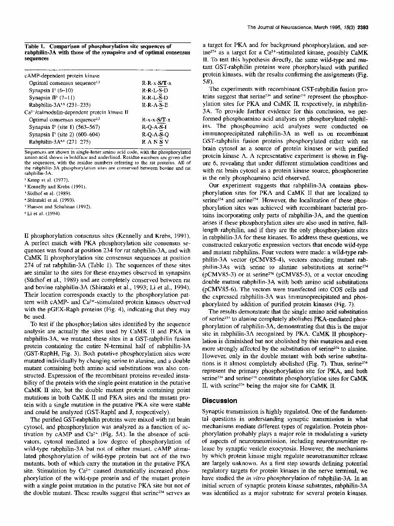
Table 1. Comparison of pbospborrlation site sequences of rabpbilin-3A with those of tbeSsy&psins and of optimal consensus sequences
CAMP-dependent protein kinase Optimal consensus sequence’,* Synapsin 1” (6-10) Synapsin II’ (7-l 1) Rabphilin-3A4,6 (231-235)
Ca2+/calmodulin-dependent protein Optimal consensus sequence2,5 Synapsin I3 (site 1) (563-567) Synapsin Is (site 2) (600-604) Rabphilin-3A4,6 (271-275)
kinase II
R-R-x-S/T-x R-R-L-S-D R-R-L-S-D R-R-A-%-E -
R-x-x-S/T-x R-Q-AX R-Q-A-S-Q R-A-N-S-V -
Sequences are shown in single-letter amino acid code, with the phosphorylated amino acid shown in boldface and underlined. Residue numbers arc given after the sequences, with the residue numbers referring to the rat proteins. All of the rabphilin-3A phosphorylation sites are conserved between bovine and rat rabphilin-3A. ’ Kemp et al. (1977). * Kennelly and Krebs (1991).
7 Siidhof et al. (1989). 4 Shirataki et al. (1993). 5 Hanson and Schulman (1992).
6 Li et al. (1994).
II phosphorylation consensus sites (Kennelly and Krebs, 1991). A perfect match with PKA phosphorylation site consensus se- quences was found at position 234 for rat rabphilin-3A, and with CaMK II phosphorylation site consensus sequences at position 274 of rat rabphilin-3A (Table 1). The sequences of these sites are similar to the sites for these enzymes observed in synapsins (Siidhof et al., 1989) and are completely conserved between rat and bovine rabphilin-3A (Shirataki et al., 1993; Li et al., 1994). Their location corresponds exactly to the phosphorylation pat- tern with CAMP- and Ca2+-stimulated protein kinases observed with the pGEX-Raph proteins (Fig. 4), indicating that they may be used.
To test if the phosphorylation sites identified by the sequence analysis are actually the sites used by CaMK II and PKA in rabphilin-3A, we mutated these sites in a GST-rabphilin fusion protein containing the entire N-terminal half of rabphilin-3A (GST-RaphH, Fig. 3). Both putative phosphorylation sites were mutated individually by changing serine to alanine, and a double mutant containing both amino acid substitutions was also con- structed. Expression of the recombinant proteins revealed insta- bility of the protein with the single point mutation in the putative CaMK II site, but the double mutant protein containing point mutations in both CaMK II and PKA sites and the mutant pro- tein with a single mutation in the putative PKA site were stable and could be analyzed (GST-RaphI and J, respectively).
The purified GST-rabphilin proteins were mixed with rat brain cytosol, and phosphorylation was analyzed as a function of ac- tivation by CAMP and Ca*+ (Fig. 5A). In the absence of acti- vators, cytosol mediated a low degree of phosphorylation of wild-type rabphilin-3A but not of either mutant. CAMP stimu- lated phosphorylation of wild-type protein but not of the two mutants, both of which carry the mutation in the putative PKA site. Stimulation by Ca2+ caused dramatically increased phos- phorylation of the wild-type protein and of the mutant protein with a single point mutation in the putative PKA site but not of the double mutant. These results suggest that serine234 serves as
a target for PKA and for background phosphorylation, and ser- ine274 as a target for a Ca2+-stimulated kinase, possibly CaMK II. To test this hypothesis directly, the same wild-type and mu- tant GST-rabphilin proteins were phosphorylated with purified protein kinases, with the results confirming the assignments (Fig. 5B).
The experiments with recombinant GST-rabphilin fusion pro- teins suggest that serine234 and serine274 represent the phosphor- ylation sites for PKA and CaMK II, respectively, in rabphilin- 3A. To provide further evidence for this conclusion, we per- formed phosphoamino acid analyses on phosphorylated rabphil- ins. The phosphoamino acid analyses were conducted on immunoprecipitated rabphilin-3 A as well as on recombinant GST-rabphilin fusion proteins phosphorylated either with rat brain cytosol as a source of protein kinases or with purified protein kinase A. A representative experiment is shown in Fig- ure 6, revealing that under different stimulation conditions and with rat brain cytosol as a protein kinase source, phosphoserine is the only phosphoamino acid observed.
Our experiment suggests that rabphilin-3A contains phos- phorylation sites for PKA and CaMK II that are localized to serine234 and serine274. However, the localization of these phos- phorylation sites was achieved with recombinant bacterial pro- teins incorporating only parts of rabphilin-3A, and the question arises if these phosphorylation sites are also used in native, full- length rabphilin, and if they are the only phosphorylation sites in rabphilin-3A for these kinases. To address these questions, we constructed eukaryotic expression vectors that encode wild-type and mutant rabphilins. Four vectors were made: a wild-type rab- philin-3A vector (pCMV85-4), vectors encoding mutant rab- philin-3As with serine to alanine substitutions at serine234 (pCMV85-3) or at serine274 (pCMV85-5), or a vector encoding double mutant rabphilin-3A with both amino acid substitutions (pCMV85-6). The vectors were transfected into COS cells and the expressed rabphilin-3A was immunoprecipitated and phos- phorylated by addition of purified protein kinases (Fig. 7).
The results demonstrate that the single amino acid substitution of serine2”4 to alanine completely abolishes PKA-mediated phos- phorylation of rabphilin-3A, demonstrating that this is the major site in rabphilin-3A recognized by PKA. CaMK II phosphory- lation is diminished but not abolished by this mutation and even more strongly affected by the substitution of serine274 to alanine. However, only in the double mutant with both serine substitu- tions is it almost completely abolished (Fig. 7). Thus, serine234 represent the primary phosphorylation site for PKA, and both serine234 and serine274 constitute phosphorylation sites for CaMK II, with serine274 being the major site for CaMK II.
Discussion
Synaptic transmission is highly regulated. One of the fundamen- tal questions in understanding synaptic transmission is what mechanisms mediate different types of regulation. Protein phos- phorylation probably plays a major role in modulating a variety of aspects of neurotransmission, including neurotransmitter re- lease by synaptic vesicle exocytosis. However, the mechanisms by which protein kinase might regulate neurotransmitter release are largely unknown. As a first step towards defining potential regulatory targets for protein kinases in the nerve terminal, we have studied the in vitro phosphorylation of rabphilin-3A. In an initial screen of synaptic protein kinase substrates, rabphilin-3A was identified as a major substrate for several protein kinases.
2394 Fykse et al. * Phosphorylation of Rabphilin-3A
Two protein kinases that phosphorylate rabphilin-3A and the sites in rabphilin-3A that they phosphorylate were identified.
The structure of rabphilin-3A predicts the presence of three major regions, of which the N- and C-terminal domains have functions in mediating rab3A binding and Ca2+/phospholipid binding, respectively. However, the function of the middle re- gion, the least conserved of the three regions of rabphilin-3A (Li et al., 1994) is unclear. As indicated in the domain model shown in Figure 8, our studies now reveal that this middle region of rabphilin-3A contains phosphorylation sites for PKA and CaMK II, suggesting that it may act as a regulatory domain.
Rabphilin-3A has several properties in common with synap- totagmin: both proteins are synaptic vesicle Ca2+-binding pro- teins with C-terminal Ca2+-binding domains and N-terminal membrane attachment sequences. Furthermore, synaptotagmin is also a phosphoprotein with phosphorylation sites N-terminal to the Ca2+-binding domains. In spite of these similarities illustrat- ed in Figure 8, there are obvious differences: synaptotagmin is an intrinsic membrane protein, whereas rabphilin-3A is only loosely attached to membranes, probably via rab3A, and syn- aptotagmin is only phosphorylated by casein kinase II, whereas rabphilin-3A is phosphorylated by at least two kinases, PKA and CaMK II, and may also be phosphorylated by cGMP-dependent protein kinases and other Ca 2+-dependent protein kinases.
Our studies were restricted to phosphorylation of rabphilin- 3A in vitro. It is unknown if rabphilin-3A is also phosphorylated in vivo but the sites defined in the current study ideally conform to the consensus phosphorylation sites used by these kinases in vivo (Table 1). Rabphilin-3A appears to be an excellent substrate for these kinases in vitro, suggesting that rabphilin-3A probably also is a substrate for these kinase in vivo, although this will have to be investigated in future experiments.
The phosphorylation of rabphilin-3A by two different protein kinases at closely linked sites is intriguing. The biochemical properties of rabphilin-3A are suggestive of a regulatory role in neurotransmission (Yamaguchi et al., 1993; Geppert et al., 1994; Li et al., 1994). As a Ca2+ binding protein localized to synaptic vesicles, rabphilin-3A may participate in a Ca2+-regulated aspect of synaptic vesicle function. Indeed, experiments with mice de- ficient in rab3A demonstrated that in synapses lacking rab3A and rab3C (which may be redundant with rab3A at some syn- apses but is not present at most synapses), rabphilin-3A is not transported to synapses. Physiological measurements on such synapses revealed a potential defect in synaptic vesicle recy- cling, suggesting a possible role for rabphilin-3A in a Ca*+- dependent step in synaptic vesicle recycling (Geppert et al., 1994). The regulation of such an activity by protein phosphor- ylation would be an attractive mechanism to modulate synaptic vesicle exocytosis as a function of Ca*+ in nerve terminals.
The phosphorylation of rabphilin-3A by multiple protein ki- nases is reminiscent of that of synapsin I which is also a pe- ripheral membrane protein of synaptic vesicles that is a substrate for PKA and CaMK II (reviewed in Greengard et al., 1993; Jahn and Stidhof, 1994). Currently rabphilin-3A and synapsin I are the only characterized major synaptic CaMK II and PKA sub- strates. Both are abundant synaptic proteins, suggesting a poten- tial regulatory role for phosphorylation in the synaptic vesicle pathway. Cah4K II has demonstrated major effects on neuro- transmitter release (Llinas et al., 1991) that, based on the ge- netics, are clearly not mediated by synapsin I (Rosahl et al., 1993). PKA also has been implicated in several synaptic regu- latory processes (Frey et al., 1993; Chavez-Noriega and Stevens,
1994). Our results suggest that rabphilin-3A may be one of the targets by which the regulatory effects of CaMK II and PKA could be mediated. This will have to be addressed in future experiments using mice lacking rab3A (which exhibit a major decrease in rabphilin-3A levels in most synapses but not a com- plete loss of rabphilin-3A; Geppert et al., 1994; Li et al., 1994) and mice that are completely deficient in rabphilin-3A.
References
Bekkers JM, Stevens CF (1990) Presynaptic mechanism for long-term potentiation in the hippocampus. Nature 346:724-729.
Bolshakov VU, Siegelbaum SA (1994) Postsynaptic induction and pre- synaptic expression of hippocampal long-term depression. Science 264:1148-l 152.
Bradford MM (1976) A rapid and sensitive method for the quantitation of microgram quantities of protein utilizing the principle of protein- dye binding. Anal Biochem 72:248-254.
Chavez-Noriega LE, Stevens CF (1994) Increased transmitter release at excitatory synapses produced by direct activation of adenylate cy- clase in rat hippocampal slices. J Neurosci 14:310-317.
Dale N, Kandel ER (1990) Facilitory and inhibitory transmitters mod- ulate spontaneous transmitter release at cultured Aplysia sensorimotor synapses. J Physiol (Lond) 421:203-222.
De Camilli P, Cameron R, Greengard P (1983a) Synapsin I (Protein I), a nerve terminal-specific phosphoprotein: I. Its general distribution in synapses of the central and peripheral nervous system demonstrat- ed by immunofluorescence in frozen and plastic sections. J Cell Biol 96:1337-1354.
De Camilli P, Harris SM, Huttner WB, Greengard P (1983b) Synapsin I (Protein I), a nerve terminal-specific nhosnhonrotein: II. Its specific association with synaptic vesicles demor&trated by immunocyto- chemistry in agarose-embedded synaptosomes. J Cell Biol 96:1355- 1373.
Dekker LV, De Graan PNE, Oestreicher AB, Versteeg DHG, Gispen WH (1989) Inhibition of noradrenaline release by antibodies to B-50 (GAP-43). Nature 342:74-76.
Frey Y, Huang Y-Y, Kandel ER (1993) Effects of CAMP simulate a late stage of LTP in hippocampal CA1 neurons. Science 260:1661- 1664.
Geppert M, Bolshakov VU, Siegelbaum SA, Takei K, De Camilli P Hammer RE, Stidhof TC (1994) Rab3A function in neurotransmitter release. Nature 369:493497.
Gorman C (1985) High efficiency gene transfer into mammalian cells. DNA cloning volume II (Glover DM, ed), pp 143-190. Oxford: IRL.
Greencard P, Valtorta E Czernik AJ. Benfenati F (1993) Svnautic ves- , d I
icle phosphoproteins and regulation of synaptic function. Science 259:780-785.
Guan KL, Dixon JE (1991) Eukaryotic proteins expressed in Es&e- richia coli: an improved thrombin cleavage and purification proce- dure of fusion proteins with glutathione S-transferase. Anal Biochem 192~262-267.
Hanson PI, Schulman H (1992) Neuronal Ca2+/calmodulin-dependent protein kinases. Annu Rev Biochem 61:559-601.
Jahn R, Stidhof TC (1994) Synaptic vesicles and exocytosis. Annu Rev Neurosci 17:219-246.
Johnston PA, Jahn R, Stidhof TC (1989) Transmembrane topography and evolutionary conservation of synaptophysin. J Biol Chem 264: 1268-l 273.
Kemp BE, Graves DJ, Benjamini E, Krebs EG (1977) J Biol Chem 252:4888-4892.
Kennelly PJ, Krebs EG (199 1) Consensus sequences as substrate spec- ificity determinants for protein kinases and protein phosphatases. J Biol Chem 266: 15555-15558.
Kishida S, Shirataki H, Sasaki T, Kato M, Kaibuchi K, Takai Y (1993) Rab3A GTPase-activating protein-inhibiting activity of Rabphilin- 3A, a putative Rab3A target protein. J Biol Chem 268:22259-22261.
Laemmli UK (1970) Cleavage of structural proteins during the assem- bly of the head of bacteriophage T4. Nature 227:680-685.
Li C, Takei K, Geppert M, Daniel1 L, Stenius K, Chapman ER, Jahn R, De Camilli P, Stidhof TC (1994) Synaptic targeting of rabphilin- 3A, a synaptic vesicle Caz+/phospholipid-binding protein, depends on rab3A/3C. Neuron 13:885-898.
Llinas R, Gruner JA, Sugimori M, McGuinness TL, Greengard P
The Journal of Neuroscience, March 1995, 75(3) 2395
(1991) Regulation by synapsin I and Ca2+-calmodulin-dependent protein kinase II of the transmitter release in squid giant synapse. J Physiol (Lond) 436:257-282.
Malinow R, Tsien RW (1990) Presynaptic enhancement shown by whole-cell recordings of long-term potentiation in hippocampal slic- es. Nature 346: 177-180.
Man-Son-Hing H, Zoran MJ, Lukowiak K, Haydon PG (1989) A neu- romodulator of synaptic transmission acts on the secretory apparatus as well as on ion channels. Nature 341:237-239.
Nichols RA, Sihra TS, Czernik AJ, Nairn AC, Greengard P (1990) Calcium/calmodulin-dependent protein kinase II increases glutamate and noradrenaline release from synaptosomes. Nature 343:647-651.
Nicoll RA, Malenka RC, Kauer JA (1990) Functional comparison of neurotransmitter receptor subtypes in mammalian central nervous system. Physiol Rev 70513-565.
Perin MS, Brose N, Jahn R, Siidhof TC (1991) Domain structure of synaptotagmin (~65). J Biol Chem 266:623-629.
Robinson PJ, Sontag JM, Liu J-P Fykse EM, Slaughter C, McMahon H, Stidhof TC (1993) Dynamin GTPase regulated by protein kinase C phosphorylation in nerve terminals. Nature 365: 163-166.
Rosahl T, Geppert M, Spillane D, Herz J, Hammer RE, Malenka RC, Sttdhof TC (1993) Short term synaptic plasticity is altered in mice lacking synapsin I. Cell 75:661-670.
Sambrook J, Fritsch EE Maniatis T (1989) Molecular cloning. A lab- oratory manual, 2d ed. Cold Spring Harbor, NY: Cold Spring Harbor Laboratory.
Sasaki T, Kikuchi A, Araki S, Hata Y, Isomura M, Kuroda S, Takai Y (1990) Purification and characterization from bovine brain cytosol of a protein that inhibits the dissociation of GDP from and the subse- quent binding of GTP to smg p25A, a ras p21-like GTP-binding protein. J Biol Chem 265:2333-2337.
Scholz Kp, Miller RJ (1992) Inhibition of quanta1 transmitter release in the absence of calcium influx by a G protein-linked adenosine receptor at hippocampal synapses. Neuron 8: 1139-l 150.
Shirataki H, Kaibuchi K, Yamaguchi T, Wada K, Horiuchi H, Takai Y (1992) A possible target protein for smg-25A/rab3A small GTP- binding protein. J Biol Chem 267:10946-10949.
Shirataki H, Kaibuchi K, Sakoda T Kishida S, Yamaguchi T, Wada K,
Miyazaki M, Takai Y (1993) Rabphilin-3A, a putative target protein for smg p25A/rab3A small GTP-binding protein related to synapto- tagmin. Mol Cell Biol 13:2061-2068.
Silinsky EM (1984) On the mechanism by which adenosine receptor activation inhibits the release of acetylcholine from motor nerve end- ings. J Physiol (Lond) 346:243-256.
Silva AJ, Stevens CF, Tonegawa S, Wang Y (1992) Deficient hippo- campal long-term potentiation in o-alcium, calmodulin kinase II mu- tant mice. Science 257:201-206.
Smith DB, Johnson KS (1988) Single-step purification of polypeptides expressed in Escherichia coli as fusions with glutathione S-transfer- ase. Gene 67:31&O.
Stevens CF (1993) Going down the way you came up. Cm-r Biol 3:891-892. Siidhof TC, Czernik AJ, Kao H, Takei K, Johnston PA, Horiuchi A, Kanazir SD, Wagner MA, Perin MS, De Camilli P Greengard P (1989) Synapsins: mosaics of shared and individual domains in a family of synaptic vesicle phosphoproteins. Science 245:1474-1480.
Sfidhof TC, Jahn R (1991) Proteins of synaptic vesicles involved in exocytosis and membrane recycling. Neuron 6:665-677.
Takeuchi A. Takeuchi N (1966) A studv of the inhibitorv action of gamma-aminobutyric acid on neuromuscular transmissionin the cray- fish. J Physiol (Lond) 183:418-432.
Weisskopf MG, Zalutsky RA, Nicoll RA (1993) The opioid peptide dynorphin mediates heterosynaptic depression of hippocampal mossy fiber synapses and modulates long-term potentiation. Nature 362: 423-427.
Yamaguchi T, Shirataki H, Kishida S, Miyazaki M, Nishikawa J, Wada K, Numata S, Kaibuchi K, Takai Y (1993) Two functionally different domains of Rabphilin3A, Rab3A p25fsmg p25A-binding and phos- pholipid- and CaZ+ binding domains. J Biol Chem 268:2716&27170.
Zalutsky RA, Nicoll RA (1990) Comparison of two forms of long-term potentiation in single hippocampal neurons. Science 248:1619-1624.
Zhuo M, Hu Y, Schultz C, Kandel ER, Hawkins RD (1994) Role of guanylyl cyclase and cGMP-dependent protein kinase in long-term potentiation. Nature 368:635-639.
Zucker RS (1989) Short-term synaptic plasticity. Annu Rev Neurosci 12:13-31.