Embed Size (px)
Citation preview
Aris Kaksis. Riga Stradin’s University 2019 http://aris.gusc.lv/06Daugavpils/Research/LipdBiLayerMembran.pdf
1
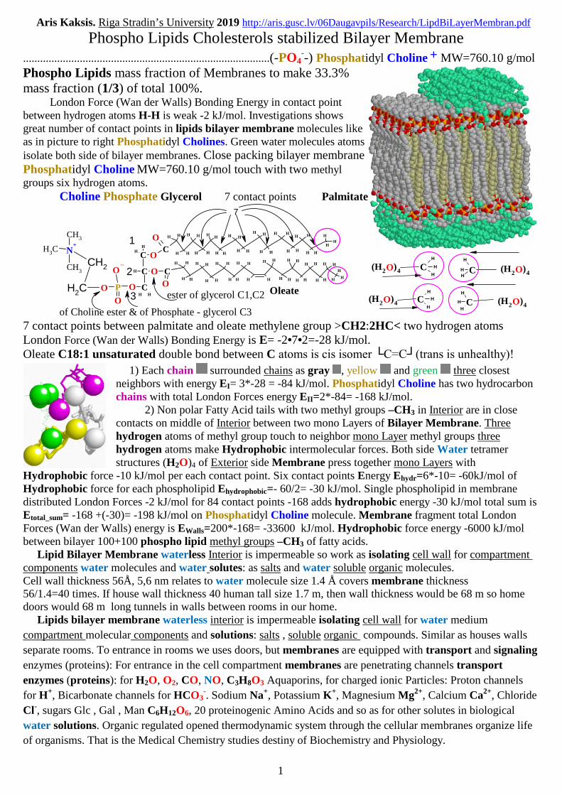
Phospho Lipids Cholesterols stabilized Bilayer Membrane.......................................................................................(-PO4
--) Phosphatidyl Choline + MW=760.10 g/mol
Phospho Lipids mass fraction of Membranes to make 33.3%mass fraction (1/3) of total 100%.
London Force (Wan der Walls) Bonding Energy in contact pointbetween hydrogen atoms H-H is weak -2 kJ/mol. Investigations showsgreat number of contact points in lipids bilayer membrane molecules likeas in picture to right Phosphatidyl Cholines. Green water molecules atomsisolate both side of bilayer membranes. Close packing bilayer membranePhosphatidyl Choline MW=760.10 g/mol touch with two methylgroups six hydrogen atoms.
Choline Phosphate Glycerol 7 contact points Palmitate
CH H
HH
H H
HH
H H
HH
H H
H H
H H
HH
H H
HH
HH
H H
HH
H H
H
C
HH
H H
HH
H H
HH
HH
H H
HH
H H
HH
H H
HH
H H
H H
HH
H
CH
HN+
CH3
CH3
CH3
CH2
CH2
O P
O
O
O CHH
CH
O
O
O
O
1
2
3Oleateester of glycerol C1,C2
of Choline ester & of Phosphate - glycerol C3
7
CH
H
H
C
H
HH
CH
H
H
CH
H
H
(H )O2 4 (H )O2 4
(H )O2 4 (H )O2 4
7 contact points between palmitate and oleate methylene group >CH2:2HC< two hydrogen atomsLondon Force (Wan der Walls) Bonding Energy is E= -2•7•2=-28 kJ/mol.Oleate C18:1 unsaturated double bond between C atoms is cis isomer └C=C┘(trans is unhealthy)!
1) Each chain surrounded chains as gray , yellow and green three closestneighbors with energy EI= 3*-28 = -84 kJ/mol. Phosphatidyl Choline has two hydrocarbonchains with total London Forces energy EII=2*-84= -168 kJ/mol.
2) Non polar Fatty Acid tails with two methyl groups –CH3 in Interior are in closecontacts on middle of Interior between two mono Layers of Bilayer Membrane. Threehydrogen atoms of methyl group touch to neighbor mono Layer methyl groups threehydrogen atoms make Hydrophobic intermolecular forces. Both side Water tetramerstructures (H2O)4 of Exterior side Membrane press together mono Layers with
Hydrophobic force -10 kJ/mol per each contact point. Six contact points Energy Ehydr=6*-10= -60kJ/mol ofHydrophobic force for each phospholipid Ehydrophobic=- 60/2= -30 kJ/mol. Single phospholipid in membranedistributed London Forces -2 kJ/mol for 84 contact points -168 adds hydrophobic energy -30 kJ/mol total sum isEtotal_sum= -168 +(-30)= -198 kJ/mol on Phosphatidyl Choline molecule. Membrane fragment total LondonForces (Wan der Walls) energy is EWalls=200*-168= -33600 kJ/mol. Hydrophobic force energy -6000 kJ/molbetween bilayer 100+100 phospho lipid methyl groups –CH3 of fatty acids.
Lipid Bilayer Membrane waterless Interior is impermeable so work as isolating cell wall for compartmentcomponents water molecules and water solutes: as salts and water soluble organic molecules.Cell wall thickness 56Å, 5,6 nm relates to water molecule size 1.4 Å covers membrane thickness56/1.4=40 times. If house wall thickness 40 human tall size 1.7 m, then wall thickness would be 68 m so homedoors would 68 m long tunnels in walls between rooms in our home.
Lipids bilayer membrane waterless interior is impermeable isolating cell wall for water medium
compartment molecular components and solutions: salts , soluble organic compounds. Similar as houses walls
separate rooms. To entrance in rooms we uses doors, but membranes are equipped with transport and signaling
enzymes (proteins): For entrance in the cell compartment membranes are penetrating channels transport
enzymes (proteins): for H2O, O2, CO, NO, C3H8O3 Aquaporins, for charged ionic Particles: Proton channels
for H+, Bicarbonate channels for HCO3-. Sodium Na+, Potassium K+, Magnesium Mg2+, Calcium Ca2+, Chloride
Cl-, sugars Glc , Gal , Man C6H12O6, 20 proteinogenic Amino Acids and so as for other solutes in biological
water solutions. Organic regulated opened thermodynamic system through the cellular membranes organize life
of organisms. That is the Medical Chemistry studies destiny of Biochemistry and Physiology.

Aris Kaksis. Riga Stradin’s University 2019 http://aris.gusc.lv/06Daugavpils/Research/LipdBiLayerMembran.pdf
2
1. 1/3 mass fraction of membranes in cells as well organelles constitute phospho lipids as
Phosphatidyl Choline. Intermolecular forces binding energy make Ebound=-198 kJ/mol phospho lipid
membranes liquid therefore can be mechanically broken, as liquid due to gravitation, pressure and
movement. Cholesterol content make membranes stronger and flexible to prevent destruction with
following cytosol leaking of water molecules as well solution of: salts , organic compound molecules.
2. Second third part of Membranes mass constitutes hydrocarbon 27 carbon steric frame steroid.
H HH
HHH
HHH
H HH
H
O
H HH
OC
H H
HH
H H
HH
H H
HH
H H
H H
H H
HH
H H
HH
HH
H H
HH
H H
H
C
HH
H H
HH
H H
HH
HH
H H
HH
H H
HH
H H
HH
H H
H H
HH
H
CH
HN+
CH3
CH3
CH3
CH2
CH2
O P
O
O
O CHH
CH
O
O
O
1
2
3 18
19
both angular methyl groups
Lipid - Cholesterol molecule. Fourrings of the steroid are labeled A, B,C and D. Angular methyl –CH3
groups labeled 18 and 19 as well tailfork, rod, splinter are good clutchfixing close hydrocarbon chains inmembrane. Double bond betweencarbon atoms >C=C< 5 and 6
to frame steroid molecule solid and inflexible. Alcohol HO- at carbon 3 but hydroxyl group HO-H
H
H
H
H HH
H HH
H HH
H O
HH
Cholesterol
10
4
912
6758
1211 13
14 1516
A
17
18
3B
C D
19
202122
23
24
2526
27
H HH
H
HH
H
HH
H HH
H HH
H
O
OCO both angular methyl groups
18
19
forms hydrogen bond >C=O...HO- with carboxyl oxygen of fattyacid Oleate or one another fatty acid carboxyl oxygen >C=O.
Cholesterol as Steroid makes membranes unbroken, flexible and so prevent following leaking of water
molecules and of water solution components: salts and water soluble organic molecules. The
Cholesterol/Phospho Lipid C/PL mole ratio of human red blood cell membranes ranges from a normal
value of 0.9–1.0 (since 1978 first publication Journal of Cellular Biochemistry 2004 V8, 4, p 413-430).
If Cholesterol amount decreases up to 0,5= C/PL , then membranes leak cell content out, but if
Cholesterol amount increases up to 1,5= C/PL , then membrane becomes solid, inflexible and squeeze
channels, aquaporins, but receptors becomes inactive due to absence conformational flexibility.
Membrane total mass 100%=33.3%+33.3%+(20%) Aquaporins +13.3% other proteins)
I) 1/3 part constitute Phospholipids which mass fraction of Membranes to make 33.3% of total mass 100%;
II) second 1/3 part Cholesterols which mass fraction of Membranes to make 33.3% of total mass 100%;
III) third 1/3 part Membranes integral Proteins which mass fraction to make 33.3% of total mass 100%
Bulk mass fraction 20% goes to Aquaporins for other remains 13.3% are constitute four type Proteins:
1. Glycoproteins with linked O- glycoside bonds Immunological marker L-fucose Fuc and Immunological
determinants including blood groups A, B, AB, 0 located outside in extra cellular space for leucocytes-scanners
host bodies recognition. Leucocytes are scavengers non-host bodies binding to remove from Host organism.
2. Cell Structural building blocks cytoskeleton and structural integral membrane proteins;
3.Transport enzymes (channels) integral membrane proteins, 20% Aquaporins for H2O,O2,NO transport;
4. Receptors enzymes (Membranes integral Proteins) of the Signal transduction Pathway components for
biological communication inside the cells, between the cells and or tissues, as well between living organisms.
Aris Kaksis. Riga Stradin’s University 2019 http://aris.gusc.lv/06Daugavpils/Research/LipdBiLayerMembran.pdf
3
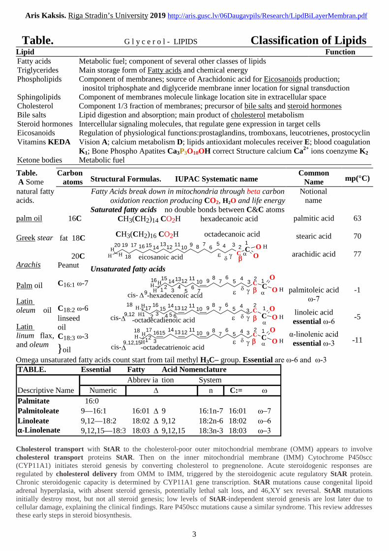
Table. G l y c e r o l - LIPIDS Classification of LipidsLipid FunctionFatty acids Metabolic fuel; component of several other classes of lipidsTriglycerides Main storage form of Fatty acids and chemical energyPhospholipids Component of membranes; source of Arachidonic acid for Eicosanoids production;
inositol triphosphate and diglyceride membrane inner location for signal transductionSphingolipids Component of membranes molecule linkage location site in extracellular spaceCholesterol Component 1/3 fraction of membranes; precursor of bile salts and steroid hormonesBile salts Lipid digestion and absorption; main product of cholesterol metabolismSteroid hormones Intercellular signaling molecules, that regulate gene expression in target cellsEicosanoids Regulation of physiological functions:prostaglandins, tromboxans, leucotrienes, prostocyclinVitamins KEDA Vision A; calcium metabolism D; lipids antioxidant molecules receiver E; blood coagulation
K1; Bone Phospho Apatites Ca3P3O10OH correct Structure calcium Ca2+ ions coenzyme K2
Ketone bodies Metabolic fuel
Table.A Some
Carbonatoms Structural Formulas. IUPAC Systematic name
CommonName mp(°C)
natural fattyacids.
palm oil
Greek stear
Arachis
Palm oil
Latinoleum oil
Latinlinum flax,and oleum
16C
fat 18C
20CPeanut
C16:1 ω-7
C18:2 ω-6
linseedoilC18:3 ω-3
}oil
Fatty Acids break down in mitochondria through beta carbonoxidation reaction producing CO2, H2O and life energy
Saturated fatty acids no double bonds between C&C atomsCH3(CH2)14 CO2H hexadecanoic acid
CH3(CH2)16 CO2H octadecanoic acid
HH
H OC
CHO
eicosanoic acid
1012 111314151617
18
1920
4
9 12
67 5
8 3
Unsaturated fatty acids
CO
C HOHH
H
-hexadecenoic acid
10
4
9 13
67 5
812 111314
1516
cis- 9
2
41 3
675
2
C OC HO
HH
H
-octadecadienoic acid
10 98
12 11131415
16
cis-9,12
1718
4
12
67 5 3
41 3 652
CO
CHO
HH
H
-octadecatrienoic acid
10
4
9 12
67 5
812 1113141516
cis-9,12,
1718
15
3
1 32
Notionalname
palmitic acid
stearic acid
arachidic acid
palmitoleic acidω-7
linoleic acidessential ω-6
α-linolenic acidessential ω-3
63
70
77
-1
-5
-11
Omega unsaturated fatty acids count start from tail methyl H3C– group. Essential are ω-6 and ω-3 TABLE. Essential Fatty Acid Nomenclature
Abbrev ia tion System
Descriptive Name Numeric D n C:= w
Palmitate 16:0
Palmitoleate 9—16:1 16:01 D 9 16:1n-7 16:01 w-7
Linoleate 9,12—18:2 18:02 D 9,12 18:2n-6 18:02 w-6α-Linolenate 9,12,15—18:3 18:03 D 9,12,15 18:3n-3 18:03 w-3
Cholesterol transport with StAR to the cholesterol-poor outer mitochondrial membrane (OMM) appears to involvecholesterol transport proteins StAR. Then on the inner mitochondrial membrane (IMM) Cytochrome P450scc(CYP11A1) initiates steroid genesis by converting cholesterol to pregnenolone. Acute steroidogenic responses areregulated by cholesterol delivery from OMM to IMM, triggered by the steroidogenic acute regulatory StAR protein.Chronic steroidogenic capacity is determined by CYP11A1 gene transcription. StAR mutations cause congenital lipoidadrenal hyperplasia, with absent steroid genesis, potentially lethal salt loss, and 46,XY sex reversal. StAR mutationsinitially destroy most, but not all steroid genesis; low levels of StAR-independent steroid genesis are lost later due tocellular damage, explaining the clinical findings. Rare P450scc mutations cause a similar syndrome. This review addressesthese early steps in steroid biosynthesis.
Aris Kaksis. Riga Stradin’s University 2019 http://aris.gusc.lv/06Daugavpils/Research/LipdBiLayerMembran.pdf
4
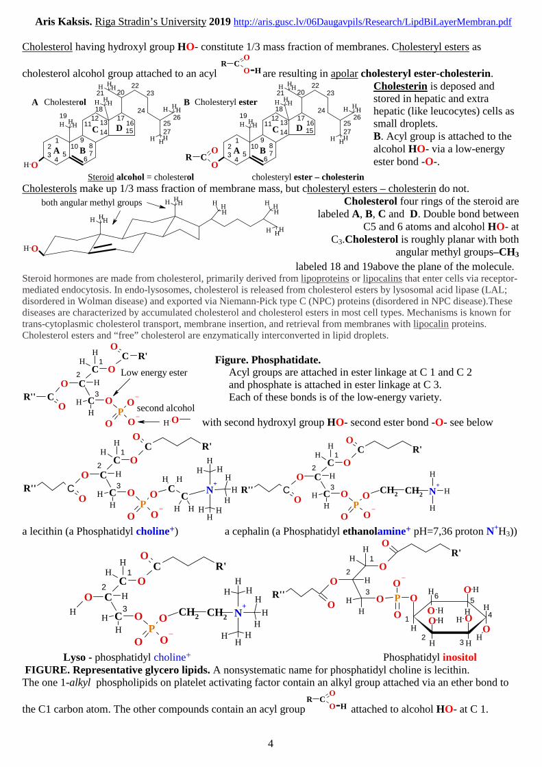
Cholesterol having hydroxyl group HO- constitute 1/3 mass fraction of membranes. Cholesteryl esters as
cholesterol alcohol group attached to an acyl
OC
OR
H are resulting in apolar cholesteryl ester-cholesterin.H
H
H
H
H HH
H HH
H HH
H O
HH
Cholesterol
10
4
912
6758
1211 13
14 1516
A
17
18
3B
C D
19
2021
A
2223
24
2526
27
HH
HH
OC
OR
H
H
H HH
H HH
H HH
Cholesteryl ester
10
4
912
6758
1211 13
14 1516
A
17
18
3B
C D
19
2021
B
2223
24
2526
27
Steroid alcohol = cholesterol cholesteryl ester – cholesterin
Cholesterin is deposed andstored in hepatic and extrahepatic (like leucocytes) cells assmall droplets.B. Acyl group is attached to thealcohol HO- via a low-energyester bond -O-.
Cholesterols make up 1/3 mass fraction of membrane mass, but cholesteryl esters – cholesterin do not.
H HH
H
HH
H
HH
H HH
H O
H HH
both angular methyl groups Cholesterol four rings of the steroid arelabeled A, B, C and D. Double bond between
C5 and 6 atoms and alcohol HO- atC3.Cholesterol is roughly planar with both
angular methyl groups–CH3
labeled 18 and 19above the plane of the molecule.Steroid hormones are made from cholesterol, primarily derived from lipoproteins or lipocalins that enter cells via receptor-mediated endocytosis. In endo-lysosomes, cholesterol is released from cholesterol esters by lysosomal acid lipase (LAL;disordered in Wolman disease) and exported via Niemann-Pick type C (NPC) proteins (disordered in NPC disease).Thesediseases are characterized by accumulated cholesterol and cholesterol esters in most cell types. Mechanisms is known fortrans-cytoplasmic cholesterol transport, membrane insertion, and retrieval from membranes with lipocalin proteins.Cholesterol esters and “free” cholesterol are enzymatically interconverted in lipid droplets.
H
H
H
O
O
C R'
O
O
CR''O
O OP
O
C
C
C
HH
OH
1
2
3
Low energy ester
second alcohol
Figure. Phosphatidate.Acyl groups are attached in ester linkage at C 1 and C 2and phosphate is attached in ester linkage at C 3.Each of these bonds is of the low-energy variety.
with second hydroxyl group HO- second ester bond -O- see below
HH
CH
H
H
N+
HHH
HHH
CR'' C
R'
H
H
H
O
O
C
O
O
O
O OP
O
C
C
C
HH
HH
1
2
3CH2
R'' C
R'
CH2
H
H
H
O
O
C
O
O
O
O OP
O
C
C
C
HH
H
H
H
N+
1
2
3
a lecithin (a Phosphatidyl choline+) a cephalin (a Phosphatidyl ethanolamine+ pH=7,36 proton N+H3))
CH2H
H
H
N+
HHH
HHH
H
R'
CH2
H
H
H
O
O
C
O
O
O OP
O
C
C
C
HH 1
2
3 H
H
H
H H
HO
H
O
OOHO H
H
O HR''
R'
H
H
H
O
O
O
O O
O
PO
HH 1
2
3
1
3
6
4
2
5
Lyso - phosphatidyl choline+ Phosphatidyl inositolFIGURE. Representative glycero lipids. A nonsystematic name for phosphatidyl choline is lecithin.The one 1-alkyl phospholipids on platelet activating factor contain an alkyl group attached via an ether bond to
the C1 carbon atom. The other compounds contain an acyl group
OC
OR
H attached to alcohol HO- at C 1.

Aris Kaksis. Riga Stradin’s University 2019 http://aris.gusc.lv/06Daugavpils/Research/LipdBiLayerMembran.pdf
5
B. Sphingolipids are derivatives of sphingosine, an amino alcohol.
Sphingosine
CC (CH2)12
CH3
O
H
H
H
H
ON
+H
H
H
H
H
H
H1
2
3 4
R
O
C CC (CH
2)
12
CH3
O
H
H
HH
ON
H
HH
H
H1
2
3 4
Ceramide
O
O HO
O H
OH
H OH
H
OO
PO
R
O
C(CH2)12
CH3
O
H
H
HH
N
H
HH
H
1
2
3 4
HH
HH
H
N+
HHH
HHH
O
O OP
O
HHR
O
C(CH
2)
12
CH3
O
H
H
HH
N
H
HH
H
1
2
3 4
Glucosyl ceramide (cerebroside) oligosaccharide ceramide(ganglioside)
FIGURE. Sphingosine andrepresentative sphingolipids.
Sphingosine is a C18compound with hydroxyl
groups —OH on C1 and C3,an amino group —NH3
+ on
C2 and a trans double bond =at C4
CC
H
H
3 4
Sphingomyelin
Arachidonic acid salt arachidonate is Phosphatidyl Choline fatty acid ester component in membranes
Four Eicosanoids are produced in enzymatic lipid peroxidation using initial compound arachidonate.Prostaglandins (PGs),Thromboxanes (TXs) andProstacyclins (PGIs),Leukotrienes (LTs).
OCH
HH
O1
23
456
789
101112
131415
1617
181920
=665
43
21
Essential ω=6 fatty acid 20-carbon compounds (Greek eikosil , "twenty") with four cis double bonds.
Almost all mammalian cells except erythrocytes produce one or more of eicosanoids,:PGA2, PGE1, PGE2, PGE3, PGF2α, PGD2, PGH2, TXA2, TXB2 PGI2, LTE4 .
Enzymatic transformation of arachidonate in Cyclo Oxygenase COX begins withcross-link between C8 —C12.
This step is target of anti-inflammatory and anti-clottinghuman blood medicine: Aspirin, Ibuprofen, Tylenol, Paracetamol, Warfarin,
which blocks cross-link between C8—C12.=>If cross-link done COX hem peroxidase iron(III) Fe3+ by donor acceptor bond
adsorbs radical oxygen singlet molecule •::O-:-O::• produce first Eicosanoids.
OCH
HH
O
HH
HO
CO
10
9 8
121113
14
1516
1718
4 26
75
19
3
20
1
PGH2 In COXI and PGD2 in COXII Peroxidation of cross-linked
O H
O
O HH
HO
CO
10
9 8
1211
13
14
1516
1718
4 26
75
19
3
20
1
O H
O O H
HH
HO
CO
10
9 8
12
11 13
14
1516
1718
4 26
75
19
3
20
1
arachidonate between C8—C12 start at C9 and C11•::O-:-O::• with following peroxidationat C15 producing hydroxyl group –OH.
Arising Prostaglandin moleculesproduce swelled size tissue inflammationphysiological reaction with strong pane.
Thromboxane is the initiating factor for blood clotting closing the damaged blood vessels.If anti-inflammatory and anti-clotting human blood medicine: Aspirin, Ibuprofen, Tylenol, Paracetamol,
Warfarin, which blocks cross-link between C8 —C12 are used than:No Prostaglandin and Thromboxane molecules arising andNo produce swelled size tissue inflammation physiological reaction with strong paneNo initiation for trombs formation in blood vessels.Symptoms of produced swelled size tissue inflammation physiological reaction with strong pane removed,Symptoms initiation for trombs formation in blood vessels are removed.

Aris Kaksis. Riga Stradin’s University 2019 http://aris.gusc.lv/06Daugavpils/Research/LipdBiLayerMembran.pdf
6
NON-ENZYMATIC Lipid peroxidation is radical initiated chain reaction providing a continues supply of freeradicals that initiate further peroxidation. The whole process (chain type reaction) is depicted as follows:
Important is to know that water plus O=O is source medium of peroxide formation agents: metal(n)+ ions,high energy ionization - radiation ~hν, peroxisomes enzymes Aldehyde OxidoReductases.
Let us start from arachidonic acid salt 4 double bonds = ω6 fatty acid Eicosanoid in Membrane Bilipid Layer:
Oxygen O=O present oxidizing power as for agent is strong and is consequently working, which Initiate in lifeorganisms bodies three different factors (1., 2., 3.) of chain reaction and its activity depends on agentsconcentration and intensity:
(2) Production of radicals R•
1. Production of radicals R• from precursor RH by metal(n)+ ion as Oxidant (Fe3+, Mn4+, Cu2+, etc).
R÷O÷O÷H + metal(n)+ (which transfer H+ and e- to Oxidant) => peroxide R÷O÷O• + metal(n--1)+ + H+
2. Production of radicals R• from precursor RH at presence of oxygen O=O high energy radiation (~hν)
Homolytic separate R÷H about H•& R• as Oxidant separate electron pair in two free electrons • • at H• and R•
R÷H + ~hν=> R• +H• similar as Oxidant metal ions hydrogen ion accept free electron H+ + e- =H• is radical.
3. Production of radicals R• from precursor RH at presence of Enzymes Aldehyde OxydoReductases inperoxisomes at presence of oxygen O=O, which concentration in cytosolic water is [O2]=6•10-5M.
2R-C=O-H + O=O (Aldehyde OxydoReductase) => peroxide 2RC÷O÷O• + 2H•
(2) Propagation (new radical R• production):
peroxide R÷O÷O• + R÷H => peroxide R÷O÷OH + R•
R• + O=O => peroxide R÷O÷O• , etc.
(3) Termination (recombination radical R• and R÷O÷O• attraction and joining):
peroxide R÷O÷O• + peroxide R-O-O• => peroxide R÷O÷O÷R + O=Operoxide R÷O÷O• + R• => peroxide R÷O÷O÷R
R• + R•=> R÷R
HH
OH
HH
O
H
H
OH
H
H
O
H
OH
HH
O
H
OH
HH
OO O
H O OH
H O OOO
HH
OO
R
R
R
OO
R
H
H
12
34
Eicosanoate~hradiation energy E
567
8910
111213
141516
1718
1920
hydrogen radical
RH
R
•
•
•
•
÷O÷O
Malonil aldehyde Endoperoxide Hydro peroxide ROOH does undergo oxidation.
FIGURE. NON-ENZYMATIC Lipid peroxidation. The reaction is initiated R•
by high energy radiation (~hν), Aldehyde OxydoReductase or by heavy metal ions Fe3+, Cu2+,Malonil aldehyde is only formed by fatty acids with 3 or more >3 double bonds and is used asmeasure of lipid peroxidation together with ethane from the terminal 2-carbon of ω3 fatty acidsand pentane from the terminal 5-carbon of ω6 fatty acids.

Aris Kaksis. Riga Stradin’s University 2019 http://aris.gusc.lv/06Daugavpils/Research/LipdBiLayerMembran.pdf
7
Apolipoproteins B-48,C-III,C-II figure 17-2
Cholesterol Triacylglycerides Phospholipids likeand Choleseryl esters
Molecularstructure
of a chylomicron.The surface is
a layer ofphospholipids-
cholesterolcomplex with headgroups facing the
aqueous phase.Triacylglyceridessequestered in theinterior (yellow)
make up more than80% of the mass.
Severalapolipoproteins
that protrude fromthe surface
(B-48, C-lll, C-ll)act as signals in the
uptake andmetabolism of
Lipoproteinvesicle content.The diameter ofChylomicrons
ranges from about100 nm to about500 nm compriseup to 106 million
molecules of Fats,
Cholesterin,Phosphatidyl
CholineThe remnants of chylomicrons, depleted of most of their triacylglycerides but still containing cholesterol
and apolipoproteins, travel in the blood to the liver, where they are taken up by endocytosis, mediated byreceptors for their apolipoproteins. Triacylglycerides that enter the liver by this route may be oxidized toprovide energy and also to provide precursors for the synthesis of ketone bodies, as described in Biochemistrystudies. When the diet contains more fatty acids in excess than are needed immediately for fuel or as ketonebodies, the liver converts them to triacylglycerides, which are packaged with specific apolipo - proteins intoVLDLs, LDL. The VLDLs, LDL are transported in the blood to adipose tissues, where the triacylglycerides are
removed and stored in lipid droplets within adipocytes. Choleseryl esters and Cholesterol metabolizingwithin HDL vesicles have been up taken in liver and extra hepatic cells.
Six blood plasma transport forms of Lipids in Lipoprotein vesicles and Lipcalins
Albumin7 Fatty acidand Waterinsoluble
drugtransportGreek Hylē -
80200 nm 2870 nm 2025 nm 812 nmChylomicrons very low density low density high densitymeans Substance lipoproteins lipoproteins lipoproteins
Chylomicron - Substance of micron size VLDL LDL HDL

Aris Kaksis. Riga Stradin’s University 2019 http://aris.gusc.lv/06Daugavpils/Research/LipdBiLayerMembran.pdf
8
600 Fats ingested in diet Part III Bioenergetics and Metabolism
Figure. Processing of dietary lipids in vertebrates. Digestion and absorption of dietary lipids occur in thesmall intestine, and the fatty acids released from triacylglycerides are packaged and delivered to muscle andadipose tissues. The eight steps are discussed in the text.
These products of lipase action diffuse into the epithelial cells lining the intestinal surface (the intestinalmucosa) (step (3)), where they are reconverted to triacylglycerides and packaged with dietary cholesterol andspecific proteins into lipoprotein aggregates called chylomicrons (Fig. 17-2; see also Fig.17-1, step (4)).
Apolipoproteins are lipid-binding proteins in the blood, responsible for the transport of triacylglycerides,phospholipids, cholesterol, and choles-teryl esters between organs. Apolipoproteins ("apo" designates the protein in itslipid-free form) combine with lipids to form several classes of lipoprotein particles, spherical aggregates with hydrophobiclipids at the core and hydrophilic protein side chains and lipid head groups at the surface. Various combinations of lipidand protein produce particles of different densities, ranging from chylomicrons and very low-density lipoproteins(VLDL) to very high-density lipoproteins (VHDL), which may be separated by ultracentrifugation. The structures androles of these lipoprotein particles in lipid transport we have studied now.
The protein moieties of lipoproteins are recognized by receptors on cell surfaces. In lipid uptake from the intestine,chylomicrons, which contain apolipoprotein C-II (apoC-II), move from the intestinal mucosa into the lymphatic system,from which they enter the blood and are carried to PS* (phospho lipase) and adipose tissue (Fig. 17-1, step (5)). In thecapillaries of these tissues, the extracellular enzyme lipoprotein lipase, activated by apoC-II, hydrolyzestriacylglycerides to fatty acids and glycerol (step (6)), which are taken up by cells in the target tissues (step (7)). Inmuscle, the fatty acids are oxidized for energy up to CO2, H2O, in adipose tissue, they are reesterified for storage astriacylglycerides (step (8)) and storing in fat droplets.

Aris Kaksis. Riga Stradin’s University 2019 http://aris.gusc.lv/06Daugavpils/Research/LipdBiLayerMembran.pdf
9
Lipocalins water transport of Cholesterol, Steroid hormones, vitamins K, E, D, AOSBP (oxy-sterol binding protein) oxy-sterol transportprotein involved in cholesterol metabolic transport acrossmembranes surface load from and unload to membranes, thatkeep homeostasis 33.3% mass fraction 1/3 of 100%membrane mass. Lipocalins like as OSBP mechanism isretinol ORPs and other Lipocalins for A,K,E,D vitamintransport proteins. Human has12 OSBP isoforms. Humanisoform OSBP4 loaded by cholesterol shown here:
Protein polypeptide chain backbone trace make 434amino acids alpha carbon atoms from N-terminus Met1 up toC-terminus Leu434 .
OSB4 lipocalin molecule exterior surface around thelid of the tunnel three tentacle helixes contains ten highlyconserved basic positive charged residues Lys15, Lys173,Lys334, Arg344, Arg347, Lys348, Lys353, Lys407, Arg410,Lys411. –NH3
+ attract to negative charged >PO4- phosphate
on surface with three tentacle alpha helixes..Published in Nature. 2005 September 1; 437(7055): 154–158
Steroids and vitamins K, E, D, A involved intotransfer by lipocalins as water insoluble molecules.
In the bound conformation of Osh4, sterol ligands are inaccessible from the outside water molecules.
Aris Kaksis. Riga Stradin’s University 2019 http://aris.gusc.lv/06Daugavpils/Research/LipdBiLayerMembran.pdf
10
1ZHWMarz =>Steroid interaction complex with protein 20-hydroxycholesterol head group dawn to beta sheetbarrel floor. 115-293 beta sheet white 12 anti parallel strands17 alpha α-helixes H1,H2,H3,H4,H5,H6,H7,H8,H9,H10,H11,H12,H13,H14,H15,H16,H17 HC2 ligand .
Water molecules HOH2003,2011,2023;HOH2104,2236,2262,2263 near O-3. 20 that is hydrogen-bonded.Receptor activation domain protein has the ligand binding LB hydrophobic tunnel.
O-3 hydroxyl buried at the bottom of the tunnel and cholesterol tail chain touches the inner surface of the lidwith amino acids Trp10,Phe13,Leu14,Ile17,Leu27,Ala29
The O-3-hydroxyl of cholesterol, ergosterol, and oxysterols binds to two water molecules and to the side-chainof Gln 96. This Gln96 is part of a hydrated cluster of polar side-chains at the bottom of the tunnel.Gln96,Trp46,Tyr97,Asn165, and Gln181 comprise the remainder of the polar cluster at the tunnel bottom, andform water-mediated interactions with the O-3-hydroxyl.
30-117 consists of a two-stranded β-sheet and three α-helixes H4,H5,H6,H7,H8,H9 that form a 50 Å longanti parallel bundle, which runs the entire length of the barrel. The portion of the bundle distal to the lid fills thecenter of the barrel and thereby plugs the far end of the tunnel. The proximal portion of the bundle forms onewall of the tunnel, replacing the missing strands of the barrel.
There is a large C-terminal region residues 308-434 following the barrel red color. The exterior surface aroundthe lid of the tunnel contains ten highly conserved basic residuesLys15,Lys173,Lys334,Arg344,Arg347,Lys348,Lys353,Lys407,Arg410,Lys411 off.
Osh4 has a novel fold, the burial of its ligands in a central hydrophobic tunnel is reminiscent of the structuresof other lipid binding and transport proteins.Pro1,Ala5,Leu24,Leu27,Ala29,Pro31,Ile33,Leu39,Phe42,Leu93,Gly105,Pro110,Leu111,Pro145,Pro146,Val147,Ala149,Ile167,Ala169,Phe171,Leu175,Leu177,Val179,Phe182,Pro198,Pro199,Pro200,Ile203,Ile206,Leu207,Val208,Ala209,Pro211,Phe212,Val213,Leu215,Leu290,Pro304,Leu305,Ala321
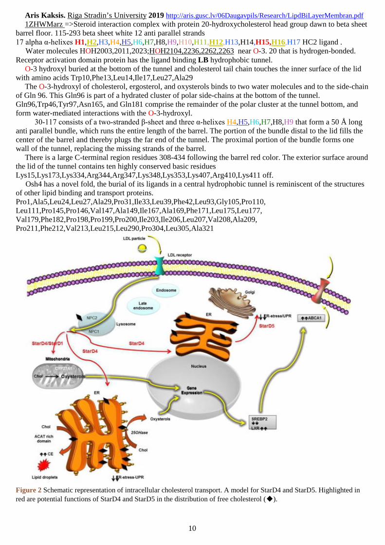
Figure 2 Schematic representation of intracellular cholesterol transport. A model for StarD4 and StarD5. Highlighted in
red are potential functions of StarD4 and StarD5 in the distribution of free cholesterol (◆).

Aris Kaksis. Riga Stradin’s University 2019 http://aris.gusc.lv/06Daugavpils/Research/LipdBiLayerMembran.pdf
11
Steroidogenic acute regulatory protein (StAR) transport (START) intracellularof steroids, bile acids. phospholipids, sphingosines, K, E, D, A vitamins transport in water
The steroidogenic acute regulatory protein-related lipid transfer (START) domain family are defined by thepresence of a conserved 195÷240 amino acid sequence that folds into an α/β helix-grip structure forming a hydrophobic pocket for ligand binding. The mammalian START proteins bind diverse ligands, such as cholesterol, oxysterols,phospholipids, sphingolipids, possibly fatty acids and K,E,D,A vitamins, and have putative roles in non-vesicular lipidtransport, thioesterase enzymatic activity, and tumor suppression. J Endocrinol March 1, 2012; 212257-275
Fifteen mammalian proteins, STARD1-STARD15, possess a START domain for six subfamilies: cholesterol,25-hydroxycholesterol, phosphatidylcholine, phosphatidylethanolamine and ceramides are ligands forSTARD1/STARD3/STARD5, STARD5, STARD2/STARD10, STARD10 and STARD11, respectively. The lipids orsterols bound by the remaining 9 START proteins are unknown. Recent studies show that the C-terminal end of thedomain plays a fundamental role, forming a lid over a deep lipid-binding pocket that shields the ligand from the externalenvironment. The START domain can be regarded as a lipid-exchange and/or a lipid-sensing domain. Mammalian STARTproteins have diverse expression patterns and can be found free in the cytoplasm, attached to membranes or in the nucleus.They appear to function in a variety of distinct physiological processes, such as lipid transfer between intracellularcompartments, lipid metabolism and modulation of signaling events. Mutation or misexpression of START proteins islinked to pathological processes, including genetic disorders, autoimmune disease and cancer.
A member of the STARD4 subfamily, was monitored. . Cholesterol stabilized STARD3-START against trypsin-catalyzed degradation, whereas cholesterol had no protective effect on STARD1-START.
Phosphorylated sphingolipids ceramide-1-phosphate (C1P) and sphingosine-1-phosphate (S1P) have emerged as keyregulators of cell growth, survival, migration and inflammation. C1P produced by ceramide kinase is an activator of groupIVA cytosolic phospholipase A2α (cPLA2α), the rate-limiting releaser of arachidonic acid used for pro-inflammatory eicosanoid production, which contributes to disease pathogenesis in asthma or airway hyper-responsiveness, cancer,atherosclerosis and thrombosis. To modulate eicosanoid action and avoid the damaging effects of chronic inflammation,cells require efficient targeting, trafficking and presentation of C1P to specific cellular sites.
Although their physiological function remain in the trafficking of cholesterol and in its cellular homeostasis [14, 15].Possible medical and industrial applications The concepts reviewed represent novel approaches to further define
intracellular cholesterol transport. We suggest that the newly described proteins of the StarD4 subfamily, with theirdistinctive regulations and localizations, alone or in association with other proteins, play unique and relevant roles inintracellular cholesterol movement and metabolism in a variety of tissues of relevance to human health. Furthermore,future studies on the role of these proteins will give a better understanding of cholesterol metabolism needed to lay thegroundwork for the development of better therapies for cholesterol related disorders (i e. atherosclerosis or Niemann-Pickdisease) and UPR related diseases (i.e. Huntington’s disease and Alzheimer’s disease).
Fig. 1. Phylogenetic tree and the 15 START-domain proteins in humans. START domain sequences were aligned by theEclustalw program (Genetics Computer Group, Madison, WI). The phylogenetic tree was drawn with the drawtreesoftware [J. Felsenstein, 1993, PHYLIP (Phylogeny Inference Package) v.3.5c, Department of Genome Sciences,University of Washington, Seattle, WA]. Abbreviations: Journal of Cell Science 2005 118: 2791-2801. Mt, mitochondrialtargeting motif; MENTAL, MLN64 N-terminal domain; PH, pleckstrin homology domain; FFAT, two phenylalanines inan acidic tract motif responsible for ER targeting; RHOGAP, Rho-GTPase-activating-protein domain; SAM, sterile alpha
motif; THIO, acyl-CoA thioesterase domain. Journal of Cell Science 2005 118: 2791-2801 1EM2, 1LN1

Aris Kaksis. Riga Stradin’s University 2019 http://aris.gusc.lv/06Daugavpils/Research/LipdBiLayerMembran.pdf
12
Table 1 Characteristics of the mammalian START 4 domain protein subfamily membersPhysical map positions (chromosome, position in megabases, Mb) in the mouse and human genomes are based on theEnsembl database (www.ensembl.org). Cellular location abbreviations used are: endoplasmic reticulum (ER). Lipidbinding abbreviations used are: 7-α-hydroxycholesterol (7-α-OHchol), 25-hydroxycholesterol (25OH), cholic acid (CA) and chenodeoxycholic acid (CDCA). StarD4 subfamily membersOther name (s) StarD4 CRSP StarD5 StarD6Chromosomme/PositionMouse 18/33.4 Mb 7/73.3 Mb 18/70.8 Mb
Human 5/110.5 Mb 15/77.6 Mb 18/52 MbSubcellular location Cytosolica, ERa,b, mitochondriab[A,B] Cytosolic, ER, Golgia,
Nucleus[C–F]Cytosolicc,
mitochondriab [G,H]Tissue distribution * Liver, macrophages, keratinocytes,
kidney*[A,B]Kupffer cells, peripheral
macrophages, kidneyproximal tubules*[D,E]
Nervous system andtestis germ cell[G, I–K]
Lipid binding Cholesterol, 7-α-OHchol, 7-hydroperoxycholesterol #,e[C, L–N]
Cholesterol, 25OH, CAand CDCA d,e[C,O,P]
Cholesterold,e [N]
Regulation Regulated in response to sterol levelsby SREBP pathway[B, Q]. Early phase
of ER stress[R]
Induced in response to ERstress [D,F,Q]
Potential regulationunder neurotoxicconditions[G,I, J]
* Tissue distribution: Restricted expression, note that STARD4 and STARD5J Endocrinol March 1, 2012; 212257-275 1EM2, 1LN1Table 1. Human START proteins, their ligands, and the available crystal structures.Group Protein Ligand PDB entry1 - StAR STARD1 cholesterol [5] 3P0L; ligand-free (this study) 212 AA
STARD3/MLN64 cholesterol [5]PLoS One. 2011;6(6)2I93+CLR,1EM2; ligand-free [5] 213 AA2 - START STARD4 cholesterol [19] 1JSS (mouse); ligand-free [7] 222 AAonly STARD5 cholesterol, 25-hydroxycholesterol [19] 2R55; ligand-free (this study) 213 AA
STARD6 cholesterol [40] 2MOU to be published 2014 222 AA3 - PCTP STARD2/PCTP phosphatidyl choline [41]1LN3,1LN1 , 1LN2; DLP complex1 [6] 210 AA
STARD7 phosphatidyl choline [42] -STARD10 phosphatidylcholine/ethanolamine [43] -234 AA 234 AASTARD11/CERT
2E3M,2E3N,2E3O,2E3P,2E3Q,2E3S,2Z9Y,2Z9Z
ceramides [8] 4K80, 4KF6, 4K85,4K84, 4K8N, 4KBS, 4KBR 214 AANature. 2013 Aug 22;500(7463):463-7
2E3R; C18-ceramide complex [8]; 2Z9Z;C10-DAG complex2 [8]; 3H3S; H15complex3 [9] and 10 more entries
4 - RhoGAP STARD8 charged lipid? -STARD12 charged lipid? -STARD13 charged lipid? 2PSO; ligand-free (this study) 195 AA
5-Thioesterase STARD14 fatty acid? 3FO5; PEG complex4 (this study) 240 AASTARD15 fatty acid? PNAS. 2008 2E3M,2E3N,2E3O,-2E3R,2E3S,2Z9Y,2Z9Z,2E3Q, 2E3P,
6 -STARD9 STARD9 ?Journal.PLoS One. 2011;6(6):e195213P0L,1LN1,1EM2,1JSS,2R55,2E3O,2PSO,3FO5
central amphiphatic tunnel is reminiscent of the structures of mentioned lipid binding .Fig. 2. Structure of the STARTdomains of MLN64 (A) and PCTPwith its ligand (B). (A) Ribbondiagram of the START domain ofMLN64 (ID code: 2I93.PDB).Secondary structural elements andthe C- and N-termini are labeled.MLN64 has a central β-sheet gripped by N-terminal (α1) and C-terminal (α4) α-helices (red), the latter being closely packed abovethe curved sheet.
(B). Cut-away view of the molecular surface of the START domain of PCTP complexed with(DLPC,dilinoleoyl-sn-glycerol-3-phosphorylcholine) a phosphatidylcholine molecule (ID code: 1LN1.PDB). The DLPC molecule(shown in stick representation) is located in the hydrophobic. Journal of Cell Science 2005 118: 2791-28011EM2.pdb, 2I93.pdb, 1LN1.pdb

Aris Kaksis. Riga Stradin’s University 2019 http://aris.gusc.lv/06Daugavpils/Research/LipdBiLayerMembran.pdf
13
The closest analogy is to the cholesterol-binding steroidogenic acute regulatoryprotein (StAR) transport (START) domain proteins MLN64 and StarD4, thephosphatidyl choline (PC) binding START domain protein PC-TP, and the mammalianphosphatidyl inositol transfer proteins. In these structures the ligands are completelysequestered from solution. For the ground prominence is water-mediated interactions.
central amphiphatic tunnel is reminiscent of the structures of mentioned lipid binding.Nature Structural Biology 9, 507 - 511 (2002) 1LN1.pdb 1LN2 and 1LN3
Fig. 6. Temperature factors and a putative entrance to the amphiphilic cavity 2E3R.pdb.(A–D) Ribbon diagrams of the CERT START domain colored according to the
crystallographic B-factors for the apo-CERT START domain and in complex withC6-, C16-, and C18-ceramide, respectively. The ceramide molecules are drawn as filled
spheres. (E) Ribbon representation of the CERT START domain in complex withC18-ceramide. The structure is rotated by 45° around the y axis with respect to those
shown in D. α3 and Ω1 loop are colored cyan and magenta, respectively. C18-ceramide is drawn as space-filling spheres, in which yellow, blue, and red spheres
represent C, N, and O atoms, respectively. (F) Molecular surface of the CERTSTART domain in complex with C18-ceramide, drawn in the same orientation as inE. The hydrophobic surface is painted green, and the residues in α3 and Ω1 loop are
highlighted as dotted spheres in cyan and magenta, respectively. C18-ceramide isdrawn as in E.
PNAS. 2008 2E3M, 2E3N, 2E3O, 2E3P, 2E3Q, 2E3R, 2E3S, 2Z9Y, and 2Z9Z
Fig. 2. Schematic representation of C16-ceramide recognition by the CERT START domain. Residueslining the amphiphilic cavity are shown. Red dashed lines, hydrogen bonds; red circles, water molecules; black,blue, and red dots, C, N, and O atoms, respectively, of the residues involved in the hydrogen network. Greenboxes indicate residues contributing to the hydrophobicity of the cavity in general, whereas green boxes withthick borders indicate those with direct hydrophobic interactions, which are represented as green dashed lines.Among these, eight amino acid residues, which are common to all of the C6-, C16-, C18-ceramide complexstructures, are indicated by thick-bordered green boxes filled with light green.eukaryotes Nature. 2013 Aug 22;500(7463):463-7. 4K80, 4KF6, 4K85, 4K84, 4K8N, 4KBS, 4KBR

Aris Kaksis. Riga Stradin’s University 2019 http://aris.gusc.lv/06Daugavpils/Research/LipdBiLayerMembran.pdf
14
Figure 2 CPTP conformation and functional recognition of C1P. a, CPTP lipid headgroup recognition center residue
O
O P
O
O
HH
H
H
H
H
H
H
H
H
H
H
H
H
H
H
H
H
H
H
H
H
H
H
H H
H
H
H
H
H
H
H
H
H
H
H
H
H
H
H
H
H
H
H
H
H
H
H
H
H
H
H H
H
H
H
H
H
H
H
H
OC
C
C
O
H H
H
HN
H
H
H
H
interaction with phosphate and amide groups ofbound 16:0-C1P (spacefill). Hydrogen-bonds =dashed lines. CPTP Cα backbone is light gray; side chains; and oxygen and nitrogen, red andblue, respectively. Water molecules are pinkspheres. b, C1P transfer by CPTP point lines ofphosphate headgroup recognition cavity. wtCPTP(gray). c, Non-polar residues forminghydrophobic pocket that accommodates 18:0-C1Psphingosine and acyl chains. d, C1P transfer byCPTP point mutants (violet) of the hydrophobicpocket. wtCPTP (gray); Side-chains shown inpanel c. Data in b and d represent the mean ± s.d.of three independent experiments. e,Conformational changes in hydrophobic pocket
upon 18:0-C1P binding. Side-chains (lavender; stick) of apo-CPTP and human CPTP with bound18:0-C1P (yellow; ball-and-stick).f, g, Surface electrostatics of hydrophobic pocket opening at lipid headgroup recognition sites inapo-CPTP (f) and CPTP/18:0-C1P complex (g). h, 18:0-C1P chemical structural formula.
i, j Crystal structures of CPTP (ribbon) with bound 18:0-C1P in sphingosine-in i (CPK);sphingosine conformations. k, Superposition of bound 18:0-C1P in sphingosine- conformation.
The hydrophobic pocket is lined by ~25 nonpolar residues, mainly Phe, Leu, Val, and Ile thatprevent water entry while ensheathing the ceramide aliphatic chains. Mutation of L43, L118, orL146 to positively-charged R or V57 or V158 to high polarity N compromises hydrophobic pocketfunctionality and strongly diminishes C1P transfer. More conservative mutation (e.g. W117A)
only moderately reduces C1P transfer, while F42A near the pocket bottom stimulates C1P transfer. Mutation near the entryportal (I53N) or in the flexible α1–2 loop (F50R) is well tolerated (75–80% active) (Fig. 2d). Ceramide entry is orientedby hydrogen bonding of the lipid amide oxygen and nitrogen with H150 and D56, respectively. Hydrogen bond disruptionbetween lipid amide nitrogen and D56 (D56V) moderately slows C1P transfer, but H150 mutation (H150L) abolishesactivity. Super positioning of apo- and 16:0-C1P/CPTP structures (rmsd 1.4 Å) shows K60, R106 and R110 nearlyidentically positioned in the positively-charged surface cavities. Yet, large conformational differences exist for VI53,W36, W119 and F52 (Fig. 2e) due to closer packing of certain α-helices in apo-CPTP (Fig. S2d,e). Many Leu and Phe arerepositioned, reducing the solvent accessible (SA) volume (40 Å3) (Table S4) and effectively collapsing the hydrophobicpocket (Fig. 2f,g) compared to 18:0-C1P/CPTP complex (364 Å3).
Nature. 2013 Aug 22;500(7463):463-7. 4K80, 4KF6, 4K85, 4K84, 4K8N, 4KBS, 4KBRCellular retinaldehyde-binding protein (CRALBP) is essential for mammalian vision by routing 11-cis-retinoids for
the conversion of photo bleached opsin molecules into photosensitive visual pigments. The arginine-to-tryptophanmissense mutation in position 234 (R234W) in the human gene RLBP1 encoding CRALBP compromises visual pigmentregeneration and is associated with Bothnia dystrophy. Our structural model of wild-type CRALBP locates R234 to apositively charged cleft at a distance of 15 A from the hydrophobic core sequestering 11-cis-retinal.
Fig. 1. Monomeric structure of CRALBP. (A) Ribbon diagram of wild-type CRALBP bound to 11-cis-retinal. Thehelices of the N-terminal domain H1,H2,H3,H4,H5 C-terminal helices H14,H15,H16; the helical gate H11,H12,H13
β-strands are indicated in yellow. The position of R234 is indicated as CPKspace fill, the 11-cis-retinal ligand is shown as CPK space fill in the cavity.The cavity surface was calculated with VOIDOO (33). (B) View afterrotation of A by 90° on the vertical axis. Images were generated withChemscape-Raswin. Proc Natl Acad Sci U S A. 2009 Nov 3;106(44):18545-50. 3HY5
CO
H
W=284.45g/mo
C20H28
11-cis-RET
111
122
133
144155
8
9 100