Embed Size (px)
Citation preview

Sid Bhende MD
Sentara Vascular
Specialists
April 21st 2017
Phlegmasia Cerulea Dolens - A Limb
Threatening Problem

Presenter name
Title
Date
Disclosures

Presenter name
Title
Date
Complications of DVT
• Early
– Pulmonary Emboli
• Life threatening “saddle embolus”
• Incidence of PE is estimated to be approximately 60 to 70 per 100,000
• PE is found in up to 50% of DVT patients (Silent PE)

Presenter name
Title
Date

Presenter name
Title
Date
Complications of DVT
• Early
– What else?????
Phlegmasia Alba Dolens
Phlegmasia Cerulea Dolens
Venous Gangrene

Presenter name
Title
Date
Complications of DVT
• Late
– PTS (Post Thrombotic Syndrome)
• Leg swelling
• Leg pain
• Skin discoloration
• Varicosities
• Venous ulceration

Presenter name
Title
Date
Early Complications of DVT
Phlegmasia Alba Dolens
Phlegmasia Cerulea Dolens
Venous Gangrene

Presenter name
Title
Date
Plegmasia Alba Dolens
• Phlegmasia “Alba” Dolens
– “Alba” means white (i.e. Albino)
– Referred to as “Milk Leg or White Leg”
– Historically seen in pregnant women (third trimester) or mothers who had just given birth
• Compression of Lt iliac vein against the pelvic rim from an enlarged uterus
– Presently it is due to venous occlusion (DVT)
• 40% of patients with phlegmasia alba dolens have an underlying malignancy

Presenter name
Title
Date
Pathophysiology
• Phlegmasia “Alba” Dolens
– Thrombosis involving ONLY the major deep venous channels but SPARING the collateral veins
– Venous drainage is decreased but still present via the superficial channels
– At this stage the leg is painful, swollen and appears white (pale)
• But no arterial compromise (either palpable or dopplerable pulses)

Presenter name
Title
Date
Pathophysiology

Presenter name
Title
Date
Phlegmasia Alba Dolens

Presenter name
Title
Date
Diagnosis
• Phlegmasia “Alba” Dolens
– Diagnosis is made with venous duplex
• Identify Acute DVT
• Location of DVT (proximal vs distal)
• Extent of DVT
– Contrast enchanced CT scan can be helpful
• To identify thrombus is pelvic veins or IVC
• Check for underlying malignancy

Presenter name
Title
Date
Diagnosis

Presenter name
Title
Date
Treatment • Phlegmasia “Alba” Dolens
– Treatment
• Admit to inpatient
• Anticoagulation (IV heparin gtt)
• Elevate affected limb
• Vascular surgery consultation – Medical management vs. Endovascular venous lysis (CDT, mechanical
thrombectomy, pharmacomechanical thrombectomy)

Presenter name
Title
Date
Phlegmasia Cerulea Dolens • Phlegmasia “Cerulea” Dolens (PCD)
– “Cerulea” means Blue
– Referred to as “Painful Blue Leg”
• Rare condition that occurs in < 1% of DVT patients
• Up to 90% of patients with PCD have underlying malignancy (50% occult malignancy)

Presenter name
Title
Date
Phlegmasia Cerulea Dolens • 50% of patients with PCD progress to Venous
Gangrene
– 30-50% Limb Amputation rate
– Overall Mortality rate of 20-40%

Presenter name
Title
Date
Pathophysiology • Phlegmasia “Cerulea” Dolens
– Thrombosis causing COMPLETE occlusion of venous drainage (deep and superficial system)
– Leads to increase in capillary pressure
– Leads to exudation of fluid into the interstitial space
– Leads to skin blistering
• Characteristics of PCD
– Pain, swelling and most importantly cyanosis “blue appearance”

Presenter name
Title
Date
Pathophysiology

Presenter name
Title
Date
Pathophysiology

Presenter name
Title
Date

Presenter name
Title
Date
Diagnosis • Phlegmasia “Cerulea” Dolens
– Based on clinical signs and symptoms
• Painful, blue, swollen leg
– Venous duplex to confirm the diagnosis and localize the thrombus (purpose of intervention)
– Contrast enchanced CT to identify centrally located and pelvic thrombus

Presenter name
Title
Date
Plegmasia Cerulea Dolens

Presenter name
Title
Date
Treatment • Phlegmasia “Cerulea” Dolens
– THIS IS AN EMERGENCY!!
– PATIENT IS GOING TO THE OR FOR SURGICAL VENOUS THROMBECTOMY
– Fluid resuscitation
• Patients are hypotensive and sometimes in shock due to fluid extravasation and loss of intravascular fluid
– Anticoagulation
• IV heparin bolus prior to OR

Presenter name
Title
Date
Treatment • Phlegmasia “Cerulea” Dolens
– Systemic tPA vs Catheter directed thrombolysis
• Literature does not support either therapy as being successful
• Time is tissue
– Some surgeons recommend a brief (6 hours) of IV heparin gtt with profound leg elevation
• If unsuccessful then will proceed with surgical venous thrombectomy

Presenter name
Title
Date
Surgical Treatment • Phlegmasia “Cerulea” Dolens
– Local, Regional or General anesthesia
– Longitudinal groin incision to expose CFV, GSV and SFA
– IV Heparin intra op if not given preop (Check ACT)
– Venotomy to facilitate Fogarty catheter thrombectomy
• American surgeons – place IVC filter (contralateral groin) before
• European surgeons – no filter, perform thrombectomy with positive pressure ventilation or Valsalva maneuver

Presenter name
Title
Date
Surgical Treatment

Presenter name
Title
Date
Surgical Treatment

Presenter name
Title
Date
Surgical Treatment • Phlegmasia “Cerulea” Dolens
– Place Fogarty balloon in Common Iliac vein
– Pass suction catheter parallel to Fogarty and try to suction out the internal iliac vein thrombus
– Must confirm iliac vein flow
• MANDATORY VENOGRAM
– If Iliac vein stenosis/compression (MTS) is noted then may need balloon angioplasty + stenting

Presenter name
Title
Date
Surgical Treatment

Presenter name
Title
Date
Surgical Treatment • Phlegmasia “Cerulea” Dolens
– Thrombus in infrainguinal region is expressed manually using an Esmarch
– Start wrapping at the base of the toes and proceed proximally to the groin incision
• Passing fogarty distally will damage venous valves

Presenter name
Title
Date
Surgical Treatment

Presenter name
Title
Date
Surgical Treatment • Phlegmasia “Cerulea” Dolens
– Once the venous outflow is restored the venotomy is closed
– An autogenous AVF is created in groin to increase iliac vein patency
• GSV is divided and proximal end is anastomosed to SFA
– Perform a 4 compartment lower leg fasciotomy

Presenter name
Title
Date
Surgical Treatment

Presenter name
Title
Date
Surgical Treatment
Presenter name
Title
Date
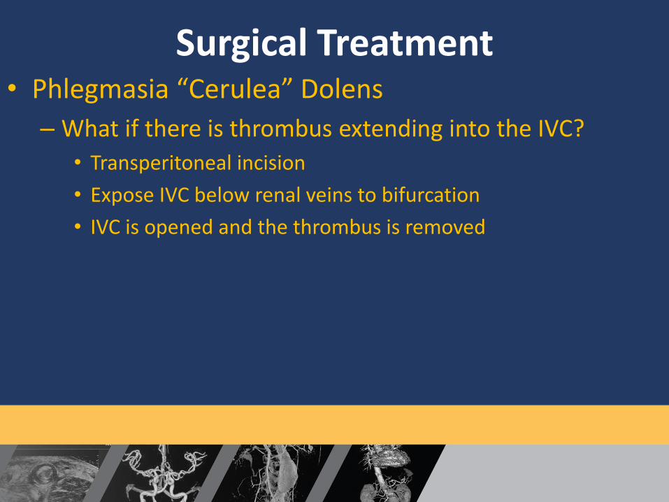
Surgical Treatment • Phlegmasia “Cerulea” Dolens
– What if there is thrombus extending into the IVC?
• Transperitoneal incision
• Expose IVC below renal veins to bifurcation
• IVC is opened and the thrombus is removed

Presenter name
Title
Date
Post Operative Treatment • Phlegmasia “Cerulea” Dolens
– Continue IV heparin drip x 5 days
– Then transition to oral anticoagulation (coumadin, xarelto, eliquis) x 6 months
– Gradient Compression Stockings
– Once swelling decreased, return to OR to close fasciotomy sites or heal with secondary intention
– Ligate groin AVF in 6-12 weeks

Presenter name
Title
Date
Venous Gangrene • Venous Gangrene
– Massive iliofemoral or IVC occlusion with extensive vascular congestion and venous ischemia
– Thrombosis causing COMPLETE occlusion of venous drainage (deep and superficial system) AND ARTERIAL COMPROMISE
– 50% of phlegmasia cerulea dolens progress to venous gangrene

Presenter name
Title
Date
Pathophysiology

Presenter name
Title
Date
Venous Gangrene • Clinical Features
– Excruciating Limb pain
– Severe edema
– Blistering with fluid extravasation
– Superficial gangrene and necrosis
– No motor or sensory to foot
– IRREVERSIBLE (Phlegmasia Alba and Cerulea Dolens are reversible)
– Treatment is AMPUTATION

Presenter name
Title
Date
Venous Gangrene

Presenter name
Title
Date
Case Study • 75 year old male presents to ED with LLE pain and
swelling x 1 day
• Denies CP or SOB
• Pt recently diagnosed with Stage IV Lung cancer and received 2 doses of chemotherapy via a mediport
• Vital signs are stable
• O/E – LLE is edematous but NO PHLEGMASIA, Pedal pulses are palpable

Presenter name
Title
Date
Case Study • Blood work is normal
• LLE venous duplex to r/o DVT
• Venous duplex demonstrated acute femoropopliteal and tibial occlusive DVT, no extension into external iliac vein

Presenter name
Title
Date
Case Study

Presenter name
Title
Date
Case Study • What do you do?
• Patient was discharged from ED with Xarelto (starter pack) x 21 days
• Instructed to follow up with vascular surgery in the next week
• Keep LLE elevated

Presenter name
Title
Date
Case Study • Patient returns to ED 2 days later complaining of
worsening pain and swelling
• A different ED physician evaluates patient
• O/E – LLE is much more swollen and appears white
• Repeat LLE venous duplex is ordered and now demonstrates Acute Left external iliac vein, femoral vein, popliteal vein and tibial vein DVT
• Vascular surgery consult obtained

Presenter name
Title
Date
Case Study • Vascular surgery evaluates patient and feels that
patient has developed Phlegmasia Alba Dolens
• Pt is admitted to ICU
• Initiate IV heparin gtt
• CT A/P with contrast to better evaluate IVC and pelvic veins
• Keep LLE elevated

Presenter name
Title
Date
Case Study • No IVC clot
• Clot extends to the Lt CIV

Presenter name
Title
Date
Case Study • Discuss options of medical management (IV Heparin
vs. Endovascular thrombolysis/thrombectomy)
– Risk of bleeding from venous lysis is high due to stage IV lung cancer
• Decision made to continue IV heparin and perform serial exams with the understanding that if this progress to phlegmasia cerulean dolens then patient will need surgery

Presenter name
Title
Date
Case Study • Next morning the LLE pain is worse and now has
spots of bluish purplish areas (new)

Presenter name
Title
Date
Case Study • Pt taken to OR for surgical venous thrombectomy
– Able to clear iliac vein clot
– Venogram was normal
– Able to express infrainguinal clot
– AVF created in left groin
– LLE prophylactic fasciotomy performed
• Post op patient continued on IV heparin gtt

Presenter name
Title
Date
Case Study • Over the course of next few days, the LLE swelling,
pain and color improved
• Fasciotomy sites closed on POD #4
• Pt given GCS
• Discharged on coumadin x 6 months

Presenter name
Title
Date
Take Home Points • Phlegmasia “Alba” Dolens – “White” leg
– Medical management (IV Heparin) vs Endovascular therapy
– Check for Malignancy (40%)
• Phlegmasia “Cerulea” Dolens – “Blue” leg
– Limb threatening emergency
– Mandatory open intervention
– Check for Malignancy (90%)

Presenter name
Title
Date
Take Home Points • Venous Gangrene – “Game Over”
– Amputation

Presenter name
Title
Date
Thank You


![Phlegmasia cerulea dolens complicated by …€¦ · International Journal of Case Reports and Images, Vol. 7 No. 5, May 2016. ISSN – [0976-3198] Int J Case Rep Imag 2016;7(5):314–317](https://img.dokumen.tips/doc/110x75/60182b9bf2349945062bf04c/phlegmasia-cerulea-dolens-complicated-by-international-journal-of-case-reports-and.jpg)