Embed Size (px)
Citation preview

Yale West Campus Materials Characterization Core (MCC)
ywcmatsci.yale.edu
PHI VersaProbe II Scanning XPS Microprobe

Materials Characterization Core (MCC)
ywcmatsci.yale.edu2/20Yale West Campus
Core Policies
• DO NOT let other people use the facility under your account.
• DO NOT try to fix parts or software issues by yourself!
• DO NOT surf web using instrument computer!
• Follow checklist and SOP! DO NOT explore program!
• Facility usage time at least twice a month, OR receive training
again (two practice sessions within one week).
• No trainings on monthly users

Materials Characterization Core (MCC)
ywcmatsci.yale.edu3/20Yale West Campus
What is XPS? X-ray Photoelectron Spectroscopy
• X-ray tube• UV lamp• Synchrotron
detector
electronoptics
Vacuum orAmbient pressure
• Photoelectric effect
• A spectroscopy that records the counts of X-ray induced secondary electrons -
photoelectrons as the function of binding energy
• A technique based on photoelectric effect:

Materials Characterization Core (MCC)
ywcmatsci.yale.edu4/20Yale West Campus
What is XPS? X-ray Photoelectron Spectroscopy
• X-ray tube• UV lamp• Synchrotron
detector
electronoptics
Vacuum orAmbient pressure
• Photoelectric effect
• A spectroscopy that records the counts of X-ray induced secondary electrons -
photoelectrons as the function of binding energy
• A technique based on photoelectric effect:

Materials Characterization Core (MCC)
ywcmatsci.yale.edu5/20Yale West Campus
What kinds of samples for XPS?
• Vacuum compatible: low vapor pressure under 10-8 Pascal
• Conductive or insulating
Freezing
Materials Characterization Core (MCC)
ywcmatsci.yale.edu6/20Yale West Campus
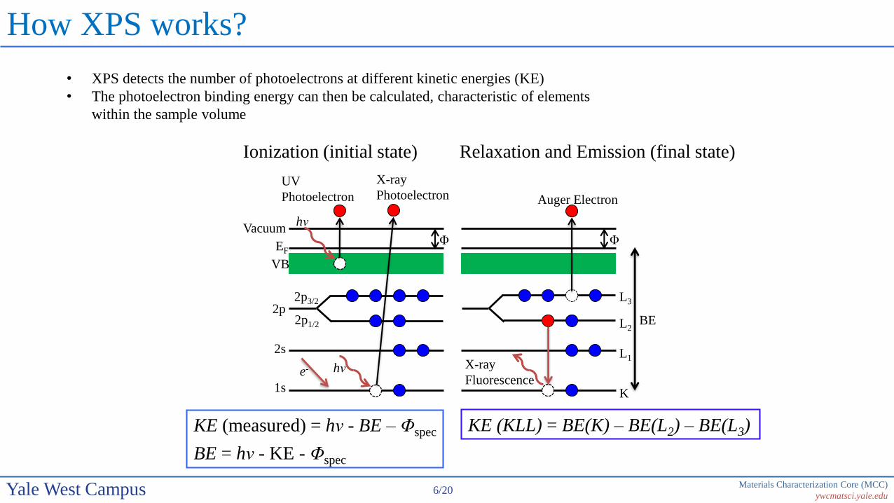
How XPS works?
• XPS detects the number of photoelectrons at different kinetic energies (KE)
• The photoelectron binding energy can then be calculated, characteristic of elements
within the sample volume
KE (measured) = hν - BE – Φspec
BE = hν - KE - Φspec
KE (KLL) = BE(K) – BE(L2) – BE(L3)
Ionization (initial state) Relaxation and Emission (final state)
Auger Electron
Φ
BE
L3
L1
L2
X-ray
FluorescenceK
UV
Photoelectron
Vacuum
VB
2p3/22p
1s
X-ray
Photoelectron
EFΦ
hν
2s
2p1/2
hν
e-
Materials Characterization Core (MCC)
ywcmatsci.yale.edu7/20Yale West Campus
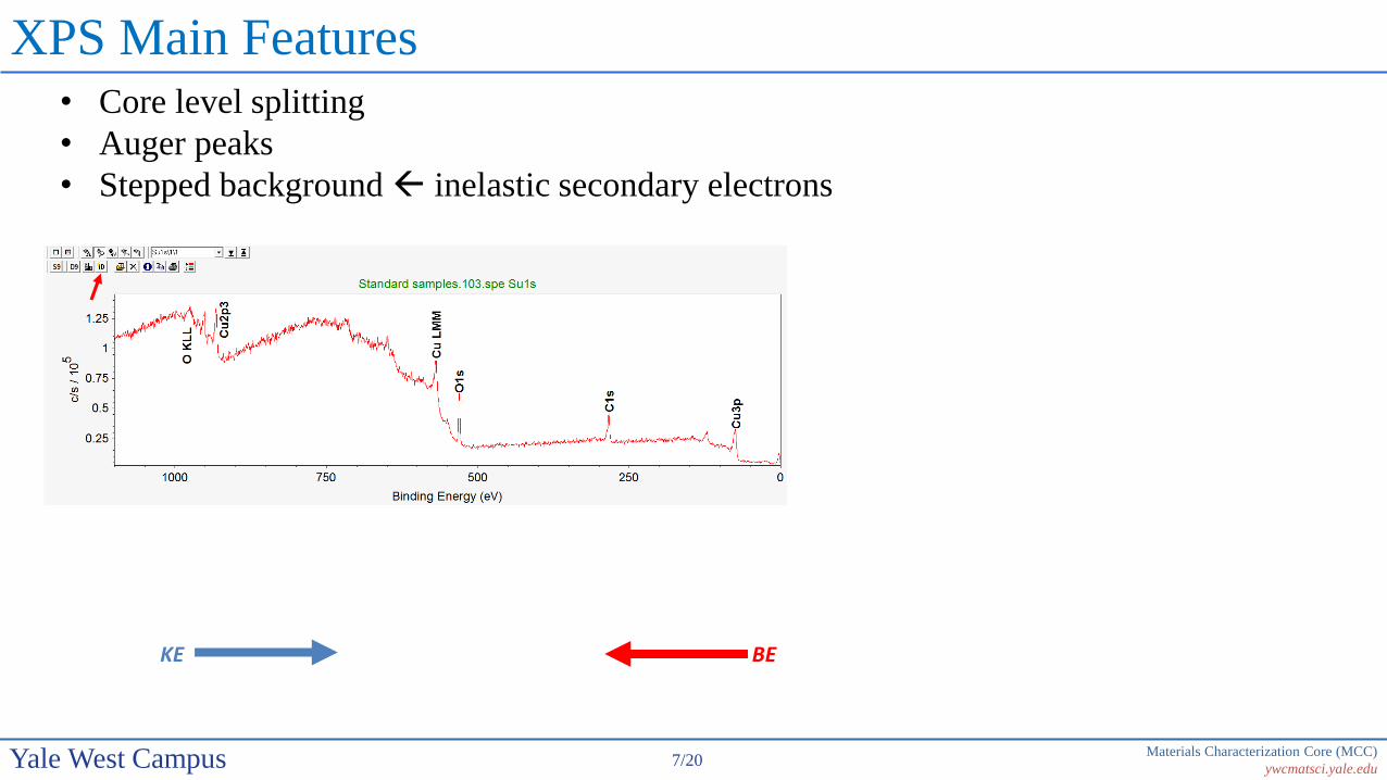
XPS Main Features• Core level splitting
• Auger peaks
• Stepped background inelastic secondary electrons
KE BE

Materials Characterization Core (MCC)
ywcmatsci.yale.edu8/20Yale West Campus
XPS Peak Notation
4f7/2
n
l = 0 s1 p2 d3 f
j = l ± s, s = 1/2
Spin-orbital splitting with l > 0
Orbital l j Degeneracy (2j + 1) Peak area ratio Electron level
s 0 1/2 1 - 1s
p 1 1/2, 3/2 2, 4 1 : 2 2p1/2, 2p3/2
d 2 3/2, 5/2 4, 6 2 : 3 3d3/2, 3d5/2
f 3 5/2, 7/2 6, 8 3 : 4 4f5/2, 4f7/2
Materials Characterization Core (MCC)
ywcmatsci.yale.edu9/20Yale West Campus
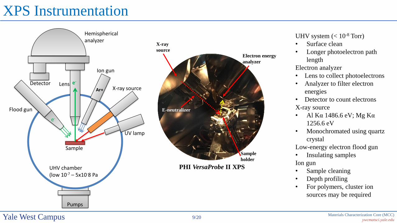
XPS Instrumentation
UV lamp
Hemisphericalanalyzer
X-ray source
Flood gun
Sample
UHV chamber(low 10-7 – 5x10⁻⁸ Pa
Ion gun
e-
e-
Ar+
Detector Lens
Pumps
UHV system (< 10-8 Torr)
• Surface clean
• Longer photoelectron path
length
Electron analyzer
• Lens to collect photoelectrons
• Analyzer to filter electron
energies
• Detector to count electrons
X-ray source
• Al Kα 1486.6 eV; Mg Kα
1256.6 eV
• Monochromated using quartz
crystal
Low-energy electron flood gun
• Insulating samples
Ion gun
• Sample cleaning
• Depth profiling
• For polymers, cluster ion
sources may be required
Sample
holder
Electron energy
analyzer
X-ray
source
PHI VersaProbe II XPS
E-neutralizer

Materials Characterization Core (MCC)
ywcmatsci.yale.edu10/20Yale West Campus
X-ray Dual Anode Source
X-ray
lines
Line Energy
(eV)
Width (eV)
Mg Kα1,2 1253.6 0.70
Al Kα1,2 1486.6 0.85
K (1s)
L (2s)
L2 (2p1/2)L3 (2p3/2)
M1 (3s)
M2,3 (3p)
M4,5 (3d)
Kα1
Kα2
Kβ
Materials Characterization Core (MCC)
ywcmatsci.yale.edu11/20Yale West Campus
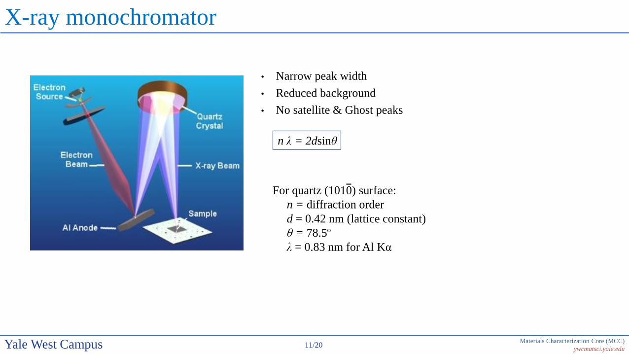
X-ray monochromator
• Narrow peak width
• Reduced background
• No satellite & Ghost peaks
n λ = 2dsinθ
For quartz (1010) surface:
n = diffraction order
d = 0.42 nm (lattice constant)
θ = 78.5º
λ = 0.83 nm for Al Kα

Materials Characterization Core (MCC)
ywcmatsci.yale.edu12/20Yale West Campus
Spherical Capacitor Analyzer (SCA)
Pass energy:
Analyzer Resolution:
V0: the median equipotential surface of radius r
V: the potential applied between inner (radius b) and outer (radios a) shells
w: entrance and exit slit widths
𝛿𝛼: angular deviation of the electron trajectories at the entrance with
respect to the center line
r =a+b
2
Where the mean radius
𝐸0 = 𝑒𝑉0 =𝑉
𝑏𝑎−𝑎𝑏
a
b
r
𝜹𝜶
V2<0
w wV
∆𝐸 = 𝐸0𝑤
𝑎 + 𝑏+𝛿𝛼2
4
For the PHI SCA : 𝐸0 = 0.56𝑉 ∆𝐸 = 0.015𝐸0and
Typical 𝐸0 = 100 eV ∆𝐸 = 1.5 eV

Materials Characterization Core (MCC)
ywcmatsci.yale.edu13/20Yale West Campus
Why are we interested in XPS?
http://www.eag.com/mc
• Surface sensitive technique
• Chemical shift detection XPS is also named as Electron Spectroscopy
for Chemical Analysis (ESCA)
Typical Analysis Depths for Techniques
XPS detects electron signals in the near surface region (0 ~ 10 nm)

Materials Characterization Core (MCC)
ywcmatsci.yale.edu14/20Yale West Campus
Analytical Resolution vs. Detection Limit
http://www.eag.com/mc
• XPS resolution can be
reached below 10 µm
• XPS detection limits: ppt
range

Materials Characterization Core (MCC)
ywcmatsci.yale.edu15/20Yale West Campus
Why XPS is Surface Sensitive?
• Inelastic scattering of photoelectrons
Materials Characterization Core (MCC)
ywcmatsci.yale.edu16/20Yale West Campus
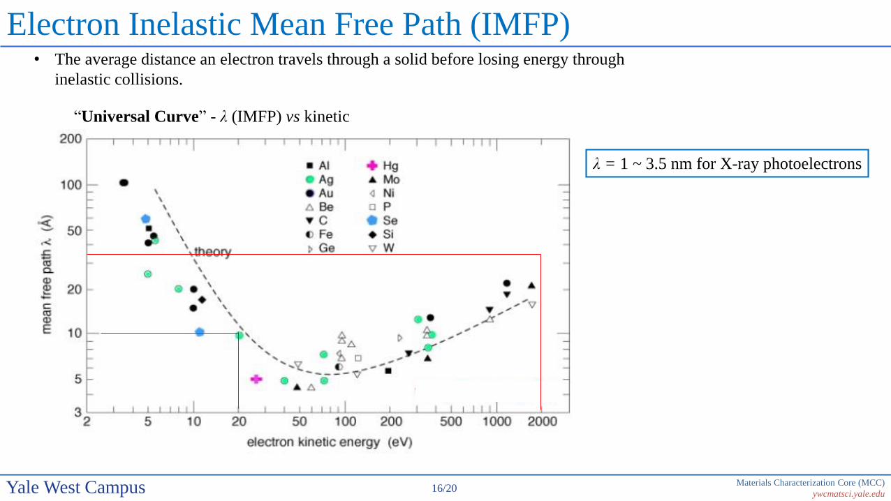
Electron Inelastic Mean Free Path (IMFP)
“Universal Curve” - λ (IMFP) vs kinetic
energy
λ = 1 ~ 3.5 nm for X-ray photoelectrons
• The average distance an electron travels through a solid before losing energy through
inelastic collisions.