Embed Size (px)
Citation preview

www.elsevier.com/locate/ydbio
Developmental Biology
Phenotypic and functional characteristics of spermatogonial
stem cells in rats
Buom-Yong Ryu, Kyle E. Orwig1, Hiroshi Kubota, Mary R. Avarbock, Ralph L. Brinster*
Department of Animal Biology, School of Veterinary Medicine, University of Pennsylvania, Philadelphia, PA 19104-6009, United States
Received for publication 12 April 2004, revised 1 July 2004, accepted 2 July 2004
Available online 7 August 2004
Abstract
Spermatogonial stem cells (SSCs) are at the foundation of the highly productive spermatogenic process that continuously produces male
gametes throughout postnatal life. However, experimental evaluation of SSCs in postnatal testes is complicated because these cells are
extremely rare and few defining morphology or biochemical characteristics are known. In this study, we used the spermatogonial
transplantation functional assay, combined with fluorescence-activated cell sorting (FACS) analysis to identify cellular, biochemical and
surface antigenic characteristics of SSCs in rat testes during development. Our results demonstrated that forward scatter (FSc)hi, side scatter
(SSc)hi, mitochondria membrane potential (DCm)lo, Ep-CAM+, Thy-1+, h3-integrin+ stem cells in neonate rat testes become SSclo, DCmhi,
Ep-CAM+, Thy-1lo, h3-integrin� stem cells in pup rat testes. Furthermore, prospective identification of rat testis cell populations (Ep-
CAM+), highly enriched for SSCs (1 in 13 for neonate; 1 in 8.5 for pup) enabled us to predict the Thy-1 and h3-integrin status of stem cells in
neonate and pup testes, which was subsequently confirmed by transplantation analyses. Systematic characterization of SSCs enabled the
production of testis cell populations highly enriched (up to 120-fold) for SSCs and will facilitate future investigations of functional and
genomic characteristics.
D 2004 Elsevier Inc. All rights reserved.
Keywords: Stem cell; Spermatogonial stem cell; Spermatogenesis; Germline; Development; Rat; Transplantation; Testis; Enrichment
Introduction
Stem cells in the male germline undergo dramatic
functional and morphological transformations during early
postnatal life, which result in the establishment of
spermatogenesis. In rats, quiescent gonocytes (primarily
T1 prospermatogonia) are the only germ cells present at
birth, and these cells are readily identified by their large
0012-1606/$ - see front matter D 2004 Elsevier Inc. All rights reserved.
doi:10.1016/j.ydbio.2004.07.004
* Corresponding author. Department of Animal Biology, School of
Veterinary Medicine, University of Pennsylvania, 3850 Baltimore Avenue,
Philadelphia, PA 19104-6009. Fax: +1 215 898 0667.
E-mail address: [email protected] (R.L. Brinster).1 Current address: Department of Obstetrics, Gynecology, and Repro-
ductive Sciences, Pittsburgh Development Center of Magee-Women’s
Research Institute, University of Pittsburgh School of Medicine, Pittsburgh,
PA 15213, United States.
size and characteristic location in the center of the
seminiferous tubules. Within a few days after birth,
proliferation resumes (marking the transition to T2
prospermatogonia) and some gonocytes develop pseudo-
pods, migrate to the seminiferous tubule basement
membrane, and give rise to adult-type spermatogonial
stem cells (SSCs; McCarrey, 1993) that are morpholog-
ically unremarkable. The SSC pool rapidly expands
during prepubertal testis development, producing one
new stem cell every 6 s (Ryu et al., 2003). At the same
time, spermatogonial stem cells give rise to progenitor
type A spermatogonia that are committed to differentiate,
but are morphologically indistinguishable from the stem
cells that produced them. Therefore, biological investiga-
tions of spermatogonial stem cells will rely in part on the
ability to identify them in heterogeneous testis cell
populations by unique molecular or functional character-
274 (2004) 158–170

B.-Y. Ryu et al. / Developmental Biology 274 (2004) 158–170 159
istics. Molecular characteristics of male germline stem
cells in the rat testis are largely unknown and probably
change in concert with their morphology, location, and
function as spermatogenesis is established during prepu-
bertal testis development.
Experimental evaluation of stem cells in postnatal tissues
is complicated because these cells are extremely rare
(comprising 1 in 3333 cells of the adult mouse testis)
(Tegelenbosch and de Rooij, 1993) and few defining
morphological or biochemical characteristics are known.
However, stem cells of all types can be identified by their
functional capacity to self-renew and produce a differ-
entiated tissue. Therefore, development of a SSC trans-
plantation technique about 10 years ago provided a
functional endpoint that now enables investigators to
examine stem-cell activity in heterogeneous testis cell
populations (Brinster and Avarbock, 1994; Brinster and
Zimmermann, 1994). Male germline stem cells are the only
cells that can produce and maintain spermatogenesis
following transplantation into recipient testes. When com-
bined with fluorescence-activated cell sorting (FACS)
technology, the functional assay can be used to evaluate
stem-cell activity in subpopulations of testis cells that have
been fractionated based on specific cellular characteristics
(e.g., size, complexity, surface antigens). Systematic eval-
uation of adult mouse testis cells using this strategy has
identified male germline stem cells in a subpopulation
defined by low side scatter of incident light (SSclo) and the
surface characteristics, a6-integrin+, Thy-1+, CD9+, av-
integrin�, c-kit�, and MHC class I (MHC-I)� (Kanatsu-
Shinohara et al., 2004; Kubota et al., 2003; Shinohara et al.,
2000).
Despite decades of morphological and physiological data
on rat spermatogenesis, comparatively little is known about
the molecular biology of rat SSCs. However, recent studies
have revealed that the rat testis has several advantages as a
model stem-cell system that is ripe for exploitation. For
example, adult rat testes have a higher concentration (9.5-
fold) and number (120-fold) of stem cells than adult mouse
testes (Orwig et al., 2002b); and stem cells can be isolated to
purity from newborn rat testes based on distinct morpho-
logical characteristics (Orwig et al., 2002a), providing a
valuable resource for biological investigation.
The current studies were guided by previous results in
mice as well as the dynamic biological changes known to
occur as gonocytes in the neonate testis differentiate into
spermatogonial stem cells in the prepubertal (pup) rat testis
(quiescent vs. proliferative, migratory vs. resident in the
stem-cell niche, large with distinct morphology vs.
bordinaryQ morphology). Changes in stem-cell size, shape,
and complexity were manifested as differences in forward
scatter (FSc) and side scatter (SSc) of incident light.
Differences in mitochondrial membrane potential (DCm)
might reflect the energetic state in quiescent vs. proliferative
cells; and cell surface characteristics were expected to
change as gonocytes migrate from the seminiferous tubule
lumen and seed the stem-cell niche on the basement
membrane.
Materials and methods
Donor rats and cell collection
Donor testis cells were obtained from neonate (0.5–5
days postpartum, dpp; day of birth is 0 dpp) and pup (8–
14 dpp) Sprague–Dawley (S/D) rats carrying a fusion
transgene composed of the mouse metallothionein I (MT)
promoter driving the expression of the lacZ structural
gene (Rhim et al., 1994) (a generous gift from R.
Hammer). The lacZ transgene is expressed in germ cells
derived from MT-lacZ donor rats and encodes a nuclear-
localized h-galactosidase protein. Donor-derived sperma-
togenesis is identified after transplantation into recipient
seminiferous tubules by staining with the h-galactosidasesubstrate, 5-bromo-4-chloro-3-indolyl h-d-galactoside (X-
gal; Nagano et al., 1999). Single-cell suspensions from
neonate and pup donor testes were produced by
enzymatic digestion as previously described (Bellve et
al., 1977; Brinster and Avarbock, 1994; Ogawa et al.,
1997; Orwig et al., 2002a). Cells for transplantation were
suspended in culture medium [Minimum Essential
Medium-a (MEMa) supplemented with 10% fetal bovine
serum (FBS) and antibiotics (50 units/ml penicillin and
50 Ag/ml streptomycin)].
Cell staining with antibodies and JC-1
The dissociated testis cells were suspended (5 � 106
cells/ml) in Dulbecco’s PBS supplemented with 1% FBS/10
mM HEPES/1 mM pyruvate/antibiotics (50 units/ml pen-
icillin and 50 Ag/ml streptomycin)/1 mg/ml glucose (PBS-
S). Cells then were incubated with primary antibodies for
20 min on ice and washed twice with excess PBS-S, and
used for FACS analysis. Primary antibodies used in this
investigation were biotin-conjugated anti-Thy-1 (HIS51;
PharMingen), R-phycoerythrin (PE)-conjugated anti-h3-integrin (2C9.G2; PharMingen), and anti-Ep-CAM (GZ1;
PickCell laboratories). All staining and washing were
performed with a similar protocol. For FACS analysis
using secondary reagents, cells were further incubated with
allophycocyanin (APC)-conjugated streptavidin (PharMin-
gen) for Thy-1 and Alexa Fluor 488-conjugated goat anti-
mouse IgG1 antibody (Molecular Probes) for Ep-CAM. For
FACS experiment with 5,5V, 6,6V-tetrachloro-1,1V, 3,3V-tetrae-thylbenzimidazolylcarbocyanine iodide (JC-1, Molecular
Probes) staining, cells were incubated with JC-1 (10 Ag/ml
in 378C culture medium) with or without valinomycin (100
nM, Sigma) for 10 min at 378C and washed twice with
excess PBS-S. Control cells were not treated with antibodies
or JC-1. After the final wash, cells were resuspended (5 �106 cells/ml) in PBS-S containing 1 Ag/ml propidium iodide

B.-Y. Ryu et al. / Developmental Biology 274 (2004) 158–170160
(PI, Sigma), filtered (35 Am, Falcon 2235), and kept in the
dark on ice until analysis.
Fluorescence-activated cell sorting
Flow cytometric analyses and sorting were performed
using a dual-laser FACStar Plus (BD Biosciences) equipped
with 488-nm argon and 633-nm helium neon laser made
available through the Cancer Center Flow Cytometry and
Cell Sorting Shared Resource at the University of Pennsyl-
vania. PE, Alexa Fluor 488, JC-1, and PI were excited at
488 nm, and emissions were collected with 530/30 for
Alexa Fluor 488 and J-monomer of JC-1, 575/26 for PE and
J-aggregate of JC-1, 610/20 for PI. APC was excited at 633
nm and emission was collected with 660/32. Cells were
sorted into 5-ml polypropylene tubes (Falcon 2063)
containing 3 ml of PBS-S. The sorted and unsorted control
cells were centrifuged and resuspended in 3 ml of culture
medium. The tubes were gassed with 5% CO2 and stored
overnight at 48C.
Recipient mice and transplantation procedure
On the day of injection, donor cells were resuspended in
culture medium and transplanted into immunological
compatible NCr nude (nu/nu, Taconic, Germantown, NY)
recipient mice that were treated with busulfan (44 mg/kg,
Sigma) at 4–6 weeks of age (Brinster and Avarbock, 1994;
Clouthier et al., 1996; Russell and Brinster, 1996).
Busulfan-treated recipient testes are virtually devoid of
endogenous germ cells, and approximately 10 Al of donortestis cell suspension can be introduced per testis at the time
of transplantation, about 6 weeks after busulfan treatment,
resulting in 70–80% filling of the tubules in each testis
(Ogawa et al., 1997). Recipient mice were anesthetized by
Avertin injection (640 mg/kg, i.p.) for transplantation. The
Animal Care and Use Committee of the University of
Pennsylvania approved all experimental procedures in
accordance with The Guide for Care and Use of Laboratory
Animals of the National Academy of Sciences (Assurance
no. A3079-01).
Analysis of recipient testes
Testes of recipient mice were collected 2–3 months after
transplantation, stained with X-gal to visualize donor-
derived spermatogenesis (Nagano et al., 1999). Transgenic
donor spermatogonial stem cells are defined by their ability
to produce blue colonies of spermatogenesis in recipient
testes and each colony is clonally derived from a single stem
cell (Zhang et al., 2003). Differentiated germ cells cannot
produce and sustain colonies of spermatogenesis, and
endogenous germ cells do not express the lacZ transgene.
Colony number was counted by using a dissecting micro-
scope to evaluate donor-derived spermatogenesis in recip-
ient testes. The efficiency of the rat to nude mouse
spermatogonial transplantation is about 5%, based on
comparisons between transplantation results and morpho-
logical estimates of stem-cell number (Orwig et al., 2002a,b;
Ryu et al., 2003). Because the number of cells that could be
recovered and injected in each experiment varied, colony
number was normalized to 105 cells injected per testis.
Statistical analyses were performed by ANOVA, and
significant differences between means were determined by
Scheffe’s multiple comparisons test.
Results
FACS analysis reveals cell size and complexity
characteristics of stem cells in the germline of neonate and
pup rats
To characterize the stem-cell populations of neonate and
pup rat testis cells, FACS analysis was performed. Initially,
the parameters of forward scatter (FSc) and side scatter
(SSc) of incident light were accessed on dispersed and
unstained populations of testis cells. FSc is an approximate
indicator of cell size, and SSc correlates with cell shape
and complexity. The FACS cell separation pattern was
divided into six fractions for the neonate based on the
characteristics of cell distribution and four fractions for the
pup (Fig. 1). Each fraction for the respective animal ages
was microinjected into the seminiferous tubules of busul-
fan-treated recipient nude mice to determine stem-cell
activity (Table 1).
Gonocytes are the only germ cells present in neonate rat
testes, and in vitro and in vivo studies demonstrate that these
cells are readily identified by their characteristic large size
(12–15 Am) and location in the seminiferous tubule lumen
(Li et al., 1997; Orth and Boehm, 1990; Orwig et al., 2002a;
van Dissel-Emiliani et al., 1989). Therefore, we expect to
find gonocytes in the cell fractions with high FSc. Func-
tional evaluations of stem-cell activity in each fraction by
transplantation analysis were consistent with this premise,
because 100% of stem-cell activity [Table 1; (4.0+1.5)/5.5]
was recovered in fractions G4 and G5, which contain c5%
of neonatal testis cells with the highest FSc (Fig. 1A). Stem-
cell concentration was highest in fraction G4 of neonatal
testis cells (FSchi, SSchi), which contained 72.7% (Table 1,
4.0/5.5) of SSCs and was enriched 23.4-fold for SSCs
compared to unselected controls (Table 1, 166.1 vs. 7.1
colonies/105 cells transplanted).
Preliminary light scatter analyses of pup testis cells with
the same six gates used to evaluate neonate testis cells
demonstrated that stem cells were scattered among five
fractions (G2–G6). Therefore, we used a modified gating
strategy to analyze the size and complexity characteristics of
stem cells in the rat pup testis (Fig. 1B). Fractions G3 and
G4, collectively characterized as SSclo, contained 92.2%
[Table 1, (11.2+17.2)/30.8] of stem cells. The highest stem-
cell concentration (115.8 colonies/105 cells transplanted)

Fig. 1. Comparison of light-scattering properties of dispersed testis cells from neonate (A) and pup (B) MT-lacZ transgenic rats by flow cytometric analysis.
Cell size and complexity affect the relative forward scatter (FSc) and side scatter (SSc) of incident light, respectively, which are presented in arbitrary units on a
linear scale. PI+ (dead) cells were excluded before analysis. Each dot represents the FSc and SSc value for a single testis cell. Cells from A and B were sorted
into six and four fractions based on their light-scattering properties and transplanted into busulfan-treated nude mouse testes to evaluate their functional ability
to generate spermatogenesis (see Table 1).
B.-Y. Ryu et al. / Developmental Biology 274 (2004) 158–170 161
was found in fraction G3 of rat pup testis cells, which was
enriched 3.7-fold (115.8/31.6) for SSCs compared to
unselected controls (Fig. 1; Table 1). In contrast, stem-cell
activity in fraction G2 was somewhat lower than unselected
controls and G1 was nearly devoid of stem-cell activity (1.4
colonies/105 cells transplanted). Stem-cell activity was
essentially absent from the FSclo fraction (G1) of neonate
and pup rat testis cells; therefore, FSclo and dead (PI+) cells
were excluded before all subsequent FACS analyses.
EP-Cam
Ep-CAM (epithelial cellular adhesion molecule) is a
calcium-independent homophilic adhesion molecule that is
expressed in most epithelia and in carcinoma (Litvinov et
al., 1997) and has been recently used to isolate and culture
murine gonocytes and type A spermatogonia (van der Wee
Table 1
Transplantation/colonization analysis of neonate and pup rat testis cells after FAC
Fractiona Neonate
Percentage of cells
recoveredb (%)
Colonies/105 cells
transplanted
Normalize
no. of colo
Control 7.1 F 2.5 (10)
G1 24.3 F 3.4 (3) 0.0 (12) 0.0
G2 4.8 F 0.8 (3) 0.0 (12) 0.0
G3 8.3 F 1.1 (3) 0.4 F 0.4 (12) 0.0
G4 2.4 F 0.0 (3) 166.1 F 30.3 (10) 4.0
G5 2.5 F 0.2 (3) 58.7 F 27.6 (10) 1.5
G6 55.1 F 2.6 (3) 0.0 (10) 0.0
Total fractionated 97.4 5.5
Values are mean F SEM. Number of observations is enclosed in parentheses.a Unsorted neonate (0.5–5 dpp) and pup (9–12 dpp) testis cells were used as conb Cell distribution as a percentage of PI� neonate and pup testis cells fractionatec Normalized no. of colonies was calculated by multiplying column 2 (% of cell
column 6.
et al., 2001; Moore et al., 2002). Therefore, FACS analysis
was used to evaluate the expression of Ep-CAM in the
neonate and pup rat testis cells (Fig. 2). Ep-CAM+ (G2) and
Ep-CAM� (G1) cell fractions from neonate and pup rat
testis cells were collected by flow cytometric sorting, and
each cell population was transplanted to evaluate sperma-
togonial stem-cell activity (Figs. 2A, B; Table 2). SSC
activity was detected almost exclusively in the Ep-CAM+
cell population in both neonate and pup testis cells, which
generated 384.7 F 55.7 and 590.0 F 96.6 colonies/105
cells, respectively (Table 2). Compared with unsorted cells
from neonate and pup rat testes, the concentrations of stem
cells in the Ep-CAM+ fractions were 69.9-fold (384.7/5.5)
and 11-fold (590/53.5) enriched in neonate and pup,
respectively. Ep-CAM+ populations contained 86.1% (Table
2, 3.1/3.6) of SSCs in neonate testis cells and 89.8% (47.8/
53.2) of SSCs in pup testis cells.
S analysis by light scatter
Pup
d
niescPercentage of cells
recoveredb (%)
Colonies/105 cells
transplanted
Normalized
no. of coloniesc
31.6 F 9.3 (10)
6.5 F 2.8 (2) 1.4 F 1.4 (8) 0.1
11.0 F 1.3 (2) 20.5 F 7.7 (8) 2.3
9.7 F 1.0 (2) 115.8 F 21.4 (12) 11.2
70.6 F 2.4 (2) 24.4 F 6.4 (10) 17.2
97.8 30.8
trols. Fractions (neonate: G1–G6; pup: G1–G4) from Figs. 1A, B.
d by FACS.
s recovered) � column 3 (colonies/105 cells transplanted) and column 5 �

Fig. 2. Flow cytometric analysis of neonate and pup rat testis cells for Ep-CAM expression. PI+ (dead) and G1 (Figs. 1A, B) cell fractions were excluded before
analysis. Neonate (A) and pup (B) testis cells were stained with Alexa Fluor 488-conjugated antibody to the Ep-CAM and examined in a flow cytometer. G1
was designated Ep-CAM�; G2 was designated Ep-CAM+. Cells from each fraction were transplanted into busulfan-treated nude mouse testes to evaluate their
functional ability to generate spermatogenesis (see Table 2). (C–F) Staining profiles of Ep-CAM vs. Thy-1 (C, neonate; D, pup) and h3-integrin (E, neonate; F,pup) are shown with quadrant statistics.
B.-Y. Ryu et al. / Developmental Biology 274 (2004) 158–170162
Analysis of Ep-CAM+ rat testis cells for Thy-1 expression
Our analyses of Ep-CAM expression by neonate and pup
rat testis cells revealed that the majority of stem cells were
recovered in the Ep-CAM+ fraction and that the stem cells
were highly concentrated in these fractions (neonate, 70-
fold; pup, 11-fold). Therefore, experiments were designed to
determine whether prospectively identified Ep-CAM+ rat
testis cells could be used to predict additional stem-cell
characteristics.
Thy-1 (CD90) is a glycoprotein of the immunoglobin
superfamily, which is expressed by a variety of stem cells,
including mouse SSCs (Baume et al., 1992; Beech et al.,
1983; Goldschneider et al., 1978; Jiang et al., 2002; Kubota
et al., 2003; Ling and Neben, 1997; Spangrude et al., 1988).
Therefore, Ep-CAM+ neonate and pup rat testis cells were
evaluated for Thy-1 expression. The results shown in Figs.
2C and D demonstrate that 100% of neonate (1.0/1.0) and
90% of pup (5.4/6.0) Ep-CAM+ rat testis cells also were
Thy-1+, suggesting that stem cells are Thy-1+.

Table 2
Spermatogenic potential of neonate and pup rat testis cells fractionated by Ep-CAM expression
Fractiona Neonate Pup
Percentage of cells
recoveredb (%)
Colonies/105 cells
transplanted
Normalized
no. of coloniescPercentage of cells
recoveredb (%)
Colonies/105 cells
transplanted
Normalized
no. of coloniesc
Control 5.5 F 1.0 (24) 53.5 F 9.9 (12)
Ep-CAM� 99.1 F 0.1 (3) 0.5 F 0.2 (18) 0.5 91.8 F 0.9 (2) 5.9 F 2.4 (9) 5.4
Ep-CAM+ 0.8 F 0.0 (3) 384.7 F 55.7 (32) 3.1 8.1 F 0.8 (2) 590.0 F 96.6 (14) 47.8
Total fractionated 99.9 3.6 99.9 53.2
Values are mean F SEM. Number of observations is enclosed in parentheses.a Unsorted neonate (3 dpp) and pup (8 dpp) testis cells were used as controls. Fractions from Figs. 2A, B.b Cell distribution as a percentage of PI� and FSchi (excluding G1 from Figs. 1A, B) of neonate and pup testis cells fractionated by FACS.c Normalized no. of colonies was calculated by multiplying column 2 (% of cells recovered) � column 3 (colonies/105 cells transplanted) and column 5 �column 6.
B.-Y. Ryu et al. / Developmental Biology 274 (2004) 158–170 163
Analysis of Ep-CAM+ rat testis cells for b3-integrinexpression
A similar strategy was used to evaluate h3-integrinexpression by Ep-CAM+ neonate and pup rat testis cells. A
previous report demonstrated the presence of a5-, av-, and
h3-integrins on primordial germ cells (Anderson et al.,
1999a), which are the precursors of gonocytes in the
neonatal testes. We evaluated Ep-CAM+ neonate and pup
rat testis cells for h3-integrin immunoreactivity. In contrast
to the Thy-1 expression profiles, Ep-CAM+ neonate and pup
rat testis cells were divergent with respect to h3-integrinexpression. While 75% (0.9/1.2) of Ep-CAM+ neonate rat
testis cells also were h3-integrin+, only 5.9% (0.3/5.1) of
Ep-CAM+ pup testis cells were h3-integrin+, suggesting thath3-integrin expression changes as gonocytes in the neonate
testis differentiate into spermatogonial stem cells in the pup
testis (Figs. 2E, F).
Functional confirmation of predicted Thy-1 and b3-integrinexpression by neonate and pup rat testis cells
Fractions designated Thy-1� (G1), Thy-1lo (G2), and
Thy-1hi (G3) were collected and transplanted to evaluate
spermatogonial stem-cell activity (Fig. 3; Table 3). The
results in Table 3 demonstrate that, relative to the
unselected controls (5.8 colonies/105 cell transplanted),
Thy-1lo (42.6 colonies/105 cells transplanted) and Thy-1hi
(139.2 colonies/105 cells transplanted) neonate rat testis cell
populations are enriched for spermatogonial stem cells.
While the Thy-1hi fraction contained the highest concen-
tration of SSCs, the normalized number of SSCs [(% of
total cells recovered/fraction) � (colonies/105 cells trans-
planted)] was similar in the Thy-1hi (6.3 colonies) and Thy-
1lo (6.9 colonies) fractions of neonate testis cells (Table 3).
No stem-cell activity was observed in Thy-1� neonate rat
testis cells. Therefore, the entire population of germline
stem cells is in a neonate testis cell population defined as
Thy-1+ (Fig. 3A, G2 + G3).
In contrast to the neonate, stem cells in rat pup testes
were more heterogeneous with respect to Thy-1 expression.
While the Thy-1lo fraction (G2) of rat pup testis cells had the
highest concentration of SSCs (151.6 colonies/105 cells
transplanted, 9.0% of total cells), the highest number of
stem cells (88.4% of total cells � 40.6 colonies/105 cells
transplanted) was found in the Thy-1� fraction. These
results likely indicate that rat pup spermatogonial stem cells
exhibit low expression of Thy-1, and this cell population
was divided by the G1/G2 gating boundary. A few stem
cells were also recovered in the Thy-1hi fraction (G3) of rat
pup testis cells (Fig. 3B; Table 3).
Experiments were then designed to evaluate function-
ally h3-integrin expression in neonate and pup rat testis
cells. For these studies, we focused on Thy-1+ (Fig. 3A,
G2 + G3) neonate and Thy-1lo (Fig. 3B, G2) pup testis
cells fractions that are enriched for spermatogonial stem
cells (see Table 3). The results shown in Fig. 3C
demonstrate that almost all Thy-1+ neonate testis cells
also express h3-integrin, suggesting that stem cells are also
h3-integrin+. This premise was supported by transplanta-
tion analyses (not shown), demonstrating that germline
stem cells are in the h3-integrin+ fraction of neonatal testis
cells and not in the h3-integrin� fraction. Because all
Thy-1+ neonatal testis cells were also h3-integrin+, h3-integrin selection is not expected to provide additional
enrichment of SSCs. In contrast, the Thy-1lo population of
rat pup testis cells was fractionated neatly into h3-integrin�
(Fig. 3D, G4) and h3-integrin+ (Fig. 3D, G5) fractions.
Stem-cell activity was found primarily in a population of
rat pup testis cells characterized as Thy-1lo, h3-integrin�
(Fig. 3D, G4 and legend), which was enriched over 17-fold
for spermatogonial stem cells (352.5 colonies/105 cells
transplanted) compared to unselected controls (20.6 colo-
nies/105 cells transplanted). In contrast, the Thy-1lo, h3-integrin+ population of rat pup testis cells contained a
lower concentration of stem cells (7.1 colonies/105 cells
transplanted) than unselected controls (Fig. 3D and
legend). Recipient testes transplanted with Thy-1lo, h3-integrin� and Thy-1lo, h3-integrin+ pup testis cells are
shown in Figs. 2E and F, respectively. These functional
results are consistent with the prospective in vitro
predictions, indicating that neonate rat testis cells are Ep-
CAM+, Thy-1+, h3-integrin+ and pup testis cells are Ep-
CAM+, Thy-1lo, h3-integrin�.

Fig. 3. Flow cytometric analysis of neonate and pup rat testis cells for Thy-1 and h3-integrin expression. PI+ (dead) and FSclo (G1, Figs. 1A, B) cell
populations were excluded before analysis. Neonate (A) and pup (B) testis cells were stained with APC-conjugated antibody to the Thy-1 and examined in a
flow cytometer. Open histograms indicate cells stained with secondary antibody only. Cells were sorted into three groups based on Thy-1 immunoreactivity.
G1 was designated Thy-1�, G2 was designated Thy-1lo, and G3 contained cells with high Thy-1 expression (Thy-1hi). Cells from each fraction were
transplanted into busulfan-treated nude mouse testes to evaluate their functional ability to generate spermatogenesis (see Table 3). Based on the transplantation
results, the stem cell enriched Thy-1+ fraction of neonate testis cells (A, G2 + G3) and Thy-1lo fraction of pup testis cells (B, G2) were evaluated for h3-integrin expression (C, D). Cell distribution in percent of neonate testis cells (C) was Thy-1+, h3-integrin� (G4), 1.8 F 0.2%, and Thy-1+, h3-integrin+ (G5),18.8 F 2.9% (mean F SEM, n = 2). Distribution of pup testis cells (D) was Thy-1lo, h3-integrin� (G4), 5.7 F 0.4% and Thy-1lo, h3-integrin+ (G5), 2.4 F1.1% (mean F SEM, n = 2). The number of spermatogenic colonies of pup testis cells generated by 105 cells transplanted into recipient testes was as follows:
G4 = 352.5 F 53.4; G5 = 7.1 F 3.1; unsorted control cells = 20.6 F 6.1 (mean F SEM, n z 6). (E, F) Comparison of recipient testes 3 months after
transplantation of Thy-1lo, h3-integrin� (E) and Thy-1lo, h3-integrin+ (F) pup testis cells. Individual blue tubules indicate colonies of spermatogenesis arising
from donor stem cells. Stain: X-gal. Scale bar = 2 mm.
B.-Y. Ryu et al. / Developmental Biology 274 (2004) 158–170164
Functional analysis of SSCs using fluorescent
mitochondrial probes
Previous studies suggested that quiescent, long-term
repopulating hematopoietic stem cells have low mitochon-
drial activity (Kim et al., 1998; Okada et al., 1993;
Spangrude and Johnson, 1990). We hypothesized that low
mitochondrial membrane potential (DCm) might be a
common characteristic of all stem cells, and tested this
hypothesis by incubating neonate and pup rat testis cells
with JC-1, which is a sensitive and reliable indicator of
mitochondrial membrane potential (Garner et al., 1997;

Table 3
Spermatogenic potential of neonate and pup rat testis cells fractionated by Thy-1 expression
Fractiona Neonate Pup
Percentage of cells
recoveredb (%)
Colonies/105 cells
transplanted
Normalized
no. of coloniescPercentage of cells
recoveredb (%)
Colonies/105 cells
transplanted
Normalized
no. of coloniesc
Control 5.8 F 1.0 (8) 24.1 F 4.8 (12)
Thy-1� 78.9 F 3.2 (2) 0.0 (8) 0.0 88.4 F 1.3 (2) 40.6 F 8.3 (12) 35.9
Thy-1lo 16.1 F 0.5 (2) 42.6 F 16.3 (8) 6.9 9.0 F 0.9 (2) 151.6 F 41.0 (12) 13.6
Thy-1hi 4.5 F 2.5 (2) 139.2 F 40.5 (6) 6.3 1.9 F 0.2 (2) 1.4 F 1.4 (12) 0.0
Total fractionated 99.5 13.2 99.3 49.5
Values are mean F SEM. Number of observations is enclosed in parentheses.a Unsorted neonate (3 dpp) and pup (12–13 dpp) testis cells were used as controls. Fractions from Figs. 3A, B.b Cell distribution as a percentage of PI� and FSchi (excluding G1 from Figs. 1A, B) of neonate and pup testis cells fractionated by FACS.c Normalized no. of colonies was calculated by multiplying column 2 (% of cells recovered) � column 3 (colonies/105 cells transplanted) and column 5 �column 6.
B.-Y. Ryu et al. / Developmental Biology 274 (2004) 158–170 165
Reers et al., 1991; Salvioli et al., 1997; Smiley et al., 1991).
JC-1 is cationic dye that produces two fluorescence
emission peaks, which reflect the existence of two physical
forms of the dye. The monomer, the predominant form at
low concentration or low DCm, emits green fluorescence
(~525 nm). The J-aggregate, which is the predominant form
at high concentrations or high DCm, emits red fluorescence
(~590 nm). Therefore, increased DCm is indicated by an
increased red/green fluorescence intensity ratio.
The JC-1 stained neonate and pup testis cells were
divided into five fractions (G1–G5) to determine the relative
stem-cell activity according to the pattern of J-monomer
(green) and J-aggregate (red) (Fig. 4A, B). Cells from each
fraction were transplanted into the seminiferous tubules of
busulfan-treated nude mice. The results in Table 4 demon-
strate that, relative to the unselected control (10.1 colonies/
105 cells transplanted), fractions G3 (414.4 colonies/105
cells transplanted) and G4 (1208.7 colonies/105 cells trans-
planted) of neonate rat testis cell populations were enriched
41- and 120-fold for SSCs, respectively. These two cell
fractions contained 85.7% [(5.4 + 3.6)/10.5] of SSCs. The
cell fraction with the highest concentration of SSCs (G4)
had the lowest J-aggregate (red) to monomer (green)
fluorescence ratio, thus supporting the hypothesis that
gonocytes have low DCm. Stem-cell activity was minimal
or absent in fractions G1, G2, and G5 of neonate rat testis
cells.
In contrast to the neonate, stem cells in rat pup testes
(Fig. 4B) were more heterogeneous with respect to JC-1
fluorescence characteristics. The majority of stem cells
(80.1%, 63.1/78.8, Table 4) were in the fraction G2 (93.3
colonies/105 cells transplanted) that contained about a 2.5-
fold (93.3/37.8) higher concentration of stem cells than
unsorted controls (37.8 colonies/105 cells transplanted,
Table 4). Fraction G3 of pup testis cells had the highest
concentration of SSCs, but because this fraction comprised
only 5.5% of sorted cells, the overall number of stem cells in
fraction G3 was less than in fraction G2, which contained
67.6% of sorted cells. Although fraction G4 (69.1 colonies/
105 cells transplanted) was enriched ~1.8-fold for SSCs
compared to unsorted controls, this fraction contained only
0.4% (0.3/78.8, Table 4) of stem cells. There was almost no
stem-cell activity in fractions G1 and G5. Based on these
results, SSCs in rat pup testes appear to have more active
mitochondria than their gonocyte precursors in neonate
testes.
The JC-1 cell fractions containing the highest concen-
tration of SSCs from neonate (Fig. 4A, G4) and pup (Fig.
4B, G3) were evaluated by light scatter in Figs. 4C and D.
The results demonstrate that the stem-cell enriched JC-1
fractions (neonate, G4; pup, G3) projected into light scatter
gates G4 (Fig. 4C, neonate) and G3 (Fig. 4D, pup) that were
shown in Fig. 1 to be enriched for SSCs.
Depolarization of mitochondrial membrane by treatment
with valinomycin caused a dramatic decrease in the red/
green fluorescence intensity ratio in JC-1-stained cells from
neonate (Fig. 4E) and pup (Fig. 4F) testes. Whereas 0.3%
(neonate) and 0.4% (pup) of JC-1-treated testis cells were
found in fraction G4 in the absence of valinomycin (Figs.
4A, B), 87.7% of neonate and 97.1% of pup testis cells were
observed in fraction G4 with valinomycin treatment (Figs.
4E, F). These results provide strong evidence that the red/
green JC-1 fluorescence characteristics of neonate and pup
rat testis cells reflect mitochondrial membrane potential.
Discussion
Stem cells in the adult testis are rare, quiescent, and
indistinguishable from committed progenitor spermatogo-
nia, which hampers biological investigations. In contrast,
the character of stem cells in the early postnatal testis
changes dramatically such that during a few days, large,
morphologically distinct, quiescent, migratory gonocytes
develop into bnormal sizedQ, morphologically unremarkable,
proliferative spermatogonial stem cells. The dynamic
changes in the stem-cell population that occur when
spermatogenesis is initiated in the postnatal testis instructed
our efforts to identify corresponding molecular and cellular
characteristics of these rare and important cells.
In the newborn rat, gonocytes are the only germ cells
present and those with stem-cell potential can be identified

Fig. 4. Flow cytometric analysis of neonate and pup rat testis cells stained with JC-1. PI+ (dead) and FSclo (G1, Figs. 1A, B) cell populations were excluded
before analysis. (A, B, E, F) Relative J-monomer (green fluorescence) and J-aggregate (red fluorescence) are measured in arbitrary units on a log scale. Neonate
(A) and pup (B) testis cells were sorted into five fractions based on differences in JC-1 staining and transplanted into busulfan-treated nude mouse testes to
evaluate stem cell activity (see Table 4). Based on the transplantation results, stem cell enriched fractions of JC-1 stained neonate (A, G4) and pup (B, G3) testis
cells were evaluated for light-scattering characteristics (C, D). Cell distribution in percent of neonate and pup testis cells was as follows: neonate, G2 = 2.3%;
G3 = 10.5%; G4 = 73.1%; G5 = 1.3%; G6 = 12.8% (n = 1); and pup, G2 = 32.9%; G3 = 51.8%; G4 = 10.4% (n = 1). JC-1 staining patterns of neonate (E) and
pup (F) testis cells in the presence of valinomycin was as follows: neonate, G1 = 1.2%; G2 = 0.5%; G3 = 8.1%; G4 = 87.7%; G5 = 1.9% (n = 1); and pup, G1 =
0.3%; G2 = 0.3%; G3 = 0.5%; G4 = 97.1%; G5 = 1.5% (n = 1).
B.-Y. Ryu et al. / Developmental Biology 274 (2004) 158–170166
as a morphologically distinct subpopulation of large cells
with distinct pseudopods (Orwig et al., 2002a). These
unique cellular characteristics were reflected in the forward
scatter (FSc) and side scatter (SSc) of incident light, which
indicate cell size and shape/complexity, respectively,
because stem cells were highly enriched in a subpopulation
of newborn rat testis cells defined as FSchi, SSchi. This
population has a stem-cell concentration of about 1 in 30
[166.1 colonies/105 cells H 5% transplantation efficiency
(see Materials and methods) = 3322 stem cells per 105 cells
transplanted], where each colony of spermatogenesis is
clonally derived from a single transplanted SSC (Zhang et
al., 2003).
It is important to emphasize the value of the rat model
and the early stage of germline development analyzed in
producing this result. Rat testes have a higher initial
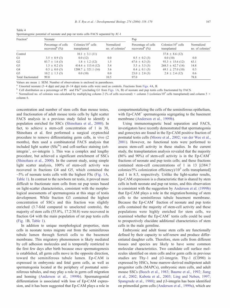
Table 4
Spermatogenic potential of neonate and pup rat testis cells FACS separated by JC-1
Fractiona Neonate Pup
Percentage of cells
recoveredb (%)
Colonies/105 cells
transplanted
Normalized
no. of coloniescPercentage of cells
recoveredb (%)
Colonies/105 cells
transplanted
Normalized
no. of coloniesc
Control 10.1 F 3.1 (11) 37.8 F 8.6 (12)
G1 3.5 F 0.9 (3) 0.0 (12) 0.0 0.5 F 0.2 (3) 0.0 (10) 0.0
G2 83.7 F 1.6 (3) 1.8 F 1.2 (12) 1.5 67.6 F 4.3 (3) 93.3 F 15.6 (12) 63.1
G3 1.3 F 0.2 (3) 414.4 F 113.4 (12) 5.4 5.5 F 3.3 (3) 268.3 F 62.7 (14) 14.8
G4 0.3 F 0.0 (3) 1208.7 F 321.1 (16) 3.6 0.4 F 0.1 (3) 69.1 F 27.0 (10) 0.3
G5 10.2 F 1.3 (3) 0.0 (10) 0.0 23.0 F 2.0 (3) 2.8 F 2.4 (12) 0.6
Total fractionated 99.0 10.5 97.0 78.8
Values are mean F SEM. Number of observations is enclosed in parentheses.a Unsorted neonate (3–4 dpp) and pup (8–14 dpp) testis cells were used as controls. Fractions from Figs. 4A, B.b Cell distribution as a percentage of PI� and FSchi (excluding G1 from Figs. 1A, B) of neonate and pup testis cells fractionated by FACS.c Normalized no. of colonies was calculated by multiplying column 2 (% of cells recovered) � column 3 (colonies/105 cells transplanted) and column 5 �column 6.
B.-Y. Ryu et al. / Developmental Biology 274 (2004) 158–170 167
concentration and number of stem cells than mouse testes,
and fractionation of adult mouse testis cells by light scatter
FACS analysis in a previous study failed to identify a
population enriched for SSCs (Shinohara et al., 2000). In
fact, to achieve a stem-cell concentration of 1 in 30,
Shinohara et al. first performed a surgical cryptorchid
procedure to remove differentiating germ cells, in vivo (2
months), then used a combinatorial FACS analysis that
included light scatter (SSclo) and cell-surface staining (a6-
integrin+, av-integrin�). This was a complex and lengthy
procedure, but achieved a significant enrichment of SSCs
(Shinohara et al., 2000). In the current study, using simple
light scatter analysis, 100% of stem-cell activity was
recovered in fractions G4 and G5, which contained the
~5% of neonate testis cells with the highest FSc (Fig. 1A,
Table 1). In contrast to the newborn rat testis, it proved more
difficult to fractionate stem cells from rat pup testes based
on light-scatter characteristics, consistent with the morpho-
logical assessments of spermatogonia at this stage of testis
development. While fraction G3 contained the highest
concentration of SSCs and this fraction was slightly
enriched (3.7-fold compared to unselected controls), the
majority of stem cells (55.8%, 17.2/30.8) were recovered in
fraction G4 with the main population of rat pup testis cells
(Fig. 1B, Table 1).
In addition to unique morphological properties, stem
cells in neonate testes migrate out from the seminiferous
tubule lumen through Sertoli cells to the basement
membrane. This migratory phenomenon is likely mediated
by cell adhesion molecules and is temporally restricted to
the first few days after birth because once spermatogenesis
is established, all germ cells move in the opposite direction,
toward the seminiferous tubule lumen. Ep-CAM is
expressed in embryonic and fetal germ cells, as well as
spermatogonia located at the periphery of postnatal semi-
niferous tubules, and may play a role in germ cell migration
and homing (Anderson et al., 1999b). Spermatogonial
differentiation is associated with loss of Ep-CAM expres-
sion, and it has been suggested that Ep-CAM plays a role in
compartmentalizing the cells of the seminiferous epithelium,
with Ep-CAM+ spermatogonia segregating to the basement
membrane (Anderson et al., 1999b).
Using immunomagnetic bead separation and FACS,
investigators have recently demonstrated that spermatogonia
and gonocytes are found in the Ep-CAM positive fraction of
postnatal testis cells (Moore et al., 2002; van der Wee et al.,
2001). However, no functional tests were performed to
assess stem-cell activity in these studies. In the current
study, the transplantation analysis revealed that the majority
(86% and 90%) of stem-cell activity is in the Ep-CAM+
fractions of neonate and pup testis cells; and these fractions
contained stem-cell concentrations of 1 in 13 [(384.7
colonies/5% colonization efficiency)/105 cells transplanted]
and 1 in 8.5, respectively. Unlike the light-scatter results,
Ep-CAM expression is a characteristic that is shared by stem
cells in both neonate and pup rat testes, and this observation
is consistent with the suggestion by Anderson et al. (1999b)
that Ep-CAM plays a role in the migration/homing of stem
cells to the seminiferous tubule basement membrane.
Because the Ep-CAM+ fraction of neonate and pup testis
cells contained the majority of stem-cell activity and these
populations were highly enriched for stem cells, we
examined whether the Ep-CAM+ testis cells could be used
to prospectively elucidate additional characteristics of stem
cells in the male germline.
Embryonic and adult tissue stem cells are functionally
defined by their capacity to self-renew and produce differ-
entiated daughter cells. Therefore, stem cells from different
tissues and species are likely to have some common
molecular characteristics. Two candidate cell surface mol-
ecules identified on stem cells and/or germ cells in previous
studies are Thy-1 and h3-integrin. Thy-1 (CD90) is
expressed by HSCs, bone marrow-derived multipotent adult
progenitor cells (MAPCs), embryonic stem cells, and adult
mouse SSCs (Beech et al., 1983; Baume et al., 1992; Jiang
et al., 2002; Kubota et al., 2003; Ling and Neben, 1997;
Spangrude et al., 1988); and h3-integrin has been identified
on primordial germ cells (Anderson et al., 1999a), which are

B.-Y. Ryu et al. / Developmental Biology 274 (2004) 158–170168
the precursors of gonocytes in neonatal testes. Our flow
cytometry results (Fig. 2) indicate that the Ep-CAM+
fraction of rat neonate testis cells is Thy-1+ and h3-integrin+
(Figs. 2C, E), while the Ep-CAM+ fraction of rat pup testis
cells is Thy-1+ and h3-integrin� (Figs. 2D, F), and these
results were subsequently confirmed by functional trans-
plantation analyses shown in Fig. 3 and Table 3. Therefore,
prospective analyses of testis cell populations enriched for
stem cells can be instructive for identifying candidate
molecular markers. However, until a pure stem-cell pop-
ulation is available, it is still necessary to confirm the
prospective results with a functional assay.
Based on these results, Thy-1 appears to be a conserved
stem-cell characteristic, while h3-integrin expression may
reflect developmental changes in the stem-cell population,
as gonocytes in the neonatal testis differentiate into
spermatogonial stem cells located on the seminiferous
tubule basement membrane in pup testes. Integrins play a
central role in the cell migration, both as receptors that
connect the extracellular matrix (ECM) to intracellular
cytoskeletal proteins, as well as receptors that transduce
information from the ECM to affect cell behavior (Clark and
Brugge, 1995; Damsky and Werb, 1992; Juliano and
Haskill, 1993; Schwartz and Ingber, 1994). In a trans-
endothelial migration assay, monocytes lacking h3-integrinsrevealed weak migratory activity, whereas monocytes
expressing h3-integrins exhibited strong migration (Weer-
asinghe et al., 1998). Interestingly, h3-integrin is a ligand
for Thy-1 on astrocytes and promotes focal adhesion
formation, cell attachment, and spreading (Leyton et al.,
2001). Perhaps Ep-CAM, Thy-1, and h3-integrin collabo-
rate to mediate germ cell migration/homing during early
testis development and once the stem cell localizes to its
niche in the pup testis, the h3-integrin status changes
commensurate with decreased migratory activity.
Spermatogenesis is a highly productive process that
produces millions of mature spermatozoa each day. This
level of production requires an active amplification com-
partment that proceeds through about 12 population dou-
blings as progenitor spermatogonia differentiate into
functional sperm (Potten, 1992). Stem cells provide the
cellular input for the amplification compartment, but are
themselves probably relatively quiescent, a feature that may
be necessary to protect the integrity of the stem-cell genome
and the germline. Indeed, previous studies of bone marrow
cells demonstrated that long-term repopulating HSCs are
enriched in a subpopulation defined by low-intensity
staining with the vital mitochondrial dye, rhodamine (Rh)-
123, which might reflect reduced mitochondrial activation
status of quiescent HSCs (Kim et al., 1998; Okada et al.,
1993; Spangrude and Johnson, 1990). Because adult SSCs
are also slow-dividing cells (de Rooij, 1998), we hypothe-
sized that low mitochondrial membrane potential (DCm)
might be a common characteristic of all stem cells. JC-1 is a
mitochondrial-specific probe that is particularly sensitive to
changes in DCm in living cells (Garner et al., 1997; Reers et
al., 1991; Salvioli et al., 1997; Smiley et al., 1991). In
contrast to Rh-123, JC-1 staining is strongly affected by
drugs that dissipate DCm, but unaffected by agents that
depolarize the plasma membrane, making JC-1 a reliable
probe for analyzing DCm changes by flow cytometry
(Salvioli et al., 1997).
The results shown in Fig. 4A and Table 4 demonstrate
that the neonate testis cell fraction with the highest
concentration of SSCs (G4) exhibited the lowest DCm
[low J-aggregate (red) to monomer (green) JC-1 fluores-
cence ratio]. Therefore, our results support the hypothesis
that relatively quiescent stem cells in the newborn rat testis,
and perhaps all stem cells, have low DCm. This population
was enriched 120-fold for SSCs (1208.7 colonies/105 cells
transplanted) compared with unselected control (10.1
colonies/105 cells transplanted), which corresponds to a
stem-cell concentration of ~1 in 4 [105 cells transplanted /
(1208.7 colonies/5% colonization efficiency)]. In contrast,
stem cells in rat pup testes were more heterogeneous with
respect to JC-1 fluorescence characteristics (Fig. 4B; Table
4). The majority of stem cells (80%, 63.1/78.8) were in the
populations, which showed relatively higher red fluores-
cence (G2; Fig. 4B; Table 4). Therefore, SSCs in rat pup
testes appear to have more active mitochondria than their
gonocyte precursors, which might reflect increased prolif-
erative activity as this population expands to fill the rapidly
increasing number of niches in the prepubertal rat testis
(Ryu et al., 2003).
Using FACS analysis to fractionate testis cells based on
specific cellular and biochemical characteristics and the
transplantation functional assay to quantify stem-cell activ-
ity in the various fractions enabled us to systematically
characterize stem cells in the developing rat testis. We took
advantage of the fact that rat testes have a higher
concentration and number of stem cells than mouse testes;
and the rapidly changing nature of germline stem cells
during early postnatal development allowed them to be
distinguished from the main testis cell population. The
results revealed that FSchi, SSchi, DCmlo, Ep-CAM+, Thy-
1+, h3-integrin+ stem cells in the newborn rat testis
differentiate into SSclo, DCmhi, Ep-CAM+, Thy-1lo, h3-integrin� stem cells in the pup rat testis. These molecular
characteristics correspond to changes in biological activity
as stem cells migrate, proliferate, and initiate spermato-
genesis in the 2 weeks after birth.
Acknowledgments
We thank C. Freeman and R. Naroznowski for assistance
with animal maintenance and experimentation, J. Hayden
provided help with photography. This project is funded, in
part, under a grant with the Pennsylvania Department of
Health. The Department specifically disclaims responsibility
for any analyses, interpretations, or conclusions. Financial
support for the research was from the National Institute of
B.-Y. Ryu et al. / Developmental Biology 274 (2004) 158–170 169
Child Health and Human Development Grant 044445, the
Commonwealth and General Assembly of Pennsylvania,
and the Robert J. Kleberg, Jr. and Helen C. Kleberg
Foundation.
References
Anderson, R., Fassler, R., Georges-Labouesse, E., Hynes, R.O., Bader,
B.L., Kreidberg, J.A., Schaible, K., Heasman, J., Wylie, C., 1999a.
Mouse primordial germ cells lacking beta1 integrins enter the
germline but fail to migrate normally to the gonads. Development
126, 1655–1664.
Anderson, R., Schaible, K., Heasman, J., Wylie, C., 1999b. Expression of
the homophilic adhesion molecule. Ep-CAM in the mammalian
germline. J. Reprod. Fertil. 116, 379–384.
Baume, C.M., Weissman, I.L., Tsukamoto, A.S., Buckle, A., Peault, B.,
1992. Isolation of a candidate human hematopoietic stem-cell pop-
ulation. Proc. Natl. Acad. Sci. U. S. A. 89, 2804–2808.
Beech, J.N., Morris, R.J., Raisman, G., 1983. Density of Thy-1 on axonal
membrane of different rat nerves. J. Neurochem. 41, 411–417.
Bellve, A.R., Cavicchia, J.C., Millette, C.F., O’Brien, D.A., Bhatnagar,
Y.M., Dym, M., 1977. Spermatogenic cells of the prepuberal mouse.
Isolation and morphological characterization. J. Cell Biol. 74, 68–85.
Brinster, R.L., Avarbock, M.R., 1994. Germline transmission of donor
haplotype following spermatogonial transplantation. Proc. Natl. Acad.
Sci. U. S. A. 91, 11303–11307.
Brinster, R.L., Zimmermann, J.W., 1994. Spermatogenesis following
male germ-cell transplantation. Proc. Natl. Acad. Sci. U. S. A. 91,
11298–11302.
Clark, E.A., Brugge, J.S., 1995. Integrins and signal transduction pathways:
the road taken. Science 268, 233–239.
Clouthier, D.E., Avarbock, M.R., Maika, S.D., Hammer, R.E., Brinster,
R.L., 1996. Rat spermatogenesis in mouse testis. Nature 381, 418–421.
Damsky, C.H., Werb, Z., 1992. Signal transduction by integrin receptors for
extracellular matrix: cooperative processing of extracellular informa-
tion. Curr. Opin. Cell Biol. 4, 772–781.
de Rooij, D.G., 1998. Stem cells in the testis. Int. J. Exp. Pathol. 79, 67–80.
Garner, D.L., Thomas, C.A., Joerg, H.W., DeJarnette, J.M., Marshall, C.E.,
1997. Fluorometric assessments of mitochondrial function and viability
in cryopreserved bovine spermatozoa. Biol. Reprod. 57, 1401–1406.
Goldschneider, I., Gordon, L.K., Morris, R.J., 1978. Demonstration of Thy-
1 antigen on pluripotent hemopoietic stem cells in the rat. J. Exp. Med.
148, 1351–1366.
Jiang, Y., Jahagirdar, B.N., Reinhardt, R.L., Schwartz, R.E., Keene, C.D.,
Ortiz-Gonzalez, X.R., Reyes, M., Lenvik, T., Lund, T., Blackstad, M.,
Du, J., Aldrich, S., Lisberg, A., Low, W.C., Largaespada, D.A.,
Verfaillie, C.M., 2002. Pluripotency of mesenchymal stem cells derived
from adult marrow. Nature 418, 41–49.
Juliano, R.L., Haskill, S., 1993. Signal transduction from the extracellular
matrix. J. Cell Biol. 120, 577–585.
Kanatsu-Shinohara, M., Toyokuni, S., Shinohara, T., 2004. CD9 is a surface
marker on mouse and rat male germline stem cells. Biol. Reprod. 70,
70–75.
Kim, M., Cooper, D.D., Hayes, S.F., Spangrude, G.J., 1998. Rhodamine-
123 staining in hematopoietic stem cells of young mice indicates
mitochondrial activation rather than dye efflux. Blood 91, 4106–4117.
Kubota, H., Avarbock, M.R., Brinster, R.L., 2003. Spermatogonial stem
cells share some, but not all, phenotypic and functional characteristics
with other stem cells. Proc. Natl. Acad. Sci. U. S. A. 100, 6487–6492.
Leyton, L., Schneider, P., Labra, C.V., Ruegg, C., Hetz, C.A., Quest,
A.F.G., Bron, C., 2001. Thy-1 binds to integrin h3 on astrocytes and
triggers formation of focal contact sites. Curr. Biol. 11, 1028–1038.
Li, H., Papadopoulos, V., Vidic, B., Dym, M., Culty, M., 1997. Regulation
of rat testis gonocyte proliferation by platelet-derived growth factor and
estradiol: identification of signaling mechanisms involved. Endocrino-
logy 138, 1289–1298.
Ling, V., Neben, S., 1997. In vitro differentiation of embryonic stem cells:
immunophenotypic analysis of cultured embryoid bodies. J. Cell.
Physiol. 171, 104–115.
Litvinov, S.V., Balzar, M., Winter, M.J., Bakker, H.A.M., Bruijn, I.H.,
Prins, F., Fleuren, G.J., Warnaar, S.O., 1997. Epithelial cell adhesion
molecule (Ep-CAM) modulates cell–cell interactions mediated by
classic cadherins. J. Cell Biol. 139, 1337–1348.
McCarrey, J.R., 1993. Development of the germ cell. In: Desjardins, C.,
Ewing, L.L. (Eds.), Cell and Molecular Biology of the Testis. Oxford
Univ. Press, New York, pp. 58–89.
Moore, T.J., Boer-Brouwer, M., Dissel-Emiliani, F.M.F., 2002. Purified
gonocytes from the neonatal rat form foci of proliferating germ cells in
vitro. Endocrinology 143, 3171–3174.
Nagano, M., Avarbock, M.R., Brinster, R.L., 1999. Pattern and kinetics of
mouse donor spermatogonial stem cell colonization in recipient testes.
Biol. Reprod. 60, 1429–1436.
Ogawa, T., Arechaga, J.M., Avarbock, M.R., Brinster, R.L., 1997.
Transplantation of testis germinal cells into mouse seminiferous tubules.
Int. J. Dev. Biol. 41, 111–122.
Okada, S., Nagayoshi, K., Nakauchi, H., Nishikawa, S., Miura, Y.,
Suda, T., 1993. Sequential analysis of hematopoietic reconstitution
achieved by transplantation of hematopoietic stem cells. Blood 81,
1720–1725.
Orth, J.M., Boehm, R., 1990. Functional coupling of neonatal rat
Sertoli cells and gonocytes in coculture. Endocrinology 127,
2812–2820.
Orwig, K.E., Ryu, B.Y., Avarbock, M.R., Brinster, R.L., 2002a. Male germ-
line stem cell potential is predicted by morphology of cells in neonatal
rat testes. Proc. Natl. Acad. Sci. U. S. A. 99, 11706–11711.
Orwig, K.E., Shinohara, T., Avarbock, M.R., Brinster, R.L., 2002b.
Functional analysis of stem cells in the adult rat testis. Biol. Reprod.
66, 944–949.
Potten, C.S., 1992. Cell lineages. In: McGee, J.O’D., Isaacson, P.G.,
Wright, N.A. (Eds.), Oxford Textbook of Pathology. Oxford Univ.
Press, Oxford, pp. 43–52.
Reers, M., Smith, T.W., Chen, L.B., 1991. J-aggregate formation of a
carbocyanine as a quantitative fluorescent indicator of membrane
potential. Biochemistry 30, 4480–4486.
Rhim, J.A., Sandgren, E.P., Degen, J.L., Palmiter, R.D., Brinster, R.L.,
1994. Replacement of diseased mouse liver by hepatic cell trans-
plantation. Science 263, 1149–1152.
Russell, L.D., Brinster, R.L., 1996. Ultrastructural observations of
spermatogenesis following transplantation of rat testis cells into mouse
seminiferous tubules. J. Androl. 17, 615–627.
Ryu, B.Y., Orwig, K.E., Avarbock, M.R., Brinster, R.L., 2003. Stem cell
and niche development in the postnatal rat testis. Dev. Biol. 263,
253–263.
Salvioli, S., Ardizzoni, A., Franceschi, C., Cossarizza, A., 1997. JC-1,
but not DiOC6(3) or rhodamine 123, is a reliable fluorescent probe
to assess [Delta][Psi] changes in intact cells: implications for studies
on mitochondrial functionality during apoptosis. FEBS Lett. 411,
77–82.
Schwartz, M.A., Ingber, D.E., 1994. Integrating with integrins. Mol. Biol.
Cell 5, 389–393.
Shinohara, T., Orwig, K.E., Avarbock, M.R., Brinster, R.L., 2000.
Spermatogonial stem cell enrichment by multiparameter selection of
mouse testis cells. Proc. Natl. Acad. Sci. U. S. A. 97, 8346–8351.
Smiley, S.T., Reers, M., Mottola-Hartshorn, C., Lin, M., Chen, A.,
Smith, T.W., Steele Jr., G.D., Chen, L.B., 1991. Intracellular
heterogeneity in mitochondrial membrane potentials revealed by a
J-aggregate-forming lipophilic cation JC-1. Proc. Natl. Acad. Sci.
U. S. A. 88, 3671–3675.
Spangrude, G.J., Johnson, G.R., 1990. Resting and activated subsets of
mouse multipotent hematopoietic stem cells. Proc. Natl. Acad. Sci.
U. S. A. 87, 7433–7437.

B.-Y. Ryu et al. / Developmental Biology 274 (2004) 158–170170
Spangrude, G.J., Heimfeld, S., Weissman, I.L., 1988. Purification and
characterization of mouse hematopoietic stem cells. Science 241,
58–62.
Tegelenbosch, R.A.J., de Rooij, D.G., 1993. A quantitative study of
spermatogonial multiplication and stem cell renewal in the C3H/101 F1
hybrid mouse. Mutat. Res. 290, 193–200.
van der Wee, K.S., Johnson, E.W., Dirami, G., Dym, T.M., Hofmann, M.C.,
2001. Immunomagnetic isolation and long-term culture of mouse type
A spermatogonia. J. Androl. 22, 696–704.
van Dissel-Emiliani, F.M.F., de Rooij, D.G., Meistrich, M.L., 1989.
Isolation of rat gonocytes by velocity sedimentation at unit gravity. J.
Reprod. Fertil. 86, 759–766.
Weerasinghe, D., McHugh, K.P., Ross, F.P., Brown, E.J., Gisler, R.H.,
Imhof, B.A., 1998. A role for the avh3 integrin in the transmigration of
monocytes. J. Cell Biol. 142, 595–607.
Zhang, X., Ebata, K.T., Nagano, M.C., 2003. Genetic analysis of the clonal
origin of regenerating mouse spermatogenesis following transplanta-
tion. Biol. Reprod. 69, 1872–1878.


![bai hoc con buom [ newssaigon.net ]](https://img.dokumen.tips/doc/110x75/55a9874f1a28ab6b248b46d9/bai-hoc-con-buom-newssaigonnet-.jpg)


