Embed Size (px)
Citation preview
346 Usman et al.
Int. J. Biosci. 2018
RESEARCH PAPER OPEN ACCESS
Phenotype analysis, molecular genetics and therapeutics of
glaucoma: Recent developments and future directions with
respect to Pakistan
Muhammad Usman*1,2, Muhammad Ali3, Qaisar Hanif4, Muhammad Qasim2,
Muhammad Ibrahim Rajoka2, Muhammad Rashid2, Zulqarnain Manzoor5,
Omer Draz3, Tehreem Anwar1, Mirza Jawad ul Hasnain1
1Department of Bioinformatics & Computational Biology, Virtual University of Pakistan,
Lahore, Pakistan
2Department of Bioinformatics and Biotechnology, Government College University, Faisalabad, Pakistan
3Department of Zoology, Government College University, Faisalabad, Pakistan
4Department of Ophthalmology Allied Hospital, Medical University, Faisalabad, Pakistan
5Department of Biotechnology, International Islamic University, Islamabad, Pakistan
Key words: Blindness, Eye disease, Glaucoma, Genetic disease, Optical nerves
http://dx.doi.org/10.12692/ijb/12.2.346-357 Article published on February 28, 2018
Abstract
Blindness is a major disability that severely compromises the life quality and makes the individuals unfit
for every job. Genetic factors play key role in many types of eye disorders, involving those disorders that are
the key reason of blindness amongst infants, children and adults. Glaucoma is a main source of sight loss
not only sight and is categorized by enlightened deterioration of the optical nerve and is usually connected
with higher intraocular pressure. Without adequate dealing, glaucoma can be developed to optical disability
and ultimately sightlessness. Scientists had determined and mapped several genes for glaucoma. Pakistani
population is relatively least investigating for genetic diseases as compare to European population. Because
of the high degree of consanguinity Pakistani population offers a priceless genetic resource for identifying
new genomic regions and to fill gaps in the existing knowledge. In this review, we provide detailed
description of genetic and phenotypic heterogeneity of glaucoma disease with respect to Pakistan and also
we shed some light on current therapeutic approaches and future directions. Furthermore, we identified 10
consanguineous glaucomatous families of Pakistan with multiple affected individuals and performed their
clinical evaluation for better understanding of disease.
* Corresponding Author: Muhammad Usman [email protected]
International Journal of Biosciences | IJB |
ISSN: 2220-6655 (Print) 2222-5234 (Online)
http://www.innspub.net
Vol. 12, No. 2, p. 346-357, 2018
347 Usman et al.
Int. J. Biosci. 2018
Introduction
Inherited visual disorders leading to blindness is a
serious problem all over the world, especially in
developing countries. Congenital cataracts (CCs),
glaucoma & Retinitis Pigmentosa (RP) are the leading
causes of blindness which can be transmitted from
parents to their offsprings in various patterns of
inheritance.
The word “glaucoma” covers a number of
miscellaneous eye disarrays, all of which comprehend
injury to the optical nerve (Khan et al., 2017) .
Glaucoma, a complicated heterogeneous set of
disorders that is the second principal reason of
blindness after cataract of visual disability and can
cause permanent blindness if not treated (Rizzo et al.,
2017). Glaucoma is categorized by the deterioration of
optic nerves related with increasing intraocular
pressure and the destruction of retinal ganglion cells
(RGC) (Faiq et al., 2013). Molecular genetic
techniques have been used to study the genetics of
glaucoma in detail. Different types of glaucoma have
been reported during the past few years. These
studies indicate that Glaucoma is genetically and
clinically heterogeneous in nature (Frick and Foster,
2003). The number of blind individuals would be
increasing from 44 million in 2000 to 76 million in
2020 worldwide (Sarfarazi, 1997) out of which about
15% cases of blindness are because of glaucoma
(Weisschuh and Schiefer, 2003).
It is an irreversible vision loss along with decreased
contrast and color sensitivity (Casson et al., 2012).
Visual disorder in glaucoma is due to optic nerve
damage because of high intra ocular pressure in
most of the cases. It leads to blindness if left
untreated (Wiggs et al., 1998). Discharge of aqueous
humour through trabecular meshwork is blocked
that cause rise in intra ocular pressure. Molecular
mechanism of normal as well as glaucomatous
aqueous humour outflow is not yet known (Farkas
and Grosskreutz, 2001). Retinal ganglionic cells die
by apoptosis resulting in degeneration of optic
nerve. Relationship of high intra ocular pressure and
apoptotic death of ganglionic cell is not well
understood. Early detection and continuous
conventionally recommended treatment can reduce
irreparable damage (Feldman, 2004).
Maintaining low intra ocular pressure by the use of
ocular hypotensive agents is the major medical
treatment for glaucoma (Sarfarazi and Stoilov, 2000).
Expanding knowledge of Molecular Biology has made
it possible us to describe the etiology of inherited
disorders through identification of genes. It is very
crucial to understand what differences exist among
genes and their loci responsible for different
hereditary disorders in Pakistan from the studies
conducted elsewhere in the World. During the course
of study, the affected individuals will be subjected to
clinical examination and a series of ophthalmological
tests for the correct diagnosis. Besides genetic
dissection of these diseases, the study is also aimed at
spreading awareness among affected individuals and
their families as most genetic disorders in Pakistani
population have a recessive mode of inheritance
which is expressed where consanguinity is common.
Prevalence of Glaucoma
It was forecasted that 161 million people globally had
optical loss and 37 million people were suffered from
blindness in 2002. Visual impairment had 12.3% of
worldwide sightlessness, though cataract accounted
for 47.8%. it is also estimated that glaucoma had
effected adults more as compared to children and
women are more effected as compared to men
(Casson et al., 2012; Faiq et al., 2013). In 2010 study,
it is founded out that 60.5 million individuals all over
the world had open-angle glaucoma (OAG) and angle-
closure glaucoma (ACG). It is roughly estimated that
in 2020 this prevalence would be increased to 79.6
million. The collective (74%) people would have OAG.
70% will be women which would be effected by ACG
and 87% will be Asians. Glaucoma is the loss of visual
ability caused more in women and Asians (Buhrmann
et al., 2000). Visual loss is practically third time in
African Americans as compared to white Americans,
and POAG is the foremost source of sightlessness in
African Americans (Rein et al., 2006). There is a more
risk to manage health care. 17.8% people who have
disease of the visual impairment in USA contribute their
self to take the medicine and bear the cost for that
purpose. On behalf of a considerable percentage which is
given that the yearly entire through medical costs for
these illnesses was assessed to be $16.2 billion and in
coming years this cost will be increased unquestionably
(Hussain and Bittles, 1998).
348 Usman et al.
Int. J. Biosci. 2018
Phenotypic Heterogeneity in Glaucoma
Glaucoma has been divided into different groups like
congenital and non-congenital, primary and secondary,
open and closed angle, infantile, juvenile and adult on
the basis of etiology, anatomy of anterior chamber
(Shastry, 2013). Primary defective molecules and all
the risk factors should be taken into account while
grouping glaucoma (Weisschuh and Schiefer, 2003). It
is now believed that almost all forms of glaucoma have
genetic basis. Inherited forms of glaucoma are
heterogeneous, both autosomal dominant and
recessive a very little literature is available about sex
linked inheritance of glaucoma. There are three major
classes of glaucoma (Khan et al., 2017).
Primary Congenital Glaucoma
Primary congenital glaucoma (PCG) is an infrequent
form of glaucoma. It appears at birth or within first
three years of life. It is due to progressive flaws in TM
and anterior chamber resulting in high intra ocular
pressure, optic disc destructions and raised corneal
diameter (Weinreb et al., 2014). It shows autosomal
recessive Mendelian inheritance.
Open-Angle Glaucoma
In open angle glaucoma (OAG) defect is in the
outflow of aqueous humour due to decreased number
of cells in filtration region and close to the interior
wall of Schlemm's canal, extra cellular materials are
accumulated. Primary open angle glaucoma (POAG)
is the most common type of glaucoma, disturbing
over 33 million persons wide-reaching (Khan et al.,
2017). Open-angle chronic glaucoma progress
gradually, it is pain-free and most of the time shows
no signs, until it has developed sufficiently. It is of
adult onset and juvenile form. It has also been
proposed that JOAG shows autosomal dominant
inheritance while the adult onset POAG exhibits non-
Mendelian inheritance (Weinreb et al., 2014).
Increased level of IOP is the main discovery related to
this disease. Additionally, on clinical trials, noticeable
focal flaws in the retinal nerve fibre layer generally
intercede optic disc modifications and visual field
damage. Changes related to age in the trabecular
region are the most probable reason of this condition.
Even though many cases of POAG are related with
increased IOP, in some cases level of IOP is shown
normal and it is stated as normal-tension glaucoma
(NTG). This is possibly because of unnoticed change
in levels of IOP but optic nerve is usually responsive
to these changes (Fraunfelder et al., 2004).
Angle Closure Glaucoma
This is a set of disorders in which there is aqueous
obstruction that results due to rescindable
(appositional) or cohesional (synechial) closure of the
anterior-chamber. There are two forms in which angle
closure can occur and that is in acute and chronic
form. In the acute form, there is sudden stoppage of
the TM by the iris via pupillary block mechanism
which results in elevation of IOP. The chronic form
may grow after acute form where synechial closure of
the angle remains, or it may grow over time as the
angle closes from lengthened or recurring contact
between the peripheral iris and the TM, which
frequently contributes to functioning obstruction to the
angle and peripheral anterior synechiae (PAS) (Kim
and Jung, 1997). Primary angle closure glaucoma
(PACG) occurs when access to the trabecular
meshwork (TM) is physically obstructed, typically by
the iris, and the drainage angle is closed.
Three main mechanisms hypothesized to be
responsible for PACG are pupillary block, anterior iris
rotation, and plateau iris. When the pressure of the
posterior chamber overrun the pressure of the anterior
chamber, pupillary block appears to be happen, and
this blocks the TM by pushing peripheral and mid
peripheral iris forward (Harasymowycz et al., 2016).
Secondary angle-closure happens by some known
genetics. Phacomorphic glaucoma is an example of
secondary angle-closure, that happens because the
angle is closed when lens pushes iris forward (Tello et
al., 2000). This might also happen in subluxation.
Secondary pupil block may be caused by Uveitis, which
is distinguished by posterior synechiae and iris bombe.
Some other reasons comprise malignant glaucoma,
neovascularisation, retinopathy of prematurity, Vogt-
Koyanagi-Harada syndrome, posterior scleritis,
acquired immunodeficiency syndrome, leukaemia,
orbital or carotid cavernous fistula and neuropathia
epidemica (Quigley and Broman, 2006).
349 Usman et al.
Int. J. Biosci. 2018
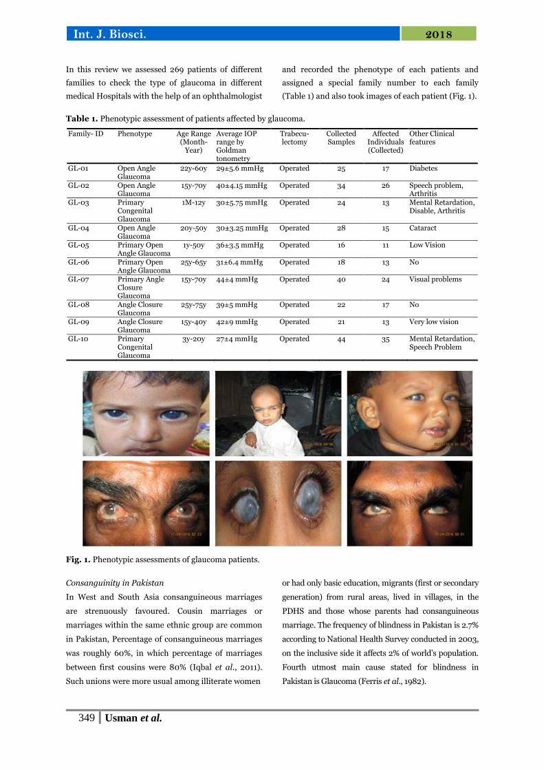
In this review we assessed 269 patients of different
families to check the type of glaucoma in different
medical Hospitals with the help of an ophthalmologist
and recorded the phenotype of each patients and
assigned a special family number to each family
(Table 1) and also took images of each patient (Fig. 1).
Table 1. Phenotypic assessment of patients affected by glaucoma.
Family- ID Phenotype Age Range (Month-
Year)
Average IOP range by Goldman tonometry
Trabecu-lectomy
Collected Samples
Affected Individuals (Collected)
Other Clinical features
GL-01 Open Angle Glaucoma
22y-60y 29±5.6 mmHg Operated 25 17 Diabetes
GL-02 Open Angle Glaucoma
15y-70y 40±4.15 mmHg Operated 34 26 Speech problem, Arthritis
GL-03 Primary Congenital Glaucoma
1M-12y 30±5.75 mmHg Operated 24 13 Mental Retardation, Disable, Arthritis
GL-04 Open Angle Glaucoma
20y-50y 30±3.25 mmHg Operated 28 15 Cataract
GL-05 Primary Open Angle Glaucoma
1y-50y 36±3.5 mmHg Operated 16 11 Low Vision
GL-06 Primary Open Angle Glaucoma
25y-65y 31±6.4 mmHg Operated 18 13 No
GL-07 Primary Angle Closure Glaucoma
15y-70y 44±4 mmHg Operated 40 24 Visual problems
GL-08 Angle Closure Glaucoma
25y-75y 39±5 mmHg Operated 22 17 No
GL-09 Angle Closure Glaucoma
15y-40y 42±9 mmHg Operated 21 13 Very low vision
GL-10 Primary Congenital Glaucoma
3y-20y 27±4 mmHg Operated 44 35 Mental Retardation, Speech Problem
Fig. 1. Phenotypic assessments of glaucoma patients.
Consanguinity in Pakistan
In West and South Asia consanguineous marriages
are strenuously favoured. Cousin marriages or
marriages within the same ethnic group are common
in Pakistan, Percentage of consanguineous marriages
was roughly 60%, in which percentage of marriages
between first cousins were 80% (Iqbal et al., 2011).
Such unions were more usual among illiterate women
or had only basic education, migrants (first or secondary
generation) from rural areas, lived in villages, in the
PDHS and those whose parents had consanguineous
marriage. The frequency of blindness in Pakistan is 2.7%
according to National Health Survey conducted in 2003,
on the inclusive side it affects 2% of world’s population.
Fourth utmost main cause stated for blindness in
Pakistan is Glaucoma (Ferris et al., 1982).
350 Usman et al.
Int. J. Biosci. 2018
Genetic Heterogeneity in Glaucoma
MYOC
The location of gene MYOC (MIM601652) is at the
GLC1A locus (MIM137750) on chromosome 1q25
(Fingert et al., 2002). It was the first OAG gene to be
defined and was associated with JOAG and POAG.
MYOC has 3 exons and codes 57 kD Myocilin protein
(Kubota et al., 1997). This protein is cytoskeletal and
usually expressed in the retinal photoreceptor cells
and is present mainly in the basal body of
photoreceptor linking cilium. Normally in eyes,
MYOC mRNA is expressed in the iris, ciliary body,
TM as well as in retinal photoreceptor cells and optic
nerve head-specifically, the astrocytes (Ohlmann and
Tamm, 2002). The contemporary purpose of Myocilin
is still unidentified. By glucocorticoids, transmuting
growth factor-β, stretch, and elevated intraocular
pressure (Filla et al., 2002). Myocilin expression is
expressed in trabecular meshwork cells. It is a
discharged glycoprotein which is lightly coupled with
the Trabecular meshwork out of the cell matrix. It
exists in the aqueous humour and they are usually
huge oligomers aggregated into themselves. The role
of myocilin and its connections regarding
pathophysiology of glaucoma. Enhanced Myocilin
expression has been identified in the trabecular
meshwork of glaucoma patients, including pseudo
exfoliation glaucoma, POAG and pigmentary
glaucoma (Bejjani et al., 1998). An additional vital
subtype of glaucoma is normal tension glaucoma
(NTG), which is considered by glaucomatous damage
to the ONH and progressive loss of vision in patients
with normal IOP. The role of Myocilin in the
physiopathology of NTG is still uncertain. It is most
frequently mutated gene in familial glaucoma cases,
accounting for 4% of adult onset POAG and 10%
JOAG (Noda et al., 2000).
Cyp1B1
Location of CYP1B1 (MIM 601771) is at the GLC3A
locus (MIM 231300) on chromosome
2p21(Beckerman, 2002). CYP1B1 contains 3 exons, in
which protein coding exons are only two and three
(Hayes et al., 1996). CYP1B1 displays the highest level
of expression in endometrial tissue. It also has
constitutive expression in extrahepatic tissues
involving mammary and lung tissue. Activation of
both polycyclic aromatic hydrocarbons and aryl
amines can be catalysed by CYP1B1 (Christou et al.,
1994). Additionally, it also has constitutive expression
in the human mammary carcinoma MCF-7 cell line and
facilitates the hydroxylation of 17 ‚-estradiol to form 4-
catecholoestrogen. CYPIBI level of expression is also
seen in a many types of malignant tumours but is not
noticeable in normal tissues, showing that this
cytochrome P450 is a specific tumour causing form of
cytochrome P450 (Sutter et al., 1994). Although
CYP1B1 mRNA has been identified in a limited number
of normal tissues, findings of an absence of detectable
CYP1BI protein in normal tissues suggest that either
CYP1B1 protein is present at a very low level in normal
tissues or the CYP1B1 mRNA is not translated.
The CYP1B1 expression and the development of 4-
hydroxy estrogens have been related with estrogen-
associated tumours in several tissues and species.
Increased 4-hydroxy estrogen formation has been
related with caner formation of several cancers
counting endometrial cancer (Jefcoate et al., 2000).
Juvenile glaucoma (JG) and congenital glaucoma
(CG) are inherently heterogeneous, and
categorization is only on small subset, with most
frequent mutations found in MYOC and CYP1B1.
These mutations are related with autosomal recessive
CG. In Pakistani patients, in 10 out of 20 families
(50%) homozygosity is expressed with CYP1B1- STR
markers (Sheikh et al., 2014).
OPTN
Optineurin (OPTN) gene is located on chromosome
10p14 at the GLC1E [55]. OPTN contains sixteen
exons; in the 5′ untranslated region, first 3 exons are
non-protein coding and then they are followed by
thirteen exons that codes a 577 amino acid (Faiq et
al., 2013). Its expression is usually in brain and ocular
tissues such as the optic nerve, TM, non-pigmented
ciliary epithelium and retina (Rezaie et al., 2007). In
broad sense it is expressed in both ocular and non-
ocular tissues. This protein interacts with apoptosis
related proteins and might show a role in protection
of neurological system as it decreases susceptibility to
apoptosis of retinal ganglion cells.
351 Usman et al.
Int. J. Biosci. 2018
OPTN increased expression blocks cytochrome c
discharge from the mitochondria and defends the cell
from H2O2-induced cell death (Kumar et al., 2007a).
The mechanical role of OPTN in the physiopathology of
glaucoma still remains vague .OPTN genes is involved in
the aetiology of adult-onset primary open-angle
glaucoma (POAG) (Kumar et al., 2007b) . Most common
variations of OPTN in Asia is M98K and T34T.
WDR36
WDR36 (MIM 609669) gene is located at the GLC1G
locus (Fingert et al., 2011; Monemi et al., 2005). WDR36
is limited in centre between positions 111,037,156 bp and
147,210,429 bp at chromosome 5q22.1-q32 using the
Human Genome Sequence.WDR36 contains 23 exons
and codes for a 951 amino acid protein with several
WD40 recursions. Its expression is seen in both human
ocular and non-ocular tissues and also in adult mouse
and embryonic tis The main function of this gene is the
formation of coordinating multiprotein complicated
assemblies, where the recurring units’ aid as a stiff
scaffold for protein interactions.
It has been anticipated that T-cell activation may be
mediated by WDR36 and currently, participation of T-
cell facilitated responses is found in glaucoma-associated
optic nerve degeneration (Bakalash et al., 2005) . In the
Asian population, mutations in WDR36 seem to play a
negligible role in POAG pathogenesis but polymorphic
variants have been found to be associated with primary
open angle glaucoma, specifically in high tension
glaucoma (Mookherjee et al., 2011). More than 150 PCG-
related mutations in CYP1B1 have been defined. One
missense variant, p. G36D, and a 12 bp in-frame deletion
mutation, p. Gly67Ala70del, have recently been found in
Pakistani Families.
Other Molecular Players of Glaucoma
A new locus (GLC3D) residing on the LTBP2 gene has
been categorized in developmental glaucoma but its
role is still to be determined in standard cases of PCG
(Bejjani et al., 2000). One intronic SNP (rs3742793)
was found in LTBP2 gene within exon six and seven
in 18 patients out of a cohort of 54 unconnected
Indian patients with PCG who were negative for
MYOC, CYP1B1 and FOXC1 mutations. This SNP
resulted in C to G mutation at position g. 75070493.
There is no pathogenic variants were recognized in
the LTBP2 (Abu-Amero et al., 2011). Tested patients
of 54 dissimilar Saudi PCG families (74 patients),
there is no mutation in these patients who were
detected as having PCG via typical ophthalmological
inspections and screened for mutations in CYP1B1
and LTBP2 by sequencing.
In exceedingly engrained populations like Slovakian
Gypsies, Iranians and Saudi Arabians 80-100%
occurrence of recessively inherited glaucoma is
testified due to mutations in CYP1B1. 7 out of 9 PCG
patients (78%) from 8 consanguineous families from
Oman showed mutations in CYP1B1 (Bashir et al.,
2014). CYP1B1 mutations are the major (75.9%) cause
of PCG in the Saudi Arabian population with G61E as
the dominant disease-associated allele. British
Infantile and Childhood Glaucoma, study shown that
the frequency of PCG in the Pakistani children is
about nine times higher than that in Caucasians.
GLC3A contribute 17% of primary congenital
glaucoma in sporadic cases besides 2 patients out of 3
families from Pakistan (Wiggs et al., 2004).
Ethnic specific OPTN mutation patterns may exist.
The wild type OPTN protein, functioning through the
TNF-α pathway, is ventured to play a neuro-
protective role in the eye and optic nerve. But when
malfunctioning, it causes visual loss and optic
neuropathy as typically seen in NTG and high-tension
glaucoma (Alward et al., 2003; Zhou et al., 2013).
Three genes at 14 chromosomal loci elucidate for less
than 5% of all POAG cases signifying that 90% of
contribution of genes in POAG cases is not known.
D384N mutation has been stated as one of the major
mutations in POAG, as reported by a Chinese family
having POAG. Whereas T353I modification was
thought as a high-risk factor for POAG. These two
changes were first reported in one juvenile -onset
POAG patient who presented with more acute clinical
appearances, signifying that T353I polymorphism
may be related with the acuteness of POAG.
352 Usman et al.
Int. J. Biosci. 2018
The first indication for a mutation linked to familial
PACG emanates from the analysis of a large family
with nanophthalmos, hyperopia, and angle closure
glaucoma (Dai et al., 2008).
This study has directed towards the documentation of
the gene nanophthalmos 1 (NNO1) which is present
on chromosome 11. Presently the only human gene
recognized which produce an angle closure glaucoma
phenotype is NNO1. In distinction, several genetic
loci have been recognized that may not be
contributing, but increase an individual’s risk to
produce PACG. A positive connotation to CYP1B1, a
gene concerned in the formation of congenital
glaucoma, was highlighted in studies of PACG in
patients of Chinese, Indian, and Canadian origin
(Cheng et al., 2013). A current meta-analysis of
genome-wide associations for ocular axial length was
shown in patients of European and Asian origin
exhibiting refractive errors, which along with
hyperopia and myopia is mainly defined by axial
length. RSPO1 C3orf26, LAMA2, GJD2, ZNRF3,
CD55, MIP, ALPPL2 and ZC3H11B were identified as
nine genome-wide significant loci for axial length
(McBrien et al., 2001). Recently, genome-wide
association study (GWAS) in an Asian population of
PACG acknowledged three PACG vulnerability loci
in PLEKHA7, COL11A1, and also in PCMTD1 and
ST18. COL11A1 is a predominantly stimulating gene as
it encodes one of the two alpha chains of type XI
collagen, which is highly articulated in the scleral
tissue. Several studies provide additional evidence for
the potential role of collagen in glaucoma.
Modifications in collagen deposition influence the
biomechanical and remodelling competences of the
sclera, thus result in glaucoma-predisposing axial
length changes and associated refractive errors
(Inamori et al., 2007). In a Japanese and Chinese Han
populations, single-nucleotide polymorphism (SNP) in
the gene COL1A1 is related towards the increased risk
of myopia and it is plausible that other genetic variants
result in conformational variations to the anterior
segment that incline toward the development of the
disease (Norman et al., 2011). Still, transformations in
collagen composition of the sclera may be associated
with suboptimal optic nerve head biomechanics,
resulting in increased exposure to axonal damage in
glaucomatous eyes (Consoli et al., 2005).
Glaucoma Treatment and concerns
Glaucoma is a serious chronic disease. Its world
widely occurrence is approximately 67 million. It is a
visual disorder which causes blindness if no care is
taken place and no proper treatment is taken place at
specific time. It is not clearly understood that how it
causes loss of visual sight and then lead to complete
blindness. The speed of glaucoma causes can be
reduced but not stopped or controlled and not specific
measurement can be taken place to find out it earlier.
It causes an attention disturbance situation analysis
and by not full focusing on it and considering it well
and due to no proper diagnostic measurements
unproductive results are accompanied. The main
reason of glaucoma increased percentage is post
ponding of visual check-ups and no availability of
proper machinery and techniques. If visual loss is
taken place once then there is no adequate optical
treatment is available to cover up it e.g. IOP is a major
risk which lead to the loss of visual ability (Fechtner
and Realini, 2004). Some other things also very
important like cost, sustainability etc. Now these days
its treatments are taken place including medication,
laser and operation. With recently doing research and
adequate medicinal treatment glaucoma can be
stopped or well-orderedly controlled and sight loss
can be recovered. In these days there is no remedy
but glaucoma required more attention and treatment
with care by doing this it can be recovered.
Glaucoma is treated with the help of specific medicine
which take a good part to control the loss of quantity
of liquid which is built by eyes. Some that kind of
treatment causes safety of eye pressure for some
years. Some medicines are used that control the
attack of aqueous humour, enlightening the
expenditure of aqueous humour, protecting the
optical nerves. Prostaglandin F2α derivatives which
causes enhancement of the uveoscleral discharges
recently used at wide level. Bimatoprost is also widely
used for glaucoma treatment as an anti-glaucoma
(Khaw et al., 2004). Acetylcholine receptor agonists is
also used for IOP so that visual loss can be controlled.
Timolol is utilized which is the major suggested drug
and betaxolol is also used which has the slightest
common harmful effects.
353 Usman et al.
Int. J. Biosci. 2018
These both medicines are together used as a β1
receptor blockers. Laser treatment is used commonly
for glaucoma by using ciliary and the pigmented
trabecular meshwork cells. 20 to 30 percent patients
are treated with laser technique and 70 percent
patients are overwhelmed with it (Lau et al., 2002).
Some kinds of surgeries are also taken place like
Minimally Invasive Glaucoma Surgery (MIGS) are
taken place through which glaucoma is detected and
then treated. Some kind of research is also taken
place which would provide help us to overcome
different kind of glaucoma and Gentler sorts of laser
Cyclophotocoagulation is also taken place to recover
the glaucoma.
The awareness of patients and their involvement in
treatment of glaucoma is very necessary and play an
important role to prevent and cure of such kind of
disease just like Glaucoma. A huge amount of study is
taken place on glaucoma that is caused in young
persons. The patients who lose their eye visual power
was studied and analysed and primary reason was
find out but due to the less knowledge about
blindness, more information was not obtained but
increment of literature and educational system can
enhance the betterment in treatment of visual loses
(Gooch et al., 2012).
Now these days’ main focus is taken place on
intraocular pressure. It is achieved by taking regular
up-to-date eye drops but due to less awareness of
patient and improper treatment glaucoma is
increased. Many constant curing treatments are
under research by some scientists. Conjunctively,
punctal plugs intravitreal inserts and sub
conjunctival, and drug sidings are recently in under
clinical improvements (Garaci et al., 2009a).
Treatment of glaucoma is more expensive but there is
a hope that dramatically improvement will come in
this disease treatment in few coming years and
improved IOP control system will be developed. Some
researcher reviled that that glaucoma is a
multifaceted neurologic disease that disturbs optic
Nerves, optic radiations, and the adjacent geniculate
nucleus also. Central nervous system (CNS) causes
impairment accompanying with glaucoma that has
been perceived by modifications in optic nerves by
using of magnetic significance imaging (Garaci et al.,
2009b). POAG affected person is treated with some
early rehabilitations which show good effect during
coming 10 years that results are obtained by utilizing
the therapies which decrease the IOP.
In some cases, glaucoma patients are treated and
cured during 5 years. Some filtration surgeries are
taken place which controls the IOP that is the basic
thing that is done with glaucoma patients and it
reduces the effect of Glaucoma and enhances the
curing chances (Ehrlich et al., 2012). Recently some
goals are in mind of scientists that glaucoma can be
stopped by some basic treatments and its causing
chances can be lowered by decreasing the IOP
(intraocular pressure). By increase of the aqueous
discharge glaucoma is caused and it reduces the
better medication of glaucoma.
Treatment should not only decrease the glaucoma but
it also should reduce the bad effects of that treatment.
Full knowledge should be given to the patient and
then according to their choice and betterment their
treatment should be done (Kass et al., 2002).
Nevertheless of the some chemical composites and or
by using some technologies of accomplishment, the
perfect ocular delivery system is that one which
achieved and enhances the operational values of
specific drug amount at the major position for
anticipated time periods and it decrease the universal
acquaintance and meet the expenses of patient which
increased the volubility of life but there is a major task
for scientist to find out technique which should be safe
so that safe transport fences of eye can be done which
should not contain the unwanted and unappealing bad
effects and it should prove as a safe therapy.
Conclusion
The biological origin of this disorder is not yet
entirely tacit, and the aspects causative to its
progression are not yet entirely characterised. The
most imperative risk factor for glaucoma is raised by
intraocular pressure. Because the optic-nerve damage
in glaucoma is yet not acquiescent to direct treatment,
354 Usman et al.
Int. J. Biosci. 2018
the provided treatment is only for the known risk
factor that can be modified, increased intraocular
pressure. We hope that further analysis of data from
this study can be used to find some defining features
of such population subsets, diagnostic risk factors, or
perhaps clues to risk factors that may be docile to
therapeutic changes.
Acknowledgment
The author is thankful to those families who were
participated in this study voluntarily. The author is
also thankful to Department of Ophthalmology Allied
Hospital, Medical University Faisalabad, Pakistan for
the phenotypic assessments of the patients.
Author’s Contributions
MU Identified glaucomatous families, proceeded with
clinical evaluation, wrote and corresponded the
manuscript, QH performed clinical evaluation, MA,
MQ and MIR helped in manuscript writing, MR, ZM,
OD, TA, and MJH helped in families’ identification.
References
Abu-Amero KK, Osman EA, Mousa A, Wheeler
J, Whigham B, Allingham RR, Hauser MA, Al-
Obeidan SA. 2011. Screening of CYP1B1 and LTBP2
genes in Saudi families with primary congenital
glaucoma: genotype-phenotype correlation. Molecular
Vision 17, 2911.
Alward WL, Kwon YH, Kawase K, Craig JE,
Hayreh SS, Johnson AT, Khanna CL,
Yamamoto T, Mackey DA, Roos BR. 2003.
Evaluation of optineurin sequence variations in 1,048
patients with open-angle glaucoma. American
Journal of Ophthalmology 136, 904-910.
Bakalash S, Shlomo GB, Aloni E, Shaked I,
Wheeler L, Ofri R, Schwart M. 2005. T-cell-
based vaccination for morphological and functional
neuroprotection in a rat model of chronically elevated
intraocular pressure. Journal of Molecular Medicine
83, 904-916.
Bashir R, Sanai M, Azeem A, Altaf I, Saleem F,
Naz, S. 2014. Contribution of GLC3A locus to Primary
Congenital Glaucoma in Pakistani population. Pakistan
Journal of Medical Sciences 30, 1341.
Beckerman M. 2002. Modeling and Simulation of
Microelectrode-Retina Interactions. Oak Ridge Y-12
Plant, TN (US).
Bejjani BA, Lewis RA, Tomey KF, Anderson
KL, Dueker DK, Jabak M, Astle WF, Otterud
B, Leppert M, Lupski JR. 1998. Mutations in
CYP1B1, the gene for cytochrome P4501B1, are the
predominant cause of primary congenital glaucoma in
Saudi Arabia. The American Journal of Human
Genetics 62, 325-333.
Bejjani BA, Stockton DW, Lewis RA, Tomey
KF, Dueker DK, Jabak M, Astle WF, Lupski
JR. 2000. Multiple CYP1B1 mutations and
incomplete penetrance in an inbred population
segregating primary congenital glaucoma suggest
frequent de novo events and a dominant modifier
locus. Human Molecular Genetics 9, 367-374.
Buhrmann RR, Quigley HA, Barron Y, West SK,
Oliva MS, Mmbaga BB. 2000. Prevalence of glaucoma
in a rural East African population. Investigative
Ophthalmology & Visual Science 41, 40-48.
Casson RJ, Chidlow G, Wood JP, Crowston
JG, Goldberg I. 2012. Definition of glaucoma:
clinical and experimental concepts. Clinical &
Experimental Ophthalmology 40, 341-349.
Cheng C-Y, Schache M, Ikram MK, Young TL,
Guggenheim JA, Vitart V, MacGregor S,
Verhoeven VJ, Barathi VA, Liao J. 2013. Nine
loci for ocular axial length identified through genome-
wide association studies, including shared loci with
refractive error. The American Journal of Human
Genetics 93, 264-277.
Christou M, Savas Ü, Spink DC, Gierthy JF,
Jefcoate CR. 1994. Co-expression of human CYP1A1
and a human analog of cytochrome P450-EF in
response to 2, 3, 7, 8-tetrachloro-dibenzo-pdioxin in
the human mammary carcinoma-derived MCF-7
cells. Carcinogenesis 15, 725-732.
355 Usman et al.
Int. J. Biosci. 2018
Consoli D, McMeekin A, Ramlogan R, Mina A,
Tampubolon G, Metcalfe J. 2005. Progress in
medicine: The structure and evolution of know-how for
the treatment of glaucomea. CRIC Discussion Paper.
Dai X, Nie S, Ke T, Liu J, Wang Q, Liu M. 2008.
Two variants in MYOC and CYP1B1 genes in a
Chinese family with primary angle-closure glaucoma.
Zhonghua yi xue yi chuan xue za zhi= Zhonghua yixue
yichuanxue zazhi= Chinese Journal of Medical
Genetics 25, 493-496.
Ehrlich JR, Radcliffe NM, Shimmyo M. 2012.
Goldmann applanation tonometry compared with
corneal-compensated intraocular pressure in the
evaluation of primary open-angle Glaucoma. BMC
Ophthalmology 12, 52.
Faiq M, Sharma R, Dada R, Mohanty K, Saluja
D, Dada T. 2013. Genetic, biochemical and clinical
insights into primary congenital glaucoma. Journal of
Current Glaucoma Practice 7, 66.
Farkas RH, Grosskreutz CL. 2001. Apoptosis,
neuroprotection, and retinal ganglion cell death: an
overview. International ophthalmology clinics 41, 111-130.
Fechtner RD, Realini T. 2004. Fixed
combinations of topical glaucoma medications.
Current Opinion in Ophthalmology 15, 132-135.
Feldman RM. 2004. An evaluation of the fixed-
combination of latanoprost and timolol for use in
open-angle glaucoma and ocular hypertension.
Expert Opinion on Pharmacotherapy 5, 909-921.
Ferris FL, Kassoff A, Bresnick GH, Bailey I.
1982. New visual acuity charts for clinical research.
American Journal of Ophthalmology 94, 91-96.
Filla MS, Liu X, Nguyen TD, Polansky JR,
Brandt CR, Kaufman PL, Peters DM. 2002. In
vitro localization of TIGR/MYOC in trabecular
meshwork extracellular matrix and binding to
fibronectin. Investigative Ophthalmology & Visual
Science 43, 151-161.
Fingert JH, Robin AL, Stone JL, Roos BR,
Davis LK, Scheetz TE, Bennett SR, Wassink
TH, Kwon YH, Alward WL. 2011. Copy number
variations on chromosome 12q14 in patients with
normal tension glaucoma. Human Molecular Genetics
20, 2482-2494.
Fingert JH, Stone EM, Sheffield VC, Alward
WL. 2002. Myocilin glaucoma. Survey of
Ophthalmology 47, 547-561.
Fraunfelder F, Keates EU. 2004. Topiramate-
associated acute, bilateral, secondary angle-closure
glaucoma. Ophthalmology 111, 109-111.
Frick KD, Foster A. 2003. The magnitude and
cost of global blindness: an increasing problem that
can be alleviated. American Journal of
Ophthalmology 135, 471-476.
Garaci FG, Bolacchi F, Cerulli A, Melis M,
Spanò A, Cedrone C, Floris R, Simonetti G,
Nucci C. 2009a. Optic nerve and optic radiation
neurodegeneration in patients with glaucoma: in vivo
analysis with 3-T diffusion-tensor MR imaging.
Radiology 252, 496-501.
Garaci FG, Bolacchi F, Cerulli A, Melis M,
Spanò A, Cedrone C, Floris R, Simonetti G,
Nucci C. 2009b. Optic Nerve and Optic Radiation
Neurodegeneration in Patients with Glaucoma: In
Vivo Analysis with 3-T Diffusion-Tensor MR Imaging
1. Radiology 252, 496-501.
Gooch N, Molokhia SA, Condie R, Burr RM,
Archer B, Ambati BK, Wirostko B. 2012. Ocular
drug delivery for glaucoma management.
Pharmaceutics 4, 197-211.
Harasymowycz P, Birt C, Gooi P, Heckle L,
Hutnik C, Jinapriya D, Shuba L, Yan D, Day R.
2016. Medical management of glaucoma in the 21st
Century from a Canadian perspective. Journal of
Ophthalmology 2016.
356 Usman et al.
Int. J. Biosci. 2018
Hayes CL, Spink DC, Spink BC, Cao JQ,
Walker NJ, Sutte TR. 1996. 17 beta-estradiol
hydroxylation catalyzed by human cytochrome P450
1B1. Proceedings of the National Academy of Sciences
93, 9776-9781.
Hussain R, Bittles A. 1998. The prevalence and
demographic characteristics of consanguineous
marriages in Pakistan. Journal of Biosocial Science
30, 261-275.
Inamori Y, Ota M, Inoko H, Okada E, Nishizaki
R, Shiota T, Mok J, Oka A, Ohno S, Mizuki N.
2007. The COL1A1 gene and high myopia susceptibility
in Japanese. Human Genetics 122, 151-157.
Iqbal S, Khan Z, Shah SA, Khan MY. 2011. Types
and presentation of glaucoma. Journal of Postgraduate
Medical Institute (Peshawar-Pakistan) 22.
Jefcoate CR, Liehr JG, Santen RJ, Sutter TR,
Yager JD, Yue W, Santner SJ, Tekmal R,
Demers L, Pauley R. 2000. Chapter 5: Tissue-
specific synthesis and oxidative metabolism of
estrogens. JNCI Monographs 2000, 95-112.
Kass MA, Heuer DK, Higginbotham EJ, Johnson
CA, Keltner JL, Miller JP, Parrish RK, Wilson
MR, Gordon MO. 2002. The Ocular Hypertension
Treatment Study: a randomized trial determines that
topical ocular hypotensive medication delays or prevents
the onset of primary open-angle glaucoma. Archives of
Ophthalmology 120, 701-713.
Khan L, Ali M, Qasim M, Jabeen F, Hussain B.
2017. Molecular basis of glaucoma and its
therapeutical analysis in Pakistan: an overview.
Biomedical Research and Therapy 4, 1210-1227.
Khaw K-T, Wareham N, Bingham S, Luben R,
Welch A, Day N. 2004. Association of hemoglobin A1c
with cardiovascular disease and mortality in adults: the
European prospective investigation into cancer in
Norfolk. Annals of Internal Medicine 141, 413-420.
Kim YY, Jung HR. 1997. Clarifying the
nomenclature for primary angle-closure glaucoma.
Survey of Ophthalmology 42, 125-136.
Kubota R, Noda S, Wang Y, Minoshima S,
Asakawa S, Kudoh J, Mashima Y, Oguchi Y,
Shimizu N. 1997. A novel myosin-like protein
(myocilin) expressed in the connecting cilium of the
photoreceptor: Molecular cloning, tissue expression,
and chromosomal mapping. Genomics 41, 360-369.
Kumar A, Basavaraj MG, Gupta SK, Qamar I, Ali
AM, Bajaj V, Ramesh T, Prakash DR, Shetty JS,
Dorairaj SK. 2007a. Role of CYP1B1, MYOC, OPTN
and OPTC genes in adult-onset primary open-angle
glaucoma: predominance of CYP1B1 mutations in Indian
Patients. Molecular Vision 13, 667.
Kumar A, Basavaraj MG, Gupta SK, Qamar I, Ali
AM, Bajaj V, Ramesh T, Prakash DR, Shetty JS,
Dorairaj SK. 2007b. Role of CYP1B1, MYOC, OPTN,
and OPTC genes in adult-onset primary open-angle
glaucoma: predominance of CYP1B1 mutations in Indian
Patients. Mol Vis 13, 667-676.
Lau J, Lee V, Fan D, Lau M, Michon J. 2002.
Knowledge about cataract, glaucoma, and age
related macular degeneration in the Hong Kong
Chinese population. British Journal of
Ophthalmology 86, 1080-1084.
Mc Brien NA, Cornell LM, Gentle A. 2001.
Structural and ultrastructural changes to the sclera in
a mammalian model of high myopia. Investigative
Ophthalmology & Visual Science 42, 2179-2187.
Monemi S, Spaeth G, DaSilva A, Popinchalk S,
Ilitchev E, Liebmann J, Ritch R, Héon E, Crick
RP, Child A. 2005. Identification of a novel adult-
onset primary open-angle glaucoma (POAG) gene on
5q22. 1. Human Molecular Genetics 14, 725-733.
Mookherjee S, Chakraborty S, Vishal M,
Banerjee D, Sen A, Ray K. 2011. WDR36 variants
in East Indian primary open-angle glaucoma patients.
Noda S, Mashima Y, Obazawa M, Kubota R,
Oguchi Y, Kudoh J, Minoshima S, Shimizu N.
2000. Myocilin expression in the astrocytes of the
optic nerve head. Biochemical and Biophysical
Research Communications 276, 1129-1135.
357 Usman et al.
Int. J. Biosci. 2018
Norman RE, Flanagan JG, Sigal IA, Rausch
SM, Tertinegg I, Ethier CR. 2011. Finite element
modeling of the human sclera: influence on optic
nerve head biomechanics and connections with
glaucoma. Experimental Eye Research 93, 4-12.
Ohlmann A, Tamm E. 2002. Die Rolle von Myocilin
bei der Pathogenese des primären
Offenwinkelglaukoms. Der Ophthalmologe 99, 672-677.
Quigley HA, Broman AT. 2006. The number of
people with glaucoma worldwide in 2010 and 2020.
British Journal of Ophthalmology 90, 262-267.
Rein DB, Zhang P, Wirth KE, Lee PP, Hoerger
TJ, McCall N, Klein R, Tielsch JM, Vijan S,
Saaddine J. 2006. The economic burden of major
adult visual disorders in the United States. Archives
of Ophthalmology 124, 1754-1760.
Rezaie T, Stoilov I, Sarfarazi M. 2007.
Embryonic expression of the optineurin (glaucoma)
gene in different stages of mouse development. Mol.
Vis 13, 1446-1450.
Rizzo MI, Greco A, De Virgilio A, Gallo A,
Taverniti L, Fusconi M, Conte M, Pagliuca G,
Turchetta R, de Vincentiis M. 2017. Glaucoma:
recent advances in the involvement of autoimmunity.
Immunologic Research 65, 207-217.
Sarfarazi M. 1997. Recent advances in molecular
genetics of glaucomas. Human Molecular Genetics
6, 1667-1677.
Sarfarazi M, Stoilov I. 2000. Molecular genetics of
primary congenital glaucoma. Eye 14, 422-428.
Shastry BS. 2013. Genetic susceptibility to primary
angle closure glaucoma (PACG). Discovery Medicine
15, 17-22.
Sheikh SA, Waryah AM, Narsani AK, Shaikh
H, Gilal IA, Shah K, Qasim M, Memon AI,
Kewalramani P, Shaikh N. 2014. Mutational
spectrum of the CYP1B1 gene in Pakistani patients
with primary congenital glaucoma: novel variants
and genotype-phenotype correlations. Molecular
Vision 20, 991.
Sutter TR, Tang YM, Hayes CL, Wo Y-Y, Jabs
EW, Li X, Yin H, Cody CW, Greenlee WF. 1994.
Complete cDNA sequence of a human dioxin-
inducible mRNA identifies a new gene subfamily of
cytochrome P450 that maps to chromosome 2.
Journal of Biological Chemistry 269, 13092-13099.
Tello C, Rothman R, Ishikawa H, Ritch R.
2000. Differential diagnosis of the angle-closure
glaucomas. Ophthalmology Clinics 13, 443-453.
Weinreb RN, Aung T, Medeiros FA. 2014. The
pathophysiology and treatment of glaucoma: a
review. Jama 311, 1901-1911.
Weisschuh N, Schiefer U. 2003. Progress in the
genetics of glaucoma, Genetics in Ophthalmology.
Karger Publishers pp. 83-93.
Wiggs JL, Allingham RR, Vollrath D, Jones KH,
De La Paz M, Kern J, Patterson K, Babb VL, Del
Bono EA, Broomer BW. 1998. Prevalence of
mutations in TIGR/Myocilin in patients with adult and
juvenile primary open-angle glaucoma. American
Journal of Human Genetics 63, 1549.
Wiggs JL, Lynch S, Ynagi G, Maselli M, Auguste
J, Del Bono E, Olson L, Haines J. 2004. A
genomewide scan identifies novel early-onset primary
open-angle glaucoma loci on 9q22 and 20p12. The
American Journal of Human Genetics 74, 1314-1320.
Zhou X-M, Yin Y, Fan N, Cheng H-B, Li X-H,
Wang Y, Yu W-H, Cai S-P, Liu X-Y. 2013. Single
nucleotide polymorphism of MYOC affected the
severity of primary open angle glaucoma.
International Journal of Ophthalmology 6, 264.