Embed Size (px)
Citation preview

Ph.D. thesisMould growth on buildingmaterialsSecondary metabolites, mycotoxins and biomarkers

By og BygStatens Byggeforskningsinstitut 2002
Mould growth on building materialsSecondary metabolites, mycotoxins and biomarkers
Ph.D. thesis
Kristian Fog Nielsen

Title Mould growth on building materialsSubtitle Secondary metabolites, mycotoxins and biomarkersEdition 1. editionYear 2002Author Kristian Fog NielsenLanguage EnglishPages 120References Page 61-80Key words Indoor climate, moulds, building materials, spores, mycotoxins
ISBN 87-563-1130-3
Price DKK 250,00 incl. 25 per cent VATCoverphoto Kristian Fog NielsenPrinter BookPartner, Nørhaven digital A/S
Publisher By og Byg, Statens Byggeforskningsinstitutdbur, Danish Building and Urban ResearchP.O. Box 119, DK-2970 HørsholmE-mail [email protected]
Extracts may be reproduced but only with reference to source: Mould growth on building materials. Secondarymetabolites, mycotoxins and biomarkers. Ph.D. thesis. (2002)
Page iii
Table of Contents
Preface _________________________________________________________________ v
Author's preface ________________________________________________________ vii
Abstract _______________________________________________________________ viii
Resume_________________________________________________________________ ix
Abbreviations and terms___________________________________________________ x
Papers prepared in connection with this thesis_______________________________ xii
1 Introduction _________________________________________________________ 1
2 Moulds in buildings ___________________________________________________ 32.1.1 The building associated funga _____________________________________________ 3
2.2 Growth of moulds on building materials________________________________ 52.2.1 "Water - the key factor"11 _________________________________________________ 52.2.2 The impact of the material on the mould growth _______________________________ 82.2.3 Modelling mould growth on materials________________________________________ 8
2.3 Health problems associated with airborne moulds _______________________ 92.3.1 In vivo and in vitro effect of fungal spores ____________________________________ 92.3.2 Epidemiologically studies ________________________________________________ 10
2.4 Biologically active metabolites from moulds ___________________________ 122.4.1 Stachybotrys__________________________________________________________ 132.4.2 Aspergillus ___________________________________________________________ 172.4.3 Penicillium____________________________________________________________ 202.4.4 Trichoderma __________________________________________________________ 222.4.5 Memnoniella __________________________________________________________ 222.4.6 Alternaria ____________________________________________________________ 222.4.7 Chaetomium __________________________________________________________ 232.4.8 Cladosporium _________________________________________________________ 232.4.9 Ulocladium ___________________________________________________________ 232.4.10 Paecilomyces _________________________________________________________ 24
2.5 Biomarkers of mould growth ________________________________________ 242.5.1 Ergosterol ____________________________________________________________ 24
2.6 Detection of fungal metabolites ______________________________________ 252.6.1 Chromatographic methods for detection of mycotoxins _________________________ 252.6.2 Sample preparation ____________________________________________________ 282.6.3 Detection of mycotoxins in air and settled dust _______________________________ 28
3 Overview of experimental work ________________________________________ 30
4 Results and discussion_______________________________________________ 31
4.1 The building associated funga _______________________________________ 314.1.1 Penicillium and Aspergillus cultures received from Finland ______________________ 31
4.2 Fungal growth on materials _________________________________________ 324.2.1 Methods for testing the susceptibility of materials _____________________________ 334.2.2 Growth assesment _____________________________________________________ 34

Page iv
4.3 Growth and metabolite production ____________________________________ 364.3.1 Stachybotrys __________________________________________________________ 364.3.2 Aspergillus____________________________________________________________ 444.3.3 Penicillium____________________________________________________________ 494.3.4 Trichoderma __________________________________________________________ 514.3.5 Memnoniella echinata ___________________________________________________ 524.3.6 Alternaria_____________________________________________________________ 524.3.7 Chaetomium globosum __________________________________________________ 534.3.8 Cladosporium _________________________________________________________ 544.3.9 Ulocladium ___________________________________________________________ 544.3.10 Paecilomyces _________________________________________________________ 544.3.11 Eurotium _____________________________________________________________ 55
4.4 Overall discussion _________________________________________________ 554.4.1 Growth on materials ____________________________________________________ 554.4.2 Metabolites ___________________________________________________________ 56
5 Conclusion__________________________________________________________ 58
6 Perspectives and future research _______________________________________ 60
References______________________________________________________________ 61
Appendix A, list of conference proceedings __________________________________ 81
Appendix B, Review of metabolite data from the examined species_______________ 82

Page v
PREFACEThis publication includes the Ph.D. thesis of Kristian Fog Nielsen exclusive the publishedpeer reviewed papers.The Danish Building Research Institute financed the Ph.D. project. It was agreed upon in1998 between the Research Academy, Technical University of Denmark (DTU) and theDanish Building Research Institute (SBI) – now Danish Building and Urban Research (By ogByg).Kristian Fog Nielsen M. Sc. (Chem. & Biotech. Eng) commenced his studies in June 1998and defended his thesis at a public hearing and evaluation on December 7, 2001.A major part of the chemical and mycological work was performed in the Mycology Group atBioCentrum – DTU.Dr. Maija Riitta Hirvonen and dr. Aino Nevalainen, the National Public Health Institute,Kuopio, Finland and dr. Olaf Adan, TNO Bouw, Delft, the Netherlands are gratefully ac-knowledged for their hospitality to Kristian Fog Nielsen, thereby giving him a broader per-spective on his project.The institute wants to thank the principal supervisor Ulf Thrane, associate professor, Ph.D.for the inspiring, valuable and always positive collaboration during the three years. DANISH BUILDING AND URBAN RESEARCHDivision of Energy and Indoor ClimateJune 2002
Erik ChristophersenHead of Division

Page vi
Mould growth on building materialsSecondary metabolites, mycotoxins and biomarkers
The Mycology Group, Biocentrum-DTUTechnical University of Denmark, Building 221Søltofts Plads, Building 221, DK-2800 Kgs. Lyngby, Denmark
Energy and Indoor Climate DivisionDanish Building Research InstituteDr. Neergaards Vej 15, DK-2970 Hørsholm, Denmark
Supervisors: Ulf Thrane, associate professor, Ph.DThe Mycology Group, Biocentrum-DTU,Technical University of Denmark
Suzanne Gravesen, Senior ResearcherEnergy and Indoor Climate Division,Danish Building Research Institute
Examiners: Ib Søndergaard, associate professor, Ph.DBiocentrum-DTU, Technical University of Denmark
Brian Flannigan, ProfessorNapier University, Edinburgh, Scotland
Leon Brimer, associate professor, Ph.DRoyal Veterinary and Agricultural UniversityFrederiksberg, Denmark
This thesis was defended at 14 o’clock the 7. December 2001, inauditorium 51, building 208, Technical University of Denmark.

Page vii
AUTHOR'S PREFACEMore than seven ago years ago I was introduced to the fascinating world of fungi and myco-toxins by Ole Filtenborg and Thomas O. Larsen. The opportunity to work on my own with anarray of sophisticated equipment, made me stay in the Mycology Group, and led to my M.Sc.project where Suzanne Gravesen and Thomas convinced me to work with satratoxins inbuildings. Successfully ending this work, it resulted in my Ph.D. project financed by the Dan-ish Building Research Institute where Erik Christophersen organised the funds for the projectas well as the many travels.However, it has also been frustrating to work in a field where you are unable to help people,often socially disadvantaged, who are forced to move from their mouldy homes due to mas-sive symptoms, often victims of the ignorance of the authorities.Working in such a multidisciplinary area requires help from a lot of people, and at the DanishBuilding Research Institute I would like to thank the librarians, especially Lillian Nielsen whohave been a tremendous help in ordering scientific papers. Gunnar Holm has been a greathelp in constructing experimental set-ups, Lotte P. Uttrup and Per Hansen have preparedmaterial samples, Jan Carl Westphall helped with various photographic problems, YelvaJensen has been a good secretary, and Solveig Nissen with the language revision of someof the papers as well as this thesis. Peter A. Nielsen is acknowledged for the fruitful discus-sions on health and building related issues.Jørgen Ø. Madsen, Department of Organic Chemistry, in preparing the 4-D2-ergosterol was agreat help in making the ergosterol analytical method work.The private consultants Mikael Ø. Hansen and Peter Thompson have kindly supplied me with"real" mouldy materials, interesting field observations, and taken their time to show me thereal mouldy world outside the laboratories.I would like to thank Dr. Aino Nevalainen, Dr. Maija-Riitta Hirvonen, and their helpful Ph.D.students for letting me work in their laboratories at the National Public Health Institute,Kuopio, Finland.Working at TNO Bouw, The Netherlands, with Dr. Olaf Adan and Gerben van der Wel helpedme understand the importance of instationary environmental conditions for mould growth.Having Professor Bruce B. Jarvis in our group for a year raised the level of my work onStachybotrys and natural products significantly and introduced me to a number of other re-search groups.The help of colleagues in the Mycology group, in particular Elisabeth Krøger, Kir Lyhne,Flemming Lund, Thomas O. Larsen, Jørn Smedsgaard, Jens C. Frisvad and Birgitte Ander-sen has been encouraging. Ulf Thrane has been the perfect Ph.D. supervisor who alwayshad time - if needed, and on the other hand never intervened much in my work.Thanks to Suzanne Gravesen who believed so much in my work and have taught me somuch about so many different things and introduced me to so many people as well as help-ing with the language revision of this report.The support and understanding of my beloved wife Hanne - especially during my stays inFinland, Holland and various conference as well as the acceptance of the large piles of pa-pers, CD, books etc. during my project cannot be overestimated.
Lyngby, June 2001
Kristian Fog Nielsen

Page viii
ABSTRACTThe aim of this study was to document if the moulds produce mycotoxins and other biologi-cally active metabolites when growing in buildings, as well as investigate the influence ofenvironmental conditions on the production of these metabolites. The growth of moulds un-der various humidities should also be investigated along with the use of chemical biomarkersfor quantitation of mould growth.It was shown that Stachybotrys chartarum produced a number of mycotoxins when growingin buildings. These components were produced in significantly higher quantities than by othermoulds investigated in this study. Only 35% of the isolates from buildings produced the ex-tremely cytotoxic satratoxins. Actually these metabolites are probably not responsible foridiopathic pulmonary hemosiderosis in infants, which is probably caused by other S. charta-rum metabolites.For the first time ever Stachybotrys metabolites were found in air-samples, where severalclasses of spiriocyclic drimanes and satratoxins were detected.Aspergillus versicolor produced high quantities of the carcinogenic mycotoxin, sterigmato-cystin at water activities (aw) > 0.95. At lower aw more than 10 unknown metabolites wereproduced, including at least 5 metabolites also produced by A. ochraceus. A. versicolor wasoften growing in mixed cultures with others moulds where it sporulated poorly, meaning thatit may evade detection based on cultivating methods. The A. ustus isolates from buildingswere macro-morphologically and chemically very different from the cereal isolates, andshould be described as a new species.Penicillium chrysogenum produced few detectable metabolites and often none when growingon materials. Combined with the no observed effects on persons experimentally exposed tohigh quantities of the spores, these observations implies that this species may not be impor-tant and is actually obscuring the detection of more toxic genera and species. P. brevicom-pactum produced mycophenolic acid and P. polonicum the tremorgenic verrucosidin whenthey were inoculated on water-damaged materials.Chaetomium globosum produced high quantities of chaetoglobosins whereas Trichodermaspecies did not produce detectable quantities of trichothecenes when growing on materials.Even on laboratory media <1% of the isolates produced trichodermol or esters of it.Ergosterol content of building materials was quickly and precisely quantified by isotope dilu-tion GC-MS/MS. Determination of ergosterol is only needed as a supplement for assessingmould growth on test materials, as visual assessment, especially supported by dissectionmicroscopy generally was just as sensitive. The minimal RH for growth on wood based mate-rials and material containing starch was just below 80% at room temperature, and increasedto about 90% at 5°C. On paper-mineral composites such as gypsumboard the minimal RHwas approx. 90% RH from room temperature to 5°C. Pure mineral based materials with feworganic additives seem to be able to support growth at RH ≥ 0.90, although ≥95% RH wasneeded to generate chemical detectable quantities of biomass.The phylloplane Cladosporium was able to outgrow P. chrysogenum on materials undertransient humidities. This is presumably why phylloplanes like Cladosporium, Ulocladium,Phoma and Aureobasidium are very common in bathrooms and other places with instation-ary humidity conditions.Mould growth in buildings is causing various health effects among the occupants, howeverthe causal components is still partly unknown making scientifically based guidelines for "howmuch is too much" and cost efficient remediation of mouldy buildings almost impossible.

Page ix
RESUMEMålet med dette studium var at dokumentere om skimmelsvampe producerer mykotoksinerog andre biologisk aktive stoffer, når de vokser på byggematerialer. Anvendelse af kemiskemarkører til kvantificering af den producerede biomasse skulle også undersøges sammenmed svampenes vækst og metabolisme under forskellige temperatur- og fugtforhold.Stachybotrys chartarum producerede en række mykotoksiner, når den voksede på bygge-materialer. Mængderne af disse stoffer i svampepartiklerne, der blev frigjort ved en let vaku-um opsamling, var klart større end de mængder, der blev produceret af andre skimmelsvam-pe. Kun 35% af isolater fra vandskadede bygninger producerede de celledræbende satra-toxiner, og inhalationen af netop disse synes næppe at være årsagen til lungeblødninger hosspædbørn; men kunne derimod skyldes andre stoffer fra Stachybotrys, eks. de spiriocykliskedrimaner. For første gang blev Stachybotrys metabolitter fundet i luftprøver. Her blev fleretyper af spiriocykliske drimaner samt satratoxiner påvist.Aspergillus versicolor producerede meget store mængder af de kræftfremkaldende sterig-matocystiner når vandaktiviteten (aw) > 0.95, hvorimod den ved lavere aw i stedet producerermindst 10 ukendte metabolitter. Heraf er 5 tidligere set i kulturekstrakter fra A. ochraceus.A. versicolor gror oftest i blandede kulturer med mange andre svampe, og da den sporulererdårligt vil den oftest ikke blive påvist ved dyrkning på vækstmedier. A. ustus isolater fra byg-ninger var både kemisk og makro-morfologisk forskellige fra stammer isoleret fra korn og børderfor beskrives som en ny art.Penicillium chrysogenum producerer stort set ingen påviselige metabolitter, når den gror påbyggematerialer. Sammenholdt med de ikke observerede effekter efter patienters ekspone-ring for meget store mængder luftbårne sporer i et enkelt forsøg indikerer det, at den langtfrahar det samme toksiske potentiale som A. versicolor og S. chartarum. I stedet forhindrer denpåvisning af mere vigtige arter. P. brevicompactum producerede mycophenolsyre og P. po-lonicum det tremorgene verrucosidin, når de voksede på vandskadede materialer. Undersamme forhold producerede Chaetomium globosum store mængder af de toksiske chae-toglobosiner, hvorimod Trichoderma spp. ikke producerede påviselige mængder trichothece-ner på materialer, og selv på laboratoriesubstrater producerede <1% af isolaterne trichoder-mol eller estre af denne.Ergosterol-indholdet i materialer kunne hurtigt og præcist blive kvantificeret med isotop for-tynding GC-MS/MS. Dette er kun nødvendigt som et supplement til bestemmelse af biomas-se på testmaterialer, idet visuel bestemmelse kombineret med stereomikroskopi generelt erlige så følsomt. Den minimale relative fugtighed (RH) for at der kan opstå vækst af skimmel-svampe ligger lige under 80% ved stuetemperatur og stiger til ca. 90% RH ved 5°C. På gips-plader kræver skimmelsvampene ca. 90% RH for at kunne vokse. Beton med nogle organi-ske additiver kan tilsyneladende give anledning til meget minimal vækst ved RH > 90%, ogved RH > 95% kan der blive produceret ikke ubetydelige mængder biomasse på dette mate-riale. Den phylloplane Cladosporium kan udkonkurrere P. chrysogenum på materialer undertransiente fugtforhold, og det er sandsynligvis årsagen til at phylloplaner som Cladosporium,Ulocladium, Phoma og Aureobasidium dominerer i badeværelser og andre steder, hvor derer skiftende fugtforhold.Skimmelsvampevækst i bygninger er sundhedsskadeligt, med en del af de aktive stoffer ogmekanismer en endnu ukendte. Derfor er det ikke muligt at fastsætte hygiejniske grænse-værdier for skimmelsvampe i bygninger, ligesom det ikke endnu er muligt at renovere an-grebne bygninger økonomisk effektivt, idet man ikke ved hvor langt man skal gå.

Page x
ABBREVIATIONS AND TERMSAFB1 Aflatoxin B1
ALK Alkaloid forming agarAntibiotic Activity against microorganisms, usually against bacteriaaw Water activityCE Capillary electrophoresisCFU Colony forming unitConidia Asexual production structure from fungi /moulds.CYA Czapek yeast extract agarDAD Diode array detectionDAS Diacetoxyscirpentriol, a type A trichotheceneDG18 Dichloran 18% glycerol agarDON Deoxynivalenol, a type B trichotheceneEI+ Positive ion, electron impact ionisationESI Electrospray ionisation, method used extensively in LC-MSFLD Fluorescence detectionGC Gas chromatographyHFB Heptafluorobuturyl, derivative-group -CO-CF2-CF2-CF3.HPLC High performance liquid chromatography, also referred to as LCIL-1 Interleukin 1, a cytokineIL-6 Interleukin 6, a cytokineIPH Idiopathic pulmonary hemosiderosisLC Liquid chromatographyLC50 Concentration lethal to 50% of the test organismsLD50 Dose lethal to 50% of the test organismsMEA Malt extract agarMS Mass spectrometryMS/MS Tandem mass spectrometryMTR Macrocyclic trichotheceneMVOC Microbial volatile organic compoundMW Molecular mass (Da)NICI Negative ion chemical ionisationNIV Nivalenol, a type B trichotheceneNO Nitrogen oxide, as inflammatory mediatorOAT Oatmeal agarPCR Polymerase chain reactionPFP Pentafluoropropionyl, derivative group, -CO-CF2-CF3.PSA Potato sucrose agarRE Roridin E, a macrocyclic trichotheceneRH Relative humidity (%)RI Retention indexRT Retention timeROS Reactive oxygen speciesSG Satratoxin G, a macrocyclic trichotheceneSH Satratoxin H, a macrocyclic trichotheceneSPE Solid phase extraction, chemical clean-up method

Page xi
Spore Reproductive structure of fungi/moulds and certain bacteriaST Sterigmatocystin5ST 5-methoxysterigmatocystinT-2 T-2 toxin, a type a trichotheceneTA Type A trichotheceneTB Type B trichotheceneTFA Trifluoroacetic acidTLC Thin layer chromatographyTMS Trimethylsilyl, derivative group, -Si(CH3)3
TNFα Tumor necrosis factor α, a cytokineTR TrichotheceneV8 V8 juice agarVB Verrucarin B, a macrocyclic trichotheceneVJ Verrucarin J, a macrocyclic trichotheceneWater-damaged Floating water present, aw ≈ 1WP WallpaperYES Yeast extract sucrose agar

Page xii
PAPERS PREPARED IN CONNECTION WITH THISTHESIS
1. Nielsen KF, Hansen MØ, Larsen TO, Thrane U. 1998. Production of trichothecene my-cotoxins on water damaged gypsum boards in Danish buildings. International Biodete-rioration & Biodegradation 42:1-7.
2. Nielsen KF, Thrane U, Larsen TO, Nielsen PA, Gravesen S. 1998. Production of my-cotoxins on artificially inoculated building materials. International Biodeterioration &Biodegradation 42:8-17.
3. Nielsen, KF, Gravesen S, Nielsen PA, Andersen B, Thrane, Frisvad JC. 1999. Produc-tion of mycotoxins on artificially and naturally infested building materials. Mycopa-thologia 145:43-56.
4. Gravesen S, Nielsen PA, Iversen R, Nielsen KF. 1999. Microfungal contamination ofdamp buildings - examples of risk constructions and risk materials. EnvironmentalHealth Perspectives 107, Supplement 3:505-508.
5. Nielsen, KF, Madsen JØ. 2000. Determination of ergosterol on mouldy building materi-als using isotope dilution and gas chromatography - tandem mass spectrometry. Jour-nal of Chromatography A 898: 227-234.
6. Andersen B, Nielsen, KF, Jarvis BB. Characterization of Stachybotrys from water-damaged buildings based on morphology, growth and metabolite production. Mycolo-gia (Accepted for publication).
7. Peltola J, Niessen L, Nielsen KF, Jarvis BB, Salkinoja-Salonen M, Möller E. Geneticdiversity of Stachybotrys chartarum and relatedness to toxicity (submitted).
8. Nielsen KF, Huttunen K, Andersen B, Jarvis BB, Hyvärinen A, Hirvonen M-R. Relation-ship between metabolite profiles of Stachybotrys isolates from water-damaged build-ings and their induction of inflammatory mediators and cytotoxicity in macrophages(submitted).
9. Nielsen KF, Huttunen K, Andersen B, Jarvis BB, Hyvärinen A, Hirvonen M-R. 2002.Relationship between metabolite profiles of Stachybotrys isolates from water-damagedbuildings and their induction of inflammatory mediators and cytotoxicity in macro-phages. Mycopathologia (in press).
10. Nielsen, KF, Nielsen PA, Holm G, Uttrup LP. Mould growth and metabolism on buildingmaterials under different humidities and temperatures (Submitted).
11. Wilkins KC, Nielsen KF, Din SL. 2002. Patterns of volatile metabolites and trichothe-cenes produced by Stachybotrys and other trichothecene producing molds. Environ-mental Science & Pollution Research (in press).
12. Reelsev M, Miller M, Nielsen, KF. Quantifying mold biomass on building materials: Acomparison of ergosterol and MycoMeter-test (submitted).
13. van der Wel GK, Nielsen KF, Adan O. Mould growth and metabolism on building mate-rials under constant and transient humidities. (in prep, crude draft)

Page xiii
Appendix A lists the conference proceedings and non peer-reviewed publications preparedduring this Ph.D. thesis.


Page 1
1 INTRODUCTIONDuring the last 10 years several studies have shown that people living and working in dampor mouldy buildings have an increased risk of airways infections, adverse health effects, res-piratory problems such as asthma, and CNS symptoms1-9. However concern of mould growthin buildings is not a new problem, already in the Bible (Leviticus Chapter 14, 33-48) it is writ-ten that contaminated spots on walls should be removed, and if consistently reappearing thehouse should be torn down.The estimated proportion of dwellings with microfungal problems in Northern Europe andNorth America is perhaps as high as 20-40% based on data from the United Kingdom with30-45%1,10, the Netherlands with 20-25%6,11,12, Finland with 20-30%9,13, USA with up to 40%2
and Canada with up to 30%3 buildings affected. No data exist for dwellings in Denmark, butthe majority of schools and day-care centres build in the late 1960s and 1970s seem to haveproblems.Mould growth only occurs in water-damaged and humid constructions. Consequently themajor part of the problems in Scandinavia and North America are due to poorly manufac-tured constructions and inadequate maintenance14-16.The health problems observed in mouldy or damp buildings can be grouped into three majorcategories as seen in Table 1.
Table 1Health problems associated with mouldy and damp buildings
General symptoms incl. CNS-symptoms Mucosal symptoms Lung symptoms
• Extreme fatigue7,8,17-20.• Lack of concentration and memory7,8,17-20, in
extreme cases as cognitive impairment21.• Nausea1,7,8,17,18
• Lowered immune function due to amisbalance in the lymphocytes sub-populations or chronic stimulation of some ofthe these22,23.
• Blocked nose.1,5,12,19
• Itching eyes19,24,25
• Burning sensation of theskin7,8,17,18,24,25
• Hoarseness.1,7,8,17,18
• Recurrent airway infections,especially sinusi-tis7,8,17,18,24,25.
• Wheeze1,2,4
• Cough2,3,5,6,8,12
• Bronchitis2,3
• Asthma2,4,6,8
• Pulmonary hemo-siderosis in in-fants26-28.
To cope effectively with the health problems it is essential to identify the causative agentsand the cellular mechanisms as it would lead to:• Clinically valid analyses documenting the patients' complaints.• Targeted analysis of causative compounds in buildings suspected of having mould prob-
lems• Consent guidelines for "how much is too much".• Economically sound cleaning procedures.
However, very little knowledge have been established on peoples' specific exposures andthe non-immunologic mechanisms, especially toxic reactions, after exposure to moulds8,29,30.Non-immune mediated chronic activation of immune competent cells22,30,31 have been sug-gested as the major mechanisms.From the agricultural occupational environment exposure-effect data exist. Here inhalation offungal spores have been shown to cause cancer32,33, premature birth34 and farmers lung25.However the levels of spores and metabolites are magnitudes higher than in indoor air35.

Page 2
A number of potentially causative agents produced by moulds in the water-damaged build-ings have been suggested:• Proteins causing the well-known immediate allergic reactions within minutes of exposure
(Type I allergy) 24,25,36 and in rare incidences type III allergy25.• β-(1,3)-d-glucans triggering inflammatory reactions very similar to symptoms observed on
exposure to endotoxin37-39.• Microbial volatile organic compounds (MVOC) released from the fungi during growth40-43.• Mycotoxins released from fungal spores and fragments after inhalation19,40,44-49.
Compounds from other organisms associated with moulds such as mites6 and bacteria50-53
should also be considered as they may play an important role in the ecosystem of theinfested materials. Additional bacteria growing on building materials also produces a numberof very potent bacterial toxins54.
The work carried out during the study and this report, focused on the secondary metabolitesand mycotoxins produced by moulds during growth on building materials, as thesemetabolites are potential risk factors for the adverse health effects observed in mouldybuildings.Consequently, a major part of the following literature review will focus on fungal growth onmaterials and the different metabolites produced by moulds during their growth in buildings.

Page 3
2 MOULDS IN BUILDINGSViable mould spores and bacteria are ubiquitous in buildings10,55 and are well adapted to in-habit this ecological niche if just sufficient water is available10,55-57.The moulds growing on a particular material are referred to as the associated funga58, andhave been divided into three groups by Grand et al55 following their water requirements onlaboratory substrates:
• Primary colonisers capable of growth below a water activity (aw) of 0.8 including speciesof Wallemia, Penicillium, Aspergillus and Eurotium59.
• Secondary colonisers, with a minimal aw between 0.8 and 0.9 including species of thephylloplanes: Cladosporium, Phoma, Ulocladium and Alternaria.
• Tertiary colonisers, demanding aw of at least 0.9, including genera such as: Stachybo-trys, Chaetomium, Trichoderma, Auraeobasidium40,60 as well as actinomycetes and otherbacteria57,61. These conditions are generally only met by incoming water and not just highhumidity or condensation on indoor surfaces25,55.
2.1.1 The building associated fungaThe associated funga reported from different countries varies considerable for a number ofreasons, such as different climates, materials, different isolation procedures and difficulties inidentifying the isolates to species level especially in Penicillium, Aspergillus and Cladospo-rium59,62-64. In Table 2, the most common fungal species isolated in buildings have beencompiled.In Europe, P. chrysogenum is the most abundant59,63,65, whereas it seems to be Penicilliumaurantiogriseum and P. viridicatum in North America. However, this is probably due to mis-identification with other species from the P. aurantiogriseum complex.For the genus Aspergillus consensus seems to exists with A. versicolor being the absolutelymost frequently isolated species, followed by A. sydowii and A. ustus63,64.
Table 2The most common part of the building associated funga*
Genus Species Natural habitat Common on
Water damage moulds
Chaetomium globosum Soil, straw, wood Mostly on wood and cellulosecontaining materials
Stachybotrys chartarum Hay and straw66, paper, soil Gypsum boards, pipe insulationUlocladium chartarum and atrum Soil, dung, grasses Wood, wallpaper, gypsum boards
Trichoderma harzianum, citrinoviride, atro-viride and longibrachiatum
Wet wood, soil Mostly on wood
Alternaria tenuissima Saprophyte on plants, foods Cereals,leaves
Wallpaper, gypsum
Aureobasidium pullulans Soil, leaves, cereals Paint especially in bathrooms,window frames, paint
Rhodulotorula rubra Paints, woodPhoma sp. Plant material, soil, Paints, wood, wall papers, caulk-
ings, especially in bathroomsHigh relative humidity moulds

Page 4
Table 2The most common part of the building associated funga*
Genus Species Natural habitat Common on
Aspergillus versicolor Cheese, cereals, spices, dried meatproducts
Most materials, primary coloniser,grows in dust
Penicillium chrysogenum Various foods, spices, dry cereals All materialsPenicillium brevicompactum Soil, nuts, fruits and juices Especially wooden materialsPenicillium corylophilum Various foods Most materials, primary coloniserAspergillus Sydowii Soil, cotton, beans, nuts and straw Most materials, primary coloniser
Aspergillus ustus Soil, cereals, groundnutsCladosporium sphaerospermum Dead plants Paints, wood, wall papers, caulk-
ings, especially in bathrooms
Cladosporium herbarum Dead plans, stored fruits Paints, wood, wall papers, caulk-ings, especially in bathrooms
Penicillium palitans Cheese, wood Most materials, but especiallywooden
Eurotium repens Cakes, dried food, cerealsWallemia sebi Dried foods, jam, cakes, dates, salted
fish, sugar, chocolatePaecilomyces variotii CompostPenicillium polonicum Cereals, meat products
Aspergillus niger Dried food, spicesPenicillium expansum Nuts, fruits (apples) Wood
*References14,16,40,49,59,63,65,67,67-72
When measuring viable fungi, as colony-forming units (CFU), the laboratory media will al-ways favour certain genera and species65, thus several media are needed for covering themajority of the building associated funga. Dichloran 18% glycerol agar (DG18) is generallyconsidered the best media for xerophilic fungi (moulds who can grow aw <0.8573), and lownutrient high aw media as V8 juice agar (V8), malt extract agar (MEA) or rose Bengal agar74-76
for the secondary and tertiary colonisers77. However MEA can be supplemented with cellu-lose agar to enhance detection of Stachybotrys who have difficulties competing with speciesof Aspergillus and Penicillium as well as having many sterile spores49,78.
2.1.1.1 Air measurementsFor investigating moulds associated health problems, air measurements would provide thebest exposure data. However, as the causal agents have not been identified, it has not beenestablished what should actually be measured. Four main techniques are used for air meas-urements: i) cultivation of viable spores; ii) collecting spores and counting them microscopi-cally79; iii) detecting chemical markers80,81; iv) using different molecular-biological (DNA)techniques82. In sections 2.5 and 2.6.3 measurements of biomarkers, including mycotoxins,will be dealt with in detail.A major problem with cultivating techniques is that a major part of the spores may not beviable83. In addition, hyphal and spore fragments are also released from the mouldy materi-als84. Aggressive air sampling methods are generally preferred over sedimentation plateswhich are unreliable 40,59,77.Even in rooms with visible mould growth aggressive sampling may give low counts85-87 al-though other authors have found clear relationships10. Especially if the ratio between out-doors and indoors is used and speciation of the collected moulds is performed, a clear rela-tionship between infested areas and air measurements can be found14. Determining the ratio

Page 5
of Penicillium and Aspergillus versus outdoors phylloplane moulds as Cladosporium and Al-ternaria has been successful for indicating a humid environment40,87,88.A totally different and very elegant approach, is to measures the personal exposure and re-sponse by determining inflammatory mediators, such as IL-1, IL-6, TNFα, NO, eosinophilcationic protein and myeloperoxidase in nasal lavage fluid89-91.Nevertheless interpretation and knowledge of the limitations of the different techniques iscrucial78 and generally no single method will provide the full picture of mould exposure andpresence in a building69.
2.2 Growth of moulds on building materialsMoulds can grow on laboratory media at temperatures as low as -7°C92 and some wood as-sociated cladosporia and penicillia are able to grow at temperatures as low as -5°C on wood,but seem to require at least 0°C to germinate93. In comparison Aspergillus restrictus94 re-quires 9°C, and A. versicolor 4°C25 for growth on a high nutrient substrate such as MEA,indicating that temperature cannot be used to avoid fungal growth in buildings.The associated funga is also selected by aw, light92 and especially the composition of thematerial, including organic carbon, pH, nutrients like trace metals, nitrogen, phosphor andsulphur10,56,57,92,95,96, consequently the most important of these factors will be reviewed in thenext subchapters.
2.2.1 "Water - the key factor"11
This is undoubtedly the most important factor for determining if mould growth will start in abuilding11. Most of the moulds have their optimal aw at 0.96-0.98 even though some are ableto grow at much lower aw
11,95,97. Some xerophilic fungi like Eurotium spp. and Wallemia willnot grow at such high aw
65,73.The germination process requires a slightly (approx. 0.02) higher aw than the critical aw forgrowth57,94,98-101, older spores also require longer time to germinate99.In Table 3, data on the minimal aw for growth on food products and agar media have beencompiled. On agar media the aw was usually regulated by adding glycerol or occasionallysugars to the media.
Table 3Minimal aw for fungal growth of selected species
Medium Aspergillusversicolor
Eurotium spp.
Penicilliumspp.
Cladosporiumspp.
Rhodotorulaspp.
Alternariaspp.
Stachybotryschartarum
Food products73 0.78 0.70-0.72 0.78-0.84MEA94 0.71 0.74MEA99 0.81MEA100 0.71 0.79-0.82 0.94MEA102 0.81 0.85Different agars56 0.80 0.70 0.80MEA94 0.74 0.71MEA103 0.81 0.76 0.82 0.85 0.92 0.89 0.95DRBC*103 0.81 0.77 0.83 0.86 0.93 0.89 0.95
* Dichloran Rose Bengal chloramphenicol agar.
Table 4 compiles data on selected mould growth on different building related materials, asthe minimal aw on these are higher than on the more nutrient rich agar substrates.

Page 6
Table 4Minimal water activity for fungal growth on materials of selected species and genera
Medium Aspergillusversicolor
Eurotiumspp.
Penicilliumspp.
Trichodermaspp.
Cladosporiumspp.
Rhodotorulaspp.
Alternariaspp.
Stachybotryschartarum
Ceiling tile104 0.85-0-90Fibre glass104 0.85-0-90Wood chip paper,25°C55
0.84 0.84-0.89 0.96 no growth
Viscose 105 0.80 0.80 0.80-0.85 0.85-0.90 0.90 0.90Woodchip paper, at12°C55
0.91 0.91-0.87 0.97 no growth
Painted woodchippaper, 25°C55
0.79 0.84 0.93 0.97
Painted woodchippaper at 12°C55
0.87 0.87 0.87-0.91 0.96
Fibre glass earthcont.106
0.69-0.72 0.53-0.60 0.85-0.87
Fibre glass106 0.85-0.87 0.96-0.98Wallpaper andpaper107
0.78-0.81
Gypsum107 0.84-0.89Cellulose filter108 0.84-0.89Woodchipwallpaper103
0.85 0.77 0.86 0.89 0.93 0.90 0.97
Table 5 shows data on building materials, where the moulds were applied as a mixture, thenatural contamination was used or where moulds growing were not identified, often referredto as mildew.
Table 5Minimal water activity for fungal growth of mixtures or unidentified fungi (mildew)
Medium MixturePinewood , planned109 0.80Painted wood109 0.80Hardened paint109 No growthDistempered wood109 0.80Brick109 0.88Cement rendered brick109 0.80 (but almost no growth)OSB, MDF, Particle board, Fibre board, wood110 Growth at 0.70 (after 8 weeks)Glass wool and cotton111 0.92-0.96Cotton fabrics112 0.70Wood and wool111 0.85Leather111 0.76Different wood113 0.80, 0.90 when temp < 5°CWhite brick109,109 0.80
It is clear from Tables 4 and 5 that no consensus exists on the minimal aw for fungal growthon materials. It is however generally known that the humidity calibrations in many studies arenot correct11. Hence studies are needed with more precisely controlled humidity and otherenvironmental conditions.

Page 7
2.2.1.1 Humidity - some definitionsThe water activity is called equilibrated relative humidity in some papers103 and is the sameas the RH/100 (at steady state). Aw is considered a better descriptor of available water57,114
than the absolute water contents in the material56,57,105. This is perhaps seen more clearlywhen the aw relation to the osmoticpressure is shown56,57,114, as it is thedifference between this pressure andthe osmotic pressure in their own cy-toplasma the moulds have to maintain:
VaTRpressureOsmotic w)ln(××−=
Where T is the absolute temperature, Rthe gas constant, and V the partial molalvolume of water.
Large local differences in ventilationand surface temperatures, especially on thermal bridges, can generate micro climates withvery high aw, even in rooms with low RH. This is due to the large temperature dependency ofRH and the water contents of air as illustrated in Figure 1. For example, if the indoor air tem-perature is 22°C and the RH is 50%. The RH at a cold wall of 15°C can be estimated toabout 80% RH, which is at the RH point where moulds starts to grow.Precise measurements of the humidity in materials and the air is difficult especially at RH >90%. Especially if the measurements are calibrated against sulfuric acid it gives inaccuratedeterminations11,57.
2.2.1.2 Transient conditionsPhysical parameters including humidity and temperature are not constant in buildings, andgreatly influence the mould growth11,115-117. A bathroom is one of the extreme examples ofthis, and in this environment the funga is different than in environments with more constantenvironmental conditions65,118 as it is being dominated by phylloplane fungi such as Phoma,Aureobasidium, and Cladosporium. This field observation is explained by data of Park119 whoshowed that after drying for one week, phylloplane fungi were able to re-attain growth fromthe hyphal tip within 60 min, whereas storage moulds (Penicillium spp.) and soil fungi (Fu-sarium spp., Verticillium, and Trichoderma) needed 1-2 days, meaning that the phylloplanescan cope with a few hours of humid environment in the bathroom.Adan11, who worked with mould growth on plaster and painted materials, introduced the termTime-of-wetness (TOW) defined as the ratio between the wet period (RH ≥ 80%) and thetotal period, and the term f, for the number of cycles per day. It was shown that using airdryer than 65% during the dry period did not affect the growth, and that growth increasedslowly with TOW from TOW=0.17 to 0.5, with a dramatic increase of higher TOWs. Theparameter f had only very limited effect on growth, unless very high values were used (f>4).Viitanen116,120 also showed that when short time fluctuations were applied (0.25 days) thegrowth occurred more slowly compared with longer periods (1, 2, 7 and 14 days).
0
5
10
15
20
25
30
-5 0 5 10 15 20 25Temperature (°C)
Wat
er c
onte
nts
of a
ir (g
/m³)
100% RH
80% RH
50% RH
Figure 5. Relations between air temperature, relative humidityand absolute humidity.

Page 8
2.2.2 The impact of the material on the mould growthMoulds are able to degrade almost all natural and many man made materials25,65, especiallyif they are hygroscopic11,111. Even totally inorganic materials will still get mouldy as they overtime absorb dust which is a good medium for especially A. fumigatus25 and A.versicolor121,122.Wood is still one of the most commonly used materials, and is highly susceptible to mouldgrowth116,123,124. It may become infested with Cladosporium, P. brevicompactum and P.expansum, already at the sawmill93. Kiln drying makes the surface of the wood moresusceptible to mould growth116,120, as the surface will get a higher content of nitrogen and lowmolecular carbohydrates125. A number of modified wood materials are commonly used, andmaterials such as OSB, plywood and MDF are more susceptible to growth of Aspergillus,Trichoderma and Penicillium than solid wood, particleboard110, acylated wood126 and wood-polyethylene composites127.Prefabricated gypsum board is the most commonly used inner-wall material in new Danishbuildings. However due to the paper, used to reinforce the material, the boards are highlysusceptible to growth of moulds especially the cellulytic S. chartarum14,25,72,107,128. Thegypsum itself (used to plaster walls) can also support fungal growth and the susceptibility isthen correlated to the relative nutrient content of gypsum11 and additives who make it morehygroscopic at lower humidities129.Many indoor surfaces are wallpapered and this increases the susceptibility of the walls, aspaper and the glue are very good media for most indoor moulds55,64,72.Plastic materials are being increasingly used, and polyethylene and PVC are vulnerable tomould growth, as the moulds utilise most plasticizers130. Already in 1957, Berk, et al131
showed that 90 of 127 different plasticizers could be degraded by moulds commonly foundon indoor surfaces. Even fibre glass insulation106,132 and fibre glass ceiling tiles (10% ureaphenol-formaldehyde resin)104 support fungal growth of especially A. versicolor andPenicillium sp. Polyurethanes has been used in many composites as well as insulationmaterials, and some of them are highly susceptible to mould attack133 and they should beroutinely tested for microbial degradation. Especially Paecilomyces variotii, T. harzianum,and Penicillium spp. are frequently growing on urea-formaldehyde foam insulationmaterials72.Paints can both increase and decrease the susceptibility of a given base material.Waterborne paints, which are the most commonly used in Europe due to occupational healthproblems with organic solvents, are highly susceptible to mould growth and should beroutinely tested11,134. However the base material for paint is also important for the mouldgrowth11,55. Aureobasidium pullulans is the absolutely most common mould on paints which itdeteriorates in contrast to Penicillium and Aspergillus species which grow superficially on thepaints135. Actually A. pullulans is often succeeding Aspergillus, Alternaria and Cladosporiumon acrylic paints136.Mould growth in air filters137 and in ventilation ducts138 is of special concern, as the ventilationsystem will act as an effective carrier of the spores. In the ventilation ducts growth generallyoccurs on painted surfaces and especially in dust139, although certain fibre glass insulationmaterials may support growth16,140.
2.2.3 Modelling mould growth on materialsValidated models which can predict fungal growth under different environmental conditionson building materials are extremely important as they can be interfaced with many of the

Page 9
current PC programs which can model humidity conditions on materials, and simulate realbuildings. However only a very few models for growth on materials have been published:• Rowan103 predicted RH limit for growth of S. chartarum, A. versicolor and Eurotium
herbariorum, as a function of temperature (3rd order polynomial), and interfaced thissimple model for transient temperature and humidity conditions in buildings.
• Hukka & Viitanen141 have an almost identical model, and have improved it120,141 to includealso growth under transient temperature and humidity conditions.
However, none of these models have been validated on a large number of real buildings withand without mould problems, which is the only way to validate these models.Adan11 have developed several models for comparing the susceptibility with buildingmaterials to both constant and transient humidity conditions. These are however moresophisticated and they are based on a non-linear logistic model, which is able to cope withthe non-linear, 6-point scale, used to compare the susceptibility of different materials.
2.3 Health problems associated with airborne mouldsA number of different approaches exist for studying the health effects associated with mouldybuildings. The two major approaches are: i) comparing the toxicity or biological activity,based on in vitro and in vivo data, with the actual exposures in the buildings; ii) epidemiologi-cal studies where symptoms in a large population are correlated to exposure in normal andmouldy buildings.
In the first studies of health problems related to mouldy buildings it was believed that peoplewere type I allergic25,142, meaning that IgE antibodies are formed against specific proteins,called allergens, from the fungal spores25. Type I allergy can be precisely diagnosed by theclassic skin prick testing (SPT) or in serum if extracts of the appropriate allergens exist25.SPT panels and antibody-allergen-kits for the outdoor air moulds, Alternaria and Cladospo-rium, as well as some penicillia and aspergilli have been commercially available for a numberof years, but are not available for many of the building associated moulds. Allergic personsare often genetically predisposed25 and may develop asthma due to inflammation of the lungas a results of large quantities of inflammatory mediators released from the different lunggranolocytes. However, as type I allergy is not the main cause of the health problems ob-served in mouldy buildings22,30, the use of SPT or specific IgE in serum has a very limiteddiagnostic value when working with mouldy buildings.A number of other types of antibodies, including the IgG antibodies, are naturally producedagainst most "non-self" proteins and help clearing these from the body. Detection of specificIgG antibodies against these "non-self" proteins can hence be used do determine if a personhave actually been exposed to a specific mould. However only positive results can be used,as not all people will develop detectable quantities of IgG antibodies23.High exposures to protein dust may result in such high levels of antigen-antibody complexes,in the blood vessels, that these are deposited in such quantities that they may trigger an im-mune response (allergic alvolistis / hypersensitivity pneumonitis) seen as malaise, elevatedtemperature, pains in joints and even astma25,143.
2.3.1 In vivo and in vitro effect of fungal sporesAs earlier mentioned, the vehicle for exposure to mycotoxins in the indoor environmentseems to be the fungal spores46 and fragments of these and the mycelia84. Hence, studies of

Page 10
the effects of whole spores are important as they contain the whole "mixture" of metabolitesand structural components as well as the particle effect of the spores144.An interesting study showed that rats exposed to naturally released spores of A. versicolor(up to 10×106 CFU/m3) originating from growth on the walls, during one month developedsevere lung damages including granulomatous lesions, partly due to high production of IL-1from the macrophages145.Instillation of P. chrysogenum spores in mice146 revealed similar results, and in additionshowed that 104 spores instilled twice a week increased total IgE in serum and IL-4 in thelung bronchoalveolar lavage, interestingly this was not seen when the spores were treatedwith methanol to kill them or extract metabolites.Shahan et al147 showed that spores from different fungal species induced very different re-sponses in rat macrophages, measured as mRNA induction of TNFα and other inflammatorymediators. Spores from A. terreus and P. spinulosum did not stimulate any inflammatory re-sponse, in contrast to A. fumigatus, A. candidus, A. niger, Eurotium amstelodami, and C.cladosporioides which stimulated production of several inflammatory mediators.Ruotsalainen148 showed that Stachybotrys isolates (21 different) induced either strong in-flammation, by releasing NO, TNFα, IL-6 and reactive oxygen species (ROS), or cytotoxicity(induced by satratoxins producing isolates) in macrophages. In vitro studies with instilledStachybotrys spores in mice have showed similar results45,45 and will be dealt with in detailsin section 2.4.1.The cytotoxicity of Penicillium and Aspergillus isolates from damp buildings in Scotland cul-tured on MEA was studied at it was found that about 50% of the investigated isolates werecytotoxic to the fibroblasts149,150. Such cytotoxicity may cause severe lung damage, especiallyif the fibroblasts and perhaps also macrophages die by necrosis, as the latter subsequentlywill liberate high quantities of inflammatory mediators.However there seems to be many different mechanisms associated with the effects in cellcultures, especially when both cytotoxicity and inflammatory potential are tested simultane-ously 151. Generally many mould spores will kill macrophages before they are able to respondwith an inflammatory response.These studies indicate that the lung damage induced by fungal spores may be partly or fullydue to the inflammation induced by the macrophages as it has been shown that excess in-duction of some cytokines, NO and ROS may play an important role in the pathogenesis ofinflammatory diseases such as asthma152-154. It is also notable, that isolates/species specificmetabolites are involved. However, except for the first, they have all used spores from agarculture, which is seriously biased as moulds usually produces much higher quantities of me-tabolites on these media64.Literature on in vitro and in vivo studies of specific metabolites will be found in respectivemould species in section 2.4.
2.3.2 Epidemiological studiesEpidemiological studies have been widely used to study health effects in mouldy build-ings6,9,155, especially combined with questionnaires to determine symptoms and/or if thehome or workplace has been damp or mouldy. However several studies have concluded thatobjective measures are needed to obtain statistically significant results6,30,86,88.A large number of studies on general health problems and the association dampness andmould growth have been published (see Table 1, p. 1), however reviewing these is outside

Page 11
the scope of this work, and only case studies of specific moulds will be further dealt with, asthey may be correlated to specific fungal metabolites.Johanning et al23,156 studied an office building heavily infested with S. chartarum. Only twopersons were IgE positive to S. chartarum, these persons worked in the same area andsome had abnormal complement function and composition of T and B lymphocytes, CD3+ Tcell (CD3 is a protein found on both Tcytotoxic and Thelper cells143) concentrations were loweramong the employees than among controls. IgG measurements showed only small differ-ences between exposed and non-exposed employees and were not significantly correlatedwith symptoms. In later papers20,157, they reported several more cases from the clinic withcorresponding surveys of their homes for mould occurrence. Specific IgG antibodies couldonly be used as exposure markers but was only positive in 25% of the cases of actual expo-sure.In a 10 years old building with severe water damages Hodgson et al19 found that the occu-pants had significantly more pulmonary problems, as eye irritations, stuffy nose, and espe-cially flue like symptoms, than normal. Air samples in one of the rooms revealed up to 8000CFU/m3 of especially A. versicolor, and in an other room up to 129 CFU/m3 but with up to50% of the colonies being S. chartarum.
2.3.2.1 Idiopathic pulmonary hemosiderosis casesA dramatic escalation in the symptoms observed in mouldy buildings has been reported fromthe Cleveland, Ohio, area158,159. Here idiopathic pulmonary hemosiderosis (IPH) in infants (<6 months old) was associated with growth of S. chartarum in the homes. It should be notedthat IPH is extremely rare in infants, with a general prevalence of 0.2-1.2 per 106 children peryear in the Western world160. Reports of such life threatening conditions including fatal caseshave great legal implications, especially in the USA, as well as economic importance for therest of the world as it could lead to guidelines not accepting growth of S. chartarum in build-ings.The first 10 IPH cases from Cleveland were reported in December 1994 by the US CDC161
however with no plausible reason. Later CDC reported additional 27 cases from Cleve-land158,159, and at this time it was concluded that IPH was associated with growth of S. char-tarum. Additional 7-9 cases were identified in Chicago162 and 6 from Detroit163, however nomould investigation of the houses has been published.An overview of the IPH cases in USA has been made by Dearborn et al26, who found 138IPH cases in USA the period 1993-1995, and that the IPH incident rate in Cleveland was 1.5IPH per 103 infants per year in the period. The association between IPH and Stachybotrysexposure has been questioned by the CDC164, after a anonymous review to which the inves-tigators have not been able to respond.An additional case has been reported from Kansas city165,166 in a home with a mould infestedventilation system, yielding 420 Stachybotrys spores/m3 (counted by microscopy) from thebedroom of the infant, however also extremely high counts of Penicillium and Aspergillus(11250 spores/m3 counted by microscopy) in the air was detected.From Houston a case has also been described28, and here Stachybotrys (= Houstonstrain167) was isolated from the lung of a 7 years old white boy with IPH, chronic cough, andchronic fatigue.From Cleveland an additional case has been reported168 with no abnormal clinical tests ex-cept from the hemosiderin laden macrophages (showing that the bleedings had been occur-

Page 12
ring over some time) recovered from the lung. The bedroom of the child was heavily infestedwith S. chartarum and air samples revealed 6×10⁴ CFU/m3 of the fungus.From St. Louis there is also a report27 on a case of IPH in an infant, who was exposed tofungi during a 2 weeks holiday. On the return trip the infant developed the hemorrhage uponexposure to tobacco smoke. Low quantities of hemosiderin in macrophages in the lung indi-cated no preceding bleedings. During the vacation (14 days) the infant had resided in a roomwith visible mould growth of Trichoderma, Penicillium, Cladosporium and Ulocladium.Recently a fatal IPH case has been discovered in Belgium (N. Nolard, personal communica-tion), in this case airborne spores were detected, they were released in high numbers due toconstructional work in the bathroom.
2.4 Biologically active metabolites from mouldsMicrofungi have the ability to produce a high number of secondary metabolites, which theyfor various reasons need in their natural habitat169,170. Most of these are produced as a re-sponse to other organisms especially other fungi171. Some of these metabolites can cause atoxic response "when introduced by a natural route in low concentrations to higher verte-brates and animals" and are referred to as mycotoxins172.Sorenson173 and Miller69 were some of the first to realise the implications of mycotoxins in theindoor environment. The latter emphasises that moulds generally produce several mycotox-ins and synergizers, which are not toxic themselves, but enhance the toxicity of some myco-toxins174. It is also stressed that repeated low concentration exposure of airborne mycotoxinswould be very difficult to diagnose.When trying to predict which biologically active compounds of microfungal origin may be pre-sent in water-damaged buildings - at least five problems have to be addressed:
• Most secondary metabolites and mycotoxins are species specific making identification ofisolates to the species level extremely important. Changes in taxonomy including intro-duction of new species and synonomisation of others have further complicated thiswork62,175.
• False positive findings of mycotoxins are often common from laboratories which use in-sufficiently specific methods, or which are not experienced in mycotoxin analysis and thelarge number of interfering compounds produced by moulds62.
• Even in fungal extracts of species, which have been extensively studied for their metabo-lites as e.g. Aspergillus flavus, A. fumigatus, and Fusarium graminearum, unknown bio-logically active metabolites are still present69.
• A large number of the secondary metabolites described in the literature have been testedin a very few assays, and extremely few compounds have been tested in full animal stud-ies. The latter method is unfortunately often the only way of finding defects in the off-spring or reveal metabolites which are activated in special organs such as liver or kid-ney144,176. For exposure assessment even fewer metabolites have been tested in inhala-tion studies where some metabolites have been magnitudes more potent177.
• The produced metabolites vary considerably between media, so the moulds may producevery different metabolites when growing on building materials63,178.

Page 13
It should be considered that on naturally infested materials the moulds do not grow in purecultures, but in mixed cultures with other moulds and often also bacteria. These conditionsmay induce production of other metabolites than in pure cultures50.The following review will target the potential fungal metabolites produced in buildings by thebuilding associated funga, and may have a varying detailing level. This reflects where mostof the experimental work has been concentrated and where less known species and generahave been reviewed.The tables of the metabolites reported from the reviewed species have been moved to Ap-pendix B, as many of them run over several pages, and hence will diminish the readability ofthis thesis.
2.4.1 StachybotrysStachybotrys chartarum (Ehrenberg ex Link) Hughes (= S. atra Corda, and S. alternansBonord.) is the only species from this genus which has been found in buildings.Jong & Davis179 made a revision of the genus in 1976 based on available cultures and foundthat the genus comprised additional ten species: S. albipes, S. bisbyi, S. cylindrospora, S.dichroa, S. kampalensis, S. microspora, S. nephrospora, S. parvispora, S. oenanthes and S.theobromae. Later McKenzie180 described three new species from decaying leaves, andDorai & Vittal 181 and Hua-zhong182, Miyazaki et al183 additional species.Most interest of S. chartarum has been due to its capacity to deteriorate organic fabric fi-bres179 and its ability to produce the highly cytotoxic macrocyclic trichothecenes184,185 whichhave given significant problems in mouldy straw in Eastern and Northern Europe184,186-188.Stachybotryotoxicosis was first described in the 1930s by Russian researchers189, and laterby Forgacs in the English written literature187,190. He assisted Russian veterinarians duringWorld War II, where stachybotryotoxicosis in horses was a severe problem for the Russianarmy191. Russian researchers showed that horses were very susceptible to Stachybotrysinfested straws192. Forgacs describes two types of toxicosis: i) A typical form with inflamma-tion of the stomach, and the mouth as well as necrosis of some areas, followed by a de-creasing number of thrombocytes and a decreases in the blood clotting, followed by a de-creasing number of leukocytes and an almost total absence of blood clotting192; ii) An atypicalform, characterised by nervous disorders, as loss of reflexes, elevated body temperatures,and altered cardiac action192.The link to indoor air appeared in 1986, when Croft et al47 described a household in Chicago,where the occupants suffered from symptoms similar to stachybotryotoxicosis. Filters fromair sampling in this home was black after sampling due to the many Stachybotrys spores (B.B. Jarvis, personal communication).
2.4.1.1 TrichothecenesIn 1973, Eppley & Bailey184 were the first to isolate trichothecenes from S. chartarum, theyfound the known components, trichodermol and roridin E (RE) and three novel components,named satratoxins H, G and F193. These three components were first structure elucidated 4-7years later185,194. All trichothecenes produced by Stachybotrys species have been compiled inTable 1 in Appendix B.The trichothecenes (TR) are divided into the following four groups195.• Type A trichothecenes (TA), easily described as the trichothecenes (TR) not included in
the following three groups. These trichothecenes have been isolated from species within

Page 14
the fungal genera Memnoniella, Trichoderma, Trichothecium, Myrothecium, Stachybo-trys, and Fusarium, are highly cytotoxic.
• Type B trichothecenes (TB) are characterised by the C8-keto group. This group is ap-proximately only 10% as toxic as the TA176,196 and is only produced by Fusarium species.
• Type C trichothecenes are characterised by the ring from R2 to the R3 alcohol group andare generally referred to as macrocyclic trichothecenes (MTR). These are at least 10times more cytotoxic than the type A, and are produced by species of Stachybotrys andMyrothecium.
• The very rare type D trichothecenes are characterised by a C-7,8 or C-9,10 epoxy group.
The sesquiterpenoid pathway of the trichothecenes produced by Fusarium has been exam-ined in detail (see Figure ) due to the economical importance of this genus197. An importantstep in the biosynthesis is the then cyclization to trichodiene by trichodiene synthase, en-coded by the TRI5 gene197-199, which has been characterised in Fusarium, Myrothecium andStachybotrys197,200,201. Trichodieneis volatile and can be used as anindicator for the synthesis oftrichothecenes202,203. The MTR isthe synthesised over trichoderminand the trichoverroids as seen inFigure 197,204.Several studies have shown thatS. chartarum strains isolated fromcereals can produce MTR107,108,205
when grown on building materialsand air filters. In naturally infestedmaterials MTR have been de-tected by Croft et al47 who found,satratoxin H (SH), verrucarin B(VB), verrucarin J (VJ), and thetrichoverrins. Several later studieshave verified these find-ings50,165,206. From an exposurepoint of view, Sorenson46 hasshown that the spores from MTRproducing isolates contained ap-prox. 10-40 ppm of the MTR ≈ 40fg MTR per spore, whereas Niku-lin et al45 found 140 fg/spore.Not all S. chartarum strains pro-duces satratoxins, Korpinen & Uoti188, showed that 67% of the S. chartarum strains isolatedfrom toxic feed in Finland were cytotoxic to fibroblasts (presumably producing satratoxins).Andrienko & Zaichenko207, showed that Russian S. chartarum isolates grown on seeds, couldbe grouped in four: i) 19% with highly dermatoxic properties (presumable being satratoxinproduces); ii) 4% with only antifungal activities; iii) 47% strains with antifungal and antibacte-rial activity also being dermatoxic; iv) 26% with no biological activity. Jarvis et al208 found that80% of the strains isolated from stachybotrytoxicosis cases in farm animals from Eastern
O
O
O
O
OO
O
OH
O
O
O
O
OH H
OO
OH
H
OH
O
O
OH
O
O
OHH
OHH
O
CH3CH3
OCH3
H
O
O
CH3
O
OHCH3CH3
OCH3
H
OH
OH
O
O
OO
O
O
OH
O
O
OHO
O
O
O
O
O
OH
HOH
O R
O
O
O
O
O
CHOHCH3O
OH
H
OH
O
O
O
O
O
O
O
O
O
O
O
O
O
O
O
O
O
-
Roridin ETrichoverrin A & B
Trichoverrol A & B
Trichodermol
Trichodermin
O-PPO-PP
Trichodiol
Trichodiene
Trichodienesynthetase(TRI5)
Trichotriol, in Fusarium and Trichecthecium
TrichothecenesNIV, DAS, DON, T2-toxin ....
Trichoderma, Stachybotrys, Memnoniella and Myrothecium
Stachybotrys andMyrothecium
Roridin L-2
?
Farnesyl pyrophosphate 3 mevalonate units
Satratoxin H
Verrucarin JVerrucarin B
Satratoxins G(R=OH)and F(R=O)
?
Figure 2. Purposed biosynthesis of the macrocyclic trichothecenes.

Page 15
Europe produced MTR. This is significantly different from the strains isolated in water-damaged buildings in the USA where only 30-40%, of the isolates from the IPH case homesproduced MTR44,209 and from Finland148 where about 50% of the strains were highly cytotoxicto murine macrophages, resembling the biological activity of MTR producing strains.
Toxicology of the trichothecenesIt has long been known that the TR generally inhibited the protein synthesis196, where someinhibited the ribosomal termination or the elongation of the translation process. Later both TRand MTR have been shown also to induce apoptosis in cells, by activating SAPK/JNK andp38 MARK kinases by ribosome binding inhibiting the protein synthesis196,210. Interestinglytrichodermol and trichodermin are the only TR that are non-cytotoxic196.Parent-Massin & Parchment211 grouped haematoxicity, abnormal blood cell grouping or mal-function, of mycotoxins in three groups:• Haematoxicity not described in literature: patuline, verrucosidin, and fumonisins.• Compounds able to cause adverse haematoxic effects, but with the principal mechanism
not being haematoxic: ochratoxin, aflatoxin and zearalenone.• Compounds giving severe haematoxic effects, with minor symptoms, like the trichothe-
cenes.At high concentrations (10-20 mM) T-2 (the most toxic TA) induces haemolysis of red bloodcells of rodents, pigs, dogs, horses and humans. Single doses of DAS (a TA) given to hu-mans as anti-cancer drug (Phase I test), resulted in vomiting and central nervous toxification.TR have a significantly lower LD50 value in new borne animals (mice, lowered by approx. 100×) in which the pathologists found severe lesions of bone marrow, thymus and spleen.As only few MTR are commercially available, only few studies on these components exist.However Hughes et al212 studied the effect of 11 different MTR on the immune system ofmice. They were administered, once, with half the LD50 dose and sacrificed after 4 days.White blood cell concentration decrease upon exposure to VJ and RE, but some of the oth-ers MTR had the opposite effects. This might be due to more circulating white blood cells atthe start. ELISA showed a decreased production of antibodies against sheep red blood cellsfor some components.Pestka & Bondy213 who showed that mice exposed to TR had up to a 10 000 lower LD50 toSalmonella typhimurium exposure. This is presumably due to the fact that leukocytes (whiteblood cells) are affected at far lower concentrations than the acute ones211. Especially TA,are cytotoxic to granulocyte, monocyte and erythroblast progenitors211 at concentrationshigher than 5×10⁻⁸ M. Effects at this level includes inhibition of antibody production, delayedskin graft rejection, and inhibiting phagocytosis of murine macrophages144.These symptoms resemble the ones observed in humans from Stachybotrys infested build-ings reported by Johanning et al23,156 (see section 2.3.2), especially when combined with thedata of Creasia et al177,214 who showed that the acute effects of the TR T-2 toxin in rodentswas 10-20 time stronger when it was inhaled than when it was administrated through thefeed. However Pang et al215 was not able to verify this effect in pigs.
Animal studies on the inhalation of Stachybotrys sporesThe association of IPH cases with S. chartarum26 has of course triggered a number of proj-ects on the influence of S. chartarum spores on cell cultures and whole animals.Lung mycotoxicosis in mice was demonstrated by Nikulin et. al.45 who used two S. chartarumstrains, isolates #72(=CBS 414.95 = IBT 9460) and #29 (=CBS 413.95 = IBT9472). It wasshown that #72 induced much more severe haemorrhage in the mice than #29. The first iso-

Page 16
late produced satratoxins and high quantities of stachybotrylacetones and the later strainproduced only minor quantities of the latter type. The same group216 also instilled mice with103 and 105 spores of the same two S. chartarum isolates 6 times during 3 weeks. In themice instilled with the highest dose, severe inflammatory changes were observed in thelungs. The mice administered with spores from #72 developed much more severe inflamma-tions than the mice instilled with spores from #29. At the lowest level only the mice instilledwith the spores from the satratoxin producing isolate, #72, developed inflammatory changesin the lungs. However due to the few isolates used and the variation in both MTR andstachybotrylacetone production the study remains partly inconclusive on the responsiblecomponents.This is the same for the study of Mason et al217,218 who instilled S. chartarum spores and iso-satratoxin F in the lungs of mice, and observed that both obstructed the lung homeostasiswhereas Cladosporium spores did not. However the S. chartarum spores and the pure toxininduced very different surfactant production (P60 and P100 phosphor lipids), but no chemicalanalyses were performed on the strain so it is unknown if it produced MTR.Rao et al219 instilled spores in rat lungs, and showed that when spores from a highly cytotoxic(presumably a MTR producer) of S. chartarum was methanol extracted, the toxicity of thespores almost disappeared compared with unextracted spores. This clearly shows that thepulmonary toxicity is due to extractable metabolites as MTR or most of the metabolites de-scribed in the following subchapter.The conclusion drawn from these studies is that S. chartarum spores indeed contains anumber of metabolites which can induce severe inflammation, haemorrhage and death inanimals. However there is no evidence that these effects are only caused by the MTR.
2.4.1.2 Non trichothecene metabolitesMost species in Stachybotrys and especially S. chartarum produces high quantities of anumber of spiriocyclic drimanes183,220 asshown in Figure 3. These componentshave a number of biological properties,inter alia inhibition of TNFα liberationfrom human macrophages221, comple-ment inhibition183,222 and antiviral activ-ity223. When considering that one of themost common symptoms in mouldybuildings is recurrent airway infections,metabolites which inhibits the complement system are interesting, as the complemet systemis an important part of our defence against bacteria143. A number of triprenyl phenol metabo-lites (see Figure 3) have been described from several Stachybotrys species. They are pre-cursors of the spiriocyclic drimanes, as the triprenyl can be cyclizied into the drimane part.These metabolites can activate and enhance the plasminogen-mediated fibrinolysis (at levelsdown to 100 µM)224-226 and could in that way be plausible components responsible for IPH,as this could account for the brittle blood vessels in the lungs.Both the triprenyl phenols and the spiriocyclic drimanes are produced by two mixed biosyn-thetic pathways, partly by the sesquiterpenoid pathway (shown in blue on Figure 3) andpartly by the polyketide pathway. Reviewing the literature on these components has beentroublesome as Russian groups have published known structures under new names, data onthe stereochemistry is often not available, and a significant number of the publications have
O
OH
XOH
O
N
O
OHOH
O
OH
Figure 3. Left, the spiriocyclic drimanes, stachybotrylactone(X=O) and stachybotrylactam (X=NH). Right, SMTP-1, one oftheir precursors.

Page 17
been Japanese patents and patent applications. Chemical and biological data on the spirio-cyclic drimanes and other non-trichotehcene metabolites are given in Table 2, Appendix B.A new type of metabolites has been isolated from non-satratoxin producing isolates of S.chartarum, and has been named atranones209. They are 227 biosynthetically related to thedolabellanes228 and altogether 8 atranones and two dolabellanes have been isolated from theS. chartarum cultures209,227. Three of these metabolites (6β-hydroxydolabella-3E,8E,12-trien-14-one, atranones B and C) have also been detected in material samples from a IPH casehome in Cleveland229. However the biological activity revealed until now on these compo-nents has been rather limited, but with the number of enzymatic steps used to synthesisesuch complicated components228 they must have a biological activity.A component belonging to the immunosuppressing cyclosporin family has also been isolatedfrom a Stachybotrys sp.230, and is of course interesting due to many infections experiencedby people living in mouldy buildings. Also components, shown not to be TR, damaging thecell membrane of boar sperm cells has also been described231 from S. chartarum, however ithas not been cleaned up and characterised.Vesper et al232 tested the haemolytic activity229 of S. chartarum isolates from the ClevelandIPH cases, grown on gypsum boards. Haemolysis was correlated to growth at 35°C and notat 23°C, and a higher percentage of the spores from IPH case homes were haemolytic. Theresponsible component has just been partly charecterised233 as a novel haemolytic protein,stachylysin. Additionally the group has showed that the S. chartarum strains from the IPHcase homes produces higher quantities of siderophores than strains from non-IPH casehomes167. Iron is a key component for pathogens and iron binding proteins and peptidesexcreted from pathogenic bacteria234 and fungi235 are well known. The authors seem to indi-cate that a mechanism might be partly due to actual growth of Stachybotrys in the lung tissueof the infants.All these findings clearly show that the biologically active components from S. chartarum arefar from known, and even when they are all known there will still be much left to do in deter-mining the actual exposure to these in mouldy buildings.
2.4.2 AspergillusAs the genus of Aspergillus Mich. ex Fr. is one of the anamorph genera placed in the familyTrichocomaceae, where it is associated with 8 holomorph genera. This is new in comparisonwith the last monograph by Raper & Fennell236, which is still extensively used, although itshould only be used in combination with "Integration of modern taxonomic methods fromPenicillium and Aspergillus classification" and “Modern concepts in Penicillium and Aspergil-lus classification” from 2000237 and 1990238 respectively.The following sub chapter has been divided into subsections on the most important species.
2.4.2.1 A. versicolorA. versicolor (Vuillemin) Tiraboschi belong to the subgenus »Nidulantes« section »Versicol-ores«, with no known teleomorphs. This species is common in many dry environments and isone of the few aspergilli, which are also common in temperate and cold environments25. It isthe absolutely most common Aspergillus in damp and water-damaged buildings.Together with several streptomycetes239 and Chaetomium globosum240 it may be one of themajor producers of the mouldy smelling component, geosmin, in buildings241.

Page 18
Fragments which are significantly smaller than the spores are liberated from A. versicolorinfested building materials84 and may account for the health problems in buildings where it isthe predominant species on the materials without being so in cultivation based air-samples.Prolonged exposure may lead to significant lung damage, maybe as a secondary effect of IL-1 liberation145, as discussed earlier in the report.
SterigmatocystinSterigmatocystin (ST) is considered the most important toxic metabolite of A. versicolor andas seen in Table 3 in Appendix B, a number of analogues have been isolated including anumber of precursors that are also biologically active.The biosynthesis of ST has been studied in detail, as ST is only two enzymatic steps fromaflatoxin B1 (AFB1) which is of great economical importance for the agriculture industry. ST isdeca polyketide synthesised over a C20 precursor, which is then transformed to norsoloricacid, averatin, 1'hydroxyversicolorone, versiconal hemiacetate acetate, versicolorin A andfinally ST242, meaning that findings of ST would also imply the presence of a significant num-ber biologically active precursors.ST is cytotoxic by itself, but the major problem is the carcinogenic properties after activationin the liver by the cytochrome P450 mono-oxidase system, to the 8,9-epoxide analoguewhich then preferable reacts with guanine as seen in Figure 4, just as for AFB1 176,243.ST has been shown to be a very strong inhibitor of the of chicken tracheal cilia244, and as thisis one of the important ways we clean our upper airways, inhibition of this system may in-crease the risk of upper airways infections.No studies of airborne ST have been found. However several exist for AFB1, which in ratsand mice (one dose 17 mg/kg) suppressed the macrophage phagocytosis for 2 weeks aswell as their release of TNFα upon exposure to endotoxin. Airborne dust containing AFB1 hasbeen shown to increase the risk of liver cancer in workers handling ABF1 infected animal
feed by two- to threefold witha 10 years latency32,33. Laterstudies have shown that theAFB1-guanine-adduct is animportant exposure bio-marker243.ST production is influenced bythe humidity on cereals, as
was only detected at RH over 88.5% and the maximum production of the toxin as well asbiomass occurred at 93% RH245, and hence the same phenomena may be found in buildings.
Other metabolitesA. versicolor produces a number of other metabolites shown in Appendix B, table 4, but theirtoxicity is generally not very well described. The production of cyclopiazonic acid by A. versi-color reported by Ohmomo et al246 was due to misidentification with A. oryzae62 and isolationof griseofulvin, dechlorogriseofulvin, and 3,8-dihydroxy-6-methoxy-1-methylxanthone from A.versicolor reported by Kingston et al247 must be due to a misidentification or contaminationwith a griseofulvin producing Penicillium (J. C. Frisvad, personal communication).
O O
O
O CH3
O
OH
O O
O
O CH3
O
OH
O
N
NNH
N
O
NH2DNA strand
O O
O
O CH3
O
OHOH
P450 mono-oxidase
Figure 4. Activation of sterigmatocystin and formation of the guanineadduct.

Page 19
2.4.2.2 A. ustusAspergillus ustus (Bainer) Thom & Church, also belongs to the subgenus »Nidulantes« but isplaced in the section »Usti«, and has no known teleomorphs.The most extensive work on A. ustus metabolites have been done in South Africa by thegroup of Steyn and Vleggaar, due to problems with mycotoxicosis in cattle caused by A. us-tus infections of grains248-250. And as seen in Appendix B, Table 5 the austamides and aust-diols251,252 are acute toxic and the austocystins both toxic and carcinogenic253. The latergroup is also deca polyketides and closely related to AFB1 and ST both in structure and bio-logical activity242 also notable is the production of versicolorin C254 which is one of the precur-sors to ST.Aspergillus ustus var. pseudodeflectus is very different from A. ustus sensu strictu and pro-duces pergillins, the TMC-120's and the ophibolins all showing various biological activities255
(Appendix 5, Table 5)254,256,257.However no studies of airborne A. ustus spores or metabolites have been published, andhence the data on airborne AFB1 found in the A. versicolor section is the best when con-cerning the austocystins.
2.4.2.3 A. niger complexThe Aspergillus section Nigri, found in the subgenus Circumdati, is also called the Black As-pergilli, and contains 8 species: A. heteromorphus, A. ellipticus, A. niger, A. bubingensis, A.brasiliensis, A. japonicus, A. carbonarious and A. aculeatus258.The literature review on metabolites deals with the first 6 species, as they are very difficult todifferentiate and seem to produce very similar metabolites (J.C. Frisvad, personal communi-cation), and can be seen in Appendix B, Table 6.When a Niger is found in buildings, there are usually significant quantities of biomass but it isnot that common that a full overview of its preferred materials has been established.Extracts of A. niger generally contains significant quantities of the low toxic259 tetra-cycliccompounds and naphtho-γ-pyrones 65, but the distribution of strains producing the highly cy-totoxic malformins260 not known. This together with the low number of isolates which actuallyproduces the highly nephrotoxic component ochratoxin A, makes a this a species whichcould pose a significant indoor problems if these two latter types of metabolites are producedin buildings.
2.4.2.4 Other aspergilliA number of other aspergilli are also found in buildings especially the teleomorph Eurotiumrepens de Bary (A. glaucus group in Raper & Fennell236), often isolated from wood containingproducts and in roof construction which are getting very hot during the day. Eurotium is notrecognised as a mycotoxin-producing genus65, but has however never been tested for theirinhalation toxicity. Many of them can grow at 37°C and may cause infections in immunosup-pressed persons.A. ochraceus is relatively rare in buildings, but in a household with indoor air problems,growth of A. ochraceus was found, and subsequently ochratoxin A was detected in dust us-ing several highly selective analytical methods261. This means that growth of this species in abuilding should be considered a very serious problem.Two studies262 have both shown that A. flavus does not produce aflatoxins when growing ondifferent building materials. The isolates used with both studies were capable of producing

Page 20
theses toxins on laboratory media262,263. However this species produces a number of othermetabolites which many pose a health risk.A. fumigatus, is commonly found in dust and thus isolated frequently on dusty materials, butrarely found growing on materials25. When cultivated on wood pieces, it produces tremor-genic components264, but the responsible components were not detected. Later, Ren et al262
showed that two isolates capable of producing verruculogen and helvolic acid, failed to pro-duce these when growing on different materials. This species is the most common infectiousmould, growing in the lungs of immunosuppressed persons.
2.4.3 PenicilliumThe genus of Penicillium Link ex Fr. is one of the anamorph genera placed in the familyTrichocomaceae. It is associated to the holomorph genera, Eupenicillium and Talaromy-ces265. In the last 10-15 years, the use of secondary metabolite profiles, isoenzyme patterns,and genetic approaches has drastically altered the taxonomy of this genus, previously classi-fied on the basis of morphology. At present 225 accepted species exist in Penicillium. Theseinclude approx. 3 times as many synonymisized species265, reflecting the difficulties experi-enced when identifying and classifying Penicillium species.Especially the terverticillate penicillia are difficult to identify to species level as they are veryclosely related, and secondary metabolite profiling seems to be most the accurate method266.Unfortunately misidentification of production cultures has obscured the connection betweenspecies and secondary metabolites62,267,268.
2.4.3.1 P. chrysogenumPenicillium chrysogenum Thom, is placed the series Chrysogena Raper & Thom ex Stolk &Samson269 in the subgenus Penicillium.P. chrysogenum has 16 synonomised species269:• P. griseoroseum Dierckx• P. brunneorubrum Dierckx• P. citreoroseum Dierckx,• P. baculatum Westling• P. notatum Westling• P. meleagrinum Biourge• P. flavidomarginatum Biourge
• P. cyaneofulvum Biourge• P. roseocitreum Biourge• P. rubens Biourge• P. camerunense Heim apud Heim• P. chrysogenum var. brevisterigma Romankova• P. harmonense Baghdadi• P. verrucosum var. cyclopium strain ananas-olens Ramire
P. chrysogenum is found in dry habitats and can grow at temperatures as low as 5°C270. It isknown to produce a large number of metabolites especially antibacterial as shown in Table 7in Appendix B. However, this also reflects the many screening projects including this speciesdue to its original production of penicillin. The most toxic metabolites isolated have been theneurotoxic roquefortine C but it may also produce secalonic acid D176,271-273 which is pro-duced by the closely related P. oxalicum, (J.C. Frisvad, personal communication).PR-toxin have been reported from this species by Frisvad & Filtenborg274 and by Dai et al275,but these findings are still discussed (J.C. Frisvad, personal communication).An inflammatory mediator may be present in the spores, as methanol extracted spores werenon active compared with non-extracted spores146.
Page 21
2.4.3.2 P. brevicompactumPenicillium brevicompactum Dierckx265 is now placed in the series Olsonii269 in the subgenusPenicillium. It is common on wood, some fruits, tropical soil and can grow at a lower aw thanother penicillia124,269. P. brevicompactum has 9 synonomised species269:• P. griseobrunneum Dierckx• P. stolenifererum Thom• P. tabescens Westling• P. szaferi K.M. Zalessky• P. hagemiii K.M. Zalessky
• P. biolowiezense K.M. Zalessky• P. patris-meii K.M. Zalessky• P. brunneostoloiferum Abe• P. brunneostoloiferum Abe ex Ramirez
As seen from Table 8 in Appendix B, this fungus is capable of producing a high number ofbiologically active metabolites, from which mycophenolic acid is the most well known andmost consistently produced268,276. This component was the first mould metabolite to be puri-fied and crystallised, as early as in 1893277. It is being used as both an antibacterial and im-munosuppressant drug. The most toxic component is the mytagenic botryodiploidin278-280, butit has not been prooven to be the principle component for a number of mycotoxicosis caseswhere this species has been the primary contaminant (J.C. Frisvad, personal communica-tion). However most of the biologically active components seem to be able to interact withthe growth of plants281-283.
2.4.3.3 P. polonicumPenicillium polonicum K.M. Zalessky also found in the subgenus Penicillium, but the seriesViridicata, some times refereed to as the Penicillium aurantiogriseum complex. The seriesare generally found on cereals and produces a high number of secondary metabolites269.P. polonium has 2 synonomised species:• P. aurantigriseum var. polonicum (K.M. Zalessky) Frisvad • P. carneolutescens G. Smidth
Some of the most toxic Penicillium species are found in the aurantiogriseum complex, but itis extremely difficult to distinguish the individual species by other means than metabolite pro-filing. Consequently literature has been obscured especially in this complex and the literaturereview will mostly be based on the paper of Frisvad & Lund284,285, and can be seen in Table9, Appendix B.P. polonicum produces at least the groups of highly toxic components, the verrucosidins286
who are tremorgenic, the cytotoxic penicillic acid, which is extremely active against rodentlung macrophages (in vitro)287, and nephrotoxic glycopeptides which may be involved in theBalkan nephropathy endemic 288.
2.4.3.4 P. expansumP. expansum is occasionally isolated in buildings, where it is often isolated from wood63. Onthis substrate, Land and Hult289 showed that some P. expansum could produce patulin. Thismycotoxin is one of the most toxic Penicillium metabolites, being very highly cytotoxic andhas a high inhibitory effect on the activity of mouse peritoneal macrophages290, as well asincreases the susceptibility of fungal and bacterial infections213.However on agar substrates this species produces patulin, citrinin, chaetoglobosin C, ro-quefortine C, and the communisins A and B, one of the most toxic mixtures in this genus65.

Page 22
2.4.4 TrichodermaTrichoderma Pers. ex Fr. is common on water-damaged wood containing materials, and fromDanish buildings six different species have been isolated: T. longibrachiatum, T. harzianum,T. citrinoviride, T. atroviride, T. viride and T. harmatum71.Trichoderma was originally known for producing trichodermin and trichodermol which wasoriginally isolated from a T. viride strain, and crystallised for the X-ray structure determinationas the first TR by Godtfredsen & Vangedal291,292. Later another ester, Harzianum A, of tricho-dermol has been isolated293. As trichodermol and esters of it are not commercially available,very few surveys for these metabolites have been performed, probably also due to its verylow toxicity.Cvetnic & Pepelnjak294 have reported the production of DAS from T. viride using TLC, but ithas not been possible to obtain any of the strains, for verification of this very unlikely finding.As seen in Appendix B, Table 10, the majority of the non-peptabol metabolites seems to beantibiotic and especially antifungal, and a large number of these seem to be small pyronesand lactones (<300 g/mol) often being volatile. Production of gliotoxin and viridin have alsobeen reported from several Trichoderma species, but these have always been shown to beT. virens295 which has not been found in mouldy buildings65.The major group of biologically active metabolites are the peptabols, and cyclic peptides,Appendix B, Table 11, who often works by making pores in the cell membranes of the targetorganisms giving free passage of ions over the cell membrane. These metabolites have bothantibacterial, and antifungal properties, but few reports exists of their effect on mammaliancells as most of the work on Trichoderma has been done to identify new antibacterial, andantifungal components. A number of proteins with severe biological properties ribosome in-activation296 and high toxicity297,298 have also been described.No studies on the inhalation toxicity of these species or their peptabols exist, but they may besignificantly more potent when inhaled.
2.4.5 MemnoniellaOnly one species from the genus Memnoniella Höhnel has been identified in buildings andthat is M. echinata Galloway, which is closely related to Stachybotrys179,299. As seen in Ap-pendix B, Table 12, a number of biosynthetic groups are produced by this species. Thegriseofulvins which is a known type of antibiotics44 and are commercially used as such, caninhibit the polymerisation of turbulin in mammalian cells176.Two mycophenolic acid analogues; memnoconol and memnoconone300 have also been iso-lated along with a number of spiriocyclic drimanes very similar to the ones produced by S.chartarum223,300-303 also the production of the simple trichothecenes; trichodermol and tricho-dermin are common with Stachybotrys304.
2.4.6 AlternariaThe genus of Alternaria Nees ex Fr. comprises more than 40 species270 and is very commonin soil and the phylloplane species A. alternata and A. tenuissima are found in very highnumbers in the outdoors air during the summer25. The toxicology of the Alternaria toxinshave been reviewed thoroughly by Woody & Chu305. The acute toxicity (LD50) in rodents ofalternariols seems to be in the range of 400 mg/kg, which is quite low even though they arereported teratogenic at 10 lower levels. They are mutagenic in the Ames test, but only fewdata in higher animals exists. Tenuazonic acid (LD50 10-200 mg/kg in rodents) is more toxic

Page 23
and the altertoxins are about 1000 more toxic (LD50, 200 µg/kg in rodents). In Appendix B,Table 13 the biologically active metabolites of this groups have been compiled. It should benoted that A. alternata in grains may to be associated to esophageal-canser in China.The most potent mycotoxins produced by this genus, are the AAL toxins which are related tothe fumonisins306,307 Alternaria alternata f.sp. lycopersici infecting tomato plants, but thesestrains have not been linked to buildings.Ren et al308 showed that only alternariols were produced on various building materials,whereas the altertoxins were not produced in detectable quantities.However if species of this genus should be the cause of the health problems observed inmouldy buildings, very different metabolites should be produced selectively in buildings,
2.4.7 ChaetomiumThe teleomorph genus of Chaetomium Kunze ex Fr. comprises about 160-180 species270
and has the genus of Acremonium Link ex Fr. as anamorph genus.The genus one of the most important deteriorators of cotton and cellulose containing materi-als270 and in buildings C. globosum Kunze ex Steud. is the most common species16.C. globosum is known from the cytohalasin mycotoxins, which are also produced by severalPenicillium species, and who inhibit cytoplasmatic cleavage, and inhibit glucose transport inthe tissue176. However the knowledge of toxicity of these components during inhalation is notknown.The major work done on the metabolites has been performed in Japan309, but as seen in Ta-bel 14 from Appendix B, many other metabolites are produced by species of the genus, es-pecially the highly cytotoxic chaetomin which inhibits protein synthesis, the cytotoxic chaeto-cins, the mutagenic mollicellins and the chaetochromins who are cytotoxic, teratogenic andinduces liver injuries310.Interestingly some species also produce sterigmatocystins311 but these species have notbeen found in buildings.
2.4.8 CladosporiumCladosporium Link ex Fr. is a phylloplane genus and during the summer there can be asmany as 10 000 spores per. m3 air of C. cladosporioides and C. herbarum25, and both con-tains many of the same allergenic proteins312.On indoor surfaces C. sphaerospermum Penz. and some times also C. herbarum (Pers.)Link ex Gray is frequently isolated from bathrooms and silicone caulkings11,25.As seen in Appendix B, Table 15 no papers on real mycotoxins have been found from thisgenus, but a number of antifungal metabolites have been isolated along with a few plantgrowth inhibitors.
2.4.9 UlocladiumThe genus of Ulocladium (Thüm.) Simmons is often isolated from water-damaged materialstogether with Stachybotrys, and generally it is Ulocladium chartarum and U. atrum which aretwo commonly species of this genus in buildings16,63.Although this genus is morphologically similar to Alternaria and share the same major-allergen, almost no secondary metabolites have been isolated as seen in Table 16 in Appen-dix B.

Page 24
2.4.10 PaecilomycesFrom the genus Paecilomyces Bain. two species are found in water damaged buildings, P.variotii Bain. and P. lilacinus (Thom) Samson, with the first being the most abundant.P. variotii is capable of growing on very low nutrient substrates as optical lenses, whereas P.lilacinus seems to prefer protein rich media270.P. variotii can produce the strongly cytotoxic mycotoxins patulin and viriditoxin65 as well themetabolites: ferrirubin, variotin, fusigen and indole-3-acetic acid25.
2.5 Biomarkers of mould growthAs earlier mentioned, not all spores are viable and hence a number of methods for measur-ing chemical and molecular biological markers are needed. These methods can also providemuch faster measurements for mould determination and significantly better quantification.For determination of moulds, four different principles can be used for material samples or airfilters:• Determination of the activity of fungal enzymes. Here two methods have been used:
i) Fluorescein diacetate hydrolytic activity (by determining fluorescein) will both detect thepresence of bacteria and/or moulds in wooden samples125,313; ii) Determination of the ac-tivity of β-N-acetylglucosaminidase314 (a enzyme in the chitinase partway) which is nowcommercially available as the Mycometer TestTM. Both methods have the advantage thatthe samples can be incubated for as long time as wanted, and hence a very high sensi-tivity can be obtained as many molecules of reactant can be obtained from each enzymemolecule. However these methods only detect functional enzymes.
• Chemical markers, which can be genus, species, or isolate specific. These include:i) extracellular polysaccharides from Penicillium and Aspergillus30,315;ii) 18:2ω-phosphorlipid fatty acids314;iii) β-glucan from the cell wall, also being an important in-flammatory marker37,39; iv) mycotoxins or secondary metabolites who are species spe-cific47; v) MVOC's in the air41,316; vi) chitin from the cell wall 317; and finally the cell mem-brane material ergosterol 318,319. All these metabolites can principally be detected usingvarious analytical methods as described for metabolites in section 2.6.
• PCR-based methods, detecting specific parts of the fungal DNA, enabling even iso-late recondition if needed, and also recently quantitation229. These methods will undoubt-edly be very important in the future especially when the DNA-chip technology comesdown in price.
• Bioassay detection, either by detection cytotoxicity320,321 or more specific mode of ac-tion, as the protein translation inhibition which is highly selective against trichothe-cenes322. Such methods give health relevant data but do not give any information on theproducing mould.
All these methods are significantly faster than cultivation techniques and can also detectdead spores and mycelia, and will be the predominant methods in the future.
2.5.1 ErgosterolErgosterol (Erg) is the predominant compound in the fungal cell membrane, making it a suit-able indicator for fungal growth, although it should be noted that also some amoebae andgreen algae are capable of producing Erg323.

Page 25
Erg is partially bound as different esters324, and thus three different extractions can be per-formed: Free Erg, extracted without saponification; Total free Erg by saponification of theesters after extraction; and Total Erg by saponification of the sample during extraction325-
327,327-330. In plant tissue extraction has shown some matrix dependency due to instability ofErg under various conditions, as exposure to sunlight or low pH327,328,331.Extraction has included reflux in methanol-water with or without NaOH/KOH for 30-90 min,generally being labour intensive compared with the microwave-assisted extraction for totalErg developed by Young330 and the extraction method of Larsson and co-workers318,332,333.These methods have been able to detect quantities down to 500 pg, corresponding to 200spores81,88.Erg is an excellent marker for quantification of fungal biomass on building materials318,334 andcan also be used as exposure marker in air or dust, where especially Larsson et al332,335 andMiller et al22,81,88,336 have used it. However here it was found not to correlate with question-naire data on mould in the home and not with as self-reported symptoms81.
2.6 Detection of fungal metabolitesAlthough perhaps more than half the time of the present work has been spent on developinganalytical methods and trouble shooting on instruments, this subchapter will only give anoverview of the analytical methods, as a more detailed literature review can be found in thepapers 1-13.
2.6.1 Chromatographic methods for detection of mycotoxinsFungal metabolites can be detected by various methods, often combined with chroma-tographic methods337,338.
2.6.1.1 Thin layer chromatographyThin layer chromatography (TLC) is the oldest of the chromatographic method, and requiresless expensive and advanced equipment than the other chromatographic methods274 and aconsiderable sample speed can be achieved compared with other methods, unless 2-D TLCis used337,339.Target components can after separation be detected visually or instrumentally under light orunder UV-light, the latter being very sensitive to fluorescent components. Usually the targetcomponents can be more or less selectively coloured by spraying with various reagents274,337.Antimicrobially components can be found by spraying the plate with a suspension of micro-organisms in a growth substrate and after incubation for some days spots with no growth canbe detected.Mass spectrometric methods such as MALDI-TOF and FAB-MS can also be used as detec-tion methods of TLC339 although it seems more troublesome and slower than LC-MS and willprobably only be used for very special applications.TLC is not specific enough for detection of TR340 although a number of "selective" colouringmethods have been published341,342.For sterigmatocystins, spraying with AlCl3 is considered quite specific when combined withsample preparation methods removing quinones and related components343.

Page 26
2.6.1.2 Gas chromatographyAs previously mentioned, mycotoxins are generally not volatile and hence gas chroma-tographic detection of them will always have to face instability problems during injection andchromatography344. On the other hand capillary GC gives 10-100 times higher separationpower345 than LC (packed column) and runs very stable with flame ionisation, electron cap-ture detection (ECD), and MS, which are significantly more sensitive than UV and refractionindex detectors used in LC.Almost all GC methods for mycotoxins start with derivatisation of free OH and N-H groups346
to decrease the thermal lability. Acid groups are usually methylated, and alcohol groups arereacted to their TMS ether or esterficied to their acetyl or pentafluoropropionyl esters340,344.GC methods on most mycotoxins have been published, inter alia AFB1 and ST346, Alternariamycotoxins347, penicillic acid348, patulin349, the backbone of the AAL toxins and the fumonis-ins350, and especially the TR340. The latter, is the only group where GC-MS or GC-ECD hasbeen the primary analytical method.351-365.
2.6.1.2.1 GC-MS for trichothecenesThe use of MS detection of the pentafluoropropionyl- (PFP) or heptafluorobuturyl (HPB) es-ters will give the highest sensitivity if the MS has a mass range > m/z 800340. These deriva-tives can be formed by the imidazoles PFPI and HFBI respectively or acid anhydrides PFPAand HFBA respectively, although the latter needs a nucleophilic catalyst such as triethyla-mine, dimethylaminopyridin or imidazole340. Unfortunately the TB react slowly with both typesof reagents and can undergo isomerisation giving more than one peak352. The fluorinatedderivatives can be detected selectively to very low levels using negative ion chemical ionisa-tion (NICI)-MS or ECD, but also positive electron impact ionisation (EI+)-MS or positivechemical ionisation (PCI)-MS can been used360,366,367.TR lacking free OH groups cannot be detected using NICI and ECD unless they are hydro-lysed to their parent alcohols prior to derivatisation353,360.
2.6.1.2.2 GC-MS for sterolsGC-MS has also been used, usually based on an internal standard, such as cholesterol ordehydrocholesterol335,368, as a isotope substituted standard is not commercially available. Erghas usually been derivatised to its tertiary-butyldimethylsilyl or trimethylsilyl (TMS) ether toreduce decomposition during injection and chromatography. On-column injection can also beused to further reduce decomposition330, but this method requires cleaner samples and is notas robust as splitless injection368.
2.6.1.3 High performance liquid chromatographySince the late 1980s and the start of the 1990s HPLC have been the most important analyti-cal method for mycotoxins337,369,370.The success of HPLC compared with the other methods have several reasons. Firstly theinstruments have decreased in price as well as getting easier to maintain. They are easilyautomated and PCs can sample and do automated detection of target compounds. Manycolumn materials are available from many different manufactures, and significantly largersamples can be injected than in a GC. More over samples do generally not demand derivati-sation as GC does.From the mid 1990s the use of HPLC with MS detection employing atmospheric pressureionisation techniques (API)371 such as electrospray372 and atmospheric pressure chemical
Page 27
ionisation (APCI) have greatly expanded the use of HPLC for mycotoxins372,373. As toxinspreviously transparent to UV detectors can now be detected, e.g. the fumonisins which pre-viously needed OPA derivatisation374,375, small peptides376,377, and especially the TR378.
2.6.1.4 Capillary electrophoresisCapillary electrophoresis (CE) has been used for detection of mycotoxins and low molecular-mass compounds since the late 1990s, due to the extreme separation power exceeding cap-illary GC by several magnitudes, as well as providing very fast separation379-381.The method has, however, a number of limitations as uncharged components cannot beseparated unless micellar CE is being used which greatly lowers its separation power382. Thevery small sample volume (<<1 µl) which can be loaded on to the column gives detection-limit problems even though several pre-concentration methods exists380,383. This means thatCE is still generally used with very sensitive detectors such as Laser-Induced Fluorescencedetection384,385. CE-MS methods have been used for low molecular-mass compounds but ithas not yet been possible to use it in combination with micellar CE371,371,381.
2.6.1.5 Immunochemical methodsFor routine analysis, enzyme linked immunoassay (ELISA) is used for detection of AFB1, theTB DON, ochratoxin A and zearalenone in cereals386,387.An increasingly important and very specific technique is to combine chromatographic detec-tion methods with immunoaffinity clean-up388.Monoclonal antibodies have also been prepared for the TB 3- and 15-acetyl-DON389,390, spo-ridesmin A391, the MTR roridin A392, the TA DAS393,394 and presumable a number of other my-cotoxins.A roridin A ELISA has been used to detect SG and SH in building material samples320, as theassay cross-reacts with SG (4-7%) and SH (15-19%)392,394. Also the MTR hydrolysis productverrucarol, was detected in serum using DAS-ELISA samples after it was derivatised to di-acetyl-verrucarol which is structurally similar to DAS.
2.6.1.6 Bioassay for detection of mycotoxinsThe first assays for measuring Stachybotrys toxins, were rabbit skin test which later havebeen shown to be highly sensitive for TR66,188,395. However due to ethical reasons this methodis not used any more, as in vitro cultivated cell lines of human or animal origin can providethe same sensitivity and specificity321. Cell cultures are the most common assay for cytotox-icity testing, and have been very successful for detection of the cytotoxic trichothe-cenes20,321,396 and cytotoxic indoor air organisms148,149. It should be noted that great differ-ences in sensitivity and specificity of different cell types exist321. Most of the methods areused in combination with the MTT test, which measures if the mitochondria are functioningusing a simple colormetric detection321,397. The cell assays can also be used for measuringother reactions than cytotoxicity, e.g. induction of inflammatory mediators such as NO anddifferent interleukins152,398.The major problem with mammalian cell cultures is the need for cultivation in highly sterileenvironments and frequent sub-cultivation which is very time and material-consuming. Hencea more simple solution can be used, e.g. sperm cells50,399-401 who are available from localinsemination centres, blood cells which can assayed for haemolysis229 and/or rings fromchicken trachea where the movement of the cilia can be monitored402. A very simples assaysis the brine shrimp Arthemia salina184,403 which has been extensively used for TR and other
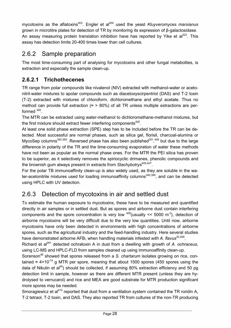
Page 28
mycotoxins as the aflatoxins403. Engler et al404 used the yeast Kluyveromyces marxianusgrown in microtitre plates for detection of TR by monitoring its expression of β-galactosidase.An assay measuring protein translation inhibition have has reported by Yike et al322. Thisassay has detection limits 20-400 times lower than cell cultures.
2.6.2 Sample preparationThe most time-consuming part of analysing for mycotoxins and other fungal metabolites, isextraction and especially the sample clean-up.
2.6.2.1 TrichothecenesTR range from polar compounds like nivalenol (NIV) extracted with methanol-water or aceto-nitril-water mixtures to apolar compounds such as diacetoxyscirpentriol (DAS) and T-2 toxin(T-2) extracted with mixtures of chloroform, dichloromethane and ethyl acetate. Thus nomethod can provide full extraction (≈ > 80%) of all TR unless multiple extractions are per-formed 340.The MTR can be extracted using water-methanol to dichloromethane-methanol mixtures, butthe first mixture should extract fewer interfering components405.At least one solid phase extraction (SPE) step has to be included before the TR can be de-tected. Most successful are normal phases, such as silica gel, florisil, charcoal-alumina orMycoSep columns340,359. Reversed phase has also been published351,406 but due to the largedifference in polarity of the TR and the time-consuming evaporation of water these methodshave not been as popular as the normal phase ones. For the MTR the PEI silica has provento be superior, as it selectively removes the spiriocyclic drimanes, phenolic compounds andthe brownish gum always present in extracts from Stachybotrys405,407.For the polar TB immunoaffinity clean-up is also widely used, as they are soluble in the wa-ter-acetonitrile mixtures used for loading immunoaffinity columns340,390, and can be detectedusing HPLC with UV detection.
2.6.3 Detection of mycotoxins in air and settled dustTo estimate the human exposure to mycotoxins, these have to be measured and quantifieddirectly in air samples or in settled dust. But as spores and airborne dust contain interferingcomponents and the spore concentration is very low 408(usually << 5000 m-3), detection ofairborne mycotoxins will be very difficult due to the very low quantities. Until now, airbornemycotoxins have only been detected in environments with high concentrations of airbornespores, such as the agricultural industry and the feed-handling industry. Here several studieshave demonstrated airborne AFB1 when handling materials infested with A. flavus32,409.Richard et al261 detected ochratoxin A in dust from a dwelling with growth of A. ochraceus,using LC-MS and HPLC-FLD from samples cleaned up using immunoaffinity clean-up.Sorenson46 showed that spores released from a S. chartarum isolates growing on rice, con-tained ≈ 4×10-14 g MTR per spore, meaning that about 1500 spores (450 spores using thedata of Nikulin et al45) should be collected, if assuming 80% extraction efficiency and 50 pgdetection limit in sample, however as there are different MTR present (unless they are hy-drolysed to verrucarol) and rice and MEA are good substrate for MTR production significantmore spores may be needed.Smoragiewicz et al410 reported that dust from a ventilation system contained the TR roridin A,T-2 tetraol, T-2 toxin, and DAS. They also reported TR from cultures of the non-TR producing

Page 29
genera Penicillium, Aspergillus, Alternaria, and Ulocladium, which demonstrates that all theirresults must be false positives.Yike et al229,322 demonstrated that dust and air samples from houses infested with S. charta-rum inhibited their protein translation assay, which strongly suggested the presence oftrichothecenes in the air.To conclude it should be possible to detect mycotoxins in air samples, but no validated sam-pling and clean-up procedures exist, meaning that this major problem has to be addressedfirst.

Page 30
3 OVERVIEW OF EXPERIMENTAL WORKThe details of the experiments performed during this study can be found in the papers 1-13.However results of a number of small experiments not included in the papers can be found inthe discussion part and identification of components initially reported as unknown.
A. versicolor, S. chartarum and Trichodermaspp. grown on building materials.Development of extraction methods for themycotoxins prior to HPLC analysis.Paper #2
Collection of S. chartarum infestedmaterials. Development of solidphase extraction and GC-MS NICImethod for trichothecenesPaper #1
Time
Chaetomium spp., Alternaria spp., Ulocladium spp., P.chrysogenum, P. polonicum, P. brevicompactum, A. ustus andA. niger grown on building materials. Naturally infested materialalso collected, including some with A. versicolorPaper #3
Growth tests of amixture of 8species ondifferentmaterials underdifferent humidityand temperatureconditionsPaper #10
Comparison of cytotoxicityand inflammatoryresponses and metaboliteprofiles of Stachybotrysisolates. Paper #9
Development of fast solidphase extraction andHPLC method formacrocyclictrichothecenes from S.chartarum. Paper #6
Development of fastGC-MS/MS methodsfor Stachybotrys andFusariumtrichothecenes.Paper #8
Development ofquantitative EI+GC-MS/MSmethods forergosterolPaper #5
Method comparison ofergosterol andMycometer testPaper #12
Genetic, toxicityand metaboliteprofiles ofStachybotrysisolatesPaper #7
Comparison ofMVOC's and satra-toxin productionPaper #11
Analysing airsamples fromStachybotrysinfested houses
and isolates fromthe Belgian IPHcase home
To be published
Mould growthand metabolismon buildingmaterials underconstant andtransienthumidities.Performed atTNO, Paper #13
Double blinded exposureof humans to spores of P.chrysogenum and T.harzianum.To be published
Figure 4. Overview of experimental work.

Page 31
4 RESULTS AND DISCUSSIONThe reason for the more detailed comments and results included in the thesis, is that a num-ber of them would not fit into any scientific papers, but that I feel they should be documentedsomewhere.
4.1 The building associated fungaAs mentioned in paragraph 2.1.1 the associated funga reported from different countries var-ies considerably. Unfortunately this study has not analysed field samples enough to makedefinite conclusions on this issue. However a number of observations are still interesting:• It was not possible to get any material samples with Alternaria infestation, all were in-
stead infested with Ulocladium. Although > 15 field samples which should contain Alter-naria were received).
• Almost all (>50%) of the material samples received were at least partly infested with P.chrysogenum and A. versicolor, although the presence of the latter could only be re-vealed by chemical detection of sterigmatocystin (ST), see section 4.3.2.1, page 44.
• When Trichoderma spp. or Chaetomium spp. were present on a material they grew inalmost pure culture and with significant biomass.
4.1.1 Penicillium and Aspergillus cultures received from FinlandA number of Aspergillus and Penicillium isolates collected from both material and air sampleswere received from National Public Health Institute, Kuopio, Finland (KTL). They were culti-vated on YES, CYA, CREA and MEA and identified by colony diameter and morphology.Isolates that could not be unquestionably identified were metabolite-profiled by HPLC-DAD(YEA and CYA).
Table 6Aspergillus and Penicillium isolates (n=131) received from KTL1
Species Frequency(%)
Species Frequency(%)
Species Frequency(%)
Aspergillus versicolor 23 P. citreonigrum 3 Penicillium charlesii 1Penicillium brevicompactum 11 Aspergillus ustus2 2 Penicillium commune 1Aspergillus fumigatus 10 Penicillium crustosum 2 Penicillium echnilatum 1Penicillium corylophilum 5 Penicillium solitum 2 Penicillium janczewskii 1Aspergillus sydowii 5 Aspergillus niger3 2 Penicillium manginii 1Eurotium repens 5 Penicillium decumbens 2 Penicillium purpugenum 1Penicillium chrysogenum 5 Penicillium expansum 2 Penicillium rosepurureum 1Penicillium palitans 5 Penicillium rugulosum 2 Penicillium variabel 1Penicillium glabrum 4 Aspergillus flavus 1 Penicillium vinaceum 1Penicillium italicum 4 Penicillium albocoremium 11From Anne Hyvärinen, National Public Health Institute, Kuopio, Finland. 2Not a classic A. ustus but Aspergillus ustus var. pseu-dodeflectus. 3Did not produce detectable quantities (HPLC-FLD) ochratoxin A on CYA or YES agar.
As seen in Table 6, A. versicolor was the most predominant mould almost contributing with25% of the isolates and more than 50% of all aspergilli. Surprisingly A. fumigatus was veryfrequently isolated (10%) which might be due to sampling on dusty surfaces, as A. fumigatusis frequently recorded in Danish house dust (S. Gravesen, personal communication).

Page 32
P. brevicompactum was the most frequently isolated Penicillium, confirming results from theon-going Danish School study, where P. brevicompactum, P. corylophilum and P. chryso-genum were the most common penicillia.
These results contradict results from North America, reporting P. aurantiogriseum and P.viridicatum as the most abundant Penicillium species. This might be due to misidentificationwith P. palitans and other species from the aurantiogriseum-complex (J.C. Frisvad, personalcommunication), but unfortunately it has not been possible to obtain any of these Penicilliumisolates to verify the identification.
4.2 Fungal growth on materialsIn papers 2 and 3, material samples were water-damaged (aw ≈ 1) and significantly highernumber of spores were applied to the materials than naturally found in buildings. The mouldswere usually able to cover the materials where they occur "naturally" within 4-10 days, clearlydemonstrating that when water damages occur, action should be taken the same day if ma-terials should be saved. This also complies with observations by several consultant engi-neers (M. Ø. Hansen, personal communication), who report complains from the inhabitantswithin 10-30 days after a single water damage, and complaints within 2-4 days in buildingswith a known water damage history.When using lower aw (papers 10 and 13) the growth rate decreased dramatically and withvery few exceptions only the primary and secondary colonisers were observed growing onthe materials. At stationary conditions (paper 10), only Penicillium and Aspergillus speciesgrew, with P. chrysogenum and A. versicolor being the most common species. However onwooden materials A. flavus and Eurotium spp. were observed quite frequently and alwayswith significant biomass. At 5°C only P. chrysogenum and A. versicolor grew, with the latterproducing conidiophores with very few conidia.Wooden and wallpapered materials supported growth down to 78% RH (paper 10), whichwas lower than the other materials. It was seen that the planned pinewood was usually notinfested on as much of the surface as the rough materials, but on the areas where growth
occurred there was not significant difference in theproduced biomass. At 5°C this limit was about 90%RH, whereas the results only indicates a limit be-tween 80 and 90% RH. These results comply per-fectly with the results of Hukka & Viitanen141 andGrant et al55, when the uncertainties of the RHmeasurements of both studies (±2%) are taken intoaccount.Interestingly, the gypsum boards did not supportgrowth <90% RH, but did support production ofsignificant quantities of biomass at 95% RH, it isalso clear that the fibre-gypsum was less suscepti-ble than the cardboard-gypsum, which is the mostcommon type in Denmark. Paper insulation mate-rial did not support fungal growth at all due to thehigh contents of borates, although it should benoted that this material when damaged supports
growth of several moulds including Stachybotrys (unpublished results). The almost totally
1
5 cm
2
2
Figure 6. Concrete after 4 months at 95% RHand 10°C, at #1 can A. versicolor conidiophoresbe seen and at #2 sterile mycelia.

Page 33
inorganic concrete materials partly supported germination of especially A. versicolor andsterilia mycelia at RH >90% and growth at 95%, as seen in Figure 6. This must be due totrace quantities of organic components in the concrete. As the concrete materials werecleaner than the ones used in buildings concrete based materials will probably support moregrowth under field conditions (paper 10).When comparing Tables 2 and 3, it is clear that the temperature drop from 10 to 5°C had adramatic effect on the fungal growth, showing no growth at 90% RH and 5°C, and that alsohere was wooden and the wallpapered materials the most susceptible (paper 10).
4.2.1 Methods for testing the susceptibility of materialsAs mentioned in the literature part, testing materials for their susceptibility is difficult, evenjust controlling the humidity is difficult. As this was the first time such a test was performed at
the Danish Building Re-search Institute, it was triedto keep the set-up as simpleas possible (paper 10), andregard the study as a pilotproject which should ex-plore difficulties such ashumidity range, inoculation,and especially assessmentof the materials. This set-upis shown in Figure 7, andwas primarily built by Gun-
nar Holm. Initial conditions may play an important role for the surface aw of the materials, andas it was desired to use steel chamber and take the materials out for assesment, it was nec-essary to use the sample boxes for reasons occupational health.This meant that the surface aw would be controlled by the RH ofthe incoming air to the sample box and primarily by the absorb-ance of water by the material until equilibrium, which small ex-periments had showed would take months. This meant that wehad to add the quantity of liquid water to the materials, assumingthat it would be equally distributed within a few days. Althoughthis should tested in future experiments.I paper 13, the set-up at TNO Bouw was used, and the initialhumidity was controlled by a high continuous airflow over thematerials (see Figure 8). This means that the absorption of waterfrom the airflow was so little that the change in humidity over the10 materials in each channel could be neglected. However thisapproach created another problem as the air speed itself caninhibits growth if the speed is too high. This is illustrated in Fig-ure 9, where the air speed was higher in one side of the channelthan in the other.The TNO approach (paper 13) had another advantage, as it ispossible to work with transient humidity conditions, and here it
1 mm
90% RH
Hygrostate
Sample
80% RH
Exhaust
Flow controllers
Sample box
Ventilator
Water bath
1.2 m3/hr air
Dry air 0.1 m3/hr air
Dry air
Figure 7. The fungal growth assesment set-up used in Paper 10.
Figure 8. The TNO set-up, 10material in each row. In thiscase infested with P. chryso-genum.

Page 34
was seen that the stress of using periods of high and low humidity during a day, trying tomimic a bathroom. This had a strong selectivity stress towards the phylloplane, Cladospo-rium, which under transient conditions was able to outgrow P. chrysogenum.The instationary conditions, with and without surface conden-sation (paper 13), can cope with conditions which keeps thestorage moulds away and still be too dry for the phylloplane. Itcan also cope with water accumulation in the surface of mate-rials, especially when working with two to three layer systems,like a painted piece of cardboard gypsum (3-layer system)11.Hence, the total growth behaviour of materials is extremelycomplicated, and to characterise a material well, the perform-ance and associated funga under at least three different envi-ronmental conditions is needed:• The lowest RH which supports growth.• The performance under transient conditions.• The performance under water damage.
4.2.2 Growth assessmentIn paper 10, the materials had to be taken out of the set-up for assesment. This was verytime consuming and obscured the environmental conditions for the samples. Using the ap-proach of Adan11 as in Paper 13 (see Figure 8), the materials can be assessed several timesa day if required. The time resolution of this process describes the susceptibility - as how fastgrowth started - better than endpoint determinations of biomass. The latter parameter seemsto describes how much of the substrate could be utilised (papers 10 and 13).Visual inspection (paper 13) has a major problem, especially using the 5 point scale de-scribed by British Standard411, as the assessed coverage area is the output parameter. Thisdoes not take the concentration of fungal biomass into account. Thus a material with verylight coverage over all of the material will be assessed to have a higher value than a materialwith heavy growth over only a part of the material.The use of image analysis was partly explored at the stay at TNO Bouw (unpublished), andthis technique will undoubtedly help in future studies, as images for assessing the materialscan be taken automatically several times a day. It can also cope with the high growth rateobserved on some paints (wherethe materials had to be assessed 2-3 times per day, paper 13). Also amore standardized assessment in-cluding the fungal "concentration"can be made. However this will re-quire that the image-analysis is cali-brated carefully against very well-described samples. Here chemicalmarkers could help to compare theappearance of different moulds.The use of dissection microscopyshowed that the moulds often grewin mixtures (see Figure 10). Looking at materials with scarce growth and a fibrous surfacewas very difficult, and only sporulation could be detected. However it was possible to distin-
Figure 9. The influence of toohigh air velocity on A. versicolorgrowth in one side of the set-up.
Figure 10. Mixture of A. flavus, A. versicolor and P. chrysogenum,growing on pinewood.

Page 35
guish A. versicolor, often seen as white to olive conidiophores with very few spores and P.chrysogenum, often as light bluish or white conidiophores with very long chains of sporesattached.When no sporulation occurred on fibrous materials such as the pinewood or gypsum boardsit was only the chemical markers which could detect growth, but in no case significant bio-mass overlooked.
4.2.2.1 Biological markersOne or both the biological markers ergosterol (Erg) and the Mycometer TestTM were used inpapers 5, 10, 12 and 13.Qualitative determination or Erg was easily performed by the standard HPLC and TLC meth-ods already used at the department, but quantitative determination proved to be painstakingprocess. External standard quantitation using HPLC or GC-MS proved unreliable, especiallyusing GC-MS where both splitless injection, various ion-trap phenomena, and the breakdownof the GCQ followed by it being rebuilt in USA, complicates things further (paper 5).All the sterol standards obtained were almost transparent to the HPLC-DAD, leaving GC-MSwith internal standard as the only possibility. LC-MS was not available at that time, and willprobably not be an alternative, as the low-polar solvents used to elute it will give problemswith the ionization process. Several sterols were tried as internal standard for GC-MS, butthe high dynamics of the external ionisation ion-trap system did not give better standard de-viations (SD) than 25% when working on standards. When using 4-D2-ergosterol as internalstandard, synthesised by Dr. J.Ø. Madsen, Erg could be determined with a SD of < 5%, asvariation in the ionization and the dynamics of the gate lens is no longer interfering with theanalysis.In paper 5 it was shown that no SPE clean-up step was needed when using GC-MS/MS andthat the sample did not need to be neutralised after the saponification when splitless injec-tions was used. Microwave assisted extraction was proven to be a very powerful and fasttechnique but very dangerous, as the vials occasionally exploded and ripped open the mi-crowave oven door.As many samples could be hydrolysed in standard laboratory vials at the same time - thismethod was preferred. It was possible to reach 120 samples per day, although the GC-MS/MS can only analyse 60-65 samples per day. The SD ranged from 1-4% in spiked sam-ples and in real samples SD ranged from 4-12%, with detection limits in the sample of 2-10ng, which is fully sufficient as the variation of fungal growth on surfaces is much higher.However during analyses of samples described in papers 10 and 13, this detection limits wasdifficult to reach and the general limit was 10-30 ng, which is significantly higher than re-ported in paper 5, which was already higher than reported elsewhere 318,330. One of the majorproblems was the daughter ion-yield (number of daughter ions / number of the parent ion inthe full scan chromatogram), decreased to 4-8% for unknown reasons, compared with a yieldof > 50% of the PFP or HFB-trichothecene derivatives. The water and oxygen levels in thesystem ware extremely low, and various q-values, ion-source temperatures and CID voltageswere explored as well as alternative tuning of the lenses, but with no increased sensitivity.Sample decomposition during injection was a particular problem while running the sampledescribed in paper 13, where the painted materials had a dramatic impact on the sensitivitydue to deposits in the liner (not visible). In the future such samples should probably, afterhydrolysis, be loaded (cold) onto a C18 cartridge which can be cleaned with water-methanol

Page 36
mixtures, before the sterols can be eluted with methanol/dichloromethane, but would com-promise sample preparation speed.In paper 12, the m/ 363 and 365 were used as parent ions and m/z 157 and 159 as daughterions for Erg and 4-D2-Erg respectively, this combined with newly cleaned ion source assem-ble and new liners in the GC every 60 injections lowered the detection limit to 1 ng (20 pginjected).In paper 10, both Erg and the Mycometer Test™ showed growth or no growth, but the cor-relation was obscured by the high variability of the fungal growth. This problem was over-come in paper 12, where the same sub-sample of material or agar were analysed for enzymeactivity with the Mycometer Test™ and for ergosterol (Erg) using GC-MS/MS. Here it wasnoticed that the enzyme activity would usually be highest during the initial growth phase andusually decreased to about 50-80% whereas Erg would only decrease to 90-70% of it peakvalue, as illustrated in Figure 11. When comparing enzyme activity, Erg and dry weight ofagar cultures it was noticed that these parameter did not always peak at the same time,
probably due to different in-duction of the enzyme.Compared with visual inspec-tion, paper 10, the chemicalmarkers generally showed thesame, but were a nice alterna-tive on very fibrous surfaceswhere even the use of a ste-reomicroscope gave problemswith distinguishing fungalgrowth from fibres. Erg has theadvance of being an absolutemethod, which can be good forcomparing different ratings ofmould growth.The secondary metaboliteswere very useful species-
specific biomarkers, and in papers 3 and 10, significant growth of A. versicolor (see section4.3.2.1, page 44) was hidden by P. chrysogenum.Metabolites may also indicate how wet the materials have been, e.g. does the presence ofthe A. versicolor metabolites PE1, XOO and ROQ but not ST, indicates 0.95 > aw > 0.90,whereas ST indicates aw > 0.95 (see section 4.3.2.1, page 44).
4.3 Growth and metabolite productionIn this chapter the most important results of the different moulds and their growth and me-tabolites production will be discussed.
4.3.1 StachybotrysExperimental growth of S. chartarum IBT 7711 could be recognised after 5 days of incuba-tion (paper 2) on new water-damaged (aw ≈ 1) gypsum boards, which is partly due to the highdose of spores (4×10³ spores/cm2), but still surprisingly fast. Growth of the other 4 isolateswas not as fast, but the gypsum boards were still totally covered with the fungus after 14days. None of the 5 isolates could grow on chipboard, acoustic ceiling tiles or pinewoodpieces.
0
500
1000
1500
2000
2500
3000
3500
4000
0 20 40 60 80 100 120 140 160Incubation time (days)
Enzy
me
activ
ity (a
rbitr
ary
units
)
0
200
400
600
800
1000
1200
1400
Ergo
ster
ol (n
g)
EnzymeErgosterol
Figure 11. Comparison of Mycometer test and ergosterol determinationson gypsum board inoculated with Stachybotrys chartarum (IBT 9694).

Page 37
These findings are in accordance with field observations14 and paper 4 where consistentlywet gypsum boards seem to be the main substrate of Stachybotrys in buildings25.In paper 10 where mixtures of commonmoulds were inoculated under regulatedhumidities and temperatures (95-70% RH)Stachybotrys was only found on the threetypes of gypsum boards when RH ≥95, andhere only with very limited production ofbiomass had occurred, as shown in Figure12. This demonstrates that this speciesgenerally require water-damaged materialsto produce significant quantities of biomassand compete with the penicillia and asper-gilli. This is in accordance with the litera-ture (see Table , p.5) where Stachybotrysis seen to require aw 0.95 or more forgrowth, although Pasanen et al107,108 havereported growth as low as aw 0.78-0.81which is highly unlikely. This is presumable due to the 1 ml spore suspension added to verysmall samples (down to 0.25 g), meaning that aw of the samples would be higher than the RHin the air controlled by the saturated salt solutions for an unknown period.
4.3.1.1 Analysis for trichothecenesIn papers 1 and 2, GC-MS negative ion chemical ionisation (NICI) was used successfully fordetection of the trichothecenes (TR) as it gave excellent matrix suppression compared withEI+ ionization, as illustrated i Figure 2 in paper 2. However, the matrix suppression could alsobe obtained with more polar extraction mixtures (methanol-water) and more effective removalof acid residues from the derivatised samples. Combined with a number of problems withNICI compared with EI+ (paper 1, 2 and 8) the latter became the preferred as NICI gave thefollowing problems:• A decrease in sensitivity after 10-15 samples was observed. This could be avoided by
changing the ion volume.• After changing to the turbo pump, it took 1-2 hr to stabilise the ionisation gas.• The MS got very dirty much quicker and needed to be cleaned every 2-3 months, which
is very time consuming.• Very short lifetime of the filament (1-2 months)• The high pressure of methane, which was necessary to obtain a high sensitivity and high
m/z ions, gave a poor mass resolution of approx. m/z 0.5-1 (10% valley), making auto-matic data processing very difficult.
• If the method should be used on the type A trichothecenes (TA) from Fusarium, the ionvolume temperature should be raised to 120°C to avoid condensation. However this gavealmost no high-mass ions (<m/z 300) especially not for the type B trichothecenes (TB).
• The number of ionised molecules is about 10 times as low in NICI as in EI+, meaningthan when MS/MS (which looses 10-90% of the ions) was used a significant drop in sen-sitivity was observed.
Figure 12. Scarce growth of S. chartarum on a gypsumboard at 95% RH and 10°C after 4 months.

Page 38
In EI+ the daughter ion spectra were significantly more reproducible than the full scan spec-tra. Thus EI+ ionisation combined with MS/MS (paper 8) was used in all other studies exceptpapers 1 and 2.In the study of TR produced by Fusar-ium (paper 8) it was observed that themethods used in papers 1 and 2, couldnot fully derivatise the type B trichothe-cenes (TB) and that a number of arte-facts were produced. A number ofmethods using imidazole or acid anhy-dride based reagents and mixtures ofthese were tried but none were suc-cessful, until the method of Langseth etal396 was applied. This method workedboth with PFP and HFB esters, but thePFP was chosen as the HFB gives MWexceeding the upper mass range of theGCQ (m/z 1000).However this derivatisation method didnot work for trichodermol and verrucarolas partly facilitated apotrichothecenerearrangements (ring breakage of the epoxy group)412, so the original method (papers 1 and2) using HFBI was not changed for these trichothecenes (Figure 13).To speed up the hydrolysis step it was tried to pass the hydrolysed sample through a strongcat-ion exchanger for neutralisation of the NaOH405, which worked fine on standards. How-ever with fungal extracts huge quantities of interfering peaks and irreproducible derivatisationwere observed, hence the liquid-liquid extraction seems to be a powerful clean-up step.Parallel to changing the GC-MS method to EI+, a clean-up method for HPLC detection wasdeveloped to detect the unhydrolysed macrocyclic trichothecenes (MTR). It was clear thatreversed phase clean-up was not effective due to the polarity difference between the MTR aproblem well known from the simple trichothecenes where normal phase clean-up is themost effective340. Silica and PEI silica (provided by B.B. Jarvis407) were tested, but it wasclear that the latter was significantly better to remove the spiriocyclic drimanes. By dry-packing the PEI in syringes between two pieces of filter-support material stamped out by asharp steel cylinder a SPE-PEI module could be manufactured on less than 30 sec. The di-chloromethane sufficient to elute the macrocyclic trichothecenes could then be placed in theRVC (takes 24 vial) and evaporated in 2 hr giving a much higher sample throughput com-pared with the method of Hinkley & Jarvis405. The effect of cleaning up the extract on the PEIcolumns is illustrated in Figure 14, where the HPLC profiles of crude extract is compared withthe cleaned up extracts on a MTR and an non-MTR producing S. chartarum on PSA agar.It was noted that the sample should be totally dry before redissolving it in dichloromethane,as water or methanol traces facilitated the breakthrough of the spiriocyclic drimanes in thedichloromethane fraction. When trying to fractionate Stachybotrys extracts it was also notedthat the dry solvent blanks from the PEI-columns were toxic to the boar sperm used by Pel-tola et al231 and the human bronchoalveolar cells used by Norn et al413.
O
O
O
O
O
CHOHCH3O
OH
H
OH
O
OH
O
OH
OO
O
OO
F
F
OF
FF
F F
F
F
FF
FF
F
O
O
OH
O
O
OH
O
OO
O
OF
F
F
F
F
FF
O
OCH3
OCH3
O
OH
CH3OCH3
O
O
CH3
OCH3
OH
O
OH
OHO F
F
F
FF
O
OOF
F
F
FF
O
CH3
OCH3
O
O
OF
F
FF
F
O F
F
F
FF
OO
OCH3
OCH3
O CH3OCH3
O
NaOH in methanol
Satratoxin H
VerrucarolBis-heptafluorobuturylverrucarol
HFBI in toluene-acetonitril
HeptafluorobuturyltrichodermolTrichodermolTrichodermin
DAS (4,15-diacethoxyscirpenol)
DON (4-Deoxynivaleol, vomitoxin)
PFPA in toluene-acetonitril-imidazole Pentafluoropropionyl-DAS
Tri(pentafluoropropionyl)-DON
Figure 13. Derivatisation of the trichothecenes.

Page 39
To study the ratio between trichodermol and trichodermin in the crude extracts, unhydrolysedcrude and PEI cleaned extracts were HFB derivatised and analysed using GC-MS/MS. ThePEI clean-up step was necessary to avoid huge quantities of interfering peaks and unrepro-ducible derivatisation. It was also shown that the trichodermol TR are predominantly presentas trichodermin (paper 6-8).
4.3.1.2 Metabolites produced by StachybotrysDuring the first part of study (papers 1 and 2) it was believed that most isolates would pro-duce MTR208. This was, however later shown not to be true, papers 6-9 and Jarvis et al44, asonly 30-40% of the isolates produces MTR.The samples collected in a school and a dwelling (paper 1) both yielded verrucarol upon hy-drolysis (up to 15 ng/cm2 SH equivalents) as well as trichodermol (not quantified). When in-cubating different strains on water-damaged new and old gypsum boards (paper 2), only 2 of5 isolates yielded high quantities of verrucarol upon hydrolysis (up to 120 ng/cm2 SH equiva-
min0 5 10 15 20 25 30 35 40
mAU
0
50
100
150
200
250
0.843
10.177
12.186 14.474
15.466
16.472
17.234
18.655
20.682
24.513
A
nm200 250 300 350 400 450 500 550
mAU
0
200
400
600
800
1000
1200
1400
Mer-NF5003analogue
Stachybotrysdialdehyde
nm200 250 300 350 400 450 500 550
mAU
0
50
100
150
200
250
Spiriocyclic drimanes(stachylactam)
nm200 250 300 350 400 450 500 550
mAU
0
10
20
30
40
min0 5 10 15 20 25 30 35 40
mAU
-10
0
10
20
30
40
50
60
70
11.41 13.40 14.37
16.49
18.65 30.305
30.881
40.145C
nm200 250 300 350 400 450 500 550
mAU
0
1
2
3
4
5
6β-hydroxydolabella-3E,8E,12-trien-14-one
nm200 250 300 350 400 450 500 550
mAU
0
2
4
6
8
Epoxydolabellane A
Mer-NF5003 analogue
Ergosterol
min0 5 10 15 20 25 30 35 40
mAU
-5
0
5
10
15
20
25
30
10.23
10.91 40.224
B
Mer-NF5003 analoguenm200 250 300 350 400 450 500 550
mAU
0
2
4
6
8
10
12
14
16
Satratoxin G
nm200 250 300 350 400 450 500 550
mAU
0
1
2
3
4
5
6
7
Roridin Enm200 250 300 350 400 450 500 550
mAU
0
5
10
15
20
25
30
35
40
Satratoxin H
Epi- and iso roridin E
Ergosterol
Verrucarin J
Iso-satratoxin F
Figure 14. HPLC-DAD chromatograms (260 nm) of Stachybotrys chartarum extracts from PSA agar: a) crudeextract (IBT 7711); b) PEI cleaned up extract from a satratoxin producing strain (IBT 7711); c) PEI cleanedup extract from an atranone producer (IBT 9466).

Page 40
lents), one isolate produced significantly less MTR. In extracts from the last two isolates ver-rucarol was perhaps present, but detection could only be performed on the non-specific m/z213 ion, [HFB-O]-, which was observed from many peaks of the chromatogram.The satratoxin levels detected (15-120 ng/cm2) are the same as reported by Johanning etal23 who found 33 ng cm-2 satratoxin H in biomass scraped off from a infested building mate-rial, but lower than found by Nikulin et al107 who detected 2-3.5 µg/cm2 satratoxins from ahighly toxic strain of S. chartarum (isolate #72 = IBT 9460).Temperature and humidity did not seem to influence the production of the spiriocyclic dri-manes (paper 10) when growing on gypsum boards, as these were detected in substantialquantities at 10°C and 95% RH at gypsum boards.In papers 6, only S. chartarum produced satratoxins G, H and iso-F whereas S. dichroastopped the trichothecene biosynthesis at roridin E (see Figure 2, p. 14). S. oenanthes pro-duced trichodermol type TR. S. bisbyi, S. albipes, S. microspora S. nilagrica S. parvispora, S.theobromae, and S. cylindrospora did not produce any detectable quantities of TR. The latterspecies has been reported to produce trichodermol and trichodermin414 and El-Maghraby etal415 have reported that S. albipes, S. kampalensis and S. microspora produced MTR. Un-fortunately these strains are not available in any culture collection. However as only 1-3strains of each species were tested, the study is not big enough for major conclusions onthese rare species, as it should also be noted that only ALK and PSA were used for thesespecies.In papers 6 and 8 it was observed that the strains capable of producing MTR did this on allsolid substrates tested (CMA, V8, YES, PSA, OAT, PSA, DRYES, ALK, CYA and rice ex-tracts agar), with PSA and ALK agar yielding the highest quantities. On the latter agar, en-hanced production of stachybotrylactones and lactams was observed, often more than 20different components with the UV spectra illustrated in Figure 14A. As these components arethe main interfering components (removed by the PEI silica SPE clean-up), PSA agar waschosen as the standard substrate for chemotaxonomic studies of S. chartarum.The rich substrates CYA and YES, where high quantities of biomass was produced, yieldedsurprisingly low quantities of MTR together with CMA, V8, OAT and rice extract agar. Thelow quantities of MTR produced on the latter substrate is surprising since whole rice is a verygood substrate used extensively by Jarvis and co-workers44,204,304. The construction card-board used in study 12 gave approx. as much of the MTR per area as PSA agar.Three taxa was found among the isolates from buildings (paper 6): i) the MTR producing S.chartarum isolates; ii) the non MTR producing S. chartarum isolates; iii) a new undescribedspecies, referred to as Stachybotrys sp. type A.The latter taxon did not show PCR amplification of the TR5 gene (paper 7) and did not pro-duce detectable quantities of trichodermol. However this was not the same in paper 6, where25% of the Type A isolates produced trichodermol or esters of it, which was also confirmed inpaper 8, where trichodermol and trichodiene was detected from the type A isolate.Study paper 6, 7 and 9 showed that and the non-MTR producing S. chartarum taxon and theType A taxon consistently produced 6β-hydroxy-dolabella-3E,8E,12-trien-14-one and oftenalso epoxydolabellane A (see Figure 14C), both precursors to the atranones227. Due to thelack of pure reference standards the identity of these had to be confirmed using LC-MS. Thiswas kindly performed by Hanne Jakobsen, on the new Micromass LCT system using electro-spray ionization (ESI) with the same or similar eluents as the general LC method370. In Figure15) the ESI spectra of these two dolabellanes can be seen.

Page 41
When growing on PSA agar, S. chartarum rarely produced the atranones themselves, whichis different to growth on rice227. However, some of the isolates tested in the spring of 2001also produced the atranones, indicating small changes in the PSA, may induce this.Two extracts of atranone producing isolates on rice (Jarvis, B.B., Hinkley, S.F., Nielsen, K.F.et al, to be published) contained trace levels of verrucarol upon hydrolysis (GC-MS) and alsowere significant more toxic that the other atranone producing isolates although still being
magnitudes less cytotoxic than the MTR pro-ducers. Trace level of verrucarol was also de-tected in the material sample from the BelgiumIPH case home from which none of 6 isolateswere MTR producers. When IBT 14915 (anatranone producer) in paper 2 was inoculatedon gypsum boards trace level of verrucarolwas also detected. Altogether these observa-tions indicate that the atranone producers maybe able to synthesise the verrucarol type TR.Hopefully a more sensitive LC-MS method canreveal if MTR are produced by these strains.The very clear distinction of atranone produc-ers and MTR-producers on agar substrates(papers 6-9) suggests that the genes from theatranone biosynthesis perhaps in inserted in,
or near the genes coding for the enzymes used for synthesising trichodermin into verrucarol-type TR or epi-genetic changes (rearrangements in the 3D structure of the genes), so thatonly of these pathways is expressed.The isolates studied by Ruotsalainen et al148 were retrieved from Public Health Institute,Kuopio Finland (KTL). Originally two large groups of isolates were observed, one which washighly cytotoxic and one which induced inflammatory responses in the murine macrophages.When analysing (paper 9) the chemical profiles of the 20 isolates, it was shown that all the11 isolates being cytotoxic, except one which was probably contaminated or confused byanother isolate, belonged to the MTR producing taxon. All the isolates inducing inflammatoryresponses belonged to the atranone producing S. chartarum taxon. In the original study148 itwas also observed that as the only isolate HT16, did not induce the strong inflammatory re-sponse as well as not being cytotoxic at 105 spores/ml. This isolate was the only Stachybo-trys sp. type A, suggesting that this taxon is not producing the inflammatory inducing sub-stance(s) and that the effects is not due to the particle effects, as the spores are almost iden-tical in size and shape (paper 6). The pure atranones B and D were did not induce any in-flammatory response in the macrophages. Thus, the specific agent triggering the inflamma-tory response in the macrophages is still unidentified. The inflammatory agent(s) may bepresent in the MTR-producing isolates, but due to the extremely potent cytotoxins themacrophages are killed before they can respond or that the spores contains componentsinhibiting the induction of TNFα221.Identification of the component inducing the inflammatory response is of special interestsince high induction of TNFα is known to be a significant factor of the induction for lung dam-age153. Nikulin et al45,216 suggested that the MTR were the main reason of the lung tissueinflammation they observed when installing mice with S. chartarum spores from a MTR pro-ducer (#72) and a non-MTR producer (#29, later shown to produce atranones, paper 6).
220 240 260 280 300 320 340 360 380 400m/z0
100
%
319.3 301.3
217.2 283.3219.2 273.3 245.2 284.3
302.3 303.3
382.3
320.3 360.4
342.4 365.4
383.3
384.3406.4
[M+H-H20]+
[M+H+CH3CN]+
[M+H]+ [M+Na+CH3CN]+
220 240 260 280 300 320 340 360 380 400m/z0
100
%
219.2
303.3
220.2285.3235.2
260.3
344.3
304.3
305.3 326.3345.4
369.3 409.3389.3
[M+H+CH3CN]+
[M+H]+
Figure 15. Positive ESI spectra of EpoxydolabellaneA and 6β-hydroxydolabella-3E,8E,12-trien-14-one.

Page 42
However #29 also produced significantly smaller quantities of the spiriocyclic drimanes,making these components an alternative agent.When looking at the number of papers dealing with possible causative agents to the IPH, it issurprising how little effort has been put into the spiriocyclic drimanes, which are produced atconsiderably higher levels than the atranones and trichothecenes, presumable 10-500 timesmore, based on their extension coefficient. Especially the spiriocyclic drimanes isolated fromS. microspora226,416 which enhances the plasminogen-mediated fibrinolysis should be takeninto consideration as they might be produced by the S. chartarum isolates in buildings. Theneurotoxic symptoms reported byJohanning et al21,23 could be dueto the neurotoxic as well as thenerve activator compounds417,418.It the present work attempts werenot made to differentiate betweenthe different spiriocyclic drimanesas their UV spectra are verysimilar. Furthermore there wasonly very limited time to run LC-MS analyses. A single subclass,including 5-7 analogues of thespiriocyclic drimanes was, how-ever, tentatively identified (paper6), by their distinct UV-spectra seen in Figure 16, as the Mer-NF5003221 components. LC-MSanalyses pointed at molecular masses (MW) of 470 Da of three analogues, two analogues of412 Da, one with 368 Da. All with slightly higher MW than the reported B and E analogue221,however one minor analogue also fitted MW, 386 Da, with the F analogue221. The attentionwas drawn to these components, as B.B. Jarvis had never seen these in Stachybotrys ex-tracts from rice. These components are produced on most agar substrates, especiallyDRYES and PSA, as well as on hay (unpublished) and in crude extracts from the buildingmaterials (paper 1, 2, 10 and 11). In paper 10, it was shown that the spiriocyclic drimanesand atranones were produced at 10°C and 95% RH on several types of gypsum boards indi-cating that these metabolites are produced under most environmental conditions.
Airborne Stachybotrys metabolitesLate in the study a number of air samples (24 hr, 60 m3/min) and material samples were re-ceived from E. Johanning. The filters and materials had already been shown to be highlycytotoxic in the pig kidney cell line used by Gareis et al321, which indicated the presence ofMTR. Analysis of these samples revealed that two air samples contained several spiriocyclicdrimanes including Mer-NF5003 analogues as seen on Figure 17, and probably also satra-toxins G and H (the UV spectra were very weak), which were confirmed by the presence ofverrucarol in the two hydrolysed samples, detected by GC-MS and GC-MS/MS (Figure 18).Based on HPLC-DAD detection of satratoxins G and H, the total MTR contents were esti-mated to 75 and 100 ng (± 50%), whereas GC-MS showed 130 and 150 ng (± 50%) respec-tively.
200 220 240 260 280 300 320 340 360 380 400 420 440nm-0
100
%
232.0
288.0
328.0
420 440 460 480 500 520 540 5600
100
%
471.3
411.3 428.4 457.6443.4
472.4
511.4473.4488.4 512.4527.4552.4557
[M+CH3CN]+
[M+H]+
200 220 240 260 280 300 320 340 360 380 400 420 440nm-0
100
%
212.0
236.0292.0
330 340 350 360 370 380 390 400 410 420 430 440 450 460 470 480 490m/z0
100
%
413.3
3533
339.3335.3325.3 343.3
3713354.3 360.3367.3
372.3389.3384.4376.3 412.4
392.3407.4399.4
414.3 453.3415.3
430.4420.5 450.5432.4440.6443.4454.4
486.5469.3458.5 476.4 493.5
[M+CH3CN]+
[M+CH3CN]+
Figure 16. UV and ESI spectra of two MER-NF500 analogues.
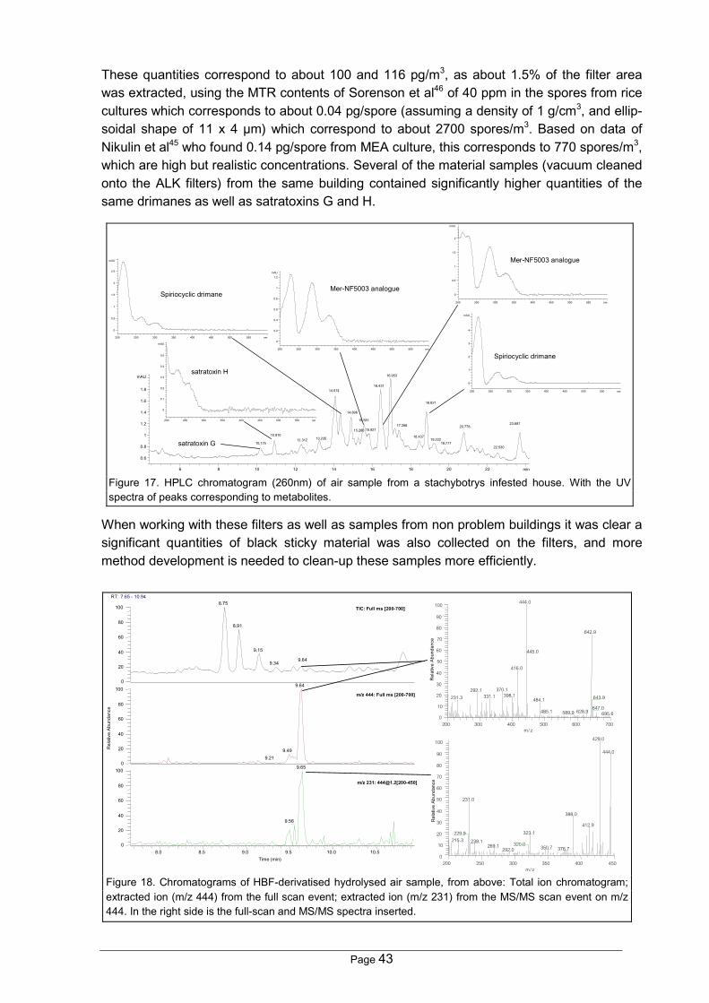
Page 43
These quantities correspond to about 100 and 116 pg/m3, as about 1.5% of the filter areawas extracted, using the MTR contents of Sorenson et al46 of 40 ppm in the spores from ricecultures which corresponds to about 0.04 pg/spore (assuming a density of 1 g/cm3, and ellip-soidal shape of 11 x 4 µm) which correspond to about 2700 spores/m3. Based on data ofNikulin et al45 who found 0.14 pg/spore from MEA culture, this corresponds to 770 spores/m3,which are high but realistic concentrations. Several of the material samples (vacuum cleanedonto the ALK filters) from the same building contained significantly higher quantities of thesame drimanes as well as satratoxins G and H.
When working with these filters as well as samples from non problem buildings it was clear asignificant quantities of black sticky material was also collected on the filters, and moremethod development is needed to clean-up these samples more efficiently.
min6 8 10 12 14 16 18 20 22
mAU
0.6
0.8
1
1.2
1.4
1.6
1.8
10.175
10.910 12.312 13.235
14.074
14.896
15.268
15.581
15.821
16.431
16.953
17.398
18.437
18.821
19.222 19.777
20.778
22.530
23.687
nm200 250 300 350 400 450 500 550
mAU
0
0.2
0.4
0.6
0.8
1
1.2
nm200 250 300 350 400 450 500 550
mAU
0
0.5
1
1.5
2
nm200 250 300 350 400 450 500 550
mAU
0
1
2
3
4
nm200 250 300 350 400 450 500 550
mAU
0
0.5
1
1.5
2
2.5
nm200 250 300 350 400 450 500 550
mAU
0
0.1
0.2
0.3
0.4
0.5
satratoxin H
Spiriocyclic drimane
Spiriocyclic drimane
Mer-NF5003 analogue
Mer-NF5003 analogue
satratoxin G
Figure 17. HPLC chromatogram (260nm) of air sample from a stachybotrys infested house. With the UVspectra of peaks corresponding to metabolites.
RT: 7.65 - 10.94
8.0 8.5 9.0 9.5 10.0 10.5Time (min)
0
20
40
60
80
1000
20
40
60
80
100
Rel
ativ
e Ab
unda
nce
0
20
40
60
80
1008.75
8.91
9.15
9.649.34
9.64
9.499.21
9.65
9.56
TIC: Full ms [200-700]
m/z 231: [email protected][200-450]
m/z 444: Full ms [200-700]
200 250 300 350 400 450m/ z
0
10
20
30
40
50
60
70
80
90
100
Rel
ativ
e Ab
unda
nce
429.0
444.0
231.0
388.0
412.9
323.1229.9
215.3 239.1320.0269.1 350.7 376.7292.0
200 300 400 500 600 700m/ z
0
10
20
30
40
50
60
70
80
90
100
Rel
ativ
e Ab
unda
nce
444.0
642.9
445.0
416.0
370.1292.1398.1331.1 643.9231.3 484.1
647.0485.1 628.9589.9 695.6
Figure 18. Chromatograms of HBF-derivatised hydrolysed air sample, from above: Total ion chromatogram;extracted ion (m/z 444) from the full scan event; extracted ion (m/z 231) from the MS/MS scan event on m/z444. In the right side is the full-scan and MS/MS spectra inserted.

Page 44
When comparing exposure data to animal studies, it seems for the non-toxicologist to bevery high quantities that have been instilled in rats and mice. Often 105 to 106 spores in sin-gle doses45,217-219,419 and down to 103 spores in several doses216, and with the concentrationsof Stachybotrys spores even in highly infested buildings generally <<1000 spores/m3.As seen in the literature part, many data on the toxicity of trichothecenes exist, however onlyaf few studies take lung toxicity, particle effects, and other metabolites into account.Using the data of Nikulin et al45 who used 106 spores (containing 140 ng MTR) as a singledose, the death of 2 of 4 animals, resembles the LD50 value (not taking the statistical uncer-tainty into account). Directly transferred to a human (70 kg) assuming the same susceptibilityper weight, corresponds to about 750 µg MTR or 3×10⁹ spores.As concentrations of Stachybotrys spores even in highly infested buildings, is generally be-low 1000 per m3 (Hunter et al10 have reported 20 times higher values) and with a human res-piratory rate of 1.5 m3/hr, about 36 000 spores can inhaled per day. This is so much lowerthan the LD50 value, that there is no reason to suspect lethal mycotoxicoses even if a 1000xlower LD50 is used, to cope with individual and species susceptibility as well as more toxicstrains into account.Workers handling infested materials, and not using the prescribed respiratory filters may beexposed to very high levels. Assuming up to 0.1-1 µg/cm2 MTR and a LD50 of 750 µg MTRthis corresponds to dust from 0.15-1.5 m2, and it is not likely that any persons will inhale somuch biomass even if working in a small unventilated room. But significant inflammation inthe lungs and decrease in platelet concentration, as well as changes in the lymphocyte sub-populations may be possible. Such effects were observed in mice when instilling 6 dose over3 weeks using 103 spores each time216. This corresponds to the dust from 2-15 cm2 infestedmaterial per day. However these does not take the higher metabolism and shorter life spanof the mice into account.To asses the risk of people living in mouldy homes repetitive exposures of small doses isneeded to estimate the No observed adverse effect level (NOAEL)420. This is especially im-portant for the MTR, as they have immunosuppressing effects at far lower levels than theacute ones and should be combined with the other metabolites.To conclude on this species, which is the far most investigated indoor contaminants, and stillnot been established any causal relationship to IPH, although is seems that the MTR are notthe principle components. The general immunosuppression of people exposed to stachybo-trys may also be explained by the spirocyclic drimanes, although exposure to MTR producingstrains must be considered to be worse.Significant is needed to characterise the health effects of spiriocyclic drimanes as they arethe primary metabolites of S. chartarum when growing on building materials. More over theyare also produced in its natural habitat, hay, and have been proven pharmacologically activein many assays (Appendix B, Table 2).
4.3.2 Aspergillus
4.3.2.1 A. versicolorIn paper 2, it was shown that this species was capable of growing on almost all the water-damaged materials, and that chipboard and wallpaper supported production of the most visi-ble biomass. Furthermore (paper 13), it was observed that A. versicolor grew especially faston paints, and transient humidity conditions greatly slowed its growth.

Page 45
Two different growth patterns were seen: either low biomass production and heavy sporula-tion or high biomass production (white-red mycelia) with very low sporulation (papers 2, 3and 10).In paper 10, A. versicolor was one ofthe most frequently observed mouldgrowing on the materials, even capa-ble of growing at 5°C. This is contra-dictory to Smith & Hill94, who ob-served that A. versicolor requires 9°Cfor growth on a high nutrient substratesuch as MEA.It was capable to grow on the threetypes of concrete at RH > 0.90 (paper10), although the produced quantitiesof biomass were very low, except fora few hotspots (illustrated in Figure19). It was notable, that it always wasA. versicolor that grew on the concretes and never P. chrysogenum, which indicates that A.versicolor may require less nutrients or is better grow at high pH. This is confirmed by privateconsultants who often see A. versicolor growing in plaster (M. Ø. Hansen, personal commu-nication).In paper 2, it was shown that all the 5 isolates of A. versicolor from buildings produced, veryhigh quantities of sterigmatocystins (ST) and 5-methoxysterigmatocystin (5ST), up to 30µg/cm2 on the material samples. In addition other ST analogues, versicolorins and othercomponents from this biosynthetic pathway wereusually also present in lower quantities esti-mated on the peak areas and the molar absorp-tivities. The amounts of biomass scraped fromthe materials were estimated to less than 100mg, which means that the total contents of ste-rigmatocystins may have been more than 1% ofthe biomass. All the materials, especially wall-paper supported production of ST, which waspositively correlated to the amount of visiblebiomass and negative correlated with sporula-tion. This is quite obvious, as substantial energyis put into sporulation.In the study on naturally infested materials (pa-per 3) it was observed that red coloured areas infested with non- or poorly sporulating iso-lates contained the largest quantities of ST, whereas areas with many conidia contained verysmall quantities of ST. This is important for exposure assesment, as the spores are consid-ered the source of exposure to ST, although new studies have shown than smaller fragmentsthan the spores are liberated from its mycelium84.
Figure 19. A "hotspot" of A. versicolor conidiophores on lowdensity concrete (bar is 1 mm).
2 0 0 3 0 0 4 0 0
1 0 0
%3 3 2
200 220 240 260 280 300 320 340 360 380 400 420 4400
100
%
355.2
327.2 206.2280.3241.1 259.2
292.2 332.5 418.3356.2 357.2 368.3 390.3 419.3 436.4
Figure 20. UV spectrum and positive ESI spec-trum of 5-methoxysterigmatocystin.

Page 46
In both paper 2 and 3, the investigated materials weresoakingley wet (aw > 0.99) and compared with paper 10and 13 where different humidities were applied, it wasapparent that ST production was correlated with RH,and that ST production was not detectable at aw < 0.90.This complies with findings on cereals245, where sterig-matocystin production started at aw 0.89 with a maxi-mum production and biomass at aw 0.93.In paper 2 and 3, 5ST was tentative detected based onits UV spectrum, especially the shoulder at 270-280 nm
(Figure 20) not seen in the UC-spectra of other sterigmatocystins. Later it was shown bypositive electrospray LC-MS, that the molecular weight was 354 Da. This is seen as the[M+H]+ ion in Figure 20. The M+64 adduct could not be accounted for but it was also ob-served for ST.A number of A. versicolor isolates produced one to five components resembling the knowncomponent versicolin421-423 (J.C. Frisvad, personal communication). Two of the UV spectraare shown in Figure 21. Al least one of these was produced in substantial quantities on wall-papered gypsum boards (paper 2).It was tried to clean up 5 of these components by growing aparticular good producer on YES agar, where they oftenare produced in very high quantities. But as the compo-nents seem to be unstable when cleaned-up, and as timewas short the project (2 weeks) it was abandoned. Later itwas tried to tentative identify the analogues by LC-MS as aliterature review showed that it might be 2,3,6-trihydroxytoluene423 and esters hereof. However this ana-logues was not commercially available, but the two ana-logues to isomers 3,4,5-trihydroxytoluene, and 2,4,6-trihydroxy toluene of were purchased. Especially the 3,5,6analogue, had a similar UV-spectrum (Figure 22) and aretention time similar to several of the "versicolins".LC-MS of extracts containing the "versicolins" indicated MW of 260 and 262 Da, and severalwith 242 Da. However other, less abounded, unexplained ions were also present, meaningthat these MW may not be correct. This was further complicated, as LC-MS of the two com-mercial analogues indicated the 2,4,6-analogue was not ionised at all, and that the 3,4,5analogue showed 6 major ions at m/z 253, 294, 311 and 481, with no apparent ionizationmechanism. Thus the ionisation conditions, including ion-polarity, have to be optimized forthis type of components before any definite MW could be assigned.In paper 10 and 13, where the sterigmatocystins and their analogues were almost never de-tected, and number of other metabolites were detected. These have previously been de-tected in extracts of A. ochraceus (J.C. Frisvad, personal communication), and are namedROQ, PE1 and XXO until their structure is known, their UV spectra can be seen in Fig. 11.Especially the latter component was often detected in material extracts. Occasional was theknown component penigequinolone detected. Looking at some of the very small peaks in thechromatograms from both agar and material extracts (paper 2 and 3) revealed that thesemetabolites were present in many A. versicolor extracts.
200 220 240 260 280 300 320 340 360 380nm-0
100
%
208.0
276.0
Figure 21. UV spectra of two peaks re-sembling versicolin.
nm200 225 250 275 300 325 350 375
mAU
0
100
200
300
400
500
nm200 225 250 275 300 325 350 375
mAU
0
50
100
150
200
250
Figure 22. UV spectra 3,4,5-trihydroxytoluene and 2,4,6-trihydroxytoluene.

Page 47
LC-MS analysis indicated that XXO had a MW of 431 Da, andan analogue eluting at 1-2 min. before a MW of 447 Da.Three analogues of ROQ were detected, however the MW asonly determined for on of them as 393 Da. It is interestingthat these presumable nitrogen-containing metabolites (nitro-gen rule) are still biosynthesised at RH < 90% while thepolyketide pathway that produced ST, is shut down. Howeverat 86% RH they were not biosynthesised by the still growingA. versicolor.Four other types of metabolites, eluting between 3 and 10min, seemed to be strongly associated with growth of thisspecies under low RH, and their UV spectra can be seen inFigure 23, all has several analogues often also seen. A few ofthe metabolites have been seen in agar culture extracts.The HPLC-DAD analyses of the samples of paper 10 and 13,also revealed a ERG analogue eluting approx. 2 min afterhaving identical UV spectra, it was only detected in samplesinfested with A. versicolor. Inspection of the full scan seg-ments of the GC-MS analyses indicated no other sterols,which together with the retention time and same UV spectrumstrongly indicates that it was an ester of ergosterol.
It is obvious that the metabolism of this species is very diverse, and needs much more atten-tion, as most of the metabolites produced at aw < 0.95 are unknown. These metabolites maybe important for the adverse health effects associated to growth of A. versicolor, especiallybecause they were liberatedfrom the materials by a light vac-uum, and hence have the capa-bility of becoming airborne.Animal tests have also shownthat A. versicolor induces in-flammatory responses in ratsand mice145,151. These thingscombined with the small liber-ated fragments from the sporesor hyphae84, means that thegrowth of A. versicolor poses ahealth risk even if air measure-ments does not show elevatedconcentrations of spores.
4.3.2.2 A. ustusIn paper 3, A. ustus were incubated on a number of materials, they grew very slowly on puregypsum boards and chipboard whereas growth was much faster on the wallpapered ones,which it covered the surface after 2 weeks. On the gypsum boards solid structures, 1-2 mmin diameter, containing up to 100 Hülle cells were observed. The A. ustus isolates studied inpaper 3, were all isolated from mouldy buildings, and produced no detectable quantities ofany known mycotoxins from this species. The highly toxic austamides, austdiols, and the
nm250 300 350 400 nm
mAU
0
0.5
1
1.5
2
2.5
3
3.5
4
250 300 350 400 nm 50
mAU
0
2
4
6
8
10
12
250 300 350 400 450 50
mAU
0
5
10
15
20
25
30
35
40
Figure 23. From above, UV spec-tra of the structurally unknownROQ, XXO, and PE1.
n m2 5 0 3 0 0 3 5 0 4 0 0 4 5 0
m A
0
2 0
4 0
6 0
8 0
1 0
1 2
n m25 0 30 0 3 5 0 4 0 0 45 0
m A
0
1 0
2 0
3 0
4 0
5 0
6 0
7 0
8 0
nm200 250 300 350 400 450
mAU
0
20
40
60
80
100
nm200 250 300 350 400 450
mAU
0
50
100
150
200
250
Figure 24. UV spectra of four unknown metabolites presumable pro-duced by A. versicolor

Page 48
austocystins (Appendix B, Table 5) who are consistentlyfound in cereal isolates (J.C. Frisvad, personal communi-cation) were not produced in detectable quantities on thematerials or in agar cultures. Instead a number of metabo-lites with UV spectra resembling the ones seen in Figure25, with varying UV-max, all eluting between from 14 minto 35 min, were observed. The low toxic kotanines, alsoseen from isolates of A. niger was the only known me-tabolites produced on the materials.The indoor isolates appears to be Aspergillus ustus var.pseudodeflectus, which should be described as a newspecies, as varieties are not accepted any more169.In agar culture extracts of the indoor air type, componentsresembling the drimane sesquiterpenes, the TMC's, per-gillins and ophibolins seem to be present (Appendix B,Table 5). These metabolites are not described as toxic,and have been isolated from Aspergillus ustus var. pseudodeflectus, but a definitive identifi-cation of these metabolites was not possible due to the lack of reference standards and LC-MS analyses.The general conclusion on the indoor air A. ustus isolates, is that studies on their in vitro andin vitro pulmonary toxicity and inflammatory potential is needed (grown on relevant materi-als), as well as knowledge on what is actually liberated from materials infested by these iso-lates.
4.3.2.3 A. niger complexGrowth of especially A. niger was extremely fast on all artificially infested materials except ongypsum board and the wallpapered gypsum boards where almost no biomass were pro-duced (paper 3). The other materials were covered by black Aspergillus heads in one to twoweeks, and substantial quantities of biomass could be scraped off the material. This indi-cates that the gypsumboards contains com-ponents inhibiting thegrowth of this species.All isolates (paper 3)produced high quanti-ties (>20 different)naphtho-γ-pyrones andtetracyclic compounds eluting from 6 to 35 min (UV spectra, see Figure 26) and the knowncompounds orlandin and nigragillin (Appendix B, Table 6). The quantities of these compo-nents made detection of the malformins and other end-absorption components impossible,and if the spores are later shown to be highly cytotoxic a more selective method againstthese small cyclic peptides should be developed. After the study the samples were reana-lysed using HPLC-FLD for the presence of ochratoxins, but these analyses were also nega-tive.When observing the spores on the media it is clear that they contains a high number of othermetabolites, but as for A. ustus, studies on their in vitro and in vitro pulmonary toxicity andinflammatory potential is needed.
nm250 300 350 400 450
mAU
0
250
500
750
1000
1250
1500
1750
nm250 300 350 400 450
mAU
0
100
200
300
400
500
Figure 25. Common UV-spectra fromAspergillus ustus var. pseudodeflectus.
nm200 250 300 350 400 450 500 550
mAU
0
10
20
30
40
nm200 250 300 350 400 450 500 550
mAU
0
20
40
60
80
100
120
Figure 26. UV spectra of a tetracyclic compound (left) and naphtho-γ-pyrones.

Page 49
4.3.2.4 A. flavusIn paper 10, A. flavus contaminated a number of the wooden materials, presumable due toineffective X- ray sterilisation (25 kGy), as A. flavus growth was seen on a piece of pinewoodreturned from the sterilisation plant. It should be noted that all boxes were marked with smallX-ray indicators to assure that they were x-rayed.However as A. flavus usually produces aflatoxins, extracts from materials where it grew wereanalysed with HPLC-FLD, where the samples had been derivatised using trifluoroacetic acidto produce the stronger fluorescent hemiacetals424, which had a 52 times lower detection limit(≈100 pg) for the B1 analogue than the underivatised. No aflatoxin was detected in of thesesamples, which complies with the results of Ren et al262 and by Rao et al263. Surprisingly, theisolate recovered from one of the materials did not produce aflatoxins on CYA or YES agar.However the in vitro and in vivo toxic and inflammatory potential of material cultures shouldstill be investigated as this species produces many other metabolites when growing on mate-rials, and hence may still be a problem.
4.3.3 PenicilliumIn papers 3 and 10, it was observed that the penicillia produced significant less biomass thanthe aspergilli, based on visual observations, what could be scraped off the materials and thesize ergosterol peak in the HPLC chromatograms.It has been a problem to obtain materials with other penicillia than P. chrysogenum, as iden-tification to the species level is not done by the consultant engineers we collaborates with,and who have a good source of naturally infested materials.
4.3.3.1 P. chrysogenumIn paper 3 it was observedthat this species grew veryfast on the wallpapered gyp-sum boards and chipboard,seen as grey to light greendiscolouring patches. Thematerials were overgrown intwo to three weeks.On the naturally infestedmaterials investigated it waspresent in almost all sam-ples, with more substantialamount of biomass than onthe artificially infested mate-rials. Isolation often alsorevealed an additional butlimited number of A. versi-color colonies, indicatingthat it often grows in mixedcultures, which was also observed in paper 10. The mixed cultures may be due to a lowerquantities of antifungal metabolites, or perhaps that the metabolites does not diffuse in thesesolid materials.
1
3
32
4
Figure 27. A MDF plate (20×14 cm) from paper 10, after 4 months at95%RH and 10°C. # 1 and 2 are P. chrysogenum growth (2 mm high) and#3 is a Eurotium Sp., #4 is Ulocladium sp., and the rest of the material ispartly covered with a mixture of A. versicolor and P. chrysogenum.

Page 50
In paper 13, P. chrysogenum (CBS 401.92) was surprisingly fast on the painted samples,where it grew faster than A. versicolor, and was visible after 2 weeks, here it appeared lightblue, but changed colour to grey over time.P. chrysogenum produced (paper 3, 10 and 13) very low quantities of secondary metabolitescompared with Aspergillus species, e.g. were no P. chrysogenum metabolites detected inany of the large areas seen on Figure 27, except for occasional detection of chrysogine, and2-pyrovoylaminobenzamide no metabolites could be associated to growth of this speciesunder non water damage conditions. A considerable number of the P. chrysogenum infestedsamples in paper 13 were contaminated with an unknown Aspergillus, producing totally un-known metabolites perhaps related to A. ustus and/or A. versicolor.On water-damaged wallpapered gypsum boards it produced chrysogine (paper 3) whereasthis metabolite was not detected on chipboard. The natural samples, meleagrin was detectedas the only metabolite, which is interesting as it is a predecessor of the neurotoxic roque-fortine C (paper 3). TLC analyses274 showed that all isolates produced an unknown metabo-lite appearing as dark blue spots after spraying with anis aldehyde. This metabolite was onlydetected from P. chrysogenum and had approximately 10% higher Rf-value than griseofulvin.Recently it was shown that double blinded placebo controlled exposure of 8 "sensitive" hu-mans to 6×10⁵ spores/m3 for 6 min of P. chrysogenum, grown on wallpapered gypsumboards, did not induce any significant changes in self reported symptoms, clinical measure-ments or blood analyses (Meyer, H.W., Kildesø, J., Nielsen, K.F. et al, to be published).As for most of the other moulds, no data no the long term toxic potential of this species ex-ists, and hence it may produce toxic metabolites, not detectable with HPLC-DAD. Howeveron the other hand, it may be one of the moulds that obscure detection of more toxic species.
4.3.3.2 P. brevicompactumAll isolates grew well on water-damaged wallpapered chipboard and chipboard (paper 3)whereas the growth was slower on the wallpapered gypsum-boards, and no growth was ob-served on gypsumboards. This observation suggest that growth it is stimulated by compo-nents present in wood, which corresponds well to field observations where it is frequentlyisolated from wooden materials124.On chipboard (paper 3), 1/3 of the isolates produced mycophenolic acid, and 1/2 of thestains also produces asperphenamate, a tanzawaid acid analogue425(UV in Figure 28), andthe structurally unknown metabolite O.In paper 10, this species was not isolated from any ofthe materials, indicating that it has difficulties withcompeting with P. chrysogenum and A. versicolor.However the dust from one MDF plate (10°C, 95% RH)contained another tanzawaid acid analogue indicationthe presence of P. brevicompactum on the material,and other possible analogue was detected from abeech wood piece (90% RH, 20°C), also indicating thepresence of P. brevicompactum.Especially the presence of the immunosuppressing mycophenolic acid, a number of metabo-lites with unknown biologically activities, and the anecdotal mycotoxicoses after ingestion ofinfected food, suggests that may have strong in vitro and in vivo toxic and inflammatory po-tential, which of needs further investigation, as it is on of the most common penicillia inmouldy buildings.
nm200 250 300 350 400 450 500
mAU
0
20
40
60
80
Figure 28. UV spectra of unknown tan-zawaid acid analogue.

Page 51
4.3.3.3 P. polonicumOnly two indoor strains were available, and they grew on all the materials, although usuallywith not very much biomass (paper 3). They both produced 3-methoxy-viridicatin, verruco-fortine, and the tremorgenic verrucosidin.However as it is not clear how common this species is in the indoor environment, there areother species which need to have their in vitro and in vivo toxic and inflammatory potentialdetermined before this species.
4.3.3.4 Penicillium expansumOnly one isolate of indoor origin was available (paper 3) and it grew very poor on all the fourmaterials, and no metabolites were detected in any of these samples. This is contradictory toLand and Hult289 who saw production of the patulin, when their strains were inoculated onwood and wood chips.As this species can produce some of the most toxic components found in this genus, it isnecessary to know if its presence in buildings is due to growth on materials or introducedfrom mouldy fruits.
4.3.4 TrichodermaIn paper 2, where the materials were inoculated at aw ≈ 1, growth of Trichoderma could bedetected after 3-4 days on wooden materials whereas it was much more slow on other mate-rials and did not occur on the new gypsum boards. These observations are fully in accor-dance with field observations (paper 4) where Trichoderma is generally found on wood prod-ucts25,71.In paper 10, where the humidity was lower, Trichoderma was only growing on a very smallfraction at the wooden materials and here not even as the dominating mould. This furtherdemonstrates that this genus requires water-damaged materials to grow.None of the T. atroviride, T. harzianum, T. longibrachiatum and T. viride infested materialsfrom, paper 2, revealed trichodermol upon hydrolysis, which was also the case for the YESextracts from the 36 isolated investigated by Lübeck et al71.In a later paper 8, 150 additional agar extract from U. Thrane's collection were screened fortrichodermol, however only one isolate (IBT 9471 = ATCC 90237) was able to producetrichodermol as also found by Corley et al293. It was not been possible to obtain other knowntrichodermol producing isolates, as the isolate used produce the first trichodermin by Godt-fredsen & Vangedal291 at LEO pharmaceuticals, Denmark, is not available.The literature review on Trichoderma showed that the peptides are some of the dominatingbiologically active metabolites (Appendix B, Table 11), but they were not targeted in this workdue to lack of LC-MS facilities.Recently it was shown that double blinded exposure of 8 "sensitive" humans to 6×10⁵
spores/m3 of T. harzianum, grown on wallpapered gypsum boards, during 6 min, did not in-duce any significant changes in self reported symptoms, clinical measurements or bloodanalyses (Meyer, H.W., Kildesø, J., Nielsen, K.F. et al, to be published). The set-up was (foreconomic reasons) partly buildt in plastic, and during characterization of the set-up it wasshown that particles smaller than the spores were absorbed to the walls of the set-up.As T. harzianum as A. versicolor, also produces smaller particles (Kildesø, J.K., Würtz, H.,Nielsen, K.F. et al, to be published) than the spores the people were not exposed to thesefine particles.

Page 52
To conclude on this genus, it is important that their in vitro and in vitro pulmonary toxicity andinflammatory potential is investigated (grown on relevant materials), as well as knowledge onwhat is actually liberated from materials infested.
4.3.5 Memnoniella echinataNo growth tests were performed with this species on materials, but an infested piece ofMemnoniella infested gypsum board received from Dr. E. Johanning. Dust, sampled by theALK mouthpiece, from this sample contained griseofulvin and its two dechlorinated deriva-tives.Chemical analysis (unpublished) were performed on 11 isolates, grown on ALK, PSA, SYES,CYA and V8 agar: Results showed that the three griseofulvins where consistently producedon all media and in significant in quantities compared to other metabolites, and combinedwith data from rice cultures44, it must concluded that these components seems to be pro-duced on all solid substrates. Spiriocyclic drimanes, similar to the ones produced by S.chartarum, were also consistent produced, however in significant (ca. 10×) lower quantitiesthan the griseofulvins, based on the UV molecular absorptivities.The production of trichodermol or esters of it, varied considerable even in the same isolateson the same media (papers 8 and 11).
To conclude on this species, it seems to be isolated the same places as S. chartarum al-though less frequently, which may be due to difficulties of distinguishing it from S. chartarum.However studies on its in vitro and in vitro pulmonary toxicity and inflammatory potential isneeded (grown on relevant materials), as well as knowledge on what is actually liberatedfrom infested materials.
4.3.6 AlternariaAll the isolates, except of the A. infectoria group isolate, inoculated on different materials(paper 3) grew well and produces alternariol, it's mono methyl ether and three to five un-known analogues with identical UV-spectra. However these metabolites very low toxic andshould not be considered mycotoxins (see section 2.4.6, p. 22). No altertoxins, tentoxin ortenuazonic acid was detected in any of the extracts, confirming results of Ren et al308.
It was tried to find naturally infested material with growth of Alternaria, but all samples (>15)brought to the department was infested with Ulocladium sp. and not Alternaria. This may in-dicate that actual Alternaria growth on materials may not be that common, and that the manyfindings of Alternaria in the indoor air environment may originate from out door sources.However it has still not been shown if the AAL toxins are produced by Alternaria in buildings,although this is very unlikely, as this type of strains are only known as endophytes inplants350,426. These metabolites are the only really mycotoxins known from this species, butneed LC-MS detection or the unspecific OPA derivatisation also used for fumonisins. Thus,the presence of this fungus in buildings only seems to pose a very limited risk, especially asthe main source of exposure comes from outdoors, where the air concentrations are magni-tudes higher than indoors.

Page 53
4.3.7 Chaetomium globosumBeing an ascomycete this genus has a very characteristic appearance (paper 3) due to itsdistinct black or dark green, hairy perithecia visible to the naked eye. Field samples were allvery wet wallpaper, gypsum board and wood products (paper 3 and 4). On the artificially in-fested water-damaged material growth was very fast covering the surface with substantialamounts of biomass after two weeks (paper 3).In paper 10 where mixtures of common moulds were inoculated under controlled humiditiesChaetomium was only found growing at one material clearly showing that this genus gener-ally requires water-damaged materials to grow.When growing on building materials (paper 3) C.globosum consistently produced high quantities ofbiomass (up to 10 mg/cm2). Very high quantities ofsecondary metabolites, especially chaetoglobosinsA, C and analogues were produced in very similarquantities, 28-50 and 5-7 µg/cm2 respectively, bysix isolates incubated on wallpapered gypsum. Anunknown metabolite, was detected in all Chaeto-mium extracts except in IBT 7029, which had visi-ble smaller peritricia than the other isolates. Hence this component may be a species-specific marker. Its UV data (see Figure 29) did not seem to match any of the componentsfound in Table 15 in Appendix B.The metabolite profile of naturally infested materials were very similar to the artificially in-fested materials, chaetoglobosin A and C production in quantities up to 3 µg/cm2. Besidesthese, three other chaetoglobosins and approximately 10 unknown metabolites were alsodetected. Analyses of two old (2-3 years) heavily black-stained materials did not revealchaetoglobosins A or C, but a number more polar analogues, which are probably hydrolysisor oxidation products of the original metabolites, however these extracts have not yet beensubjected to LC-MS analyses.C. globosum grew very well together with gram negative bacteria on various building materi-als without it was possible to se this in the stereo microscope. However when determiningthe inflammatory potential of this mixture in mice macrophages huge quantities of TNFα andNO was liberated due to endotoxin from the bacteria. This observation may point againstmore studies on the interaction between bacteria and moulds as the bacteria may need themoulds powerful enzyme-systems. However spores from the pure cultures did not induceany inflammatory response in macrophages, but was cytotoxic to the cells (unpublished).
When concerning risk assessment of this fungus in buildings, it is clear the cytotoxic and cy-toplasmatic cleavage inhibiting chaetoglobosins is the main concern. However as no data oninhalation of the spores have been found in the literature. As the spores of this fungus ispacked in the Peritrecia the exposure to airborne spores may be rather limited and smallerparticles released from dry mycelia, as it has been shown for A. versicolor, must be studiedbefore any attempt on risk assesment is tried.
nm250 300 350 400 450 500 550
mAU
0
10
20
30
40
50
60
Figure 29. Unknown metabolite from C. globo-sum.

Page 54
4.3.8 CladosporiumC. sphaerospermum seems to be the most predominant species growing in buildings, andwas able to out grow P. chrysogenum even in a ratio of 1 to 100 (paper 13). This is a resultof their ability to re-attain growth from the hyphal tip much faster than penicillia119, which ispresumable why this and other phylloplanes are so common in bathrooms and on siliconcaulkings in the window frame, environments which often have very different humidities dur-ing the day.No secondary metabolites were detected in any of the samples in paper 13, and this genus isknown for the very few metabolites it produces, and it ability to grow under these specialconditions may account for this.The high number of spores of C. cladosporioides (up to 15000m-3) found out doors during thesummer and the apparent lack of metabolite production on materials, and no inflammatoryresponse when in instilled mice lungs217, suggests that this genus should generally not beconsidered more than a humidity indicator.
4.3.9 UlocladiumAll four isolates inoculated on materials (paper 3) grew well, but did not produce any detect-able quantities of secondary metabolites, which is in accordance with the literature whereextremely few metabolites from this genus have been described. Analyses of natural sam-ples with excessive growth have not either revealed any metabolites in the polarity range welook at using present HPLC-DAD method.In Study 10, it was only isolates at RH ≈ 0.95, indication that is also requires very high RH,and not metabolite seemed to be associated to it.To conclude on this genus, it seems to be isolated the same places as S. chartarum althoughmore frequently, as it is presumable able to grow under the same humidities as Alternaria,but the low production of metabolites no known knowledge of mycotoxicoses indicates that itis not a major problem. However this should be confirmed by testing its in vitro and in vitropulmonary toxicity and inflammatory potential.It is a good indicator for wet environments and hence water-damage, but requires significantmicroscopic training if it should be differentiated from Alternaria.
4.3.10 PaecilomycesP. lilacinus was found to grow poorly on all the artificially infested materials except on gyp-sum boards, where the characteristic pink cotton-like mycelium was produced, but only inmodest amounts, and no metabolites were detected in the extract (paper 3).P. variotii grew well on the artificially infested chipboards and wallpapered chipboard, but thequantities of biomass were very limited, however at the chipboard did support production ofseven components with totally unknown UV-spectra.These two species, both needs to have their in vitro and in vitro pulmonary toxicity and in-flammatory potential determined (grown on relevant materials) besides determining the sizeof particles liberated from materials infested with them.

Page 55
4.3.11 EurotiumIn study 10, Eurotium sp. was isolated from several of the wooden materials inoculated at 25,20 and 10°C (materials were not sterilised prior to inoculation at 10°C).The HPLC analyses revealed thepresence of very high quantities ofmetabolites compared with A. versi-color and P. chrysogenum. Thesemetabolites were probably present inthe ascoma (see Figure 30) as thesestructures were very predominant.Echinulin and several derivatives weredetected along with a high number ofantraquinones, including questin andquestinol, several benzoquinones,including flavoglausin, produced bymany Eurotium species427. Also twoisochromane toxin analogues425,428
were detected.The high quantities of metabolites sampled on the filters indicates that this genus may beimportant, and the in vitro and in vitro pulmonary toxicity and inflammatory potential of dustfrom infested materials is needed.
4.4 Overall discussion
4.4.1 Growth on materialsIt has been demonstrated that the tertiary colonisers in practise require almost liquid water togrow on the materials. Hence the name water-damage moulds is very appropriate. To avoidgrowth of these is theoretically very simple, as it just requires dry constructions. However dueto insufficient quality assurance in the building industry and partly also insufficient mainte-nance, these five problems are observed:• The climate screen, especially the roof, should be watertight, which should not pose any
problem. However this is actually a major problem in Denmark, as seen in paper 4, espe-cially due to leaking flat roofs.
• Leaking pipes, especially when they are hidden in the constructions, constitutes a sub-stantial part of the severe water damages (papers 3 and 4).
• Rising damp from the ground, due to lack of drainage or capillary breaking layer.• Water build into the construction either from hardening concrete, which had not had the
time to dry out. Also materials which have been placed on the ground or out in the rain isbecoming an increasing problem and a number of these cases have been reported in thepress.
• When parts of the building is exposed to rain during the building process, this an engi-neering or planning problem which demand that the partly finish construction is shieldedduring the building process.
Figure 30. The ascomata from Eurotium sp. on a piece ofpinewood after 4 months at 10°C and 95% RH.

Page 56
Solving these problems is a not a matter of choosing or developing other materials, as al-most all materials was able to support fungal growth (paper 4 and 10). Instead it is a matterof creating new quality assurance systems, which can be practically implemented during thebuilding process.
4.4.2 MetabolitesThe water-damage moulds produced very high quantities of secondary metabolites, and assecondary metabolites are signal components169, these metabolites must be very importantfor the fungi, as they must use significant energy and internal resources on producing them.Trichoderma spp. produced very low quantities of detected metabolites, which is probablydue to the poor extraction and UV-absorption of the peptabols which this genus is so knownfor.When comparing the quantities and number of metabolites produced the aspergilli and peni-cillia on agar substrates with the metabolites produced on building materials, it was clear thatthe rich agar substrates short circuits the metabolism of these genera. It appeared that theaspergilli produced significant higher quantities on moisture saturated materials, whereas thepenicillia seemed to need the many nutrients found in the rich agar media.When comparing the metabolism of aspergilli and penicillia with Stachybotrys, it is seems likethis short circuiting is correlated to the extensively mycelia production, which seemed to benegatively correlated the sporulation. Thus, when trying to predict the production of metabo-lites on various matrixes such as building materials, by using agar substrates, media en-hancing sporulation and decreasing pure mycelia growth should be used.Especially when working with A. versicolor, it was frustrating to see the numbers of unknownmetabolites in the extracts. Hopefully accurate-mass LC-MS, will make identification of somemetabolites which is probably known in literature possible.It is essential to investigate the toxicity and inflammatory responses of all appropriate mouldsas function of the humidity, as it may only under certain humidities that they are toxic or in-flammagenious. Perhaps it is also isolate depended or perhaps loses some of the strains theability of producing important metabolites when held in the lab, as it observed when workingon the membrane-toxin (paper 7).The total microbial flora still needs to be established, at bacteria presumable means a lotunder certain conditions and there may be several types of bacteria present at the materialswhich have not been determined. The bacteria may need the moulds to degrade the materi-als, and especially genera as Stachybotrys, Chaetomium and Trichoderma that are very wellknown producers of cellulases and as they requires almost saturated conditions they willhave to compete with bacteria which can also grow at these aw.
4.4.2.1 Analytical methodsExtraction of the metabolites was the most important step, and it was clear that extractingwhole materials in organic solvents is not a suitable technique as: i) very huge quantities ofinterfering material was also extracted from the material (paper 1-3) which may obscure de-tection or even react with the target components; ii) mycotoxins as ST and TR, could only beextracted from the material-matrix in very limited quantities, <1% of spiked quantities (paper2); iii) the detected metabolites may not become airborne, especially if they are excreted intothe material. It is important to evade interfering components, as it will require a number ofdifferent solid phase extracting (SPE) techniques for each or a group of components. This

Page 57
makes screening impossible, as it perhaps needed to make 2-6 different SPE clean-up stepsper analysis, which is very time and resource consuming.Scraping off the material using a blade worked better (paper 3) but material is easily lost if itis dry and if the material is very wet then significant quantities of interfering material may stillbe present from the material. Hence the vacuum sampling technique used in paper 10 and13, is much better as it only samples metabolites that may become airborne and samples aminimum of matrix.The used detection methods have unfortunately only worked on medium to apolar sub-stances, which could be detected using HPLC-UV, except for the trichothecenes, which weretargeted by GC-MS. However important substances as the nephrotoxic glycopeptides, AALtoxins, malformins, cyclosporin analogues, and many peptides would have evaded detection,this also includes unknown toxins and of course metabolites present below the detectionlimits.Appropriate bioassays would cope with these problems as these, but it would still need a lotof assays for the different activities, so it would have been perfect to combine this study witha lot of assay tests, unfortunately this was not possible.

Page 58
5 CONCLUSIONMoulds growing on water-damaged materials produce a high number of very toxic metabo-lites. The production of these metabolites is very dependent on the humidity of and for somemoulds also the substrate.Three taxa of Stachybotrys are found in buildings of these only one taxon, approx. 35% ofthe isolates, actually produced satratoxins whereas most other strains stop the trichothecenebiosynthesis at trichodermin. All three taxa produced high quantities of the pharmacologicallyactive spiriocyclic drimanes and their precursors. These two groups of metabolites and theatranones, which are produced by the two non-satratoxin producing taxa, are produced onbuilding materials under field conditions. Idiopathic pulmonary hemasiderosis in infants didnot seem to be associated with satratoxin production. Stachybotrys biomass collected onfilters from naturally infested materials samples, contained significant higher quantities ofmetabolites than any other fungal species investigated during the study.This study has shown that spiriocyclic drimanes and satratoxins were present on filters fromair-samplings in a building with severe health problems due to growth of S. chartarum.Aspergillus versicolor, which is perhaps the most common species found in mouldy buildings,had a very diverse metabolism, where many of its biosynthetic pathways were only occa-sionally expressed. When growing on materials under high water activity (>0.95%) most iso-lates produced high quantities of the carcinogenic mycotoxin, sterigmatocystin, whereasaw<0.90 a number of structurally unknown metabolites, presumably alkaloids, also knownfrom A. ochraceus were produced. A. versicolor grew often in mixed cultures with othermoulds where it sporulated poorly, meaning that it may evade detection by cultivation basedmethods.Penicillium chrysogenum is the most frequently detected species, but it produces surprisinglyfew detectable metabolites and often none. Combined with the lack of observed effects inpatients experimentally exposed to high quantities of the spores it indicates that it may not beimportant from a heath point of view and is actually obscuring the detection of more toxicgenera and species.On water-damaged materials Chaetomium globosum produces high quantities of chaetoglo-bosins, whereas P. brevicompactum produces mycophenolic acid and P. polonicum the tre-morgenic verrucosidin. The A. ustus isolates from buildings are macro-morphologically andchemically very different from cereal isolates, and should be described as a new species.Trichoderma longibrachiatum, T. harzianum, T. citrinoviride, T. atroviride and T. viride doesnot produce detectable quantities of trichothecenes when growing on materials, and even onlaboratory media <1% of the isolates produce trichodermol or esters of it.When mouldy material are investigated for mycotoxins they should be vacuum sampled ontofilters, as it dramatically decreases the number of interfering substances from the materialsand gives a more precise description of peoples exposure.Except from Stachybotrys, almost no data on the pulmonary toxicity and inflammatory poten-tial of the spores and fragments exist from indoor moulds grown on appropriate materials,and not on agar media where the secondary metabolism is often short circuited.The minimal RH for growth on wood-based materials and material containing starch is justbelow 80% at room temperature, and increases to about 90% at 5°C. On paper-mineralcomposites such as gypsumboard the min RH is approx. 90% RH from room temperature to
Page 59
5°C. Pure mineral based materials with few organic additives seems to be able to supportgrowth at RH ≥ 0.90, although 95% RH was needed to generate chemical detectable quanti-ties of biomass. S. chartarum requires RH ≥ 95% to grow on gypsum boards.When applying transient humidities to material samples during a day, the phylloplane Clado-sporium was able to outgrow P. chrysogenum, even though it can grow under lower RH thanCladosporium under constant humidity. This is presumably why phylloplanes like Cladospo-rium, Ulocladium, Phoma and Aureobasidium are very common i bathrooms and otherplaces with instationary humidity conditions.Ergosterol content of building materials could be very precisely be quantified and quicklyanalysed (60-100 samples per day) by isotope dilution GC-MS/MS, although not being assensitive as other GC-MS based methods due to interference from the isotope marked stan-dard and instrumental problems on the ion-trap used. Visual assessment, especially sup-ported by dissection microscopy, was however just a sensitive and is able to cope with thehigh variation in mould biomass on materials, but should occasionally be supplemented withchemical markers for comparison with other studies.

Page 60
6 PERSPECTIVES AND FUTURE RESEARCHAlthough excellent research is being done in this field especially in Scandinavia, The Neth-erlands and North America, we are far away from a major breakthrough, and hence muchmore research is needed.
Smaller projectsStudies on the inhalative toxicity of the spiriocyclic drimanes found in the spores of Stachy-botrys are needed. This should be followed by development of specialised methods for de-tecting these metabolites as they are present in substantial quantity and have shown to begood biomarkers in this study.Studies on the three different Stachybotrys taxa are needed, to se if atranone and satratoxinproducing isolates coexists, and/or also to see if the atranone producing taxa over long timealso can produce low quantities of satratoxins.An immunochemical assay for detection of sterigmatocystin-guanine and other adducts ofthis toxin could be used to determine if people are exposed to the toxin through the air oronly through the food.The unknown A. versicolor metabolites needs to be structure elucidated and produced insufficient quantities for a toxicological evaluation.As exposure to mycotoxins may originate from particles significant smaller than the spores itshould be determined if filters or adsorption materials, like activated carbon, should be usedfor sampling of the mycotoxins. These studies should also face clean-up problems due tointerfering substances collected from the air.
Major projectsGeneral microbiological work is needed so the total indoor funga can be established pre-cisely and to the species level, as a lot of confusion exists, especially in Penicillium.A lot of work is needed on the ecology of the moulds on the materials and the performance ofdifferent materials, especially under dynamic conditions. It is also very important to know theresponse of the moulds to both short and long periods of increased humidity still close totheir critical RH.Co-ordinated and comparable tests of the inflammatory potential and the toxicity of particlesreleased from infested materials, including materials from problem buildings and materialsincubated at realistic conditions, should be performed. These tests should be performed froma representative number of isolates from all relevant species. Such results can be used torate the health impact of the moulds in epidemiological studies and for risk handling inmouldy buildings.Inhalation experiments where the cognitive performance on mice or rats and their off-springare measured should also be tried. This was very successful for low concentration exposureto xylene429, and if combined with exposures to known areas of naturally infested and artifi-cially infested materials kept under well characterised ventilation conditions, this may be theway to a major breakthrough.

Page 61
REFERENCES1. Platt,SD, Martin,CJ, Hunt,SM, Lewis,CW. 1989. Damp Housing, Mold Growth and Sympto-
matic Health State. Brit.Med.J 298: 1673-1678.2. Brunekreef,B, Dockery,DW, Speizer,FE, Ware,JH , Spengler,JD, Ferris,BG. 1989. Home
dampness and respiratory morbidity in children. Am.Rev.Resp.Dis. 140: 1363-1367.3. Dales,RE, Zwanenburg,H, Burnett,R, Franklin,CA. 1991. Respiratory health effects of home
dampness and moulds among Canadian children. Am.J.Epidem. 134: 196-203.4. Dekker,C, Dales,RE, Bartlett,S, Brunekreef,B , Zwanenburg,H. 1991. Childhood asthma and
the indoor environment. Chest 100: 922-926.5. Brunekreef,B. 1992. Association between questionnaire reports of home dampness and child-
hood respiratory symptoms. Sci.Tot.Env. 127: 79-89.6. Verhoeff,AP, Strien,RT, Wijnen,JTM, Brunekreef,B. 1995. Damp Housing and Childhood Res-
piratory Symptoms: The role of sensitization to dust mites and moulds. Am.J.Epidem. 141:103-110.
7. Koskinen,O, Husman,T, Hyvärinen,A, Reponen,T, Nevalainen,A. 1997. Two Moldy Day-careCenters: a Follow up study of respiratory symptoms and infections. Indoor Air 7: 262-268.
8. Koskinen,O. 1999. Moisture, Mold and Health. Ph.D. dissertation, KTL National Public HealthInstitute, Kuopio, Finland.
9. Koskinen,O, Husman,T, Meklin,T, Nevalainen,A. 1999. Adverse health effects in children as-sociated with moisture and mold observations in houses. Int.J.Env.Health Res. 9: 143-156.
10. Hunter,CA, Grant,C, Flannigan,B, Bravery,AF. 1988. Moulds in buildings: the air spora of do-mestic dwellings. Int.Biodet. 24: 81-101.
11. Adan,OCG. 1994. On the fungal defacement of interior finishes. Dr. Dissertation, EindhovenUniversity, The Netherlands.
12. Brunekreef,B. 1992. Damp housing and adult respiratory symptoms. Allergy 47: 498-502.13. Nevalainen,A, Partanen,P, Jääskeläinen,E, Hyvärinen,A, Koskinen,O, Meklin,T, Vahteristo,M,
Koivisto,J, Husman,T. 1998. Prevalence of moisture problems in Finnish houses. Indoor AirSuppl. 4: 45-49.
14. Miller,JD, Haisley,PD, Reinhardt,JH. 2000. Air sampling result in relation to extend of fungalcolonization of building materials in some water-damaged buildings. Indoor Air 10: 146-151.
15. Hilling,R. 1998. 220 Schools. Damage and defects in school buildings. SP Swedish NationalTesting and Research Institute, Borås [In Swedish].
16. Gravesen,S, Nielsen,PA, Nielsen,KF. 1997. Skimmelsvampe i vandskadede bygninger. SBIrapport 282. Statens Byggeforskningsinstitut, Hørsholm [In Danish].
17. Koskinen,O, Husman T, Hyvärinen A, Reponen T, Ruuskanen J, Nevalainen A. 1994. Respi-ratory Symptoms and Infections in Mouldy Dwellings and Day-care Centers. In Samson RA,Flannigan B, Flannigan ME, Verhoeff AP, Adan OCG, Hoekstra ES. (eds.), Health Implicationsof Fungi in Indoor Environments. p. 211-214. Elsevier, Amsterdam.
18. Koskinen,O, Husman,T, Hyvärinen,A. 1995. Respiratory symptoms and infections amongchildren in a day-care center with mould problems. Indoor Air 5: 3-9.
19. Hodgson,MJ, Morey,PR, Leung,WY, Morrow,L, Miller,JD, Jarvis,BB, Robbins,H, Halsey,JF,Storey,E. 1998. Building-associated pulmonary disease from exposure to Stachybotrys charta-rum and Aspergillus versicolor. J.Occup.Env.Med. 40: 241-249.
20. Johanning,E, Landsbergis,P, Gareis,M, Yang,CS, Olmsted,E. 1999. Clinical experience andresults of a sentinel health investigation related to indoor fungal exposure. Env.Health Persp.107: 489-494.
21. Gordon,WA, Johanning,E, Haddad,L. 1999. Cognitive impairment associated with exposure totoxigenic fungi. In Johanning E (Ed.), Bioaerosols, Fungi and Mycotoxins: Health effects, As-sessment, Prevention and Control. Eastern New York Occupational & Environmental HealthCenter, New York, p. 94-98.

Page 62
22. Dales,RE, Miller,JD, White,J, Dulberg,C, Lazarovits,AI. 1998. Influence of residential fungalcontamination on peripheral blood lymphocyte population in children. Arch.Env.Health 53:190-195.
23. Johanning,E, Biagini,RE, Hull,D, Morey,PR, Jarvis,BB, Landsbergis,P. 1996. Health and im-munology study following exposure to toxigenic fungi (Stachybotrys chartarum) in a water-damaged office environment. Int.Arch.Occup.Health 68: 207-218.
24. Gravesen,S. 1994. Allergic and non-allergic manifestations related to indoor fungal exposure -management of cases. In Samson RA, Flannigan B, Flannigan ME, Verhoeff AP. (eds.),Health implications of fungi in indoor air environment. p. 241-248. Elsevier, Amsterdam.
25. Gravesen,S, Frisvad,JC, Samson,RA. 1994. Microfungi. Munksgaard, Copenhagen.26. Dearborn,DG, Yike,I, Sorenson,WG, Miller,MJ, Etzel,RA. 1999. Overview of investigations into
pulmonary hemorrhage among infants in Cleveland, Ohio. Env.Health Persp. 107: S495-S49927. Novotny,WE, Dixit,A. 2000. Pulmonary hemorrhage in an infant following 2 weeks exposure of
fungal exposure. Arc.Ped.Adol.Med. 154: 271-275.28. Elidermir,O, Colasurdo,GN, Rossmann,SN, Fan,LL. 1999. Experience and reason - Stachy-
botrys and Pulmonary Hemorrhage. Isolation of Stachybotrys from the lung of a child withpulmonary hemosiderosis. Pediatrics 104: 964-966.
29. Gravesen,S. 2000. Microbiology on Indoor Air '99- what is new and interesting? An overviewof selected papers presented in Edinburgh August, 1999. Indoor Air 10: 74-80.
30. Douwes,J. 1998. Respiratory health effects of indoor microbial exposure. A contribution to thedevelopment of exposure assesment methods. Dr. Dissertation, LandbouwuniversiteitWageningen, Holland.
31. Larsen,FO, Christensen,G, Clementsen,P, Gravesen,S, Skov,P, Norn,S. 1996. Microfungi inindoor air are able to trigger histamine release by non-IgE-mediated mechanisms. Inflamma-tion Research 45: S23-S24
32. Olsen,JH, Dragstead,L, Autrup,H. 1988. Cancer risk and occupational exposure to aflatoxinsin Denmark. Brit.J Cancer 58: 392-396.
33. Autrup,JL, Schmidt,J, Seremet,T, Autrup,H. 1991. Determination of exposure to aflatoxinsamong Danish worker in animal-feed production through the analysis of aflatoxin B1 adductsto serum albumin. Scand.J.Work.Env.Health 17: 436-440.
34. Kristensen,P, Irgens,LM, Andersen,A, Bye,AS, Sundheim,L. 1997. Gestational age, birthweight and perinatal deaths amongst births to Norwegian farmers 1967-1981. Am.J.Epidem.146: 329-338.
35. Lappalainen,S, Pasanen,A-L, Pasanen,P, Kalliokoski,P. 1997. Production of fungal volatilecompounds in bedding material. Agric.Food Sci.Finland 6: 219-227.
36. Gravesen,S. 1979. Fungi as a cause of allergic disease. Allergy 34: 135-154.37. Rylander,R, Northall,M, Engdal,U, Tunsäter,A, Holt,PG. 1998. Airways inflammation, atopy,
and (1-3)-β-D-glucan exposure in two schools. Am.J.Resp.Crit.Care Med. 158: 168538. Rylander,R. 1997. Investigation of the relationship between disease and airborne (1-3)-β-D-
glucan in buildings. Mediators of Inflammation 6: 275-277.39. Goto,H, Yuasa,K, Rylander,R. 1994. (1-3)-β-Glucans in indoor air, Its measurements and In
Vitro activity. Am J Ind.Med. 25: 81-83.40. Miller,JD, Laflamme,AM, Sobol,Y, Lafontaine,P, Greenhalgh,R. 1988. Fungi and Fungal prod-
ucts in Canadian houses. Int.Biodet. 24: 103-120.41. Ström,G, West J, Wessen B, Palmgren U. 1994. Quantitative analysis of microbial Volatiles in
damp Swedish houses. In Samson RA, Flannigan B, Flannigan ME, Verhoeff AP. (eds.),Health implications of fungi in indoor air environment. p. 291-305. Elsevier, Amsterdam.
42. Wilkins,CK, Nielsen,EM, Wolkoff,P. 1997. Patterns in volatiles organic compounds in dustfrom mouldy buildings. Indoor Air 7: 128-134.
43. Bock,R, Schleibinger,HW, Ruden,H. 1998. Volatile secondary metabolites as indicators forfungal growth indoors. Umweltmed.Forsch.Prax 3: 359-364.
Page 63
44. Jarvis,BB, Sorenson,WG, Hintikka,E-L, Nikulin,M, Zhou,Y, Jiang,J, Wang,S, Hinkley,SF, Et-zel,RA, Dearborn,DG. 1998. Study of Toxin Production by Isolates of Stachybotrys chartarumand Memnoniella echinata isolated during a study of pulmonary hemosiderosis in infants.Appl.Env.Microb. 64: 3620-3625.
45. Nikulin,M, Reijula,K, Jarvis,BB, Hintikka,E-L. 1997. Experimental lung mycotoxicosis inducedby Stachybotrys atra. Int.J.Experim.Pathol. 77: 213-218.
46. Sorenson,WG, Frazer,DG, Jarvis,BB, Simpson,J, Robinson,VA. 1987. Trichothecene Myco-toxins in Aerosolized Conidia of Stachybotrys atra. Appl.Env.Microb. 53: 1370-1375.
47. Croft,WA, Jarvis,BB, Yatawara,CS. 1986. Airborne outbreak of Trichothecene Mycotoxicosis.Atmos.Env. 20: 549-552.
48. Flannigan,B, McCabe,EL, McGarry,F. 1991. Allergenic and toxigenic micro-organisms inhouses. J.Appl.Bacter. 70: 61-73.
49. Flannigan,B, Miller JD. 1994. Health implications of fungi in indoor environments - An over-view. In Samson RA, Flannigan B, Flannigan ME, Verhoeff AP, Adan OCG. (eds.), Health im-plications of fungi in indoor air environment. p. 3-26. Elsevier, Amsterdam.
50. Anderson,MA, Nikulin,M, Köljalg,U, Anderson,MC, Rainey,F, Reijula,K , Hintikka,E-L, Salki-noja-Salonen,M. 1997. Bacteria, Moulds, and Toxins in Water-Damaged Building Materials.Appl.Env.Microb. 63: 387-393.
51. Hirvonen,M-R, Nevalainen,A, Makkonen,N, Mönkkönen,J, Salvolainen,K. 1997. Strecptomy-ces spores from mouldy houses induce nitric oxide, TNF-α and IL-6 secretion from RAW264.7macrophage cell line without causing subsequent cell death. Env.Tox.Pharm. 3: 57-63.
52. Räty,K, Raatikainen,O, Holmalahti,J, Wright,A, Joki,S, Pitkaenen,A , Saano,V, Hyvärinen,A,Nevalainen,A, Buti,I. 1994. Biological activities of actinomycetes and fungi isolated from indoorair of problem houses. Int.Biodet.Biodegr. 34: 143-154.
53. Brunekreef,B, Douwes,J, Doekes,G, van Strien,R. 1999. Health effects of mould and bacterialcomponents in the home environment. Proceedings of the 8th International Conference on In-door Air Quality and Climate, Indoor Air `99. p. 897-898.
54. Andersson,M. 1999. Bacterial diversity and toxicity in air, indoor environments and food. Ph.D.Thesis, Department of Applied Chemistry and Microbiology, Dicision of Microbiology, Univer-sity of Helsinki.
55. Grant,C, Hunter,CA, Flannigan,B, Bravery,AF. 1989. The Moisture Requirements of MouldsIsolated from Domestic Dwelling. Int.Biodet. 25: 259-284.
56. Ayerst,G. 1966. Influence of physical factors on the deterioration by moulds.Soc.Chem.Indust.Monograph. 23: 14-20.
57. Scott,WJ. 1957. Water relations of food spoilage micro-organisms. Adv.Food.Res. 7: 83-127.58. Filtenborg,O, Frisvad,JC, Thrane,U. 1996. Moulds in food spoilage. Int.J Food Microbiol 33:
85-102.59. Samson,RA. 1985. Occurrence of moulds in modern living and working environments. Eur.J
Epidemiol. 1: 54-61.60. Nielsen,KF, Thrane,U, Larsen,TO, Nielsen,PA, Gravesen,S. 1998. Production of mycotoxins
on artificially inoculated building materials. Int.Biodet.Biodegr. 42: 8-17.61. Gaylarde,CC, Morton,LHG. 1999. Deteriogenic biofilms on buildings and their control: a re-
view. Biofouling 14: 59-74.62. Frisvad,JC. 1989. The connection between the penicillia and aspergilli and mycotoxins with
special emphasis on misidentified isolates. Arch.Env.Contam.Tox. 18: 452-467.63. Frisvad,JC, Gravesen S. 1994. Penicillium and Aspergillus from Danish homes and working
places with indoor air Problems: Identification and mycotoxin determination. In Samson RA,Flannigan B, Flannigan ME, Verhoeff AP. (eds.), Health implications of fungi in indoor air envi-ronment. p. 281-290. Elsevier, Amsterdam.
64. Larsen,TO, Frisvad JC. 1994. Production of volatiles and presence of mycotoxins in conidia ofcommon indoor Penicilia and Aspergillii. In Samson RA, Flannigan B, Flannigan ME, Verhoeff

Page 64
AP. (eds.), Health implications of fungi in indoor air environment. p. 251-279. Elsevier, Am-sterdam.
65. Samson,RA, Hoekstra,ES, Frisvad,JC, Filtenborg,O. 2000. Introduction to Food- and AirBorne Fungi. 6th Edition. In Samson RA, Hoekstra ES, Frisvad JC, Filtenborg O(Eds.). Cen-traalbureau voor Schimmelcultures, Utrecht.
66. Korpinen,E-L. 1973. Studies on Stachybotrys alternans. I. Isolation of toxigenic strains fromFinnish grains and feeds. Acta.Pathol.Microbiol.Scand.Sect.B 81: 191-197.
67. Hansen,MØ. 2000. Database on moulds and the materials they grow on. Østergaard Miljø,Havdrup [In Danish].
68. Gravesen,S. 1972. Identification and quantitation of indoor airborne micro-fungi during 12month from 44 Danish houses. Acta Allergologia 27: 337-354.
69. Miller,JD. 1992. Fungi as contaminant of indoor air. Atmos.Env. 26A: 2163-2172.70. Tobin,RS, Baranowski,E, Gilman,AP, Kuiper-Goodman,T, Miller,JD, Giddings,M. 1987. Sig-
nificance of fungi in indoor air: Report of a working group. Can.J.Public.Health 78: S1-S3271. Lübeck,M, Poulsen,SK, Lübeck,PS, Jensen,DF, Thrane,U. 2000. Identification of Trichoderma
strains from building materials by ITS1 ribotypning, UP-PCR fingerprinting and UP-PCR crosshybridization. FEMS micro.biol.lett. 185: 129-134.
72. Bissett,J. 1987. Fungi associated with urea-formaldehyde foam insulation in Canada. My-copath. 99: 47-56.
73. Pitt,JI. 1975. Xerophilic fungi and the spoilage of foods of plant origin. In Duckworth RB. (ed.),Water relations of foods. Academic press, New York.
74. Andersen,B, Nissen,A. 2000. Evaluation of media for detection of Stachybotrys and Chaeto-mium species associated with water-damaged buildings. Int.Biodet.Biodegr. 46: 111-116.
75. Biological Particles in Indoor Environments. Report No. 12. European Collaborative Action.Indoor Air Quality & its Impact on Man1993. In. Verhoeff AP(ed.). Commission of the Euro-pean Communities, Brussels.
76. Field Guide for the Determination of Biological Contaminants in Environmental Samples1996.In Dillon HK, Heinsohn PA, Miller JD(Eds.). American Industrial Hygiene Association,
77. Wanner,H, Verhoeff,AP, Colombi,A, Flannigan,B, Gravesen,S, Mouilleseaux,A, Nevalainen,A,Papadakis,J, Seidel,K. 1993. Biological Particles in indoor environments.
78. Dillon,HK, Miller,JD, Sorenson,WG, Douwes,I, Jacobs R.R. 1999. Review of methods applica-ble to the assessment of mold exposure to children. Env.Health Persp. 107: 473-480.
79. Palmgren,U, Ström,G, Blomquist,G, Malmberg,P. 1986. Collection of airborne micro-organismems on Nucleopore filters and analysis - CAMNEA method. J.Appl.Bacter. 61: 401-406.
80. Larsson,L, Saraf,A. 1997. Use of Gas Chromatography-Ion Trap Tandem Mass Spectrometryfor the Detection and Characterization of Microorganisms. Molec.Biotech. 7: 279-287.
81. Dales,RE, Miller,JD, White,J. 1999. Testing the association between residential fungus andhealth using ergosterol measures and cough recordings. Mycopath. 147: 21-27.
82. Haugland,RA, Vesper,SJ, Wymer,LJ. 1999. Quantitative measurements of Stachybotryschartarum conidia using realtime detection of PCR products with the TaqMan fluorogenicsystem. Mol.Cell.Prob. 13: 329-340.
83. Pasanen,A-L, Kujanpää,L, Pasanen,P, Kalliokoski,P, Blomquist,G. 1997. Culturable and totalfungi in dust accumulated in air ducts in single-family houses. Indoor Air 7: 121-127.
84. Kildesø,J, Würtz,H, Nielsen,KF, Wilkins,CK, Gravesen,S, Nielsen,PA, Thrane,U, Schneider,T.2000. The release of fungal spores from water damaged building materials. Proceedings ofHealthy Buildings 2000, Espoo. p. 313-318.
85. Pasanen,A-L, Nhninnen,M, Kalliokoski,P, Nevalainen,A, Jantunen,MJ. 1992. Airborne Clado-sporium and other fungi in damp versus reference residences. Atmos.Env. 26B: 121-124.
86. Garrett,MH, Rayment,PR, Hooper,MA, Abramson,MJ, Hooper,BM. 1998. Indoor airborne fun-gal spores, house dampness and associations with environmental factors and respiratoryhealth in children. Clin.Exp.Allergy 28: 459-467.

Page 65
87. Nevalainen,A, Pasanen,A-L, Niininen,M, Reponen,T, Kalliokoski,P, Jantunen,MJ. 1991. Theindoor air quality in Finnish homes with mold problems. Env.Int. 17: 299-302.
88. Dales,RE, Miller,JD, McMullen,ED. 1997. Indoor air quality and health: Validity and determi-nation of reported home dampness and mould. Int.J.Epidemol. 26: 120-125.
89. Gosset,P, Malaquin,F, Delneste,Y, Wallaert,B, Capron,BA, Joseph,M , Tonnel,A. 1993. Inter-leukin-6 and interleukin-1α production is associated with antigen-induced late nasal response.J.Allergy Clin.Immunol. 92: 878-890.
90. Hirvonen,M-R, Ruotsalainen,M, Roponen,M, Hyvärinen,A, Husman,T, Kosma,V-M, Komu-lainen,H, Savolainen,K, Nevalainen,A. 1999. Nitric Oxide and proinflammatory cytokines innasal lavage fluid associated with symptoms and exposure to moldy building microbes.Am.J.Resp.Crit.Care Med. 160: 1943
91. Wålinder,R, Wieslander,G, Norbäck,D, Wessen,B. 2001. Nasal lavage biomarkers: Effects ofwater damage and microbial growth in an office building. Arch.Env.Health 56: 30-36.
92. Panasenko,VT. 1967. Ecology of microfungi. The Botanical review 33: 189-215.93. Land,CJ, Banhidi,ZG, Albertson,AC. 1985. Surface discoloring and blue stain by cold-tolerant
filamentous fungi on outdoor softwood in Sweden. Mat.Organis. 20: 133-156.94. Smith,SL, Hill,ST. 1982. Influence of temperature and water activity on germination and
growth of Aspergillus restrictus and A. versicolor. Trans.Brit.Myc.Soc. 79: 558-559.95. Gervais,P, Grajek,W, Bensoussan,M, Molin,P. 1988. Influence of the water activity of a solid
substrate on the growth rate and sporogenesis of filamentous fungi. Biotechnol.Bioeng. 31:457-463.
96. Joffe,AZ. 1962. Mycopath.Mycl.Appl. 16: 20197. Gervais,P, Berlin,JM, Sarrette,M. 1988. Influence of water activity on aroma production by
Trichoderma viride growing on solid substrate. J.Ferment.Tech. 66: 403-407.98. Clayton,CN. 1942. The germination of fungus spores in relation to controlled humidity. Phyto-
pathology 32: 921-943.99. Groom,P, Panisset,T. 1933. Studies of Penicillium chrysogenum Thom in relation to tem-
perature and relative humidity of the air. Ann.Appl.Biol. 20: 633-660.100. Ayerst,G. 1969. The effect of moisture and temperature on growth and spore germination of
some fungi. J.Stored Prod.Res. 5: 127-141.101. Pitt,JI, Hocking,AD. 1977. Influence of solute and hydrogen ion concentration on the water
relations of some xerophilic fungi. J.General Microb. 101: 35-40.102. Hocking,AD, Miscamble,BF, Pitt,JI. 1994. Water relations of Alternaria alternata, Cladospo-
rium sphaerospermum, Curvularia lunata, and Curvularia pallescens. Mycol.Res. 98: 91-94.103. Rowan,NJ, Johnstone,CM, McLean,RC, Anderson,JG, Clarke,JA. 1999. Prediction of toxi-
genic fungal growth in buildings by using a novel modelling system. Appl.Env.Microb. 65:4814-4821.
104. Chang,JCS, Forade,KK, Vanosdell,DW. 1995. Growth evaluation of fungi (Penicillium spp.and Aspergillus spp.) on ceiling Tiles. Atmos.Env. 29: 2331-2337.
105. Galloway,LD. 1935. The moisture requirements of molds fungi with special reference to mil-dew in textiles. J.Textile Inst. 26: 123-129.
106. Ezeonu,IM, Noble,JA, Simmons,RB, Prince,DL, Crow,SA, Ahearn,DG. 1994. Effects of Rela-tive Humidity on Fungal Colonization of Fiberglass Insulation. Appl.Env.Microb. 60: 2149-2151.
107. Nikulin,M, Pasanen,A-L, Berg,S, Hintikka,E-L. 1994. Stachybotrys atra growth and toxin pro-duction in some Building Materials and Fodder under different Relative Humidities.Appl.Env.Microb. 60: 3421-3424.
108. Pasanen,A-L, Nikulin,M, Berg,S, Hintikka,E-L. 1994. Stachybotrys atra Corda may produceMycotoxins in Respiratory Filters in Humid Environments. Am Ind.Hyg.Assoc.J 55: 62-65.
109. Coppock,JBM, Cookson,ED. 1951. The effect of humidity on mould growth on constructionmaterials. J.Sci.Food Agric. 534-537.

Page 66
110. Chung,WY, Wi,SG , Bae,HJ, Park,BD. 1999. Microscopic observation of wood-based com-posites exposed to fungal deterioration. J.Wood Sci. 45: 64-68.
111. Block,SS. 1953. Humidity requirements for mould growth. Appl.Env.Microb. 1: 287-293.112. Illman,WI, Weatherburn,MW. 1940. Development of mould on cotton fabrics. American Dye-
stuff Reptr. 36: 343-344.113. Viitanen,HA. 1994. Factors affecting the development of biodeterioration in wooden construc-
tions. Mater.Struct. 27: 483-493.114. Atkins,PW. 1990. Physical Chemistry. Oxford University Press, Oxford.115. Becker,R. 1984. Condensation and mould growth in dwellings-parametric and field study.
Build.Env. 19: 243-250.116. Viitanen,HA, Bjurman,J. 1995. Mould growth on wood at fluctuating humidity conditions.
Mat.Organis. 29: 27-46.117. Bjurman,J. 1989. Effect of humidity fluctuation on mould on pine sapwood. International Re-
search on Wood Preservation,118. Moriyama,Y, Nawata,WC, Tsuda,T, Nitta,M. 1992. Occurrence of moulds in Japanese bath-
rooms. Int.Biodet.Biodegr. 30: 55119. Park,D. 1982. Phylloplane fungi: tolerance of hyphal tips to drying. Trans.Brit.Myc.Soc. 79:
174-179.120. Viitanen,HA. 1997. Modelling the time factor in the development of mould fungi - Effects of
critical humidity and temperature conditions in pine and spruce sapwood. Holzforschung 51: 6-14.
121. Pasanen,P, Korpi,A, Kalliokoski,P, Pasanen,A-L. 1997. Growth and volatile metabolite pro-duction of Aspergillus versicolor in house dust. Env.Int. 23: 425-432.
122. Korpi,A, Pasanen,A-L, Pasanen,P, Kalliokoski,P. 1997. Microbial Growth and Metabolism inhouse dust. Int.Biodet.Biodegr. 40: 19-27.
123. Bjurman,J. 1994. Ergosterol as an Indicator of Mould Growth on Wood in Relation to CultureAge, Humidity Stress and Nutrient Level. Int.Biodet.Biodegr. 33: 355
124. Seifert,KA, Frisvad JC. 2000. Penicillium on solid wood products. In Samson RA, Pitt JI.(eds.), Integration of modern taxonomic methods from Penicillium and Aspergillus classifica-tion. p. 285-298. Harwood academic publishers, Amsterdam.
125. Thelander,O, Bjurman,J, Boutelje,J. 1993. Increase in the content of low molecular carbonhydrates at lumber suracces during drying and correlation with nitrogen content, yellowing andmould growth. Wood Sci.Tech. 27: 381-389.
126. Suttie,ED, Hill,CAS, Jones,D, Orsler,RJ. 1998. Chemically modified solid wood - I. Resistanceto fungal attack. Mat.Organis. 32: 159-182.
127. Mankowski,M, Morrell,JJ. 2000. Patterns of fungal attack in wood-plastic composites followingexposure in a soil block test. Wood.Fibre Sci. 32: 340-345.
128. Pasanen,A-L, Juutinen,T, Jantunen,MJ, Kalliokoski,P. 1992. Occurrence and moisture re-quirements of microbial growth in building materials. Int.Biodet.Biodegr. 30: 273-283.
129. Morgenstern,J. 1982. Einfluss von polyvinylacetat-zusätzen in potzmörtel auf die Schimmel-bildung. Mat.Organis. 17: 241-251.
130. Webb,JS, Nixon,M, Eastwood,IM, Greenhalg,M , Robson,GD, Handley,PS. 2000. Fungalcolonization and biodeterioration of plasticized polyvinyl chloride. Appl.Env.Microb. 66: 3194-3200.
131. Berk,S, Ebert,H , Teitell,L. 1957. Utilisation of plasticisers and related organic compounds byfungi. Ind.Eng.Chem. 49: 1115-1124.
132. Ezeonu,IM, Prince,DL, Simmons,RB, Crow,SA , Ahearn,DG. 1994. Fungal Production ofVolatiles during Growth on Fibreglass. Appl.Env.Microb. 60: 4172-4173.
133. Seal,KJ, Pathirana,RA. 1982. The microbiological susceptibility of polyurethanes: a review.Int.Biodet.Bull. 18: 81-85.
134. Bravery,AF. 1988. Biodeterioration of paints a state of the art review. In Houghton DR, SmithRN, Eggins HOW. (eds.), Biodeteroriation 7. p. 466-485. Elsevier, London.

Page 67
135. Jakubowski,J. 1971. The preweather Technique. Paint mildrewcide evaluation in the labora-tory parallels field performance. Am.Paint J. 56: 56-76.
136. Winters,H, Isquith,IR, Coll,M. 1975. A study of the ecological succession in Biodegradation ofa vinyl acrylic paint film. Dev.Ind.Microbiol. 17: 167-171.
137. Simmons,RB, Price,DL, Noble,JA, Crow,SA, Ahearn,DG. 1997. Fungal colonization of air fil-ters from hospitals. Am Ind.Hyg.Assoc.J 58: 900-904.
138. Chang,JCS, Forade,KK, Vanosdell,DW. 1996. Assessment of fungal (Penicillium chryso-genum) growth on three HVAC duct materials. Env.Int. 22: 425-431.
139. Ahearn,DG, Simmons,RB, Switzer,F, Ajello,L , Pierson,DL. 1991. Colonization by Cladospo-rium spp. of painted metal surfaces associated with heating and air conditioning. J.Ind.Microb.8: 277-280.
140. Ahearn,DG, Crow,SA, Simmons,RB, Prince,DL, Noble,JA, Mishra,SK, Pierson,DL. 1996. Fun-gal colonization of fiberglass insulation in the air distribution system of a multi-story officebuilding: VOC production and possible releation to a sick building syndrome. J.Ind.Microb. 16:280-285.
141. Hukka,A, Viitanen,HA. 1999. A mathematical model of mould growth on wooden materials.Wood Sci.Tech. 33: 475-485.
142. Gravesen,S, Larsen,L, Gyntelberg,F, Skov,P. 1986. Demonstration of Microorganisms andDust in Schools and Offices. Allergy 41: 520-525.
143. Kuby,J. 1997. Immunology. Freeman, New York.144. Sorenson,WG. 1997. Aerosolized Mycotoxins: Implications for Occupational settings. In Jo-
hanning E, Yang CS (Eds.), Eastern New York Occupational Program, New York, p. 57-67.145. Sumi,Y, Nagura,H, Takeuchi,M. 1994. Granulomatous lesions in the lung induced by inhala-
tion of mold spores. Virchow Arc. 424: 661-668.146. Cooley,JD, Wong,WC, Jumper,CA, Straus,DC. 1999. Cellular and humorale responses in an
animal model inhaling Penicillium chrysogenum spores. In Johanning E (Ed.), Bioaerosols,Fungi and Mycotoxins: Health effects, Assessment, Prevention and Control. Eastern NewYork Occupational & Environmental Health Center, New York, p. 304-310.
147. Shahan,TA, Sorenson,WG, Paulauskis,JD, Morey,R, Lewis,DM. 1998. Concentration- andtime-dependent upregulation and release of the cytokines MIP-2, KC, TNF, and MIP-1 alphain rat alveolar macrophages by fungal spores implicated in airway inflammation.Am.J.Resp.Cell Mol.Biol. 18: 435-440.
148. Ruotsalainen,M, Hirvonen,M-R, Nevalainen,A, Meklin,T, Savolainen,K. 1998. Cytotoxicity,production of reactive oxygen species and cytokines induced by different strains of Stachybo-trys sp. from mouldy buildings in RAW264.7 macrophages. Env.Tox.Pharm. 6: 193-199.
149. Smith,JE, Anderson,JG, Lewis,CW, Murad,YM. 1992. Cytotoxic fungal spores in the indooratmosphere of damp domestic environment. FEMS micro.biol.lett. 200: 337
150. Smith,JE, Lewis,CW, Murad,YM, Anderson,JG. 1993. A survey of mycotoxin-associated fun-gal spora with cytotoxic properties collected from the home environment. In Scudamore KA(Ed.), Occurrence and Significance of mycotoxins. Central Science Laboratory, Berks, p. 109-117.
151. Murtoniemi,T, Nevalainen,A, Suutari,M, Toivola,M, Komulainen,H, Hirvonen,M-R. 2001. In-duction of cytotoxicity and production of inflammatory mediators in RAW264.7 macrophagesby spores grown on six different plasterboards. Inhalation Toxicology 13: 233-247.
152. Hirvonen,M-R, Nevalainen,A, Makkonen,N, Mönkkönen,J, Salvolainen,K. 1997. Induced pro-duction of nitric oxide, tumor necrosis factor, and interleukin-6 in RAW 264.7 macrophages bystreptomycetes from indoor air of mouldy houses. Arch.Env.Health 52: 426-432.
153. Abraham,E, Jesmok,G, Tuder,R, Allbee,J, Chang,Y-H. 1995. Contribution of tumor necrosisfactor-alpha to pulmonary cytokine expression and lung injury after hemorrhage and resusci-tation. Critical Care Medicine 23: 1319-1326.
154. Cantry,D, Feldman,M. 1990. The role of cytokines in autoimmunity. Biotech.Ther. 1: 361-409.

Page 68
155. Strachan,DP, Sanders,CH. 1989. Damp housing and childhood asthma. Respiratory effects ofindoor air temperature and relative humidity. Journal of Epidemiology and Community Health,43: 7-14.
156. Johanning,E, Morey,PR, Jarvis,BB. 1993. Clinical-epidemiological investigation of health ef-fects caused by Stachybotrys atra building contamination. In Jaakkola JJK, Ilmarinen R, Sep-pänen O (Eds.), Indoor Air '93, Helsinki, p. 225-230.
157. Johanning,E. 1995. Health problems related to fungal exposure - the example of toxigenicStachybotrys chartarum (atra). In Johanning E, Yang CS. (eds.), Fungi and Bacteria in IndoorAir Environments. p. 169-182. Eastern New York Occupational Program, New York.
158. Dearborn,DG, Infeld,MD, Smidth,PG. 1997. Update. Pulmonary hemorrhage/hemosiderosisamong infants 1993-1996. MMWR 46: 33-35.
159. Montana,E, Etzel,RA, Allan,TM. 1997. Environmental risk factors associated with pediatricidiopatic pulmonary hemosiderosis in a Cleveland community. Pediatrics 99: 117
160. Dearborn,DG. 1997. Pulmonary hemorrhage in infants and children. Curr.Opin.Pediatr. 9: 219-224.
161. CDC. 1994. Acute pulmonary hemorrhage/hemasiderosis among infants - Cleveland, January1993 - November 1994. MMWR 43: 881-883.
162. Anonymous1995. Acute pulmonary hemorrhage among infants. Chicago, April 1992 to No-vember 1994. MMWR 44: 67-74.
163. Pappas,MD, Sarnaik,AP, Meert,KL, Hasan,RA, Lieh-Lai,MW. 1996. Idiopathic PulmonaryHemorrhage in Infancy: Clinical Features and Management With High Frequency Ventilation.Chest 110: 553-555.
164. CDC. 2000. Update: Pulmonary hemorrhage/hemosiderosis among infants - Cleveland, Ohio,1993-1996 (Reprinted from MMWR, vol 49, pg 180-184, 2000). JAMA 283: 1951-1953.
165. Flappan,SM, Portnoy,J, Jones,P, Barnes,C. 1999. Infant pulmonary hemorrhage in a subur-ban home with water damage and mold (Stachybotrys atra). Env.Health Persp.Supp. 107:927-930.
166. Knapp,JF, Michael,JG, Hegenbartk,MA, Jones,PE, Black,PG. 1999. Case records of the Chil-dren's Mercy Hospital, case 02-1999: A 1-moth-old infant with respiratory distress and chock.Ped.Emerg.Care 15: 288-293.
167. Vesper,SJ, Dearborn,DG, Elidermir,O, Haugland,RA. 2000. Quantification of siderophore andhemolysin from Stachybotrys chartarum strains, including a strain isolated from the lung of achild with pulmonary hemorrhage and hemosiderosis. Appl.Env.Microb. 66: 2678-2681.
168. Tripi,PA, Modlin,S, Sorenson,WG, Dearborn,DG. 2000. Acute pulmonary haemorrhage in aninfant during induction of general anaesthesia. Paed.Anesthes. 10: 92-94.
169. Frisvad,JC. 1998. Secondary metabolites and species models in Penicillium and Aspergillus.Dr. Tech. Dissertation. Department of Biotechnology, Technical University of Denmark,Lyngby.
170. Williams,DH, Stone,MJ, Hauck,PR, Rahman,SK. 1989. Why are secondary metabolites (natu-ral products) biosynthesised? J.Nat.Prod. 52: 1189-1208.
171. Gloer,JB. 1995. The chemistry of fungal antagonism and defense. Canadian Journal Of Bot-any-Revue Canadienne De Botanique 73: S1265-S1274
172. Smith,JE, Solomons,GL. 1994. Mycotoxins in Human Nutrition and health. European Com-mision, Directorate-General XII,
173. Sorenson,WG. 1989. Health implications of mycotoxins in the home and workplace: an over-view. In O'Rear CE, Llewellyn GC. (eds.), Biodeteroriation Research 2. p. 201-215. Plenum,New York.
174. Dowd,PF, Miller,JD, Greenhalgh,R. 1989. Toxicity and interactions of some Fusariumgraminearum metabolites to caterpillars. Mycologia 81: 646-650.
175. Frisvad,JC, Thrane U, Filtenborg O. 1998. Role and use of secondary metabolites in fungaltaxonomy. In Frisvad JC, Bridge PD, Arora DK. (eds.), Chemical fungal taxonomy. p. 289-319.Marcel Dekker, New York.

Page 69
176. Ueno,Y. 1985. Toxicology of Mycotoxins. Crit.Rev.Toxicol 14: 99-132.177. Creasia,DA, Thurmann,JD, Wannemacher,RWJ. 1990. Acute inhalation toxicity of T-2 toxin in
the rat and guinea pig. Fund.Appl.Tox. 14: 54-59.178. Smedsgaard,J, Frisvad,JC. 1997. Terverticillate penicillia studied by direct electrospray mass
spectrometric profiling of crude extracts. I. Chemosystematics. Biochem.Syst.Ecol. 25: 51-64.179. Jong,SC, Davis,EE. 1976. Contribution to the knowledge of Stachybotrys and Memnoniella in
cultures. Mycotaxon 3: 409-485.180. McKenzie,EHC. 1991. Dematiaceous hyphomycetes on freycinetia (pandanaceae). 1. Stachy-
botrys. Mycotaxon 41: 179-188.181. Dorai,M, Vittal,BPR. 1986. A new Stachybotrys from eucalyptus litter. Trans.Brit.Myc.Soc. 87:
642-644.182. Hua-zhong,K. 1997. Stachybotrys yunnanensis sp. Nov. and Neosatorya delicata sp. Nov.
isolated from yunna, China. Mycotaxon 62: 427-433.183. Miyazaki,W, Tamaoka,H, Shinohara,M, Kaise,H, Izawa,T, Nakano,Y, Kinoshita,T, Inoue,K.
1980. A complement inhibitor produced by Stachybotrys complementi, nov. sp. K-76, a newspecies of fungi imperfecti. Microbiol.Immunol 24: 1091-1108.
184. Eppley,RM, Bailey,WJ. 1973. 12,13-Epoxy-∆9-Trichothecenes as the Probable MycotoxinsResponsible for Stachybotryotoxicosis. Science. 181: 758-760.
185. Eppley,RM, Mazzola,EP, Highet,RJ, Bailey,WJ. 1977. Structure of Satratoxin H, a metaboliteof Stachybotrys atra. Application of Proton and Carbon-13 Nuclear Magnetic Resonance.J.Org.Chem. 42: 240-243.
186. Bata,A, Harrach,B, Ujszászi,K, Kis-Tamás,A, Lasztity,R. 1985. Macrocyclic TrichotheceneToxins Produced by Stachybotrys atra Strains Isolated in Middle Europe. Appl.Env.Microb. 49:678-681.
187. Forgacs,J, Carl,WT, Herring,AS, Hinshaw,WR. 1958. Toxicity of Stachybotrys atra for animals.Trans.New York Acad.Soc. 20: 787-808.
188. Korpinen,E-L, Uoti,J. 1974. Studies on Stachybotrys alternans. II. Occurence, morphology andtoxigenity. Acta Path.Microbiol.Scand.Sec.B. 82: 1-6.
189. Sarkisov,AK, Orshanskaiya,VN. 1944. Laboratory diagnosis of toxic strains Stachybotrys al-ternans. Veterinariya 21: 38-40.
190. Forgacs,J. 1972. Stachybotrytoxicosis. In Kadis S, Ciegler A, Ajl SJ. (eds.), Microbial toxins.Vol. VIII. p. 95-128. Academic Press, New York.
191. Khrushchev,N. 1970. Khrushchev Remembers. Little Brown, Boston.192. Forgacs,J, Carll,WT. 1962. Mycotoxicosis. Adv.Vet.Sci. 7: 273-293.193. Gutzwiller,J, Mauili,R, Sigg,HP, Tamm,C. 1964. Die konstitutionen von verrucarol und roridin
C. Helv.Chim.Acta 47: 2234-2262.194. Eppley,RM, Mazzola,EP, Stack,ME, Dreifuss,PA. 1980. Structure of Satratoxin F and Satra-
toxin G, a metabolites of Stachybotrys atra. Application of Proton and Carbon-13 NuclearMagnetic Resonance Spectroscopy. J.Org.Chem. 45: 2522-2523.
195. Ueno,Y. 1983. Trichothecenes - chemical, biological and toxicological aspects. In. UenoY(ed.). Elsevier, Amsterdam.
196. Bamburg,JR. 1976. Mycotoxins and Other Fungal Related Food Problems. In Rodricks JV.(ed.), Advances in Chemistry Series 149. p. 144-162. American Chemical Society,
197. Trapp,SC, Hohn,TM, McCormick,S, Jarvis,BB. 1998. Characterization of the gene cluster forbiosynthesis of macrocyclic trichothecenes in Myrothecium roridum. Mol Gen Genet 257: 421-432.
198. Rizzo,I, Varavky,E, Haidukowski,M. 1997. Macrocyclic trichothecenes in Baccharis coridifoliaplants and endophytes and Baccharis artemissioides Plants. Toxicon 35: 753-757.
199. Zamir,LO. 1989. Biosynthesis of 3-acetylnivalenol and sambucinol. Tetrahedron 45: 2277-2305.
200. Desjardins,AE, Hohn,T, McCormick,SP. 1993. Trichothecene Biosynthesis in Fusarium Spe-cies: chemistry, genetics, and significance. Microb.Rev. 57: 595-604.

Page 70
201. Fekete,C, Logrieco,A, Giczey,G, Hornok,L. 1997. Screening of fungi for the presence of thetrichodiene synthase encoding sequence by hybridization to the Tri5 gene from Fusariumpoae. Mycopath. 138: 91-97.
202. Wilkins,CK. 2000. Volatile sesquiterpenes from Stachybotrys chartarum. Indicators for tricho-thecene producing mold species? Env.Sci.Pollut.Res. 7: 77-78.
203. Jelen,HH, Latus-Zietkiewicz,D, Wasowicz,E, Kaminski,E. 1997. Trichodiene as a volatilemarker for trichothecenes biosynthesis. J.Microbiol.Meth. 31: 45-49.
204. Jarvis,BB. 1991. Macrocyclic trichothecenes. In Sharma RP, Salunkhe DK. (eds.), Mycotoxinsand Phytoalexins. p. 361-421. CRC Press, Boca Raton.
205. Pasanen,A-L, Nikulin,M, Tuomainen,M, Parikka,P, Hintikka,E-L. 1993. Laboratory Experi-ments on membrane filter sampling of airborne mycotoxins produced by Stachybotrys atraCorda. Atmos.Env. 27A: 9-13.
206. Tuomi,T, Reijula,K, Johnsson,T, Hemminki,K, Hintikka,E-L, Lindroos,O, Kalso,S, Koukila-Kähköla,P, Mussalo-Rauhamaa,H, Haahtela,T. 2000. Mycotoxins in crude building materialsfrom water-damaged buildings. Appl.Env.Microb. 66: 1899-1904.
207. Andriienko,OV, Zaichenko,OM. 1997. Certain peculiarities of biological action of the stachy-botryotoxin preparations. Mikrobiol.Z. 59: 41-46.
208. Jarvis,BB, Lee,Y-W, Comezoglu,SN, Yatawara,CS. 1986. Trichothecenes Produced byStachybotrys atra from Eastern Europe. Appl.Env.Microb. 51: 915-918.
209. Hinkley,SF, Jiang,J, Mazzola,EP, Jarvis,BB. 1999. Atranones: Novel diterpenoids from thetoxigenic mold Stachybotrys atra. Tetrahedron Lett. 40: 2725-2728.
210. Yang,GH, Jarvis,BB, Chung,YJ, Pestka,JJ. 2000. Apoptosis induction by the satratoxins andother trichothecene mycotoxins: relationship to ERK, p38 MARK, and SAPK/JNK activation.Toxicol.Appl Pharmacol. 164: 149-160.
211. Parent-Massin,D, Parchment,RE. 1998. Haematotoxicity of mycotoxins. Rev.Med.Vet. 149:591-598.
212. Hughes,BJ, Hsieh,GC, Jarvis,BB. 1989. Effects of macrocyclic Trichothecene Mycotoxins onthe Murine immune System. Arch.Env.Contam.Tox. 18: 388
213. Pestka,JJ, Bondy,GS. 1990. Alternations of immune function following dietary mycotoxin ex-posure. Can.J.Physiol.Pharmac. 68: 1009-1016.
214. Creasia,DA, Thurmann,JD, Jones III,LJ, Nealley,ML, York,CG, Wannemacher,RWJ, Bun-ner,DL. 1987. Acute Inhalation Toxicity of T-2 mycotoxin i Mice. Fund.Appl.Tox. 8: 230-235.
215. Pang,VF, Lambert,RJ, Felsburg,PJ. 1988. Experimental T-2 toxicosis in swine following inha-lation exposure: clinical signs and effect on hematology, serum biochemistry and immune re-sponse. Fund.Appl.Tox. 11: 100-109.
216. Nikulin,M, Reijula,K, Jarvis,BB, Veijalaninen,P, Hintikka,E-L. 1997. Effects of intranasal expo-sure to spores of Stachybotrys atra in mice. Fund.Appl.Tox. 35: 182-188.
217. Mason,CD, Rand,TG, Oulton,M, MacDonald,JM, Scott,JE. 1998. Effects of Stachybotryschartarum (atra) conidia and isolated toxin on lung surfactant production and homeostasis.Natural Toxins 6: 27-33.
218. Mason,CD, Rand,TG, Oulton,M, MacDonald,J, Anthes,M. 2001. Effects of Stachybotrys char-tarum on surfactant convertase activity in juvenile mice. Toxicol.Appl Pharmacol. 172: 21-28.
219. Rao,CY, Brain,JD, Burge,HA. 2000. Reduction of pulmonary toxicity of Stachybotrys charta-rum spores by methanol extraction of mycotoxins. Appl.Env.Microb. 66: 2817-2821.
220. Roggo,BE, Hug,P , Moss,S, Stämpfli,A, Kriemler,H-P, Peter,HH. 1996. Novel spirodihydro-benzofuranlactams as antagonists of endothelin and as inhibitors of HIV-1 protease producedby Stachybotrys sp. II. Structure determination. J.Antib. 49: 374-379.
221. Tomishige,T, Miyazaki,K. 1992. Tumor necrosis factor-liberation inhibitors containing ses-quiterpene analogs or their salts. Patent
222. Kaise,H, Shinohara,M, Miyazaki,W, Izawa,T , Nakano,Y, Sugawara,M, Sugiura,K, Sasaki,K.1979. Structure of K-76, a complemet inhibitor. J.Chem.Soc.,Chem.Com. 726-727.

Page 71
223. Kaneto,R, Dobashi,K, Kojima,I, Sakai,K, Shibamoto,N, Yoshioka,T, Nishid,AH, Okamoto,R,Akagawa,H, Mizuno,S. 1994. Mer-nf5003b, mer-nf5003e and mer-nf5003f, novel sesquiterpe-noids as avian-myeloblastosis virus protease inhibitors produced by Stachybotrys sp. J.Antib.47: 727-730.
224. Hasumi,K, Ohyama,S, Kohyama,T, Ohsaki,Y , Takayasu,R, Endo,A. 1998. Isolation of SMTP-3, 4, 5 and -6, novel analogs of staplabin, and their effects on plasminogen activation and fi-brinolysis. J.Antib. 1059-1068.
225. Hu,W, Ohyama,S, Hasumi,K. 2000. Activation of fibrinolysis by SMTP-7 and -8, novel stapla-bin analogs with a pseudosymmetric structure. J.Antib. 53: 241-247.
226. Kohyama,T, Hasumi,K, Hamanaka,A, Endo,A. 1997. SMTP-1 and -2, novel analogs of stapla-bin produced by Stachybotrys microspora IFO30018. J.Antib. 50: 172-174.
227. Hinkley,SF, Mazzola,EP, Fettinger,JC, Lam,YKT, Jarvis,BB. 2000. Atranones A-G, from thetoxigenic mold Stachybotrys chartarum. Phytochem 55: 663-673.
228. Rodriguez,AD, González,E, Ramírez,C. 1998. The structural chemistry, reactivity, and totalsynthesis of dolabellane diterpenes. Tetrahedron 54: 11683-11729.
229. Vesper,SJ, Dearborn,DG, Yike,I, Allen,T, Sobolewski,J, Hinkley,SF, Jarvis,BB, Haugland,RA.2000. Evaluation of Stachybotrys chartarum in the House of an Infant with Pulmonary Hem-morrhage: Quantitative Assessment before, during, and after Remediation. J.Urb.Health 77:68-85.
230. Sakamoto,K, Tsujii,E, Miyauchi,M. 1993. FR901459, a novel immunosuppressant isolatedfrom Stachybotrys chartarum No. 19392. Taxonomy of the producing organism, fermentation,isolation, physicochemical properties and biological activities . J.Antib. 46: 1788-1798.
231. Peltola,J, Anderson,MA, Raimo,M, Mussalo-Rauhamaa,H, Salkinoja-Salonen,M. 1999. Mem-brane toxic substances in water-damaged construction materials and fungal pure cultures. InJohanning E (Ed.), Eastern New York Occupational & Environmental Health Center, NewYork, p. 432-443.
232. Vesper,SJ, Dearborn,DG, Yike,I, Sorenson,WG , Haugland,RA. 1999. Hemolysis, toxicity, andrandomly amplified polymorphic DNA analysis of Stachybotrys chartarum strains.Appl.Env.Microb. 3175-3181.
233. Vesper,SJ, Magnuson,ML, Dearborn,DG, Yike,I , Haugland,RA. 2001. Initial Characterizationof the Hemolysin Stachylysin from Stachybotrys chartarum. Infect.immun. 69: 912-916.
234. Bullen,JJ. 1981. The significance of ion in infections. Rev.Infect.Dis. 3: 1127-1138.235. Holzberg,M, Artis,WM. 1983. Hydroxamate siderophore production by opportunistic and sys-
temic fungal phatogens. Infect.immun. 40: 1134-1139.236. Raper,KB, Fennell,DI. 1977. The Genus Aspergillus. Robert E. Kriger Publishing Company,
New York.237. Integration of modern taxonomic methods from Penicillium and Aspergillus classification2000.
In Samson RA, Pitt JI(Eds.). Harwood academic publishers, Amsterdam.238. Modern concepts in Penicillium and Aspergillus classification1990. In Samson RA, Pitt
JI(Eds.). Plenum Press, New York.239. Sunesson,A-L, Nilsson,C-A, Carlson,R, Blomquist,G, Andersson,B. 1997. Production of vola-
tile metabolites from Streptomyces albidoflavus cultivated on gypsum board and tryptone glu-cose extract agar - Influence of temperature, oxygen and carbon dioxide levels.Ann.Occup.Hyg. 41: 393-413.
240. Kikuchi,T, Shigetoshi,K, Suehara,H, Nishi,A, Tsubaki,K. 1981. Odorous metabolite of a fun-gus, Chaetomium globosum, Kinze ex Fr. Identification of geosmin, a musty-smelling com-pound. Chem.Pharm.Bull. 29: 1782-1784.
241. Bjurman,J, Kristensson,J. 1992. Volatile production by Aspergillus versicolor as a possiblecause of odor in houses affected by fungi. Mycopath. 118: 173-178.
242. Steyn,PS. 1998. The biosynthesis of mycotoxins. Rev.Med.Vet. 149: 469-478.243. McConnell,IR, Garner RC. 1994. DNA adducts of aflatoxins, sterigmatocystins and other my-
cotoxins. In Hemminki K, Dipple A, Shuker DEG, Kadlubar FF, Segerbäck D, Bartsch H.

Page 72
(eds.), DNA adducts: Identification and Biological Significance. IARC Scientific publication No.125. p. 29-55. IARC, Lyon.
244. Piecková,E, Jesenská,Z. 1998. Mold on house walls and the effect of their chloroform-extractable metabolites on the respiratory cilia movement of one-day-old chicks in vitro. FoliaMicrobiologica 63: 672-678.
245. Bokhari,FM, Flannigan B. 1996. Effect of storage environments on growth of Aspergillus versi-color and sterigmatocystin production in cereals. Proceedings of the Tenth InternationalBiodeterioration and Biodegradation Symposium. p. 287-293. DECHEMA, Frankfurt am Main.
246. Ohmomo,S, Sugita,M, Abe,M. 1973. Isolation of cyclopiazonic acid, cyclopiazonic acid imineand bissecodehydrocycpiazonic acid from cultures of Aspergillus versicolor (Vuill.) Tiraboschi.J.Agric.Chem.Jpn. 47: 57-63.
247. Kingston,GD, Chen,PN, Vercellotti,JR. 1976. Metabolites of Aspergillus versicolor: 6,8-dimethylnidurufin, griseofulvin, dechlorogriseofulvin and 33,8.dihydroxy-6-methylxanthone.Phytochem 1037-1039.
248. Jesus,AE, Horak,RM, Steyn,PS, Vleggaar,R. 1987. Metabolites of Aspergillus ustus. Part 4.Stable isotope labeling on the biosyntheisi of the austalides. J.Chem.Soc.Perkin I 2253-2257.
249. Steyn,PS, Vleggaar,R. 1975. Dihydrofuro[3',s': 4,5]furo,3,2-b-xanthenones: the structures ofaustocystins G, H and I. Journal of South Afric.Chem.Inst. XXVIII: 375-377.
250. Vleggaar,R, Steyn,PS, Nagel,DW. 1974. Constitution and absolute configuration of austdiol,the main toxic metabolite from Aspergillus ustus. J.Chem.Soc.Perkin I 1: 45-49.
251. Steyn,PS. 1973. The structures of five diketopiperazines from Aspergillus ustus. Tetrahedron29: 107-120.
252. Steyn,PS, Vleggaar,R. 1976. The structure of dihydrodeoxy-8-epi-austdiol and the absoluteconfiguration of the azaphilones. J.Chem.Soc.Perkin I 204-206.
253. Steyn,PS, Vleggaar,R. 1974. Austocystins. Six novel dihydrofuro[3',s': 4,5]furo,3,2-b-xanthenones: from Aspergillus ustus. J.Chem.Soc.Perkin I 1: 2250-2256.
254. Singh,SB, Smith,JL, Sabnis,GS, Dombrowski,AW, Schaeffer,JM, Goetz,MA, Bills,GF. 1991.Structure and conformation of ophiobilin K and 6-epiophioboiln K from Aspergillus ustus as anematocidal agent. Tetrahedron Lett. 47: 6931-6938.
255. Cutler,HG, Crumley,FG, Cox,RH, Springer,JP , Arrendale,RF, Cole,RJ, Cole,PD. 1984. Ophi-bolins G and H: New fungal metabolites from a novel source Aspergillus ustus. J Agr.FoodChem. 32: 778-782.
256. Kohno,J, Sakurai,M, Kameda,N, Shi,N, Matsubara,S. 1999. Production, isolation and biologi-cally properties of TMC-120A, B and C, novel inhibitors of eosinophil survival from Aspergillusustus. J.Antib. 52: 913-916.
257. Kohno,J, Hiramatsu,H, Nishio,M. 1999. Structures of TMC-120A, B and C, Novel IsoquinolineAlkaloids from Aspergillus ustus TC 1118. Tetrahedron 55: 11247-11252.
258. Varga,J, Kevei F, Hamari Z, Toth B, Teren J, Croft JH, Kozakiewicz Z. 2000. Genotypic andphenotypic variability among black Aspergilli. In Samson RA, Pitt JI. (eds.), Integration ofmodern taxonomic methods from Penicillium and Aspergillus classification. p. 397-411. Har-wood academic publishers, Amsterdam.
259. Kobbe,B, Cushman,M, Wogan,GN, Demain,AL. 1977. Production and antibacterial activity ofmalformin C, a toxic metabolite of Aspergillus niger. Appl.Env.Microb. 33: 996-997.
260. Kim,K-W, Sugawara,F, Yoshida,S, Murofushi,N, Takahashi,N, Curtis,RW. 1993. Structure ofmalformin B, a phytotoxic metabolite produced by Aspergillus niger. Biosci.Biotech.Biochem.57: 787-791.
261. Richard,JL, Plattner,RD, Mary,J, Liska,SL. 1999. The occurrence of Ochratoxin A in dust col-lected from a problem household. Mycopath. 146: 99-103.
262. Ren,P, Ahearn,DG, Crow,SA. 1999. Comparative study of Aspergillus mycotoxin productionon enriched media and construction material. J ind.microbiol.biotechnol. 23: 209-213.
263. Rao,CY, Fink,RC , Wolfe,LB, Liberman,DF, Burge,HA. 1997. A study of aflatoxin productionby Aspergillus flavus growing on wallboard. J.Am.Biolog.Safety Assoc. 2: 36-42.

Page 73
264. Land,CJ, Hult,K , Fuchs,R, Hagelberg,S, Lundström,H. 1987. Tremorgenic mycotoxins fromAspergillus fumigatus as a possible occupational health problems in sawmills.Appl.Env.Microb. 53: 787-790.
265. Pitt,JI, Samson RA, Frisvad JC. 2000. List of accepted species and their synonyms in thefamily Trichocomaceae. In Samson RA, Pitt JI. (eds.), Integration of modern taxonomic meth-ods from Penicillium and Aspergillus classification. p. 9-49. Harwood academic publishers,Amsterdam.
266. Skouboe,P, Frisvad,JC, Taylor,JW, Lauritsen,D, Boysen,M, Rossen,L. 1999. Phylogeneticanalysis of nucleotide sequences from the ITS region of terverticillate Penicillium species. My-col.Res. 103: 873-881.
267. Frisvad,JC, Filtenborg O. 1990. Revision of Penicillium Furcatum based on secondary me-tabolites and conventional characters. In Samson RA, Pitt JI. (eds.), Modern concepts in Peni-cillium and Aspergillus classification. p. 159-172. Plenum Press, New York.
268. Frisvad,JC, Filtenborg O. 1990. Secondary metabolites as consistent criteria in Penicilliumtaxonomy and a synoptic Penicillium subgenus Penicillium. In Samson RA, Pitt JI. (eds.),Modern concepts in Penicillium and Aspergillus classification. p. 373-384. Plenum Press, NewYork.
269. Frisvad,JC, Filtenborg O, Lund F, Samson RA. 2000. The momogeneous species and seriesin subgenus Penicillium are related tom mammal nutrition and excretion. In Samson RA, PittJI. (eds.), Integration of modern taxonomic methods from Penicillium and Aspergillus classifi-cation. p. 265-283. Harwood academic publishers, Amsterdam.
270. Domsch,KH, Gams,W, Anderson,T-H. 1980. Compendium of soil fungi. Academic Press,London.
271. Steyn,PS. 1970. The isolation, structure and absolute configuration of secalonic acid D, thetoxic metabolite of Penicillium oxalicum. Tetrahedron 26: 51-57.
272. Andersen,R, Büchi,G, Kobbe,B, Demain,AL. 1977. Secalonic acids D and F are toxic metabo-lites of Aspergillus aculeatus. J.Org.Chem. 42: 352-353.
273. Kurobane,I, Vining,LC, McInnes,AG. 1979. Biosynthetic relationships among the secalonicacids. Isolation of emodin, endocrin and secalonic acids from Pyrenochaeta terrestris and As-pergillus aculeatus. J.Antib. 31: 1256-1266.
274. Frisvad,JC, Filtenborg,O. 1983. Classification of Terverticillate Penicilia based on profile ofMycotoxins and other Secondary Metabolites. Appl.Env.Microb. 46: 1301-1310.
275. Dai,MC, Tabacchi,R, Saturnin,C. 1993. Nitrogen-containing aromatic compounds from theculture medium of Penicillium chrysogenum Thom. Chimai 47: 226-229.
276. Stolk,AC, Samson RA, Frisvad JC, Filtenborg O. 1990. The systematics of the terverticillatePenicillia. In Samson RA, Pitt JI. (eds.), Modern concepts in Penicillium and Aspergillus clas-sification. p. 121-137. Plenum Press, New York.
277. Bentley,R. 2000. Mycophenolic acid: A one hundred year Odyssey from antibiotic to immuno-suppressant. Chem.Rev. 100: 3801-3825.
278. Fujimoto,Y, Kamiya,M, Tsunoda,H, Ohtsubo,K, Tatsuno,T. 1980. Reserche toxicologique dessubstances metaboliques de Penicillium carneo-lutescens. Chem.Pharm.Bull. 248: 1062-1067.
279. Moreau,S, Lablache-Combier,A, Biguet,J, Foulon,C, Delfosse,M. 1982. Botrydiplodin, a my-cotoxin synthetised by a strain of P. roqueforti. J.Org.Chem. 47: 2358-2359.
280. Moule,Y, Decloitre,F, Hamon,G. 1981. Mutagenicity of the mycotoxin botryodiplodin in theSalmonella typhimurium/Microsomal activation test. Env.Mutagen. 3: 287-291.
281. Castillo,M-A, Moya,P, Couillaud,F, Carcera,MD, Marinez-Pardo,R. 1998. A heterocyclic oximefrom a fungus with anti-juvenile hormone activity. Arch.Insect Biochem.Physiol 37: 287-294.
282. Macias,FA, Varela,RM, Simonet,AM, Cutler,HG, Cutler,SJ, Ross,SA, Dunbar,DC, Dugan,FM,Hill,RA. 2000. Allelochemicals from New Zealand fungi. (+)-Brevione A. The first member of anovel family of bioactive spiroditerpenoids isolated from Penicillium brevicompactum Dierckx.Tetrahedron Lett. 41: 2683-2686.

Page 74
283. Moya,P, Primo-Yufera,E. 1997. Brevioxime: A New Juvenile Hormone Biosynthesis InhibitorIsolated from Penicillium brevicompactum. J.Org.Chem. 62: 8544-8545.
284. Frisvad,JC, Lund F. 1993. Toxic and secondary metabolite production by Penicillium speciesgrowing in stored cereals. In Scudamore KA. (ed.), p. 146-171. Central Science Laboratory,Berks.
285. Lund,F, Frisvad,JC. 1994. Chemotaxonomy of Penicillium aurantiogriseum and related spe-cies. Mycol.Res. 98: 481-492.
286. Steyn,PS, Vleggaar,R. 1985. Tremorgenic mycotoxins. Fortschr.Chem Org.Naturst. 48: 1-80.287. Sorenson,WG, Simpson,JP. 1986. Toxicity of penicillic acid for rat alveolar macrophages.
Environmental Research 41: 505-513.288. Nielsen,KF, Gravesen,S, Nielsen,PA, Andersen,B, Thrane,U, Frisvad,JC. 1999. Production of
mycotoxins on artificially and naturally infested building materials. Mycopath. 145: 43-56.289. Land,CJ, Hult,K. 1987. Mycotoxin production by some wood-associated Penicillium spp.
Lett.Appl.Microbiol 4: 41-44.290. Bourdiol,D, Escolula,L, Salvayre,R. 1990. Effects on patulin on microbicidal activity of mouse
peritoneal macrophages. Food Chemical Toxicology 28: 29-33.291. Godtfredsen,WO, Vangedal,S. 1964. Trichodermin, a new Antibiotic, Related to Trichothecin.
Proc.Chem.Soc. 188292. Godtfredsen,WO, Vangedal,S. 1965. Trichodermin, a new Sesquiterpene Antibiotic. Acta
Chem.Scand. 19: 1088-1102.293. Corley,DG, Miller-Wideman,M, Durley,RC. 1994. Isolation and structure if harzianum A: A new
Trichothecene from Trichoderma harzianum. J.Nat.Prod. 57: 442-425.294. Cvetnic,Z, Pepelnjak,S. 1997. Distribution and Mycotoxin-producing ability of some fungal
isolates from the air. Atmos.Env. 31: 491-495.295. Dennis,C, Webster,J. 1971. Antagonistic properties of species-groups of Trichoderma. I. Pro-
duction of non-volatile antibiotics. Trans.Brit.Myc.Soc. 57: 25-39.296. LIN,A, Chen,CK, Chen,YJ. 1991. Molecular action of tricholin, a ribosome-inactivating protein
isolated from Trichoderma viride. Mol.Micro. 5: 3013297. Hou,CT, Ciegler,A, Hesseltine,CW. 1972. New mycotoxin, trichotoxin A, from Trichoderma
viride isolated from southern leaf blight-infected corn. App.Microb. 23: 183-185.298. Brückner,H, König,WA, Greiner,M, Jung,G. 1979. The sequences of the membrane-modifying
peptide antibiotic trichotoxin A-40. Angew.Chem Int Ed.Engl. 18: 476-477.299. Haugland,RA, Vesper,SJ, Harmon,SM. 2001. Phylogenetic relationship of Memnoniella and
Stachybotrys species and evaluation of morphological features for Memnoniella species iden-tification. Mycologia 93: 54-65.
300. Hinkley,SF, Fettinger,JC, Dudley,K, Jarvis,BB. 1999. Memnobotrins and Memnoconols: NovelMetabolites from Memnoniella echinata. J.Antib. 52: 988-997.
301. Lam,YKT, Wichman,CF, Huang,L, Burg,RW, Monagham,RL, Jackson,R, Reid,G, Maguire,JJ,McKnight,AJ, Ragan,CI. 1992. A novel inositol mono-phosphatase inhibitor from Memnoniellaechinata. J.Antib. 45: 1397-1403.
302. Giocobbe,RA, Huang,L, Kong,YL, Lam,YT, Del,SM, Wichman,CF. 1991. Drug for treatingmanic depression. Patent US 4981980.
303. Stefanelli,S, Sponga,F, Ferrari,P, Sottani,C, Corti,E, Brunati,C, Islam,K. 1996. Inhibitors ofmyo-inositol monophosphatase, ATCC 20928 factors A and C isolation, physicochemicalcharacterization and biological properties. J.Antib. 49: 611-616.
304. Jarvis,BB, Zhou,Y, Jiang,J, Wang,S, Sorenson,WG, Hintikka,E-L, Nikulin,M, Parikka,P, Et-zel,RA, Dearborn,DG. 1996. Toxigenic Moulds in Water-Damaged Buildings: Dechlorogriseo-fulvins from Memnoniella echinata. J.Nat.Prod. 59: 553-554.
305. Woody,MA, Chu FS. 1992. Toxicology of Alternaria mycotoxins. In Chelkowski J, Visconti A.(eds.), Alternaria. Biology, plant disease and metabolites. p. 409-434. Elsevier, Amsterdam.
306. Winter,CK, Gilchrist,DG, Dickman,MB, Jones,C. 1996. Chemistry and biological activity ofAAL toxins. Adv.Exp.Med.Biol. 392: 307-316.

Page 75
307. Shier,WT, Abbas,H, Mirocha,CJ. 1991. Toxicity of the mycotoxins fumonisins B1 and B2 andAlternaria alternata f.sp. lycopersici toxin (AAL) in cultured mammalian cells. Mycopath. 116:97-104.
308. Ren,P, Ahearn,DG, Crow,SA. 1998. Mycotoxins of Alternaria alternata produced on ceilingtiles. J.Ind.Microb. 20: 53-54.
309. Udagawa,S. 1984. Taxonomy of mycotoxin-producing Chaetomium. In Kurata H, Ueno Y.(eds.), Toxigenic Fungi - Their toxins and health hazard. p. 139-147. Elsevier, Amsterdam.
310. Koyama,K, Natori,S. 1987. Chaetochromins B, C and D, bis(naphtho-gamma-pyrone) deriva-tives from Chaetomium gracile. Chem.Pharm.Bull. 35: 578-584.
311. Sekita,S, Yoshihira,K, Natori,S, Udagawa,S , Muroi,T, Sugiyama,Y, Kurata,H, Umeda,M.1981. Mycotoxin production by Chaetomium spp. and related fungi. Can.J.Microbiol. 27: 766-772.
312. Achatz,G, Oberkofler,H, Lechenauer,E. 1995. Molecular cloning of major and minor allergensof Alternaria alternata and Cladosporium herbarum. Mol.Immunol. 32: 213-227.
313. Bjurman,J. 1993. Determination of microbial activity in moulded wood by the use of fluorescindiacetate. ? 1-16.
314. Miller,M, Palojärvi,A, Rangger,A, Reeslev,M, Kjøller,A. 1998. The use of fluorogenic sub-strates to measure fungal presence and activity in soil. Appl.Env.Microb. 64: 613-617.
315. Notermans,S. 1988. (1-5)-linked beta-D-galactosidases are immunodominant in extracellularpolysaccharides of Penicillium and Aspergillus species. Mol.Immunol. 25: 975-979.
316. Sunesson,A-L, Nilsson,C-A, Andersson,B, Blomquist,G. 1996. Volatile metabolites producedby two fungal species cultivated on building materials. Ann.Occup.Hyg. 40: 397-410.
317. Swift,MJ. 1973. The estimation of mycelial biomass by determination of hexosamine contentof wood tissue decayed by fungi. Soil Biol Biochem. 5: 321-332.
318. Szponar,B, Larsson,L. 2000. Determination of Microbial Colonisation in Water-Damagedbuildings using chemical Marker analysis by gas chromatography - mass spectrometry. IndoorAir 10: 13-18.
319. Miller,JD, Young,JC. 1997. The use of ergosterol to measure exposure to fungal propagules inindoor air. Am Ind.Hyg.Assoc.J 58: 39-43.
320. Johanning,E, Gareis,M, Chin,YS, Hintikka,E-L, Jarvis,BB, Dietrich,R. 1998. Toxicity screeningof materials from buildings with fungal indoor air quality problems (Stachybotrys chartarum).Mycotoxin Res. 14: 60-73.
321. Hanelt,MM, Gareis,M, Kollarczik,B. 1994. Cytotoxicity of mycotoxin evaluated by the MTT cell-culture assay. Mycopath. 128: 167-174.
322. Yike,I, Allan,T , Sorenson,WG, Dearborn,DG. 1999. Highly sensitive protein translation assayfor trichothecene toxicity in airborne particulates: Comparison with cytotoxicity assays.Appl.Env.Microb. 65: 88-94.
323. Mercer,EI, Barlett,K. 1974. Sterol esters of Phycomyces blakesleeanus. Phytochem 14: 1099-1105.
324. Rezanka,T. 1992. Analysis of sterol esters from alga and yeast by high- performance liquidchromatography and capillary gas chromatography-mass spectrometry with chemical ioniza-tion. J.Chrom. 598: 219
325. Bermingham,S, Maltby,L, Cooke,RC. 1995. A critical assessment of the validity of ergosterolas an indicator of fungal biomass. Mycol.Res. 99: 479
326. Gessner,MO, Chauvet,E. 1993. Ergosterol-to-biomass conversion factors for aquatic hypho-mycetes. Appl.Env.Microb. 59: 502-507.
327. Newell,SY. 1995. Minimizing Ergosterol Loss during Preanalytical Handling and Shipping ofSamples of Plant Lit. Appl.Env.Microb. 61: 2794
328. Suberkopp,K, Gessner,MO, Chauvet,E. 1993. Comparison of ATP and ergosterol as indica-tors of fungal biomass associated with decomposing leaves in streams. Appl.Env.Microb. 59:3367-3372.

Page 76
329. Davis,MW, Lamar,RT. 1992. Evaluation of methods to extract ergosterol for quantification ofsoil fungal biomass. Soil Biol Biochem. 24: 189
330. Young,JC. 1995. Microwave-assisted extraction of the fungal metabolites ergosterol and totalfatty acids. J Agr.Food Chem. 43: 2904-2910.
331. Gessner,MO, Schmitt,AL. 1996. Use of Solid-Phase Extraction To Determine Ergosterol Con-centrations in Plant Tissue Colonized by Fungi. Appl.Env.Microb. 62: 415
332. Axelsson,B-O, Saraf,A, Larsson,L. 1995. Determination of ergosterol in organic dust by gaschromatography-mass spectrometry. J.Chrom.B 666: 77-84.
333. Saraf,A. 1999. Microorganisms in the indoor environment. Analysis and structure-bioactivityrelationships. Ph.D. Dissertation, Department of infectious diseases and microbiology, Univer-sity of Lund, Sweden.
334. Pasanen,A-L, Yli-Peitla,K, Pasanen,P, Kalliokoski,P, Tarhanen,J. 1999. Ergosterol content invarious fungal species and biocontaminated building materials. Appl.Env.Microb. 65: 138-142.
335. Saraf,A, Larsson,L. 1996. Use of gas chromatography/ion-trap tandem mass spectrometry forthe determination of chemical markers of microorganisms in organic dust. Journal of Massspectrometry 31: 389-396.
336. Dales,RE, Miller,JD. 1999. Residential Fungal Contamination and Health: Microbial Cohabi-tants as Covariates. Env.Health Persp.Supp. 107: 481-483.
337. Chu,FS. 1992. Resent Progress on analytical techniques for mycotoxins in feedstuffs. JAnim.Sci 70: 3950-3963.
338. Gilbert,J. 1993. Recent advances in analytical methods for mycotoxins. Food addit.contam.10: 37-48.
339. Lin,L, Zhang,J, Wang,P, Wang,Y, Chen,J. 1998. Thin-layer chromatography of mycotoxinsand comparison with other chromatographic methods. J.Chrom.A 815: 3-20.
340. Langseth,W, Rundberget,T. 1998. Instrumental methods for determination of nonmacrocyclictrichothecenes in cereals, foodstuffs and cultures. J.Chrom.A 815: 103-121.
341. Novak,TJ, Quinn-Dogget,K. 1991. 2-(Diphenylacetyl)-1,3-indanedione-1-hydrozone (dipain)derivatives for detection of trichothecene mycotoxins. Analytical Letters 24: 913-924.
342. Ramakrishna,Y, Bhat,RV. 1987. Comparison of different spray reagents for the identificationof trichothecenes. Curr Sci 56: 524-526.
343. Septien,I, Blanco,JL, Suratz,G, Cutuli.,MT. 1994. Solubility and stability of sterigmatocystin inaqueous solutions. Mycotoxin Res. 10: 15-20.
344. Scott,PM. 1993. Gas Chromatography of Mycotoxins. In Betina V. (ed.), Chromatography ofMycotoxins: Techniques and applications. Journal of Chromatography Library, Vol 54. p. 3-11.Elsevier, Amsterdam.
345. Giddings,JC. 1991. Unified Separation Science. John Wiley & Sons, Inc, New York.346. Scott,PM. 1993. Methods for the detection and determination of nitrogen-containing mycotox-
ins. ? 617-625.347. Scott,PM, Weber,D, Kanhere,SR. 1997. Gas chromatography-mass spectrometry of some
Alternaria mycotoxins. J.Chrom.A 765: 255-263.348. Thorpe,CW, Johnson,RL. 1974. Analysis of penicillic acid by gas-liquid chromatography. J
Assoc.Off.Anal.Chem 57: 861-865.349. Rychlik,M, Schieberle,P. 1999. Quantitation of the mycotoxin patulin by a stable isotope dilu-
tion assay. J Agr.Food Chem. 47: 3749-3755.350. Shepard,GS, Thiel,PG, Marasas,WFO, Sydenham,EW. 1993. Isolation and determination of
AAL phytotoxins from corn cultures of the fungus Alternaria alternata f. sp. lycopersici.J.Chrom. 641: 95-100.
351. Begley,P, Foulger,BE, Jeffery,PD, Black,RM, Read,RW. 1986. Detection of trace levels oftrichothecenes in human blood using capillary gas chromatography-electron-capture negativeion chemical ionisation mass spectrometry. J.Chrom. 367: 87-1014.
352. Black,RM, Clarke,RJ, Read,RW. 1986. Detection of trace levels of trichothecene mycotoxinsin human urine by gas chromatography-mass spectrometry. J.Chrom. 367: 103-115.

Page 77
353. D'agostino,PA, Provost,LR, Drover,DR. 1986. Analysis of trichothecene mycotoxins in humanblood by capillary column gas chromatography-ammonia chemical ionization mass spec-trometry. J.Chrom. 367: 77-86.
354. Mirocha,CJ, Pawliszyn J, Hewetson DW. 1986. Gas chromatography/mass spectral analysisof trichothecenes. In Richard JL, Thurston JR. (eds.), p. 305-322. Kluwer, Dordrecht.
355. Rosen,J, Rosen,RT, Hartman,TG. 1986. Capillary gas chromatography-mass spectrometry ofseveral macrocyclic trichothecenes. J.Chrom. 355: 241-251.
356. Krishnamurthy,T, Sarver,WE, Greene,SL, Jarvis,BB. 1987. Mass Spectral Investigation onTrichothecene Mycotoxins. II. Detection and Quantification of Macrocyclic Trichothecenes byGas Chromatography/Negative Ion Chemical Ionization Mass Spectrometry. J.AOAC. 70: 132-140.
357. Scott,PM, Lombaert,GA, Pellaers,P, Bacler,S, Kanhere,SR, Sun,WF, Lau,P-Y, Weber,D.1989. Application of capillary gas chromatography to a survey of wheat for five trichothecenes.Food addit.contam. 6: 489-500.
358. Schwandorf,K, Müller,H-M. 1991. Determination of Trichothecenes in Cereals by Gas Chro-matography with Ion Trap Detection. Chromatographia 32: 137-143.
359. Möller,TE, Gustavsson,HF. 1992. Determination of Type A and Type B Trichothecenes incereal by gas chromatography with electron capture detection. J AOAC Int. 75: 1049-1053.
360. Seidel,V, Fraissler,S, Lang,C, Schiller,K, Lindner,FW. 1993. Analysis of trace levels of tricho-thecene mycotoxins in Austrian cereals by gas chromatography with electron capture detec-tion. Chromatographia 37: 191-201.
361. Croteau,SM, Prelusky,DB, Trenholm,HL. 1994. Analysis of Trichothecene Mycotoxins by gaschromatography with Electron Capture Detection. J Agr.Food Chem. 42: 926-933.
362. Furlong,EB, Soares,LMV. 1995. Gas Chromatographic method for Quantitation and Confirma-tion of Trichothecenes in Wheat. J.AOAC. 78: 386-390.
363. Ryu,J-S, Yang,J-S, Song,Y-S, Kwon,O-S, Park,J, Chang,I-M. 1997. Survey of natural occur-rence of trichothecene mycotoxins and zearaleone in Korean cereals harvested in 1993 usinggas Chromatography/mass spectrometry. Food addit.contam. 13: 333-341.
364. Schollenberger,M, Lauber,U, Jara,HT, Suchy,S, Drocher,W, Müller,H-M. 1998. Determinationof eight trichothecenes by gas chromatography-mass spectrometry after sample clean-up by atwo stage solid-phase extraction. J.Chrom.A 815: 123-132.
365. Lee,RK, Kee,SYK , Ng,WF, Gopalakrishnakone,R. 1999. Determination of TrichotheceneToxin (T2 Mycotoxin) in Aqueous Sample with Solid Phase Microextraction Technique Fol-lowed by Gas Chromatography with Flame Ionization Detection. J.High Res.Chrom. 122: 424-426.
366. Hewetson,DW, Mirocha,CJ. 1987. Development of Mass Spectra Library of TrichothecenesBased on Positive Chemical Ionization Mass Spectra. J.AOAC. 70: 647-653.
367. Krishnamurthy,T, Sarver,WE. 1987. Detection and Quantification of Picogram Amounts ofMacrocyclic Trichothecenes in Brazilian Baccharis Plants by Direct Chemical Ionization Tan-dem Mass Spectrometry. Analytical Chemistry 59: 1272-1278.
368. Grob,K. 1993. Split and Splitless Injection in Capillary Gas Chromatography. Hüthig, Heidel-berg.
369. Frisvad,JC, Thrane U. 1993. Liquid chromatography of mycotoxins. In Betina V. (ed.), Chro-matography of mycotoxins: Techniques and applications. Journal of Chromatography Library.p. 253-372. Elsevier, Amsterdam.
370. Smedsgaard,J. 1997. Micro-scale extraction procedure for standardized screening of fungalmetabolite production in cultures. J.Chrom.A 760: 264-270.
371. Aschroft,AE. 1997. Ionization methods in organic mass spectrometry. The Royal SocietyChemistry, Cambridge.
372. Jørgensen,K, Vahl,M. 1999. Analysis of ochratoxin A in pig kidney and rye flour using liquidchromatography tandem mass spectrometry (LC/MS/MS). Food Addit.Contam 16: 451-456.

Page 78
373. Rosenberg,E, Krska,R, Wissiack,R, Kmetov,V, Josephs,R, Razzazi,E, Grasserbauer,M. 1998.High-Performance liquid chromatography-atmospheric-pressure chemical ionization massspectrometry as a new tool for the determination of the mycotoxin zearalenone in foof andfeed. J.Chrom.A 819: 277-288.
374. Josephs,J. 1996. Detection and characterization of fumonisin mycotoxins by liquid chromatog-raphy electrospray ionization using ion trap and triple quadrupole mass spectrometry. Rapidcommunications in mass spectrometry 10: 1333-1344.
375. Shepard,GS. 1998. Chromatographic determination of the fumonisin mycotoxins. J.Chrom.A815: 31-39.
376. Sewram,V, Nieuwoudt,TW, Marasas,WFO, Shephard,CS, Ritieni,A. 1999. Determination ofFusarium mycotoxins, fusaproliferin and beauvericin by high-performance liquid chromatogra-phy-electrospray ionization mass spectrometry. J.Chrom.A 858: 175-185.
377. Sewram,V, Nieuwoudt,TW, Marasas,WFO, Shephard,CS, Ritieni,A. 1999. Determination ofthe mycotoxin moniliformin in cultures of Fusarium subglutinans and in naturally contaminatedmaize by high-performance liquid chromatography - atmospheric pressure chemical ionizationmass spectrometry. J.Chrom.A 848: 185-191.
378. Berger,U, Oehme,M, Kuhn,F. 1999. Quantitative determination and structure elucidation oftype A- and B-trichothecenes by HPLC/ion trap multiple mass spectrometry. J Agr.FoodChem. 47: 4240-4245.
379. McComb,ME, Krutchinsky,AN, Ens,W, Standing,KG, Perreault,H. 1998. Sensitive high-resolution analysis of biological molecules by capillary zone electrophoresis coupled with re-flecting time-of-flight mass spectrometry. J.Chrom.A 800: 1-11.
380. Tomlinson,AJ, Benson,LM, Braddock,WD, Oda,RP. 1994. On-line preconcentration-capillaryelectrophoresis-mass spectrometry (PC-CE-MS). Journal of High Resolution Chromatography17: 729-731.
381. Wheat,TE, Lilley,KA, Banks,JF. 1997. Capillary electrophoresis with electrospray mass spec-trometry detection for low molecular-mass compounds. J.Chrom.A 781: 99-105.
382. Nielsen,MS, Nielsen,PV, Frisvad,JC. 1996. Micellar electrokinitic capillary chromatography offungal metabolites resolution optimized by experimental design. J.Chrom.A 721: 337-344.
383. Naylor,S, Tomlinson,AJ. 1998. Membrane preconcentation-capillary electrophoresis tandemmass spectrometry (mPC-CE-MS/MS) in the sequence analysis of biologically derived pep-tides. Talanta 45: 603-612.
384. Corneli,S, Maragos,CM. 1994. Capillary electrophoresis with laser-induced fluorescence:Method for the mycotoxin ochratoxin A. J Agr.Food Chem. 3162-3165.
385. Maragos,CM, Greer,JI. 1997. Analysis of Aflatoxin B1 in Corn Using Capillary Electrophoresiswith Laser-Induced Fluorescence Detection. J Agr.Food Chem. 45: 4337-4341.
386. Park,JJ, Chu,FS. 1996. Assessment of Immunochemical Methods for the Analysis of Tricho-thecene Mycotoxins in Naturally Occurring Moldy Corn. J AOAC Int. 79: 465
387. Pestka,JJ, Abouzied,MN, Sutikno. 1995. Immunological assays for mycotoxin detection. FoodTechnol. 120-128.
388. Visconti,A, Pascale,M. 1998. Determination of zearalenone in corn by means of immunoaffin-ity clean-up and high-performance liquid chromatography with fluorescence detection.J.Chrom.A 815: 133-140.
389. Usleber,E, Märtlbauer,E, Dietrich,R, Terplan,G. 1991. Direct enzym-linked immunosorbentassay for the detection of the detection of the 8-ketotrichothecene mycotoxins deoxynivalenol,3-acetyldeoxynivaleol, and 15-acetyldeoxynivaleol in buffer solutions. J Agr.Food Chem. 39:2091-2095.
390. Usleber,E, Schneider,E, Märtlbauer,E, Terplan,G. 1993. Two formats of enzyme immunoas-say for 15-acetyldeoxynivaleol applied to wheat. J Agr.Food Chem. 41: 2019-2023.
391. Collin,R, Schneider,E, Briggs,L, Towers,N. 1998. Development of immunodiagnostic field testsfor the detection of the mycotoxin, sporidesmin A. Food And Agricultural Immunology 10: 91-104.
Page 79
392. Hack,R, Märtlbauer,E, Terplan,G. 1988. Production and characterization of monoclonal anti-bodies to the macrocyclic trichothecene roridin A. Appl.Env.Microb. 54: 2328-2330.
393. Dietrich,R, Johanning,E, Gareis,M, Schneider,E, Usleber,E, Märtlbauer,E. 1999. Immuno-chemical detection of mycotoxins associated with Stachybotrytoxicosis. In Johanning E (Ed.),Eastern New York Occupational & Environmental Health Center, New York, p. 482-491.
394. Märtlbauer,E, Gareis,M, Terplan,G. 1988. Enzyme Immunoassay for the Macrocyclic Tricho-thecene Roridin A: Production, Properties, and Use of Rabbit Antibodies. Appl.Env.Microb. 54:225-230.
395. Bodon,L, Palyusik,M. 1970. Cytotoxicity of toxic extracts from the fungus Stachybotrys alter-nans. Acta.Vet.Acad.Sci.Hung. 20: 289-294.
396. Langseth,W, Bernhoft,A, Rundberget,T, Kosiak,B, Gareis,M. 1999. Mycotoxin production andcytotoxicity of Fusarium strains isolated from Norwegian cereals. Mycopath. 144: 103-113.
397. Mosmann,T. 1983. Rapid colorimetric assay for cellular growth and survival: application toproliferation and cytotoxicity assay. J.Immunol.Methods 65: 55
398. Saraf,A, Larsson,L, Larsson,B-M, Larsson,K, Palmberg,L. 1999. House dust induces IL-6 andIL-8 response in A549 epithelial cells. Indoor Air 9: 219-225.
399. Anderson,MA, Mikkola,R, Kroppensted,RM, Rainey,FA, Peltola,J. 1998. The mitochondrialtoxin produced by Streptomyces griseum strains isolated from an indoor environment is vali-nomycin. Appl.Env.Microb. 64: 4767-4773.
400. Mikkola,R, Anderson,JM, Peltola,J, Saris,N-E, Grigoriev,P, Salkinoja-Salonen,M. 1999. Purifi-cation and properties of toxins isolated from Streptomyces griseus and Bacillus cereus foundin construction materials of buildings. Proceedings of the 8th International Conference on In-door Air Quality and Climate, Indoor Air `99, p. 1110-1111.
401. Peltola,J, Andersson,M, Haatela,T, Mussalo-Rauhamaa,H, Salkinoja-Salonen,M. 1999. Toxi-genic indoor Actinimyceres and fungi case study. The 8th International Conference on IndoorAir Quality and Climate, p. 560-561.
402. Piecková,E, Jesenská,Z. 1995. The Effect of Chloroform-Extractable Secondary Metabolitesof Filamentous Fungi on the Movement of Respiratory Tract Cilia from One-Day- Old Chicks invitro. Folia Microbiologica 40: 123-127.
403. Brown,RF, Wildman,JD. 1968. Temperature-Dose Relationship with Aflatoxin on the BrineShrimp Arthemia salina. J.AOAC. 51: 905-906.
404. Engler,KH, Coker,R, Evans,IH. 1994. A novel colorimetric yeast bioassay for detecting tricho-thecene mycotoxins. J.Microbiol.Meth. 207-218.
405. Hinkley,SF, Jarvis BB. 2000. Chromatographic method for Stachybotrys toxins. In Pohland A,Trucksess MW. (eds.), Methods Molecular Biology. 157. Mycotoxin Protocols. p. 173-194.Humana Press, Totowa.
406. Black,RM, Clarke,RJ, Read,RW. 1987. Detection of trace levels of trichothecene mycotoxinsin environmental residue and foodstuffs using gas chromatography with mass spectrometric orelectron-capture detection. J.Chrom. 388: 365-378.
407. Jarvis,BB. 1992. Macrocyclic trichothecenes from Brazilian Baccharis species: from micro-analysis to large-scale isolation. Phytochem.Anal. 3: 241-249.
408. Palmgren,U, Ström,G, Malmberg,P, Blomquist,G. 1986. The Nucleopore Method: a techniquefor enumaration of viable and non viable airborne microorganisms. Am J Ind.Med. 10: 325-327.
409. Sorenson,WG. 1990. Mycotoxins as potential occupational hazards. Developments IndustrialMicrobiol. 31: 205-211.
410. Smoragiewicz,W, Cossette,B, Boutard,A, Krzystyniak,K. 1993. Trichothecene mycotoxins inthe dust ventilation system in office buildings. Int.Arch.Occup.Health 65: 113-117.
411. Anonymous. 1990. Fungal Resistance of panel Products made of or containing materials oforganic origin, Part 3. British Standard 1982.
412. Grove,JF. 1988. Non-macrocyclic trichothecenes. Nat.Prod.Rep. 5: 187-209.

Page 80
413. Larsen,FO, Clementsen,P, Hansen,M, Maltbæk,N, Gravesen,S, Skov,P, Norn,S. 1996. Themicrofungi Trichoderma viride potentiates histamine release from human bronchoalveloarcells. APMIS 104: 673-679.
414. Ayer,WA, Miao,S. 1993. Secondary metabolites of the aspen fungus Stachybotrys cylindro-spora. Can.J.Chem. 71: 487-493.
415. El-Maghraby,OMO, Bean,GA, Jarvis,BB, Aboul-Nasr,MB. 1991. Macrocyclic trichothecenesproduced by Stachybotrys isolated from Egypt and Eastern Europe. Mycopath. 113: 109-115.
416. Shinohara,C, Hasumi,K, Hatsumi,W, Endo,A. 1996. Staplabin, a novel fungal triprenyl phenolwhich stimulates the binding of plasminogen to fibrin and U937 cells. J.Antib. 49: 961-966.
417. Orii,Y, Kawamura,Y, Mizobe,F, Sakai,N, Hanada,K. 1994. Extraction of nerve growth factorenhancer from cultured Stachybotrys parvispora F-4708. Patent
418. Nozawa,Y, Yamamoto,K, Ito,M, Sakai,N, Mizoue,K, Mizobe,F, Hanada,K. 1997. StachybotrinC and parvisporin, novel neuritogenic compounds .1. Taxonomy, isolation, physico-chemicaland biological properties. J.Antib. 50: 635-640.
419. Rao,CY, Burge,HA, Brain,JD. 2000. The time course of response to intratracheally instilledtoxic Stachybotrys chartarum spores in rats. Mycopath. 149: 27-34.
420. Caserett and Doull's Toxicology. The basic Science of Poisons1996. In. Klaassen CD(ed.).McGraw-Hill, New York.
421. Dhar,AK, Bose,SK. 1968. Studies on versicolin, a new antifungal antibiotic from Aspergillusversicolor. II. Isolation and purification. Appl.Microbiol 16: 749-752.
422. Dhar,AK, Bose,SK. 1969. Studies on versicolin, a new antifungal antibiotic from Aspergillusversicolor. Tetrahedron Lett. 55: 4871-4874.
423. Rickards,RW. 1971. Structure of the antifungal antibiotic versicolin. J.Antib. 24: 715-716.424. Anonymous. 1999. Aflatoxins in corn and peanut butter. AOAC official method 990.33. In
Cunniff P. (ed.), Official methods of analysis of AOAC international. AOAC International,Gaithersburg.
425. Malmstrøm,J, Christophersen,C, Frisvad,JC. 2000. Secondary metabolites characteristic ofPenicillium citrinum, Penicillium steckii and related species. Phytochem 54: 301-309.
426. Grogan,RG, Kimble,KA, Misaghi,I. 1975. A stem canker disease of tomato caused by Alter-naria alternata f. sp. lycopersici. Phytopathology 65: 880-886.
427. Enk,M. 1996. Chemotaxonomy of Eurotium. M.Sc. Thesis, Department of Biotechnology,Technical University of Denmark.
428. Malmstrøm,J. 2000. Marine fungal chemistry. Ph.D. Thesis. The Marine Chemistry section.University of Copenhagen, Denmark.
429. Hass,U, Lund,SP , Simonsen,L, Fries,AS. 1995. Effects of prenatal exposure to xylene onpostnatal development behavior in rats. Neurotoxicology and Teratology 17: 341-349.

Page 81
APPENDIX AConference proceedings and papers in Danish non peer reviewed journals.
Nielsen, KF. 2001. A review on mycotoxins in the indoor environment. In Juuti S (Ed.), Thesecond Finnish-Danish Workshop on Moulds in buildings. Finnish Research Programme onEnvironmental health, Kuopio, p. 8-13.
Meyer, HW, Nielsen, KF, Kildesø, J, Jensen, KA, Norn, S, Permin, H, Poulsen, LK, Malling,HJ, Gravesen, S, Gyntelberg, F. 2001. Double blind placebo controlled exposure to moulds.In Juuti S (Ed.), The second Finnish-Danish Workshop on Moulds in buildings. Finnish Re-search Programme on Environmental health, Kuopio, p. 30-31.
Thrane, U., Andersen, B., and Nielsen, K. F. Skimmelsvampe og indeklimakvallitet. DanskKemi 82(2), 33-35. 2001.
Nielsen, KF, Nielsen, PA, Holm, G. 2000. Growth of moulds on building materials under dif-ferent humidities. Proceedings of Healthy Buildings 2000, Espoo. p. 283-288.
Kildesø, J, Würtz, H, Nielsen, KF, Wilkins, CK, Gravesen, S, Nielsen, PA, Thrane, U,Schneider, T. 2000. The release of fungal spores from water damaged building materials.Proceedings of Healthy Buildings 2000, Espoo. p. 313-318.
Nielsen, KF. 2000. Mycotoxins from Mould infested building materials. Mycotoxin Res. 16A:113-116.
Nielsen, KF, Thrane, U. 2000. Fast methods for screening of trichothecenes in fungal cul-tures using GC-MS/MS. Mycotoxin Res. 16A: 252-256.
Jarvis, BB, Hinkley, SF, Nielsen, KF. 2000. Stachybotrys: An unusual mold associated withwater-damaged buildings. Mycotoxin Res. 16A: 105-108.
Wilkins, CK, Nielsen, KF, Hoekstra, ES, Larsen, K. 1999. MVOC analysis for detection ofmicrobial growth in buildings I. Variation of Mvoc mycotoxin patterns in Aspergillus versicolor. Proceedings of the 8th International Conference on Indoor air quality and Climate, Indoor Air´99, p. 897-901.
Nielsen, KF. 1999. Mycotoxins and other biologically active compounds in water damagedbuildings. International Journal of immunopathology and pharmacology p. S20-S21.
Nielsen, KF, Gravesen, S. 1999. Production of mycotoxins on water damaged Building mate-rials. In Johanning E (Ed.), Eastern New York Occupational & Environmental Health Center,New York, p. 423-431.
Nielsen, KF. 1999. Mykotoksiner og biologisk aktive stoffer i vandskadede bygninger. Miljøog Sundhed 10: 4-9.
Nielsen, K. F. Mykotoksinproduktion på vandskadede byggematerialer. Dansk Kemi 79(6/7),30-34. 1998.
Larsen, FO, Clementsen, P, Hansen, M, Maltbæk, N, Larsen, TO, Nielsen, KF, Gravesen, S,Skov, P, Norn, S. 1998. Volatile organic compounds from the indoor mould Trichodermaviride cause histamine release from human bronchoalveolar cells. Inflammation Research47: S5-S6

Page 82
APPENDIX BHere are listed chemical and biological data from the components the different mould generaand species covered in section 2.4. When the mono isotopic mass was not listed in the paperit was calculated1. This appendix has its own reference list.
Table 1Trichothecenes from Stachybotrys species from Stachybotrys species
Component Producing species Biologically effect Elementarycomposition
λ(log ε) M (Da)1
Satratoxin H 2 S. chartarum Highly cytotoxic C29H36O9 225(14700), 255(10400) 528.24Iso-Satratoxin H 3 S. chartarum Highly cytotoxic C29H36O9 528.24Satratoxin G4 S. chartarum Highly cytotoxic C29H36O10 256(6500) 544.23Iso- satratoxin G 3 S. chartarum Highly cytotoxic C29H36O10 260 544.23Satratoxin F 4 S. chartarum Highly cytotoxic C29H34O10 250 (5900) 542.33Iso- satratoxin F 3 S. chartarum Highly cytotoxic C29H34O10 251(17700) 542.33Roridin E 5 S. chartarum Highly cytotoxic C29H38O8 195(15800), 223(25100), 263(19900) 514.26Iso-roridin E6 S. chartarum Highly cytotoxic C29H38O8 223(24000), 262(16000) 514.26Epi-roridin E6 S. chartarum Highly cytotoxic C29H38O8 223(24000), 262(16000) 514.26Verrucarin J7,8 S. chartarum Highly cytotoxic C27H32O8 196(15500), 219(19900), 262(14500) 484.21Verrucarin B7,8 S. chartarum Highly cytotoxic C27H32O9 258.5(23400) 500.20Roridin L-23,9 S. chartarum Highly cytotoxic C29H38O9 259(24650) 530.25Trichoverrol A7,8 S. chartarum Highly cytotoxic C23H32O7 260(36300) 420.21Trichoverrol B7,8 S. chartarum Highly cytotoxic C23H32O7 260(33900) 420.21Trichoverrin A 7,8 S. chartarum Highly cytotoxic C29H40O9 260(39800) 532.27Trichoverrin B7,8 S. chartarum Highly cytotoxic C29H40O9 260(33800) 532.27Trichodermin10 S. chartarum,
S. cylindrosporaC17H24O4 End abs. 292.38
Trichodermol10 S. chartarum,S. cylindrospora
C15H22O3 End abs. 250.34
1Molecular ion according to
Table 2Non-trichothecene secondary metabolites and mycotoxins from Stachybotrys species
Component Producingspecies
Biologically effect Elementarycomposition
λ(log ε) M (Da)
Atranone A11 S. chartarum C24H32O6 224(10500) 416.22Atranone B11 S. chartarum C25H34O7 231(10800) 446.23Atranone C11 S. chartarum C24H32O6 End abs 416.22Atranone D11 S. chartarum C24H34O4 231(14800) 386.25Atranone E11 S. chartarum C24H34O4 226(12500) 386.2457Atranone F12 S. chartarum C24H32O7 237(4169) 432.2148Atranone G12 S. chartarum C25H34O8 462.2254Epoxydolabellane A6,12 S. chartarum C20H30O3 235(8870) 318.22066β-hydroxydolabella-3E,8E,12-trien-14-one 13
S. chartarum C20H30O2 236(8710) 302.2246
Mellin5 S. chartarum C10H10O3 246, 314 178.063K-76 COOH 14-16 Anti diabetics, necrosis factor-
liberation inhibitorsC23H30O7 418.1992
6,8-dihydroxy-3,5,7-trimethylisochroman10
S. cylindrospora C11H13O3 202(38000), 25(8000),275(41007), 281(1400)
208.1091
Bisbynin17 S. bisbyi Enhances fibrinolytical activity C15H22O5 End abs. 282.1467Epi-cochlioquinone18,19 S. bisbyi Inhibitor of cholesterol acetyltrans-
fereceC30H44O8 270(10200), 386(1100) 523.3004
F1839-A20,21 Inhibitor of Pancreatic cholesterolesterase
C23H31NO5 218(37424), 262(6553),301(3182)
401.2202
F1839-B20,21 Inhibitor of Pancreatic cholesterolesterase
C24H33NO6 218(41271), 271(6230),308(2910)
431.2308
F1839-C20,21 Inhibitor of Pancreatic cholesterolesterase
C24H33NO6 217(33069), 267(5293),302(2525)
431.2308
F1839-D20,21 Inhibitor of Pancreatic cholesterolesterase
C23H31NO5 225(31615), 255(10385),298(4924)
401.2202
F1839-E20,21 Inhibitor of Pancreatic cholesterolesterase
C25H35NO6 217(31462), 262(6408),302(2314)
445.2464
F1839-F20,21 Inhibitor of Pancreatic cholesterol C28H39NO7 217(33069), 267(5293), 501.2727

Page 83
Table 2Non-trichothecene secondary metabolites and mycotoxins from Stachybotrys species
Component Producingspecies
Biologically effect Elementarycomposition
λ(log ε) M (Da)
esterase 300(2525)F1839-I20,21 Inhibitor of Pancreatic cholesterol
esteraseC23H32NO4 229(11384), 282(9412),
325(3683)386.2331
F1839-J20,21 Inhibitor of Pancreatic cholesterolesterase
C29H41NO6 218(32041), 261(7357),301(2323)
499.2934
FR90145922 23 S. chartarum Immunosuppressant, cyclosporinanalogue
C62H111N11O13 End abs 1217.8363
K-7624,25 S. complementi Complement inhibitorInhibitor of Pancreatic cholesterolesterase
C23H30O6 246(16500), 307(6700),359(5400)
402.2043
Kampanol A26 S. kampalensis RAS farnasyl-potein transferaseinhibitor
C25H32O6 223(13330), 262(5118),308(2408)
428.2199
Kampanol B 26 S. kampalensis RAS farnasyl-potein transferaseinhibitor
C27H38O7 474.2617
Kampanol C 26 S. kampalensis C25H25O6 208(23369), 230(sh), 282(7480),314(3360)
428.2199
L-671 77627-29 S. chartarum,S. cylindrospora
Complement inhibitor, antimanicdepressive drug
C23H32O5 230 (230%), 286(187%),330(118%)
388.2250
Mer-NF5003 B28,30 Cytotoxic, antiviral C23H32O6 229(12200), 281(9410),328(4700), PP*
404.2199
Mer-NF5003 E28,30 Cytotoxic, antiviral C23H32O5 225(15100), 286(10800),328(5430), PP
388.2250
Mer-NF5003 F, 28,30,31 Cytotoxic, antiviral, cholesterollowering
C23H30O5 245(14900), 301(5610),352(4090), PP
386.2093
NG-24232,33 S. parvispora Activator of nerve growth C27H37NO6 214(41800), 258(9600),306(3300)
471.2622
NG-24332,33 S. parvispora Activator of nerve growth C25H35NO5 214(40800), 257(8900),302(3300)
429.2515
NG-24534 S. parvispora Activator of nerve growth C31H39NO5 222(27800), 261(10500),285(4900), 296(4700)
505.2828
Parvisporin35 S. parvispora Neurotoxic C23H34O5 206(16700), 295(5800) 390.2406SMTP-1 36 S. microspora Enhances plasminogen activity C25H35NO5 214(39700), 254(7200),
299(2800)429.2515
SMTP-2 36 S. microspora Enhances plasminogen activity C25H37NO7 212(34000), 252(8000),294(3000)
463.2570
SMTP-337 S. microspora Enhances plasminogen activity C26H35NO7 214(43000), 256(9500),298(3400)
473.2414
SMTP-437 S. microspora Enhances plasminogen activity C32H39NO6 214(40700), 259(8400),302(2500)
533.2777
SMTP-537 S. microspora Enhances plasminogen activity C29H41NO6 215(36700), 259(7600),300(2300)
499.2934
SMTP-637 S. microspora Enhances plasminogen activity C34H40N2O6 216(50300), 260(10100),282(6200), 290(5400),308(2500)
572.2886
SMTP-738 S. microspora Enhances plasminogen activity C51H68N2O10 213(82500), 257(18800),302(5820)
868.4876
SMTP-838 S. microspora Enhances plasminogen activity C52H70N2O10 213(79500), 257(18200),302(5600)
882.5033
Spirodihydrobenzofuran-lactam 1 39,40
S. chartarum HIV protease inhibitorAnti-manic depression drug
C23H31NO4 385.2253
Spirodihydrobenzofuran-lactam 2 39,40
S. chartarum HIV protease inhibitor C25H35NO5 429.2515
Spirodihydrobenzofuran-lactam 3 39,40
S. chartarum HIV protease inhibitor C28H39NO6 485.2777
Spirodihydrobenzofuran-lactam 4 39,40
S. chartarum HIV protease inhibitor C28H37NO8 515.2519
Spirodihydrobenzofuran-lactam 5 39
S. chartarum HIV protease inhibitor C23H29NO5 399.2046
Spirodihydrobenzofuran-lactam 6 39,40
S. chartarum HIV protease inhibitor C52H70N2O10 882.5030
Stachybocin A41-43 S. chartarum Endothelium receptor antagonister C52H70N2O10 218(70500), 266(15400), 300-310(5300) PP
882.5030
Stachybocin B41-43 S. chartarum Endothelium receptor antagonister C52H70N2O11 218(70500), 265(15700), 300-310(5400) PP
898.4980
Stachybocin C41-43 S. chartarum Endothelium receptor antagonister C52H70N2O11 218(69100), 265(15300), 300-310(5400) PP
898.4980
Stachybocin D44 S. chartarum C52H70O12N2 218(77100), 265(17100), 295-315(5800)
914.4929

Page 84
Table 2Non-trichothecene secondary metabolites and mycotoxins from Stachybotrys species
Component Producingspecies
Biologically effect Elementarycomposition
λ(log ε) M (Da)
Stachybotramide6,10 S. chartarum,S. cylindrospora
C25H37O5N 431.2672
Stachybotrin 45 S. alternans C25H35NO5 429.2383Stachybotrin A46 S. alternans C26H37NO5 443.2673Stachybotral46 S. alternans C23H32O4 372.2302RF-4667A47 S. elegans ACAT inhibitor C29H44O10 End abs. 552.66RF-4667D47 S. elegans ACAT inhibitor C25H40O8 468.2723RF-4667F47 S. elegans ACAT inhibitor C25H40O8 468.2723RF-4667G47 S. elegans ACAT inhibitor C23H38O7 426.2618RF-4667H47 S. elegans ACAT inhibitor C23H38O7 426.2618RF-4667I47 S. elegans ACAT inhibitor C23H38O7 426.2618RF-4667J47 S. elegans ACAT inhibitor C21H36O6 384.2512Q-11270 A48 S. chartarum C28H39NO6 PP 485.2777Q-11270 B48 S. chartarum C28H39NO7 PP 501.2726BR-01149 Bone resorption suppressant C23H32O4 230(120000), 240(73000),
300(84000), 340(3000)372.2302
Stachybotrin A50 Antibacterial, antifungal C23H31NO5 220(17000), 254(6400),302(2900)
401.2202
Stachybotrin B50 Antibacterial, antifungal C23H31NO4 385.2254Stachybotrin C35 S. parvispora C31H39NO5 216(52800), 258(12600),
301(4000)505.2828
Stachybotrydial6,10 S. chartarum Complement inhibitor, Inhibitor ofPancreatic cholesterol esterase
C23H30O5 248, 307, 359,PP 386.21
Stachybotrylactone6,10
(Stachybotrolide46)S. chartarum Complement inhibitor C23H30O5 218(28000), 268(5300),
309(2600)386.21
Stachyflin51 Antiviral C23H31NO4 385.229152 S. cylindrospora C25H26O5 260(10600), 320(3700) 406.1780252 S. cylindrospora C25H26O5 265(8500), 278(6900) 406.1780352 S. cylindrospora C25H26O5 330(7700) 406.1780153 HIV protease inhibitor C28H37NO9 218, 265, 300 531.2468154, several tautomer S. parvispora Chymase inhibitor, antifungal C26H30O5 222(31700), 255(13400),
315(5100)422.2093
Stachylysin55 S. chartarum Haemolytic Pentamer 11920,monomer
Staplabin36,56,57 S. microspora Enhances plasminogen activity C28H39NO6 216(33500), 258(7300),300(500)
485.2777
* Printed in paper
Table 3Secondary metabolites and mycotoxins from Aspergillus versicolor
related to sterigmatocystin and its biosynthesisComponent Biologically effect Elementary
compositionλ(log ε) M (Da)
5-Methoxysterigmatocystin 58,59 Cytotoxic C19H14O7 232(24100), 248(26800), 279(11200), 331(12100) 354.0739Aversin59 C20H16O7 224(36700), 251(13400), 285(33600), 313(8900), 363(4960),
440(7830)368.0896
Averufin 59-61 C20H16O7 223(33000), 256(16500), 265(18500), 286(24800),294(30800), 319(12500), 454(10500)
368.0895
Deoxyaverufinon 62 C20H16O7 210(21900), 222.7(30150), 254.5(13400), 266(14500),292.4(23900), 320.5(7050), 454.5(6900)
368.0895
Deoxyaverufin62 C20H14O7 366.0739Demethylsterigmatocystin63 C17H10O6 310.0477Dihydrodemethylsterigmatocystin59,64
C17H12O6 233(26800), 250(33100), 259(30300), 335(19400) 312.0633
Dihydrosterigmatocystin 59,64 Cytotoxic C18H14O6 208(20400), 232(26300), 247(30900), 325(16300) 326.0790Norsolorinic acid 59 C20H18O7 234(23667), 265(16650), 283(17352), 297(19872),
313(23763), 465(7336)370.1052
Demethyl solorinic acid65 C20H18O7 223(30903), 280(33884), 335(4786), 400(4571) 370.1052O-methylaversin C21H18O7 222(33800), 285(39200), 348(4700), 407(4200) 382.1052O-Methylsterigmatocystin 59 C19H14O6 236(40700), 310(16500) 338.0790Sterigmatin66 C17H10O6 310.0477Sterigmatocystin 59 LD50 30-120 mg/kg in
animals, carcinogenicC18H12O6 208(19000), 235(24500), 249(27500), 329(13100) 324.0633
Averufanin63,67 C20H18O7 370.1952Averufin67,68 C20H16O7 223(33000), 256(16500), 265(18500), 286(24800), 368.0896
Page 85
Table 3Secondary metabolites and mycotoxins from Aspergillus versicolor
related to sterigmatocystin and its biosynthesisComponent Biologically effect Elementary
compositionλ(log ε) M (Da)
294(30800), 319(12500), 454(10500)Versiconol 59,63 C18H16O8 224(30100), 266(14100), 294(23900), 315(12300), 455(7499) 360.0845Versicolorin A 59,69 C18H10O7 222(28100), 255(13500), 267(18200), 290(25100), 326(6700),
450(7000)338.0426
Versicolorin B 59,69 C18H12O7 223(23900), 255(13500), 266(19500), 291(24000),324(12800), 450(8700)
340.0582
Versicolorin C 59,69-71 Toxic to ducklings C18H12O7 223(23800), 255(15800), 267(20400), 292(28800),326(10000), 450(10700)
340.0582
6-Deoxyversicolorin A72 C18H10O6 322.04775,6-Dimethoxydihydrosterig-matocystin 73
C20H18O8 233(25100), 248(33500), 276(7600), 330(18000) 386.1001
5,6-Dimethoxysterigmatocystin 73 Cytotoxic C20H16O8 233(27200), 248(34000), 275(7700), 330(19200) 384.08456,8-dimethylnidurufin 59,74 C22H20O8 224(48200), 251(19000), 288(30900), 314(8540), 444(8790) 412.11576,8-O-Dimethylversicolorin A75 C20H14O7 223(47900), 250(18200), 282(46800), 312(10700), 435(11200) 366.0739Versicolo<rone 76 C20H16O7 224(23988), 254(11749), 263(13800), 291(25500), 320(8320),
454(10715)368.0896
Table 4Secondary metabolites not related to the sterigmatocystin biosynthesis from A. versicolor
Component Biologically effect Elementarycomposition
λ(log ε) M (Da)
A 77 Cholesterol lowering C31H40O8 240, 340 540.2724B 77 Cholesterol lowering C31H40O8 554.2881SIPI-1-278 Cholesterol lowering C21H22O7 223, 263, 292, 451 386.1366SIPI-1-378 Cholesterol lowering C22H24O7 223, 263, 292, 449 400.1522SIPI-1-478 Cholesterol lowering C24H28O7 223, 263, 293 428.1835Aspercolorin71 Toxic to ducklings C25H28N4O5 210(28080), 226(18920), 260(13620),
315(4170)464.206
Versiol 79 C16H22O3 262.157Ferricrocin 80 Iron III binding peptide C29H44FeN9O13 782.2408Ferrirhodin 81 Iron III binding peptide C41H64FeN9O17 1010.377MM 4084 82
Versimide (MM4086)83-85 Insecticide C9H11NO4 204.5(10260) 197.0688Nidulotoxin86,87
Nitrobenzoyl ester 288 C22H25NO8 257(21100) 431.1581Nitrobenzoyl ester 388 C22H25NO7 256(8200) 415.1631Nitrobenzoyl ester 488 C22H25NO8 258(7600) 431.1581FK17-P2a89 C13H16O5 252.0998FK17-P2b189 C13H16O4 236.1049FK17-P2b289 C13H16O4 236.1049FK17-P389 C13H15ClO4 270.066Insulicolide A88,90 Cytotoxic to cancer cells
in NCI assay (0.3-1µg/ml)C22H25NO8 255(16900) 431.1581
Lactone 191 C16H12O6 300.0643Lactone 291 C16H12O7 316.0583Versicolin = 2,3,6-trihydroxy toluene 92-95 Antifungal C7H803 222(25120), 256(7943), 390(400), 520(150) 140.0473Versilin96 Antifungal C7H6O4 212, 257, 387 154.0266Mycoversilin 97-99 Antifungal C18H16O8 End, 269(1290), PP 360.0845
Table 5Secondary metabolites and mycotoxins from Aspergillus ustus
Component Biologicallyeffect
Elementarycomposition
λ(log ε) M (Da)
10,20-dehydro[12,13-dehydroproly]-2-(1',1'-dimethylallyltrytophyl) dike-topiperazine100
C21H21O2N3 224(27540), 272(8510), 284(6920), 292(5130) 347.1635
12,13-dehydroproprolyl-2-(1',1'-dimethylallyltrytophyl) diketopiper-azine100
C21H23N3O2 223(34674), 268(10715), 283(10000), 292(7760) 349.1792
12,13-dihydro-12- C21H23N3O4 231(30200), 255(10965), 390(2950) 381.169

Page 86
Table 5Secondary metabolites and mycotoxins from Aspergillus ustus
Component Biologicallyeffect
Elementarycomposition
λ(log ε) M (Da)
hydroxyaustamide101
12,13-dihydro-austamide 100 C21H23O3N3 238(30900), 256(13490), 390(3310) 365.17414,6-bisdemethylautocystin A67 C17H9ClO6 231(31623), 241(32359), 254(37154), 261(35481),
270(32359), 325(25119), 360(12300)224.0088
4-O-demethylautocystin A67 C18H11ClO6 225(26300), 249(26915), 257(25120), 269(15488),314(12023), 361(4075)
344.0088
6-demethylautocystin A67 C18H11ClO6 239(35480), 254(32359), 261(31625), 270(24547),312(16980), 362(5888)
358.0244
6-epi-Ophiobolin K102 Antibiotic C25H36O3 239(21395) 384.26317-chloro-2-(3-furyl)-1,3,8-trimethoxyxanhone67
C20H15ClO6 386.0558
8-deoxy-6-O-methylversicolorin A67 C19H12O6 221(22387), 240(14125), 274(20893), 412(7080), 436(2344) 336.0634Austalide A102,103 C28H36O9 222(35400), 267(17140) 516.236Austalide B102,103 C26H34O9 223(28700), 269(16800) 490.2203Austalide C102,103 C30H38O11 221(26900), 265(13800) 574.242Austalide D102,103 C28H36O10 222(32700), 268(17700) 532.2308Austalide E102,103 C28H36O10 222(34600), 266(19600) 532.2308Austalide F102 C26H34O9 220(30900), 264(16800) 490.2203Austalide G104 C28H38O9 221(26500), 267(14100) 518.251Austalide H104 C26H36O8 221, 267 476.240Austalide I104,105 C27H34O8 221(35600), 266(17000) 486.225Austalide J105 C25H32O7 221(31900), 267(16200) 444.214Austalide K104,105 C25H32O5 222(18900), 267(11000) 412.224Austalide L104,105 C25H32O6 223(31200), 269(16600) 428.219Austamide100 Acute toxic to
ducklingsC21H21O3N3 234(26300), 256(117), 256(1100), 282(8709), 392(2691) 363.1584
Austdiol106,107 Acute toxic toducklings
C12H12O5 256(15100), 376(24000) 236.0685
Austin108 C27H32O9 243(11900) 500.2047Austinol107 C25H30O8 246(8080) 458.1941Austocystin A68 Mutagenic,
cytotoxicC19H13ClO6 229( 25120), 239(33110), 247(34675), 258(10715),
303(12300), 335(5623)372.0401
Austocystin B68 Mutagenic,cytotoxic
C22H20O7 227(24550), 240(25120), 254(33113), 263(21878),274(23442), 328(16218), 372(7244)
396.1209
Austocystin C68 Mutagenic,cytotoxic
C23H22O7 229(20417), 238(24000), 255(20895), 277(18200),304(12590), 368(4570)
398.1366
Austocystin D68 Mutagenic,cytotoxic
C22H20O8 227(19500), 254(31625), 263(22910), 273(23440),329(12883), 370(4365)
412.1158
Austocystin E68 Mutagenic,cytotoxic
C22H22O8 228(19500), 236(23442), 257(22910), 262(22390),273(21380), 304(10233), 368(4365)
414.1315
Austocystin F68 Mutagenic,cytotoxic
C17H10O7 224(27542), 252(34675), 259(30200), 268(25120),325(14791), 368(4266)
326.0426
Austocystin G109 Mutagenic,cytotoxic
C18H11ClO7 225(18620), 249(23442), 309(9772), 340(3236) 374.0193
Austocystin H109 Mutagenic,cytotoxic
C22H18O7 226(11482), 253(14791), 272(10233), 328(6166), 360(2455) 394.1053
Austocystin I109 Mutagenic,cytotoxic
C18H12O7 229(21878), 238(27542), 252(29302), 264(19953),300(13800), 355(5129)
340.0583
Aversin 59 C20H16O7 224(36700), 251(13400), 285(33600), 313(8900), 363(4960),440(7830)
368.0896
Averufanin63,67 C20H18O7 370.1952Averufin67,68 C20H16O7 223(33000), 256(16500), 265(18500), 286(24800),
294(30800), 319(12500), 454(10500)368.0896
Dehydroaustin107 C27H30O9 End, 239(3800) 498.189Deoxybrevianamide EDihydropergilin110 C15H18O4 218(21400), 260(12000) 262.1204
Dihydroxy-8-epi-austdiol106 C12H14O4 231(6920), 352(20000) 222.0892Drimane sesquitepene 1111 Binds to endo-
thelium receptorC23H30O5 304(36600) 386.2094
Drimane sesquitepene 2111 Binds to endo-thelium receptor
C23H32O4 303(37000) 372.2302
Drimane sesquitepene 3111 Binds to endo-thelium receptor
C23H32O5 205(6000), 303(32000) 388.2251
Drimane sesquitepene 4111 Binds to endo-thelium receptor
C23H32O4 202(8400), 294(34500) 372.2302
Drimane sesquitepene 5111 Binds to endo- C21H28O5 255(24100) 360.1937

Page 87
Table 5Secondary metabolites and mycotoxins from Aspergillus ustus
Component Biologicallyeffect
Elementarycomposition
λ(log ε) M (Da)
thelium receptorDrimane sesquitepene 6111 Binds to endo-
thelium receptorC23H30O5 300(36200) 386.2094
Drimane sesquitepene 7111 Binds to endo-thelium receptor
C15H24O3 232(1800 252.1726
Iso-austin107 C27H32O9 240(7300) 500.2047Mer-NF8054X112 C38H42O5 242(20400) 458.3032Ophibolin G113 Inhibiting wheat
and bacteriaC25H34O2 235(36300) 366.2559
Ophibolin H113 Antibiotic C25H38O3 243(19500) 386.2821Ophiobolin K102 Antibiotic C25H36O3 240(23470) 384.2631Pergillin114 Plant growth
inhibitorC15H16O4 225(9550), 287(10700) 260.105
Phenylahistin115,116 Mammalian cellcycle inhibitor,anti-tumoractivity
C20H22N4O2 202(24000), 233(11220), 320(26900) 350.1743
Prolyl-2-(1',1'-dimethylallyl) trypto-phyldiketopiperazine100
C21H25N3O2 225(32359), 275(7079), 283(8128), 291(7079) 351.1949
TMC-120A117,118 Inhibitor of IL-5mediated pro-longed eosiophilsurvival
C15H14NO2 210(31520), 250(33880), 323(7080), 340(11220), 354(12880) 241.1103
TMC-120B117,118 Inhibitor of IL-5mediated pro-longed eosiophilsurvival
C15H12NO2 212(34670), 233(12880), 239(12880), 271(23440),295(21380), 302(21380), 365(7760)
239.0946
TMC-120C117,118 Inhibitor of IL-5mediated pro-longed eosiophilsurvival
C15H14NO3 211(32360), 253(31620), 354(10230), 413(1120) 257.1052
Ustic acid119,120 C21H18O6Cl2 436.0481Nornidulin (Ustin) 119 C19H15O5Cl3 427.9987Versicolorin C 59,102 C18H12O7 223(23800), 255(15800), 267(20400), 292(28800),
326(10000), 450(10700)340.0582
PR-163686
Table 6Secondary metabolites and mycotoxins from Aspergillus niger
Component Biologically effect Elementarycomposition
λ(log ε) M (Da)
10, 23-Dihydro-24,25,dehydro-21-oxo-aflavinine121
Antiinsectan C28H37NO2 224(18200), 267(4200), 273(1400), 283(2100),291(1750)
419.2824
10, 23-Dihydro-24,25,dehydro-aflavinine121
C28H39NO 226(31600), 284(4400), 291(4100) 405.3032
14-Epi-14-hydroxy-10, 23,dihydro-24,25,dehydro-aflavinine121
C28H39NO2 226(16400), 284(2290), 292(2100) 421.2980
16-Keto-aspergillimide 122 Anthelmintic C20H27N3O4 373.20031-Hydroxyyanuthone A123 Antifungal, antibacterial C24H32O5 234(8510), 290(220) 400.22511-Hydroxyyanuthone C123 Antibacterial C24H32O5 232(4680), 282(468) 400.225122-Deacetylyanuthone A123 Antibacterial C22H32O4 238(16220), 302(480) 360.23022-Methylene-3-(6-hydroxyhexyl)-butanedioic acid 124,125
Plant growth regulator C11H18O4 214.1225
2-Methylene-3-hexyl-bubutanedioic acid124
C11H18O5 230.1154
2-Methylene-3-hexyl-maleic acid anhy-dride 124,126
Plant growth regulator C12H16O5 253 240.0998
4-hyroxymandelic acid* C8H8O4 168.0423Orbole*Antifumicin A*Antifumicin B*Aspereyellone*6'-O-demethylnigerone127 C31H24O10 226(39810), 278(63095), 408(10000) 556.139Aflavinine121 C28H39NO 225(18700), 283(3010), 291(2710) 405.3032Antafumicin A128 Antifungal C13H14O6 217(21000), 279(15500), 317(8720) 266.0786

Page 88
Table 6Secondary metabolites and mycotoxins from Aspergillus niger
Component Biologically effect Elementarycomposition
λ(log ε) M (Da)
Antafumicin B128 Antifungal C13H14O6 217(21000), 279(15500), 317(8720) 266.0786Asnipyrone A129 C21H22O3 240(18600), 290(19055), 378(31625) 322.1570Asnipyrone B129 C20H20O3 235(16600), 265(15490), 376(33885) 308.1413Asperazine130,131 Cytotoxic, anti leukemic C40H36N6O4 225, 275, 300Asperenone132 C22H24O3 242(13800), 300(9200), 414-416(94200) 312.1726Asperic acid130 C16H28O4 225, 280Asperparaline A (Aspergillimide)122,133,134
Paralyses silkworms,Anthelmintic
C20H29N3O3 206 359.2209
Asperparaline B133 Paralyses silkworms C19H27N3O3 345.2053Asperparaline C133 Antiins Paralyses
silkworms ectanC19H27N3O3 345.2053
Asperrubrol132,135 C20H22O 242(9800), 293(11200), 400(63200) 278.1672Aurasperone A 136-138 Acute toxic C32H26O10 225(51300), 258(53700), 280(100000),
325(8710), 406(12880) , PP570.1526
Aurasperone B 136,138 Acute toxic C32H30O12 235(51000), 282(87700), 321(20100),334(20000), 410(13600) , PP
606.1737
Aurasperone C 136,138 Acute toxic C31H28O12 236, 283.5, 323, 336, 412 , PP 592.1581Aurasperone D 137,138 Depress central nerv-
ous systemC31H24O10 235-240(50119), 280(51286), 320-325(15136),
380(7080)556.139
Aurasperone E137 Depress central nerv-ous system
C32H28O11 230(72444), 282(147910), 322(22387),330(20418), 400(19500)
588.1631
BMS-192548139
BMS-192548140 Neuropeptide Y recep-tor inhibitor
C21H18O9 280(4440), 320(35548), 414(38822) , PP 414.0950
Dianhydro-aurasperone C141 Acute toxic, Anti-cancercell drug
C31H24O10 225(22908), 255(26915), 280(85113), 325(3715),405(5754)
556.1369
Flavasperone 136-138 C16H14O5 225(28184), 254(47863), 406(5495) , PP 286.0841Fonsecin monomethylether138,142 C16H16O6 232(28500), 277(40500), 317(9100), 330(10000),
395(8400)304.0947
Fonsecinone A143 C32H26O10 228(45700), 256(44670), 278(67600),325(11480), 398(7080)
570.1526
Fonsecinone B143 C32H28O11 229(38900), 255(41700), 280(70800),320(14125), 328(14125), 403(9120)
588.1631
Fonsecinone C143 C32H28O11 234(51285), 254(38000), 279(60255),315(19500), 327(14800), 398(7080)
588.1631
Fonsecinone D143 C32H26O10 227(26900), 279(48975), 316(8128), 328(6920),403(6310)
570.1526
Heminigerone 127 C16H14O5 225(25119), 242(32360), 247(31623),277(33884), 378(4467)
286.0841
Hexylitaconic acid125,130 Plant growth inhibitorHydroxyhexyl-butanedioic acid 125
Iso-aurasperone A137,138 Depress central nerv-ous system
C32H26O10 255(33884), 275(38905), 385-390(3467) 570.1626
Iso-nigerone 127 C32H26O10 228(50119), 248(56234), 279(66069), 390(9550) 570.1626Kotanin86,144 C23H20O8 203(92%), 208(100), 237(35), 259(15%),
296(36%), 306(41%), 317(35%)424.1158
Malformin A159,145 Cytotoxic C23H39O5N5S2 End abs. 529.2396
Malformin A259,145 Cytotoxic C22H37O5N5S2 End abs. 515.2239
Malformin B1a, B1b, B3, B559,145,146 Cytotoxic C23H39O5N5S2 End abs. 529.2396
Malformin B2, B559,145,146 Cytotoxic C22H37O5N5S2 End abs. 515.2239
Malformin C59,147 Cytotoxic C23H39O5N5S2 End abs. 529.2396Nigerazine A148 Plant growth inhibitorNigerazine B148 Plant growth inhibitor C16H22N2O 280(22400) 258.1732Nigerone 127 C32H26O10 226(51286), 278(83186), 407(14125) 570.1626Nigragillin59 Toxic to silkworms C13H22ON2 262(26200) 222.1739Ochratoxin A86,149 Cytotoxic, nephrotoxic C20H18ClNO6 205(94%), 215(98%), 218(21%), 283(2%),
332(17%)403.0823
Orlandin144 Plant growth inhibitor C22H18O8 311(29500), 321(25120) 410.1002Oxalic acid 86,150 ToxicPyrophen130,151 C16H17NO4 287.1158Rubrofusarin B137,141 Anti-cancer cell drug C16H14O5 225(39810), 250(37153), 375(79433), 320(5248),
395(9120)286.0841
Rubrofusarin127
SB200437122,122,152 AnthelminticSB203105122,122,152 AnthelminticTubingensin A153 AntiviralTubingensin B153 Cytotoxic C28H35NO 218(17200), 237(25500), 260(10100), 401.2720

Page 89
Table 6Secondary metabolites and mycotoxins from Aspergillus niger
Component Biologically effect Elementarycomposition
λ(log ε) M (Da)
299(10100), 325(2200), 338(6700)VM54159122 122,152 AnthelminticYanuthone A123 Antibacterial C24H34O5 232(7950), 310(500) 402.2407Yanuthone B123 Antifungal, antibacterial C24H32O5 244(4470), 290(1950), 338(830) 400.2251Yanuthone C123 Antibacterial C24H34O5 232(13180), 280(2040), 322(1070) 402.2407Yanuthone D123 Antifungal, antibacterial C28H38O8 226(7940), 254(3715), 298(1150) 502.2567Yanuthone E123 Antibacterial C29H42O8 234(9120), 280(1000), 340(450) 518.2881* J.C. Frisvad, personal communication.
Table 7Secondary metabolites and mycotoxins from Penicillium chrysogenum
Component Biologically effect Elementarycomposition
λ(log ε) M (Da)
ω-hydroxyemodine 154 C15H10O6 286.04772-Acetyl-4(3H)-quinazolinone 155,156 C10H8N2O2 188.05862-pyrovoylaminobenzamide 154,157,158 C10H10N2O3 211(16500), 242(9700), 305(6930) 206.06902-[(2-hydroxypropionyl)amino] benzamide159,160
C10H12O3N2 249(23440), 293(5838) 208.0849
Chrysogine 154,155,161 C10H10N2O2 226(21877), 230(20892), 238(12882), 265(7244),273(6606), 292(2884), 305(3890), 316(3235)
190.0743
Emodic acid 154 C15H8O6 284.032Meleagrin 154 218(60%), 229(67%), 259(20%), 283(25%),
329(66%)N-acetyl-amino-3H-phenoxazine-3-one 154
N-formyl-O-aminophenol 154
Questiomycin A 154,162,163 C12H8N2O2 425(23200) 212.0586Questiomycin B 154
Roquefortine C 59,154 C22H23O2N5 209(29500), 240(16200), 328(27000) 389.1854Roquefortine D 59,154 203(67%), 218(100%), 283(43%), 288(45%),
294(44%), 302(45%)Sorbicillin 164-167 Antioxidant C14H16O3 320(22900), 380(8000) 232.1100Xanthocillin X (Brevicid) 154,168,169 Antibiotic C18H12O2N2 221(18%), 239(21%), 265(5%), 294(12%),
362(100%)288.0900
Xanthocillin X monomethylether 170 Antibiotic C19H14O2N2 243, 303, 368, 385170
236(14200), 295(17200), 360(30000)171302.1056
Xanthocillin X dimethylether 170 Antibiotic C20H16O2N2 230, 296, 363, 380 316.1212Methoxy xanthocillin X dimethylether 170 Antibiotic 243, 303, 368, 385 346α-Penitrin172 C14H17NO3 PP 247.1208β-Penitrin164,172 C14H17NO3 PP 247.1208Penitrinic acid164,172 C15H17NO5 PP 291.1107Tiglic acid164 C5H8O2 100.0524d-α-methylbutyric acid164
Furoic acid164
β-Indoleacetic acid164
Phenylacetic acid164
2-Decenedioic acid164 C10H10O4 194.0579Xanthocillin Y1 (Brevicid) 168,173 C18H12N2O3 304.0849Xanthocillin Y2 (Brevicid) 168,173 C18H12N2O4 320.07981,4-Di-4-hydroxyphenyl-2,3-diformamid-(1,3)-butandien168
2-Aminophenol163,168 C6H7NO 109.0528N-formyl-2-amino-phenol168 C12H8N2O2 237(27990), 422(21827) 212.05862-Amino-3H-phemoxazon-(3) 168
N-Acetyl-2-amino-3H-phenoxazon-(3)168
A168 C12H18N2O2 237(28184), 422(21827), 434(22029) 222.1368e168 C14H10N2O3 243(20417), 406(15136) 254.0691B168 218(10233), 245(12023), 286(6310)d168 C18H16N2O4 324.1111C168
Penicillin G174 154 208(100%), 244(4%), 259(4%)Penicillin F154 C14H20N2O4S 312.1145Chrysogenin175 C18H22O6 334.1417Sorrentanone167 C14H14O4 194(5300), 272(8700) 246.0892Secalonic acid D176-179** Toxic, teratogenic,
toxic to mitochon-C32H30O14 239(18200), 267(16200), 338(33885) 638.1635

Page 90
Table 7Secondary metabolites and mycotoxins from Penicillium chrysogenum
Component Biologically effect Elementarycomposition
λ(log ε) M (Da)
driaBisvertinolone167,180 Antibacterial 512Lumdidin = met ØProduced by the closely related P. oxalicum, making it very likely that it is also produced by P. chrysogenum (J. C. Frisvad,personal communication).
Table 8Secondary metabolites and mycotoxins from Penicillium brevicompactum
Component Biologically effect Elementarycomposition
λ(log ε) M (Da)
1,4-Dimethyl-hydroxy-3-[4'-methoxyphenylmethyl]-2-5piperazinedione 5 181
C14H18N2O4 213(6600), 228(8700), 276(1200) 278.1267
11-(5'-epoxy-4'-hydroxy-3'-hydroxymethylcyclo-2'-hexenone)-∆-drimene 181
Antifungal C22H32O4 221(4650), 239(8250) 360.2301
1-Deoxypebrolide 182 C24H30O6 414.20422,4-Dihydroxy-6-(1,2-dioxopropyl)benzoic acid120,183-187**
C10H8O6 214(100%), 239(18%), 261(46%),279(15%), 296 (23%)
224.0320
2,4-Dihydroxy-6-(1-hydroxy-oxopropyl)benzoic acid120,183-186**187
C10H10O6 212(100%), 240(22%), 261(33%),287(18%), 3098(20%)
226.0477
2,4-Dihydroxy-6-(2-oxopropyl)benzoic acid183,184,188**
C10H10O5 214(100%), 239(18%), 263(37%),287(18%), 298(21%)
210.0528
2,4-Dihydroxy-6-propylbenzoic acid 154
2-Carboxy-3,5-dihydoxybenzylmethylketone 154
2-Carboxy-3,5-dihydoxyphenylethyl carbinol 154
3,5-Dihydroxyphthalic acid 154,184,188 C8H6O6 198.01643-Thiomethyl-3-[4'-(3''-methyl-2''-butenoyl)phenylmethyl]-2-5-piperazinedione 181
C17H22O3S 207(14125), 229(15136), 277(4000) 334.1352
6-Hydroxy-3-methylthio-3-[4'-(3''-methyl-2''-butenoyl)phenylmethyl]-2-5-piperazinedione 4 181
C17H22N2O4S 350.1301
Adenophostin A 189-191 Ca2+ release in mus-cle189
C14H26N5O18P3 256 645.0486
Adenophostin B 189-191 Ca2+ release in mus-cle189
C18H28N5O19P3 256 711.0591
Asperphenamate 192,193 C30H26N2O4 478.1894Botryodiploidin 154,194-196 Genotoxic C7H12O3 278 144.0787Brevianamide A154,197-199 C21H23N3O3 235(28600), 256(7100), 404(3260) 365.1739Brevianamide B154,197,200 C21H23N3O3 As Brevianamide A 365.1745Brevianamide C*197,200 C21H23N3O3 234, 259, 450 365.1741Brevianamide D*197,200 C21H23N3O3 235, 264, 306, 479 365.1741Brevianamide E 198,199 C21H15N3O3 239(7500), 296(2050) 357.1113Brevianamide F197,200 C16H17N3O2 223, 275, 280, 288 283.1321Brevicompanine A 201 Inhibit plant growth C22H29N3O2 210, 245, 303 367.2236Brevicompanine B 201 Inhibit plant growth C22H29N3O2 208, 245, 300 367.2246Brevigellide 154
Brevigellin A 202 C31H41N5O7 230(10000) 595.3019Brevione A 203 Inhibit plant growth C27H34O4 422.2457Brevioxime204,205 Plant hormone activity C15H22N2O3 278.1630Cis-bis(methylthio)silvatins181,206 207
Cis-bis(methylthio)silvatins181,206
parent alcohol
207
Compactin 208 C23H34O5 230(19050), 237(19950), 246(12882) 390.2403Deoxybrevianamide E 197 C21H25N3O2 227, 279, 291.5 351.1947Desacetylpebrolide182 C22H28O6 230(11900) 388.1886Erthryo mycophenolic acid 209 C17H20O7 336.1209Ethyl mycophenolat 209 C19H24O6 303(4700) 348.1573Iso Brevianamide A 154
Metabolites OMycochromenic acid 154,209 C17H18O6 246(20500), 280(3200), 321.5(3500),
332.5(300)318.1103
Mycophenolic acid 154,184,210 Anti-cancer211 C17H20O6 214(100%), 235(14%), 249(22%),273(2%), 303(11%)
320.1260
Mycophenolic acid diol lactone 154,209
N-(2-methyl-3-oxodec-8-enoyl)-pyrrole212 Plant hormone activity C15H21NO2 247.1572N-(2-methyl-3-oxodecanoyl)-2-pyrroline213 Plant hormone activity C15H25NO2 251.1885N-(2-methyl-3-oxodecanoyl)-pyrrole212 Plant hormone activity C15H23NO2 249.1729N-Benzoylphenylalanine193,214,215 C17H17NO3 283.1209

Page 91
Table 8Secondary metabolites and mycotoxins from Penicillium brevicompactum
Component Biologically effect Elementarycomposition
λ(log ε) M (Da)
N-benzoylphenylalaninol193,214,215 C16H17NO2 255.126Pebrolide182,216 C24H30O7 230(9700) 430.1992* Only produced when extract of culture are irradiated with light. **Has a tautomer.
Table 9Secondary metabolites and mycotoxins from Penicillium polonicum
Component Biologicallyeffect
Elementarycomposition
λ(log ε) M (Da)
3-methoxyviridicatin217 203(100%), 212(88%), 221(98%), 263(16%), 281(21%),300(14%), 315(19%), 323(23%), 335(17%)
Anacine217
Cyclopenin 154 203(93%), 210(100%), 231(59%), 251(25), 275(5),288(7%)
Cyclopenol 154 201(100%), 214(67%), 233(35%), 269(5%), 285(7%)Cyclopetin217
Dehydrocyclopeptin217
Leucyltryptophanyldiketopiperazine217
Met AA4Met B217
Met C217
Met DO217
met FO217
Nephrotoxic glycopeptides, NephrotoxicNormeyhylverrucosidin 154
Orsellinic acid 154
Aspterric acidAnasinePseurotins4-methyl-4-[2-(2R)-hydroxyl-3-butynyloxy]RuglosuvinePenicillic acid 154 CytotoxicMet POLX217
Puberuline217
Verrucofortine 154
Verrucosidin 154,218 Tremorgenic C24H32O6 241(21000), 294(13000) 416.22Viridicatin 154 205(83%), 219(81%), 223(100%), 239(55%), 265(14%),
288(21%), 297(20%), 308(24%), 318(29%), 329(22%)Viridicatol 154
Table 10Secondary metabolites and mycotoxins from Trichoderma species
Component Producing species Biologicallyeffect
Elementarycomposition
λ(log ε) M (Da)
(2S,3S,4R)-2-[(2R)-2-hydroxytetracosanoylamino]-1,3,4-octadecatriol 219
T. koningii C42H85O5N 683.6428
24,25-Dihydro trichotetronine 220 C28H34O8 257(35500), 368(39800) 498.22543-(2-hydroxypropenyl)-4-(hexa-2E,4E-dien-6-yl)furan-2(5H)-one221
T. harzianum C13H18O3 222.1256
3-(propenyl)-4-(hexa-2E,4E-dien-6-yl)furan-2(5H)-one221
T. harzianum C13H20O4 240.1362
3-3'-isocyanocyclopent-2-enylidene)propionic acid222
3-Dimethylamino-5-hydroxy-5-vinyl-2-cyclopenten-1-one223
T. koningii Antifungal C9H13NO2 Unstable 167.0952
4-Hydroxyphenetyl alcohol 219 T. koningii C8H10O2 138.06814β-Epi- exahydrobenzopyran-5-one221,224
T. harzianum Antifungal C16H26O5 298.1780
4β-Epi-dihydropyran 221,224 T. harzianum C16H26O4 282.18316-n-pentenyl-2H-pyran-2-one225,226 Antifungal C10H12O2 302 166.09946-n-pentyl-2H-pyran-2-one225,226 Antifungal C10H14O2 302 164.0845Bisorbibutenolide227 Antioxidant C28H32O8 263(14700), 293(16300),
369(14500)496.2097
Bisorbicillinolide227 Antioxidant C28H32O8 293(20900), 374(17900), 496.2097

Page 92
Table 10Secondary metabolites and mycotoxins from Trichoderma species
Component Producing species Biologicallyeffect
Elementarycomposition
λ(log ε) M (Da)
Bisvertinol180,228 T. longibrachiatum 227(9900), 274(15400),300(15000), 313sh, 400(24000)
Bisvertinolone180,228 T. longibrachiatum 272(23000), 294(23000),366(24000)
Chrysophanol86,229 T. viride C15H10O4 208(51%), 225(100%), 237(33%),259(65%), 275(30%), 279(31%),283(30), 287(31%), 308(30%),428(30%)
254.0579
Cyclo-(L-Pro-L-Leu)219,230,231 T. koningii C11H18O2N2 210.1368Cyclonerodiol 219,230,231 T. harzianum , T. koningii Plant growth
regulatorC18H28O2 276.2089
Cyclonerodiol-keto-diol224 T. harzianum , T. koningii C16H26O4 282.1831Demethylsorbicillin232 Antioxidant C13H14O3 206(16300), 325(13800) 218.0946Diene isocyanide222,233 T. harmatumDihydropyran 221,224 T. harzianum C16H26O4 260(79400) 282.1831Diterpenoid diketone 4 234 T. harzianumEmodin86,229 T. viride C15H10O5 207(58%), 223(100%), 237(37%),
255(51%), 267(56%), 275(52%),288(60%), 331(6%), 441(33%)
270.0528
Ergokonin A235 T. koningii Antifungal C34H53NO10S 667.3390Ergokonin B235 T. koningii Antifungal C28H42O5 458.3032Harziandione 224,231 T. harzianumHarzianolide236 T. harzianum C13H18O3 222.125Harzianum A 237 T. harzianum Antifungal C23H28O6 204(9200), 303(21600) 400.1886Hexahydrobenzopyran-5-one 221 T. harzianum Antifungal C16H26O5 298.1780Homothallin II233 T. koningii Antibacterial,
antifungalC8H7NO2 257 149.0527
Iso nitril acid 1238 T. harmatum C9H7NO3 177.0426Iso nitril acid 2238 T. harmatum C9H9NO2 163.06344,4-Dihydroxy-2-(1-hydroxyheptyl)-3,4,5,6,7,8-hexahydro-2H-1-benzopyran-5-one239
T. koningii Antifungal C16H26O5 260(7943) 298.1780
Isoharzandione240 T. viride Antifungal C20H28O2 255.5(1760) 300.2089Isonitrin D233 T. koningii C8H7NO2 236 149.0527Koninginin A 241,242 T. koningii Inhibiting wheat
growthC16H28O4 End abs. 284.1988
Koninginin B243,244 T. koningii Inhibiting wheatgrowth
C16H26O4 262(17400) 282.1831
Koninginin D243 T. koningii C16H26O5 298.1780Koninginin E243 T. koningii Inhibiting wheat
growthC16H26O4 262 282.1831
Koninginin G245 T. aureoviride Inhibiting wheatgrowth
C16H30O5 212(8510), 260(7590) 302.2069
Methyl Benzoate219 T. koningii C8H8O2 136.25MR-304A 246 T. harzianum Melanin synthe-
sis inhibitorC8H11NO4 209(4204), 268(726) 174.9906
MR-93A 247 T. harzianum C8H11NO3 226(8991) 169.0751Octaketide-acetat diol 3 221 T. harzianum Antifungal C16H28O4 284.1988Octaketide-triol 10 224 T. harzianum Antifungal C16H26O5 298.1780Oxosorbicillinol232 Antioxidant C14H16O5 231(11000), 301(9000),
377.5(17600)264.0996
Pachybasin229 T. viride C15H10O3 327(3.47), 403(3.78) 238.0630Sorbicillin165,227,228 T. longibrachiatum Antioxidant C14H16O3 318(27000) 232.1099Sorbiquinol 248 T. longibrachiatum 292(23000), 359(22500),
374(19500)480.2132
Tricho-acorenol219 T. koningii C15H26O 222.1984Trichodermin 249 T. longibrachiatum, T.
harzianum, T. virideCytotoxic C17H24O4 End abs. 292.1675
Trichodermol 249 T. longibrachiatum, T.harzianum, T. viride
Cytotoxic C15H22O3 End abs. 250.1569
Trichodermolide 248 T. longibrachiatum C24H28O5 280(52480), 217(14125) 396.1937Trichodimerol220,228 T. longibrachiatum C28H32O8 362(4.48), 307(4.20), 295(4.17),
240(4.0)496.2090
Trichotetronine220 C28H32O8 271(38000), 291(42660),384(19500)
496.2098
Trichoviridin233,250 C8H9NO4 220(2500) 183.0532U-21963251 T. viride Antibiotic C9H7NO3 223(15115) 177.0426
Page 93
Table 10Secondary metabolites and mycotoxins from Trichoderma species
Component Producing species Biologicallyeffect
Elementarycomposition
λ(log ε) M (Da)
Harzianic acid252 T. harzianum Antibiotic C19H27NO6 244(10964), 299(10471),343(23422), 359(25120),376(23422), 398(10471)
365.1838
Table 11Peptides and peptabols from Trichoderma species
Component Producing species Biologically effect Amino acids M (Da)Atroviridin A, B, C 253 T. atroviride 20 aa 1863Harzianin HB I254 T. harzianum Antibacterial, membrane
modifying11aa 1160
Harzianin HC: I, III, IX, VI, VIII, X, XI,XII, XIII, XIV, XV 255
T. harzianum Antibacterial, membranemodifying
14aa 1401, 1415, 1413, 1385, 1399,1413, 1415, 1429, 1443, 1399,1427
Harzianin HK VI 256 T. psedokoningii Membrane modifying 11aa 1174Hypomurocin A-1, A-2, A-3, A-4, A-5257
Hypocreamuroiana
Antibacterial 11aa 1160, 1174, 1174, 1174, 1188
Hypomurocin B-1, B-2, B-3a, B-3b,B-5, B-5 257
Hypocreamuroiana
Antibacterial 18 aa 1718, 1722, 1702, 1732, 1732,1746
Trichocellin A , A-I, A-II, A-III, A-IV,A-V, A-VI, A-VII, A-VIII 258
T. viride Membrane modifying 20aa 1921, 1935, 1921, 1935, 1949,1963, 1949, 1963
Trichocellin B-I, B-II 258 T. viride Membrane modifying 20aa 1922, 1936Trichokindin TK-Ia, Ib, IIa, IIb, IIIa,IIIb, IV, Va, Vb, VI, VII 259
T. harzianum Membrane modifying 18aa 1748, 1748, 1762, , 1762, 1762,1762, 1776, 1776
Tricholin260,261 T. viride Fungicide Protein (14200 Da)Trichorozin I, II, III, IV 262 T. harzianum Membrane modifiyng 11aa 1161, 1175, 1175, 1189Trichorzianin B, IIa, IIIc, IVb, Vb,VIa, VIb, VII 263
T. harzianum Antifungal 19aa (UV data86)
Trichorzianin A IIA, IIIA, IIIB, IIIC,IVB, VB, VIA, VIB VII 263
T. harzianum Antifungal 19aa (UV data86)
Trichorzin HA I ,II, III, V, VII 264,265 T. harzianum Antibacterial, membranemodifying
18aa
Trichorzin HC VIII, XV 264 T. harzianum Antibacterial, membranemodifying
14aa
Trichorzin KA V 264 T. harzianum Antibacterial, membranemodifying
19aa
Trichorzin MA I, II, III 264,265 T. harzianum Antibacterial, membranemodifying
18aa
Trichorzin PA VI, II, IV, V, VII, VIII,IX 266,267
T. harzianum Membrane modifying 18aa
Trichorzin SA II 264 T. harzianum Antibacterial, membranemodifying
20aa
Trichorzin TA IIIc 264 T. harzianum Antibacterial, membranemodifying
19aa
Trichotoxin A268 T. viride 191 aaTrichotoxin A-40269 T. viride Membrane modifiyng 18 aaTrichotoxin A-50269 T. viride Membrane modifiyngTrikoningin KA V270 T. koningii Antibacterial, membrane
modifying19aa 1889
Trikoningin KB I, II 270 T. koningii Antibacterial, membranemodifying
11aa 1037, 1051
Trichokonin TK-IIa, IIb, IIc, V, VI, VII,VIII271,272
T. koningii 20 aa 1907, 1923, 1921, 1865, 1936,1950, 1950
Table 12Secondary metabolites and mycotoxins from Memnoniella species
Component Biologically effect Elementarycomposition
λ(log ε) M (Da)
Dechlorogriseofulvin3 C17H18O6 235(21400), 253(15100), 290(24500),322(4100)
318.1104
Epidechlorogriseofulvin3 C17H18O6 248(21880), 288(28840), 320(7244) 318.1104Griseofulvin3 C17H17O6Cl 236(21300), 252(12500), 291(21800),
324(5200)352.0714
Memnobotrin A273 Cytotoxic to breast cells in NCI assays C25H33NO5 217(26200), 257.5(5600), 300(2300) 427.2359

Page 94
Table 12Secondary metabolites and mycotoxins from Memnoniella species
Component Biologically effect Elementarycomposition
λ(log ε) M (Da)
Memnobotrin B273 Cytotoxic to cells in NCI assays C27H37NO6 218.5(37200), 259(10600), 302(3300) 471.2621Memnoconol273 Cytotoxic to cells in NCI assays. Related to
mycophenolic acidC23H32O6 219(28700), 261.5(11100), 290(3000) 404.2199
Memnoconone273 Cytotoxic to cells in NCI assays Related tomycophenolic acid
C23H30O5 219(38000), 261(14600), 289(2900) 386.2093
Norlicxanthone86,274-276 C14H12O5 205(68%), 218847%), 241(100%),267(26%), 281(19%), 313(60%),346(29%)
260.0685
L-671 77627-29,277 Complement inhibitor, antimanic depressivedrug, inhibitor of myo-inositol monophos-phatase
C23H32O5 230 (230%), 286(187%), 330(118%) 388.2250
Trichodermin274 Cytotoxic C17H24O4 End abs 292.1675Trichodermol274 Cytotoxic C15H22O3 End abs 250.1569Factor A 277 Inhibitor of myo-inositol monophosphatase C23H30O5 247, 308, 356 386.2093Factor C 277 inhibitor of myo-inositol monophosphatase C23H30O6 247, 308, 356 402.2042
Table 13Secondary metabolites and mycotoxins from Alternaria species
Component Producing species Biologically effect Elementarycomposition
λ(log ε) M (Da)
Altenuene 278 A. alternata, sp.(= A. tenuis) Cytotoxic C15H16O6 319(6600), 278(10000),240(30000)
292.0946
Altenusin278 A. alternata, sp.(= A. tenuis) C15H14O6 217, 247, 290 290.0790Alterlosin I278 A. alternata C20H14O7 256(31500), 285(15700),
366(5100)366.0752
Alterlosin II278 A. alternata, C20H16O7 225(30400), 348(5000) 368.0669Alternariol 278-280 A. alternata, A. tenuissima Teratogenic, 100 mg/kg in mice C14H10O5 218, 258(38000), 302, 330 258.0528Alternariol monomethyl ether 278,279
A. alternata, A. tenuissima Cytotoxic C15H12O5 335-342, 301, 290, 257, 230 272.0684
Altenuisol 278 A. alternata, sp.(= A. tenuis) C14H19O6 216(9000), 256(11000),278(3500)
274.0476
Altertoxin I 280,281 A. alternata, A. tenuissima Mutagenic, LC50 200µg/ml to brineshrimp, toxic to fibroblast >5µg/ml
C20H16O6 215(25500), 256(34600),285(16300), 296(13300),356(6000)
352.0946
Altertoxin II 281 A. alternata Mutagenic, LC50 200µg/ml brineshrimp, toxic to fibroblast >0.02µg/ml
C20H14O6 215(27000), 258(31700),286(17000), 297(13500),358(5300)
350.0790
Altertoxin III 281 A. alternata Mutagenic, , toxic to fibroblast >0.2µg/ml
C20H12O6 210(19500), 265(14500),352(5200)
348.0636
Dehydroaltenusin278
A. alternata, sp.(= A. tenuis) C15H12O6 217(33800), 249(12600),300(7500)
288.0633
Dihydrotentoxin278 A. alternata Interferes with chlorophyl forma-tion
C22H32N4O4 258 416.2392
Helmidiol282 A. alternata C16H24O6 207(17850) 312.1573Iso-altenuene278 A. alternata, sp. Phytotoxic. C15H16O6 243(25800), 281.4(8490),
325.7(4863)292.0946
Muculosin 283 A. alternata C14H16N2O3 260.1161Stemphyltoxin 278 A. alternata Mutagenic C20H12O6 215(29000), 269(24600),
287(20000), 300(16500),374(4700)
348.0636
Tentoxin278 A. alternata Interferes with chlorophyl forma-tion
C22H30N4O4 285(17500) 414.2270
Tenuazonic acid A. alternata, A. tenuissima Monkey LD50 50mg/kg C10H15NO3 218(5000), 277.5(12500) 197.1051
Table 14Secondary metabolites and mycotoxins from Chaetomium species
Component Producing species Biologically effect Elementarycomposition
λ(log ε) M (Da)
Aureonitol 284 C. cochlioides C13H18O2 206.1307Spiroketal 4 284 C. cochlioides C13H20O4 240.1362Chaetochromin A285,286 C. gracile Cytotoxic, delayed liver
injuries, teratogenicC30H26O10 235(43680), 270(48230), 292(59150),
325(15470), 335(10740), 410(10470)546.1526
Chaetochromin B287 C. gracile Cytotoxic, delayed liver C30H26O16 230(37200), 270(52100), 292(62900), 546.1526

Page 95
Table 14Secondary metabolites and mycotoxins from Chaetomium species
Component Producing species Biologically effect Elementarycomposition
λ(log ε) M (Da)
injuries, teratogenic 325(10300), 340(6500), 412(9500)Chaetochromin C287 C. gracile Cytotoxic, delayed liver
injuries, teratogenicC29H24O10 233(33700), 270(37700), 293(45200),
325(12000), 338(8100), 414(7900)532.1369
Chaetochromin D287 C. gracile Cytotoxic, delayed liverinjuries, teratogenic
C30H24O10 234(36300), 270(38800), 293(47500),325(13000), 338(8200), 416(8700)
544.1323
Chaetocin B.288 C. virescens cytotoxic in Hela, IC50
30ng/mlC30H28N6O6S5
UnstabileTriacetyl-derivat: 209(50100),237(20000), 299(6300)
760.0575
Chaetocin C288 C. virescens cytotoxic in Hela, IC50
20ng/mlC30H28N6O6S6
UnstabileTriacetyl-derivat: 210(31600),238(12600), 301(5000)
760.0575
Chaetocin 59 C. minutum Cytostatic, cytotoxic in Hela,IC50 40ng/ml
C30H28O6N6S4 306(6000) 696.0938
Chaetoglobosin A289 C. globosum LD50 6-18 mg/kg in mice,multinucleated cells
C32H36O5N2 223(40700), 274(6600), 282(6600),291(5300)
528.262
Chaetoglobosin B289 C. globosum ED50 3-10 µg/ml in HeLacells, multinucleated cells,multinucleated cells
C32H36O5N2 222(43600), 274(7900), 281(7900),290(6700)
528.262
Chaetoglobosin C289 C. globosum ED50 10-32 µg/ml in HeLacells, multinucleated cells
C32H36O5N2 222(36300), 273(6700), 281(6700),291(5700)
528.262
Chaetoglobosin D289 C. globosum ED50 3-10 µg/ml in HeLacells, multinucleated cells
C32H36O5N2 221(43600), 273(9100), 281(9100),290(7500)
528.262
Chaetoglobosin E289 C. globosum ED50 3-10 µg/ml in HeLacells, multinucleated cells
C32H38O5N2 221(56200), 275(7100), 281(7000),291(6300)
530.278
Chaetoglobosin F289 C. globosum ED50 10 µg/ml in HeLa cells,multinucleated cells
C32H38O5N2 222(47800), 276(6900), 283(6700),292(6000)
530.278
Chaetoglobosin G289 C. globosum ED50 3 µg/ml in HeLa cells,multinucleated cells
C32H36O5N2 222(32300), 275(6000), 282(6000),291(5300)
530.278
Chaetoglobosin J289 C. globosum ED50 3 µg/ml in HeLa cells C32H36O4N2 224(47800), 270(7000), 280(7000),290(6000)
512.258
20-DihydrochaetoglobosinA290
C. subaffe C32H32N2O5 212(51286), 221(63100), 266(8510),272(8320), 291(6600)
530.2728
20-Dihydroprochaeto-globosin II 290
C. subaffe C32H36N2O2 221(21880), 291(2400) 480.2779
20-Dihydroprochaeto-globosin III290
C. subaffe C32H38N2O4 222(42660), 291(5250) 514.2833
Chaetohalasin A291 C. brasiliense Cytotoxic in NCI's humancell line 8µg/ml, antibacterial
C27H39NO2 212 (6200) 409.2971
Chaetomin 59,292 C. globosum, C.cochliodes
20ng/ml inhibits proteinsynthesis in HeLA cells
C31H30O6N6S4 295(8250), 284(9600), 275(9100) 710.1094
Chaetoviridin A293 C. globosum C23H25ClO6 305(26000), 365(19000), 450(4600) 432.1341Chaetoviridin B293 C. globosum C23H29ClO7 290(14000), 385(27000), 405(24000),
430(11200)452.1604
Chaetoviridin C293 C. globosum C23H27ClO6 294(7600), 386(14000), 406(12600),430(5500)
434.1498
Chaetoviridin D293 C. globosum C23H28ClO8 290(8900), 395(16000), 410(13800),430(6200)
468.1552
Chetracin A 288 C. abuense cytotoxic in Hela, IC50
70ng/mlC30H28N6O8S8 855.9738
11α,11'α-dihydroxychaetocin288
C. retardatum cytotoxic in Hela, IC50
40ng/mlC30H28O8N6S4 242(17400), 302(2400) 728.0854
Cochliodinol294295 C. globosum andC. cochliodes
Antibiotic. C32H32N2O4 272(11500), 293(11200), 352(5400),470(22400)
508.2362
Colletodiol 296,297 C. funicola C14H20O6 210(20500), 215(17500), 220(13400),225(9700), 230(6400)
284.126
Differanisole A298 Induces cell differentiation inmouse leukemia cells
C11H12Cl2O4 318(2700), 257(4060), 220(23000) 278.0114
Chrysophanol86,229 C. elatum C15H10O4 208(51%), 225(100%), 237(33%),259(65%), 275(30%), 279(31%),283(30), 287(31%), 308(30%),428(30%)
254.24
Flavipin 289 196.037Heptelidic acid299,300 Antibiotic C15H20O5 End abs. 280.1311Isocochliodinol300,301 C32H32N2O4 508.2362Mollicellin A 302 C. mollicellins C21H18O7 382.1052Mollicellin B 302 C. mollicellins C21H18O7 382.1052Mollicellin C 302 C. mollicellins Mutagenic, antibacterial C22H20O8 412.1158Mollicellin D 302 C. mollicellins Antibacterial C21H21ClO6 404.1027Mollicellin E 302 C. mollicellins Mutagenic, antibacterial C22H19ClO8 446.0769Mollicellin F 302 C. mollicellins Antibacterial C21H17ClO8 432.0612

Page 96
Table 14Secondary metabolites and mycotoxins from Chaetomium species
Component Producing species Biologically effect Elementarycomposition
λ(log ε) M (Da)
Mollicellin G301,302 C. amygdalispo-rum,C mollicellins
Mutagenic, antibacterial C21H20O6 368.1285
Mollicellin H 302 C. mollicellins C21H10O6 368.126Neococochliodinol300,301 C. amygdalispo-
rumC32H32N2O4 506.0
O-methylsterigmatocystin301 C. thielavioideum Carcinogenic C19H14O6 236(40700), 310(16500) 338.0790Oosporein303,304 = Chaeto-midin305
C. aureum, C.trilaterale
LD50 6 mg/mg in cockerels C14H10O8 208(33500), 291(23000) 306.0375
Prenisatin295 C. globosum Antifungal C13H13O2N 247(19000), 299(3900), 422(1000) 215.0946Spiroketal 284 C. cochlioides C13H20O2 208.1464Sterigmatocystin301 C. udagawae, C.
thielavioideumCarcinogenic C18H12O6 208(19000), 235(24500), 249(27500),
329(13100)324.0633
Chaetoatrosin A306 C. atrobrunneum Inhibitor of chitin synthase II C14H14O5 225(2400), 272(1625), 408(250) 262.0841Tomichaedin305 C12H8O5 276.9984
Table 15Secondary metabolites and mycotoxins from Cladosporium species
Component Producing species Biologically effect Elementarycomposition
λ(log ε) M (Da)
Cladosporol307 C. cladosporioides β-glucan inhibitor C20H16O6 214(19500), 260(11200),338(6000)
352.0947
Cladospolide A308 C. cladosporioides,C. tenuissimun
Plant growth regulator C12H20O4 217(10650) 228.1362
Cladospolide B 308 C. cladosporioides,C. tenuissimun
Plant growth regulator C12H20O4 211(5730) 228.1362
Cladospolide C309 C. tenuissimun Plant growth inhibitor C12H20O 214(3.9) 228.1362Calphostin A310,311 C. cladosporioides Inhibitor of protein kinase C C44H38O12 225(56199), 269(25300),
476(20400), 543(10200),686(9880)
758.2363
Calphostin B310,311 C. cladosporioides Inhibitor of protein kinase C C37H34O11 225(49500), 269(26600),476(21800), 543(11000),585(10900)
654.2101
Calphostin C310,311 C. cladosporioides Inhibitor of protein kinase C C44H38O14 223(58200), 270(29400),476(22900), 543(11600),586(11200)
790.2262
Calphostin D310,311 C. cladosporioides Inhibitor of protein kinase C C30H30O10 226(44800), 269(29500),474(23500), 539(11900),582(12000)
550.1839
Calphostin I310,311 C. cladosporioides Inhibitor of protein kinase C C44H38O15 217, 258, 348, 475, 540,582
806.2211
Indoleacetic acid86
Cladosporide A312 Antifunga C25H40O3 284(162) 388.297723,24,25,26,27-Pentanor-lanost-8ene-3β-22-diol312
Antifunga C25H42O2 374.3187
Isocladosporin313 C. cladosporioides Antifungal, inhibiting growth of wheat C16H20O5 218(20900), 231(11950),269(13030), 306(5984)
292.1312
Cladosporin 313,314 C. cladosporioides Antifungal, inhibiting growth of wheat C16H20O5 217(20900), 230(11950),270(13030), 303(5984)
292.1312
Table 16Secondary metabolites and mycotoxins from Ulocladium species
Component Producing species Biologically effect Elementarycomposition
λ(log ε) M (Da)
1-hydroxy-6-methyl-8-(hydroxymethyl)xanthone 315,316
U. botrytis Antibacterial C15H14O4 258.0892
Ulocladol 317 U. botrytis Tyrosin kinase inhibitor C16H14O7 251(12880),299(3465
318.074
Page 97
Reference List
1. Anonymous1991. Atomic Weights of the elements 1989. Pure and Applied Chemistry 63: 975-990.2. Eppley, RM, Mazzola, EP, Highet, RJ, Bailey, WJ. 1977. Structure of Satratoxin H, a metabolite of Stachybotrys atra.
Application of Proton and Carbon-13 Nuclear Magnetic Resonance. J.Org.Chem. 42: 240-243.3. Jarvis, BB, Sorenson, WG, Hintikka, E-L, Nikulin, M, Zhou, Y, Jiang, J, Wang, S, Hinkley, SF, Etzel, RA, Dearborn, DG.
1998. Study of Toxin Production by Isolates of Stachybotrys chartarum and Memnoniella echinata isolated during astudy of pulmonary hemosiderosis in infants. Appl.Env.Microb. 64: 3620-3625.
4. Eppley, RM, Mazzola, EP, Stack, ME, Dreifuss, PA. 1980. Structure of Satratoxin F and Satratoxin G, a metabolites ofStachybotrys atra. Application of Proton and Carbon-13 Nuclear Magnetic Resonance Spectroscopy. J.Org.Chem. 45:2522-2523.
5. Eppley, RM, Bailey, WJ. 1973. 12,13-Epoxy-∆9-Trichothecenes as the Probable Mycotoxins Responsible for Stachy-botryotoxicosis. Science. 181: 758-760.
6. Hinkley, SF, Jarvis BB. 2000. Chromatographic method for Stachybotrys toxins. In Pohland A, Trucksess MW. (eds.),Methods Molecular Biology. 157. Mycotoxin Protocols. p. 173-194. Humana Press, Totowa.
7. Croft, WA, Jarvis, BB, Yatawara, CS. 1986. Airborne outbreak of Trichothecene Mycotoxicosis. Atmos.Env. 20: 549-552.8. Jarvis, BB, Lee, Y-W, Comezoglu, SN, Yatawara, CS. 1986. Trichothecenes Produced by Stachybotrys atra from East-
ern Europe. Appl.Env.Microb. 51: 915-918.9. Bloem, RJ, Smitka, TA, Bunge, RH, French, JC. 1983. Roridin L-2, a new trichothecene. Tetrahedron Lett. 24: 249-252.
10. Ayer, WA, Miao, S. 1993. Secondary metabolites of the aspen fungus Stachybotrys cylindrospora. Can.J.Chem. 71:487-493.
11. Hinkley, SF, Jiang, J, Mazzola, EP, Jarvis, BB. 1999. Atranones: Novel diterpenoids from the toxigenic mold Stachybo-trys atra. Tetrahedron Lett. 40: 2725-2728.
12. Hinkley, SF, Mazzola, EP, Fettinger, JC, Lam, YKT, Jarvis, BB. 2000. Atranones A-G, from the toxigenic mold Stachy-botrys chartarum. Phytochem 55: 663-673.
13. Vesper, SJ, Dearborn, DG, Yike, I, Allen, T, Sobolewski, J, Hinkley, SF, Jarvis, BB, Haugland, RA. 2000. Evaluation ofStachybotrys chartarum in the House of an Infant with Pulmonary Hemmorrhage: Quantitative Assessment before, dur-ing, and after Remediation. J.Urb.Health 77: 68-85.
14. Tomishige, T, Miyazaki, K. 1992. Tumor necrosis factor-liberation inhibitors containing sesquiterpene analogs or theirsalts. Patent
15. Miyazaki, K, Saigo, S. 1991. Antidiabetics containing sesquiterpenes. Patent16. Hong, K, Kinoshita, T, Miyazaki, W, Izawa, T, Inoue, K. 1979. An anticomplementary agent, K-76 monocarboxylic acid:
its site and mechanism of inhibition of the complement activation cascade. J.Immunol. 122: 2418-2423.17. De Silva, LB, Herath, WHMW, Gunawardena, DSS, Wijesundera, RLC, Medis, SA, Choudhari, MI, Clardy, J. 1997.
Bisbynin, a novel secondary metabolite from the fungus Stachybotrys bisbyi (srinivasan) barron. Tetrahedron Lett. 36:1997-2000.
18. Fujioka, T, Yao, K, Hamano, K, Hosoya, T, Kagasaki, T, Furkuva, Y, Haruyama, H, Sata, S, Koga, T, Tsuita, Y. 1996.Epi-cochlioquinone A, a novel Acyl-CoA: Chlesterol Acyltransferace Inhibitor produced by Stachybotrys bisbyi. J.Antib.49: 409-413.
19. Kiyoshi, H. 1992. New compound epi-coclioquinone A. Patent JP4334383.20. Sakai, K, Watanabe, K, Masuda, K, Tsuji, M , Hasumi, K, Endo, A. 1995. Isolation, characterization and biological activi-
ties of novel triprenyl phenols as pancreatic cholesterol esterase inhibitors produced by Stachybotrys sp. F-1839.J.Antib. 48: 447-456.
21. Endo, A. 1994. Cholesterol esterase inhibitor. Patent JP6184133.22. Sakamoto, K, Tsujii, E, Miyauchi, M. 1993. FR901459, a novel immunosuppressant isolated from Stachybotrys charta-
rum No. 19392. Taxonomy of the producing organism, fermentation, isolation, physicochemical properties and biologicalactivities . J.Antib. 46: 1788-1798.
23. Shizue, I. 1993. Fr901459 substance, and its production and use. Patent JP5271267.24. Kaise, H, Shinohara, M, Miyazaki, W, Izawa, T, Nakano, Y, Sugawara, M, Sugiura, K, Sasaki, K. 1979. Structure of K-
76, a complemet inhibitor. J.Chem.Soc.,Chem.Com. 726-727.25. Miyazaki, W, Tamaoka, H, Shinohara, M, Kaise, H, Izawa, T, Nakano, Y , Kinoshita, T, Inoue, K. 1980. A complement
inhibitor produced by Stachybotrys complementi, nov. sp. K-76, a new species of fungi imperfecti. Microbiol.Immunol 24:1091-1108.
26. Singh, SB, Zink, DL, Willians, M, Polishook, JD, Sanchez, M, Silverman, KC, Lingham, RB. 1998. Kampanols: novel rasfarnesyl-protein transferase inhibitors from Stachybotrys kampalensis. Bioorg.Medicin.Let. 8: 2071-2076.
27. Lam, YKT, Wichman, CF, Huang, L, Burg, RW, Monagham, RL, Jackson, R, Reid, G, Maguire, JJ, McKnight, AJ, Ra-gan, CI. 1992. A novel inositol mono-phosphatase inhibitor from Memnoniella echinata. J.Antib. 45: 1397-1403.
28. Kaneto, R, Dobashi, K, Kojima, I, Sakai, K , Shibamoto, N, Yoshioka, T, Nishid, AH, Okamoto, R, Akagawa, H, Mizuno,S. 1994. Mer-nf5003b, mer-nf5003e and mer-nf5003f, novel sesquiterpenoids as avian-myeloblastosis virus proteaseinhibitors produced by Stachybotrys sp. J.Antib. 47: 727-730.
29. Giocobbe, RA, Huang, L, Kong, YL, Lam, YT, Del, SM, Wichman, CF. 1991. Drug for treating manic depression. Patent4981980.
30. Dobashi, K. 1995. New sesquiterpene derivative. Patent JP7145161.
Page 98
31. Endo, A. 1995. New cholesterol esterase inhibitor. Patent JP7070102.32. Orii, Y, Kawamura, Y, Mizobe, F, Sakai, N, Hanada, K. 1994. Extraction of nerve growth factor enhancer from cultured
Stachybotrys parvispora F-4708. Patent33. Orii, Y. 1994. NGF action enhancer. Patent JP6239869.34. Nozawa, Y. 1995. Benzopyran-based compound. Patent JP7215958.35. Nozawa, Y, Yamamoto, K, Ito, M, Sakai, N, Mizoue, K, Mizobe, F, Hanada, K. 1997. Stachybotrin C and parvisporin,
novel neuritogenic compounds .1. Taxonomy, isolation, physico-chemical and biological properties. J.Antib. 50: 635-640.36. Kohyama, T, Hasumi, K, Hamanaka, A, Endo, A. 1997. SMTP-1 and -2, novel analogs of staplabin produced by Stachy-
botrys microspora IFO30018. J.Antib. 50: 172-174.37. Hasumi, K, Ohyama, S, Kohyama, T, Ohsaki, Y, Takayasu, R, Endo, A. 1998. Isolation of SMTP-3, 4, 5 and -6, novel
analogs of staplabin, and their effects on plasminogen activation and fibrinolysis. J.Antib. 1059-1068.38. Hu, W, Ohyama, S , Hasumi, K. 2000. Activation of fibrinolysis by SMTP-7 and -8, novel staplabin analogs with a pseu-
dosymmetric structure. J.Antib. 53: 241-247.39. Roggo, BE, Hug, P, Moss, S, Stämpfli, A, Kriemler, H-P, Peter, HH. 1996. Novel spirodihydrobenzofuranlactams as
antagonists of endothelin and as inhibitors of HIV-1 protease produced by Stachybotrys sp. II. Structure determination.J.Antib. 49: 374-379.
40. Roggo, BE, Petersen, F, Sills, M, Roesel, JL, Moerker, T, Peter, HH. 1996. Novel spirodihydrobenzofuranlactams asantagonists of endothelin and as inhibitors of HIV-1 protease produced by Stachybotrys sp. I. Fermentation, isolationand biological activity. J.Antib. 49: 13-19.
41. Ogawa, K, Nakamura, M, Hayashi, M. 1995. Stachybocin, novel endothelin receptor antagonists, produced by Stachy-botrys sp. M6222 II. Structure determination of stachybocins A, B and C. J.Antib. 48: 1396-1400.
42. Nakamaru, M, Ito, Y, Ogawa, K, Michisuji, Y, Sato, S-I, Takada, M, Hayashi, M, Yaginuma, S, Yamamoto, S. 1995.Stachybocins, novel endothelin receptor antagonists, produced by Stachybotrys sp. M6222. J.Antib. 48: 1389-1395.
43. 1994. Substance am6222, its production and use thereof. Patent JP6184181.44. Yaginuma, S. 1995. New physiologically active substance stachybocin d and production thereof. Patent JP7330622.45. Kamalov, LS, Aripova, SF, Isaev, MI. 1997. Low-molecular-mass metabolites of fungi. I. Stachybotrin from Stachybotrys
alternans. Chem.Nat.Comp. 33: 462-468.46. Kamalov, LS, Aripova, SF, Isaev, MI. 1999. Low-molecular-mass metabolites of fungi IV. The structures of stachybotrin
A and stachybotral. Chem.Nat.Comp. 35: 82-85.47. Tadashi, Y. 1993. ACAT inhibitor. Patent JP5310771.48. Sato, T, Nagai, K, Fukunaga, Y, Sugita, Y. 1993. New Q-11270 compounds with anti-complement activity-used for
treatment of nephritis, rheumatism & SLE, obtained by culturing Stachybotrys chartarum Q-11270. Patent JP5170769.49. 1994. Physiologically active substance BR-011. Patent JP6206843.50. Xu, X, Deguzman, FS, Gloer, JB, Shearer, CA. 1992. Stachybotrin-a and stachybotrin-b - novel bioactive metabolites
from a brackish water isolate of the fungus Stachybotrys sp. J.Org.Chem. 57: 6700-6703.51. Taishi, T, Takechi, S, Mori, S. 1998. First total synthesis of stachyflin. Tetrahedron Lett. 39: 4347-4350.52. Kamigaito, T. 1998. New benzoxacyclotridecyne compound and medicinal composition containing the same. Patent
JP10101666.53. Sakai, K. 1998. Protease-inhibiting substance and its production. Patent JP10251262.54. Kamigaito, T. 1999. 2H-indene spiro-(3'-cyclohexene) compound. Patent JP11158109.55. Vesper, SJ, Magnuson, ML, Dearborn, DG, Yike, I, Haugland, RA. 2001. Initial Characterization of the Hemolysin
Stachylysin from Stachybotrys chartarum. Infect.immun. 69: 912-916.56. Shinohara, C, Hasumi, K, Hatsumi, W, Endo, A. 1996. Staplabin, a novel fungal triprenyl phenol which stimulates the
binding of plasminogen to fibrin and U937 cells. J.Antib. 49: 961-966.57. Endo, A. 1997. Fibrinolytic activator. Patent JP9003070.58. Holker, JSE, Kagal, SA. 1968. 5-Methoxysterigmatocystin, a metabolite from a mutant strain of Aspergillus versicolor.
Chem.Commun 1574-1575.59. Cole, RJ, Cox, RH. 1981. Handbook of Toxic Fungal Metabolites. Academic Press, London.60. Grandjean, J. 1971. Structure of averufin. Metabolite of Aspergillus versicolor. J.Chem.Soc.D. 106061. Katsube, Y, Tsukihara, T, Tanaka, N, Ando, K, Hamaski, T, Hatsuda, Y. 1972. Crystal and molecular structure of
averufin. Bull.Chem.Soc.Jap 45: 2091-2096.62. Berger, Y, Jadot, J, Ramaut, J. 1976. Isolation and structure of deoxyaverufinon and dehydroaverufin: two metabolites
of Aspergillus versicolor (Vuillemin) Tiraboschi. Bull.Soc.Chim.Belg. 85: 161-166.63. Pohland, AE. 1977. Studies concerning the metabolites produced by Stachybotrys atra, Penicillium islandicum, Penicil-
lium viridicatum and Aspergillus versicolor. Ann.Nutr.Aliment 31: 663-684.64. Hatsuda, Y, Hamaski, T, Ishida, M, Hara, S. 1972. Dihydrosterigmatocystin and dihydrodemethylsterigmatocystin, new
metabolites from Aspergillus versicolor. Agric.Biol.Chem. 36: 521-522.65. Hamaski, T, Renbutsu, M, Hatsuda, Y. 1976. Red pigment from Aspergillus versicolor (Vuillemin) Tiraboschi. Ag-
ric.Biol.Chem. 31: 1513-1514.66. Fukuyama, K, Tsukihara, T, Hatsube, Y, Hamasaki, T, Hatsuda, Y, Tanaka, N, Ahida, T, Kakudo, M. 1975. Crystal and
molecular structure of sterigmatin, a metabolite of Aspergillus versicolor. Bull.Chem.Soc.Jpn 48: 1639-1640.67. Horak, RM, Steyn, PS, Vleggaar, R. 1982. Investigation of a toxiogenic strain of Aspergillus ustus: the structure of 8-
deoxy-6-o-methylversicolorin and of four new xanthones. S.Afr.J.Chem. 35: 60-62.68. Steyn, PS, Vleggaar, R. 1974. Austocystins. Six novel dihydrofuro[3',s': 4,5]furo,3,2-b-xanthenones: from Aspergillus
ustus. J.Chem.Soc.Perkin I 1: 2250-2256.

Page 99
69. Hamaski, T, Hatsuda, Y, Terashima, N, Renbutsu, M. 1967. Metabolic products of Aspergillus versicolor. V. Isolationand structures of three new metabolites, versicolorins A, B, and C. Agric.Biol.Chem. 31: 11-17.
70. Fukuyama, K, Tsukihara, T, Katsube, Y, Hamaski, T, Hatsuda, Y. 1975. Crystal and molecular structure of versicolorinC, a metabolite of Aspergillus versicolor. Bull.Chem.Soc.Jpn. 48: 2648-2652.
71. Aucamp, PJ, Holzapfel, CW. 1969. Toxic substances from Aspergillus versicolor. J.S.Afr.Chem.Inst. 22(Spec. Issue):35S-40S.
72. Fukuyama, K, Ashida, T, Katsube, Y, Kakudo, M. 1979. The crystal and molecular structures of 6-deoxyversicolorin Aand versicolorin A, metabolites from Aspergillus versicolor. Bull.Chem.Soc.Jpn 52: 677-683.
73. Hamaski, T, Nakagomi, T, Hatsuda, Y, Fkuyama, K, Katsube, Y. 1980. 5,6-Dimethoxysterigmatocystin and relatedmetabolites from Aspergillus multicolor. Agric.Biol.Chem. 44: 1149-1155.
74. Kingston, GD, Chen, PN, Vercellotti, JR. 1976. Metabolites of Aspergillus versicolor: 6,8-dimethylnidurufin, griseofulvin,dechlorogriseofulvin and 33,8.dihydroxy-6-methylxanthone. Phytochem 1037-1039.
75. Hatsuda, Y. 1971. 6,8-O-dimethylversicolorin A, a new metabolite from Aspergillus versicolor. Agric.Biol.Chem. 35: 44476. Berger-Deguee, M, Berger, Y. 1982. Structure of versicolorone isolated from Aspergillus versicolor. Phytochem 21:
1449-1451.77. Helms, GL, Linemeyer, DL, Horn, WS, Dombrowski, AW, Jones, ETT, Koupal, L, Bartizal, KF, Rozdilsky, W. 1992.
Cholesterol lowering agents and antifungal agents prepared with Aspergillus versicolor. Patent EP 92-302267.78. Hu, H, Zhu, B, Gong, B. 1999. Studies on new bioactive compounds from Aspergillus versicolor SIPI-17-SIPI-1-2, 3 and
4. Zhongguo Yaoxue Zazhi 34: 337-339.79. Fukuyama, K, Tsukihara, T, Katsube, Y. 1976. Structure of versiol, a new metabolite from Aspergillus versicolor. Tetra-
hedron Lett. 3: 189-190.80. Barnes, CL, Eng-Wilmot, DL, Van der Helm, D. 1984. Ferricrocin (C29H44FeN9O13.7H2O), an iron(III)-binding peptide
from Aspergillus versicolor. Acta Crystal.Sec.C. C40: 922-926.81. Fidelis, K, Hossain, MB, Jalal, MAF, Van der Helm, D. 1990. Structure and molecular mechanics of ferrirhodin. Acta
Crystal.Sec.C. C46: 1612-1617.82. Cole, M, Rolinson, GN. 1969. New antibiotic from Aspergillus versicolor fermentation. Patent83. Cole, M, Newbolt, R. 1970. Methyl α-(methylsuccimido)acrylate. Patent GB1187070.84. Cole, M, Rolinson, GN. 1972. Microbial metabolites with insecticidal properties. Appl.Microbiol 24: 660-662.85. Brown, AG. 1970. Versimide, a metabolite of Aspergillus versicolor. J.Chem.Soc.(C) 2572-2573.86. Frisvad, JC, Thrane U. 1993. Liquid chromatography of mycotoxins. In Betina V. (ed.), Chromatography of mycotoxins:
Techniques and applications. Journal of Chromatography Library. p. 253-372. Elsevier, Amsterdam.87. Lafont, P, Lafont, J. 1970. Production de nidulotoxin par des aspergilli apparteneant a diverse especes. Experientia 37:
807-808.88. Belofsky, GN, Jensen, PR, Renner, MK, Fenical, W. 1998. New Cytotoxic Sesquiterpenoid Nitrobenzoyl Esters from a
Marine Isolate of the Fungus Aspergillus versicolor. Tetrahedron 54: 1715-1724.89. 1994. New Suncreeen compound (Aspergillus versicolor). Patent JP06329576.90. Rahbæk, L, Christophersen, C, Frisvad, JC, Bengaard, HS, Larsen, S, Rassing, BR. 1997. Insulicolide A: A new nitro-
benzoyloxy-substituted sesquiterpene from the marine fungus Aspergillus insulicola. J.Nat.Prod. 60: 811-813.91. Carvalho, MR, Barbosa, LCA, Queiroz, JH, Howarth, OW. 2001. Novel lactones from Aspergillus versicolor. Tetrahedron
Lett. 42: 809-811.92. Dhar, AK, Bose, SK. 1969. A new metabolite antibiotic from Aspergillus versicolor. J.Antib. 19: 156-157.93. Dhar, AK, Bose, SK. 1969. Studies on versicolin, a new antifungal antibiotic from Aspergillus versicolor. Tetrahedron
Lett. 55: 4871-4874.94. Rickards, RW. 1971. Structure of the antifungal antibiotic versicolin. J.Antib. 24: 715-716.95. Dhar, AK, Bose, SK. 1968. Studies on versicolin, a new antifungal antibiotic from Aspergillus versicolor. II. Isolation and
purification. Appl.Microbiol 16: 749-752.96. Basu, S, Das, SK , Majumdar, S, Bose, SK. 1987. Multiplicity of antibiotic production in Aspergillus versicolor N5 under
mutagenesis: versilin a new antifungal antibiotic. Indian J.Exp.Biol. 25: 207-208.97. Samanta, AK, Bose, SK, Mahato, SB. 1984. Mycoversilin, a new antifungal antibiotic. II. Structure elucidation. J.Antib.
37: 73398. Samanta, AK, Kole, HK, Goswami, SK, Bose, SK. 1983. Mycoversilin, a new antifungal antibiotic from a mutant deriva-
tive of Aspergillus versicolor. Indian J.Exp.Biol. 21: 577-578.99. Samanta, AK, Bose, SK. 1984. Mycoversilin, a new antifungal antibiotic. I. Fermentation, isolation and biological prop-
erties. J.Antib. 37: 728-732.100. Steyn, PS. 1973. The structures of five diketopiperazines from Aspergillus ustus. Tetrahedron 29: 107-120.101. Steyn, PS, Vleggaar, R. 1976. 12,13-dihydro-12-hydroxyaustamide, a new dioxopiperazine from Aspergillus ustus.
Phytochem 15: 355-356.102. Singh, SB, Smith, JL, Sabnis, GS, Dombrowski, AW, Schaeffer, JM, Goetz, MA, Bills, GF. 1991. Structure and confor-
mation of ophiobilin K and 6-epiophioboiln K from Aspergillus ustus as a nematocidal agent. Tetrahedron Lett. 47: 6931-6938.
103. Horak, RM, Steyn, PS, Rooyen, PH, Vleggaar, R. 1981. Structures of the austalides A-E, five novel toxic metabolitesfrom Aspergillus ustus. J.Chem.Soc.,Chem.Com. 1265-1267.
104. Horak, RM, Steyn, PS, Vleggaar, R, Rabie, CJ. 1985. Metabolites of Aspergillus ustus. Part 3. Structure elucidation ofaustalides G-L. J.Chem.Soc.Perkin I 363-367.

Page 100
105. Horak, RM, Steyn, PS, Vleggaar, R. 1985. Metabolites of Aspergillus ustus. Part 1. Application of the heteronuclearselective populationinversion (SPI) NMR technique to the structure elucidation of the austalides A-F, novel ortho estersmeroterpenoids. J.Chem.Soc.Perkin I 1: 345-356.
106. Steyn, PS, Vleggaar, R. 1976. The structure of dihydrodeoxy-8-epi-austdiol and the absolute configuration of theazaphilones. J.Chem.Soc.Perkin I 204-206.
107. Simpson, TJ, Stenzel, DJ, Bartlett, AJ, Holker, JES. 1982. Studies on fungal metabolites. Part 3. 13C NMR spectral andstructural studies on austin and new related meroterpenoids from Aspergillus ustus. J.Chem.Soc.Perkin I 2687-2692.
108. Chexal, KK, Spinger, JP, Clardy, J, Cole, RJ, Kirksey, JW, Dorner, JW, Cutler, HG, Strawter, BJ. 1976. Austin, a novelpolyisoprenoid mycotoxin from Aspergillus ustus [letter]. J Am Chem Soc. 98: 6748
109. Steyn, PS, Vleggaar, R. 1975. Dihydrofuro[3',s': 4,5]furo,3,2-b-xanthenones: the structures of austocystins G, H and I.Journal of South Afric.Chem.Inst. XXVIII: 375-377.
110. Cutler, HG, Crumley, FG, Springer, JP, Cox, RH. 1981. Dihydropergillin: A new fungal metabolite with moderate plantgrowth inhibiting properties from Aspergillus ustus. J Agr.Food Chem. 29: 981-983.
111. Hayes, MA, Wrigley, SK, Chetland, I, Reynolds, EE, Ainsworth, AM, Renno, DV, Latif, MA, Cheng, XM, Hupe, DJ,Charlton, P, Doherty, AM. 1996. Novel drimane sesquiterpene esters from Aspergillus ustus var. pseudodeflectus withendothelin receptor binding activity. J.Antib. 49: 505-512.
112. Mizuno, R, Kawahara, N, Nozawa, K, Yamazaki, M, Nakajima, S, Kawai, K. 1995. Steriochemistry of an 18,22-cyclosterol, Mer-NF8054X, from Emericella heterothallica and Apsergillus ustus. Chem.Pharm.Bull. 43: 9-11.
113. Cutler, HG, Crumley, FG, Cox, RH, Springer, JP, Arrendale, RF, Cole, RJ, Cole, PD. 1984. Ophibolins G and H: Newfungal metabolites from a novel source Aspergillus ustus. J Agr.Food Chem. 32: 778-782.
114. Cutler, HG, Crumley, FG, Springer, JP, Cox, RH, Cole, RJ, Dorner, JD, Thean, JE. 1980. Pergillin: A nontoxic fungalmetabolite with moderate plant growth inhibiting properties from Aspergilus ustus. J Agr.Food Chem. 28: 989-991.
115. Kanoh, K, Kohno, S, Asai, T, Hrada, T, Katada, J, Muramatsu, M, Kawashma, H, Sekiya, H, Uno, I. 1997. -(-)phenylahistin: a new mammalian cell cycle inhibitor produced by Aspergillus ustus. Bioorg.Medicin.Lett. 7: 2847-2852.
116. Kanoh, K, Kohno, S, Katada, J, Hayashi, Y, Muramatsu, M, Uno, I. 1994. Antitumor activity of phenylahistin in vitro andin vivo. Biosci.Biotech.Biochem. 1130-1133.
117. Kohno, J, Hiramatsu, H, Nishio, M. 1999. Structures of TMC-120A, B and C, Novel Isoquinoline Alkaloids from Aspergil-lus ustus TC 1118. Tetrahedron 55: 11247-11252.
118. Kohno, J, Sakurai, M, Kameda, N, Shi, N, Matsubara, S. 1999. Production, isolation and biologically properties of TMC-120A, B and C, novel inhibitors of eosinophil survival from Aspergillus ustus. J.Antib. 52: 913-916.
119. Raistrick, H, Stickings, CE. 1951. Studies in the biochemistry of micro-organisms. Ustic acid, a metabolic product ofAspergillus ustus (bainer) Thom & Church. Biochem. 48: 53-66.
120. Raistrick, H, Stickings, CE. 1952. Studies in the biochemistry of micro-organisms. Biochem.J. 48: 53121. Tepaske, MR, Gloer, JB, Wicklow, DT, Dowd, PF. 1989. Three new aflavinines from the sclerotia of Aspergillus turbin-
gensis. Tetrahedron 45: 4961-4968.122. Banks, RM, Blanchflower, SE, Everett, JR, Manger, BR, Reading, C. 1997. Novel anthelmintic metabolites from an
Aspergillus species; the aspergillimides. J.Antib. 50: 840-846.123. Bugni, TS, Abbanat, D, Bernan, VS, Maisese, WM, Greenstein, M, Van Wagoner, RM, Ireland, CM. 2000. Yanuthones:
Novel metabolites from a marine isolate of Aspergillus niger. J.Org.Chem. 65: 7195-7200.124. Almassi, F, Ghisalberti, EL, ROWLAND, CY. 1994. ALKYLCITRATE-DERIVED METABOLITES FROM ASPERGILLUS-
NIGER. J.Nat.Prod. 57: 833-836.125. Isogai, A, Washizu, M, Kondo, K, Murakoshi, S, Suzuki, A. 1984. Isolation and identification of (+)-hexylitaconic acid as
a plant growth regulator. Agric.Biol.Chem. 48: 2607-2609.126. Weidenmüller, H-L, Cavagna, F, Fehlhaber, HW, Präve, P. 1972. 2-Carbocymethyl-3-N-hexyl-malec acid anhydride, a
novel metabolite from an Aspergillus. Tetrahedron Lett. 33: 3519-3522.127. Gorst-Allman, CP, Steyn, PS, Rabie, CJ. 1980. Structure elucidation of the nigerones, four new naphtopyrones from
cultures of Aspergillus niger. J.Chem.Soc.Perkin I 2474-2479.128. Fujimoto, Y, Miyagawa, H, Tsurushima, T, Irie, H, Okamura, K, Ueno, T. 1993. Structure of antafumicins A and B, novel
antifungal substances produced by the fungus Aspergillus niger NH-401. Biosci.Biotech.Biochem. 57: 1222-1224.129. Guang-yi, L, Lenz, J, Franck, B. 1989. Asnipyrones, A and B, two novel metabolites from Aspergillus niger. Heterocycles
28: 899-904.130. Varoglu, M, Crews, P. 2000. Biosynthetically diverse compounds from a saltwater culture of sponge-derived Aspergillus
niger. J.Nat.Prod. 63: 41-43.131. Varoglu, M, Corbett, TH, Valeriote, FA, Crews, P. 1997. Asperazine, a selective cytotoxic alkaloid from sponge-derived
culture of Aspergillus niger. J.Org.Chem. 62: 7078-7079.132. Rabache, M, Neumann, J, Lavollay, J. 1974. Phenylpolyenes D'Aspergillus niger: Structure er properties de'Asperrubrol.
Phytochem 1974: 637-642.133. Hayashi, H, Nishimoto, Y, Akiyama, K, Nozaki, H. 2000. New paraytic alkaloids, asperparalines A, B and C, from Asper-
gillus japonicus JV-23. Biosci.Biotech.Biochem. 64: 111-115.134. Tanimori, S, Fukubayashi, K, Kirihata, M. 2000. Synthesis of the spirosuccinimide moity of asperparaline A. Bio-
sci.Biotech.Biochem. 64: 1758-1760.135. Rabache, M, Laborey, F, Neumann, J, Lavollay, J. 1970. Chimie vegetale - Isolement et determination de la structure
d'un pigment phenylpolyenique du mycelium s'Aspergillus niger: ester methylique de l'acide demethyl-2,12phenyl-13-tridecahexene-2,4,6,8,10,12 olique. C.R.Acad.Paris 271: 795-797.
136. Tanaka, H, Wang, P-L, Yamada, O, Tamura, H. 1966. Yellow pigments of Aspergillus niger and A. awamori.I. Isolationof aurasperone A and related pigments. Agric.Biol.Chem. 30: 107-113.

Page 101
137. Ghosal, S, Biswas, K, Chakrabarti, DK. 1979. Toxic naphtho-gamma-pyrones from Aspergillus niger. J Agr.Food Chem.27: 1347-1351.
138. Ehrlich, KC, DeLucca, AJ, Ciegler, A. 1984. Naphtho-gamma-pyrone production by Aspergillus niger isolated fromstored cottonseed. Appl.Env.Microb. 48: 1-4.
139. Kodukula, K, Arcuri, M, Cutrone, JQ, Hugill, RM, Lowe, SE, Pirnik, DM, Shu, Y-Z. 1995. BMS-192548, a tetracyclicbinding inhibitor of neuropeptide Y receptors, from Aspergillus niger WB2346. Taxonomy, fermatation, isolation and bio-logically activity. J.Antib. 48: 1055-1059.
140. Shu, Y-Z, Cutrone, JQ, Klohr, SE, Huang, S. 1995. BMS-192548, a tetracyclic binding inhibitor of neuropeptide Y re-ceptors from Aspergillus niger WB2346. II. Physico-chemical properties and structural characterization. J.Antib. 48:1060-1065.
141. Ikeda, S, Sugita, M, Yoshimura, A, Sumizawa, T, Douzono, H, Nagata, Y, Akiyama, S. 1990. Aspergillus species strainM39 produces two naphtho-gamma- pyrones that reverse drug resistance in human KB cells. Int J Cancer 45: 508-513.
142. Galmarini, OL, Stodola, FH. 1965. Fonsecin, a pigment from an Aspergillus fonsecaeus. J.Org.Chem. 30: 112-115.143. Priestap, HA. 1984. New naphtopyrones from Aspergillus fonsecaeus. Tetrahedron 40: 3617-3624.144. Cutler, HG, Crumley, FG, Cox, RH, Hernandez, O, Cole, PJ, Dorner, JW. 1979. Orlandin: A novel nontoxic metabolite
with plant growth inhibiting properties. J Agr.Food Chem. 27: 592-595.145. Frisvad, JC. 1989. The connection between the penicillia and aspergilli and mycotoxins with special emphasis on mis-
identified isolates. Arch.Env.Contam.Tox. 18: 452-467.146. Kim, K-W, Sugawara, F, Yoshida, S, Murofushi, N, Takahashi, N, Curtis, RW. 1993. Structure of malformin B, a phyto-
toxic metabolite produced by Aspergillus niger. Biosci.Biotech.Biochem. 57: 787-791.147. Kobbe, B, Cushman, M, Wogan, GN, Demain, AL. 1977. Production and antibacterial activity of malformin C, a toxic
metabolite of Aspergillus niger. Appl.Env.Microb. 33: 996-997.148. Iwamoto, T, Shima, S, Hirota, A, Isogai, A, Sakai, H. 1983. Nigerazine B, a new metabolite from Aspergillus niger
screening, isolation and chemical and biological properties. Agric.Biol.Chem. 47: 739-743.149. Varga, J, Kevei F, Hamari Z, Toth B, Teren J, Croft JH, Kozakiewicz Z. 2000. Genotypic and phenotypic variability
among black Aspergilli. In Samson RA, Pitt JI. (eds.), Integration of modern taxonomic methods from Penicillium andAspergillus classification. p. 397-411. Harwood academic publishers, Amsterdam.
150. Turner, WB, Aldridge, DC. 1983. Fungal metabolites II. Academic Press, London.151. Barnes, CL, Steiner, JR, Torres, E, Pacheco, R, Marquez, H. 1990. Structure and absolute configuration of pyrophen, a
novel pyrone derivative of L-phenylalanine from Aspergillus niger. Int.J.Peptide Protein Res. 36: 292-296.152. Yamazaki, M, Okuyama, E, Kobayashi, M, Inoue, H. 1981. The structure of paraherquamide, a toxic metabolite from
Penicillium paraherquie. Tetrahedron Lett. 22: 135-136.153. Tepaske, MR, Gloer, JB, Wicklow, DT, Dowd, PF. 1989. The structure of turbingensin B: a cytotoxic carbazole alkaloid
from the sclerotia of Aspergillus turbingensis. Tetrahedron Lett. 30: 5965-5968.154. Frisvad, JC, Filtenborg, O. 1989. Terverticillate penicillia: Chemotaxonomy and mycotoxin production. Mycologia 81:
837-861.155. Bergman, J, Brynolf, A. 1990. Synthesis of chrysogine, a metabolite of Penicillium chrysogenum and some related 2-
substituted 4-(3H)- quinazolinones. Tetrahedron 46: 1295-1310.156. Chadwick, DJ, Easton, IW. 1983. 2-Acetyl-4(3H)-quinazolinone, C10H8N2O2. Acta Cryst. C39: 454-456.157. Kimura, Y, Inoue, T, Tamura, S. 1973. Isolation of 2-pyrovylaminobenzamide as an antiauxin from Colletotrichum lage-
narium. Agric.Biol.Chem. 37: 2213-2214.158. Suter, PJ, Turner, WB. 1967. 2-Pyruvoylaminobenzamide, a metabolite of Penicillium chrysogenum and Penicillium
notatum. J.Chem.Soc.(C) 2240-2242.159. Dai, MC, Tabacchi, R, Saturnin, C. 1993. Nitrogen-containing aromatic compounds from the culture medium of Penicil-
lium chrysogenum Thom. Chimai 47: 226-229.160. Ru-Dong, W, Still, PE, Smalley, HK, Schoes, FM. 1973. Isolation and partial characterization of a mycotoxin from Peni-
cilium roqueforti. Appl.Microbiol 25: 111-114.161. Hikino, H, Nabetani, S, Takemoto, T. 1973. Structure and biosynthesis of chrysogine, a metabolite of Penicillium chryso-
genum. Yakugaku Zasshi 93: 619-623.162. Schlunegger, US, Kuchen, A, Clemencon, H. 1976. Phenoxazin-derivate in Calocybe gambosa. Helv.Chim.Acta 59:
1383-1388.163. Zarnack, HBJ, Pfeifer, S. 1971. Phenoxazinones in kulturlösungen von Penicilllium notatum Westl. Chem.Ber. 314-314.164. Cram, DJ, Tishler, M. 1948. Mold metabolites. I. Isolation of several compounds from clinical penicilin. Biochem.J. 70:
4238-4239.165. Cram, DJ. 1948. Mold metabolites. II. The structure elucidation of sorbicillin, a pigment produced by the mold Penicillium
notatum. J Am Chem Soc. 70: 4240-4241.166. Trifonov, LS, Bieri, JH, Prewo, R, Dreiding, AS, Hoesch, L, Rast, DM. 1983. Isolation and structure elucidation of three
metabolites from Verticillium intertextum: sorbicillin, dihyrosrbicillin and bisvertinoquinol. Tetrahedron 39: 4243-4256.167. Miller, RF, Huang, S. 1995. Isolation and structure of sorrentanone: A new tetrasubstituted quinone from Penicillium
chrysogenum. J.Antib. 48: 520-521.168. Pfeifer, S, Bär, H, Zarnack, J. 1972. Über stoffwechlenprodukte der Xanthocillin (Brevicid) bildenden mutante von
Peniciilium notatum Westl. Pharmazie 27: 536-542.169. Hagedorn, I, Tönjes, H. 1957. Konstitutionsaufklärung von Xanthocillin - einem neuen antibioticum. Pharmazie 12: 567-
580.170. Takatsuki, A, Suzuki, S, Ando, K, Tamura, G, Arima, K. 1968. New antivaral antibiotics: Xanthocillin X mono- and di-
methylether and methoxy-xanthocillin X dimethylether. J.Antib. XXI: 671-675.

Page 102
171. Ando, K, Tamura, G, Arima, K. 1968. A new antibiotic. 1-(p-hydroxyphenyl)-2,3-diisocyano-4-(p-meyhoxyphenyl)-buta-1,3-diene. II. Elucidation of the structure. J.Antib. 21: 587-591.
172. Stodola, FH, Wachtel, JL, Moyer, AJ, Coghill, RD. 1945. Penitrinic acid, a new pigment from Penicillium notatum.J.Biochem. 159: 67-70.
173. Achenback, H, Strittmatter, H, Kohl, W. 1972. Die strukturen der xanthocilline Y1 und Y2. Chem.Ber. 105: 3061-3066.174. Usher, JJ, Hughes, DW, Lewis, MA, Chiang, SJD. 1992. Determination of the rate-limiting step(s) in the biosynthetic
pathways leading to penicillin and cephalosporin. J.Ind.Microb. 10: 157-163.175. Clutterbuck, PW, Lovell, R, Raistrick, H. 1932. The formation from glucose by members of the Penicillium chrysogenum
series of a pigment, an alkali-soluble protein and penicillin - the antibacterial substance of Flemming. Biochem. XXVI:1907-1918.
176. Steyn, PS. 1970. The isolation, structure and absolute configuration of secalonic acid D, the toxic metabolite of Penicil-lium oxalicum. Tetrahedron 26: 51-57.
177. Andersen, R, Büchi, G, Kobbe, B, Demain, AL. 1977. Secalonic acids D and F are toxic metabolites of Aspergillus acu-leatus. J.Org.Chem. 42: 352-353.
178. Ueno, Y. 1985. Toxicology of Mycotoxins. Crit.Rev.Toxicol 14: 99-132.179. Kurobane, I, Vining, LC, McInnes, AG. 1979. Biosynthetic relationships among the secalonic acids. Isolation of emodin,
endocrin and secalonic acids from Pyrenochaeta terrestris and Aspergillus aculeatus. J.Antib. 31: 1256-1266.180. Trifonov, LS, Hilpert, H, Floersheim, AS, Dreid, M, Rast, R. 1986. Bisvertinols: A new group of dimeric vertinoids from
Verticillium intertextum. Tetrahedron 42: 3157181. Ayer, WA, van Altena, I, Browne, LM. 1990. Three pipperazinediones and a drimane diterpenoid from Penicillium brevi-
compactum. Phytochem 29: 1661-1665.182. McCorkindale, NJ, Calzadilla, CH. 1981. The structure and chemistry of pebrolide, desacetylpebrolide, and 1-
deoxypebrolide. Sesquiterpene benzoates from Penicillium brevicompactum. Tetrahedron 37: 649-653.183. Andersen, B. 1991. Consistent production of phenolic compounds by Penicillium brevicompactum from chemotaxonomic
characterization. Ant.van Leeuwenhoek 60: 115-123.184. Oxford, AE, Rastrick, H. 1932. 3,5-Dihydroxyphthalic acid, a new product of the metabolism of glucose by Penicillium
brevicompactum and relate species. Biochem.J. 27: 1902-1906.185. Godin, P. 1953. Etude do metabolisme ternaire de Penicillium brevicompactum. Biochim.Biophys.Acta 11: 114-118.186. Godin, P. 1953. Etude du metabolisme ternaire de Penicillium brevicompactum. II. Analyse chromatographique de la
solution glucosee substituee au milieu culture initial. Biochim.Biophys.Acta 11: 119-112.187. Grove, JF. 1979. The synthesis and reactions of some derivatives of C-acetylorellic acid. J.Chem.Soc.Perkin I 337-343.188. Godin, P. 1955. Separation, par chromatographie sur colonne de cellulose, des substances phenoliques produites par
Penicillium brevicompactum et analyse chinique sommaire de L'une D'entre elles. Ant.van Leeuwenhoek 21: 362-366.189. Takahashi, T, Kazukiko, T, Shuji, T. 1994. Adenophosins, newly discovered metabolites of Penicillium brevicompactum,
act as agonists of the inositol 1,4,5-triphosphate receptor. J Biol Chem 269: 369-272.190. Takahashi, S, Kinoshita, T, Takahashi, M. 1994. Adenophostins A and B: potent agonists of inositol-1,4,5-trisphosphate
receptor produced by Penicillium brevicompactum: structure elucidation. J.Antib. 47: 95-100.191. Murphy, CT, Riley, AM, Lindley, CJ, Jenkins, DJ, Westwick, J, Potter, BVL. 1997. Structural analogs of D-myo-inositol-
1,4,5-trisphosphate and adenophostin A: recognition by cerebellar and platelet inositol-1,4,5-trisphosphate receptors.Mol.Pharmacol. 52: 741-748.
192. Bird, BA, Campbell, IM. 1982. Disposition of mycophenolic acid, brevianamide A, asperphenamate, and ergosterol insolid cultures of Penicillium brevicompactum. Appl.Env.Microb. 42: 345-358.
193. Bird, BA, Campbell, IM. 1982. Occurrence and biosynthesis of asperphenamate in solid cultures of Penicillium brevi-compactum. Phytochem 21: 2405-2406.
194. Moreau, S, Lablache-Combier, A, Biguet, J, Foulon, C, Delfosse, M. 1982. Botrydiplodin, a mycotoxin synthetised by astrain of P. roqueforti. J.Org.Chem. 47: 2358-2359.
195. Fujimoto, Y, Kamiya, M, Tsunoda, H, Ohtsubo, K, Tatsuno, T. 1980. Reserche toxicologique des substances me-taboliques de Penicillium carneo-lutescens. Chem.Pharm.Bull. 248: 1062-1067.
196. Moule, Y, Decloitre, F, Hamon, G. 1981. Mutagenicity of the mycotoxin botryodiplodin in the Salmonella typhimu-rium/Microsomal activation test. Env.Mutagen. 3: 287-291.
197. Sanzcervera, JF, Glinka, T, Williams, RM. 1993. Biosynthesis of brevianamide-a and brevianamide-b - in search of thebiosynthetic diels-alder construction. Tetrahedron 49: 8471-8482.
198. Birch, AJ, Wright, JJ. 1970. Biosynthesis. XLII. Structural elucidation and some aspects of the biosynthesis of thebrevianamides-A and -E. Tetrahedron 26: 2329-2344.
199. Birch, AJ, Wright, JJ. 1969. The brevianamides: a new class of fungal alkaloid. J.Chem.Soc.,Chem.Com. 644-645.200. Birch, AJ, Russel, RA. 1972. Studies in relation to biosynthesis- XLIV. Structural elucidation of Brevianamides-B, -C -D
and -F. Tetrahedron 28: 2999-3008.201. Kusano, M, Sotoma, G, Koshino, H, Fujioka, S. 1998. Brevicompanines A and B: new plant growth regulators produced
by the fungus, Penicillium brevicompactum. J.Chem.Soc.Perkin I 2823-2826.202. McCorkindale, NJ, Baxter, RL. 1981. Brevigellin, a benzoylated cyclodepsipeptide from Penicillium brevicompactum.
Tetrahedron 37: 1795-1801.203. Macias, FA, Varela, RM, Simonet, AM, Cutler, HG, Cutler, SJ, Ross, SA, Dunbar, DC, Dugan, FM, Hill, RA. 2000. Allelo-
chemicals from New Zealand fungi. (+)-Brevione A. The first member of a novel family of bioactive spiroditerpenoidsisolated from Penicillium brevicompactum Dierckx. Tetrahedron Lett. 41: 2683-2686.
204. Moya, P, Primo-Yufera, E. 1997. Brevioxime: A New Juvenile Hormone Biosynthesis Inhibitor Isolated from Penicilliumbrevicompactum. J.Org.Chem. 62: 8544-8545.
Page 103
205. Castillo, M-A, Moya, P, Couillaud, F, Carcera, MD, Marinez-Pardo, R. 1998. A heterocyclic oxime from a fungus withanti-juvenile hormone activity. Arch.Insect Biochem.Physiol 37: 287-294.
206. Hanson, JR, O'Leary, MA. 1981. New piperazinedione metabolite from Gliocladium deliquesens. J.Chem.Soc.Perkin I218
207. Kirby, GW, Rao, GV, Robins, DJ. 1988. New co-metabolites of Gliocladium virens. J.Chem.Soc.Perkin I 301208. Brown, AE, Smale, TC, King, TJ, Hasenkamp, R, Thompson, RH. 1976. Crystal and molecular structure of compactin, a
new antifungal metabolite from Penicillium brevicompactum. J.Chem.Soc.Perkin I 11: 1165-1170.209. Campbell, IM, Hernandez Calzadilla, C, McCorkindale, NJ. 1966. Some new metabolites related to mycophenolic acid.
Tetrahedron Lett. 42: 5107-5111.210. Birkinshaw, JH, Bracken, A, Morgan, EN, Raistrick, H. 1948. The molecular constitution of mycophenolic acid, a meta-
bolic product of Penicillium brevicompactum Dierckx. Part 2. Possible structural formulae for mycophenolic acid. Bio-chem.J. 43: 216-223.
211. Hayakawa, Y, Sohda, K, Seto, H. 1995. Selective cytotoxicity of mycophenolic acid against transformed cells. J.Antib.48: 1182-1184.
212. Cantin, A, Moya, P, Miranda, MA, Primo, J, Primo-Yufera, E. 1998. Isolation of N-(2-methyl-3-oxodecanoyl)pyrrole andN-(2-methyl-3-oxodec-8-enoyl)pyrrole, two new natural products from Penicillium brevicompactum, and synthesis ofanalogs with insecticidal and fungicidal activity. J Agr.Food Chem. 46: 4748-4753.
213. Moya, P, Castillo, M-A, Primo, J, Miranda, MA, Primo-Yufera, E. 1998. Isolation, Structural Assignment, and Synthesisof N-(2-Methyl-3-oxodecanoyl)-2-pyrroline, a New Natural Product from Penicillium brevicompactum with in Vivo Anti-Juvenile Hormone Activity. J.Org.Chem. 63: 8530-8535.
214. Doerfler, DL, Bird, BA, Campbell, IM. 1981. N-benzoylphenylananine and N-benzoylpehnylalaninol and their biosynthe-sis in Penicillium brevicompactum. Phytochem 20: 2303-2304.
215. McCorkindale, NJ, Baxter, RL, Roy, TP, Shields, HS, Steward, RM, Hutchinson, SA. 1978. Synthesis and chemistry ofN-benzoyl-o-[N'-benzoyl-l-pennylanyl]-l-pennylalaninol, the major mycelial metabolite of Penicillium canadense. Tetra-hedron 34: 2791
216. McCorkindale, NJ, Calzadilla, CH, Baxter, RL. 1981. Biosynthesis of pebrolide. Tetrahedron 37: 1991-1993.217. Frisvad, JC, Lund F. 1993. Toxic and secondary metabolite production by Penicillium species growing in stored cereals.
In Scudamore KA. (ed.), p. 146-171. Central Science Laboratory, Berks.218. Steyn, PS, Vleggaar, R. 1985. Tremorgenic mycotoxins. Fortschr.Chem Org.Naturst. 48 : 1-80.219. Huang, Q, Tezuka, Y, Hatanka, Y, Kikuchi, T, Nishu, A. 1995. Studies on metabolites of mycoparasitic fungi III. New
sesquiterpene alkohols from Trichoderma koningii. Chem.Pharm.Bull. 43: 1035-1038.220. Shirota, O, Pathak, V, Hossain, CF, Sekita, S, Takatori, K, Satake, M. 1997. Structural elucidation of trichotetronines:
polyketides possessing a bicyclo[2,2,2]octane skeleton with a tetronic acid moity isolated from Trichoderma sp.J.Chem.Soc.Perkin I 2961-2964.
221. Almassi, F, Ghisalberti, EL, Narbey, MJ, Sivasithamparam, K. 1991. New antibiotics from strains of Trichoderma harzia-num. J.Nat.Prod. 54: 396-402.
222. Baldwin, JE, Bansal, HS, Chondrogianni, J, Field, LD, Taha, AA, Rhaller, V, Brewer, D, Taylor, A. 1985. Biosynthesis of3-3'-isocyanocyclopent-2-enylidene)propionic acid by Trichoderma hamatum (Bon.) Bain. Tetrahedron 41: 1931-1938.
223. Mukhopadhyay, T, Roy, K, Sawant, SN, Deshmukh, SK, Ganguli, BN, Fehlhaber, HW. 1996. On an unstable antifungalmetabolite from Trichoderma koningii - Isolation and structure elucidation of a new cyclopentenone derivative (3-dimethylamino-5-hydroxy-5-vinyl-2-cyclopenten-1-one). J.Antib. 49: 210-211.
224. Ghisalberti, EL, ROWLAND, CY. 1993. Antifungal metabolites from Trichoderma harzianum. J.Nat.Prod. 56: 1799-1804.225. Claydon, N, Allan, M, Hanson, JR, Avent, AG. 1987. Antifungal alkyl pyrones of T. harzianum. Trans.Brit.Myc.Soc. 88:
503-513.226. Parker, SR, Cutler, HG, Jacyno, JM, Hill, RA. 1997. Biological activity of 6-pentyl-2h-pyran-2-one and its analogs. J
Agr.Food Chem. 45: 2774-2776.227. Abe, N, Murata, T, Hirota, A. 1998. Novel oxidized sorbicillin dimers with 1,1-diphenyl-2- picrylhydrazyl-radical scaveng-
ing activity from a fungus. Biosci.Biotech.Biochem. 62: 2120-2126.228. Andrade, R, Ayer, WA, Mebe, P. 1992. The metabolites of Trichoderma longibrachiatum. Part 1. Isolation of the metabo-
lites and the structure of trichodimerol. Can.J.Chem. 70: 2526229. Slater, GP, Haskins, RH, Hogge, LR, Nesbitt, LR. 1967. Metabolic products from a Trichoderma viride Pers. ex Fries.
Can.J.Chem. 45: 92-96.230. Schmidtz, FJ, Vanderah, DJ, Hollenbeak, KH, Enwall, CEL, Gropichand, Y, Sengupta, PK, Hossain, MB, Helm, D. 1983.
Metabolites from the marine sponge Tedania ingis. A new antisanediol and severl known diketopiperazines.J.Org.Chem. 48: 3941
231. Cutler, HG, Jacyno, JM, Phillips, RS, VonTersch, RL, Cole, PD. 1991. Cyclonerodiol from a novel source, Trichodermakoningii: Plant growth regulator activity. Agric.Biol.Chem. 55: 243-244.
232. Abe, N, Yamamoto, K, Hirota, A. 2000. Novel fungal metabolites, demethylsorbicillin and oxosorbicillin, isolated fromTrichoderma sp. USF-2690. Biosci.Biotech.Biochem. 64: 620-622.
233. Faull, JL, Graemecook, KA, Pilkington, BL. 1994. PRODUCTION OF AN ISONITRILE ANTIBIOTIC BY AN UV-INDUCED MUTANT OF TRICHODERMA-HARZIANUM. Phytochem 36: 1273-1276.
234. Hodgson, MJ. 2000. Sick building syndrome. Occup.Med.Rev. 15: 571-585.235. Augustiniak, H, Forche, E, Reichenbach, H. 1991. Isolation and Structure Elucidation of Ergokonin A and B; Two New
Antifungal Sterol Antibiotics from Trichoderma koningii. Liebigs Annalen Der Chemie 1991: 361-366.236. Claydon, N, Hanson, JR, Truneh, A. 1991. Harzianolide, a butenolide metabolite from cultures of Trichoderma harzia-
num. Phytochem 30: 3802

Page 104
237. Corley, DG, Miller-Wideman, M, Durley, RC. 1994. Isolation and structure if harzianum A: A new Trichothecene fromTrichoderma harzianum. J.Nat.Prod. 57: 442-425.
238. Brewer, D, Gabe, EJ, Hanson, AW, Taylor, A, Keeping, JW, Thaller, V, Das, EC. 1979. Isonitrile acids from cultures ofthe fungus Trichoderma hamatum (bon) Bain. aggr., X-ray structure. J.Chem.Soc.,Chem.Com. 1061-1063.
239. Dunlop, RW, Simon, A, Sivasithamparam, K, Ghisalberti, EL. 1989. An antibiotic from Trichoderma koningii activeagainst soilborne plant phathogenes. J.Nat.Prod. 52 : 67-74.
240. Mannina, L, Segre, L, Ritieni, A, Foglina, V, Vinale, FV, Randazzo, G. 1997. A new fungal growth inhibitor from Tricho-derma viride. Tetrahedron 53: 3135-3144.
241. Cutler, HG, Himmelsbach, DS, Arrendale, RF, Cole, PD. 1989. Konongin A: A novel plant growth regulator from Tricho-derma koningii. Agric.Biol.Chem. 53: 2605-2611.
242. Cutler, HG, Himmelsbach, DS, Yagen, B, Arrendale, RF, Jacyno, JM, Cole, PD, Cox, RH. 1991. Koningin B: A biologicalActive Congener of Koningin A from Trichoderma harzianum. J Agr.Food Chem. 39: 977-980.
243. Parker, SR, Cutler, HG, Schreiner, PR. 1995. Koninginin E: Isolation of a Biological active Natural Product from Tricho-derma koningii. Biosci.Biotech.Biochem. 59: 1747-1749.
244. Cutler, HG, Himmelsbach, DS, Yagen, B, Arrendale, RF, Jacyno, JM, Cole, PD, Cox, RH. 1991. Koningin B: Biologicallyactive congener of Koningin A from Trichoderma koningii. J Agr.Food Chem. 39: 977-980.
245. Cutler, HG, Cutler, SJ, Ross, SA, El Sayed, K, Dugan, FM, Bartlett, MG, Hill, AA, Hill, RA, Parker, SR. 1999. KoningininG, a new metabolite from Trichoderma aureoviride. J.Nat.Prod. 62: 137-139.
246. Lee, CH, Koshino, H, Chung, MC, Lee, HJ, Kho, YH. 1995. MR304A, a new Melanin synthesis inhibitor produced byTrichoderma harzianum. J.Antib. 48: 1168-1171.
247. Lee, C, Chung, M, Lee, H, Kho, Y. 1995. MR-93A, New oxazole from Trichoderma harziamun. J.Nat.Prod. 58: 1605-1607.
248. Andrade, R, Ayer, WA, Trifonov, LS. 1996. The metabolites of Trichoderma longibrachiatum. Part II. The structure oftrichodermolide and sorbiquinol. Can.J.Chem. 74 : 371-379.
249. Ichinoe, M, Kurata H. 1983. Trichothecene-producing Fungi. In Ueno Y. (ed.), Trichothecenes - Chemical, Biological andToxicological Aspects. p. 73-81. Elsevier, Amsterdam.
250. Ollis, WD, Rey, M. 1980. The constitution of the antibiotic trichoviridin. Tetrahedron 36: 515-520.251. Meyer, CE. 1966. U-21,963 a new antibiotic. II. Isolation and characterization. Appl.Env.Microb. 14: 511-512.252. Sawa, Y, Mori, Y, Iinuma, H, Naganawa, H, Hamada, M, Yoshida, S, Furutani, H, Kajimura, Y, Fuwa, T, Takeucji, T.
1994. Harzianic acid, a new antimicrobial antibiotic from a fungus. J.Antib. 47: 731253. Oh, S, Lee, S, Kim, J, Yoo, I. 2000. Structure determination of new antibiotic peptides, atroviridins, A, B and C from
Trochoderma atroviride. Tetrahedron Lett. 41: 61-64.254. Augeven-Bour, I, Rebuffat, S, Auvin, C, Goulard, C, Prigent, Y, Bodo, B. 1997. Harzianin HB I, an 11-residue peptabol
from Trichoderma harzianum: Isolation, sequence, solution synthesis and membrane activity. J.Chem.Soc.Perkin I 1:1587-1594.
255. Rebuffat, S, Goulard, C, Bodo, B. 1995. Antibiotic peptides from Trichoderma harzianum - harzianins hc, proline-rich 14-residue peptaibols. J.Chem.Soc.Perkin I 1849-1855.
256. Rebuffat, S, Hlimi, S, Prigent, Y, Goulard, C, Bodo, B. 1996. Isolation and structural elucidation of the 11-residue peptai-bol antibiotic, harzianin HK VI. J.Chem.Soc.Perkin I 2021-2027.
257. Becker, D, Kiess, M, Bruckner, H. 1997. Structures of peptaibol antibiotics hypomurocin A and B from the ascomycetousfungus Hypocrea muroiana Hino et Katsumoto. Liebigs Annalen-Recueil 767-772.
258. Wada, S, Nishimura, T, Iida, A, Toyama, N, Fujita, T. 1994. Primary structures of antibiotic peptides, trichocellin-a andtrichocellin-b from Trichoderma viride. Tetrahedron Lett. 35: 3095-3098.
259. Iida, A, Sanekata, M, Fujita, T, Tanaka, H, Enoki, A, FUSE, G, Kanai, M, Rudewicz, PJ, Tachikawa, E. 1994. Fungalmetabolites .16. Structures of new peptaibols, trichokindins i-vii, from the fungus Trichoderma harzianum.Chem.Pharm.Bull. 42: 1070-1075.
260. LIN, A, Lee, TM , Rern, JC. 1994. Tricholin, a new antifungal agent from Trichoderma viride, and its action in biological-control of Rhizoctonia solan. J.Antib. 47: 799-805.
261. LIN, A, Chen, CK, Chen, YJ. 1991. Molecular action of tricholin, a ribosome-inactivating protein isolated from Tricho-derma viride. Mol.Micro. 5: 3013
262. Iida, A, Sanekata, M, Wada, S, Fujita, T, Tanaka, H, Enoki, A, FUSE, G, Kanai, M, Asami, K. 1995. Fungal metabolites.18. New membrane-modifying peptides, trichorozins i-iv, from the fungus Trichoderma harzianum. Chem.Pharm.Bull.43: 392-397.
263. Lorito, M, Farkas, V, Rebuffat, S, Bodo, B, Kubicek, DP. 1996. Cell wall synthesis is a major target of mycoparasiticanatgonism by Trichoderma harzianum. J bacteriol. 178: 6382-6385.
264. Goulard, C, Hlimi, S, Rebuffat, S, Bodo, B. 1995. Trichorzins HA and MA, antibiotic peptides from Trichoderma harzia-num .1. fermentation, isolation and biological properties. J.Antib. 48: 1248-1253.
265. Hlimi, S, Rebuffat, S, Goulard, C, Duchamp, S, Bodo, B. 1995. Trichorzins HA and MA, antibiotic peptides from Tricho-derma harzianum .2. sequence determination. J.Antib. 48: 1254-1261.
266. Duval, D, Cosette, P, Rebuffat, S, Duclohier, H, Bodo, B, Molle, G. 1998. Alamethicin-like behaviour of new 18-residuepeptaibols, trichorzins PA. Role of the C-terminal amino-alcohol in the ion channel forming activity. Bio-chim.Biophys.Acta-Biomemb. 1369: 309-319.
267. Duval, D, Riddell, FG, Rebuffat, S, Platzer, N, Bodo, B. 1998. Ionophoric activity of the antibiotic peptaibol trichorzin PAVI: a Na-23- and Cl-35-NMR study. Biochim.Biophys.Acta-Biomemb. 1372: 370-378.
268. Hou, CT, Ciegler, A, Hesseltine, CW. 1972. New mycotoxin, trichotoxin A, from Trichoderma viride isolated from south-ern leaf blight-infected corn. App.Microb. 23: 183-185.

Page 105
269. Brückner, H, König, WA, Greiner, M, Jung, G. 1979. The sequences of the membrane-modifying peptide antibiotictrichotoxin A-40. Angew.Chem Int Ed.Engl. 18: 476-477.
270. Auvinguetta, C, Rebuffat, S, Vuidepot, I, MASSIAS, M, Bodo, B. 1993. Structural elucidation of trikoningin-ka and tri-koningin-kb, peptaibols from Trichoderma-koningii. J.Chem.Soc.Perkin I 249-255.
271. Huang, Q, Tezuka, Y, Kikuchi, T, Nishi, A, Tubaki, K, Tanaka, K. 1995. Studies on metabolites of mycoparasitic fungi. II.Metabolites of Trichoderma koningii. Chem.Pharm.Bull. 43: 223-229.
272. Huang, Q, Tezuka, Y, Hatanka, Y, Kikuchi, T, Nishi, A, Tubaki, K. 1996. Studies on metabolites of mycoparasetic fungi.V. Ion-spray ionization mass spectrometric analysis of trichokonin-II, a peptabiol mixture obtained from the culture brothof Trichoderma koningii. Chem.Pharm.Bull. 44: 590-593.
273. Hinkley, SF, Fettinger, JC, Dudley, K, Jarvis, BB. 1999. Memnobotrins and Memnoconols: Novel Metabolites from Mem-noniella echinata. J.Antib. 52: 988-997.
274. Jarvis, BB, Zhou, Y, Jiang, J, Wang, S, Sorenson, WG, Hintikka, E-L, Nikulin, M, Parikka, P, Etzel, RA, Dearborn, DG.1996. Toxigenic Moulds in Water-Damaged Buildings: Dechlorogriseofulvins from Memnoniella echinata. J.Nat.Prod. 59:553-554.
275. Sundholm, EG. 1978. 13C NMR spectra of lichen xanthons. Temperature dependent colapse of long range couplings tohydrogen-bonded hydroxyl protons. Acta Chem.Scand. B32: 117-181.
276. Huneck, S, Höfle, G. 1978. Struktur und 13C-NMR-spektroskopie von chlorhaltigen flechtenxanthonen. Tetrahedron 34:2491-2500.
277. Stefanelli, S, Sponga, F, Ferrari, P, Sottani, C, Corti, E, Brunati, C, Islam, K. 1996. Inhibitors of myo-inositol monophos-phatase, ATCC 20928 factors A and C isolation, physicochemical characterization and biological properties. J.Antib. 49:611-616.
278. Montemurro, N, Visconti A. 1992. Alternaria metabolites. In Chelkowski J, Visconti A. (eds.), Alternaria. Biology, plantdisease and metabolites. p. 449-557. Elsevier, Amsterdam.
279. Ren, P, Ahearn, DG, Crow, SA. 1998. Mycotoxins of Alternaria alternata produced on ceiling tiles. J.Ind.Microb. 20: 53-54.
280. Panigrahi, S, Dallin, S. 1994. Toxicity of the Alternaria spp metabolites, tenuazonic acid, alternariol, altertoxin-i, andalternariol, monomethyl ether to brine shrimp (Artemia salina l) larvae. J Sci.Food Agric. 66: 493-496.
281. Stack, ME, Prival, MJ. 1986. Mutagenicity of the Alternaria metabolites altertoxins I, II, and III. Appl.Env.Microb. 52: 718-722.
282. Kind, R, Zeeck, A, Grabley, S, Thiericke, R , Zerlin, M. 1996. Secondary metabolites by chemical screening .30. Hel-midiol, a new macrodiolide from Alternaria alternata. J.Nat.Prod. 59: 539-540.
283. Park, SH, Stierle, A, Strobel, GA. 1994. Metabolism of maculosin, a host-specific phytotoxin produced by Alternariaalternata on spotted knapweed (centaurea-maculosa). Phytochem 35: 101-106.
284. Abraham, WR, Arfmann, HA. 1992. Rearranged tetrahydrofurans from Chaetomium cochlioides. Phytochem 31: 2405-2408.
285. Koyama, K, Natori, S, Litaka, Y. 1987. Absolute configuration of chaetochromin A and related bis(naphto-γ-pyrone) moldmetabolites. Chem.Pharm.Bull. 35: 4049-4055.
286. Koyama, K, Natori, S. 1989. Biosynthesis of chaetochromin A, a bis(naphtho-gamma-pyrone) , in Chaetomium spp.Chem.Pharm.Bull. 37: 2022-2025.
287. Koyama, K, Natori, S. 1987. Chaetochromins B, C and D, bis(naphtho-gamma-pyrone) derivatives from Chaetomiumgracile. Chem.Pharm.Bull. 35: 578-584.
288. Saito, T, Suzuki, Y, Koyama, K, Natori, S, Iitaka, Y, Kinoshita, T. 1988. Chetracin A and Chaetocin B and C, threeepipolythiodioxo-pperazines from Chaetomium spp. Chem.Pharm.Bull. 36: 1942
289. Sekita, S, Yoshihira, K, Natori, S, Udagawa, S, Sakabe, F, Kurata, H, Umeda, M. 1982. Chaetoglobosins, cytotoxic 10-(indol-3-yl)-[13]cytochalasans from Chaetomium spp. I. Production, isolation and some cytological effects of chaetoglo-bosins A-J. Chem.Pharm.Bull. 30: 1609-1617.
290. Oikawa, H, Murakami, Y, Ichihara, A. 1997. 20-Ketoreductase activity of chaetoglobosin A and prochaetoglobosin in afree-cell system of Chaetomium subaffine and the isolation of new chaetoglobosins. Biosci.Biotech.Biochem. 57: 628-631.
291. Oh, H, Swenson, DC, Gloer, JB, Wicklow, DT, Down, PF. 1998. Chatochalasin A: a new bioactive metabolite fromChaetomium brasiliense. Tetrahedron Lett. 39: 7633-7636.
292. Safe, S, Taylor, A. 1972. Sporidesmins. 8. Ovine ill-thrift in Nova Scotia. 3. The characterisation of chetomin a toxicmetabolite of Chaetomium cochliodes and Chaetomium globosum. J.Chem.Soc.Perkin I 4: 472-479.
293. Takahashi, M, Koyama, K, Natori, S. 1990. Four new azaphilones from Chaetomium globosum var flavo-viridae.Chem.Pharm.Bull. 38: 625-628.
294. Brewer, D, Jerram, WA, Taylor, A. 1968. The production of cochliodinol and a related metabolite by Chaetomium spe-cies. Can.J.Microbiol. 14: 861-866.
295. Breinholt, J, Demunt, H, Heide, M, Jensen, GW, Møller, IL, Nielsen, RI, Olsen, CE, Rosendahl, CN. 1996. Prenisatin (5-(3-Methyl-2-butenyl)-indole-2,3-dione): an Antifungal Isatin Derivative from Chaetomium globosum. Acta Chem.Scand.50: 443-445.
296. Grove, JF, Speake, RN, Ward, G. 1966. Metabolic products of Colletotrichum capsici. Isolation and characterisation ofacetylcolletotrichin and colletodiol. J.Chem.Soc.(C) 230-234.
297. Powell, JW, Whalley, WB. 1969. The chemistry of fungi. 58. Structures of colletodiol, a metabolite of Chaetomium funi-cola. J.Chem.Soc.Perkin I 6: 911-912.
298. Oka, H, Asahi, K, Morishima, H, Sanada, M, Shiratori, K, Iimura, Y, Sakurai, T, Uzawa, J, Iwadare, S, Takahashi, N.1985. Differanisole A, a new differentiation inducing substance. J.Antib. 38: 1100-1102.

Page 106
299. Itoh, Y, Kodama, K, Furuya, K, Takahashi, S, Haneishi, T, Takiguchi, Y, Arai, M. 1980. A new sesquiterpene antibiotic,heptelidic acid producing organisms, fermentation, isolation and characterization. J.Antib. 33: 468-473.
300. Sekita, S, Yoshihira, K, Natori, S. 1981. Isocochliodinol and neococochliodinol, indolylbenzoquinones from Chaetomiumsp. Chem.Pharm.Bull. 29:
301. Sekita, S, Yoshihira, K, Natori, S, Udagawa, S, Muroi, T, Sugiyama, Y, Kurata, H, Umeda, M. 1981. Mycotoxin produc-tion by Chaetomium spp. and related fungi. Can.J.Microbiol. 27: 766-772.
302. Stark, AA, Kobbe, B, Matsuo, D, Büchi, G, Wogan, GN, Demain, AL. 1978. Mollicellins: mutagenic and antibacterialmycotoxins. Appl.Env.Microb. 36: 412-420.
303. Kögl, F, Wessem, C. 1944. Untersuchungen uber pilzfarbstoffe.XIV Uber oosporein,den farbstoffe von Oospora coloransvan Beyma. Recl.Trav.Chim.Pays-bas Belg. 63: 5-12.
304. Cole, RJ, Kirksey, JW, Cutler, HG, Davis, EE. 1974. Toxic effects of oosporein from Chaetomium trilaterale. J Agr.FoodChem. 22: 517-520.
305. Itahashi, M, Murakami, Y, Nichikawa, H. 1955. On the structure of tomichaedin (biochemistry of filamentous fungi X).Tohoku J.Agric.Res. 5: 281-283.
306. Kwang, E-I, You, B-S, Kim, Y-K, Kwong, B-M, Kim, H-G, Lee, H-B, Bae, K-S, Kim, S-U. 2000. Chaetoatrosin A, an novelchitn synthase II inhibitor produced by Chaetomium atrobrunneum F449. J.Antib. 53: 248-255.
307. Sakagami, Y, Sano, A, Hara, O, Mikawa, T, Marumo, S. 1995. Cladosporol, β-1,3-glucan biosynthesis inhibitor, isolatedfrom the fungus, Cladosporium cladosporiodes. Tetrahedron Lett. 36: 1469-1472.
308. Hirota, A, Sakai, H, Isogai, A. 1985. New plant growth regulators, cladospolide A and B, macrolides produced by Clado-sporium cladosporioides. Agric.Biol.Chem. 49: 731
309. Fujii, Y, Fukuda, A, Hamasaki, T, Ichimoto, I, Nakajima, H. 1995. 12-Membered lactones produced by Cladosporium-tenuissimum and the plant-growth retardant activity of cladospolide-b. Phytochem 40: 1443-1446.
310. Kobayashi, E, Ando, K, Nakano, H, Lida, T, Ohno, H, Morimoto, M, Tamaoki, T. 1989. Calphostins (UCN-1028), noveland specific inhibitors of protein kinase C. I. Fermentation, isolation, physico-chemical properties and biological activi-ties. J.Antib. 42: 1470-1474.
311. Lida, T, Kobayashi, E, Yoshida, M, Sano, A. 1989. Calphostins (UCN-1028), novel and specific inhibitors of proteinkinase C.II. Chemical structures. J.Antib. 42: 1475-1481.
312. Hosoe, T, Okada, H, Itabashi, T, Nozawa, K, Okada, K, Takaki, GMD, Fukushima, K, Miyaji, M, Kawai, K. 2000. A newpentanorlanostane derivative, cladosporide A, as a characteristic antifungal agent against Aspergillus fumigatus, isolatedfrom Cladosporium sp. Chem.Pharm.Bull. 48: 1422-1426.
313. Jacyno, JM, Harwood, JS, Cutler, HG, Lee, MK. 1993. Isocladosporin, a biologically active isomer of cladosporin fromCladosporium cladosporioides. J.Nat.Prod. 56: 1397-1401.
314. Scott, PM, Walbeek, W, MacLean, WM. 1971. Cladosporin, a new metabolite from Cladosporium cladosporioides.J.Antib. 24: 747
315. Hein, SM, Gloer, JB, Koster, B, Malloch, D. 1998. Arugosin F: A new antifungal metabolite from the Corophilous fungusAscodesmis sphaerospora . J.Nat.Prod. 61: 1566-1567.
316. Ayer, WA, Taylor, DR. 1976. Metabolites of bird's nest fungi. Part 5. the isolation of 1-hydroxy-6-methyl-8-hydroxymethylxanthone, a new xanthone, from Cyathus intermedius. Synthesis via photoenolisation. Can.J.Chem. 54:1703-1707.
317. Holler, U, Konig, GM, Wright, AD. 1999. A new tyrosine kinase inhibitor from a marine isolate of Ulocladium botrytis andnew metabolites from the marine fungi Asteromyces cruciatus and Varicosporina ramulosa. Eur.J.Org.Chem. 2949-2955.

Mould growth in buildings is associated with adverse he-alth effects among the occupants of the building. Howe-ver actual growth only occurs in damp and water-dama-ged materials, and is an increasing problem in Denmark,due to less robust constructions, inadequate maintenan-ce, and too little ventilation. This project was started todetermine if mycotoxins are produced in damp and wa-ter-damaged buildings as well investigating the influenceof environmental conditions (humidity and temperature)on the production of fungal growth and secondary meta-bolism.
1. edition, 2002ISBN 87-563-1130-3