Embed Size (px)
Citation preview

University of Pavia
Department of Molecular Medicine
PhD course in Translational Medicine XXXIII cycle
PhD thesis on
Identifying pathogenic, prognostic and theragnostic
factors in cancer-associated gastrointestinal
inflammation
Tutor: Candidate:
Prof. Antonio Di Sabatino Dr. Paolo Giuffrida
Academic year 2019-2020

2
Dedicated to Benim Askim

3
Index
Abstract ................................................................................................................................................................ 6
Introduction........................................................................................................................................................ 8
Small bowel adenocarcinomas: introduction................................................................................... 8
Table 1 ......................................................................................................................................................... 9
Table 2 ......................................................................................................................................................... 9
Epidemiology and risk factors for small bowel adenocarcinoma ......................................... 10
Table 3 ...................................................................................................................................................... 12
Histopathology and molecular biology of small bowel adenocarcinoma .......................... 16
Table 4 ...................................................................................................................................................... 18
Pathogenesis and preneoplastic lesions of small bowel adenocarcinoma ........................ 19
Figure 1. ................................................................................................................................................... 20
Clinical presentation and diagnosis of small bowel adenocarcinoma ................................ 21
Figure 2. ................................................................................................................................................... 22
Prognosis and treatment for small bowel adenocarcinoma ................................................... 23
Table 5 ...................................................................................................................................................... 27
Objective of the thesis ................................................................................................................................. 28
Materials and Methods ................................................................................................................................ 29
Study population....................................................................................................................................... 29
Histology ...................................................................................................................................................... 29
Definition and evaluation of Tb ..................................................................................................... 30
Definition and evaluation of PDCs ................................................................................................ 30
Definition of combined invasive front (CIF) ............................................................................. 30
Immunohistochemistry.......................................................................................................................... 30
MSI analysis ................................................................................................................................................ 32
Definition of Teng tumour microenvironment immune types............................................... 32

4
EBV encoded RNAs in situ hybridization ........................................................................................ 32
Gene mutation analysis .......................................................................................................................... 33
Statistical analysis .................................................................................................................................... 33
Results................................................................................................................................................................ 35
Patient demographics and clinico-pathologic features ............................................................. 35
Table 6 ...................................................................................................................................................... 36
Immunohistochemical expression of PD-L1 and association with clinico-pathologic
features ......................................................................................................................................................... 37
Figure 3 .................................................................................................................................................... 38
Table 7 ...................................................................................................................................................... 39
Table 8 ...................................................................................................................................................... 40
Immunohistochemical expression of PD-1 .................................................................................... 41
Teng tumour microenvironment immune types ......................................................................... 42
Table 9 ...................................................................................................................................................... 43
EBV+ SBAs .................................................................................................................................................... 44
Survival analysis ....................................................................................................................................... 44
Table 10 ................................................................................................................................................... 45
Figure 4 .................................................................................................................................................... 47
Figure 5 .................................................................................................................................................... 48
Figure 6 .................................................................................................................................................... 49
Figure 7. ................................................................................................................................................... 50
Tb and PDCs ................................................................................................................................................ 51
Table 11 ................................................................................................................................................... 53
Table 12. .................................................................................................................................................. 53
Figure 8. ................................................................................................................................................... 54
Table 13 ................................................................................................................................................... 55
Table 14. .................................................................................................................................................. 56

5
Figure 9 .................................................................................................................................................... 57
Figure 10 ................................................................................................................................................. 58
Discussion......................................................................................................................................................... 59
Conclusions and future perspectives .................................................................................................... 64
References ........................................................................................................................................................ 66
Scientific production arisen from this thesis ..................................................................................... 79
Peer-reviewed publications ................................................................................................................. 79
Abstracts at international meetings .................................................................................................. 83
Acknowledgements ...................................................................................................................................... 84

6
Abstract
Background and aims
Small bowel adenocarcinomas (SBAs) are frequently associated with severe prognosis
and have restricted therapeutic options. Programmed cell death protein-1 (PD-
1)/programmed cell death ligand 1 (PD-L1) pathway blockade is an effective treatment
in many microsatellite instability-high (MSI-H) solid tumours. Additionally, a minority of
Crohn’s disease-associated SBAs (CrD-SBAs) show a relatively favourable behaviour, thus
highlighting the need to improve the histopathologic prediction of CrD-SBA prognosis. We
aimed at investigating PD-L1 and PD-1 expression in non-hereditary, non-ampullary
SBAs, associated with coeliac disease (CoeD), Crohn’s disease (CrD) or sporadic, recruited
through the Small Bowel Cancer Italian Consortium. Secondary aim was to assess the
invasive front markers tumor budding (Tb) and poorly differentiated clusters (PDCs) on
CrD-SBAs investigated also for the primary aim.
Methods
We evaluated PD-L1 and PD-1 by immunohistochemistry in a cohort of 121 surgically
resected SBAs, i.e. 34 CoeD-SBAs, 49 CrD-SBAs, and 38 sporadic SBAs. PD-L1 and PD-1
expression was correlated with several clinico-pathological features, including the
aetiology, microsatellite instability status and tumour-infiltrating lymphocyte (TIL)
density. We then systematically analysed the Tb and PDCs in the invasive front of 47 CrD-
SBAs.
Results
The prevalence of PD-L1 positivity according to combined positive score (CPS) was 25.6%
in the entire cohort of SBAs, with significantly (p=0.001) increased percentage (35%) in
both CoeD-SBAs and CrD-SBAs compared to sporadic SBAs (5%). CPS≥1 SBAs were
significantly (p=0.013) more frequent in MSI-H cases (41%) than in non-MSI-H ones
(18%); however, 15 CPS≥1 microsatellite stable SBAs were also found. CPS≥1 SBAs
displayed higher TIL and PD-1+ immune cell density, more often medullary histotype, as
well as a better outcome compared to CPS<1 cases. Both Tb and PDC analyses proved
highly effective in prognostic assessment of CrD-SBA. In addition, they retained
prognostic power when combined with two other parameters, i.e. glandular histology and
stage I/II, both known to predict a relatively favourable SBA behaviour. In particular,
association of Tb and PDCs in a combined invasive front score allowed to find a minor

7
subset of cancers (12/47, 25%), characterised by combined invasive front-low grade
associated with a glandular histology and a low stage (I or II) and displaying no cancer-
related death over a median follow-up of 73.5 months.
Conclusions
This study demonstrates an increased proportion of PD-L1+ cases in both CoeD-SBAs and
CrD-SBAs in comparison with sporadic SBAs. In addition, the identification of a subset of
PD-L1+ microsatellite stable SBAs supports the need to ascertain additional biomarkers
of response to immune checkpoint inhibitors along with MSI-H. The improved separation
of lower from higher grade CrD-SBAs provided by invasive front analysis should
represent an additional help in choosing appropriate therapy for these rare and
frequently ominous cancers.

8
Introduction
Small bowel adenocarcinomas: introduction
Small bowel adenocarcinomas (SBAs) are remarkably uncommon neoplasms,
frequently sporadic. However, there are several predisposing conditions including
hereditary syndromes, namely familial adenomatous polyposis, Lynch syndrome, Peutz-
Jeghers syndrome, and juvenile polyposis syndrome, and chronic immune-mediated
intestinal disorders, i.e. coeliac disease (CoeD) and Crohn’s disease (CrD) (Table 1) [1].
The underlying gut disorder, that is CoeD or CrD, has been shown to be a stage-
independent prognostic factor in patients undergoing surgery for SBA [2]. Although both
CoeD and CrD are sustained by analogous immune-mediated mechanisms, namely T
helper 1 and 17 responses [3], CoeD-associated SBA (CoeD-SBA) and CrD-associated SBA
(CrD-SBA) represent distinct cancers in terms of clinical, histopathological, and molecular
features (Table 2). The jejunum is the most frequent location for SBA in CoeD [2], an
immune-mediated enteropathy triggered by dietary gluten in genetically susceptible
individuals [4]. CoeD-SBA exhibits a high frequency of microsatellite instability (MSI),
increased tumour-infiltrating T lymphocytes (TIL), a glandular histotype, and an
intestinal phenotype [2,5–7]. Conversely, SBA often localises in the inflamed ileum in CrD
[2,8], one of the two main forms of inflammatory bowel disease due to an excessive
immune response towards commensal microbiota [9]. In particular, SBA always arises in
CrD patients with small bowel involvement [8,10]. Unlike CoeD-SBA, most CrD-SBAs are
microsatellite stable, have low TILs and frequently show a non-glandular histotype
associated with a non-intestinal phenotype [2,7,8,10–12].
9
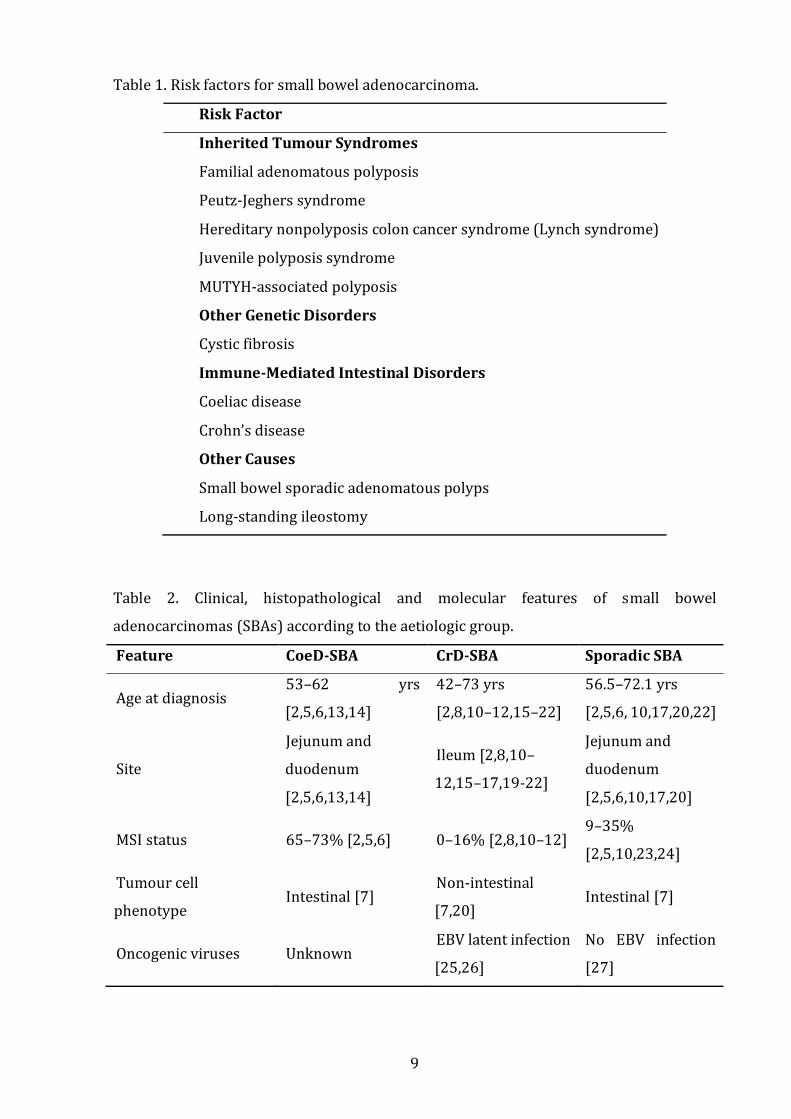
Table 1. Risk factors for small bowel adenocarcinoma.
Risk Factor
Inherited Tumour Syndromes
Familial adenomatous polyposis
Peutz-Jeghers syndrome
Hereditary nonpolyposis colon cancer syndrome (Lynch syndrome)
Juvenile polyposis syndrome
MUTYH-associated polyposis
Other Genetic Disorders
Cystic fibrosis
Immune-Mediated Intestinal Disorders
Coeliac disease
Crohn’s disease
Other Causes
Small bowel sporadic adenomatous polyps
Long-standing ileostomy
Table 2. Clinical, histopathological and molecular features of small bowel
adenocarcinomas (SBAs) according to the aetiologic group.
Feature CoeD-SBA CrD-SBA Sporadic SBA
Age at diagnosis 53–62 yrs
[2,5,6,13,14]
42–73 yrs
[2,8,10–12,15–22]
56.5–72.1 yrs
[2,5,6, 10,17,20,22]
Site
Jejunum and
duodenum
[2,5,6,13,14]
Ileum [2,8,10–
12,15–17,19-22]
Jejunum and
duodenum
[2,5,6,10,17,20]
MSI status 65–73% [2,5,6] 0–16% [2,8,10–12] 9–35%
[2,5,10,23,24]
Tumour cell
phenotype Intestinal [7]
Non-intestinal
[7,20] Intestinal [7]
Oncogenic viruses Unknown EBV latent infection
[25,26]
No EBV infection
[27]

10
CoeD-SBA, small bowel adenocarcinoma associated with coeliac disease; CrD-SBA, small
bowel adenocarcinoma associated with Crohn’s disease; EBV, Epstein-Barr Virus; MSI,
microsatellite instability; yr, year.
Epidemiology and risk factors for small bowel adenocarcinoma
Although small bowel corresponds to the 75% of digestive tract length and the 90% of
digestive absorptive surface [28], SBAs are relatively rare cancers and account for less
than 5% of all gastrointestinal neoplasms [29]. Notwithstanding, they represent around
40% of all small intestine malignancies [29]. The estimated incidence of SBA ranges
between 3,250 and 5,300 cases each year in the USA [30,31], whereas it is about 3,600
annual new cases in Europe [32]. The relative risk of developing SBA in CoeD and CrD
raises 14 and 33 times in comparison to the general population, respectively [33,34].
Amongst all SBAs, the 13% and the 7% seem to be associated with CoeD and CrD,
respectively [13,34]. The epidemiological features of SBA differ on the basis of underlying
chronic immune-mediated intestinal disorder. The median age at CoeD-SBA diagnosis has
been estimated from 53 to 62 years in American, British, Dutch and Italian patients
[2,5,6,13,14], while that at CrD-SBA diagnosis seems to be younger varying from 42 to 53
years in most studies (Table 3) [2,8,10–12,15–21]. Conversely, in an American large-scale
retrospective cohort study, CrD-SBA patients presented at a median age of 72.9 years [22].
Recently, the Small Bowel Cancer Italian Consortium also showed an older median age at
CrD-SBA diagnosis, i.e. 59 years [2]. We speculate that this discrepancy might be explained
by an older age at CrD diagnosis in the latter cohort (50 years) [2] and by a better clinical
management of CrD over the last two decades. Sporadic SBA patients often have a higher
median age at diagnosis -between 56.5 and 72.1 years- in comparison to both CoeD-SBA
and CrD-SBA [2,5,6,10,17,20,22]. Risk factors for CoeD-SBA and CrD-SBA have not
thoroughly assessed. According to the well-established protective effects of adherence to
gluten-free diet against malignant complications in CoeD [13,35], a strict gluten-free diet
also seems to reduce the risk of CoeD-SBA development. Accordingly, Elfström and
colleagues [36] demonstrated that the risk of small intestine neoplasms in coeliac patients
decreases, though not disappearing, after the first year of follow-up, likely by reducing
intestinal inflammation and mucosal damage [37].

However, several CoeD-SBAs have been described in patients under gluten-free diet;
therefore, other factors have to be involved. Interestingly, the median age at CoeD
diagnosis in patients developing SBA varies from 49 to 59 years (Table 3) [2,5,6], around
two-three decades higher than that of coeliac patients not evolving into neoplastic
complications [38]. Therefore, the diagnostic delay, a known risk factor for refractory
CoeD and, thus, for enteropathy-associated T-cell lymphoma [39], has been supposed to
play a role in CoeD-SBA. Notwithstanding, only one SBA case has been hitherto reported
in association with refractory CoeD [2], a finding suggesting a different pathogenesis
between SBA and enteropathy-associated T-cell lymphoma in CoeD. On the other hand,
there is no evidence for a role of diagnostic delay in CrD-SBA development in most studies
(Table 3), although diagnostic delay is often associated with more aggressive CrD
phenotypes, such as stricturing and penetrating behaviours [40]. Risk factors observed
for CrD-SBA encompass a long disease duration, a small bowel involvement, a stricturing
phenotype and bypassed segment(s) of small bowel [41]. As regards long disease
duration, in a French study involving 1,935 patients with small bowel location at CrD
diagnosis a cumulative risk of SBA has been assessed as 0.2% and 2.2% after 10 and 25
years of follow-up, respectively [17]. Although use of 6-mercaptopurine seemed to be a
risk factor for CrD-SBA in an American study including seven cases [42], no medical
treatment has been unquestionably found to be coupled with SBA in larger cohorts
[18,43]. Conversely, small bowel resection and use of salicylates for more than two years
protect against SBA in patients with CrD [18]. As regards gender, the rates of female
prevalence are extremely heterogeneous in both CoeD-SBA (25–62%) [2,5,6,13,14] and
CrD-SBA (29–60%) [2,8,10–12,15–22] so that it is hard to assess a gender predominance
in either conditions. However, considering the strong prevalence of CoeD in women [4],
these data may suggest that male gender is at higher risk to develop CoeD-SBA.
The incidence of SBA cases as a whole is doubled in African Americans (from 10.2 to 14.1
per 1,000,000) compared to Caucasians (from 4.5 to 7.2 per 1,000,000) [44,45]. On the
other hand, CrD-SBA has been shown to affect more frequently Caucasians in an American
large-scale retrospective study from 1992 to 2010 [22]. Similarly, CoeD-SBAs have been
described exclusively in Caucasians in the only study analyzing ethnic differences in this
aetiologic group [13]. It seems that more extensive investigations of epidemiology and
risk factors are needed.

Table 3. Studies on small bowel carcinomas associated with coeliac disease or Crohn’s disease.
Authors,
Year Pt
Age at SBC
dgn (Median,
Range, yrs)
Age at CoeD
or CrD dgn
(Median,
Range, yrs)
CoeD or CrD
duration at SBA
dgn (Median,
Range, yrs)
Stage
III/IV (%)
Overall
Survival
(%)
Main Findings
Small bowel adenocarcinoma associated with coeliac disease (CoeD-SBA)
Bruno JC et
al., 1997 [14] 6 62, 45–75 NA 17, 0–40 NA NA No evidence of flat dysplasia was present
Howdle PD
et al., 2003
[13]
23 62*, 47–80 NA 8.2, 0.8–36 NA NA CoeD-SBAs account for 13% of all SBAs
Potter DD et
al., 2004 [5] 17 59.5, 42–78 53, 25–77 NA 8/17 (47)
64.2 at 5
yrs
CoeD-SBAs have a high incidence of mismatch
repair deficiency
Diosdado B
et al., 2010
[6]
15 61, 47–79 59, 18–79 2.5, 0–32.3 NA NA CoeD-SBAs have promoter hypermethylation
of the APC gene

13
Vanoli A et
al., 2017
[2,7]
26 53, 28–80 49, 7–79 1.4, 0–25 8/26 (31) 83 at 5 yrs
CoeD-SBAs harbour MSI, high TILs and
nuclear β-catenin expression frequently and
show a better outcome in comparison with
CrD-SBAs
Small bowel adenocarcinoma associated with Crohn’s disease (CrD-SBA)
Michelassi F
et al., 1993
[15]
7 47.7*, 33–73 24, 11–57 20, 10–30 NA 6 mos
(mean)
Survival is worse in CrD-SBA than in colorectal
cancer complicating CrD
Rashid A et
al., 1997 [11] 8 45.5, 35–71 33.5 NA, 0–30 0/7 (0)
28.5 mos
(median) CrD-SBAs have RAS and TP53 mutations
Sigel JE et al.,
1999 [16] 8 42, 35–71 35, 23–52 12, 0.6–19 2/8 (25) NA
Most CrD-SBAs have dysplasia adjacent to
carcinoma
Palascak-Juif
V et al., 2005
[17]
20 47, 33–72 36, 15–54 16, 0–37 11/20 (55) 35 at 5 yrs Signet-ring cells were found in 7/20 CrD-SBAs
Piton G et al.,
2008 [18] 29 45, 29–74 34, 13–63 7, 0–52 NA NA
Small bowel resection and salicylate intake ≥2
yrs protect against CrD-SBA

14
Widmar M et
al., 2011 [19] 29 55.4, 22–81 25, 13–63 25.2, 0.8–51.3 16/29 (55) NA
Two clinical indicators of SBA were symptoms
in longstanding quiescent CrD and obstruction
refractory to medical therapy
Svrcek M et
al., 2014 [8] 41 47 NA 13.5 19/41 (46) NA
40/41 CrD-SBAs were observed in inflamed
mucosal areas. Flat or raised dysplasia was
found in 20/41 patients with CrD-SBA
Whitcomb E
et al., 2014
[20]
11 47, 42–77 24, 6–33 25, 10–40 NA NA
10/11 CrD-SBAs expressed at least a gastric
marker and 8/11 CrD-SBAs expressed the
pancreatobiliary marker CK7
Weber NK et
al., 2015 [21] 34 52.9, 32–74
22.4, 13.0–
69.3 22.3, 0–50.6 NA 52 at 2 yrs
Imaging features suggestive for CrD-SBA
included annular mass, nodularity at the
extraluminal margins of mass, and perforation
Grolleau C et
al., 2017 [10] 9 46, 37–67 36, 10–67 15, 0–32 5/9 (56) 56 at 2 yrs
Adjacent dysplasia was present in 9/9 CrD-
SBAs
Bojesen RD
et al., 2017
[12]
23 53, 37–85 NA NA NA 26 at 5 yrs 79% of CrD-SBAs showed inflammation-
dysplasia-carcinoma sequence

15
Wieghard N
et al., 2017
[22]
179 72.9 NA NA 71/179
(40)
3.9 yrs
(median)
CrD-SBAs have similar overall survival
compared to sporadic SBAs
Vanoli A et
al., 2017
[2,7]
25 59, 33–84 50, 22–84 13, 0–41 13/25 (52) 38 at 5 yrs
CrD-SBAs exhibit a low rate of MSI and TILs
CrD-SBAs are associated with dysplasia and
metaplasia, both showing
gastropancreatobiliary phenotype
Vanoli A et
al., 2017 [26] 31 59, 33–84 NA NA 17/31 (55) NA EBV+ CrD-SBAs may occur
CoeD, coeliac disease; CK, cytokeratin; CrD, Crohn’s disease; dgn: diagnosis; EBV, Epstein-Barr Virus; mo, month; MSI, microsatellite instability;
NA, not available; Pt, patient; SBA, small bowel adenocarcinoma; TIL, tumour-infiltrating lymphocyte: yr, year. *, mean.

Histopathology and molecular biology of small bowel adenocarcinoma
In general, SBAs as a whole have a predominance (52–60%) of glandular histotype [7].
However, medullary-type cancers have been observed in association with CoeD-SBA
[7,46], whereas poorly cohesive, diffuse-type cancers or mixed glandular/diffuse cases
are more frequent in CrD-SBA in comparison to CoeD-SBA and sporadic SBA [7,17]. Most
CoeD-SBAs and sporadic SBAs express intestinal phenotype markers, such as the caudal-
related homeobox transcription factor (CDX)2, the goblet cell marker mucin (MUC)2,
cytokeratin (CK)20 and/or the small bowel brush border marker CD10. Conversely, CrD-
SBAs frequently present metaplastic gastropancreatobiliary changes, characterised by
positivity for the gastric foveolar marker MUC5AC and/or the pancreatobiliary duct
marker CK7 [7,20].
A high density of CD3+ and CD8+ TILs is typical in CoeD-SBA, while TILs are frequently low
in both CrD-SBAs and sporadic SBAs [2]. This finding points to a greater host immune
response against tumour in CoeD-SBA in comparison with CrD-SBA and sporadic SBA,
thus leading to a better clinical outcome reported in CoeD-SBA (Table 3). Nevertheless,
this does not prevent tumour growth, probably due to an increased immune tolerance. In
particular, it has been hypothesized that programmed cell death ligand 1 (PD-L1) and
programmed cell death protein-1 (PD-1), crucial immune checkpoints aimed at inhibiting
and escaping immune surveillance, are also implicated in non-hereditary SBAs as well as
in colorectal and gastric cancers with MSI and/or Epstein-Barr Virus (EBV) infection
[47,48]. An American study on 42 sporadic SBAs showed PD-1 expression on
intratumoural and peritumoural lymphocytes in most cases, and PD-L1 expression on
neoplastic cells and immune cells, mainly histiocytes, in a minority of cases [49]. To the
best of our knowledge, no study assessed clonality of TILs in CoeD-SBA.
As shown in Table 4, molecular alterations were investigated in some studies recruiting
cohorts with at least 5 cases of CoeD-SBA and/or CrD-SBA. MSI, which is a consequence
of defective DNA mismatch repair and is verified by mean of molecular and/or
immunohistochemical analysis, is present in around one third of all non-hereditary SBAs
with significant differences between CoeD-SBA (65–73% MSI) [2,5,6], CrD-SBA (0–16%
MSI) [2,8,10–12], and sporadic SBA (9–35%) [2,5,10,23,24] (Tables 2 and 4). MSI causes
the anti-tumour immune response supposed to play a pivotal role in inducing a more
favourable outcome in these cancers. Genomic profiling of sporadic SBA showed some
genetic alterations affecting most frequently TP53 (mutated 58% of cases), KRAS (53.6%),

17
APC (26.8%), SMAD4 (17.4%) and PIK3CA (16%) [50,51]. Overexpression of the TP53
gene product has been reported in roughly half of cases in both CoeD-SBA and CrD-SBA,
thus confirming the crucial role of TP53 alterations in small bowel carcinogenesis [2,8,10],
as well as in inflammatory bowel disease-associated colorectal cancers [52]. KRAS
mutation, which is an early change in the adenoma–carcinoma sequence of colorectal
cancer, has been also described in 31% of CoeD-SBA and in 12–43% of CrD-SBA
[2,8,10,12,53].
Promoter hypermethylation of APC has been found in 73% of CoeD-SBA, whereas
nonsense APC mutations have not been observed in CoeD-SBA [6]. Similarly, allelic loss of
APC gene was infrequent in CrD-SBA [10]. Nevertheless, the involvement of Wnt/β-
catenin pathway has been described in most CoeD-SBAs and sporadic SBAs, as suggested
by aberrant nuclear β-catenin expression [7,54,55]. Conversely, nuclear translocation of
β-catenin has been demonstrated only in few CrD-SBAs [7,8]. To the best of our
knowledge, there are no data on SMAD4 mutation frequency in CoeD-SBA and CrD-SBA.
BRAF V600E mutation, which is remarkably infrequent in sporadic SBA [54], is also absent
in both CoeD-SBA and CrD-SBA [2,10,11] or identified up to 7% of CrD-SBA in other
studies [8,12]. Thus, unlike colorectal cancer [56,57], BRAF mutation does not seem to
play a pivotal role in inducing MLH1 gene methylation, a frequent finding in CoeD-SBA
[2,6]. No significant difference has been observed in PIK3CA or NRAS mutation rate
amongst CoeD-SBA, CrD-SBA and sporadic SBA [2,8]. Additionally, genomic profiling
showed potentially targetable genetic alterations in most SBA cases (91%) [50]. Laforest
and colleagues [58] described ERBB2/HER2 alterations in 12% of sporadic SBAs, through
mutations (7 cases) or amplifications (3 cases). HER2 amplification was also observed in
two CoeD-SBAs and in two CrD-SBAs [2].

Table 4. Molecular alterations in small bowel adenocarcinomas associated with coeliac disease or Crohn’s disease.
Authors, year Pt MSI
status
N (%)
KRAS
mutation
N (%)
NRAS mutation
N (%)
BRAF mutation
N (%)
PIK3CA mutation
N (%)
HER2 amplification
N (%)
p53 overexpression
N (%)
Nuclear β-catenin
expression
N (%) Small bowel adenocarcinoma associated with coeliac disease (CoeD-SBA)
Potter DD et al., 2004 [5]
17 8/11 (73)
NA NA NA NA NA NA NA
Diosdado B et al., 2010 [6]
15 6/9 (67) NA NA NA NA NA NA NA
Vanoli A et al., 2017 [2,7]
26 17/26 (65)
8/26 (31) 1/26 (4) 0/26 (0) 4/26 (15) 2/26 (8) 12/26 (46) 24/26 (92)
Small bowel adenocarcinoma associated with Crohn’s disease (CrD-SBA)
Rashid A et al., 1997 [11]
8 1/7 (14) 3/7 (43) NA NA NA NA 4/7 (57) NA
Svrcek M et al., 2014 [8]
41 1/36 (3) 7/30 (23) NA 1/29 (4) 0/23 (0) NA 21/35 (60) 16/31 (52)
Grolleau C et al., 2017 [10]
9 1/9 (11) 1/8 (12.5) NA 0/8 (0) NA NA NA NA
Bojesen RD et al., 2017 [12]
23 0/14 (0) 2/14 (14) NA 1/14 (7) NA NA NA NA
Vanoli A et al., 2017 [2,7]
25 4/25 (16)
4/25 (12) 1/25 (4) 0/25 (0) 2/25 (8) 2/25 (8) 12/25 (48) 6/24 (25)
MSI, microsatellite instability; NA, not available; Pt, patient.

Pathogenesis and preneoplastic lesions of small bowel adenocarcinoma
The exact pathogenesis of non-hereditary SBA is mostly unknown due to their rarity.
Dysplastic lesions close to CoeD-SBA are quite rare [7,14,59], whereas the recurrent
presence of dysplasia in the superficial part of both CrD-SBA and sporadic SBA has been
observed [7,8,60,61]. In addition, either in CrD-SBA or in CoeD-SBA, dysplasia has been
reported as flat or raised [7,8,10-12,59]. Dysplasia is distant or adjacent to CrD-SBA
[7,8,10-12], while no dysplasia has been described far from CoeD-SBA [7,59].
Dysplasia close to both CoeD-SBA and CrD-SBA is characterised by overexpression of p53
and retained reactivity for mismatch repair proteins [7]. Of note, loss of MLH1 is
infrequently observed in dysplasia associated with MSI CoeD-SBA [7], thus suggesting
that MLH1-hypermethylation-related MSI is a late event along small bowel carcinogenesis
in coeliac patients. Furthermore, the rare dysplastic foci adjacent to the invasive CoeD-
SBA have been reported to express nuclear β-catenin, whereas CrD dysplasia shows a
preserved membranous expression of β-catenin [7]. Therefore, Wnt pathway activation
seems to be an early process in CoeD-SBA carcinogenesis. Accordingly, overexpression of
the Wnt-related transcription factor and stem cell marker Sex-determining Region Y-Box
(SOX) 9 has been described in hyperplastic crypts of coeliac patients at CoeD diagnosis
[62], as well as in CoeD-SBA tumour cells, in continuity with SOX-9+ close dysplastic and
hyperplastic crypts (Figure 1) [7]. This may suggest a histogenetic association between
crypt hyperplasia and CoeD-SBA. On the other hand, a gastropancreatobiliary metaplastic
phenotype has been predominantly described in dysplastic or non-dysplastic mucosa
adjacent to CrD-SBA [7,20]. Although small bowel dysplasia has been found to have a low
sensitivity (33%) at enteroscopy in CrD patients at high risk of SBA [63], MUC5AC-positive
or CK7-positive metaplastic changes at perendoscopic biopsies should lead CrD patients
to a strict endoscopic follow-up. Immature crypt hyperplasia and gastropancreatobiliary
metaplasia might be reckoned as possible preneoplastic lesions, likely evolving into
dysplasia and carcinoma, in CoeD and CrD, respectively (Figure 1). Thus, an inflammation-
hyperplasia-dysplasia-carcinoma sequence may take place in CoeD-SBA development,
whereas an inflammation-metaplasia-dysplasia-carcinoma sequence may occur in CrD-
SBA pathogenesis. Further extensive and prospective studies are necessary to confirm
these models of cancerogenesis in order to recognise early preneoplastic lesions, which
may aid in early cancer diagnosis.

20
Figure 1 - Schematic representation of the pathogenic mechanisms underlying small bowel adenocarcinomas associated with chronic intestinal disorders.
Legend to Figure 1. In coeliac disease villous atrophy induces crypt hyperplasia, characterised by increased intraepithelial lymphocytes (IEL) similarly to atrophic epithelium. Nuclear Sex-determining Region Y-Box (SOX)-9-positive immature hyperplastic crypts evolve into flat nuclear β-catenin-positive dysplasia, thus leading to coeliac disease-associated adenocarcinoma (CoeD-SBA). CoeD-SBA is associated with microsatellite instability (MSI) and high number of tumour-infiltrating lymphocytes (TIL). In Crohn’s disease gastric (MUC5AC+)/pancreatobiliary (CK7+) metaplasia evolves into dysplastic polypoid growth, which lastly becomes Crohn’s disease-associated adenocarcinoma (CrD-SBA). CrD-SBA is almost always microsatellite stable (MSS).
Lytic phase of EBV infection frequently occurs in inflammatory bowel disease, particularly
in patients who have overused immunomodulators, mostly corticosteroids [64]. Recently,
latent phase of EBV infection, known to have a key role in gastroesophageal EBV
carcinogenesis [65], has been demonstrated in two microsatellite-stable T-cell rich CrD-
SBA [25,26]. In both cases EBV has been also detected in dysplastic lesions associated with
CrD-SBA and in small foci of iuxta-tumoural epithelium apparently devoid of dysplasia
[25,26]. Therefore, rarely EBV latent infection might be a very early, pivotal process along
SBA pathogenesis in those patients. Up-to-now, no latent infection with EBV has been

21
reported in CoeD-SBA, while EBV does not seem to be implicated in the carcinogenesis of
sporadic SBA, as suggested by its lack in a cohort of 56 sporadic SBA [27].
Clinical presentation and diagnosis of small bowel adenocarcinoma
Duration of the underlying inflammatory intestinal disorder before SBA diagnosis differs
(Table 3). CoeD-SBA presented after a median of 1.4–17 years from CoeD diagnosis in
comparison to 7–25.2 years from CrD diagnosis in CrD-SBA in studies with widest cohorts
of patients with CoeD-SBA and CrD-SBA, respectively [2,6,8,10,13–21]. However, SBA
might be diagnosed in a few cases at the same time of underlying immune-mediated
disorder for both CoeD and CrD [2,6,10,11,14,17,18,21]. The clinical spectrum of SBA at
onset is wide, including bleeding with subsequent iron-deficiency anaemia, positive fecal
occult blood test, maelena or coffee ground vomiting, obstruction with symptoms of
nausea, vomiting, abdominal pain and unexplained weight loss, or to intussusception and
perforation in the locally advanced neoplasms [1].
In coeliac patients any of the aforementioned symptoms apart from an isolated anaemia
should raise the suspicion for SBA. Additionally, when diarrhoea and fever are
simultaneously present, first of all enteropathy-associated T-cell lymphoma needs to be
considered [39], Likewise, in case of diarrhoea and intestinal obstruction, ulcerative
jejuno-ileitis has to be ruled out [4]. Once CoeD-SBA is suspected, an upper endoscopy is
recommended in coeliac patients in order to identify and sample the lesion, if it is
proximal to the ligament of Treitz (Figure 2). Notwithstanding, as most CoeD-SBA are
jejunal, additional diagnostic tests, such as device-assisted enteroscopy, computed
tomography enterography and magnetic resonance enterography, are generally needed
[66]. On the contrary, capsule endoscopy should not be encouraged in symptomatic
patients with SBA due to its several limitations, such as the impossibility to take biopsies
for histologic diagnosis and the risk of capsule retention and of missing SBA, in particular
in case of proximal site.

22
Figure 2 - Radiologic and histologic images of a coeliac disease-associated small bowel carcinoma.
Legend to Figure 2. (A) Computed tomography shows a circumferential mass with shouldered borders causing the wall thickening in the duodenum (arrows). (B) Haematoxylin and eosin staining shows a glandular-type carcinoma with a high tumour-infiltrating lymphocyte density. Original magnification: 100x.
In CrD patients, obstruction is more likely expected to be the manifestation of
fibrostricturing phenotype [67]. Similarly, anaemia and positive fecal occult blood test are
often related to active CrD [68]. Thus, apart from acute upper bleeding, all the other
symptoms of SBA are hard to differentiate the neoplasm from a relapse of CrD [69]. This
accounts for the fact that most CrD-SBA are diagnosed during the surgery or even post-
operatively by the pathologist [70]. Failure to respond to anti-inflammatory therapies
should not be considered per se an indicator of CrD-SBA, as it often happens in CrD
patients with fibrotic strictures devoid of an inflammatory component [67]. On the

23
contrary, obstructive symptoms and anaemia in a patient with longstanding quiescent
CrD should raise the suspicion for SBA [19]. Ileocolonoscopy is a diagnostic procedure
only in CrD-SBA located in the last tract of terminal ileum. Otherwise, as nearly all CrD-
SBA are more proximal, retrograde per anal device-assisted enteroscopy is the best
procedure to find and sample CrD-SBA. Computed tomography enterography and
magnetic resonance enterography might help in finding the correct location of SBA before
enteroscopy and/or laparoscopic surgery [21]. Nevertheless, both these imaging
techniques are highly indicative of CrD-SBA only in a few cases showing small bowel mass
with localised lymphadenopathy and/or evidence of distant spread, such as liver
metastasis or peritoneal carcinomatosis [10,21]. Notwithstanding, the review of imaging
data by a gastrointestinal radiologist could improve the identification of CrD-SBA-related
features, such as annular mass, nodularity at the extraluminal margins of mass and
perforation [21].
In conclusion, in the absence of inherited tumour syndrome (Table 1), both CoeD and CrD
should be ruled out in any patient at SBA diagnosis.
Prognosis and treatment for small bowel adenocarcinoma
SBA prognosis is frequently worse than that of large bowel cancers [71]. This seems to
happen in CrD patients too, in whom SBAs have been reported to be more aggressive than
colorectal carcinomas [15]. In an American retrospective study, recruiting 491 SBAs
predominantly sporadic SBAs, but also CoeD-SBAs (n = 13), CrD-SBAs (n = 23) and SBAs
due to familial adenomatous polyposis (n = 10), the median overall survival and the 5-
year overall survival rate were 20.1 months and 26%, respectively [72]. The main primary
reason for this poor outcome is that patients are generally symptom-free until late
disease, when metastases are frequently already present at SBA diagnosis. Tumour stage
has been reckoned the single most crucial prognostic factor in all SBAs [72]. Reduced
prognosis is also related to additional features, including poor differentiation, positive
margins, lymphovascular/perineural invasion, duodenal site, male gender, black ethnicity
and older age at SBA diagnosis [31,73-76]. High positive lymph nodes-to-total lymph node
ratio and a low number of investigated lymph nodes have been associated with a poor
survival [72,76,77].
Overall survival significantly differs between patients with CoeD-SBA and those with CrD-
SBA (Table 3). In particular, the predisposing immune-mediated intestinal disorder, i.e.,

24
CoeD or CrD, has been shown to be a stage-independent prognostic factor in patients
undergoing surgery for SBA in the largest study systematically comparing CoeD-SBAs,
CrD-SBAs and sporadic SBAs [2]. Five-year overall survival rate is relatively high in CoeD-
SBA, that is 64.2% and 83% in an American study and in an Italian study enrolling 17 and
26 patients, respectively [2,5]. On the contrary, five-year overall survival rate seems to be
poorer in CrD-SBA patients, varying from 26% to 38%, in French, Danish and Italian
studies [2,12,17]. Accordingly, two-year overall survival in CrD-SBA has been observed to
be 52% and 56% in an American study and in a French study, respectively [10,21], also
lower than five-year overall survival in CoeD-SBA. Overall survival has been found to be
more favourable in CoeD-SBC in comparison with sporadic SBA [2,5], whereas no survival
difference has been shown between CrD-SBA and sporadic SBA [2,10,17,22]. Recently,
Axelrad JE and colleagues [78], dealing with small bowel cancer-related mortaliy rate in
patients with inflammatory bowel disease in a binational population-based cohort study
from Denmark and Sweden, concluded that small bowel cancer death is higher in CrD
patients than in sporadic cases. This is not in keeping with all studies published so far
showing a similar death rate between patients with CrD-SBA and those with sporadic SBA
(Table 5). In particular, Palascak-Juif V and colleagues [17] demonstrated a slightly, but
not significantly, higher survival rate, in CrD-SBAs (54% at 2 years and 35% at 5 years)
than in sporadic SBAs (37% at 2 years and 30% at 5 years) both at 2 years and 5 years.
Wieghard and colleagues [22] demonstrated a better 5-year overall survival in 179 CrD-
SBAs (43%) than in 1,944 sporadic SBAs (34%). In this large American study patients
with CrD-SBA were diagnosed at an earlier stage (I/II) compared with sporadic SBA (55%
vs. 32%, p < 0.0001) and were more likely to undergo surgery (81% vs. 72%, p = 0.0016).
However, a similar cancer-specific survival was observed between the two groups,
namely 65% versus 64%. Indeed, multivariate analysis confirmed that CrD was not
significantly associated with overall survival [22]. Recently, another American
investigation [79] using the National Cancer Database demonstrated a similar overall
survival at 5 years between 493 CrD-SBAs (41%) and 2,175 sporadic SBAs (35%).
Additionally, at multivariate analysis CrD was not a risk factor for reduced survival [79].
The disagreement between Axelrad JE and colleagues [78] and all the other studies
[17,22,79] might be secondary to the fact that in the former one the death rate was
calculated in the small bowel cancer cumulatively, including SBAs, neuroendocrine
tumours, sarcomas and others. Mortality rate was not analysed for each small bowel
cancer subtype. Furthermore, the controls recruited by Axelrad JE and colleagues was

25
defined as “free of IBD” [78], but this does not rule out coeliac disease or hereditary SBAs.
Although Axelrad JE and colleagues [78] excluded patients with CoeD before the onset of
follow-up, small bowel cancers were diagnosed after that. Moreover, CoeD diagnosis may
be simultaneous to that of SBA [2], thus, if patients with CoeD were enrolled in the control
group, obviously the relative mortality rate would be higher in CrD-SBA, as well as it is
known in literature (Table 3). However, Axelrad JE and colleagues [80] then clarified that
limiting the analysis to patients with pre-existing CrD-SBA compared to control groups
the death rate was similar to that one of previous studies [17,22,79]. Although prospective
studies are necessary to evaluate the impact of small bowel cancer on CrD patient
survival, it is already evident that SBA is not the main cause of death in CrD patients.
Regardless of the aetiologic group, CoeD or CrD, prognostic factors for SBA include stage,
tumour histotype and high TILs [2,7]. Tumour histology by itself is clinically relevant, as
it has been demonstrated that diffuse-, mixed- and solid-types considered as a whole tend
to have a poorer prognosis compared to glandular-type and medullary-type SBAs [7,46].
Amongst prognostic factors within the CoeD-SBAs, either MSI or high TIL density have
been also found and they correlate one each other [2]. Notwithstanding, only TIL density
retains a prognostic power in a multivariable model, likely due to the fact that several
high-TIL SBAs showing a favourable outcome miss MSI [2]. High TIL density in SBA can
be induced by further factors besides MSI status, such as oncogenic viruses. As this
regards, non-MSI high-TIL SBAs with EBV latent infection reported in two CrD patients
seem to be have a good prognosis [25,26], presumably due to the anti-tumour immune
response triggered by abnormal peptide production from EBV. Briefly, although these
findings have to be confirmed more-in-depth, EBV latent infection should be considered
in CrD-SBA for a better prognostic assessment.
Currently, treatment for CoeD-SBA and CrD-SBA widely derives from recommendations
for sporadic SBA [81]. Surgery is the mainstay of curative therapy for SBA without distant
metastasis (M0), whose potential benefits from adjuvant chemotherapy are debated, in
particular for SBA at stage II [1]. Surgical resection with appropriate lymph node sampling
is mandatory for long-term survival in resectable SBA. Surgery is the unique curative
treatment for SBA at stage I, whereas it should be followed by adjuvant chemotherapy,
including FOLFOX4 or LV5FU2 or oral fluoropyrimidine for SBA at stage II or -to a higher
extent- for SBA at stage III [81]. In particular, as the mismatch repair deficient (MMR-d),
leading to MSI phenotype and high immune response in solid neoplasms, is related to a
better cancer-specific survival in resected SBAs at stage II [2,51,82,83], this confirms the

26
National Comprehensive Cancer Network Clinical Practice guidelines, Small Bowel
Adenocarcinoma, not recommending adjuvant chemotherapy for patients with resected
MMR-d SBA at stage II [84]. On the contrary, within patients with mismatch repair
proficient SBA at stage II, T4 neoplasms may require a more aggressive therapeutic
approach [83]. Systemic chemotherapy is the therapy for non-resectable or metastatic
SBC, namely those at stage IV [81]. In a meta-analysis of 14 studies, adjuvant
chemotherapy provided no significant survival benefit in SBA patients [85]. Nevertheless,
a recent study demonstrated that adjuvant chemotherapy was associated with a better
overall survival in patients with SBA at stage II-IV in a multivariate analysis stratified by
stage [86]. The international phase 3 clinical trial PRODIGE 33-BALLAD, assessing the
possible benefits of adjuvant chemotherapy in patients with SBA at stage I-III, is
underway [87,88].
Some molecular alterations may suggest responsiveness to novel treatments. KRAS wild-
type mutational status has been shown to predict the response to anti-epidermal growth
factor receptor monoclonal antibodies cetuximab and panitumumab alone or combined
with chemotherapy in metastatic SBA in a few cases [89,90]. On the contrary, a phase 2
clinical trial demonstrated no response of panitumumab in nine patients with metastatic
KRAS wild-type SBA, one associated to inflammatory bowel disease and two to Lynch
syndrome [91]. In particular, in this study seven patients showed SBA progression,
whereas the other two ones had stable SBA [91]. It has been assumed that SBA, as well as
right-sided colon carcinoma, benefit less from anti-epidermal growth factor receptor
agents than left-sided colon carcinomas due to their different embryologic origin, i,e,
midgut for small bowel and right-sided colon and hindgut for left-sided colon [91,92].
Although HER2 amplification is infrequent in CoeD-SBA and CrD-SBA [2], it is worth being
assessed as a possible therapeutic target of anti-HER2 receptor monoclonal antibody
trastuzumab [58,93]. Expression of PD-L1 on tumoural and immune cells in SBA should
support clinical trials in order to investigate efficacy of anti-PD-L1 monoclonal antibodies
avelumab and atezolizumab [49]. As this regards, an open-label phase 2 clinical trial of
avelumab is ongoing in patients with advanced and metastatic SBA [94]. Similarly, an
open-label phase 2 clinical trial has been assessing the response to atezolizumab together
with the MEK inhibitor cobimetinib in advanced rare cancers, including SBAs [95]. Anti-
PD-1 monoclonal antibodies pembrolizumab and nivolumab might be suitable in a subset
of patients with metastatic MSI SBA [96]. An open-label phase 2 clinical trial of
pembrolizumab is underway in patients with non-resectable metastatic or locally

27
advanced SBA [97]. Additionally, pembrolizumab has been evaluating in a large phase 1b
clinical trial in combination with the Hsp90 inhibitor XL888, inhibiting Hsp90 chaperone
function and promoting the proteasomal degradation of several oncogenic signaling
proteins, including Her-2 and Met [98]. This study was designed for several advanced
gastrointestinal cancers, including SBA, to find out the best phase 2 dose for the
combination of XL888 and pembrolizumab [98]. Another clinical trial has been testing
efficacy of the combination immunotherapy with nivolumab and the anti-CTLA-4
monoclonal antibody ipilimumab in advanced rare cancers, such as SBA [99]. Briefly,
immunotherapy has been modifying the therapeutic approach in some solid tumours, in
particular PD-1/PD-L1 pathway blockade may be considered in patients with advanced
MSI SBA, as mismatch repair deficiency has been shown to predict efficacy of anti-PD-1
antibodies in eleven types of solid tumours, including SBA [100].
Table 5. Survival rate in patients with Crohn’s disease-associated small bowel
adenocarcinoma in comparison to that in patients with sporadic small bowel
adenocarcinoma
Authors, year Group Patients
(N)
Overall
survival
at 5 years
(%)
P-value HR 95% CI P-value
Palascak-Juif V
et al., 2005 [17]
CrD-SBA
Sporadic SBA
20
40
35
30
NS
NA
NA
NA
NA
Wieghard N et
al., 2017 [22]
CrD-SBA
Sporadic SBA
179
1,944
43
34
0.0121
0.97
0.79-1.20
NS
Fields AC et al.,
2020 [79]
CrD-SBA
Sporadic SBA
493
2,175
41
35
NS
1.01
0.99-1.02
NS
CI, confidence interval; CrD-SBA, Crohn’s disease-associated small bowel adenocarcinoma; HR,
hazard ratio; NA, not available; NS, not significant; SBA, small bowel adenocarcinoma.

28
Objective of the thesis
Recent studies showed a positive correlation between PD-L1 expression and MSI-high
(MSI-H) in SBAs [49,101,102]. Although MSI-H is the main determinant of tumour
mutation load, causing PD-L1 expression in gastrointestinal neoplasms, other factors
might be involved [103]. A recent study [104] demonstrated that PD-L1 is also expressed
in several microsatellite stable endometrial carcinomas with high TILs. Furthermore, TIL
density, EBV infection, and CDX2 negativity have been associated with PD-L1 positivity in
gastrointestinal cancers [48,105,106]. Therefore, tumour immune microenvironment, in
particular PD-L1 expression, TIL density and tumour mutation load, are under
investigation, in order to identify potential markers of response to immune checkpoint
blockades [107]. On the other hand, recent studies on colorectal and other
gastrointestinal cancers have found that the invasive front markers tumor budding (Tb)
and poorly differentiated clusters (PDCs) may significantly improve their prognostic
evaluation [108-115].
On this basis, the primary objective of this thesis was to assess PD-L1 and PD-1 expression
in a relatively large and well-characterised cohort of non-hereditary SBAs, associated
with CoeD or CrD or sporadic, enrolled through the Small Bowel Cancer Italian
Consortium. PD-L1 and PD-1 expression was then correlated with several clinical and
pathological features, including the predisposing immune-mediated intestinal disorder,
the MSI or EBV status, the intestinal phenotype markers CDX2 and liver fatty acid-binding
protein (L-FABP), and cancer-specific survival. The secondary objective of this thesis was
to investigate the invasive front markers Tb and PDCs on CrD-SBAs evaluated for the
primary aim too.

29
Materials and Methods
Study population
This retrospective and longitudinal study involved 21 tertiary referral Italian Coeliac
and/or IBD Centers taking part in the Small Bowel Cancer Italian Consortium.
CoeD diagnosis was based on positivity of serum IgA anti-endomysial and anti-tissue
transglutaminase antibodies along with typical duodenal histological lesions [4]. CrD
diagnosis was verified according to internationally agreed criteria [116], and the site and
extent of the disease were confirmed by endoscopy, histology and imaging. A group of
patients with sporadic SBA, namely without a concomitant chronic intestinal immune-
mediated disorder, were recruited as controls. In patients with sporadic SBA, CoeD was
ruled out (negativity of serum IgA anti-endomysial and anti-tissue transglutaminase
antibodies, coupled with normal serum total IgA), while CrD was excluded by the lack of
classic clinical and biochemical features. Re-assessment of the sporadic surgical samples
further confirmed the absence of histologic lesions indicative of either CoeD or CrD. The
main exclusion criteria for all SBA groups were Lynch syndrome, Peutz-Jeghers
syndrome, familial adenomatous polyposis and juvenile polyposis. This study was
approved by the Ethics Committee of the San Matteo Hospital Foundation of Pavia
(protocol number 20140003980).
Histology
Tissue samples were fixed in 4% formaldehyde and processed in paraffin wax. Four μm-
thick sections were stained with haematoxylin–eosin (H&E) for morphological
evaluation. All cases were investigated for the following conventional histologic
parameters: tumour histotype, World Health Organization (WHO) tumour grade (for the
entire tumour), TILs and all parameters required for TNM staging [117]. Tumour
histotype was classified as: a) glandular, b) diffuse, c) mixed (glandular plus diffuse), d)
medullary and e) non-medullary solid types, as previously described [7,118]. WHO
tumour grade was based on the proportion of gland formation and categorized as grade 1
(well differentiated, >95%), grade 2 (moderately differentiated, 50% to 95%), or grade 3
(poorly differentiated, 0% to 49%). All available H&E–stained slides from CrD-SBAs,
including full-thickness sections of the tumor and encompassing the invasive front, were
reviewed. In carcinomas with mucinous features, WHO grade, Tb and PDCs were assessed

30
in the area outside the mucinous component. An Eclipse Ci microscope (Nikon) with a
standard 22-mm diameter eyepiece (specimen area of 0.950 mm2 under an objective lens
with a magnification of ×20) was used and the number of buds/PDCs was divided by 1.21
to achieve the number of buds per area of 0.785 mm2 as recommended for colorectal
cancer [111].
Definition and evaluation of Tb
A tumour bud is defined as a single tumor cell or a cell cluster of up to 4 tumour cells
which develops from neoplastic glands. Tb was analyzed along the invasive parts of the
tumour using the hotspot method, which is considered to be the most useful method for
assessing Tb in colorectal cancer [111]. Initially, the invasive front of the tumour was
screened using low magnification to find the areas with most Tb. For this purpose,
cytokeratin 8-18 (monoclonal, clone EP17/EP30, Dako) immunohistochemistry was
helpful in some challenging cases (ie. glandular fragmentation, strong peritumoral
inflammation) to allow a better visualization of Tb-rich areas. Tb was assessed from
several H&E areas and the single field with the most budding was used for quantitation.
The number of buds was counted in all cancers on H&E from a single field of view using
×200 total magnification (the hotspot method). Following the International Tumor
Budding Consensus Conference (ITBCC) group recommendation for colorectal cancer, we
used a three-tier system: low budding (Tb1): 0-4 buds; intermediate budding (Tb2): 5-9
buds and high budding (Tb3): 10 or more buds [111].
Definition and evaluation of PDCs
PDCs were defined as clusters of ≥5 cancer cells that lacked a gland-like structure. The
whole tumour was first scanned at low-power magnification to identify areas with the
greatest number of PDCs at the invasive front. The number of PDCs in a single field of
highest activity was then determined and graded as PDC1 (<5 PDCs), PDC2 (5 to 9 PDCs),
or PDC3 (≥10 PDCs) under an objective lens with a magnification of ×20 [110,117,119].
Definition of combined invasive front (CIF)
A CIF grade was developed as high in the presence of grade 3 for either Tb or PDCs or both
and as low in the remaining cases.
Immunohistochemistry

31
Four μm-thick sections were stained on a Dako Omnis platform with the following
antibodies: CD3 (polyclonal, Dako, Carpinteria, CA), CD8 (polyclonal, Dako), MLH1
(monoclonal, clone ES05, Dako), MSH2 (monoclonal, clone FE11, Dako), MSH6
(monoclonal, clone EP49, Dako), PMS2 (monoclonal, clone EP51, Dako), PD-L1
(monoclonal, clone 22C3, Dako), PD-1 (monoclonal, clone NAT, Dako), CDX2 (monoclonal,
clone DAK-CDX2, Dako) and L-FABP (monoclonal, clone EPR20464, Dako).
Immunoreactions were developed using 0.03% 3,3’ diaminobenzidine
tetrahydrochloride and sections were then counterstained with Harris’ haematoxylin.
TILs were stained using CD3 and CD8 antibodies and counted in ten consecutive high-
power fields (HPFs), as previously described [2]. A tumour was classified as having “high
TIL density” when the mean number of TILs/HPF was greater than 15 for CD3 or greater
than 9.5 for CD8 [120]. Immunostaining of DNA mismatch repair proteins MLH1, MSH2,
MSH6 and PMS2 in tumour cells was evaluated as proficient (retained expression) or
deficient (absent expression); only tumours showing absence of nuclear staining of all
neoplastic cells in the presence of an internal positive control (intra-tumour stromal and
inflammatory cells or non-tumour mucosa) were considered deficient [2]. In parallel, MSI
molecular analysis was performed
PD-L1 membranous expression was evaluated using the combined positive score (CPS)
measuring both tumoral cells and peritumoural/intratumoural immune cells, the
mononuclear immune cell density score (MIDS) measuring peritumoural/intratumoural
immune cells only and the tumour proportion score (TPS) measuring tumoral cells only,
as previously described [121]. In particular, CPS was calculated as the ratio of the number
of PD-L1 stained cells (tumour cells and immune cells) to the total number of viable
tumour cells, multiplied by 100. Tumours were considered negative if CPS<1, positive if
CPS≥1. TPS was the ratio of the number of PD-L1 stained tumour cells divided by the total
number of viable tumour cells, multiplied by 100. Tumours were regarded as negative if
TPS<1, positive if TPS≥1. MIDS was calculated as the ratio of the number of PD-L1 stained
immune cells to the total number of viable tumour cells, multiplied by 100; the result was
then scored in a scale from 0 to 4. MIDS 0 was defined as absent PD-L1 staining, while
MIDS 1, MIDS 2, MIDS 3 and MIDS 4 corresponded to PD-L1+ immune cells per 100 viable
tumour cells <1, ≥1 but <10, ≥10 but <100, ≥100, respectively. MIDS scores 2, 3 and 4 were
regarded as positive, whereas scores 0 and 1 as negative. PD-1-positive immune cells
were counted separately in intratumoural and peritumoural areas in ten consecutive
HPFs and the mean number of PD-1-positive cells per HPF was recorded for each case.

32
PD-1+ cells situated inside the tumour were considered as intratumoural, while PD-1+
cells located in the areas adjacent to the tumour invasive front as peritumoural. In
addition, the total number of PD-1-positive cells per HPF, corresponding to the sum of
intratumoural and peritumoural PD-1-positive cell counts was given. L-FABP or CDX2
staining was considered positive in cases with >10% moderate-to-intense staining in
tumour cells [122]. A central pathology review of each case was performed.
MSI analysis
Tumour DNA was obtained from formalin-fixed and paraffin-embedded tissues using
three representative 8 μm-thick sections of tumor samples. DNA was extracted after
manual microdissection using a QIAamp DNA formalin-fixed, paraffin-embedded tissue
kit according to the manufacturer’s protocol (Qiagen, Hilden, Germany). Microsatellite
instability analysis was performed using a pentaplex panel of monomorphic
mononucleotide repeats (BAT25, BAT26, NR21, NR-22 and NR-24) by the ABI PRISM 310
Genetic Analyzer (Applied Biosystems, Foster City, CA), as previously reported [2].
Definition of Teng tumour microenvironment immune types
Tumour microenvironment immune types were defined based on CD3+ TIL density (low
versus high) and PD-L1 expression, evaluated with the CPS. Tumors were classified in four
different Teng types, i.e. type I (high TIL density, CPS≥1), II (low TIL density, CPS<1), III
(low TIL density, CPS≥1), IV (high TIL density, CPS<1) [123].
EBV encoded RNAs in situ hybridization
The formalin-fixed, paraffin-embedded tissue sections were pretreated with proteinase K
(DAKO) for 30 min at room temperature, then hybridized with a FITC-labeled peptic
nucleic acid probe complementary to EBV-encoded RNAs (EBER-1 and 2;
DakoCytomation, Glostrup, Denmark), markers of latent phase EBV infection, and
incubated overnight at 55 °C. After washing in restricting conditions for 35 min, the
hybridized cells were visualized with an in situ hybridization detection kit (K5201; DAKO)
according to the manufacturer’s instructions. The sections were then counterstained with
Kernechtrot, dehydrated through graded alcohols, immersed in xylene and mounted with
a permanent medium. The present in situ hybridization method stained the nuclei of EBV-
infected cells dark blue, while the nuclei of non-infected cells appeared red. Specificity

33
controls were performed by omitting the EBER probe and by running in parallel EBV
positive and negative CrD-SBAs characterized in a previous investigation [26].
Gene mutation analysis
Mutation analysis of KRAS, NRAS and PIK3CA genes was performed using the Sequenom
MassARRAY system (Diatech Pharmacogenetics, Jesi, Italy), based on matrix-assisted
laser desorption/ionization time-of-flight mass spectrometry, together with the
Myriapod Colon Status Kit (Diatech Pharmacogenetics). This kit includes a series of
multiplexed assays designed to interrogate a total of 153 non-synonymous hotspot
mutations in the four genes. DNA amplification was done in a 5-μL reaction mixture
containing 10 to 20 ng of tumour DNA. PCR, Shrimp Alkaline Phosphatase reaction and
single base pair extension steps were carried out following the protocols provided by
Diatech Pharmacogenetics. Completed genotyping reactions were spotted in nanoliter
volumes onto a matrix-arrayed silicon SpectroCHIP with 96 elements using the
MassARRAY Nanodispenser (Diatech Pharmacogenetics). SpectroCHIP was analysed
using the Sequenom MassARRAYs Analyzer 4 spectrometer and the spectra were
processed by the MassARRAY Typer Analyzer 4.0 software (Diatech Pharmacogenetics).
All automated system mutation calls were confirmed by manual review of the spectra. We
investigated TP53 mutations at exons 5-8 which correspond to the core domain involved
in protein-protein interaction (tetramerization) and in binding to DNA and represent the
region where the vast majority of TP53 mutations are detected. Briefly, exons 5-8 were
amplified by PCR using sets of primers reported in IARC TP53 database tools
(http://p53.iarc.fr/ProtocolsAndTools.aspx). In detail, we used primer pairs that amplify
small (poor DNA quality) fragments (IARC code: P-312 and P-271 for exon 5; P-239 and
P-240 for exon 6; P-237 and P-238 for exon 7; P-316 and P319 for exon 8). PCR products
were subjected to automated sequencing by ABI PRISM 310 (Applied Biosystems, Foster
City, CA). All mutated cases were confirmed at least twice starting from independent PCR
reactions. In each case, the detected mutation was confirmed in the sequence as sense and
antisense strands.
Statistical analysis
Stata 15 (StataCorp, College Station, TX, USA) was used to perform all computations. We
considered a 2-sided p-value<0.05 as statistically significant. For post-hoc comparisons
between etiologic groups, the significance was set at 0.017 (Bonferroni correction).

34
Continuous data were reported as median and 25th-75th percentiles, categorical variables
are reported as counts and percent; they were compared between etiologic groups using
the Kruskal Wallis test or the Fisher exact test, respectively. The Spearman R and 95%
confidence intervals (CI) were computed to measure the correlation between continuous
variables. For the purpose of the analysis continuous variables were dichotomized at the
median value. Median follow-up (25th-75th) was computed with the reverse Kaplan Meier
method. Follow-up was computed from diagnosis of cancer to death or last available
follow-up for censored patients. Cumulative survival curves were plotted according to the
Kaplan Meier method and compared with the logrank test. The strength of the association
between series of candidate risk factors and cancer-specific mortality was assessed using
Cox regression; hazard ratios (HR) and 95% CI were derived from the models. Mortality
rates per 100 person year and 95% CIs were reported. We checked the proportional
hazard assumption with a test based on residuals. We computed the Harrell’s c statistic
for discrimination (the closer to 1, the better, the closer to 0.5, the worse). Given the low
number of deaths, we did not fit multivariable survival models, but only bivariable models
were fitted to adjust, in turn, for aetiologic group and stage.

35
Results
Patient demographics and clinico-pathologic features
This retrospective study included a cohort of 121 patients with pathologically-confirmed
primary non-hereditary, non-ampullary SBA, who underwent surgical resection and had
complete survival data. Demographic and clinico-pathologic data of all patients
investigated are reported in Table 6. We recruited 34 patients with CoeD-SBA, 49 with
CrD-SBA, and 38 with sporadic SBA, a fraction of them entered previous studies from the
Small Bowel Cancer Italian Consortium [2,7,26,124]. Median age at the time of SBA
diagnosis among coeliac (median 53.5 years) and CrD patients (median 58 years) was
significantly (p<0.001) lower than that of sporadic cases (median 69 years), and median
duration of inflammatory disorder at cancer diagnosis was significantly (p=0.016) lower
in CoeD-SBA (median 23.5 months) compared to CrD-SBA (median 156 months). A higher
rate of male gender was found in CrD-SBA (73%) and sporadic SBA (63%) compared to
the CoeD-SBA group (47%). In keeping with what is already known (Table 2), the ileum
was the commonest small bowel location for CrD-SBA (94%), whereas it was the jejunum
in both CoeD-SBA (70%) and sporadic SBA (63%). No significant difference was identified
among the three groups regarding tumour stage at diagnosis. The majority of stage III and
IV patients received systemic chemotherapy with platinum-based and 5-fluorouracil
regimens after surgical intervention.
Histologically, most SBAs showed glandular differentiation in all aetiologic groups;
however, medullary and diffuse/poorly cohesive cancers were more common in CoeD
(17%) and CrD patients (20%), respectively. CoeD-SBAs displayed a significantly
(p<0.001) greater number of TILs (median 25.1 TILs/HPF) compared to CrD-SBAs and
sporadic SBAs (median 7.1 TILs/HPF for both). MSI-H was identified in 39 cases (32.2%),
including 37 cases with loss of MLH1/PMS2 expression and two SBAs, both associated
with CrD, with isolated loss of MSH6. No discordance between immunohistochemistry for
mismatch repair proteins and MSI molecular analysis was observed in any case. MSI-H
rate was significantly (p<0.001) higher in CoeD-SBAs (65%) than in both CrD-SBAs (18%)
and sporadic SBAs (21%). As regards markers of intestinal differentiation, CDX2 loss was
significantly (p=0.012) more common in CrD-SBAs (46%) compared to CoeD-SBAs (15%),
while the absence of L-FABP expression was significantly (p<0.001) more frequent in
both CoeD-SBAs (88%) and CrD-SBAs (81%) compared to sporadic SBAs (45%).

36
Table 6. Demographic and clinico-pathologic features of all 121 SBA patients
CoeD-SBA CrD-SBA Sporadic
SBA
Overall p-value
Post-hoc comparison
p-value
Number 34 49 38
Age at SBA diagnosis
Median [25th–75th IQR], yrs
53.5
[42.7-66]
58
[51-67.5]
69
[62-77]
<0.001 CoeD vs CrD: 0.129
CoeD vs Sporadic: <0.001
CrD vs Sporadic: <0.001
Duration of inflammatory disorder at SBA diagnosis
Median [25th–75th IQR], mo
23.5
[12-110.25]
156
[6-288]
NA 0.016
Sex, N (%)
Female
Male
18 (53)
16 (47)
13 (27)
36 (73)
14 (37)
24 (63)
0.049
CoeD vs CrD: 0.014
CoeD vs Sporadic: 0.169
CrD vs Sporadic: 0.302
Site, N (%)*
Duodenum
Jejunum
Ileum
7 (21)
23 (70)
3 (9)
1 (2)
2 (4)
46 (94)
3 (8)
24 (63)
11 (29)
<0.001
CoeD vs CrD: <0.001
CoeD vs Sporadic: 0.053
CrD vs Sporadic: <0.001
Stage, N (%)**
I
II
III
IV
3 (9)
19 (60)
8 (25)
2 (6)
6 (12)
19 (39)
18 (37)
6 (12)
2 (5)
17 (46)
15 (41)
3 (8)
0.550
Histotype, N (%)
Glandular
Medullary
Diffuse
Mixed
Solid
19 (56)
6 (17)
2 (6)
4 (12)
3 (9)
24 (50)
2 (4)
10 (20)
12 (24)
1 (2)
22 (58)
1 (3)
2 (5)
10 (26)
3 (8)
0.032
CoeD vs CrD: 0.032
CoeD vs Sporadic: 0.187
CrD vs Sporadic: 0.228
CD3+ TILs/HPF
Median [25th–75th IQR]
25.1
[12.3-75.4]
7.1
[2-20.6]
7.1
[2.2-20.9]
<0.001 CoeD vs CrD: <0.001
CoeD vs Sporadic: <0.001
CrD vs Sporadic: 0.962
MSI status, N (%)
Non-MSI
MSI-H
CDX2 expression, N (%)***
Negative
Positive
12 (35)
22 (65)
5 (15)
28 (85)
40 (82)
9 (18)
22 (46)
26 (54)
30 (79)
8 (21)
11 (29)
27 (71)
<0.001
0.012
CoeD vs CrD: <0.001
CoeD vs Sporadic: <0.001
CrD vs Sporadic: 0.754
CoeD vs CrD: 0.003
CoeD vs Sporadic: 0.165
CrD vs Sporadic: 0.109
L-FABP expression, N (%)****
Negative
Positive
30 (88)
4 (12)
39 (81)
9 (19)
17 (45)
21 (55)
<0.001
CoeD vs CrD: 0.393
CoeD vs Sporadic: <0.001
CrD vs Sporadic: <0.001

37
CoeD-SBA, coeliac disease-associated small bowel adenocarcinoma; CrD-SBA, Crohn’s disease-associated small bowel adenocarcinoma; HPF, high-power field; IQR, interquartile range; L-FABP, liver fatty acid-binding protein; mo, month; MSI, microsatellite instability; MSI-H, microsatellite instability-high; NA, not applicable; SBA, small bowel adenocarcinoma; TIL, tumour-infiltrating lymphocyte; yr, year.
*In one CoeD-SBA the precise tumor site within small bowel was unknown.
**In two CoeD-SBAs and in one sporadic SBA the precise stage was unknown.
***In one CoeD-SBA and in one CrD-SBA, no section for CDX2 immunohistochemistry was available.
****In one CrD-SBA, no section for L-FABP immunohistochemistry was available.
Immunohistochemical expression of PD-L1 and association with clinico-pathologic
features
PD-L1 staining was found in immune cells and to a variable extent in tumour cells (Figure
3). PD-L1 expression according to CPS and MIDS was positively associated with male sex,
while no significant association was observed between PD-L1 expression and age at SBA
diagnosis, small bowel site and tumour stage at diagnosis (Table 7, Table 8).
Per CPS cut-off of 1 or more (CPS≥1), the prevalence of PD-L1 expression in non-
hereditary SBAs as a whole was 26%, with significantly (p=0.001) higher percentage in
both CoeD-SBA (35%) and CrD-SBA (35%) than in sporadic SBAs (5%). Among CPS≥1
SBAs, 65% (20 out of 31 cases, including 8 CoeD-SBAs, 10 CrD-SBAs and 2 sporadic SBAs),
13% (four cases, including one CoeD-SBA and three CrD-SBAs) and 23% (seven cases,
including three CoeD-SBAs and four CrD-SBAs) showed a 1≤CPS<10, 10≤CPS<50, and
CPS≥50, respectively. Per TPS≥1, the prevalence of PD-L1 expression was 8%, with
significantly (p=0.035) higher percentage in both CoeD-SBA (15%) and CrD-SBA (10%)
than in sporadic SBA (0%). Per MIDS>1, the prevalence of PD-L1 expression was 22%,
with significantly (p=0.005) higher percentage in both CoeD-SBAs (29%) and CrD-SBAs
(31%) than in sporadic SBAs (5%). No case with MIDS 4 was identified in the whole
cohort.
Patients with MSI-H SBAs had a significantly (p=0.013) higher prevalence of CPS≥1 (41%)
than those with non-MSI SBAs (18%). Patients with MSI-H SBAs had a significantly
(p=0.002) higher prevalence of TPS>1 (20%) than those with microsatellite stable SBAs
(2%). No significant difference in terms of PD-L1-positivity according to MIDS was
observed between MSI-H and microsatellite stable SBAs. In addition, PD-L1 positivity was
associated with several features known to be more frequently coupled with MSI-H
phenotype, including high CD3+ and CD8+ TIL density and medullary histotype.
SBAs with CPS≥1 exhibited L-FABP negativity significantly more frequently (p=0.036)
compared to those with CPS<1. A similar trend, despite not being significant, was

38
observed in SBAs showing TPS≥1 or MIDS>1. L-FABP negativity was more frequent in
MSI-H SBAs (82%) than in microsatellite stable cases (66%), although this did not reach
statistical significance (p=0.065). No significant difference in PD-L1 expression was
identified in CDX2-positive and CDX2-negative SBAs.
Figure 3 - PD-L1 and PD-1 expression in small bowel adenocarcinomas (SBAs)
Legend to Figure 3. A glandular coeliac disease-associated SBA showing a strong and diffuse PD-L1 membranous staining on both tumoural and immune cells (A) (PD-L1 staining; original magnification 200X) and a high number of PD-1+ intratumoural lymphocytes (B) (PD-1 staining; original magnification 400X). (C) An EBV+ lymphoepithelioma-like Crohn's disease-associated SBA with a diffuse PD-L1

39
membranous expression by tumour cells (PD-L1 staining; original magnification 200X). (D) A glandular Crohn's disease-associated SBA with a low intratumoural infiltration of PD-1+ cells. (PD-1 staining; original magnification 400X). (E) A glandular sporadic SBA showing a faint membranous and cytoplasmatic PD-L1 staining on immune cells, localized either in the tumoural stroma or in the lumen of neoplastic glands. (PD-L1 staining; original magnification 200X). (F) The same sporadic case exhibiting a low number of intratumoural PD-1+ lymphocytes (PD-1 staining; original magnification 400X).
Table 7. Association of PD-L1 expression according to CPS with clinico-pathologic
features
PD-L1-
(CPS < 1)
PD-L1+
(CPS ≥ 1)
p-value
Number, N (%) 90 (74) 31 (26)
Age at SBA diagnosis
Median [25th–75th IQR]
62 (52-71) 56 (52-67) 0.421
Sex, N (%)
Female
Male
38 (86)
51 (67)
6 (14)
25 (33)
0.029
Aetiologic group, N (%)
CoeD-SBA
CrD-SBA
Sporadic SBA
22 (65)
32 (65)
36 (95)
12 (35)
17 (35)
2 (5)
0.001
Site, N (%)
Duodenum
Jejunum
Ileum
8 (73)
41 (84)
41 (68)
3 (27)
8 (16)
19 (32)
0.172
Stage, N (%)
I
II
III
IV
8 (73)
39 (71)
30 (73)
11 (100)
3 (27)
16 (29)
11 (27)
0 (0)
0.212
Histotype, N (%)
Glandular
Medullary
Diffuse
Mixed
Solid
49 (75)
2 (22)
12 (86)
21 (81)
6 (86)
16 (25)
7 (78)
2 (14)
5 (19)
1 (14)
0.011
PD-1+ intratumoural cells/HPF
Median [25th–75th IQR]
1.1
(0.1-5.5)
7.2
(2.8-28.5)
<0.001
PD-1+ peritumoural cells/HPF
Median [25th–75th IQR]
1.9
(0.4-7.8)
13.8
(5.3-29.3)
<0.001

40
PD-1+ total cells/HPF
Median [25th–75th IQR]
2.8
(1.0-16.2)
23.8
(8.0-64.3)
<0.001
CD3+ TILs/HPF
Median [25th–75th IQR]
7.0
(2.1-24.2)
21.3
(15.1-75.2)
<0.001
CD8+ TILs/HPF
Median [25th–75th IQR]
5.2
(1.0-19.3)
52.5
(10.7-93.0)
0.008
MSI status, N (%)
Non-MSI
MSI-H
67 (82)
23 (59)
15 (18)
16 (41)
0.013
CDX2 expression, N (%)
Negative
Positive
29 (76)
60 (74)
9 (24)
21 (26)
1.000
L-FABP expression, N (%)
Negative
Positive
59 (69)
30 (88)
27 (31)
4 (12)
0.036
CoeD-SBA, coeliac disease-associated small bowel adenocarcinoma; CPS, combined positive score; CrD-SBA, Crohn’s disease-associated small bowel adenocarcinoma; HPF, high-power field; IQR, interquartile range; L-FABP, liver fatty acid-binding protein; MSI, microsatellite instability; MSI-H, microsatellite instability-high; PD-1, programmed cell death protein-1; PD-L1, programmed cell death ligand 1; SBA, small bowel adenocarcinoma; TIL, tumour-infiltrating lymphocyte.
Table 8. Association of PD-L1 expression according to TPS and MIDS with clinico-
pathologic features
PD-L1-
(TPS < 1)
PD-L1+
(TPS ≥ 1)
p-value PD-L1-
(MIDS ≤ 1)
PD-L1+
(MIDS > 1)
p-value
Number, N (%) 111 (92) 10 (8) 94 (78) 27 (22)
Age at SBA diagnosis
Median [25th–75th IQR]
60 (52-70) 59 (52-67) 0.732 62 (52-71) 55 (52-67) 0.209
Sex, N (%)
Female
Male
42 (95)
68 (89)
2 (5)
8 (11)
0.322
39 (89)
54 (71)
5 (11)
22 (29)
0.04
Aetiologic group, N (%)
CoeD-SBA
CrD-SBA
Sporadic SBA
29 (85)
44 (90)
38 (100)
5 (15)
5 (10)
0 (0)
0.035
24 (71)
34 (69)
36 (95)
10 (29)
15 (31)
2 (5)
0.005
Site, N (%)
Duodenum
Jejunum
Ileum
11 (100)
45 (92)
55 (92)
0 (0)
4 (8)
5 (8)
1.000
8 (73)
43 (88)
43 (72)
3 (27)
6 (12)
17 (28)
0.095

41
Stage, N (%)
I
II
III
IV
11 (100)
51 (93)
36 (88)
11 (100)
0 (0)
4 (7)
5 (12)
0 (0)
0.568
8 (73)
41 (75)
32 (78)
11 (100)
3 (27)
14 (25)
9 (22)
0 (0)
0.292
Histotype, N (%)
Glandular
Medullary
Diffuse
Mixed
Solid
63 (97)
3 (33)
13 (93)
25 (96)
7 (100)
2 (3)
6 (67)
1 (7)
1 (4)
0 (0)
<0.001
49 (75)
5 (56)
12 (86)
22 (85)
6 (86)
16 (25)
4 (44)
2 (14)
4 (15)
1 (14)
0.416
PD-1+ intratumoural cells/HPF
Median [25th–75th IQR]
1.3
(0.2-9.3)
3.3
(1.7-44.4)
0.061 1.2
(0.1-5.5)
10.9
(2.8-34)
<0.001
PD-1+ peritumoural cells/HPF
Median [25th–75th IQR]
3.6
(0.6-12)
6
(5.2-50.1)
0.041 2.2
(0.4-7.8)
18
(8-33.5)
<0.001
PD-1+ total cells/HPF
Median [25th–75th IQR]
5.5
(1.2-21.2)
8.9
(6.9-100.4)
0.055 3.4
(1-16.2)
25.6
(12-67.7)
<0.001
CD3+ TILs/HPF
Median [25th–75th IQR]
10.6
(2.8-26)
77.6
(23.2-121.4)
0.002 7.9
(2.1-26)
20.1
(15.1-47.2)
0.002
CD8+ TILs/HPF
Median [25th–75th IQR]
5.7
(1-23.1)
82
(18-100)
0.023 5.7
(1-21)
57.5
(3.5-94)
0.037
MSI status, N (%)
Non-MSI
MSI-H
80 (98)
31 (80)
2 (2)
8 (20)
0.002
67 (82)
27 (69)
15 (18)
12 (31)
0.161
CDX2 expression, N (%)
Negative
Positive
35 (92)
75 (93)
3 (8)
6 (7)
1.000
32 (84)
61 (75)
6 (16)
20 (25)
0.345
L-FABP expression, N (%)
Negative
Positive
76 (88)
34 (100)
10 (12)
0 (0)
0.06
63 (73)
30 (88)
23 (27)
4 (12)
0.092
CoeD-SBA, coeliac disease-associated small bowel adenocarcinoma; CrD-SBA, Crohn’s disease-associated small bowel adenocarcinoma; HPF, high-power field; IQR, interquartile range; L-FABP, liver fatty acid-binding protein; MIDS, mononuclear immune cell density score; MSI, microsatellite instability; MSI-H, microsatellite instability-high; PD-1, programmed cell death protein-1; PD-L1, programmed cell death ligand 1; SBA, small bowel adenocarcinoma; TIL, tumor-infiltrating lymphocyte; TPS, tumour proportion score.
Immunohistochemical expression of PD-1
Data on PD-1 were obtained in 118 patients, 34 with CoeD-SBAs, 49 with CrD-SBAs, and
35 with sporadic SBAs. PD-1 positivity was found in both intratumoural and peritumoural
immune cells. PD-1+ intratumoural immune cells were significantly higher in CoeD-SBAs

42
(median 5.4/HPF, 25th–75th IQR: 1.3-16.4) than in CrD-SBAs (1.2, 25th–75th: 0.5-3.7;
p=0.002) and sporadic SBAs (0.9, 25th–75th: 0-7.7; p=0.001). Similarly, PD-1+
peritumoural immune cells were significantly higher in CoeD-SBAs (8.25, 25th–75th: 3.6-
20.2) than in CrD-SBAs (3.6, 25th–75th: 1.0-9.0; p=0.007) and sporadic SBAs (1.9, 25th–
75th: 0.1-9.3; p=0.001). As regards PD-1+ immune cells, no significant difference was
identified between CrD-SBAs and sporadic SBAs. PD-1+ immune cells were significantly
(p<0.001) higher in CPS≥1 cases in comparison with CPS<1 cases (Table 7), as well as in
MIDS>1 cases compared to MIDS≤1 cases. Only PD-1+ peritumoural immune cells were
significantly (p=0.041) higher in TPS≥1 cases in comparison with TPS<1 cases (Table 8).
PD-1+ intratumoural, peritumoural and total cells density resulted associated with CD3+
TIL density (R=0.46, 95% CI 0.3-0.59, p<0.001; R=0.37, 95% CI 0.2-0.52, p<0.001; R=0.42,
95% CI 0.26-0.56, p<0.001, respectively).
Teng tumour microenvironment immune types
Amongst 121 non-hereditary SBAs, Teng type I, II, III and IV included 24 (20%), 62 (51%),
7 (6%), and 28 (23%) cases, respectively. Teng types according to MSI status and
aetiologic group are summarised in Table 9. Distribution of Teng types was significantly
(p<0.001) different amongst MSI-H and microsatellite stable SBAs. In particular, MSI-H
SBAs encompassed an increased number of Teng types with high TIL density, i.e. type I
(31%) and IV (46%). Most microsatellite stable SBAs (69%) had no PD-L1 expression and
low TIL density, i.e. type II. However, we also found 12 microsatellite stable SBAs with
high TIL density and positive PD-L1 expression (type I), including three CoeD-SBAs, eight
CrD-SBAs and one sporadic SBA, as well as three type III microsatellite stable SBAs, all of
them associated with CrD. Additionally, five MSI-H SBAs with low TIL density and
negative PD-L1 expression (i.e. type II) were identified. Likewise, distribution of Teng
types was significantly (p<0.001) different amongst CoeD-SBAs, CrD-SBAs and sporadic
SBAs. In particular, an increased proportion of type I was found in patients with CoeD-
SBAs (32%) and CrD-SBAs (23%) compared to sporadic SBAs (5%).
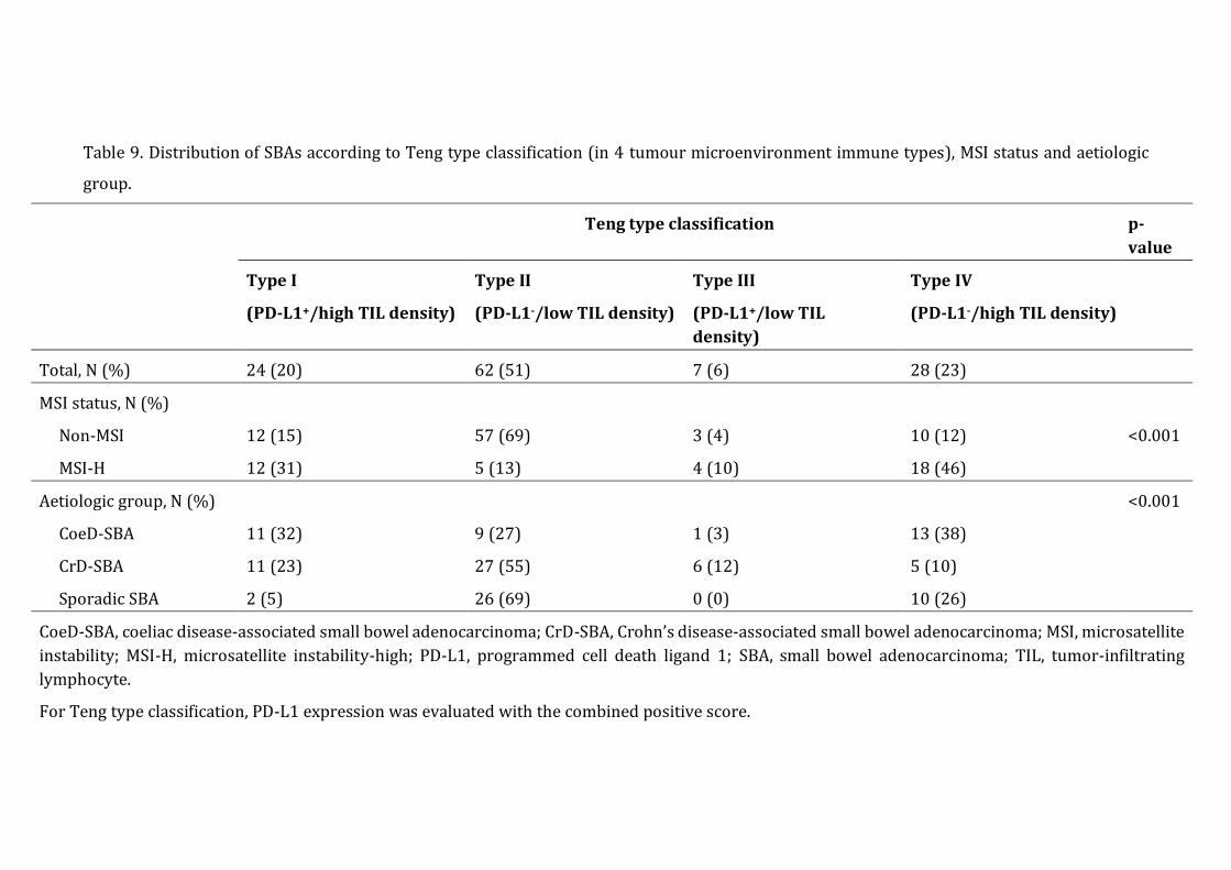
Table 9. Distribution of SBAs according to Teng type classification (in 4 tumour microenvironment immune types), MSI status and aetiologic
group.
Teng type classification p-
value
Type I
(PD-L1+/high TIL density)
Type II
(PD-L1-/low TIL density)
Type III
(PD-L1+/low TIL
density)
Type IV
(PD-L1-/high TIL density)
Total, N (%) 24 (20) 62 (51) 7 (6) 28 (23)
MSI status, N (%)
Non-MSI
MSI-H
12 (15)
12 (31)
57 (69)
5 (13)
3 (4)
4 (10)
10 (12)
18 (46)
<0.001
Aetiologic group, N (%)
CoeD-SBA
CrD-SBA
Sporadic SBA
11 (32)
11 (23)
2 (5)
9 (27)
27 (55)
26 (69)
1 (3)
6 (12)
0 (0)
13 (38)
5 (10)
10 (26)
<0.001
CoeD-SBA, coeliac disease-associated small bowel adenocarcinoma; CrD-SBA, Crohn’s disease-associated small bowel adenocarcinoma; MSI, microsatellite
instability; MSI-H, microsatellite instability-high; PD-L1, programmed cell death ligand 1; SBA, small bowel adenocarcinoma; TIL, tumor-infiltrating
lymphocyte.
For Teng type classification, PD-L1 expression was evaluated with the combined positive score.

EBV+ SBAs
Amongst the 118 tumours investigated for EBER, namely 34 CoeD-SBAs, 49 CrD-SBA, and
35 sporadic SBA, only two were EBV+, both CrD-SBAs, one showing a lymphoepithelioma-
like and the other a glandular histology, as described in a previous study [26]. The
lymphoepithelioma-like carcinoma was strongly PD-L1+ with a CPS≥50, while the
glandular CrD-SBA was negative for PD-L1.
Survival analysis
Three patients died peri-operatively, while the remaining 118 patients were followed-up
for a median of 68 months (25th-75th: 35-117) and their cancer-specific survival data are
reported in Table 10. Univariate survival analysis identified the following parameters as
significant related to a better post-operative cancer-specific survival: etiologic group -in
particular CoeD-SBA-, female sex, tumour site - jejunum being better-, pathological stage
I and II, MSI-H, high CD3+ TIL density, medullary and glandular histotype; positivity for
CDX2 expression, high intratumoural PD-1+ cell density, peritumoural PD-1+ cell density
and total PD-1+ cell density (Figure 4, Figure 5). PD-L1+ cases according to a CPS showed
better outcome in comparison with PD-L1- cases (p=0.046, Cox analysis and Figure 6A).
Teng type classification proved to be a significant prognostic parameter (p<0.001, Figure
6B); in particular, type I and IV tumours showed a significantly better cancer-specific
survival in comparison to type II. However, PD-L1 positivity lost its prognostic value when
combined with TIL density in Teng type classification. TPS, MIDS and L-FABP positivity
were not found to be significant predictors of cancer-specific survival (Figure 7).
Bivariable survival analysis inclusive of stage confirmed the significant prognostic value
of the following parameters: aetiologic group (p=0.017), sex (p=0.007), histotype
(p=0.01), MSI status (p=0.002), CD3+ TIL density (p=0.001), CPS (p=0.022) and Teng type
classification (p=0.015). At bivariable analysis inclusive of aetiologic group, the following
factors retained prognostic significance: stage (p<0.001), sex (p=0.008), histotype
(p<0.001), MSI status (p=0.016), CD3+ TIL density (p=0.002), and Teng type classification
(p=0.018). PD1+ immune cell density lost its prognostic value in a bivariable model
adjusted for CD3+ TIL density or MSI status.

45
Table 10. Cancer-specific survival of small bowel adenocarcinomas followed-up
Parameter N. of cases
N. of deaths (%)
Rate per 100 person-year (95% CI)
HR (95% CI) p-value (Cox)
Aetiologic group
CoeD-SBA
CrD-SBA
Sporadic SBA
33
47
38
5 (15)
23 (49)
23 (61)
2.66 (1.10-6.40)
13.14 (8.73-19.78)
14.49 (9.62-21,80)
1
4.37 (1.65-11.53)
4.73 (1.79-12.48)
p<0.001
Sex
Female
Male
44
74
13 (29)
38 (51)
4.32 (2.51-7.44)
17.40 (12.66-23.92)
1
3.00 (1.55-5.57)
p<0.001
Tumor site
Duodenum
Jejunum
Ileum
11
49
58
4 (36)
17 (35)
30 (52)
8.02 (3.01-21.36)
6.25 (3.89-10.06)
15.02 (10.51-21.49)
1
0.83 (0.28-2.48)
1.86 (0.66-5.29)
p=0.023
Stage
I
II III
IV
10
55
40
11
0 (0)
15 (27)
24 (60)
11 (100)
0
4.93 (2.97-8.18)
23.39 (15.68-34.89)
76.98 (42.63-138.99)
Not evaluable
1
4.66 (2.39-9.11)
14.61 (6.24-34.24)
p<0.001
MSI status
Non-MSI
MSI-H
82
36
44 (54)
7 (19)
14.55 (10.83-19.56)
3.19 (1.52-6.70)
1
0.24 (0.11-0.54)
p<0.001
CD3+ TIL density
≤15/HPF
>15/HPF
67
51
41 (61)
10 (20)
15.85 (11.67-21.52)
3.81 (2.04-7.07)
1
0.25 (0.13-0.51)
p<0.001
Histotype
Glandular
Medullary
Mixed
Diffuse
Solid
62
9
26
14
7
18 (29)
1 (11)
19 (73)
10 (71)
3 (43)
5.5 (3.47-8.74)
2.01 (0.28-14.28)
27.56 (17.58-43.20)
25.14 (13.52-46.72)
8.34 (2.69-25.86)
1
0.34 (0.05-2.53)
4.22 (2.23-10.34)
3.57 (1.19-11.1)
1.48 (0.27-3.63)
p<0.001
CDX2 expression
Negative
Positive
36
80
24 (67)
27 (34)
13.27 (8.89-19.81)
8.00 (5.49-11.67)
1
0.51 (0.29-0.89)
p=0.018
L-FABP expression

46
Negative
Positive
83
34
36 (43)
14 (41)
9.66 (6.97-13.39)
9.43 (5.59-15.92)
1
0.90 (0.49-1.67)
p=0.7391
PD-L1 expression
CPS<1
CPS≥1
89
29
46 (52)
5 (17)
10.86 (8.13-14.49)
5.11 (2.13-12.29)
1
0.43 (0.17-1.09)
p=0.046
PD-L1 expression
TPS<1
TPS≥1
109
9
49 (44)
2 (22)
10.25 (7.75-13.57)
4.59 (1.15-18.39)
1
0.43 (0.1-1.77)
p=0.182
PD-L1 expression
MIDS 0-1
MIDS 2-3
93
25
47 (50)
4 (16)
10.27 (7.71-13.67)
6.28 (2.36-16.74)
1
0.51 (0.18-1.43)
p=0.16
PD-1+ IICs*
<1.6/HPF
≥1.6/HPF
59
56
34 (58)
15 (27)
13.08 (9.35-18.31)
6.09 (3.67-10.11)
1
0.43 (0.23-0.78)
p=0.004
PD-1+ PICs*
<4.8/HPF
≥4.8/HPF
59
56
32 (54)
17 (30)
11.57 (8.18-16.37)
7.41 (4.60-11.91)
1
0.56 (0.31-1.01)
p=0.05
PD-1+ TICs*
<6.8/HPF
≥6.8/HPF
59
56
32 (54)
17 (30)
11.94 (8.44-16.89)
7.14 (4.43-11.49)
1
0.54 (0.30-0.97)
p=0.034
Teng type classification
Type I
Type II
Type III
Type IV
23
61
6
28
3 (13)
39 (64)
2 (33)
7 (25)
3.44 (1.11-10.65)
15.71 (11.48-21.5)
19.08 (4.77-76.33)
3.99 (1.9-8.37)
1
4.64 (1.43-15.03)
4.1 (0.68-24.7)
1.27 (0.33-4.91)
p<0.001
CoeD-SBA, coeliac disease-associated small bowel adenocarcinoma; CPS, combined positive score; CrD-SBA, Crohn’s disease-associated small bowel adenocarcinoma; HPF, high-power field; IIC, intratumoural immune cell; L-FABP, liver fatty acid-binding protein; MIDS, mononuclear immune cell density score; MSI, microsatellite instability; MSI-H, microsatellite instability-high; PD-1, programmed cell death protein-1; PD-L1, programmed cell death ligand 1; PIC, peritumoural immune cell; TIC, total immune cell; TIL, tumour-infiltrating lymphocyte.
*The cut-off of 1.6/HPF, 4.8/HPF and 6.8/HPF were the respective medians of PD-1+ IICs, PICs and TICs in the whole cohort.

47
Figure 4 – Kaplan-Meier cancer-specific survival estimates for small bowel adenocarcinoma (SBA) patients by aetiologic group (A), stage (B), tumour-infiltrating lymphocyte (TIL) density (C), microsatellite instability (MSI) status (D), histotype (E)
and CDX2 expression (F). CoeD-SBA, coeliac disease-associated small bowel adenocarcinoma; CrD-SBA, Crohn’s disease-associated small bowel adenocarcinoma.

48
Figure 5 - Kaplan-Meier cancer-specific survival estimates for small bowel adenocarcinoma patients by intratumoural (A), peritumoural (B) and total (C) PD-1-
positive immune cell counts.

49
Figure 6 - Kaplan-Meier cancer-specific survival estimates for small bowel
adenocarcinoma patients by PD-L1 expression according to combined positive score (CPS)
(A) and Teng type classification (B).

50
Figure 7 - Kaplan-Meier cancer-specific survival estimates for small bowel
adenocarcinoma patients by PD-L1 expression according to tumor proportion score (TPS)
(A) and mononuclear immune cell density score (MIDS) (B), and by liver fatty acid-binding
protein (L-FABP) expression (C).

51
Tb and PDCs
Two CrD-SBA cases resembling in part medullary cancers, one of which EBV+ and
reinterpreted as lymphoepithelioma-like cancer [25], were omitted from this study, as
recommended by Lugli and colleagues [111], owing to technical difficulties in assessing
Tb and PDC status. The histologic classification of the remaining 47 CrD-SBAs and related
clinico-pathologic features are reported in Table 11. A general predominance of male sex
and a median age of 57 years at SBA diagnosis are worth of note. Fifteen cases showed
high TIL density, while only eight were MMR-d, including seven cases with loss of
MLH1/PMS2 and one case showing isolated loss of MSH6. p53 overexpression was found
in 53.2% of CrD-SBCs without significant difference among histotypes. KRAS, NRAS and
PIK3CA mutations were observed in three (12.5%), one (4%) and two (8%) out of 24 cases
tested. Glandular-type cancers were predominantly well-to-moderately differentiated
according to WHO grading system.
The histologic analysis of tumour invasion front parameters (Table 12 and Figure 8)
showed a significant (p<0.001) association between histotypes and either Tb or PDC
grades with a predominance of Tb1 (50%) and PDC1 (52%) among glandular-type cases,
as well as of Tb3 (100%) and PDC3 (62%) among diffuse/mixed cancers. As for both Tb
and PDC survival analysis (see below) gave poor separation (not significant p-values) of
the relatively few grade 2 cases from the remaining grades, a two-tiered combined
invasive front (CIF) grade was developed where grade 3 cases for either Tb or PDC or both
defined CIF-high grade while all the remaining cases formed CIF-low grade. All diffuse and
mixed tumors were placed in the CIF-high grade group, in contrast to a minority (10/24,
42%) of glandular cases. In Table 13 data on depth of tumor invasion (pT) and AJCC stage
as a function of Tb, PDC and CIF grade are reported. An overall correlation was found
between each of the three invasive front grading systems and invasion/stage parameters.
Importantly, Tb, PDC or CIF grade were significantly (p=0.001, p=0.023 and p<0.001,
respectively) associated with lymph node metastases, with increasing rate of lymph node
metastases across grades). On the other hand, no association between invasive front
markers and KRAS, NRAS, PIK3CA mutations, p53 overexpression or MMR-d/MSI status
was observed.
Two patients died peri-operatively while the remaining 45 patients were followed for a
median of 85 months (25th-75th: 31-121) and their cancer-specific survival data are
reported in Table 14. As expected, stage and histotype were highly coupled with survival
(Figure 9), while high TILs, found in 27% of cases, and WHO grade, with only very few

52
grade 1 cases, were less contributive. Both Tb and PDC invasive front analysis gave
effective patient prognostication; in particular, their combination into a CIF grade
separated 14 low-grade from 31 high-grade SBC patients with highly divergent outcomes
(Figure 10 and Table 14). In addition, when CIF grade was applied to the 22 glandular
histology cases (by themselves showing significantly better survival than 23 non-
glandular cases), 13 CIF low- from 9 CIF high-grade tumors were identified, with a trend
for divergent outcomes (HR=6.54, 95% CI:0.73-58.6, p=0.054), despite the limited
number of available cases. CIF grade also gave significant results when applied to 24 stage
I+II tumors, thus separating 13 low- (12 of which present in the CIF-low grade glandular
group) from 11 high-grade cases, with significantly different outcomes (HR=7.78, 95% CI:
0.90-67.3, p=0.027). Indeed, no cancer-related death was observed, during a median
follow-up of 73.5 months, among the 12 patients with CrD-SBA showing CIF-low grade,
glandular structure and stage I or II. Of note, six (50%) of such tumours also showed high
TILs and three were MMR-d.

Table 11. Histologic classification and clinico-pathologic features of the 47 Crohn’s disease-associated SBA cases
Histotype n (%) Male sex,
Age at SBA diagnosis,
P53 overexpression (>50%)
MSI/MMR-d High TILs WHO grade, n (%)
n (%) median (25th-75th) n (%) n (%) n (%) 1 2 3
Glandular 24 (51.1) 18 (75) 59 (54.5-69) 14 (58.3) 6 (25) 9 (37.5) 6 (25) 17 (70.8) 1 (4.2)
Mixed 11 (23.4) 9 (81.8) 56 (46-68) 6 (54.5) 2 (18.2) 2 (18.2) 0 4 (36.4) 7 (63.6)
Diffuse 10 (21.3) 7 (70) 51 (39-59) 3 (30) 0 4 (40) 0 0 10 (100)
Solid 2 (4.2) 0 53 (44-62) 2 (100) 0 0 0 0 2 (100)
Total 47 (100) 34 (72) 57 (50-68) 25 (53.2) 8 (17) 15 (31.9) 6 (12.8) 21 (44.7) 20 (42.5)
MMR-d, mismatch repair deficient; MSI, microsatellite instability; TIL, tumour-infiltrating lymphocyte. WHO grade distribution among histotypes: p<0.001. All MMR-d SBAs also showed MSI by molecular analysis.
Table 12. Classification of 47 Crohn’s disease-associated SBA cases by invasive front-based grading systems.
Histotype Tumor budding (Tb), n (%) Poorly differentiated clusters (PDC), n (%) Combined invasive front grade (CIF), n (%)
Tb1 Tb2 Tb3 PDC1 PDC2 PDC3 Low High
Glandular 12 (50) 4 (16.7) 8 (33.3) 13 (54.2) 6 (25) 5 (20.8) 14 (58.3) 10 (41.7)
Mixed 0 0 11 (100) 2 (18.2) 1 (9.1) 8 (72.7) 0 11 (100)
Diffuse 0 0 10 (100) 1 (10) 4 (40) 5 (50) 0 10 (100)
Solid 0 1 (50) 1 (50) 0 1 (50) 1 (50) 1 (50) 1 (50)
Total 12 (25.5) 5 (10.6) 30 (63.8) 16 (34) 12 (25.5) 19 (40.4) 15 (31.9) 32 (68.1)
Tb and CIF grade distribution among histotypes: p<0.001. PDC distribution among histotypes: p=0.016.

Figure 8 - A) A glandular Crohn’s disease-associated small bowel adenocarcinoma (CrD-
SBA) showing grade 1 tumor budding (Tb1) and grade 1 poorly differentiated clusters
(PDC1) at the tumour invasive front (on the right) (haematoxylin and eosin; original
magnification, 200x). B) A mixed-type CrD-SBC with grade 3 PDCs (arrows; haematoxylin
and eosin; original magnification, 200x). C, D) A glandular CrD-SBC showing grade 3 Tb
(arrows; C, haematoxylin and eosin; D, pan-cytokeratin immunostaining; original
magnification, 200x).

Table 13. Distribution of Tb, PDC and CIF scores among the 47 Crohn’s disease-associated SBA cases classified according to pT and AJCC stage
Grading system N. of cases,
(%)
pT, n (%) p-value Stage, n (%) p-value
pT1 pT2 pT3 pT4
I II III IV
Tb Tb1 12 (25.5) 2 (16.7) 4 (33) 5 (41.7) 1 (8.3) 0.001 6 (50) 5 (41.7) 1 (8.33) 0 <0.001
Tb2 5 (10.7) 0 0 2 (40) 4 (60)
0 3 (60) 2 (40) 0
Tb3 30 (63.8) 0 0 16 (53.3) 14 (46.7) 0 11 (36.7) 13 (43.3) 6 (20)
PDC PDC1 16 (34.1) 2 (12.5) 4 (25) 7 (43.7) 3 (18.8)
0.016
6 (37.5) 6 (37.5) 4 (25) 0
0.002 PDC2 12 (25.5) 0 0 8 (66.7) 4 (33.3) 0 8 (66.7) 2 (16.7) 2 (16.7)
PDC3 19 (40.4) 0 0 8 (42.1) 11 (57.9) 0 5 (26.3) 10 (52.6) 4 (21.1)
CIF grade CIF low grade 15 (31.9) 2 (13.3) 4 (26.7) 6 (40) 3 (20) 0.002
6 (40) 8 (53.3) 1 (6.7) 0 <0.001
CIF high grade 32 (68.1) 0 0 17 (53.1) 15 (46.9) 0 11 (34.4) 15 (46.9) 6 (18.7)
Tb, tumor budding; PDC, poorly differentiated clusters; CIF, combined invasive front; pT: extent of the tumor into the layers of the wall of the
small intestine (according to the 8th ed. AJCC TNM staging system)

56
Table 14. Cancer-specific survival of 45 Crohn’s disease-associated SBAs classified according to their invasion front pattern and other
predictive parameters.
Parameter N. of cases N. of deaths
(%) Rate per 100 person-year (95% CI) HR (95% CI)
p-value (Cox)
Harrell's c (95% CI)
Tumor budding
Tb1 11 1 (9.1) 1.33 (0.19-9.44) 1
<0.001 0.68 (0.59-0.77) Tb2 5 2 (40) 20.25 (5.06-80.97) 10.6 (0.95-118.7)
Tb3 29 20 (68.9) 25.18 (16.24-39.03) 14.72 (1.95-111.18)
PDC
PDC1 15 3 (20) 3.69 (1.19-11.43) 1
0.004 0.69 (0.58-0.80) PDC2 12 8 (66.7) 23.84 (11.92-47.67) 4.88 (1.29-18.46)
PDC3 18 12 (66.7) 24.23 (13.76-42.67) 5.94 (1.67-21.15)
CIF grade low 14 2 (14.3) 2.45 (0.61-9.78) 1
<0.001 0.66 (0.57-0.75) high 31 21 (67.7) 25.38 (16.55-38.92) 8.27 (1.91-35.90)
WHO
G1 5 0 0 not evaluable (-∞)
0.001 0.67 (0.56-0.78) G2 20 8 (40) 10.14 (5.07-20.27) 1
G3 20 15 (75) 29.97 (18.07-49.71) 2.08 (0.88-4.91)
Histotype glandular 22 5 (22.7) 4.59 (1.91-11.03) 1
<0.001 0.68 (0.58-0.78) non-glandular 23 18 (78.3) 32.38 (20.4-51.39) 5.02 (1.85-13.63)
TILs high 15 4 (26.7) 5.62 (2.11-14.98) 1
0.027 0.60 (0.50-0.70) low 30 19 (63.3) 20.36 (12.99-31.92) 3.01 (1.02-8.92)
Stage
I 5 0 0 not evaluable (-∞)
<0.001 0.80 (0.72-0.87) II 19 6 (31.6) 5.81 (2.61-12.94) 1
III 15 11 (73.3) 50.24 (27.82-90.72) 8.29 (2.51-27.31)
IV 6 6 (100) 81.39 (36.57-181.18) 13.65 (3.67-50.83)
Tb, tumor budding; PDC, poorly differentiated clusters; CIF, combined invasive front; TILs, tumour-infiltrating lymphocytes; HR, hazard ratio; CI: confidence interval.

57
Figure 9 - Kaplan-Meier survival estimates on the 45 CrD-SBCs by stage (A) and histotype (B).

Figure 10 - Kaplan-Meier survival estimates on the 45 CrD-SBCs by tumour budding (A),
poorly differentiated clusters (B) and combined invasive front grade (C).

59
Discussion
We herein systematically investigated the tumour immune microenvironment in a large
cohort of SBAs associated with CoeD or CrD. In keeping with previous findings [49,101],
we found that PD-L1 was less frequently expressed in tumour cells than in immune cells
in SBAs. Moreover, in agreement with Thota and colleagues [49], we observed that PD-
L1+ SBAs have a higher number of intratumoural, peritumoural, and total PD-1+ immune
cells. We demonstrated that PD-L1 expression in non-hereditary SBAs as a whole was
25.6%, 8.3% and 22.3% according to CPS, TPS and MIDS, respectively. This is the first
investigation to identify a higher proportion of PD-L1+ cases in both CoeD-SBAs and CrD-
SBAs in comparison with sporadic SBAs, regardless of the score used, i.e. CPS, TPS or
MIDS. Additionally, we showed a significant increase in PD-1+ immune cells in CoeD-SBAs
in comparison with CrD-SBAs and sporadic SBAs. Indeed, a recent transcriptomic
profiling study demonstrated a predominant MSI-immune subtype of CoeD-SBA
characterized by high PD-1 activation [124]. Therefore, response to anti-PD-L1 or anti-
PD-1 monoclonal antibodies might be more often expected in CoeD-SBAs and –likely to a
lesser extent- in CrD-SBAs compared to sporadic cases. A number of clinical trials with
immune checkpoint inhibitors for the treatment of SBA patients are underway. In
particular, the ZEBRA trial assessed the activity of pembrolizumab in 40 patients with
previously treated advanced SBA [125]. Preliminary results of the ZEBRA trial found that
pembrolizumab did not significantly improve the overall response rate, although efficacy
was shown in MSI-H patients and, of note, in some microsatellite stable patients too [125].
Data related to the tumour immune microenvironment are still unavailable from this
study [125]. Further trials are recommended in these settings of patients, including those
with CoeD-SBA and CrD-SBA.
The higher expression of PD-L1 shown in CoeD-SBAs and CrD-SBAs in comparison with
sporadic SBAs and the increase in PD-1+ immune cells found in CoeD-SBAs might be
secondary to the underlying immune responses. In patients with untreated CoeD without
SBA, PD-L1 is up-regulated in enterocytes and lamina propria mononuclear cells, whereas
PD-1 is not present in the small intestine [126]. In CrD patients without SBA, PD-L1 is
reduced in CD4+ T cells because of high expression of the T helper 1 transcription factor
T-bet via transfection [127], while another study [128] demonstrated overexpression of
PD-L1 in macrophages and PD-1 in T cells, predominantly CD4+, and B cells from the

60
lamina propria. It must be outlined that the expression of PD-L1 and PD-1 we scored in
our cohort was strictly related topographically, within tumour tissue or immediately
surrounding it, to SBA, not to the underlying inflammatory CoeD or CrD background.
Whether the use of PD-L1/PD-1 pathway blockade treatments in CoeD and/or CrD
patients induces changes of the underlying immune-mediated intestinal disorder, which
might also affect SBA, requires more-in-depth study [129].
As the pro-inflammatory cytokine interferon-γ, known to be involved in the pathogenesis
underlying both CoeD and CrD [4,9], decreased the expression in vitro of L-FABP from
colorectal cancer cell lines [122], we investigated L-FABP in our cohort. In agreement with
colorectal cancer [122], we found that L-FABP negativity is more common in MSI-H SBAs
than in microsatellite stable cases. Additionally, we observed that PD-L1+ SBAs correlated
with L-FABP negativity. As in colorectal carcinomas the absence of expression of the
intestinal marker CDX2 is coupled with PD-L1 expression [106], we also correlated PD-L1
with CDX2 expression. Nevertheless, we did not observe a significant correlation between
PD-L1 expression with CDX2 negativity.
We have also confirmed the previously demonstrated association between expression of
PD-L1 and MSI-H in SBAs [49,101,102], in agreement with gastric and colorectal
carcinomas [47,48]. At first glance, these findings might lead to consider MSI-H as a
predictor of clinical response to anti-PD-L1 or anti-PD-1 monoclonal antibodies in
patients with SBA and in those with colorectal cancer [96,130] Nevertheless, in keeping
with endometrial carcinoma [104], we described 15 PD-L1+ microsatellite stable cases, 11
CrD-SBAs, three CoeD-SBAs, and one sporadic SBA, thus suggesting that MSI-H on its own
is not enough to identify all PD-L1+ SBAs. These results support the need to find additional
predictors of response to anti-PD-L1 or anti-PD-1 monoclonal antibodies. TILs have been
considered as a marker of host immune response against tumour [131], and their density
has been described as an independent prognostic factor in non-hereditary SBAs [2,102].
High TIL density, which is typical in both Teng types I and IV, plays a role in adaptive
resistance in tumours through the secretion of pro-inflammatory cytokines, which cause
PD-L1 expression and ultimately induce immunosuppression in the tumour
microenvironment [132]. We here showed that the majority of PD-L1+ cases have a high
CD3+ and CD8+ TIL density. However, we described seven out of 31 PD-L1+ SBAs with low

61
TIL density, namely Teng type III. We think that tumour-specific molecular alterations
might promote PD-L1 positivity in SBAs with low TIL density.
PD-L1 expression is frequently associated with EBV positivity in gastric cancer and two
EBV+ CrD-SBAs have been demonstrated [26,48]. We here evaluated latent phase EBV
infection in our entire SBA cohort and confirmed EBV positivity of the two previously
described SBAs, while no EBER was displayed in the other 116 cases tested. Within the
two EBV+ CrD-SBAs, only the lymphoepithelioma-like carcinoma was PD-L1+ whereas the
remaining case, the glandular one, was PD-L1-negative. This is the first study reporting
no latent infection of EBV in CoeD-SBA. In agreement with Von Rahden and colleagues
[27]. we confirmed EBV negativity in all sporadic SBAs. With regard to histotypes, PD-L1+
SBAs was associated with medullary histotype, which does have a favourable outcome not
only in SBAs but also in gastric and colorectal carcinomas [46,133,134].
In our entire SBA cohort, PD-L1 expression according to CPS showed a trend towards a
better prognosis. This is not surprising, as PD-L1 expression was higher in SBAs with
increased CD3+ and CD8+ TIL density, and in SBAs with medullary histotype, both features
related to a more favourable prognosis [2,46]. Moreover, our results confirmed aetiologic
group, histotype, TIL density, Teng tumour microenvironment immune types and stage
as prognostic parameters in SBAs [2,7,101,102,135]. Although univariate analysis
reported a more favourable prognosis in PD-L1+ SBAs according to a CPS than that in PD-
L1- cases, PD-L1 expression lost its prognostic power once adjusted for TIL density.
As regards the second objective of this thesis, we confirmed the favourable prognostic
impact of glandular histotype compared to its loss to form diffuse or mixed cancerous
growths in CrD-SBA, as already suggested in a previous study recruiting a smaller cohort
[7]. Along with this architectural assessment on the whole cancer tissue, we
demonstrated that selective evaluation of the tumour invasive front for foci of Tb and/or
PDCs substantially improved separation of more from less aggressive CrD-SBAs.
Furthermore, a significant association of Tb and PDC with lymph node metastases was
observed. In particular, invasive front analysis was effective, in glandular type CrD-SBAs,
in finding cases with many foci of cell dissociation or structural dedifferentiation -high-
grade Tb or PDCs-, which were significantly associated with survival shortening

62
compared to those with low CIF grade, namely low-to-intermediate grade Tb and PDCs.
The use of a novel CIF two tier grading system, which includes both Tb and PDC, makes
this system easier to apply in comparison with the separate assessment of Tb and PDC.
This is the first study assessing Tb and PDCs in non-ampullary SBAs in a cohort of 47 CrD
patients. Tb was also studied in ampullary adenocarcinomas, in which high-grade Tb was
found an independent predictor of overall survival [112]. Recently, Jun SY and colleagues
[136] confirmed our findings, showing that high-grade Tb or PDCs are associated with an
aggressive behaviour and, thus, with a worse patient outcome in SBA. Therefore, the main
novelty of this Korean study was to identify the invasive front markers Tb and PDCs as
prognostic indicators in sporadic SBAs [136], whereas we obtained the same results in
CrD-SBAs. Finally, we do acknowledge that Jun SY and colleagues [136] are the first to
analyse Tb and PDCs in the intratumoural area of SBAs.
Our findings extend to CrD-SBA the prognostic significance of Tb and PDC assessment, so
far mainly described for colorectal carcinoma, mostly gland-forming, usual-type
adenocarcinoma. Moreover, we demonstrated that the usefulness of CIF grade is limited
to the CrD-SBA glandular histotype, which is in agreement with recent findings on gastric
cancer [137]. It seems that both processes implicated in loss of structural differentiation,
one occurring massively within the whole neoplasm, leading to the diffuse and mixed
tumour histotypes, and the other one selectively acting at its invasive front -the high-
grade Tb or PDCs-, are strongly coupled with a worse patient outcome. Briefly,
persistence, even at the invasive front, of the “canonical” glandular-type structure defines
a relatively less aggressive subgroup of CrD-SBAs, predominantly non-metastatic and
with a restricted invasiveness.
Of note, loss of cellular/glandular differentiation to form “poorly cohesive” tumors has
long been recognised within gastric cancers and suggested to worsen patient prognosis
[138-140]. Molecular studies have highlighted that diffuse desmoplastic cancers of the
stomach, pancreas and colon likely represent the histologic consequence of the epithelial-
to-mesenchymal transition present in such neoplasms, with a severe prognostic effect
[141-146]. It appears evident, particularly from recent studies on colorectal carcinoma,
that cancer evaluation at its invasive front may identify an otherwise undetectable
tumour proneness to cell dissociation and invasion, thus predicting a worse post-

63
operative behaviour [108-111,147]. From our results it seems that the same phenomenon
happens in CrD-SBAs, which may add more information to stage histologic assessment of
surgical samples.
High TIL density was coupled with favourable survival in CrD-SBAs too; notwithstanding,
TIL density less effective in CrD-SBAs than in CoeD-SBAs [2], being present in a lower
number of cases. TIL-rich medullary-type CrD-SBAs, including the EBV+
lymphoepithelioma-like carcinoma with medullary-like histology [25], are known by
themselves to generally have a better outcome [46,133,134], thus overcoming the
technical difficulty for Tb and PDC evaluation reported in this rare cancer type.

64
Conclusions and future perspectives
As regards the primary objective of this thesis, PD-L1 expression in non-hereditary SBAs
is coupled with its aetiologic group, being more frequent in CoeD-SBAs and CrD-SBAs
compared to sporadic SBAs, as well as with its MSI status and TIL density. The
identification of a subset of PD-L1+ non-MSI cases in our cohort suggests that PD-L1
expression, together with MSI status, TIL density, and tumour mutation burden, should
be recognised as potential biomarkers of response to immune checkpoint inhibitors in
well-designed clinical trials focused on SBA, including CoeD-SBA and CrD-SBA.
With regard to the secondary objective of this thesis, analysis of tumour cell
dissociation/de-differentiation markers at the invasive front, such as Tb and PDCs, may
improve the identification of highly malignant CrD-SBAs. Indeed, as survival rate is
similarly low in patients with CrD-SBA and those with sporadic SBA (Table 5), it is
clinically relevant to find prognostic markers for this neoplasm, in particular in these two
aetiologic groups. Briefly, these findings should encourage pathologists to describe Tb and
PDCs at SBA diagnosis in both CrD patients and sporadic cases in order to separate highly
malignant cancers from less aggressive SBAs. Indeed, Tb and PDCs may represent pivotal
histological features together with mismatch repair status, histotype and pT [83] to
identify stage II SBAs needing adjuvant chemotherapy or further surgery, as well as they
have been demonstrated in colon adenocarcinoma [148].
The main limitation of this study is its retrospective nature. Notwithstanding, as SBA is a
rare neoplasm, this was a necessary choice in order to recruit a relatively large cohort of
SBAs. Additionally, the involvement of tertiary Italian Coeliac and/or IBD Centers with
long-term referral experience in CoeD and in inflammatory bowel disease, which were
following internationally agreed guidelines, was a guarantee of data quality.
Either the association of PD-L1 expression with MSI status in non-hereditary SBAs or the
finding of Tb and PDCs as able to separate lower from higher CrD-SBAs confirmed some
similarities between SBAs and colorectal carcinomas. Although clinico-radiologic and
endoscopic distinctions between SBA and colorectal carcinoma are usually
straightforward, it should be recalled that colorectal carcinoma may infiltrate or
metastasise to the small bowel, mimicking a primary SBA, or be predominantly located
around the ileocaecal valve, simulating a primary neoplasm of the terminal ileum,
especially in CrD patients. In these challenging situations, as well as in the setting of

65
metastasis of unknown primary origin, immunohistochemistry may play a certain role in
suggesting the site of origin. The typical immunophenotypic profile of lower
gastrointestinal tract (colorectal or appendiceal) carcinomas, is CK7-/CK20+/CDX2+,
whereas about 60% of SBAs have been reported to co-express CK7 and CK20 and around
a half of SBAs is negative for CDX2 [149-151]. Nevertheless, on one hand, CK7 is also
expressed in about 10% of colorectal carcinomas [152] and CK20 and CDX2 markers can
be lost in some colorectal carcinoma, especially in those harbouring MMR-d [153], while,
on the other hand, a fraction of SBAs (34%) has been reported to show a colorectal cancer-
like immunohistochemical profile (CK7-/CK20+/CDX2+) [154]. Thus, it is pivotal to find
markers to discriminate SBAs from colorectal cancers in the aforementioned challenging
contexts. The next step of this thesis is to investigate the immunohistochemical
expression of special AT-rich sequence-binding protein 2 and other gastrointestinal
phenotypic markers (CK7, CK20, CDX2, AMACR), in a large and etiologically well-
characterized series of SBAs and to correlate the observed immunophenotypic profiles
with clinico-pathologic and prognostic features, as well as with MSI status.

66
References
1. Raghav K, Overman MJ. Small bowel adenocarcinomas—Existing evidence and
evolving paradigms. Nat Rev Clin Oncol 2013;10:534–544.
2. Vanoli A, Di Sabatino A, Furlan D, et al. Small Bowel Carcinomas in Coeliac or Crohn’s
Disease: Clinico-pathological, Molecular, and Prognostic Features. A Study from the
Small Bowel Cancer Italian Consortium. J Crohns Colitis 2017;11:942–953.
3. Di Sabatino A, Lenti MV, Giuffrida P, et al. New insights into immune mechanisms
underlying autoimmune diseases of the gastrointestinal tract. Autoimmun Rev
2015;14:1161–1169.
4. Di Sabatino A, Corazza GR. Coeliac disease. Lancet 2009;373:1480–1493.
5. Potter DD, Murray JA, Donohue JH, et al. The role of defective mismatch repair in small
bowel adenocarcinoma in celiac disease. Cancer Res 2004;64:7073–7077.
6. Diosdado B, Buffart TE, Watkins R, et al. High-resolution array comparative genomic
hybridization in sporadic and celiac disease-related small bowel adenocarcinomas.
Clin. Cancer Res. 2010, 16, 1391–1401.
7. Vanoli A, Di Sabatino A, Martino M, et al. Small bowel carcinomas in celiac or Crohn’s
disease: Distinctive histophenotypic, molecular and histogenetic patterns. Mod
Pathol 2017;30:1453–1466.
8. Svrcek M, Piton G, Cosnes J, et al. Small bowel adenocarcinomas complicating Crohn’s
disease are associated with dysplasia: A pathological and molecular study. Inflamm
Bowel Dis 2014;20:1584–1592.
9. Giuffrida P, Corazza GR, Di Sabatino A. Old and New Lymphocyte Players in
Inflammatory Bowel Disease. Dig Dis Sci 2018;63:277–288.
10. Grolleau C, Pote NM, Guedj NS, et al. Small bowel adenocarcinoma complicating
Crohn’s disease: A single-centre experience emphasizing the importance of screening
for dysplasia. Virchows Arch 2017;471:611–617.
11. Rashid A, Hamilton SR. Genetic alterations in sporadic and Crohn’s-associated
adenocarcinomas of the small intestine. Gastroenterology 1997;113:127–135.
12. Bojesen RD, Riis LB, Høgdall E, et al. Inflammatory Bowel Disease and Small Bowel
Cancer Risk, Clinical Characteristics, and Histopathology: A Population-Based Study.
Clin Gastroenterol Hepatol 2017;15:1900–1907.
13. Howdle PD, Jalal PK, Holmes GK, et al. Primary small-bowel malignancy in the UK and
its association with coeliac disease. QJM 2003;96:345–353.

67
14. Bruno CJ, Batts KP, Ahlquist DA. Evidence against flat dysplasia as a regional field
defect in small bowel adenocarcinoma associated with celiac sprue. Mayo Clin Proc
1997;72:320–322.
15. Michelassi F, Testa G, Pomidor WJ, et al. Adenocarcinoma complicating Crohn’s
disease. Dis. Colon Rectum 1993, 36, 654–661.
16. Sigel JE, Petras RE, Lashner BA, et al. Intestinal adenocarcinoma in Crohn’s disease: A
report of 30 cases with a focus on coexisting dysplasia. Am J Surg Pathol.
1999;23:651–655.
17. Palascak-Juif V, Bouvier AM, Cosnes, J, et al. Small bowel adenocarcinoma in patients
with Crohn’s disease compared with small bowel adenocarcinoma de novo. Inflamm
Bowel Dis 2005;11: 828–832.
18. Piton G, Cosnes J, Monnet E, et al. Risk factors associated with small bowel
adenocarcinoma in Crohn’s disease: A case-control study. Am J Gastroenterol
2008;103:1730–1736.
19. Widmar M, Greenstein AJ, Sachar DB, et al. Small bowel adenocarcinoma in Crohn’s
disease. J Gastrointest Surg 2011;15:797–802.
20. Whitcomb E, Liu X, Xiao SY. Crohn enteritis-associated small bowel adenocarcinomas
exhibit gastric differentiation. Hum Pathol 2014;45:359–367.
21. Weber NK, Fletcher JG, Fidler JL, et al. Clinical characteristics and imaging features of
small bowel adenocarcinomas in Crohn’s disease. Abdom Imaging 2015;40:1060–
1067.
22. Wieghard N, Mongoue-Tchokote S, Young JI, et al. Prognosis of small bowel
adenocarcinoma in Crohn’s disease compares favourably with de novo small bowel
adenocarcinoma. Colorectal Dis 2017;19:446–455.
23. Warth A, Kloor M, Schirmacher P, et al. Genetics and epigenetics of small bowel
adenocarcinoma: The interactions of CIN, MSI, and CIMP. Mod Pathol 2011;24:564–
570.
24. Overman MJ, Pozadzides J, Kopetz S, et al. Immunophenotype and molecular
characterisation of adenocarcinoma of the small intestine. Br J Cancer 2010;102:144–
150.
25. Vanoli A, Di Sabatino A, Biancone L, et al. Small bowel Epstein-Barr virus-positive
lympho-epithelioma-like carcinoma in Crohn’s disease. Histopathology
2017;70:837–839.

68
26. Vanoli A, Di Sabatino A, Martino M, et al. Epstein-Barr virus-positive ileal carcinomas
associated with Crohn’s disease. Virchows Arch 2017;471:549–552.
27. Von Rahden BH, Langner C, Brücher BL, et al. No association of primary
adenocarcinomas of the small bowel with Epstein-Barr virus infection. Mol Carcinog
2006;45:349–352.
28. DeSesso JM, Jacobson CF. Anatomical and physiological parameters affecting
gastrointestinal absorption in humans and rats. Food Chem Toxicol 2001;39:209–
228.
29. Aparicio T, Zaanan A, Svrcek M, et al. Small bowel adenocarcinoma: Epidemiology,
risk factors, diagnosis and treatment. Dig Liver Dis 2014;46:97–104.
30. Kummar S, Ciesielski TE, Fogarasi MC. Management of small bowel adenocarcinoma.
Oncology 2002;16:1364–1369.
31. Bilimoria KY, Bentrem DJ, Wayne JD, et al. Small bowel cancer in the United States:
Changes in epidemiology, treatment, and survival over the last 20 years. Ann Surg
2009;249:63–71.
32. Faivre J, Trama A, De Angelis R, et al. Incidence, prevalence and survival of patients
with rare epithelial digestive cancers diagnosed in Europe in 1995–2002. Eur J Cancer
2012;48:1417–1424.
33. Han Y, Chen W, Li P, et al. Association Between Coeliac Disease and Risk of Any
Malignancy and Gastrointestinal Malignancy: A Meta-Analysis. Medicine
2015;94:e1612.
34. Canavan C, Abrams KR, Mayberry J. Meta-analysis: Colorectal and small bowel cancer
risk in patients with Crohn’s disease. Aliment Pharmacol Ther 2006;23:1097–1104.
35. Silano M, Volta U, Vincenzi AD, et al. Effect of a gluten-free diet on the risk of
enteropathy-associated T-cell lymphoma in celiac disease. Dig Dis Sci 2008;53:972–
976.
36. Elfström P, Granath F, Ye W, et al. Low risk of gastrointestinal cancer among patients
with celiac disease, inflammation, or latent celiac disease. Clin Gastroenterol Hepatol
2012;10:30–36.
37. Mukherjee R, Kelly CP, Leffler DA. Gastrointestinal cancer in celiac disease: “The first
days are the hardest days, don’t you worry anymore?”. Clin Gastroenterol Hepatol
2012;10:4–6.
38. Silano M, Volta U, Mecchia AM, et al. Delayed diagnosis of coeliac disease increases
cancer risk. BMC Gastroenterol 2007;7:8.

69
39. Di Sabatino A, Biagi F, Gobbi PG, et al. How I treat enteropathy-associated T-cell
lymphoma. Blood 2012;119:2458–2468.
40. Cantoro L, Di Sabatino A, Papi C, et al. The Time Course of Diagnostic Delay in
Inflammatory Bowel Disease Over the Last Sixty Years: An Italian Multicentre Study.
J Crohns Colitis 2017;11:975–980.
41. Cahill C, Gordon PH, Petrucci A, et al. Small bowel adenocarcinoma and Crohn’s
disease: Any further ahead than 50 years ago? World J Gastroenterol 2014;20:11486–
11495.
42. Lashner BA. Risk factors for small bowel cancer in Crohn’s disease. Dig Dis Sci
1992;37:1179–1184.
43. Solem CA, Harmsen WS, Zinsmeister AR, et al. Small intestinal adenocarcinoma in
Crohn’s disease: A case-control study. Inflamm Bowel Dis 2004;10:32–35.
44. Schottenfeld D, Beebe-Dimmer JL, Vigneau, FD. The epidemiology and pathogenesis
of neoplasia in the small intestine. Ann Epidemiol 2009;19:58–69.
45. Goodman MT, Matsuno RK, Shvetsov YB. Racial and ethnic variation in the incidence
of small-bowel cancer subtypes in the United States, 1995–2008. Dis Colon Rectum
2013;56:441–448.
46. Brcic I, Cathomas G, Vanoli A, et al. Medullary carcinoma of the small bowel.
Histopathology 2016;69:136–140.
47. Rosenbaum MW, Bledsoe JR, Morales-Oyarvide V, et al. PD-L1 expression in colorectal
cancer is associated with microsatellite instability, BRAF mutation, medullary
morphology and cytotoxic tumor-infiltrating lymphocytes. Mod Pathol
2016;29:1104–1112.
48. De Rosa S, Sahnane N, Tibiletti MG, et al. EBV⁺ and MSI Gastric Cancers Harbor High
PD-L1/PD-1 Expression and High CD8⁺ Intratumoral Lymphocytes. Cancers
2018;10:102.
49. Thota R, Gonzalez RS, Berlin J, et al. Could the PD-1 Pathway Be a Potential Target for
Treating Small Intestinal Adenocarcinoma? Am J Clin Pathol 2017;148:208–214.
50. Schrock AB, Devoe CE, McWilliams R, et al. Genomic Profiling of Small-Bowel
Adenocarcinoma. JAMA Oncol 2017;3:1546–1553.
51. Aparicio T, Svrcek M, Zaanan A, et al. Small bowel adenocarcinoma phenotyping, a
clinicobiological prognostic study. Br J Cancer 2013;109:3057–3066.

70
52. Yaeger R, Shah MA, Miller VA, et al. Genomic Alterations Observed in Colitis-
Associated Cancers Are Distinct from Those Found in Sporadic Colorectal Cancers and
Vary by Type of Inflammatory Bowel Disease. Gastroenterology 2016;151:278–287.
53. Younes N, Fulton N, Tanaka R, et al. The presence of K-12 ras mutations in duodenal
adenocarcinomas and the absence of ras mutations in other small bowel
adenocarcinomas and carcinoid tumors. Cancer 1997;79:1804–1808.
54. Bläker H, Helmchen B, Bönisch A, et al. Mutational activation of the RAS-RAF-MAPK
and the Wnt pathway in small intestinal adenocarcinomas. Scand J Gastroenterol
2004;39:748–753.
55. Lee HJ, Lee OJ, Jang KT, et al. Combined loss of E-cadherin and aberrant β-catenin
protein expression correlates with a poor prognosis for small intestinal
adenocarcinomas. Am J Clin Pathol 2013;139:167–176.
56. Fang M, Ou J, Hutchinson L, et al. The BRAF oncoprotein functions through the
transcriptional repressor MAFG to mediate the CpG Island Methylator phenotype. Mol
Cell 2014;55:904–915.
57. Serra RW, Fang M, Park SM, et al. A KRAS-directed transcriptional silencing pathway
that mediates the CpG island methylator phenotype. Elife 2014;3:e02313.
58. Laforest A, Aparicio T, Zaanan A, et al. ERBB2 gene as a potential therapeutic target in
small bowel adenocarcinoma. Eur J Cancer 2014;50:1740–1746.
59. Rampertab SD, Forde KA, Green PH. Small bowel neoplasia in coeliac disease. Gut
2003;52:1211–1214.
60. Perzin KH, Bridge MF. Adenomas of the small intestine: A clinicopathologic review of
51 cases and a study of their relationship to carcinoma. Cancer 1981;48:799–819.
61. Genta RM, Feagins LA. Advanced precancerous lesions in the small bowel mucosa.
Best Pract Res Clin Gastroenterol 2013;27:225–233.
62. Senger S, Sapone A, Fiorentino MR, et al. Celiac Disease Histopathology Recapitulates
Hedgehog Downregulation, Consistent with wound Healing Processes Activation.
PLoS ONE 2015;10:e0144634.
63. Simon M, Cosnes J, Gornet JM, et al. Endoscopic Detection of Small Bowel Dysplasia
and Adenocarcinoma in Crohn’s Disease: A Prospective Cohort-Study in High-Risk
Patients. J Crohns Colitis 2017;11:47–52.
64. Ciccocioppo R, Racca F, Scudeller L, et al. Differential cellular localization of Epstein-
Barr virus and human cytomegalovirus in the colonic mucosa of patients with active
or quiescent inflammatory bowel disease. Immunol Res 2016;64:191–203.
71
65. Genitsch V, Novotny A, Seiler CA, et al. Epstein-barr virus in gastroesophageal
adenocarcinomas—single center experiences in the context of current literature.
Front Oncol 2015;5:73.
66. Tomba C, Sidhu R, Sanders DS, et al. Celiac Disease and Double-Balloon Enteroscopy:
What Can we Achieve? The Experience of 2 European Tertiary Referral Centers. J Clin
Gastroenterol 2016;50:313–317.
67. Rieder F, Latella G, Magro F, et al. European Crohn’s and Colitis Organisation Topical
Review on Prediction, Diagnosis and Management of Fibrostenosing Crohn’s Disease.
J Crohns Colitis 2016;10:873–885.
68. Bergamaschi G, Di Sabatino A, Corazza GR. Pathogenesis, diagnosis and treatment of
anaemia in immune-mediated gastrointestinal disorders. Br J Haematol
2018;182:319-329.
69. Baars JE, Thijs JC, Bac DJ, et al. Small bowel carcinoma mimicking a relapse of Crohn’s
disease: A case series. J Crohns Colitis 2011;5:152–156.
70. Collier PE, Turowski P, Diamond DL. Small intestinal adenocarcinoma complicating
regional enteritis. Cancer 1985;55:516–521.
71. Overman MJ, Hu CY, Kopetz S, et al. A population-based comparison of
adenocarcinoma of the large and small intestine: Insights into a rare disease. Ann Surg
Oncol 2012;19:1439–1445.
72. Halfdanarson TR, McWilliams RR, Donohue JH, et al. A single-institution experience
with 491 cases of small bowel adenocarcinoma. Am J Surg 2010;199:797–803.
73. Mahul B. Amin, Donna M. Gress. AJCC Cancer Staging Manual, Eight Edition. New York:
Springer, 2017.
74. Aydin D, Sendur MA, Kefeli U, et al. Evaluation of prognostic factors and adjuvant
chemotherapy in patients with small bowel adenocarcinoma who underwent curative
resection. Clin Colorectal Cancer 2017;16:220-227.
75. Huffman BM, Jin Z, Yadav S, et al. Novel Prognostic Factors in Resected Small Bowel
Adenocarcinoma. Clin Colorectal Cancer. 2019;18:218-225.
76. Overman MJ, Hu CY, Wolff RA, et al. Prognostic value of lymph node evaluation in
small bowel adenocarcinoma: Analysis of the surveillance, epidemiology, and end
results database. Cancer 2010;116:5374–5382.
77. Dabaja BS, Suki D, Pro B, et al. Adenocarcinoma of the small bowel: Presentation,
prognostic factors, and outcome of 217 patients. Cancer 2004;101:518–526.

72
78. Axelrad JE, Olén O, Sachs MC, et al. Inflammatory Bowel Disease and Risk of Small
Bowel Cancer: A Binational Population-Based Cohort Study From Denmark and
Sweden. Gut 2020 May 30. [Epub ahead of print]
79. Fields AC, Hu FY, Lu P, et al. Small Bowel Adenocarcinoma: Is There a Difference in
Survival for Crohn's Versus Sporadic Cases? J Crohns Colitis 2020;14:303-8.
80. Axelrad JE, Olén O, Sachs MC, et al. Reply: Survival in Crohn's disease-associated small
bowel adenocarcinoma. Gut 2020 Sep 1 [Epub ahead of print]
81. Locher C, Batumona B, Afchain P, et al. Small bowel adenocarcinoma: French
intergroup clinical practice guidelines for diagnosis, treatments and follow-up
(SNFGE, FFCD, GERCOR, UNICANCER, SFCD, SFED, SFRO). Dig Liver Dis 2018;50:15–
19.
82. González I, Goyal B, Xia MD, et al. DNA mismatch repair deficiency but not ARID1A
loss is associated with prognosis in small intestinal adenocarcinoma. Hum Pathol
2019;85:18-26.
83. Vanoli A, Grillo F, Guerini C, et al. Prognostic Role of Mismatch Repair Status,
Histotype and High-Risk Pathologic Features in Stage II Small Bowel
Adenocarcinomas. Ann Surg Oncol 2020 Aug 5 [Epub ahead of print]
84. Benson AB, Venook AP, Al-Hawary MM, et al. Small Bowel Adenocarcinoma, Version
1.2020, NCCN Clinical Practice Guidelines in Oncology. J Natl Compr Canc Netw
2019;17:1109–1133.
85. Ye X, Zhang G, Chen H, et al. Meta-analysis of postoperative adjuvant therapy for small
bowel adenocarcinoma. PloS One 2018;13:e0200204.
86. Akce M, Jiang R, Zakka K, et al. Clinical Outcomes of Small Bowel Adenocarcinoma. Clin
Colorectal Cancer 2019;18:257–268.
87. Evans J, Aparicio T, Le Malicot K, et al. GLOBAL BALLAD: an International Rare
Cancers Initiative trial to evaluate the potential benefit of adjuvant chemotherapy for
small bowel adenocarcinoma (IRCI 002). J Clin Oncol 2016;34:15_suppl. TPS4154.
88. U.S. National Library of Medicine. Phase III Trial Investigating the Potential Benefit of
Adjvant Chemotherapy for Small Bowel Adenocarcinoma (BALLAD).
ClinicalTrials.gov: 2015. https:// clinicaltrials.gov/ct2/show/NCT02502370.
Accessed 07 Sep 2020.
89. Santini D, Fratto ME, Spoto C, et al. Cetuximab in small bowel adenocarcinoma: A new
friend? Br J Cancer 2010;103:1305.

73
90. Falcone R, Roberto M, Filetti M, et al. Anti epidermal growth factor receptor therapy
in small bowel adenocarcinoma: Case report and literature review. Medicine
2018;97:e9672.
91. Gulhati P, Raghav K, Shroff R, et al. Phase II Study of Panitumumab in RAS Wild-Type
Metastatic Adenocarcinoma of Small Bowel or Ampulla of Vater. Oncologist
2018;23:277-e26.
92. Venook AP. Right-sided vs left-sided colorectal cancer. Clin Adv Hematol Oncol
2017;15:22–24.
93. Hamad A, Singhi AD, Bahary N, et al. Neoadjuvant Treatment with Trastuzumab and
FOLFOX Induces a Complete Pathologic Response in a Metastatic ERBB2 (HER2)-
Amplified Duodenal Cancer. J Natl Compr Canc Netw 2017;15:983–988.
94. U.S. National Library of Medicine. ClinicalTrials.gov. 2018. Available online:
https://clinicaltrials.gov/ct2/show/NCT03000179. Accessed 07 Sep 2020.
95. U.S. National Library of Medicine. ClinicalTrials.gov. 2018. Available online:
https://clinicaltrials.gov/ct2/show/NCT03108131. Accessed 07 Sep 2020.
96. Le DT, Uram JN, Wang H, et al. PD-1 Blockade in Tumors with Mismatch-Repair
Deficiency. N Engl J Med 2015;372:2509–2520.
97. U.S. National Library of Medicine. ClinicalTrials.gov. 2018. Available online:
https://clinicaltrials.gov/ct2/show/NCT02949219. Accessed 07 Sep 2020.
98. U.S. National Library of Medicine. ClinicalTrials.gov. 2018. Available online:
https://clinicaltrials.gov/ct2/show/NCT03095781. Accessed 07 Sep 2020.
99. U.S. National Library of Medicine. ClinicalTrials.gov. 2018. Available online:
https://clinicaltrials.gov/ct2/show/NCT02834013. Accessed 07 Sep 2020.
100. Le DT, Durham JN, Smith KN, et al. Mismatch repair deficiency predicts response of
solid tumors to PD-1 blockade. Science 2017;357:409–413.
101. Watari J, Mitani S, Ito C, et al. Molecular alterations and PD-L1 expression in non-
ampullary duodenal adenocarcinoma: Associations among clinicopathological,
immunophenotypic and molecular features. Sci Rep 2019;9:10526.
102. Noh BJ, Hong SM, Jun SY, et al. Prognostic implications of immune classification in
a multicentre cohort of patients with small intestinal adenocarcinoma. Pathology
2020;52:228-235.
103. Salem ME, Puccini A, Grothey A, et al. Landscape of Tumor Mutation Load,
Mismatch Repair Deficiency, and PD-L1 Expression in a Large Patient Cohort of
Gastrointestinal Cancers. Mol Cancer Res 2018;16:805-812.

74
104. Crumley S, Kurnit K, Hudgens C, et al. Identification of a subset of microsatellite-
stable endometrial carcinoma with high PD-L1 and CD8+ lymphocytes. Mod Pathol
2019;32:396-404.
105. Lee LH, Cavalcanti MS, Segal NH, et al. Patterns and prognostic relevance of PD-1
and PD-L1 expression in colorectal carcinoma. Mod Pathol 2016;29:1433-1442.
106. Inaguma S, Lasota J, Wang Z, et al. Clinicopathologic profile, immunophenotype,
and genotype of CD274 (PD-L1)-positive colorectal carcinomas. Mod Pathol
2017;30:278-285.
107. Taube JM, Galon J, Sholl LM, Rodig SJ, Cottrell TR, Giraldo NA, et al. Implications of
the tumor immune microenvironment for staging and therapeutics. Mod Pathol
2018;31:214-234.
108. Puppa G, Senore C, Sheahan K, et al Diagnostic reproducibility of tumour budding
in colorectal cancer: a multicentre, multinational study using virtual microscopy.
Histopathology 2012;61:562-575.
109. Ueno H, Hase K, Hashiguchi Y, et al. Site-specific tumor grading system in colorectal
cancer: multicenter pathologic review of the value of quantifying poorly
differentiated clusters. Am J Surg Pathol 2014;38:197-204.
110. Barresi V, Reggiani Bonetti L, Branca G, et al. Colorectal carcinoma grading by
quantifying poorly differentiated cell clusters is more reproducible and provides
more robust prognostic information than conventional grading. Virchows Arch
2012;461:621-628.
111. Lugli A, Kirsch R, Ajioka Y, et al. Recommendations for reporting tumor budding in
colorectal cancer based on the International Tumor Budding Consensus Conference
(ITBCC) 2016. Mod Pathol 2017;30:1299–1311.
112. Ohike N, Coban I, Kim G E, et al. Tumor budding as a strong prognostic indicator in
invasive ampullary adenocarcinomas. Am J Surg Pathol 2010;34:1417-1424.
113. Cappellesso R, Luchini C, Veronese N, et al. Tumor budding as a risk factor for nodal
metastasis in pT1 colorectal cancers: a meta-analysis. Hum Pathol 2017;65:62-70.
114. Karamitopoulou E, Wartenberg M, Zlobec I, et al. Tumour budding in pancreatic
cancer revisited: validation of the ITBCC scoring system. Histopathology
2018;73:137-146.
115. Dawson H, Galuppini F, Träger P, et al. Validation of the International Tumor
Budding Consensus Conference 2016 recommendations on tumor budding in stage I-
IV colorectal cancer. Hum Pathol 2019;85:145-151.

75
116. Gomollón F, Dignass A, Annese V, et al. 3rd European Evidence-based consensus
on the diagnosis and management of Crohn's disease 2016: Part 1: Diagnosis and
medical management. J Crohns Colitis 2017;11:3-25.
117. Ueno H, Mochizuki H, Hashiguchi Y, et al. Risk factors for an adverse outcome in
early invasive colorectal carcinoma. Gastroenterology 2004;127:385-394.
118. Chiaravalli A M, Klersy C, Vanoli A, et al. Histotype-based prognostic classification
of gastric cancer. World J Gastroenterol 2012;18: 896-904.
119. Konishi T, Shimada Y, Lee L H, et al. Poorly Differentiated Clusters Predict Colon
Cancer Recurrence: An In-Depth Comparative Analysis of Invasive-Front Prognostic
Markers. Am J Surg Pathol 2018;42:705-714.
120. Chiaravalli AM, Feltri M, Bertolini V, et al. Intratumor T cells, their activation status
and survival in gastric carcinomas characterized for microsatellite instability and
Epstein-Barr virus infection. Virchows Arch 2006;448:344-353.
121. Kulangara K, Zhang N, Corigliano E, et al. Clinical Utility of the Combined Positive
Score for Programmed Death Ligand-1 Expression and the Approval of
Pembrolizumab for Treatment of Gastric Cancer. Arch Pathol Lab Med 2019;143:330-
337.
122. Wood SM, Gill AJ, Brodsky AS, et al. Fatty acid-binding protein 1 is preferentially
lost in microsatellite instable colorectal carcinomas and is immune modulated via the
interferon γ pathway. Mod Pathol 2017;30:123-133.
123. Teng MW, Ngiow SF, Ribas A, et al. Classifying cancers based on T-cell infiltration
and PD-L1. Cancer Res 2015;75:2139-2145.
124. Rizzo F, Vanoli A, Sahnane N, et al. Small-bowel carcinomas associated with celiac
disease: transcriptomic profiling shows predominance of microsatellite instability-
immune and mesenchymal subtypes. Virchows Arch 2020;476:711-723.
125. Pedersen K, Foster N, Overman M, et al. ZEBRA: an ACCRU/IRCI multicenter phase
2 study of pembrolizumab in patients with advanced small bowel adenocarcinoma
(SBA). Ann Oncol 2019;30(Suppl 4).
126. Ponce de León C, Angel López-Casado M, Lorite P, et al. Dysregulation of the PD-
1/PD-L1 pathway contributes to the pathogenesis of celiac disease. Cell Mol Immunol
2019;16:777-779.
127. Ma F, Zhao M, Song Z, et al. T-bet interferes with PD-1/PD-L1-mediated
suppression of CD4(+) T cell inflammation and survival in Crohn's disease. Clin Exp
Pharmacol Physiol 2019;46:798-805.

76
128. Kanai T, Totsuka T, Uraushihara K, et al. Blockade of B7-H1 suppresses the
development of chronic intestinal inflammation. J Immunol 2003;171:4156-4163.
129. Cortellini A, Buti S, Santini D, et al. Clinical Outcomes of Patients with Advanced
Cancer and Pre-Existing Autoimmune Diseases Treated with Anti-Programmed
Death-1 Immunotherapy: A Real-World Transverse Study. Oncologist 2019;24:327-
337.
130. Chen C, Zhang F, Zhou N, et al. Efficacy and safety of immune checkpoint inhibitors
in advanced gastric or gastroesophageal junction cancer: a systematic review and
meta-analysis. Oncoimmunology 2019;8:e1581547.
131. Mantovani A, Romero P, Palucka AK, et al. Tumor immunity: effector response to
tumor and role of the microenvironment. Lancet 2008;371:771-83.
132. Sharpe AH, Pauken KE. The diverse functions of the PD1 inhibitory pathway. Nat
Rev Immunol 2018;18:153-167.
133. Minamoto T, Mai M, Watanabe K, et al. Medullary carcinoma with lymphocytic
infiltration of the stomach. Clinicopathologic study of 27 cases and
immunohistochemical analysis of the subpopulations of infiltrating lymphocytes in
the tumor. Cancer 1990;66:945-952.
134. Friedman K, Brodsky AS, Lu S, et al. Medullary carcinoma of the colon: a distinct
morphology reveals a distinctive immunoregulatory microenvironment. Mod Pathol
2016;29:528-541.
135. Jun SY, Park ES, Lee JJ, et al. Prognostic Significance of Stromal and Intraepithelial
Tumor-Infiltrating Lymphocytes in Small Intestinal Adenocarcinoma. Am J Clin Pathol
2020;153:105-118.
136. Jun SY, Chung JY, Yoon N, et al. Tumor Budding and Poorly Differentiated Clusters
in Small Intestinal Adenocarcinoma. Cancers (Basel) 2020;12:E2199
137. Kemi N, Eskuri M, Ikäläinen J, et al. Tumor Budding and Prognosis in Gastric
Adenocarcinoma. Am J Surg Pathol 2019;43:229-234.
138. Lauren P. The two histological main types of gastric carcinoma: diffuse and so-
called intestinal-type carcinoma. An attempt to a histo-clinical classification. Acta
Pathol Microbiol Scand 1965;64:31-49.
139. Carneiro F, Seixas M, Sobrinho-Simões M. New elements for an updated
classification of the carcinomas of the stomach. Pathol Res Pract 1995;191:571-584.

77
140. Solcia E, Klersy C, Mastracci L, et al. A combined histologic and molecular approach
identifies three groups of gastric cancer with different prognosis. Virchows Arch
2009;455:197-211.
141. Kim M A, Lee H S, Lee H E, et al. Prognostic importance of epithelial–mesenchymal
transition‐related protein expression in gastric carcinoma. Histopathology
2009;54:442-451.
142. Collisson EA, Sadanandam A, Olson P, et al. Subtypes of pancreatic ductal
adenocarcinoma and their differing responses to therapy. Nat Med 2011;17:500-503.
143. Wu Y, Grabsch H, Ivanova T, et al. Comprehensive genomic meta-analysis identifies
intra-tumoural stroma as a predictor of survival in patients with gastric cancer. Gut
2013;62:1100-1111.
144. Cancer Genome Atlas Research Network. Comprehensive molecular
characterization of gastric adenocarcinoma. Nature 2014;513:202-209.
145. Cristescu R, Lee J, Nebozhyn M, et al. Molecular analysis of gastric cancer identifies
subtypes associated with distinct clinical outcomes. Nat Med 2015;21:449-456.
146. Guinney J, Dienstmann R, Wang X, et al. The consensus molecular subtypes of
colorectal cancer. Nat Med 2015;21:1350-1356.
147. Reggiani Bonetti L, Barresi V, Bettelli S, et al. Poorly differentiated clusters (PDC)
in colorectal cancer: what is and ought to be known. Diagn Pathol 2016;11:31.
148. Lee VWK, Chan KF. Tumor budding and poorly-differentiated cluster in
prognostication in Stage II colon cancer. Pathol Res Pract 2018;214:402-407.
149. Chen ZM, Wang HL. Alteration of cytokeratin 7 and cytokeratin 20 expression
profile is uniquely associated with tumorigenesis of primary adenocarcinoma of the
small intestine. Am J Surg Pathol 2004;28:1352–1359.
150. Xue Y, Vanoli A, Balci S, et al. Non-ampullary-duodenal carcinomas:
clinicopathologic analysis of 47 cases and comparison with ampullary and pancreatic
adenocarcinomas. Mod Pathol 2017;30:255–266.
151. Zhang MQ, Lin F, Hui P, et al. Expression of mucins, SIMA, villin, and CDX2 in small-
intestinal adenocarcinoma. Am J Clin Pathol 2007;128:808–816.
152. Lee MJ, Lee HS, Kim WH, et al. Expression of mucins and cytokeratins in primary
carcinomas of the digestive system. Mod Pathol 2003;16:403–410.
153. Lugli A, Tzankov A, Zlobec I, et al. Differential diagnostic and functional role of the
multi-marker phenotype CDX2/CK20/CK7 in colorectal cancer stratified by
mismatch repair status. Mod Pathol 2008;21:1403–1412.

78
154. Kim CJ, Baruch-Oren T, Lin F, et al. Value of SATB2 immunostaining in the
distinction between small intestinal and colorectal adenocarcinomas. J Clin Pathol
2016;69:1046–1050.

79
Scientific production arisen from this thesis
Peer-reviewed publications
1. Giuffrida P, Vanoli A, Arpa G, Bonometti A, Luinetti O, Solcia E, Corazza GR, Paulli M,
Di Sabatino A. Small bowel carcinomas associated with immune-mediated intestinal
disorders: the current knowledge. Cancers (Basel) 2018;11:31.
2. Giuffrida P, Arpa G, Grillo F, Klersy C, Sampietro G, Ardizzone S, Fociani P, Fiocca R,
Latella G, Sessa F, D'Errico A, Malvi D, Mescoli C, Rugge M, Nesi G, Ferrero S, Furlan D,
Poggioli G, Rizzello F, Macciomei MC, Santini D, Volta U, De Giorgio R, Caio G, Calabrò
A, Ciacci C, D'Armiento M, Rizzo A, Solina G, Martino M, Tonelli F, Villanacci V,
Cannizzaro R, Canzonieri V, Florena AM, Biancone L, Monteleone G, Caronna R, Ciardi
A, Elli L, Caprioli F, Vecchi M, D'Incà R, Zingone F, D'Odorico A, Lenti MV, Oreggia B,
Reggiani Bonetti L, Astegiano M, Biletta E, Cantoro L, Giannone AG, Orlandi A, Papi C,
Perfetti V, Quaquarini E, Sandri G, Silano M, Usai P, Barresi V, Ciccocioppo R, Luinetti
O, Pedrazzoli P, Pietrabissa A, Viglio A, Paulli M, Corazza GR, Solcia E, Vanoli A, Di
Sabatino A. PD-L1 in small bowel adenocarcinoma is associated with etiology and
tumor-infiltrating lymphocytes, in addition to microsatellite instability. Mod Pathol
2020;33:1398-1409.
3. Arpa G, Grillo F, Giuffrida P, Nesi G, Klersy C, Mescoli C, Lenti MV, Lobascio G, Martino
M, Latella G, Malvi D, Macciomei MC, Fociani P, Villanacci V, Rizzo A, Ferrero S, Sessa
F, Orlandi A, Monteleone G, Biancone L, Cantoro L, Tonelli F, Ciardi A, Poggioli G,
Rizzello F, Ardizzone S, Sampietro G, Solina G, Oreggia B, Papi C, D'Incà R, Vecchi M,
Caprioli F, Caronna R, D'Errico A, Fiocca R, Rugge M, Corazza GR, Luinetti O, Paulli M,
Solcia E, Di Sabatino A, Vanoli A. Separation of low versus high grade Crohn's disease-
associated small bowel carcinomas is improved by invasive front prognostic marker
analysis. J Crohns Colitis 2020;14:295-302.
4. Giuffrida P, Vanoli A, Di Sabatino A. Survival in Crohn’s disease-associated small
bowel adenocarcinoma. Gut 2020 Jul 24. [Epub ahead of print]
5. Giuffrida P, Arpa G, Vanoli A, Di Sabatino A. Comment on Jun, S.Y. et al., “Tumor
Budding and Poorly Differentiated Clusters in Small Intestinal Adenocarcinoma”
Cancers 2020, 12, E2199. Submitted for publication.

80
Additionally, during my PhD I also published the following peer-reviewed papers:
1. Di Sabatino A, Aronico N, Giuffrida P, Cococcia S, Lenti MV, Vanoli A, Guerci M, Di
Stefano M, Corazza GR. Association between defective spleen function and primary
eosinophilic gastrointestinal disorders. J Allergy Clin Immunol Pract 2018;6:1056-8.
2. Giuffrida P, Corazza GR, Di Sabatino A. Old and new lymphocyte players in
inflammatory bowel disease. Dig Dis Sci 2018;63:277-88.
3. Giuffrida P, Vanoli A, Biletta E, Klersy C, Bergamaschi G, Sampietro GM, Ardizzone S,
Luinetti O, Martino M, Lenti MV, Aronico A, Curti M, Manca R, Fociani P, Truffi M, Corsi
F, Corazza GR, Di Sabatino A. Increase in chromogranin A- and serotonin-positive cells
in pouch mucosa of patients with ulcerative colitis undergoing proctocolectomy. Dig
Liver Dis 2018;50:1205-13.
4. Naviglio S, Giuffrida P, Stocco G, Lenti MV, Ventura A, Corazza GR, Di Sabatino A. How
to predict response to anti-tumour necrosis factor agents in inflammatory bowel
disease. Expert Rev Gastroenterol Hepatol 2018;12(8):797-810.
5. Giuffrida P, Caprioli F, Facciotti F, Di Sabatino A. The role of interleukin-13 in chronic
inflammatory intestinal disorders. Autoimmun Rev 2019;18:549-555.
6. Giuffrida P, Cococcia S, Delliponti M, Lenti MV, Di Sabatino A. Controlling gut
inflammation by restoring anti-inflammatory pathways in inflammatory bowel
disease. Cells 2019 30 Apr;8(5).
7. Lenti MV, Miceli E, Cococcia S, Klersy C, Staiani M, Guglielmi F, Giuffrida P, Vanoli A,
Luinetti O, De Grazia F, Di Stefano M, Corazza GR, Di Sabatino A. Determinants of
diagnostic delay in autoimmune atrophic gastritis. Aliment Pharmacol Ther
2019;50:167-175.
8. Giuffrida P, Curti M, Al-Akkad W, Biel C, Crowley C, Frenguelli L, Telese A, Hall A,
Tamburrino D, Spoletini G, Fusai G, Tinozzi FP, Pietrabissa A, Corazza GR, De Coppi P,
Pinzani M, Di Sabatino A, Rombouts K, Mazza G. Decellularized human gut as a natural
3D platform for research in intestinal fibrosis. Inflamm Bowel Dis 2019;25:1740-
1750.
9. De Re V, Repetto O, De Zorzi M, Casarotto M, Tedeschi M, Giuffrida P, Lenti MV, Magris
R, Miolo G, Mazzon C, Zanette G, Alessandrini L, Canzonieri V, Caggiari L, Zanussi S,
Steffan A, Di Sabatino A, Cannizzaro R. Polymorphism in Toll-like receptors and

81
Helicobacter pylori motility in autoimmune atrophic gastritis and gastric cancer.
Cancers (Basel) 2019 May 10;11(5).
10. Fiorino G, Sturniolo GC, Bossa F, Cassinotti A, Di Sabatino A, Giuffrida P, Danese S. A
Phase 2a, Multicenter, Randomized, Double-Blind, Parallel-Group, Placebo-Controlled
Trial of IBD98-M Delayed-Release Capsules to Induce Remission in Patients with
Active and Mild to Moderate Ulcerative Colitis. Cells 2019 May 30;8(6).
11. Giuffrida P, Aronico N, Rosselli M, Lenti MV, Cococcia S, Roccarina D, Saffioti F,
Delliponti M, Thorburn D, Miceli E, Corazza GR, Pinzani M, Di Sabatino A. Defective
spleen function in autoimmune gastrointestinal disorders. Intern Emerg Med
2020;15:225-229.
12. Franzè E, Monteleone I, Laudisi F, Rizzo A, Dinallo V, Di Fusco D, Colantoni A, Ortenzi
A, Giuffrida P, Di Carlo S, Sica GS, Di Sabatino A, Monteleone G. Cadherin-11 is a
regulator of intestinal fibrosis. J Crohns Colitis 2020;14:406-417.
13. Barbaro MR, Cremon C, Morselli-Labate AM, Di Sabatino A, Giuffrida P, Corazza GR,
Di Stefano M, Caio G, Latella G, Ciacci C, Fuschi D, Mastroroberto M, Bellacosa L,
Stanghellini V, Volta U, Barbara G. Serum zonulin and its diagnostic performance in
non-coeliac gluten sensitivity. Gut 2020 Feb 14. [Epub ahead of print]
14. Giuffrida P, Di Sabatino A. MicroRNAs in celiac disease diagnosis: a miR curiosity or
game-changer? Dig Dis Sci 2020;65:1877-1879.
15. Curciarello R, Sobande T, Jones S, Giuffrida P, Di Sabatino A, Docena GH, MacDonald
TT, Kok K. Human neutrophil elastase proteolytic activity in ulcerative colitis favours
loss of function of anti-TNF-α agents. J Inflamm Res 2020;13:233-243.
16. Schiepatti A, Sanders DS, Aziz I, De Silvestri A, Goodwin J, Key T, Quaye L, Giuffrida P,
Vanoli A, Paulli M, Cross SS, Vergani P, Betti E, Maiorano G, Ellis R, Snowden JA, Di
Sabatino A, Corazza GR, Biagi F. Clinical phenotype and mortality in patients with
idiopathic small bowel villous atrophy: a dual-centre international study. Eur J
Gastroenterol Hepatol 2020 Apr 10. [Epub ahead of print]
17. Holm Nielsen S, Mortensen JH, Willumsen N, Rasmussen DGK, Mogensen DJ, Di
Sabatino A, Mazza G, Jørgensen LN, Giuffrida P, Pinzani M, Klinge L, Kjeldsen J,
Leeming DJ, Karsdal MA, Genovese F. A Fragment of Collagen Type VI alpha-3 chain is
Elevated in Serum from Patients with Gastrointestinal Disorders. Sci Rep
2020;10(1):5910.

82
18. Franzè E, Dinallo V, Laudisi F, Di Grazia A, Di Fusco D, Colantoni A, Ortenzi A, Giuffrida
P, Di Carlo S, Sica GS, Di Sabatino A, Monteleone G. Interleukin-34 Stimulates Gut
Fibroblasts to Produce Collagen Synthesis. J Crohns Colitis 2020 Apr 9. [Epub ahead of
print]
19. Sedda S, Dinallo V, Marafini I, Paoluzi OA, Izzo R, Giuffrida P, Di Sabatino A, Corazza
GR, Monteleone G. mTOR sustains inflammatory response in celiac disease. Sci Rep
2020;10(1):10798.
20. Giuffrida P, Di Sabatino A. Targeting T cells in inflammatory bowel disease. Pharmacol
Res 2020;159:105040.
21. Vanoli A, Grillo F, Guerini C, Neri G, Arpa G, Klersy C, Nesi G, Giuffrida P, Sampietro G,
Ardizzone S, Fociani P, Fiocca R, Latella G, Sessa F, D’Errico A, Malvi D, Mescoli C, Rugge
M, Ferrero S, Poggioli G, Rizzello F, Macciomei MC, Santini D, Volta U, De Giorgio R, Caio
G, Calabrò A, Ciacci C, D’Armiento M, Rizzo A, Solina G, Martino M, Tonelli F, Villanacci
V, Cannizzaro R, Canzonieri V, Florena AM, Biancone L, Monteleone G, Caronna R,
Ciardi A, Elli L, Caprioli F, Vecchi M, D’Incà R, Zingone F, D’Odorico A, Oreggia B,
Reggiani Bonetti L, Giannone AG, Orlandi A, Barresi V, Ciccocioppo R, Amodeo G,
Biletta E, Luinetti O, Pedrazzoli P, Pietrabissa A, Corazza GR, Paulli M, Solcia E, Di
Sabatino A. Prognostic role of mismatch repair status, histotype and high-risk
pathologic features in stage II small bowel adenocarcinomas. Ann Surg Oncol 2020
Aug 5. [Epub ahead of print]
22. Di Fusco D, Marafini I, Stolfi C, Troncone E, Onali S, Lolli E, Caprioli F, Mazza S, Cascella
R, Manzo L, Borgiani P, Giuffrida P, Di Sabatino A, Monteleone I, Monteleone G. A
Novel Smad7 Genetic Variant Mapping on the Genomic Region Targeted by Mongersen
Is Associated with Crohn’s Disease. Biomedicines 2020;8:E234.

83
Abstracts at international meetings
Giuffrida P, Vanoli A, Martino M, Persichella A, Viglio A, Furlan D, Klersy C, Grillo F, Fiocca
R, Mescoli C, Rugge M, Nesi G, Fociani P, Sampietro GM, Ardizzone S, Luinetti O, Calabrò A,
Tonelli F, Volta U, Santini D, Caio G, Elli L, Ferrero S, Latella G, Ciardi A, Caronna R, Solina
G, Rizzo A, Ciacci C, D'Armiento FP, Salemme M, Villanacci V, Cannizzaro R, Canzonieri V,
Reggiani Bonetti L, Biancone L, Monteleone G, Orlandi A, Santeusanio G, Macciomei MC,
D'Incà R, Perfetti V, Sandri G, Silano M, Florena AM, Giannone AG, Papi C, Coppola L, Usai
P, Maccioni A, Astegiano M, Migliora P, Lenti MV, Rovedatti L, Sessa F, Solcia E, Corazza
GR, Paulli M, Di Sabatino A. Programmed cell death ligand-1 expression is associated with
microsatellite instability in non-hereditary small bowel carcinomas: results from the
Small Bowel Cancer Italian Consortium. 26th United European Gastroenterology Week,
October 20th-24th 2018 in Vienna, Austria.

84
Acknowledgements
I would like to acknowledge all people helping me during my PhD project, in particular
Prof. Antonio Di Sabatino, my Academic father, my mentor and supervisor of this PhD
thesis, who has been always teaching how to make research;
Prof. Enza Maria Valente, Coordinator of the PhD programme in Translational Medicine;
Dr Alessandro Vanoli, pathologist colleague, who led me from a histologic point of view
and whom I shared these studies with;
Prof. Enrico Solcia, wise and great gastrointestinal pathologist;
Dr Giovanni Arpa, whom I shared these studies with;
Prof. Massimo Pinzani, Prof. Krista Rombouts and Dr Giuseppe Mazza, for their
supervision at UCL;
Small Bowel Cancer Italian Consortium Investigators: Prof. Sandro Ardizzone
(Gastroenterology, Luigi Sacco University Hospital, Milan, Italy); Dr Marco Astegiano
(General and Specialistic Surgery, Città della Salute e della Scienza-Molinette Hospital,
Turin, Italy), Prof. Valeria Barresi (Department of Diagnostics and Public Health, Section
of Anatomical Pathology, University and Hospital Trust of Verona, Verona, Italy), Prof.
Livia Biancone (Department of Systems Medicine, University of Tor Vergata, Rome, Italy),
Dr Elena Biletta (Anatomia Patologica ASL Biella, Biella, Italy), Dr Giacomo Caio
(Department of Medical Sciences, University of Ferrara, Ferrara, Italy), Prof. Antonio
Calabrò (Department of Experimental and Clinical Biomedical Sciences, University of
Florence, Florence, Italy), Dr Laura Cantoro (Gastroenterologia Aziendale USL Umbria 1,
Perugia, Italy), Prof. Vincenzo Canzonieri (Departments of Pathology, National Cancer
Institute, Aviano, Italy), Prof. Renato Cannizzaro (Department of Gastroenterology,
National Cancer Institute, Aviano, Italy), Prof. Flavio Caprioli (Gastroenterology and
Endoscopy Unit, Fondazione IRCCS Ca’ Granda Ospedale Maggiore Policlinico, Milan,
Italy), Dr Roberto Caronna (Surgical Sciences, Umberto I Hospital, La Sapienza University,
Rome, Italy), Prof. Carolina Ciacci (Department of Medicine and Surgery, University of
Salerno, Salerno, Italy), Dr Antonio Ciardi (Department of Radiological, Oncological,
Pathological Sciences, Umberto I Hospital, La Sapienza University, Rome, Italy), Prof.
Rachele Ciccocioppo (Gastroenterology Unit, Department of Medicine, AOUI Policlinico
G.B. Rossi, University of Verona, Verona, Italy), Prof. Luigi Coppola (Unit of Pathologic

85
Anatomy, San Filippo Neri Hospital, Rome, Italy), Prof. Gino Roberto Corazza (Department
of Internal Medicine, IRCCS San Matteo Hospital, University of Pavia, Pavia, Italy), Prof.
Francesco Paolo D’Armiento (Department of Advanced Biomedical Sciences, Federico II
University of Naples, Naples, Italy), Dr Maria D’Armiento (Public Health Department,
Federico II University of Naples, Naples, Italy), Dr Roberto De Giorgio (Department of
Medical Sciences, University of Ferrara, Ferrara, Italy), Prof. Antonietta D’Errico (DIMES,
Institute of Oncology and Transplant Pathology, University of Bologna, St. Orsola-Malpighi
Hospital, Bologna, Italy), Dr Renata D’Incà (Gastroenterology Section, Department of
Surgery, Oncology and Gastroenterology, University of Padua, Padua, Italy), Dr Anna
D’Odorico (Gastroenterology Section, Department of Surgery, Oncology and
Gastroenterology, University of Padua, Padua, Italy), Dr Luca Elli (Center for Prevention
and Diagnosis of Celiac Disease, Ca' Granda-Ospedale Maggiore Policlinico, Milan, Italy),
Prof. Stefano Ferrero (Department of Biomedical, Surgical and Dental Sciences, University
of Milan, Milan, Italy), Prof. Roberto Fiocca (Pathology Unit, Department of Surgical and
Diagnostic Sciences, San Martino/IST University Hospital, Genova, Italy), Prof. Ada M.
Florena (Institute of Pathologic Anatomy, Giaccone University Hospital, University of
Palermo, Palermo, Italy), Dr Paolo Fociani (Unit of Pathology, Luigi Sacco University
Hospital, Milan, Italy), Prof. Daniela Furlan (Pathology Unit, Department of Medicine and
Surgery, University of Insubria, Varese, Italy), Dr Antonino G. Giannone (Institute of
Pathologic Anatomy, Giaccone University Hospital, University of Palermo, Palermo, Italy),
Dr Federica Grillo (Pathology Unit, Department of Surgical and Diagnostic Sciences, San
Martino/IST University Hospital, Genova, Italy), Prof. Giovanni Latella (Gastroenterology
Unit, Department of Life and Enviromental Sciences, University of L’Aquila, L’Aquila,
Italy), Dr Ombretta Luinetti (Pathology Unit, IRCCS San Matteo Hospital, Pavia, Italy), Dr
Maria C. Macciomei (Pathology Unit, San Camillo-Forlanini Hospital, Rome, Italy), Dr
Antonio Maccioni (Pathology Unit, SS. Trinità Hospital, Cagliari, Italy), Dr Deborah Malvi
(DIMES, Institute of Oncology and Transplant Pathology, University of Bologna, St. Orsola-
Malpighi Hospital, Bologna, Italy), Dr Michele Martino (Department of Internal Medicine,
IRCCS San Matteo Hospital, University of Pavia, Pavia, Italy), Dr Claudia Mescoli
(Pathology Unit, Department of Medicine, University of Padua, Padua, Italy), Prof.
Giovanni Monteleone (Department of Systems Medicine, University of Tor Vergata, Rome,
Italy), Dr Paola Migliora (Unit of Pathological Anatomy, Sant’Andrea Hospital, Vercelli,
Italy), Prof. Gabriella Nesi (Division of Pathological Anatomy, Department of Surgery and
Translational Medicine, University of Florence, Florence, Italy), Dr Barbara Oreggia

86
(General Surgery Unit, Ca' Granda-Ospedale Maggiore Policlinico, Milan, Italy), Prof.
Augusto Orlandi (Department of Biopathology and Image Diagnostics, University of Tor
Vergata, Rome, Italy), Dr Claudio Papi (Unit of Inflammatory Bowel Diseases, San Filippo
Neri Hospital, Rome, Italy), Prof. Marco Paulli (Department of Molecular Medicine,
University of Pavia and Pathology Unit, IRCCS San Matteo Hospital, Pavia, Italy), Prof.
Paolo Pedrazzoli (Department of Oncology San Matteo Hospital, University of Pavia, Pavia,
Italy), Dr Vittorio Perfetti (Unit of Internal Medicine, S.S. Annuziata Hospital of Varzi,
Pavia, Italy), Prof. Andrea Pietrabissa (Department of Surgery, General Surgery II, San
Matteo Hospital, University of Pavia, Pavia, Italy), Prof. Gilberto Poggioli (Surgery of the
Alimentary Tract, Department of Medical and Surgical Sciences, Sant'Orsola - Malpighi
Hospital, University of Bologna, Bologna, Italy), Dr Erica Quaquarini (Medical Oncology
Unit, IRCCS ICS Maugeri and Department of Internal Medicine and Therapeutics,
University of Pavia, Pavia, Italy), Dr Luca Reggiani Bonetti (Section of Pathology,
Department of Diagnostic Medicine and Public Health, University of Modena and Reggio
Emilia, Modena, Italy), Prof. Fernando Rizzello (Intestinal Chronic Bowel Disease Unit,
Department of Medical and Surgical Sciences, Sant'Orsola - Malpighi Hospital, Alma Mater
Studiorum University of Bologna, Bologna, Italy), Prof. Aroldo Rizzo (Unit of Pathology,
Cervello Hospital, Palermo, Italy), Prof. Massimo Rugge (Pathology Unit, Department of
Medicine, University of Padua, Padua, Italy), Dr Giancarlo Sandri (Clinical Nutrition Unit,
Sant’Eugenio Hospital, Rome, Italy), Prof. Gianluca Sampietro (Inflammatory Bowel
Disease Surgery, Luigi Sacco University Hospital, Milan, Italy), Prof. Giuseppe Santeusanio
(Department of Biopathology and Image Diagnostics, University of Tor Vergata, Rome,
Italy), Dr Donatella Santini (Division of Pathology, Sant’Orsola-Malpighi Hospital,
Bologna, Italy), Prof. Fausto Sessa (Pathology Unit, Department of Medicine and Surgery,
University of Insubria, Varese, Italy), Dr Marco Silano (Unit of Human Nutrition and
Health, Istituto Superiore di Sanità, Rome, Italy), Dr Gaspare Solina (Units of General
Surgery, Cervello Hospital, Palermo, Italy), Prof. Francesco Tonelli (Department of
Surgery and Translational Medicine, University of Florence, Florence, Italy), Prof. Paolo
Usai (Department of Internal Medicine, University of Cagliari, Cagliari, Italy), Prof.
Maurizio Vecchi (Gastroenterology and Endoscopy Unit, Fondazione IRCCS Ca’ Granda
Ospedale Maggiore Policlinico, Milan, Italy), Dr Vincenzo Villanacci (Pathology Section,
Spedali Civili Hospital, Brescia, Italy), Prof. Umberto Volta (Division of Gastroenterology,
Sant’Orsola-Malpighi Hospital, University of Bologna, Bologna, Italy), Fabiana Zingone

87
(Gastroenterology Section, Department of Surgery, Oncology and Gastroenterology,
University of Padua, Padua, Italy).
Last but not least, I have the pleasure to acknowledge:
Benim Askim Seb, whom I dedicated this thesis to, for her love and daily encouragement,
for helping me to make the hardest issues easy at work;
my parents for their support, despite being far;
my brother Nicola and my sister Agata;
Abbas Amca and Reyhan Teyze for their daily support in London;
Eniste Ayhan.