Embed Size (px)
Citation preview

1
PhD Thesis
Inflammatory bowel disease associated with virulence
factors in Escherichia coli
Hengameh Chloé Mirsepasi-Lauridsen, M.Sc. (Eng)
Department of Microbiology and Infection Control, Statens Serum Institut
Faculty of Sciences, University of Copenhagen
Supervisors:
Professor Anders Løbner-Olesen, M. Sc., PhD
Professor Karen A. Krogfelt, M. Sc. (Eng), PhD
Clinical Associate Professor, Andreas Munk Petersen, MD, Ph.D.
Submitted: 01-06-2015

2
Table of Contents
Acknowledgements ...................................................................................................................................... 5
Summary ......................................................................................................................................................... 6
English ............................................................................................................................................................ 6
Danish (Dansk sammendrag) ......................................................................................................................... 7
List of Manuscripts ...................................................................................................................................... 9
Abbreviations .............................................................................................................................................. 10
1. Introduction .......................................................................................................................................... 12
1.1 Research Objectives ........................................................................................................................ 13
2 Background .......................................................................................................................................... 14
2.1 IBD and the gut microbiota ............................................................................................................. 14
2.2 Escherichia coli in intestinal disorders ............................................................................................ 15
2.2.1 Enteropathogenic Escherichia coli (EPEC) ............................................................................... 15
2.2.2 Enterotoxinogenic Escherichia coli (ETEC) ............................................................................... 16
2.2.3 Entroinvasive Escherichia coli (EIEC)........................................................................................ 16
2.2.4 Enteroaggregative Escherichia coli (EAEC) .............................................................................. 17
2.2.5 Verocytotoxigenic Escherichia coli (VTEC) ............................................................................... 17
2.2.6 Extra-intestinal Pathogenic Escherichia coli (ExPEC) ............................................................... 18
2.3 Escherichia coli strains associated with Crohn’s disease ................................................................ 18
2.4 Escherichia coli strains associated with Ulcerative Colitis .............................................................. 19
2.5 Probiotic and antibiotic treatment in IBD ....................................................................................... 20
2.6 IBD and Escherichia coli in vivo models ........................................................................................... 22
2.7 Suggested mechanisms in IBD associated Escherichia coli pathogenesis ....................................... 23
3 Materials and Methods ...................................................................................................................... 26
3.1 General Protocols ............................................................................................................................ 26
3.1.1 Research Ethical Approvals ...................................................................................................... 27
3.2 Aim I: Microbial diversity in fecal samples depends on DNA extraction method: easyMag DNA
extraction compared to QIAamp DNA stool mini kit extraction ................................................................. 27
3.2.1 DNA extraction by the QIAamp DNA stool minikit .................................................................. 27
3.2.2 DNA extraction by NucliSENS® easyMag ................................................................................. 27
3.2.3 PCR amplification for denaturing gradient gel electrophoresis .............................................. 27
3.2.4 Denaturing gradient gel electrophoresis ................................................................................. 28

3
3.3 AIM II: Secretion of alpha-hemolysin by Escherichia coli disrupts tight junctions in inflammatory
bowel disease patients ................................................................................................................................ 29
3.3.1 Cell infection assay and measurement of transepithelial electric resistance (TER) ................ 29
3.3.2 Detection of occludin by immunofluorescence and Western blotting ................................... 29
3.3.3 Hemolysis determination by titration assay ............................................................................ 30
3.3.4 Quantification of hemolysin expression .................................................................................. 30
3.4 AIM III: Comparison of three commercial fecal calprotectin ELISA test kits in patients with
Inflammatory Bowel Disease ....................................................................................................................... 31
3.4.1 Study material ......................................................................................................................... 31
3.4.2 Study design............................................................................................................................. 31
3.4.3 Statistical approach ................................................................................................................. 31
3.5 AIM IV: Extra intestinal pathogenic Escherichia coli are associated with intestinal inflammation in
patients with Ulcerative Colitis.................................................................................................................... 32
3.5.1 Study material ......................................................................................................................... 32
3.5.2 Statistical Analysis ................................................................................................................... 32
4 Results and Conclusion .................................................................................................................... 33
4.1 Aim I: Microbial diversity in fecal samples depends on DNA extraction method: easyMag DNA
extraction compared to QIAamp DNA stool mini kit extraction ................................................................. 33
4.1.1 Aim and Hypothesis ................................................................................................................. 33
4.1.2 Results and Conclusion ............................................................................................................ 33
4.2 Aim II: Secretion of alpha-hemolysin by Escherichia coli ................................................................ 37
disrupts tight junctions in inflammatory bowel disease patients ............................................................... 37
4.2.1 Aim and Hypothesis ................................................................................................................. 37
4.2.2 Results and Conclusion ............................................................................................................ 37
4.3 Aim III: Comparison of three commercial fecal calprotectin ELISA test kits used in patients with
inflammatory Bowel Disease ....................................................................................................................... 40
4.3.1 Aim and Hypothesis ................................................................................................................. 40
4.3.2 Results and Conclusion ............................................................................................................ 40
4.4 Aim IV: Extra intestinal pathogenetic Escherichia coli are associated with intestinal inflammation
in patients with Ulcerative Colitis ................................................................................................................ 44
4.4.1 Aim and Hypothesis ................................................................................................................. 44
4.4.2 Results and Conclusion ............................................................................................................ 44
5. Discussion and Perspectives .......................................................................................................... 48
6 Conclusion ........................................................................................................................................... 51
7 References ............................................................................................................................................ 52
Manuscript I: ................................................................................................................................................ 66

4
Microbial diversity in fecal samples depends on DNA extraction method: easyMag DNA extraction
compared to QIAamp DNA stool mini kit extraction ................................................................................... 66
Manuscript II: ............................................................................................................................................... 67
Secretion of alpha-hemolysin by Escherichia coli disrupts tight junctions in inflammatory bowel disease
patients ........................................................................................................................................................ 67
Manuscript III: .............................................................................................................................................. 68
Comparison of three commercial fecal calprotectin ELISA test kits in patients with Inflammatory Bowel
Disease ......................................................................................................................................................... 68
Manuscript IV:.............................................................................................................................................. 69
Extra intestinal pathogenic Escherichia coli are associated with intestinal inflammation in patients with
Ulcerative Colitis .......................................................................................................................................... 69
Appendices .................................................................................................................................................. 70
Conferences and Poster Presentations ....................................................................................................... 70

5
Acknowledgements This thesis will would never have become reality without the help and suggestions of many
supportive family, friends and colleagues. The work presented in this thesis was carried out at the
Department of Microbiology and Infection Control at Statens Serum Institut (SSI) in the
collaboration with Faculty of Sciences, University of Copenhagen (KU); Department of
Gastroenterology, Hvidovre Hospital of Denmark (KAS Hvidovre) and Host Micorbe Interactomics
group, Wageningen University of Netherland.
I would Hereby like to express my deepest gratitude to everyone, who has contributed to the being
of this thesis. Firstly, my phD supervisor Anders Løbner-Olesen (KU) and Karen A. Krogfelt (SSI)
are thanked for offering me the great opportunity to work very independently throughout the project
and for encouraging me and given me moral support through the project, when needed. The
freedom I was given to pursue my ideas truly learned me about project management and how to
generate new ideas. My greatest appreciation goes to my supervisor Andreas Munk Petersen (KAS
Hvidovre), who introduced me, to inflammatory bowel disease. Most of the work performed in this
thesis would not have succeeded without Andreas Munk Petersen guidance, inspiration and support.
I am also very thankful to Betina Hebbelstrup Jensen, Carsten Struve, Tine Dalby and Erik Juncker
Boll, who have been a great support during my phD and always maintaining a positive spirit and
especially for fruitful scientific discussions. I would like to direct a warm and broad thank you to all
colleagues in “Karens lab” and building 211, specifically to Susanne Jespersen for an informal,
friendly and helpful working environment. In this line I should mention Marian Jørgensen for her
linguistic review of the manuscripts included in this thesis beside all her other responsibilities.
I am also very thankful to prof. Bruce A. Vallance from British Columbia University hospital and to
prof. Jerry M. Wells from Wageningen University, for giving me the opportunity to working in their
lab and learning new skills, for their guidance and inspiration.
I am also thankful to my partner Erik Juhl, for his constant support, when I most needed it.
Last, by not least, I am indebted, beyond words, to my mother, sisters and my 2 brothers, I am very
thankful for their thoughtfulness, support and encouragement in any and everything I do. I dedicate
this thesis to my father, who passed away 3 years ago and who always believed in me and
encouraged me.

6
Summary
English
Inflammatory Bowel Disease (IBD) is a chronic inflammatory disease of the gastrointestinal tract,
traditionally divided into Crohn’s disease (CD) and ulcerative colitis (UC). UC is a relapsing non-
transmural chronic inflammatory disease that is restricted to the colon and during flares the disease
is characterised by bloody diarrhoea. CD is a chronic, segmental localised granulomatous disease
that can affect any part of the entire gastrointestinal tract from the mouth to the anus. The aetiology
of IBD is still unknown, but studies indicate several possible aetiologies such as the host immune
system and influence of the gastrointestinal microbiota.
The gut microbiota of IBD patients contributes to initiation and/ or maintaining the inflammatory
state by providing antigens or co-stimulatory factors that drive the immune response in a
misdirection in these genetically susceptible hosts. Alterations in the makeup of the intestinal
microbiota in IBD patients include a reduced diversity of intestinal microbial species in comparison
to healthy controls, which is linked to the pathogenesis of IBD (Manuscript I). In addition, we
showed that highest amount of DNA from the stool was extracted using manual Qiagen in
comparison to semi-automated easyMag® method (Manuscript I). Bacteriological analysis of
intestinal biopsies and faecal samples from UC patient show an increased number of Escherichia
coli (E. coli) species in the B2 and D phylogenetic groups, harbouring O1, O2, O6, O18 and O75
antigens. These antigens are often expressed by the Extra-intestinal Pathogenic E. coli (ExPEC).
Faecal calprotectin is a marker of gastrointestinal inflammation and is used in order to determine
mucosal healing and disease relapses/ remission in IBD patients. Our study showed that CALPRO
faecal calprotectin kit is most optimal method to analyse inflammation/ infection in IBD patients
(Manuscript III). Patients colonized with E. coli from the B2 phylogenetic group display increased
burdens of inflammation, as measured by colitis activity index (CAI) scores and faecal calprotectin
levels, in comparison to patients not colonized with B2 E. coli (Manuscript IV). Previous studies
have shown that the UC-associated E. coli strain p19A, which belongs to the B2 phylogenetic
group and harbours ExPEC genes, induces cell death in dendritic cells, as well as stimulates the
TNF-α, IL-6 and IL-23 cytokine production (Poster 1). p19A harbours two alpha-hemolysin genes,
which causes rapid loss of tight-junction integrity to monolayers of differentiated Caco2 cells
(manuscript II). These results suggest that IBD-associated E. coli might play a role in the
pathophysiology of IBD.

7
In summary, this thesis presents original experimental data characterising IBD-associated E. coli in
hopeful anticipation of inciting further interesting studies.
Danish (Dansk sammendrag)
Inflammatorisk tarmsygdom (IBD) er en kronisk inflammatorisk sygdom, og den opdeles
traditionelt i Crohn’s sygdom (CD) og Colitis ulcerosa (UC). UC er en ikke-transmural kronisk
inflammatorisk sygdom, der er afgrænset til tyktarmen, og som ved opblussen kendetegnes ved
blodig diarré. CD er en kronisk, segmentært lokaliseret granulomatøs sygdom, der kan påvirke hele
mavetarmkanalen fra mund til anus. IBD rammer især børn og yngre voksne og giver anledning til
betydelig forringet livskvalitet allerede i en tidlig alder. Ætiologien bag denne sygdom er fortsat
ukendt, men IBD er karakteriseret ved et opreguleret immunrespons i tarmen, som formodes at være
udløst af ændringer i sammensætningen af den mikrobielle tarmflora.
Tarmbakterier opretholder inflammatoriske processer i IBD patienter ved at agerer som antigener
eller co-stimulerende faktorer, som driver immunresponset i en uhensigtsmæssig retning i disse
modtagelige værter. Sammenlignet med raske individer, ses der i IBD-patienter en ændring i
sammensætning af tarmfloraen samt en reduceret mangfoldighed af de -mikrobielle arter i tarmen,
som er knyttet til patogenesen af IBD (manuscript I). Desuden viste vi, at største mængde DNA fra
afføringen blev ekstraheret ved hjælp af manuel Qiagen sammenlignet med halvautomatisk
easyMag® metoden (manuscript I). Internationale studier og vores egne undersøgelser har vist en
øget forekomst af specifikke virulente Escherichia coli (E. coli), der tilhører de fylogenetiske
grupper B2 og D, og som udtrykker O1, O2, O6, O18 og O75 antigener. Disse overfladmarkører er
kendetegnende for Extra-intestinal Pathogenic E. coli (ExPEC). Fæces calprotectin er en markør for
aktiviteten i mave-tarm inflammationen/ infektion, og denne markør anvendes for at monitorere
slimhinde-heling og sygdoms / remission i IBD-patienter (manuscript III). Vores undersøgelse
viste, at CALPRO faecal calprotectin er mest optimale metode til at analysere inflammation /
infektion i IBD patienter (manuscript III). IBD-patienter, som ikke er koloniseret med B2 E. coli,
sammenlignet med patienter der er koloniseret med E. coli fra fylogenetiske gruppe B2 ses der øget
inflammation i tarmen. Denne øgede inflammation er bestemt ved colitis aktivitet indeks (CAI)
score og faecal calprotectin (manuscript IV). Tidligere undersøgelser har vist, at den UC-
associerede E. coli stamme p19A, som tilhører fylogenetiske gruppe B2 og som udtrykker ExPEC
gener, inducerer celledød i dendritiske celler, samt stimulerer TNF-α, IL-6 og IL-23 cytokin-
produktion (Poster 1). p19A udtrykker to alpha-hæmolysin gener, som forårsager hurtigt tab af

8
tight-junction integritet i monolaget af differentierede Caco2-celler (manuskript II). Disse resultater
antyder, at IBD associerede E. coli kan spille en rolle i patofysiologien i IBD.
Sammenfattende præsenterer denne afhandling originale eksperimentelle data inkluderende
karakterisering af IBD associerede E. coli stammer, og det er forfatterens håb, at denne afhandling
kan danne grundlag for yderligere interessante studier inden for dette område.

9
List of Manuscripts
I Hengameh Mirsepasi, Søren Persson, Carsten Struve, Lee O B Andersen,
Andreas M Petersen and Karen A Krogfelt. Microbial diversity in fecal samples
depends on DNA extraction method: easyMag DNA extraction compared to QIAamp
DNA stool mini kit extraction. BMC Research Notes 2014, 7:50.
II Hengameh Mirsepasi, Zhengyu Du, Carsten Struve, Godefroid Charbon, Jurgen
Karczewski, Karen Angeliki Krogfelt, Andreas Munk Petersen, Jerry M. Wells.
Secretion of alpha-hemolysin by Escherichia coli disrupts tight junctions in
inflammatory bowel disease patients. Submitted to American Journal of
Gastroenterology.
III Hengameh Chloé Mirsepasi-Lauridsen, Ulla Bachmann Holmetoft, Sofie Ingdam
Halkjær, Karen Angeliki Krogfelt, Andreas Munk Petersen. Comparison of three
commercial fecal calprotectin ELISA test kits in patients with Inflammatory Bowel
Disease. Revision submitted for publication in Scandinavian Journal of
Gastroenterology.
IV Hengameh Chloé Mirsepasi-Lauridsen, Sofie Ingdam Halkjaer, Esben Munk
Mortensen, Magnus Christian Lydolph, Inge Nordgaard-Lassen, Karen Angeliki
Krogfelt and Andreas Munk Petersen. Extra intestinal pathogenic Escherichia coli
are associated with intestinal inflammation in patients with Ulcerative Colitis.
(Manuscript for submission in Journal of PLOS pathogens).

10
Abbreviations
AIEC: Adherent-Invasive E. coli
Caco-2 cells: Heterogeneous human epithelial colorectal adenocarcinoma cells
CAI: Colitis Activity Index
CEACAM6: Carcinoembryonic Antigen-related Cell Adhesion Molecules 6
Cipro: Ciprofloxacin
CD: Crohn’s disease
CDA: Active Crohn’s disease
CFU: Colony forming unit
cnf1: Cytotoxic necrotizing factor type 1
DAEC: Diffusely Adherent E. coli
DGGE: Denaturing Gradient Gel Electrophoresis
DMEM: Dulbecco’s modified Eagle medium
EcN: E. coli Nissle 1917
ExPEC: Extra Pathogenic E. coli
E. coli: Escherichia coli
ELISA: Enzyme-linked immunoassay method
GIT: Gastro Intestinal Tract
HBI: Harvey-Bradshaw Index
hly: Hemolysin
IBD: Inflammatory Bowel Disease
IL: Interleukin
LB: Louria broth
MOI: Multiplicity of infection
MDS: Multidimensional scaling
MPDAI: Modified Pouchitis Disease Activity Index
OD: Optical density
PA: Pouchitis active
PI: Pouchitis inactive

11
RBC: Red blood cells
ROC: Receiver Operating Characteristic
SCCAI: Simple Clinical Colitis Activity Index
TJ: Tight junction
TNF: Tumour-necrosis factor
UC: Ulcerative Colitis
UCA: Active Ulcerative Colitis
UTI: Urinary Tract Infection

12
1. Introduction
Inflammatory Bowel Disease (IBD) is a chronic inflammatory disease of the gastrointestinal tract.
IBD has traditionally been divided into crohn’s disease (CD) and ulcerative colitis(UC) 1. Crohn’s
disease and Ulcerative Colitis are differentiated by their clinical manifestations and hypothesized
pathogenic mechanisms1. UC is a relapsing non-transmural chronic inflammatory disease that is
restricted to the colon and during disease flares characterised by bloody diarrhoea 1. CD is a
chronic, segmental localised granulomatous disease that can affect anywhere in the entire
gastrointestinal tract from the mouth to the anus. Ulcerative Colitis and Crohn’s disease can appear
at any age, but most patients are diagnosed in their third decade of life2. The prevalence of IBD in
Northern Europe varies from 35 to 50 per 100,000 inhabitants for Ulcerative Colitis and 30 to100
per 100,000 inhabitants for Crohn’s disease3.
The aetiology of IBD is still unknown but studies indicate several possible causes such as genetics3–
6, immunology7–9, nutrition10,11, bacteria12, viruses13,14, and other environmental factors15,16. Animal
model studies suggest that inflammation in IBD patients likely occurs as a result of either excessive
effector T-cell function or deficient regulatory T-cell function, associated with the overproduction
of pro-inflammatory cytokines, such as Tumour-necrosis factor (TNF), interleukin-12 (IL 12) or a
deficiency in the production or function of known regulatory/immunosuppressive cytokines such as
IL-107,17. A special composition of gut microbiota is required to initiate or maintain the intestinal
inflammatory process by providing antigens or co-stimulatory factors, which can drive the immune
response to react in a genetically susceptible individual 7. However, so far there is no specific
pathogenic microorganism directly linked to IBD7. Experimental models reveal that intestinal
microbiota play a role in IBD while placebo-controlled studies of antibiotic treatment in IBD
patients show some effect in promoting remission in IBD. However many of the trials reported to
date have involved small numbers of patients, treated for a short time. Therefore, to determine the
effectiveness of antibiotic therapy in the treatment of IBD, larger RCT of antibiotics need to be
undertaken 18,19. Studies have also noted an aberrant faecal microbiota in IBD patients, as compared
to that found in healthy controls, while a reduced diversity of conventional intestinal microbiota has
been linked to IBD20–22. Interestingly, bacteriological analysis of biopsies and faecal samples from
ulcerative colitis patients shows an increased number of Escherichia coli (E. coli) species belonging
to the B2 phylogenetic groups, that harbour Extra-intestinal Pathogenic E. coli (ExPEC ) genes 23,24.

13
1.1 Research Objectives
The Overall objectives of the present dissertation were to understand host-bacterial interaction
during intestinal colonization process in IBD patients and to classify IBD-associated E. coli.
The main objectives were therefore:
I To study bacterial colonization in IBD patients in comparison to healthy controls.
(Manuscript I)
II Using a combination of classical microbiology with gene characterization, in order to
classify IBD-associated E. coli and cellular response. (Manuscript II)
III Optimization for measuring faecal calprotectin by ELISA. (Manuscript III)
IV To study host-bacterial interaction during intestinal colonization process in IBD
patients specifically in development of disease relapses and remission. Clinical
outcome will be assessed using faecal calprotectin, clinical activity indices and
statistical tools. (Manuscript IV)
14
2 Background
2.1 IBD and the gut microbiota
Because IBD is an inflammatory disease of the gastrointestinal tract, it has been speculated that
luminal factors are involved. Therefore, gastrointestinal bacteria have frequently been suspected as
the cause of IBD relapses. Some IBD patients experience clinical improvement when they receive
antibiotics such as ciprofloxacin or rifaximin25. Microbiological findings in IBD patients show a
reduction of the resident aerobic and anaerobic microbiota (as compared to healthy controls) and an
increase in potentially pathogenic microorganisms such as Klebsiella, Enterobacter, Proteus and
fungi19. Studies also show a significant reduction in the bacterial species Lactobacilli and
Bifidobacteria as well as an increase in Bacteroides in the intestinal mucosa of IBD patients 21, 22.
The decreased prevalence of Lactobacilli and Bifidobacteria might have an important role in the
aetiology of IBD, since these bacteria have immunoregulatory effects, and therefore contribute to
intestinal host defences through their interactions with the immune system26.
Several microorganisms have been suggested to play a role in the pathogenesis of IBD, but none
convincingly. Mycoplasma27, Mycobacteria species28, Clostridium difficile29, Salmonella spp.30,
Listeria monocytogenes31, Aeromonas hydrophila32, Proteus dysbioses33, E. coli and viruses13 have
all been isolated from the intestines of IBD patients and have been suspected as the reason for
relapses of the disease.
M. paratuberculosis is suspected as an etiologic factor for CD, because the disease’s histology
includes epithelioid granulomas as well as macroscopic lesions with segmental and fibrosing
stenotic areas. One of the reasons for considering M. paratuberculosis as an etiologic agent of CD is
CD’s similarity to Johne´s disease34, a chronic ileitis in cattle caused by M. paratuberculosis. M.
paratuberculosis have been isolated from 3% of the patients with CD, nevertheless, detection of
acid-fast M. paratuberculosis in granuloma and other tissues is very difficult. Additionally, in one
study, remission was shown to occur in the majority of patients with CD after treatment by anti-
mycobacterial antibiotics35.

15
2.2 Escherichia coli in intestinal disorders
E. coli is a predominant facultative anaerobic Gram-negative bacterium which colonizes the
gastrointestinal tract of human infants within a few hours of birth and helps maintain normal
intestinal homeostasis36. E. coli strains are - on the basis of genetic and clinical criteria, classified
into 3 major groups: (1) commensal strains found in the human and animal GIT (lacking specialized
virulence factor), (2) intestinal pathogenic strains (diarrheagenic), and (3) extra-intestinal
pathogenic E. coli (ExPEC)37. Six well known intestinal pathogenic E. coli strains are:
Enteropathogenic E. coli (EPEC), Enterohemorrhagic E. coli (EHEC), Enterotoxinogenic E. coli
(ETEC), Enteroaggregative E. coli (EAEC), Enteroinvasive E. coli (EIEC) and Diffusely adherent
E. coli (DAEC)38. These E. coli strains can cause gastrointestinal disease, ranging from self-limiting
diarrhoea to hemorrhagic colitis38.
E. coli consists of many different serotypes. Serotyping of E. coli utilizes somatic (O) antigen,
capsular (K) antigen, and flagellar (H) antigens, which can all be detected by agglutination assays
using specific rabbit antibodies39. E. coli as well as other enterobacteriaceae carries fimbrial/pilus
structures, which mediate adhesion to a wide range of human and animal cells containing
mannose40.
2.2.1 Enteropathogenic Escherichia coli (EPEC)
EPEC was the first pathotype of E. coli discovered in 1945, and was isolated from the intestines of
infants with diarrhoea in the UK38. EPEC adheres to epithelial cells via the BFP type IV pilus41,42,
followed by activation of its type III secretion system. As a result of its activation, protein kinase C,
protein tyrosine kinases(s), phospholipase Cγ, myosin light-chain kinase and mitogen-activated
protein (MAP) accumulate under the attached bacteria41. Hereafter various effector proteins-
including EspF, Map, EspG, EspH and Tir are translocated into the infected host cell, which
increases intracellular calcium (Ca2+) and triggers depolymerisation of microvilli actin, leading to
the formation of the characteristic pedestal complex43. This leads to increased permeability due to
loosened tight junctions and the activation of Nuclear Factor (NF)-κβ followed by production of
interleukin (IL) 8 and the transmigration of polymorphonuclear leukocytes (PMNs) across the
epithelium and into the intestinal lumen38. Diarrhoea usually results from increased ion secretion,
increased intestinal permeability, intestinal inflammation and loss of absorptive surface area
resulting from microvillus effacement38.

16
2.2.2 Enterotoxinogenic Escherichia coli (ETEC)
ETEC is mostly associated with high mortality in children under 5 years of age, as well as being a
frequent cause of diarrhoea in tourists visiting developing countries44. The reasons for ETEC
infections predominantly occurring in countries with warm climate are still unknown, but it is likely
that water contaminated by human or animal sewage may be an important means of spreading the
infection44. ETEC strains produce a heat-stable enterotoxin (ST) and a heat-labile (LT) cholera
toxin-like enterotoxin38. The heat-labile toxin consists of a single A subunit and five identical B
subunits. B subunits mediate binding of the holotoxin to the cell surface gangliosides GM1 and
GD1b, while the A subunit is responsible for the enzymatic activity of the toxin38. LT
regulates/activate adenylate cyclase, which leads to increased levels of intracellular cAMP and
activation of cAMP-dependent kinesis followed by activation of the main chloride channel of
epithelial cells- the cystic fibrosis trans-membrane conductance regulator (CTFR)45. Increased
phosphorylation of CFTR leads to increased secretion of Cl- from secretary crypt cells, which
culminates in diarrhoea45. Heat-stable enterotoxin STb has also been associated with human
disease38. It activates the intestinal brush border guanylate-cyclase-C (GC-C) receptor, which
increases intracellular messenger cyclic GMP44. Cyclic GMP mediates decreased absorption of
sodium and chloride ions and increased secretion of bicarbonate and chloride ions, ultimately
resulting in watery diarrhoea44.
2.2.3 Entroinvasive Escherichia coli (EIEC)
Entroinvasive E. coli (EIEC) shares many properties with Shigella including virulence
mechanisms46. They both possess a large invasion plasmid encoding the Mxi-Spa type III secretion
system and invasion plasmid antigen (Ipa) effectors, which enable bacterial invasion of eukaryotic
cells46. The plasmid also encodes IcsA, which make it possible for bacteria to travel from cell to cell
in vivo, while eluding the immune system46. EIEC/Shigella pathogenesis mechanisms initially
involve epithelial cell penetration, followed by lysis of the endocytic vacuole, intracellular
multiplication, directional movement through the cytoplasm, extension into adjacent epithelial
cells47 and finally they induce the apoptosis in infected macrophages and the release of IL-148.
Enteroinvasive E. coli (EIEC) may on occasion cause inflammatory colitis, and occasionally
dysentery but in most cases it causes watery diarrhoea48.

17
2.2.4 Enteroaggregative Escherichia coli (EAEC)
EAEC is mostly associated with chronic diarrhoea in developing countries and in
immunocompromised patients such as AIDS/HIV patients48. EAEC was first defined in 198749 as
an E. coli strain with the ability to adhere to Hep-2 cells in a stacked-brick like configuration50.
EAEC pathogenic mechanisms initially involve bacterial adherence to the intestinal mucosa via
fimbriae known as aggregative adherence fimbriae (AAFs), which are associated with the Dr-family
of adhesins. EAEC surface structures along with its release of flagellin causes inflammation, by
inducing the release of IL-8, thereby stimulating neutrophil transmigration across the epithelium,
which then leads to tissue disruption and fluid secretion51. Additionally, several toxins have been
associated with EAEC:
Pic: an autotransporter protease with mucinase activity52.
EAST1: homologue to STa toxin, which can cause watery diarrhoea53.
Pet: an autotransporter with enterotoxic activity. Potentially it can also lead to cytoskeletal
changes and epithelial-cell rounding by cleavage of the cytoskeletal protein spectrin54.
2.2.5 Verocytotoxigenic Escherichia coli (VTEC)
VTEC is known as the cause of haemorrhagic colitis and haemolytic uraemic syndrome, where
contaminated food is an important source of infection55. Escherichia coli producing a toxin similar
to shiga toxin (Stx, produced by Shigella dysenteriae) is the common cause of the haemolytic
uraemia syndrome in children and often manifests with bloody diarrhoea and acute renal failure56,57.
First discovered in 1982, Karmali58 found increased Stx activity in the f
ecal filtrates of children with haemolytic uraemia syndrome infected with E. coli serotype O157:H7.
Thereafter, additional VTEC serotypes have been discovered such as serotype O145 and O12156,59.
The pathogenic mechanisms of VTEC start when the Stx-E. coli are ingested and then adhere to
gastrointestinal epithelial cells via the bacterial outer membrane protein, intimin60,61, similar to the
adherence mechanisms of EPEC. Thereafter, the VTEC toxin is transported into the kidney via
blood, or by transmigration of neutrophils (PMN)62,63 or it binds to blood platelets64 and
erythrocytes65.

18
2.2.6 Extra-intestinal Pathogenic Escherichia coli (ExPEC)
ExPEC cause diseases outside of the GIT, such as in the urinary tract, as well as causing central
nervous system infections, circulatory system infections, and respiratory tract infections37,66–68.
Clermont et al divided E. coli into four main phylogenetic groups: A, B1, B2, and D69 based on the
following three genes; ChuA (heme transport gene), yjaA (function is unknown) and TspE4.C2
(anonymous DNA fragments). The most virulent extra-intestinal E. coli strains belong to the
phylogenetic group B2 (harbouring chuA and yjaA). The E. coli strains displaying less virulence
during extra-intestinal infections belong to phylogenetic group D (harbouring yjaA) and finally, the
various commensal strains of E. coli belong to groups A (harbour none of the above mentioned
genes) and B1 (harbouring TspE4.C2)37,70. Recently E. coli phylogenetic groups E (harbouring
arpA (function is unknown), chuA and TspE4.C2 genes) and F (harbouring chuA), known as a sister
groups to phylogenetic group B2, and phylogenetic groups C (harbouring arpA and yjaA genes),
which are closely related to phylogenetic group B1, have been documented71.
Johnson et al72 defined E. coli isolates harboring at least 2 of the following virulence genes: sfa/foc
(S and FIC fimbriae subunit), papA or papC (P fimbriae), afa/dra (Dr-antigen-binding adhesins),
iutA (aerobactin, iron-acquisition system) and kpsMT II (capsule, host defense-avoidance
mechanisms) as ExPEC strains 70,73. Virulence markers such as Hly (toxin, hemolysin) and/or OmpT
(Outer membrane protease T subunit) genes have also been linked to ExPEC strains74.
2.3 Escherichia coli strains associated with Crohn’s disease
Since the 1970´s E. coli strains have been suspected as a possible reason for the onset of disease in
IBD76. Several studies have found increased numbers of E. coli species with virulence properties
isolated from IBD patients in comparison to healthy control group, especially when one focused on
IBD patients undergoing disease relapses75,76. In 1978 Keighley77 observed a modification of
luminal bacteria concentrations in CD patients with evidence of a dramatic increase in E. coli
species. In 1988 Burke75 showed a significantly higher proportion of adhesive E. coli species
present in active CD (CDA) patients in comparison to a control group76. In 1997 Ilnyckyj78
published a case report study, in which infection with E. coli O157:H7 79mimicked right-sided
colonic CD. Darfeuille-Michaud80 in 1998 showed high prevalence of E. coli isolated from ileal
biopsies of CD patients, i.e. 100 % prevalence in early lesions and 65 % in chronic lesions. These

19
findings suggested that E. coli might participate in the initiation as well as a chronic inducer of the
inflammatory processes in CD.
Martin et al81 showed increased mucosal associated Gram-negative bacteria in colonic biopsies
obtained from patients with CD, of which 73% were identified as E. coli spp.. A number of studies
indicate that there is a link between the prevalence of E. coli spp. and IBD relapses82,83. AIEC has
been associated with CD. One of the histological characteristics of CD is the presence of epithelioid
granulomatous inflammation of the intestine84,85. An in vitro model of human granulomas showed
that AIEC (strain LF82) has the ability to survive and replicate within infected macrophages,
inducing aggregation of infected macrophages as well as their fusion to form multinucleated giant
cells, along with the subsequent recruitment of lymphocytes86, which are histologically linked to
CD.
Epithelium-associated invasive E. coli have been isolated from the ileal and colonic mucosa of CD
patients, and have been shown to often possess the ability to bind to intestinal epithelial cell
monolayers as well as synthesize alpha-hemolysin87,88. Colonic biopsies from the patients with CD
shows specific pathogenic strains of E. coli with the ability to infect and invade host cells, where
they multiply as well as damage the host tissues82,83.
In 1976 Schussler88 showed a significant elevation of the lipid A and O antibody titers against E.
coli in CD patient groups, in comparison to control, UC and acute enteritis groups and suggested it
as a potential marker to differentiate between CD and UC. Antigens against E. coli have been
detected in 57% of CD patient biopsies or resection specimens31 while polyclonal antibodies against
E. coli were detected in macrophages within the lamina propria, in the germinal centres of
mesenteric lymph nodes, and in giant cells along fissures, below ulcers and in granulomas.
Additionally, increased numbers of antibodies against the E. coli outer-membrane protein C were
detected in 37-55% of patients with CD89,90. However, Petersen et al 2012 showed no difference in
anti-Omp C levels in patients with active CD compared with controls and thus this observation did
not support the use of Omp C serology testing, either in disease activity assessment, or in screening
for active CD91.
2.4 Escherichia coli strains associated with Ulcerative Colitis
Tabaqchal et al in 197892 showed that the majority of IBD patients have increased positive antibody
reactions to a variety of Escherichia coli O-antigens, such as O1, O2, O6, O18(O18ac, O18ab) and

20
O75 serotypes when compared to a control group. These serotypes are mostly associated with
urinary tract infections and originate from the faecal microbiota79,92. Serotyping of E. coli strains
isolated from IBD patients24 shows that: 83% of E. coli isolates from active UC (UCA), 33% from
inactive UC (UCI), 50% from CDA and 33% of E. coli isolates from inactive CD (CDI), harbours
O1, O2, O6, O18 (O18ab,O18ac) or O75 genes, which are inked to urinary tract infection.
However, only 22% of E. coli isolates from control groups harboured one of the above-mentioned
O-antigen.
Burke et al in 198794 showed that isolated E. coli strains from the stool of UC patients were
predominantly diffusely adherent E. coli (median adhesion index= 43.5 (range 18-68)) with both
enterotoxigenic95 and enteropathogeneic96 abilities, in comparison to healthy persons (median
adhesion index=2.0, rang 0-15)94. DAEC has been linked to UC95. Bacteriological analysis of rectal
biopsies from active UC (UCA) patients showed a characteristic individual variability amongst the
mucosal bacteria and a greater number of E. coli species belonging to the B2 and D phylogenetic
groups23,24. Our study showed that UCA patients colonized with B2 E. coli have increased burden
of inflammation as measured using the CAI score and faecal calprotectin levels (Manuscript IV). A
previous study showed that UC associated E. coli p19A belongs to the B2 phylogenetic group
harbouring alpha-hemolysin, dissolves occludin and thereby disrupts tight junction (TJ) function in
Caco-2 cells in vitro, followed by increasing barrier permeability97 (Manuscript II). Additionally,
the UC associated E. coli strain p19A induces cell death in DC98 and stimulates the release of the
cytokines TNF-α, IL-6 and IL-23 (poster).
2.5 Probiotic and antibiotic treatment in IBD
Several studies in the past decade suggest both environmental factors as well as genetic
susceptibility influence the abnormal immune responses in IBD. Luminal bacteria are suspected to
have a central role in the pathogenesis of IBD99. Therefore, antibiotics have often been used as a
therapeutic option18,100,101 despite the fact that antibiotic therapy might induce disease relapse due to
Clostridium difficile infection. Some of the antibiotic combinations used as a therapeutic option in
CD are clofazimine together with clarithromycin and rifabutin, because of its activity against
Mycobacterium paratuberculosis (MAP, known from Johne´s disease in animals), which is
considered a potential cause of CD102. Conclusion of this antibiotic trial showed that it was effective
in inducing remission when used concurrently with a course of corticosteroids. Nevertheless usage
of this combination is very limited, since there are several disadvantages103 using corticosteroids,

21
such as the masking of the infection by suppressing the symptoms and signs of inflammation as
well as the increased risk of bleeding. However, a meta-analysis performed on the efficacy of broad
spectrum antibiotics in CD patients showed the most effective antibiotic therapies are metronidazole
and ciprofloxacin 104, which enables clinical improvement 105 .
Placebo-controlled studies in IBD or irritable bowel syndrome patients indicate that probiotic
treatment significantly reduced small bowel permeability (lactulose/mannitol ratio) and induced
remission in IBD patients103,106. The probiotic E. coli strain Nissle 191719,107 was isolated from the
faeces of a German soldier who seemed protected from infectious diarrhoeal disease108. Genomic
studies of E. coli strain Nissle 1917 have shown that in contrast to other non-pathogenic E. coli
strains, it exhibits microcins, adhesins and at least six different iron-uptake system (enterobactin,
salmochelin, aerobactin, yersiniabactin, EfeU) for the generation of energy through ATP.
Nevertheless it lacks prominent virulence factors such as HlyA and P-fimbrial adhesions109–113.
Studies have shown that E. coli Nissle 1917 has immunoregulatory properties such as decreasing
the number of T-cells within the intestinal mucosa, as well as reducing the secretion of pro-
inflammatory cytokines such as IL-2, IFN-γ and TNF-α. It also stimulates the secretion of
regulatory proteins such as IL-10 and IL-1 β113–115. Schlee et al116 showed that E. coli Nissle 1917
induces β-defensin 2 (hBD-2 human antimicrobial peptide), which helps reinforce the intestinal
mucosal barrier, limiting bacterial adherence, as well as invasion of the gut mucosa. In vivo models
have shown that E. coli Nissle 1917 protects against infections with Salmonella117 and Candida
albicans118. In vitro models using various cell have shown that E. coli Nissle 1917 inhibits invasion
by Salmonella, Yersinia enterocolitica, Shigella flexneri, Listeria pneumophila, L.
monocytogenes119, and invasion of host cells by AIEC120. A number of clinical trials have
demonstrated that E. coli Nissle 1917 is equally as effective as mesalazine at maintaining
remission121–125 in UC patients. It has been also demonstrated that E. coli Nissle 1917 is effective at
preventing colitis in different murine models of colitis122,125–127, yet larger studies are needed to
confirm the potential beneficial effects of E. coli Nissle 1917 in IBD13,33,128. However, a
randomized double blind study of E. coli Nissle 1917 given as an add-on treatment to patients with
active UC showed that significantly fewer of the patients treated with E. coli Nissle 1917 reached
symptomatic remission and that significantly more patients treated with E. coli Nissle 1917
withdrew from the study129(Manuscript IV).

22
2.6 IBD and Escherichia coli in vivo models
The most important aspects discovered through IBD animal models are: germ-free animals
generally do not develop intestinal inflammation, spontaneous gut inflammation requires a certain
genetic background, T-cells are involved in most IBD animal models and interactions between T-
cells and dendritic cells seems to be crucial for the initiation and perpetuation of inflammation130.
Overall, IBD animal models can be divided into 5 different categories130;
(1) Antigen-induced colitis and colitis induced by microbiota (e.g. ovalbumin-specific T- cell
receptor transgenic model)131.
(2) Other inducible forms of colitis (e.g. Chemical: Dextran sulfate sodium (DSS) model)132.
(3) Genetic colitis models (e.g. mice genetically deficient in IL-10)133.
(4) Adaptive transfer models134 (e.g. EPEC causes UC in Cotton-top tamarin (CTT: Saguinus
oedipus135)).
(5) Spontaneous colitis models (e.g. NFIL3 (a transcription factor that regulates multiple
immunologic function) deficiency, akin to IL-10 deficiency, results in the development of
spontaneous colitis in mice)136.
Ulceration of lymphoid follicles and the involvement of Peyer's patches have been reported in CD
137,138, which might be caused by the passage of particulate-matter/bacteria from the bowel lumen
into the lymphoid tissue of the mucosa and thence into the lymphatic system of the gut139. Benoit
Chassaing et al 2011140 showed that AIEC harbouring long polar fimbriae (lpf) colonize Peyer's
patches, using lpf expressing type 1 pili to adhere. Experimental animal models have revealed that
intestinal microbiota play a major role in IBD. Genetically susceptible IBD mouse models indicate
a key role for dysfunctional, unregulated, T-cell-mediated immune responses in IBD
pathogenesis141,142. IL-10 promotes development of humoral Th2 cytokine-driven immune
responses and it inhibits the development of Th1 immune responses by reducing the capacity of
macrophages to produce IL-12, which is a key inducer of Th1 immune responses143. In vivo models
have also shown that interleukin 10 deficient mice colonised with non-pathogenic conventional E.
coli strains develop distal colitis and produce high levels of interferon γ and interleukin 4 as a
result144. E. coli HlyA impairs intestinal barrier function by causing focal leaks in the intestinal
epithelium, thereby intensifying antigen uptake and triggering intestinal inflammation in vulnerable
mouse models, as well as disrupting epithelial tight junctions via an HlyA-dependent mechanism145.
Disrupted intestinal epithelial tight junctions in IBD patients with active disease have been

23
associated with a reduction in several tight junction proteins including claudins 1 and 4, occludin
and tricellulin 146. Moreover the synthetic octapeptide (AT1001), which prevents the opening of
tight junctions improves colitis in susceptible IL-10-/- mice 147 145,148. IBD animal models have so far
given us a better understanding of IBD pathogenesis, such as the involvement of intestinal
microbiota and the importance of T-helper cells in driving GI inflammation in vulnerable hosts.
2.7 Suggested mechanisms in IBD associated Escherichia coli
pathogenesis
AIEC has been isolated in 36.4% of CD patients and is associated specifically with the ileal
mucosa149. It interferes with host cell processes including protein synthesis, signal transduction, cell
division, ion secretion, transcription, cytoskeletal function, and mitochondrial function38,150. It is
able to adhere to the intestinal mucosa, invade intestinal epithelial cells, replicate intracellularly,
translocate across the human intestinal barrier and move into deeper tissues151,152.
AIEC invasion of host epithelial cells is initiated by its binding to the Carcinoembryonic Antigen-
related Cell Adhesion Molecules 6 (CEACAM6)153, which is up-regulated in CD patient’s tissues in
response to inflammation. AIEC use their type 1 pilus to adhere, and via a macropinocytosis-like
process enter, survive, and replicate inside the host cell-cytoplasm (Figure 1). AIEC uses host cell-
actin-microfilaments and microtubules to invade the host cell and translocate to other cells154,155. It
is able to survive extensively within macrophages, induce the secretion of large amounts of TNF-α
and promote a granulomatous inflammatory response86,155.

24
Figure 1 Invasion of host cell by AIEC in CD. Abnormal colonisation of the ileal mucosa is initiated by the
interaction of AIEC with intestinal epithelial cells. 1) AIEC binds to CEACAM6, which is up-regulated in the CD
patient’s ileum. 2) By using a macropinocytosis-like process AIEC enter, survive, and replicate inside the host cell
cytoplasm after lysis of the endocytic vacuole. 3) By using its invasive ability and host cell-actin microfilaments and
microtubules, it crosses the intestinal barrier through intestinal epithelial cells or through M cells, invades host cells and
translocates to other cells. 4) AIEC is able to survive extensively within resident macrophages and dendritic cells and it
induces the secretion of high amounts of TNF-α and granulomatous inflammation.
DAEC expressing the Afimbrial adhesins gene (Afa)/Dr causes intestinal as well as Urinary Tract
Infections (UTI) and might impact the pathogenesis of UC24,95,156. Involvement of DAEC
harbouring Afa/Dr in diarrhoea has been controversially demonstrated in polarized monolayers of
intestinal T84 cells157,158. DAEC have also been shown to adhere to the mucosa of UC patients and
promote pro-inflammatory responses via interaction of bacterial adhesins with membrane-bound
receptors158.
DAEC harbouring the Afa/Dr gene initiate their interactions with fully differentiated epithelial cells
through bacterial recognition of decay/accelerating factor (DAF), CEACAM1 or CEACAM6 (by
afa/DrCEA adhesins) (figure 2). DAEC interferes with host cell-signalling pathways, involving
protein tyrosine kinases(s), phospholipase Cγ, phosphatidylinositol 3-kinase, and protein kinase
C156. As a result Ca2+ levels increase in infected host cells, inducing the rearrangement of brush
border/associated F-actin and villin cytoskeletal proteins and the loss of the epithelial cell
microvilli156. Activated MAP kinase-dependent signalling pathways in the host cell induce the
ActA
Luminal capture by dendritic cell?
Translocation through M cell
M cell
Paracellular entry
Trans-epithelial migrationSpacious vacuole
Survival and growth in macrophages
TNFα
NF-κβ
LPS
IL-8CEACAM6
AIEC
Polymorphonuclear Leukocyte
1
3
2
4
ActA
Luminal capture by dendritic cell?
Translocation through M cell
M cell
Paracellular entry
Trans-epithelial migrationSpacious vacuole
Survival and growth in macrophages
TNFα
NF-κβ
LPS
IL-8CEACAM6
AIEC
Polymorphonuclear Leukocyte
1
3
2
4

25
secretion of cytokines including IL-8, TNF-α, and IL-1β followed by the up-regulation of DAF and
MHC class I chain/like gene-A. Additionally, DAEC induce changes in the distribution of tight
junction/associated proteins that leads to increased paracellular permeability156. Interactions with
and internalization of afa/Dr DAEC into undifferentiated cells occurs by mechanisms involving
lipid rafts and dynamic microtubules. They start by recognition of DAF via Afa/Dr adhesins and
invasion of DAEC into undifferentiated cells, followed by the survival of DAEC within a large
vacuole and the spreading of DAEC into other epithelial cells after the host cell undergoes
apoptosis156.
Figure 2 Infection with DAEC in UC. Infection with Afa/Dr DAEC starts by bacterial interaction with fully
differentiated epithelial cells via 1) Bacterial recognition of DAF, CEACAM1 or CEACAM6. 2) DAEC interferes with
host cell-signalling pathways involving protein tyrosine kinases(s), phospholipase Cγ, phosphatidylinositol 3-kinase,
and protein kinase C followed by an increase of Ca2+ in the host cell. Increased Ca2+ in the host cell induces
rearrangements of brush border/associated F-actin and villin cytoskeletal proteins, which results in the loss of the
epithelial cell-microvilli. 3) Changes in the distribution of tight junction/associated proteins leads to paracellular
permeability 4) Activated MAP kinase-dependent signalling pathways induce secretion of the cytokines such as
interleukin 8 (IL8), TNF-α, and IL-1β that causes an up-regulation of DAF and MHC class I chain/like gene-A. 5)
DAEC interacts with undifferentiated cells via recognition of DAF by Afa/Dr adhesins followed by invasion of the host
cell. DAEC survives within a large vacuole and spreads to other epithelial cells after host cell apoptosis.
Paracellular entry
Survival within
a large vacuole
Macrophages
TNFα
IL-8DAF, CEACAM6, CEACAM1
DAEC
Polymorphonuclear Leukocyte
Luminal capture by dendritic cell
MAP
kinasese
NF- κβ
Distribution of
thight junction
proteinsLPS
Interaction with
undifferentiated cell
PLCγ, Ca 2+,
PI3 kinase
12 3
4
5
Paracellular entry
Survival within
a large vacuole
Macrophages
TNFα
IL-8DAF, CEACAM6, CEACAM1
DAEC
Polymorphonuclear Leukocyte
Luminal capture by dendritic cell
MAP
kinasese
NF- κβ
Distribution of
thight junction
proteinsLPS
Interaction with
undifferentiated cell
PLCγ, Ca 2+,
PI3 kinase
Paracellular entry
Survival within
a large vacuole
Macrophages
TNFα
IL-8DAF, CEACAM6, CEACAM1
DAEC
Polymorphonuclear Leukocyte
Luminal capture by dendritic cell
MAP
kinasese
NF- κβ
Distribution of
thight junction
proteinsLPS
Interaction with
undifferentiated cell
PLCγ, Ca 2+,
PI3 kinase
12 3
4
5

26
3 Materials and Methods
Methods are described in detail in each individual manuscript. This section is a short description of
all the methods used in Manuscript I, II, III and IV (figure 3).
Figure 3 Schematic outline of four project parts in this thesis. Manuscripts (MS)
3.1 General Protocols
Work performed in the laboratory was according to current guidelines at Statens Serum Institut and
University of Wageningen, Netherlands as well as stated by manufacturers of material and products.
The media and product were stored according to manufacturer’s recommendations. Guidelines were
followed in agreement with previously published papers and current literature.

27
3.1.1 Research Ethical Approvals
For this study, research ethical approvals were obtained from Government institutions. Collection of
patient samples and data were approved by; the Danish Health and Medicines Authority, statistic
Denmark and the Scientific Ethics Committee for Copenhagen Regional Hospitals (Permission no.
KA-20060159), (Permission no. H-1-2009-110) and (Permission no. KA03019).
3.2 Aim I: Microbial diversity in fecal samples depends on DNA
extraction method: easyMag DNA extraction compared to
QIAamp DNA stool mini kit extraction
3.2.1 DNA extraction by the QIAamp DNA stool minikit
DNA extraction was performed according to the instructions of the manufacturer with the following
modification: 100 mg faecal sample was mixed with 1.4 ml ASL buffer in a 2 ml tube and vortexed
until the sample was thoroughly homogenized. Samples were subsequently mixed with 0.2 g
steriale zirconia/silica beads. Hereafter, the samples were processed on a TissueLyser for 6 minutes
at 30 Hz. Lysis was completed at a temperature of 95° C for 5 minutes. Finally, DNA was extracted
according to the instruction of the QIAamp DNA stool MiniKit and eluted in 100 µl elution buffer
provided in the kit.
3.2.2 DNA extraction by NucliSENS® easyMag
DNA extraction was performed according to the manufacturer’s instructions with some
modifications. Briefly, 100 mg faecal sample was mixed with 400 µl Lysis Buffer 1 and vortexed
using Mylab for 10 minutes until the faecal sample was thoroughly homogenized. The samples
were subsequently centrifuged for 5 minutes at 13.000 rpm. Hereafter, 140 µl magnetic silica was
added to each tube and thoroughly mixed with the sample. The remaining steps of the DNA
extraction process were performed by the robot according to protocol A and eluted in 110 µl elution
buffer.
3.2.3 PCR amplification for denaturing gradient gel electrophoresis
The V2-V3 region of the 16S rDNA gene was amplified by universal primer set HDA 1 position
338-357: (5′ACT CCT ACG GGA GGC AGC AGT′3) and HDA 2 position 539–561: (5′GTA TTA
CCG CGG CTG CTG GCA C–′3) [8]. The forward primer, HDA 1, was at the 5′end labeled with

28
GC clamp (5′CGC CCG GGG CGC GCC CCG GGC GGG GCG GGG GCA CGGGGG G ′3).
PCRs were performed in a total volume of 50 μL containing 20 μL of 5 PRIME Mastermix, 0.8 μM
primer HDA 1-GC, 0.8 μM primer HDA 2, 10 μL of DNA template and, finally, 4 μL RNase free
water. The PCR was performed using the following conditions: preheating at 94°C for 4 minutes
proceeded by 30 cycles of denaturing at 94°C for 30 sec, annealing at 56°C for 30 sec, elongation at
68°C for 45 sec, and finally a single step of 68°C for 7 minutes; the PCR products were run on a
0.8% agarose gel.
3.2.4 Denaturing gradient gel electrophoresis
PCR fragments were separated by Denaturing Gradient Gel Electrophoresis (DGGE) with DCode
System according to the manufacturer’s instructions. 8% Polyacrylamide (vol/vol) (ratio of
acrylamid:bisacrylamide (37.5:1)) were diluted in 0.5xTAE buffer with pH 8.0 using a gradient
ranging from 35% to 65% (100% acrylamide corresponds to 7 M urea and 40% (vol/vol)
formamide) [17]. Gels were cast using a gradient maker and a pump with a flow speed of 5 mL per
minute. After polymerization of the gel (2 hours), a 3% stacking gel without denaturing chemicals
was cast, and an appropriate comb was subsequently inserted and left for 30 minutes for
polymerization. Gels were run at 60°C for 16 hours at a constant voltage of 70 V in 0.5 × TAE
buffer. After electrophoresis, gels were stained with GelRED
for 45 minutes and analysed using an ultraviolet trans-illuminator.

29
3.3 AIM II: Secretion of alpha-hemolysin by Escherichia coli
disrupts tight junctions in inflammatory bowel disease patients
3.3.1 Cell infection assay and measurement of transepithelial electric
resistance (TER)
In order to characterize the role of hlyA in damaging TJ the following mutants were created (by
Carsten struve) from the p19A E. coli isolate:
1: CAS965: hly cluster II upstream cytotoxic necrotizing factor type 1 (cnf1)
2: CAS954: hly cluster I mutant
3: CAS956: knock out both loci of hly
4: CAS964: cnf1 mutant
and 2 wild types "CAS963" and "CAS939" used to create the mutants.
E. coli clinical isolates from from 3 healthy controls and 9 IBD patients were tested in this study.
Transepithelial/-endothelial electric resistance (TER) was used to monitor establishment of
modulation of barrier-forming cell-to-cell contact. Caco-2 cells were grown in 24-well/ transwell
for 14 days and exposed to E. coli clinical isolates, at MOI 10 for 24 h. Cytotoxicity of p19A,
mutants and commensally E. coli isolates was studied as neutral red uptake in Caco-2 cells co-
cultured with p19A WT and mutants at MOI 10 for 4 hrs compared to Caco-2cells without added
bacteria.
3.3.2 Detection of occludin by immunofluorescence and Western blotting
In order to visualize the effect of bacteria on Caco-2 cells, occludin was detected by
immunofluorescence microscopy and Western blotting. Caco-2 cell monolayers were grown as
described above and infected with bacteria for up to15 h at an MOI 100. Caco2 cell monolayers
were either fixed for immunofluorescence or lysed in 100 µl of lysis buffer (Promega) on ice for 5
to 10 min. The cell lysate was centrifuged at 13.000 g for 12 min. to pellet debris, and the
supernatant was used for Western blotting. 50 µg Caco-2 cell proteins were resolved by 10% SDS-
PAGE and transferred to 0.2 µm polyvinylidene fluoride (PVDF) membranes. Membranes were
blocked for 1 h. with 3% non-fat milk powder diluted in 0.05 % Tween 20 (TBST), then incubated
with primary antibody in 3% non-fat milk powder diluted in TBST overnight at 4°C. Hereafter,

30
membranes were visualized with secondary (SIGMA-ALDRICH ) anti-body for 1 h. at room
temperature. Rabbit polyclonal anti-actin antibody (SIGMA-ALDRICH, A2066) and anti-Occludin
antibody (Merck KGaA, Darmstadt, Germany, ABT146) was used in this study.
3.3.3 Hemolysis determination by titration assay
Defibrinated horse blood (Statens Serum Institute Diagnostica, SSI nr. 23699) was washed twice in
hemolysis buffer (0.0077 M Tris-HCl, 0.137M NaCl and 0.02M CaCl pH 7.4) and centrifuged at
3000 g for 5 min. Washed red blood cells (RBC) were re-suspend in hemolysis buffer to a final
concentration of 2 % RBC suspension, 150 µl of RBC suspension was added to each well in 96 well
plates. Overnight bacteria pellet (grown in LB 37 °C) was diluted in 5 ml hemolysis buffer.
Hereafter, 150 µl of bacteria pellet suspension and /or supernatant was re-suspended in the first well
containing 2% RBC suspension, two-fold serial dilutions (1:2 to 1:1024) in microtiter plates were
done and incubated for 1 hr at 37 °C. After incubation the plate was centrifuged 10 min at 700 g,
150 µl of supernatant was transferred to a new microtiter plate and optical density measured at 562
nm. Hemolytic titration assays were performed twice.
3.3.4 Quantification of hemolysin expression
Total RNA was phenol and chloroform extracted from LB growing cultures at OD600 of 0.8 x 10⁶
followed by DNase I digestion (Thermoscientific # EN0525). The RNA was then purified using
Qiagen column (cat. no 74104) treated with a dsDNase (Thermoscientific # EN0771) and directly
used for cDNA preparation using First Strand cDNA synthesis kit (Thermoscientific # K0702).
Primers: hlyA forward and hlyA reverse (table 1), were used 159. The gene RpoA was used as
housekeeping/reference gene; RrpoA1 RrpoA2 (table 1) 160. The Quantitative-PCR was performed
using Takara SYBR Premix Ex Taq II (RR820A) in a BioRAD CFX96. The PCR was performed
using manufacturer recommendation conditions: preheating at 95°C for 30 sec followed by 40
cycles of 95°C for 5 sec and 60°C for 30 sec for elongation.

31
3.4 AIM III: Comparison of three commercial fecal calprotectin
ELISA test kits in patients with Inflammatory Bowel Disease
3.4.1 Study material
This study includes 148 faecal samples, 96 from patients with a previously confirmed IBD
diagnosis and 52 from healthy controls, aged between 25 to 86 and 18 to 67 years, respectively.
Disease activity was measured using the following clinical activity indices: the Simple Clinical
Colitis Activity Index (SCCAI), the Harvey Bradshaw Index (HBI) and the Modified Pouchitis
Disease Activity Index (MPDAI). Three ELISA calprotectin tests (EK-CAL, CALPRO and HK325)
were compared.
3.4.2 Study design
In order to evaluate the ELISA HK325 method in comparison to ELISA CALPRO method, the first
part of the study was designed, including 29 healthy controls and 21 UCA patients from an, at that
time, ongoing study (Experiment 1). Results of this evaluation showed pour performance of
HK325. Therefore, in the second part of the study ELISA CALPRO method was evaluated in
comparison to the newer ELISA EK-CAL method by including 23 healthy controls and 54 IBD
patients (Experiment 2). Of 54 patients included in second part of the study, 28 were diagnosed with
UCA, 6 with UCI, 6 with CDA, 7 with CDI, 2 with PI and 5 with PA. In order to evaluate
correlation between faecal calprotectin values and clinical activity indices using CALPRO method,
additionally 21 IBD patients were included in the study, 11 with UCI, 2 with CDA, 3 with CDI, 2
with PI and 3 with PA. These samples were obtained in a previous study.
3.4.3 Statistical approach
The software "GraphPad Prism 5" was used for statistical analysis. The differences between the
faecal calprotectin levels among patients with active or inactive disease were analyzed using the
Mann-Whitney test. MS Excel tools were used to produce Receiver Operating Characteristic (ROC)
curves showing the distribution between specificity and sensitivity and to perform regression
analysis between clinical activity indices and faecal calprotectin values in CD, UC and pouch
patients. A p<0.05 is considered significant.

32
3.5 AIM IV: Extra intestinal pathogenic Escherichia coli are
associated with intestinal inflammation in patients with
Ulcerative Colitis
3.5.1 Study material
One hundred patients with a flare of UC were randomized to Ciprofloxacin or placebo for 1 week
followed by EcN or placebo for 7 weeks. All 4 treatments were given as add-on treatments. Stool
samples were collected; at week 0, 1, 8 and 12 and cultured for E coli. Phylogenetic E. coli groups
(A, B1, B2 and D) were determined by a simple PCR. Faecal calprotectin was measured with
CALPROLABTM Calprotectin ELISA (ALP) assay.
3.5.2 Statistical Analysis
Kaplan-Meier curves were used to compare groups. Test of equality of survival distributions for the
different clinical treatment groups was performed using the Mantel-Cox (log-rank) test. The
software "SAS 9.4" and "GraphPad Prism 5" was used for statistical analysis. The differences
between the faecal calprotectin levels in the four patient groups among patients treated/ not treated
with EcN, were analyzed using SAS- Two-way ANOVA test. The differences between faecal
calprotectin levels at week 0 among patients colonized/ not colonized with B2 E. coli, were
analysed using t-test.

33
4 Results and Conclusion
4.1 Aim I: Microbial diversity in fecal samples depends on DNA
extraction method: easyMag DNA extraction compared to QIAamp
DNA stool mini kit extraction
4.1.1 Aim and Hypothesis
There are challenges, when extracting bacterial DNA from specimens for molecular diagnostics,
since faecal samples also contain DNA from human cells and many different substances derived
from food, cell residues and medication that can inhibit downstream PCR. The purpose of the study
was to investigate the DNA extraction quality of two systems, a semi-automatic and a manual DNA
extraction system in order to choose the most efficient method for studying IBD patients’ intestinal
bacterial diversity using DGGE.
4.1.2 Results and Conclusion
The automated easyMag® protocol recommends the use of 140 μL silica161 to extract faecal DNA.
In this study, both 140 and 35 μL silica were tested and the amount of extracted faecal DNA was
compared to the amount of extracted faecal DNA using the QIAamp DNA Stool Mini Kit (Qiagen).
Faecal DNA extraction using easyMag® with 140 and 35 μL silica yielded an average of 0.8 μg/mL
(Standard Deviation (SD) 0.5) and 1.8 μg/mL (SD 2.0) DNA, respectively. Faecal samples purified
using QIAamp DNA Stool Mini Kit (Qiagen) yielded an average of 6.3 μg/mL (SD 9.4) DNA. 16S
rDNA PCR products were visualized using electrophoresis on 2% agarose gel and analysed by
DGGE. Lanes exhibiting 16S rDNA PCR products obtained from easyMag® DNA were blank,
while PCR products obtained from DNA extracted by Qiagen were clearly visible (Figure 4). It was
noted that using less silica than suggested by the easyMag® manufacturer resulted in a higher DNA
yield. An explanation could be that when using the semi-automatic easyMag® with 140 μL silica
relatively more non-DNA compounds such as protein will be extracted from the sample compared
to 35 μL silica. These non-DNA compounds probably interfere with the DNA extraction.

34
Figure 4 DGGE gel pictures show, 16S rDNA PCR products on faecal DNA extracts obtained using easyMag®
with140 μL silica and Qiagen methods. Lanes 1, 3, 5, 7, 9, 11, 13, and 15 are 16S rDNA PCR products using DNA
extracted by easyMag®. Lanes 2, 4, 6, 8, 10, 12, 14, and 16 are 16S rDNA PCR products using DNA extracted by
Qiagen.
In order to investigate whether impurities and/or inhibitory compounds had any effect on the
visualization of the DNA, DNA extractions from HC-1, HC-2, HC-3, IBD1, IBD2 and IBD3 were
diluted 5, 10, 15, and 20 times. PCR was performed and the PCR-products were analysed by
DGGE. 16S rDNA PCR products obtained using easyMag® revealed more bands and showed
bands with higher densities in the lanes where the DNA was diluted 10 and 15 times. Additionally,
almost identical bands appear for each faecal sample in DGGE gel lane in both DNA extracts;
diluted easyMag® DNA extracts and Qiagen DNA extracts. However, the lanes in DGGE gels
representing DNA extracted by Qiagen show brightest bands in comparison to DNA extracted by
easyMag®.
Interestingly, CD patients frequently have a reduced intestinal bacterial diversity (bacterial DNA)
with predominant prevalence of uncultured conventional intestinal bacteria in comparison to
healthy controls162–164. This was confirmed, when analysing IBD patients’ faecal DNA extraction by
DGGE, using 16S rDNA PCR products. When DGGE band patterns were analysed for similarities
DG
GE
Mar
ker
1-H
C 8
Eas
yMag
2-
HC
8 Q
iage
n
3-H
C 1
0 E
asyM
ag
4-H
C 1
0 Q
iage
n
6-
HC
3 Q
iage
n
5-H
C3
Eas
yMag
DG
GE
Mar
ker
DG
GE
Mar
ker
7-H
C2
Eas
yMag
8-H
C2
Qia
gen
10
-HC
1 Q
iage
n
9-H
C1
Eas
yMag
DG
GE
Mar
ker
11
- U
CI E
asyM
ag
12
- U
CI Q
iage
n
14
- C
DA
Qia
gen
13
- C
DA
Eas
yMag
15
- C
DI E
asyM
ag
16
- C
DI Q
iage
n
DG
GE
Mar
ker

35
by BioNumerics 7.0, the results show a reduced number of DGGE gel bands/reduced bacterial
diversity presented in IBD patients in comparison to control group (figure 5). Multidimensional
scaling (MDS) analysis of control group versus CD and UC patients shows, 72% of IBD patients
are clustered together in the blue circle, while only 28% of control group are clustered among
IBD patients (Figure 5A) in the blue circle. MDS image of control group versus CDA and CDI
patients shows, 77% of CD patients are clustered in the blue circle, while only 22% of control
group is clustered among CD patients (Figure 5B) in the blue circle. These results indicate that
bacterial colonisation in the GI of the IBD patients differs from the control group.
Figure 5 Multidimensional scaling (MDS) analysis performed using BioNumerics 7.0 A) MDS image of control
group versus CD and UC patients shows IBD patients are mostly clustered together in the blue circle. B) MDS image
of control group versus CDA and CDI patients shows CD patients mostly clustered together in the blue circle.
Additionally, when analysing GI bacterial diversity in the Pouch patients compared to control groups,
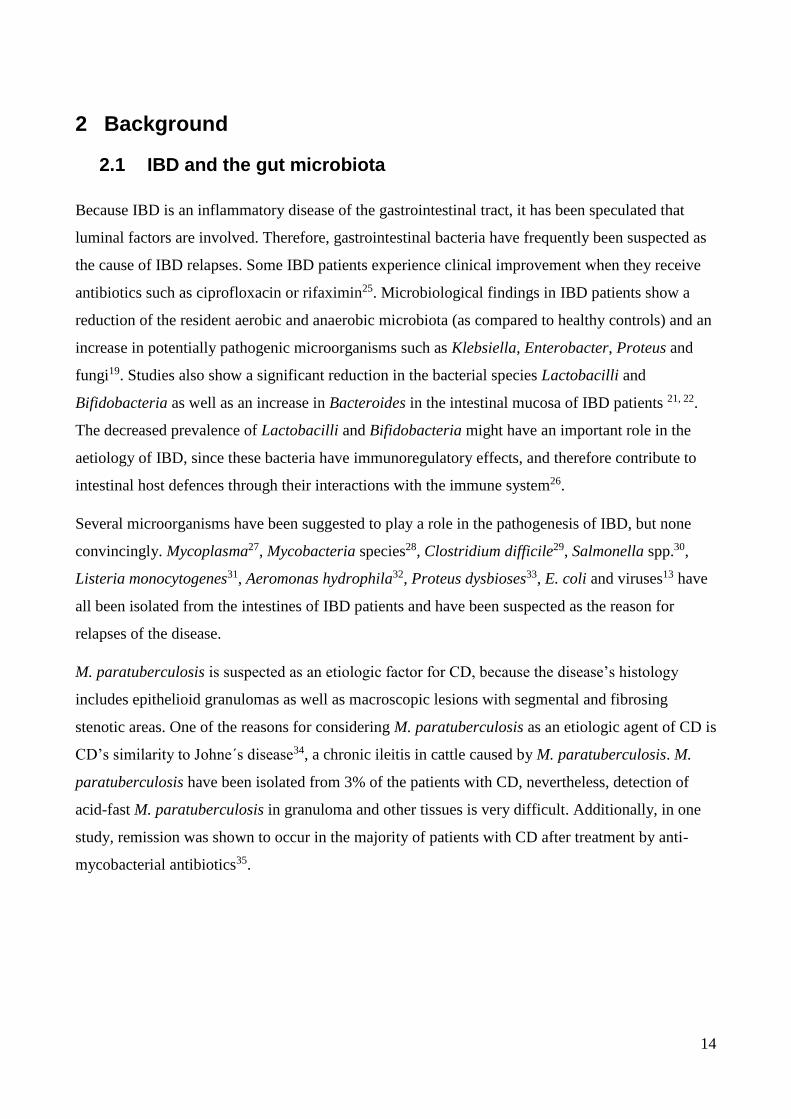
using MDS, 89% of pouch patients are clustered together in the white circle, while none of control
group are clustered among pouch patients in the white circle (Figure 6). Interestingly, DGGE gel
picture shows an identical very bright band (equal to high bacteria concentration) among pouch
patients (marked with red line around). Furthermore, the diversity is seen to be diminished in patients
with active vs. inactive pouchitis. Studies reports up to 50% of pouch patients develop at least one
episode of pouchitis165. Additionally, within 12 months of ileostomy surgery, 40% of the pouch
26 Control A
42 CD
38 UC
23 Control B
18 CDA
20 CDI
36
patients develop pouchitis165. Evidence suggests that an abnormal mucosal immune response to altered
microbiota in the pouch leads to acute and/ or chronic inflammation166.
Figure 6 Multidimensional scaling (MDS) analysis performed using BioNumerics 7.0 A) 89% of pouch patients are
clustered together in the white circle, while none of the control groups are clustered among pouch patients. B) one
identical brighter band appears in DGGE gel of all pouch patients (marked with red line around).
17 Control 7 PA
11 PI A B

37
4.2 Aim II: Secretion of alpha-hemolysin by Escherichia coli
disrupts tight junctions in inflammatory bowel disease patients
4.2.1 Aim and Hypothesis
An increased GI prevalence of E. coli has been associated with IBD. Many of the IBD-associated E.
coli belong to the phylogenetic group B2 and harbour virulence genes present in extra intestinal
pathogenic E. coli including alpha-hemolysin (hly) and cnf1. In this study, the goal was to
investigate the potential of E. coli, isolated from IBD patients with active and inactive disease, to
damage the integrity of the intestinal epithelium and the potential contribution of hly and cnf1in
vitro.
4.2.2 Results and Conclusion
Three of five E. coli strains from patients with active IBD (strains p7, p19A and p22) completely
disrupted epithelial cell TJ after only 3-6 hours of co-culture at an MOI of 10. This was not
observed with EcN, LF82, the four E. coli isolated from patients with inactive UC and three E. coli
strains from healthy controls. The three strains disrupting TJ in epithelial cells, all belonged to the
phylogenetic group B2 and they were all alpha-hemolysin positive. IBD associated E. coli p19A
was chosen for further experiments to elucidate the role of alpha-hemolysin and cnf1. P19A was
found to possess two copies of the hly gene cluster, one of which was located upstream of the cnf1
gene. Deletion mutants of the individual hemolysin genes as well as a cnf1 retained the ability to
damage Caco-2 cell TJ after less than 6 hrs of co-culture. However, this phenotypic characteristic
was lost in a double knockout mutant of both hly genes (Figure 7). Interestingly, there were no
differences in cytotoxicity between p19A WT and mutants.

38
Figure 7 p19A WT and mutants effect on Caco-2 cell TJ, using the TER method. p19A WT followed by ∆ hlyI, ∆ hlyII
and ∆ cnf1 mutant all disrupted the epithelial barrier in less than 6 hrs of co- culture with Caco-2 cells. Whereas p19A ∆
hlyI, II mutant did not disrupt epithelial barrier until after 12 hrs of co-incubation. As control TER was also performed
on Caco-2 cells without any bacteria added.
Hemolytic titration assays were performed with bacteria and bacterial culture supernatants in order
to investigate the hemolytic activity of clinical isolate E. coli p19A WT and the hly and cnf1 mutants.
The hemolytic activity was only completely abolished in the double mutant lacking hly clusters I and
II (p<0.05). Deletion of hly cluster II only partially decreased hemolytic activity, compared to the WT
suggesting hly cluster I does contribute to the overall hemolytic activity of the WT strain, despite the
fact that no reduction in hemolysis was observed in the hly cluster I mutant. RT-PCR was used to
quantify the relative amounts of the hly transcript in E. coli p19A WT and the different hly deletion
mutants. Relative expression of hly was two-fold higher in the WT p19A than in the ∆hlyI, mutant
and four-fold higher than in the ∆hlyII mutant. As expected only the double ∆hlyI, II mutant of p19A
lacked hly gene expression. A significant reduction in immunofluorescent staining of occludin was
evident, from 2 to 3 hours of co-culture with wild type p19A (P<0.001), as confirmed by Western
blotting with antibodies to occludin (Figure 8A). Similar results were obtained with all three Hly-
producing E. coli strains p7, p19A or p22, but not with the p19A ∆hlyI, II double mutant
demonstrating a link between hly expression and loss of TJ occludin (Figure 8B).

39
Figure 8 A) Western blot analysis of occludin expression in Caco-2 cell monolayers infected with p19A, p19A ∆ clbA,
p19A ∆ cnf1 and p19A WT. The occludin expression is high in p19A mutant ∆ hlyI, II, single p19A mutant ∆hlyI and
∆hlyII. The beta actin controls are equal for all. B) Confocal images of Caco-2 cell-monolayers immunofluorescence
stained for occludin (red) and nuclei (blue) for 3 hrs (MOI: 50) during apical incubation with E. coli p 19A from a
patient with active Ulcerative colitis. P-values for differences were, after 2 and 3 hrs, P <0.01 and P<0.001,
respectively. Notice, there appears a non-specific band on top of occluding band in p19A ∆hlyI, II.
IBD associated E. coli producing alpha-hemolysin can cause rapid loss of TJ integrity in differentiated
Caco-2 cell monolayers. This effect is abolished in a mutant unable to express alpha-hemolysin.
These results indicate that high alpha-hemolysin expression may be a mechanism by which strains of
E. coli isolated from IBD patients can contribute to epithelial barrier dysfunction and pathophysiology
of IBD.
A
B

40
4.3 Aim III: Comparison of three commercial fecal calprotectin
ELISA test kits used in patients with inflammatory Bowel Disease
4.3.1 Aim and Hypothesis
Faecal calprotectin is a non-invasive marker of intestinal inflammation used to distinguish between
functional and organic bowel diseases and to evaluate disease activity among patients with IBD.
The goal of this study was to compare three different ELISA tests measuring calprotectin in their
accuracy to detect IBD and to distinguish between IBD patients with active or inactive disease.
4.3.2 Results and Conclusion
Comparison of HK325 VS CALPRO ELISA kit: When measuring faecal calprotectin using
CALPRO, 92% of the samples from healthy controls and 9% of the samples from IBD patients had
a faecal calprotectin result below the cutoff value of 50 mg/kg. With HK325, only 7% of the
samples from healthy controls and none of the samples from IBD patients had a faecal calprotectin
result below the cutoff value (50 mg/kg). Based on a ROC curve, HK325 calprotectin
measurements (cutoff value of 50 mg/kg) had 95% sensitivity and 28% specificity. CALPRO
calprotectin measurements (cutoff value of 50 mg/kg) had 90% sensitivity and 93% specificity.
Comparison of EK-CAL VS CALPRO ELISA kit: Measuring faecal calprotectin using EK-
CAL, 70% of the samples from healthy controls and 9% of samples from IBD patients had a faecal
calprotectin result below the cutoff value of 50 mg/kg. Using the CALPRO calprotectin ELISA test,
96% of the samples from healthy controls and 25% of the samples from IBD patients had faecal
calprotectin value below the cutoff value of 50 mg/kg (Figure 9). Based on a ROC curve, CALPRO
calprotectin measurements (calprotectin cutoff value of 50 mg/kg) had 72% sensitivity and 96%
specificity (Figure 10). The stated EK-CAL calprotectin cutoff value of 50 mg/kg, gave 92%
sensitivity and 74% specificity. The CALPRO ELISA test performed best in measuring calprotectin
in faeces.

41
Figure 9 Frequency ratio for the EK-CAL and CALPRO ELISA: calprotectin results for healthy controls and IBD
patients. As it is shown, the faecal calprotectin values of 16 of 23 healthy controls are seen below the cutoff value
(log10≈ 1.7) compared to only 5 of 54 IBD patients for EK-CAL. For CALPRO ELISA method, the faecal calprotectin
values of 22 of 23 healthy controls and 7 of 54 IBD patients are seen below the cutoff value (log10≈ 1.7).

42
Figure 10 ROC curve of false-positive ratio for the HK325 and CALPRO ELISA: False-positive calprotectin results
of the HK325 versus the CALPRO calprotectin ELISA methods. As it is shown, EK-CAL has 26% false-positive
results, while CALPRO has 4% false-positive calprotectin results.
When comparing active vs inactive disease based on clinical activity indices, a statistically
significant difference in the CALPRO levels was found in patients with UC with median
(interquartile range [IQR]) faecal calprotectin levels of 150 mg/kg (39-1843) in UCA and of 39
mg/kg (38-47) in UCI, p<0.05. In patients with an ileoanal pouch, the median (interquartile range
[IQR]) faecal calprotectin levels were higher in active disease, 296 mg/kg (103-1783) in PA, and
117 mg/kg (60-187) in PI. The median (interquartile range [IQR]) level of faecal calprotectin was
222 mg/kg (149-789) in CDA and 125 mg/kg (57-850) in CDI, NS.
A significant correlation between clinical activity indices and faecal calprotectin in patients with
UC (p<0.05***) and patients with an ileoanal pouch (p<0.05) was shown (Figure 11). However,
regression analysis shows no correlation between faecal calprotectin and disease activity indices, in
patients with CD (p=0.078). In healthy controls, the median (interquartile range [IQR]) level of
faecal calprotectin of 8 mg/kg (8-17) was significantly lower than all active or inactive IBD patient
groups, p<0.05 (Figure 11).

43
Figure 11 Faecal calprotectin in IBD-patients: Faecal calprotectin results using the CALPRO calprotectin ELISA
method in different IBD-patient groups during active and inactive disease compared to controls. Error bars show the
mean and standard division (SD). As it is shown, faecal calprotectin concentrations are associated with clinical disease
activity indices in patients with UC and pouchitis. Controls are significantly below the cutoff value. Faecal calprotectin
is not a suitable marker for differentiating between disease activities in Crohn’s disease patients.

44
4.4 Aim IV: Extra intestinal pathogenetic Escherichia coli are
associated with intestinal inflammation in patients with Ulcerative
Colitis
4.4.1 Aim and Hypothesis
E. coli of the phylogenetic group B2 frequently colonize the intestine of patients with IBD. We have
previously tested the effect of Ciprofloxacin (Cipro) and/or orally administered E. coli EcN as add-
on treatment, to conventional therapies in patients with UCA. Studies show that IBD patients
between relapses have a calprotectin value between 123-213 mg/kg167. Therefore, in this follow-up
study a calprotectin value of >200 mg/kg was used as criteria for active UC. In this study, the goal
was to investigate the influence of EcN treatment and initial colonization with phylogenetic group
B2 E. coli on intestinal inflammation evaluated by measurement of faecal calprotectin.
4.4.2 Results and Conclusion
As described previously129 100 patients with UC were included in this study, and randomized to
four treatment groups: 1 week of Cipro or placebo followed by 7 weeks of EcN or placebo, with 25
patients in each group. Activity was primarily evaluated by a CAI score>/= 6.
When faecal calprotectin of patients with an initial faecal calprotectin level >200 mg/kg and a CAI
score≥ 6 were analysed (UCA), we confirmed that in the UCA patient group receiving placebo/EcN
fewer patients, 17 %, reached remission defined by a faecal calprotectin <200 mg/kg compared to
the UCA patient group receiving placebo/placebo, 61 %, p<0.05 (Figure 12). When comparing
UCA patient groups receiving EcN (Cipro/EcN and placebo/EcN), with UCA patient groups not
receiving EcN, (Cipro/placebo and placebo/placebo), it was demonstrated that groups receiving
EcN reached remission (faecal calprotectin < 200 mg/kg) less frequent (31.4% ) than groups not
receiving EcN (59% ), p≤0.05 (figure 12).
However, in order to see whether EcN has an overall effect on the level of calprotectin, groups
treated with EcN were compared to groups not treated with EcN. Faecal calprotectin values from
weeks 0, 1, 8 and 12 of 100 UC patients with CAI score≥ 6 (regardless of the calprotectin value of
the initial stool sample) was evaluated using the Two-way ANOVA test. When comparing the
experimental groups receiving EcN (Cipro/EcN and placebo/EcN), with groups not receiving EcN,

45
(Cipro/placebo and placebo/placebo), higher faecal calprotectin values were found in patients
treated with EcN, however this was not statistically significant, p=0.053.
Figure 12 Comparison of the UC patient groups reaching faecal calprotectin values <200 mg/kg during 12 weeks
follow-up (intention to treat- analysis) (A) Treated with: placebo-placebo (D), Cipro/EcN (A), Cipro/placebo (B) or
placebo/EcN (C). (B) 31% of the UC patients treated with EcN (AC) as add-on treatment reach calprotectin value <200
mg/kg, while 59% of the patients not treated with EcN (BD) reach a calprotectin value <200 mg/kg, p<0.05*.
When analysing all 100 UC patients with CAI score≥ 6, in the patient group treated with
Cipro/placebo, 48% (12/25) were initially colonized with B2 E. coli at week 0, prior to
ciprofloxacin treatment. After antibiotic treatment, the number of the patients colonized with B2 E.
coli was reduced to 18% (4/22), 33% (7/21) and 36% (5/14) at week 1, 8 and 12, respectively
(figure 13). These results demonstrates, that even though one week of Cipro treatment did reduce
the number of UCA patients colonized with B2 E. coli immediately after the treatment, no long
term effect on B2 E. coli colonization could be demonstrated. In the group treated with
placebo/placebo, 52 % (13/25) of the patients were colonized with B2 E. coli at week 0. In this
group, the number of patients colonized with B2 E. coli were, 42% (10/24), 45% (9/20) and 38%
(6/16) at week 1, 8 and 12, respectively (figure 13).

46
Figure 13 Project flow of all patient groups, with number of the submitted stool samples, Median faecal calprotectin
value, the number of the B2 E. coli isolates and number of the patients with a faecal calprotectin <200 mg/kg, at week
0, 1, 8 and 12.
Interestingly, UC patients, initially colonized with B2 E. coli (before treatment with EcN) had an
increased faecal calprotectin value in comparison to UC patients not colonized with B2 E. coli,
median (interquartile range [IQR]) 1336 mg/kg (672-1818) and 386 (126-787), respectively,
p<0.05*** (figure 14).
Our results demonstrate that there are no beneficial effect of using EcN as add-on treatment in UCA
patients. On the contrary, an increased number of patients treated with EcN as add-on treatment
were unable to reach remission. UC patients included in this study, initially colonized with B2 E.
coli at week 0, have increased burden of inflammation measured using CAI score and faecal
calprotectin compared to UC patient not colonized with B2 E. coli.

47
Figure 14 week 0 faecal calprotectin value in mg/kg in experimental groups (25 patients in each group) included in the
study. There is significant difference between faecal calprotectin values of group initially colonized with B2 E. coli
(46/87) in comparison to group not colonized with B2 E. coli (41/87), p<0.05*(Median with rang) (there was no
bacterial growth in 13 patients faecal samples).

48
5. Discussion and Perspectives
The colon and the distal ileum are colonized with a complex mixture of microorganisms that are
highly metabolically active. Enteric bacteria and fungi provide a constant antigenic stimulus that
drives a pathogenic immune response seen in genetically susceptible individuals such as IBD
patients. A variety of genetic defects in either mucosal barrier function or in the innate bacterial
killing lead to an abnormally aggressive T-cell activation by bacterial antigens and/or the loss of
normal immunologic tolerance by the mucosal immune system. Recently developed concepts of
IBD pathogenesis include defective innate immune function resulting in diminished bacterial killing
as well as functional alterations in the composition of the commensal bacteria such as E .coli, which
in turn leads to enhanced epithelial adherence and invasion, bacterial persistence within intestinal
epithelial and phagocytic cells, and metabolic derangements that negatively influence the epithelial
function. However, this might explain reduced intestinal bacterial diversity in IBD patients in
comparison to healthy controls162–164(manuscript I), which might be caused by intestinal overgrowth
of aggressive bacteria in IBD patients, which are normally in symbiotic balance with other intestinal
bacteria in healthy people.
Studies show increased numbers of E. coli strains from the B2 phylogenetic group harbouring
ExPEC genes within the intestines of IBD patients. Patients colonized with E. coli of the B2
phylogenetic group display increased burdens of inflammation as measured using the CAI score and
faecal calprotectin levels, in comparison to patients not colonized with B2 E. coli (manuscript IV).
DAEC have been associated with UC, while AIEC has been linked to CD. Both AIEC and DAEC
are known as enteropathogenic96 E. coli and often belongs to the B2 phylogenetic group168. The B2
phylogenetic E. coli is frequently isolated from the intestines of IBD patients169. Martinez-Medina93
et al show in a collection of 22 AIEC isolates, 14 from CD and 6 from healthy controls, 63% of the
strains were from the B2 phylogenetic group and here of 79% of the stains was isolated from CD
patients93. However, Escobar-Paramo170 et al showed that of 16 DAEC strains isolated from patients
with diarrhoea, 31.3% was from B2 and 31.3% was from D phylogenetic group. Petersen et al
200924 reported the prevalence of E. coli isolated from a UC patient harbouring afa/Dr gene, which
is related to DAEC, while other studies showed the high prevalence of E. coli isolates from CD
patients sharing ExPEC genes and the strains belonged to the AIEC pathovar93. The question
remains whether B2 E. coli strains isolated from faecal samples of IBD patients generally, are
related to the AIEC and DAEC pathovars. The role of E. coli pathobionts in the pathophysiology in

49
IBD has been attributed to their capability to adhere and invade epithelial cells and replicate in
macrophages and the most well-studied prototype strain is LF82. Previous studies have shown that
UC associated E. coli (p19A) from the B2 phylogenetic group, harbouring two alpha-hemolysin
genes, induce cell death in dendritic cells, stimulate the release of TNF-α, IL-6, IL-23 cytokines98
(poster) and causes rapid loss of TJ integrity in differentiated Caco2-cell monolayer (manuscript II).
In contrast to p19A, strain LF82 does not cause rapid dissolution of epithelial TJ, clearly indicating
that B2 phylotype of IBD-associated strains differ markedly in pathogenic mechanisms.
The IBD-associated E. coli strains included in our study causing loss of TJ in epithelial cell
monolayers were hemolytic (manuscript II). Four types of hemolysin have been demonstrated in E.
coli: alpha-hemolysin (hly A), plasmid- and phage- carried enterohemolysin (EhxA, a-hlyA) and
silent hemolysin (SheA); EhxA and HlyA belong to the RTX (repeat in toxin) related family, which
lyse erythrocytes from different mammalian species171–173. It is known that a number of E. coli
pathotypes, i.e. urinary tract pathogenic (UPEC), Verocytotoxin-producing E. coli (VTEC), EPEC
and Enterotoxigenic E. coli (ETEC) all harbor alpha-hemolysin174. The E. coli alpha-hemolysin is
known to be able to lyse erythrocytes through binding to the surface protein glycoporin175–177 but
also other cell types including leucocytic cells, bladder and renal tubular cells in a dose dependent
manner153,171,172,178,179.
The role of hlyA in TJ disruption was further investigated in E. coli strain p19A, which possessed
two hlyA clusters as previously reported for isolates of uropathogenic E. coli belonging to phylotype
B2180. We showed that both hyl A gene clusters in p19A contributed to the damaging effects on
epithelial integrity, suggesting that intestinal E. coli strains expressing possessing more than one
hlyA locus can have increased pathological consequences in intestinal inflammation. Although our
IBD-associated strains did not induce epithelial cell apoptosis, a hly-expressing uropathogenic E.
coli was previously shown to cause localized regions of apoptosis in HT29/B6 cell monolayers. The
difference between these findings and our results may be due to use of a higher MOI than in our
study, the use of different strains or the amount of hlyA expressed181 (manuscript II).
These findings suggest that IBD associated E. coli might play a role in pathogenesis of IBD and
might play a role in disease relapses in IBD patients. Additional studies of gut microbiota and IBD
associated E. coli species are needed to confirm whether IBD reflects an abnormal host response to
commensal bacteria or if the acquisition of pathogenic features by specific E. coli strains drives the
onset of the IBD. However, clinical experience with treatment effects (although varying) of both

50
immune-suppressants and antibiotics (and probiotics) would suggest that the truth about IBD
pathogenesis lies in between the interaction between the predisposing host genetic factors and the
host immune response to intestinal bacteria.

51
6 Conclusion
There is a reduced diversity of intestinal microbial species in IBD patients in comparison to healthy
controls (Manuscript I). Patients colonized with E. coli from the B2 phylogenetic group display
increased burdens of inflammation, as measured by CAI scores and faecal calprotectin levels, in
comparison to patients not colonized with B2 E. coli (Manuscript IV). UC associated E. coli (p19A)
from the B2 phylogenetic group, harbouring two alpha-hemolysin genes, induce cell death in
dendritic cells, stimulate the release of TNF-α, IL-6, IL-23 cytokines98 (poster) and causes rapid
loss of TJ integrity in differentiated Caco2-cell monolayer (manuscript II).

52
7 References
1. Baumgart DC, Sandborn WJ. Inflammatory bowel disease: clinical aspects and established and evolving therapies. Lancet. 2007 May 12;369(9573):1641–57.
2. Loftus CG, Loftus Jr. E V, Harmsen WS, Zinsmeister AR, Tremaine WJ, Melton III LJ, et al. Update on the incidence and prevalence of Crohn’s disease and ulcerative colitis in Olmsted County, Minnesota, 1940-2000. InflammBowelDis. Inflammatory Bowel Disease Clinic, Division of Gastroenterology and Hepatology, Mayo Clinic College of Medicine, Rochester, Minnesota 55905, USA. [email protected]; 2007 Mar;13(1078-0998 (Print)):254–61.
3. Annese V, Latiano A, Andriulli A. Genetics of inflammatory bowel disease: the beginning of the end or the end of the beginning? DigLiver Dis. 2003 Jun;35(6):442–9.
4. Gaya DR, Russell RK, Nimmo ER, Satsangi J. New genes in inflammatory bowel disease: lessons for complex diseases? Lancet. Gastrointestinal Unit, Molecular Medicine Centre, University of Edinburgh, Western General Hospital, Edinburgh EH4 2XU, UK; 2006 Apr 15;367(1474-547X (Electronic)):1271–84.
5. Siminovitch KA. Advances in the molecular dissection of inflammatory bowel disease. SeminImmunol. Department of Medicine, University of Toronto, Mount Sinai Hospital Samuel Lunenfeld and Toronto General Hospital Research Institutes, 600 University Avenue, Room 778D, Toronto, Ont., Canada M5G 1X5. [email protected]; 2006 Aug;18(1044-5323 (Print)):244–53.
6. Strober W, Murray PJ, Kitani A, Watanabe T. Signalling pathways and molecular interactions of NOD1 and NOD2. NatRevImmunol. 2006 Jan;6(1):9–20.
7. Bouma G, Strober W. The immunological and genetic basis of inflammatory bowel disease. NatRevImmunol. The Mucosal Immunity Section, National Institutes of Health, Mucosal Immunity Section, Laboratory of Clinical Investigation, National Institutes of Allergy and Infectious Diseases, 10 Center Drive, Room 11N238, Bethesda, Maryland 20892, USA; 2003 Jul;3(1474-1733 (Print)):521–33.
8. Keshavarzian A, Fusunyan RD, Jacyno M, Winship D, MacDermott RP, Sanderson IR. Increased interleukin-8 (IL-8) in rectal dialysate from patients with ulcerative colitis: evidence for a biological role for IL-8 in inflammation of the colon. Am J Gastroenterol. 1999 Mar;94(3):704–12.
9. Nuding S, Fellermann K, Wehkamp J, Stange EF. Reduced mucosal antimicrobial activity in Crohn’s disease of the colon. Gut. 2007 Sep;56(9):1240–7.
10. Esteve-Comas M, Ramirez M, Fernandez-Banares F, Abad-Lacruz A, Gil A, Cabre E, et al. Plasma polyunsaturated fatty acid pattern in active inflammatory bowel disease. Gut. Department of Gastroenterology, Hospital Universitari Germans Trias i Pujol, Badalona, Spain; 1992 Oct;33(0017-5749 (Print)):1365–9.
11. Hebuterne X, Filippi J, Al Jaouni R, Schneider S. Nutritional consequences and nutrition therapy in Crohn’s disease. GastroenterolClin Biol. Centre Hospitalier Universitaire de Nice, Pole Digestif, Service de Gastro-enterologie et Nutrition Clinique, Hopital de l’Archet 2, CHU de Nice, 06202 Nice cedex 03, France. [email protected]; 2009 Jun;33 Suppl 3(2210-7401 (Electronic)):S235–44.
53
12. Png CW, Linden SK, Gilshenan KS, Zoetendal EG, McSweeney CS, Sly LI, et al. Mucolytic Bacteria With Increased Prevalence in IBD Mucosa Augment In Vitro Utilization of Mucin by Other Bacteria. AmJ Gastroenterol. Mucin and IBD Research Teams, Mucosal Diseases Program, Mater Medical Research Institute and Department of Medicine, University of Queensland, Mater Health Services, South Brisbane, Queensland, Australia; 2010 Jul 20;(1572-0241 (Electronic)).
13. Kangro HO, Chong SK, Hardiman A, Heath RB, Walker-Smith JA. A prospective study of viral and mycoplasma infections in chronic inflammatory bowel disease. Gastroenterology. 1990 Mar;98(3):549–53.
14. Khan RR, Lawson AD, Minnich LL, Martin K, Nasir A, Emmett MK, et al. Gastrointestinal norovirus infection associated with exacerbation of inflammatory bowel disease. J PediatrGastroenterolNutr. Department of Pediatrics, West Virginia University Health Sciences Center, Charleston, WV, USA; 2009 Mar;48(1536-4801 (Electronic)):328–33.
15. Hume G, Radford-Smith GL. The pathogenesis of Crohn’s disease in the 21st century. Pathology. Department of Gastroenterology and Hepatology, Royal Brisbane Hospital, Brisbane, Queensland, Australia; 2002 Dec;34(0031-3025 (Print)):561–7.
16. Karlinger K, Gyorke T, Mako E, Mester A, Tarjan Z. The epidemiology and the pathogenesis of inflammatory bowel disease. EurJ Radiol. 2000 Sep;35(3):154–67.
17. Westall FC. Integrating theories of the etiology of Crohn’s disease on the etiology of Crohn's disease: questioning the hypotheses. William M. Chamberlin, Saleh A. Naser Med Sci Monit, 2006; 12(2): RA27-33. Med SciMonit. 2006 May;12(5):LE5–6.
18. Macfarlane S, Steed H, Macfarlane GT. Intestinal bacteria and inflammatory bowel disease. Crit RevClinLab Sci. 2009;46(1):25–54.
19. Schultz M, Munro K, Tannock GW, Melchner I, Gottl C, Schwietz H, et al. Effects of feeding a probiotic preparation (SIM) containing inulin on the severity of colitis and on the composition of the intestinal microflora in HLA-B27 transgenic rats. ClinDiagnLab Immunol. 2004 May;11(3):581–7.
20. Manichanh C, Rigottier-Gois L, Bonnaud E, Gloux K, Pelletier E, Frangeul L, et al. Reduced diversity of faecal microbiota in Crohn’s disease revealed by a metagenomic approach. Gut. 2006;55:205–11.
21. Braun A, Treede I, Gotthardt D, Tietje A, Zahn A, Ruhwald R, et al. Alterations of phospholipid concentration and species composition of the intestinal mucus barrier in ulcerative colitis: A clue to pathogenesis. Inflamm Bowel Dis. 2009;15:1705–20.
22. Mirsepasi H, Persson S, Struve C, Andersen LOB, Petersen AM, Krogfelt K a. Microbial diversity in fecal samples depends on DNA extraction method: easyMag DNA extraction compared to QIAamp DNA stool mini kit extraction. BMC Res Notes [Internet]. 2014;7:50. Available from: http://www.pubmedcentral.nih.gov
23. Kotlowski R, Bernstein CN, Sepehri S, Krause DO. High prevalence of Escherichia coli belonging to the B2+D phylogenetic group in inflammatory bowel disease. Gut. 2007 May;56(5):669–75.

54
24. Petersen AM, Nielsen EM, Litrup E, Brynskov J, Mirsepasi H, Krogfelt KA. A phylogenetic group of Escherichia coli associated with active left-sided inflammatory bowel disease. BMC Microbiol. 2009 Aug;%20;9:171.:171–9.
25. Rahimi R, Nikfar S, Rezaie A, Abdollahi M. A meta-analysis of broad-spectrum antibiotic therapy in patients with active Crohn’s disease. ClinTher. 2006 Dec;28(12):1983–8.
26. Garcia-Lafuente A, Antolin M, Guarner F, Crespo E, Salas A, Forcada P, et al. Incrimination of anaerobic bacteria in the induction of experimental colitis. Am J Physiol. 1997 Jan;272(1 Pt 1):G10–5.
27. Sokol H, Seksik P, Rigottier-Gois L, Lay C, Lepage P, Podglajen I, et al. Specificities of the fecal microbiota in inflammatory bowel disease. InflammBowel Dis. 2006 Feb;12(2):106–11.
28. Hermon-Taylor J, Barnes N, Clarke C, Finlayson C. Mycobacterium paratuberculosis cervical lymphadenitis, followed five years later by terminal ileitis similar to Crohn’s disease. BMJ. Department of Surgery, St George’s Hospital Medical School, London; 1998 Feb 7;316(0959-8138 (Print)):449–53.
29. Dorman SA, Liggoria E, Winn Jr. WC, Beeken WL. Isolation of Clostridium difficile from patients with inactive Crohn’s disease. Gastroenterology. 1982 Jun;82(6):1348–51.
30. Taylor-Robinson S, Miles R, Whitehead A, Dickinson RJ. Salmonella infection and ulcerative colitis. Lancet. 1989 May;20;1(8647):1145.
31. Liu Y, van Kruiningen HJ, West AB, Cartun RW, Cortot A, Colombel JF. Immunocytochemical evidence of Listeria, Escherichia coli, and Streptococcus antigens in Crohn’s disease. Gastroenterology. 1995 May;108(5):1396–404.
32. Doman DB, Golding MI, Goldberg HJ, Doyle RB. Aeromonas hydrophila colitis presenting as medically refractory inflammatory bowel disease. Am J Gastroenterol. 1989 Jan;84(1):83–5.
33. Kanareykina SK, Misautova AA, Zlatkina AR, Levina EN. Proteus dysbioses in patients with ulcerative colitis. Nahrung. 1987;31(5-6):557–61.
34. Collins MT, Lisby G, Moser C, Chicks D, Christensen S, Reichelderfer M, et al. Results of multiple diagnostic tests for Mycobacterium avium subsp. paratuberculosis in patients with inflammatory bowel disease and in controls. J ClinMicrobiol. 2000 Dec;38(12):4373–81.
35. Prantera C, Kohn A, Mangiarotti R, Andreoli A, Luzi C. Antimycobacterial therapy in Crohn’s disease: results of a controlled, double-blind trial with a multiple antibiotic regimen. Am J Gastroenterol. 1994 Apr;89(4):513–8.
36. Rolhion N, Darfeuille-Michaud A. Adherent-invasive Escherichia coli in inflammatory bowel disease. InflammBowel Dis. 2007 Oct;13(10):1277–83.
37. Russo TA, Johnson JR. Proposal for a new inclusive designation for extraintestinal pathogenic isolates of Escherichia coli: ExPEC. J Infect Dis. 2000 May;181(5):1753–4.

55
38. Kaper JB, Nataro JP, Mobley HL. Pathogenic Escherichia coli. NatRevMicrobiol. Center for Vaccine Development, Department of Microbiology and Immunology, University of Maryland School of Medicine, Baltimore, Maryland 21201, USA. [email protected]; 2004 Feb;2(1740-1526 (Print)):123–40.
39. Orskov I, Orskov F, Jann B, Jann K. Serology, chemistry, and genetics of O and K antigens of Escherichia coli. Bacteriol Rev. 1977 Sep;41(3):667–710.
40. Okeke IN, Lamikanra A, Czeczulin J, Dubovsky F, Kaper JB, Nataro JP. Heterogeneous virulence of enteroaggregative Escherichia coli strains isolated from children in Southwest Nigeria. J Infect Dis. 2000 Jan;181(1):252–60.
41. Mora A, Blanco M, Yamamoto D, Dahbi G, Blanco JE, Lopez C, et al. HeLa-cell adherence patterns and actin aggregation of enteropathogenic Escherichia coli (EPEC) and Shiga-toxin-producing E. coli (STEC) strains carrying different eae and tir alleles. Int Microbiol. 2009 Dec;12(4):243–51.
42. Hernandes RT, Elias WP, Vieira MA, Gomes TA. An overview of atypical enteropathogenic Escherichia coli. FEMS Microbiol Lett. 2009 Aug;297(2):137–49.
43. Kalman D, Weiner OD, Goosney DL, Sedat JW, Finlay BB, Abo A, et al. Enteropathogenic E. coli acts through WASP and Arp2/3 complex to form actin pedestals. NatCell Biol. 1999 Oct;1(6):389–91.
44. Taxt A, Aasland R, Sommerfelt H, Nataro J, Puntervoll P. Heat-stable enterotoxin of enterotoxigenic Escherichia coli as a vaccine target. Infect Immun. 2010 May;78(5):1824–31.
45. Sears CL, Kaper JB. Enteric bacterial toxins: mechanisms of action and linkage to intestinal secretion. Microbiol Rev. 1996 Mar;60(1):167–215.
46. Okeke IN. Diarrheagenic Escherichia coli in sub-Saharan Africa: status, uncertainties and necessities. J Infect DevCtries. 2009 Nov 27;3(11):817–42.
47. Sansonetti P. Host-pathogen interactions: the seduction of molecular cross talk. Gut. 2002 May;50 Suppl 3:III2–8.
48. Kaper JB. Pathogenic Escherichia coli. Int J Med Microbiol. 2005 Oct;295(6-7):355–6.
49. Nataro JP, Kaper JB, Robins-Browne R, Prado V, Vial P, Levine MM. Patterns of adherence of diarrheagenic Escherichia coli to HEp-2 cells. Pediatr Infect Dis J. 1987 Sep;6(9):829–31.
50. Nataro JP, Kaper JB. Diarrheagenic Escherichia coli. ClinMicrobiol Rev. 1998 Jan;11(1):142–201.
51. Steiner TS, Nataro JP, Poteet-Smith CE, Smith JA, Guerrant RL. Enteroaggregative Escherichia coli expresses a novel flagellin that causes IL-8 release from intestinal epithelial cells. J ClinInvest. 2000 Jun;105(12):1769–77.
52. Henderson IR, Czeczulin J, Eslava C, Noriega F, Nataro JP. Characterization of pic, a secreted protease of Shigella flexneri and enteroaggregative Escherichia coli. Infect Immun. 1999 Nov;67(11):5587–96.
56
53. Savarino SJ, Fasano A, Watson J, Martin BM, Levine MM, Guandalini S, et al. Enteroaggregative Escherichia coli heat-stable enterotoxin 1 represents another subfamily of E. coli heat-stable toxin. ProcNatlAcadSciUSA. 1993 Apr 1;90(7):3093–7.
54. Navarro-Garcia F, Eslava C, Villaseca JM, Lopez-Revilla R, Czeczulin JR, Srinivas S, et al. In vitro effects of a high-molecular-weight heat-labile enterotoxin from enteroaggregative Escherichia coli. Infect Immun. 1998 Jul;66(7):3149–54.
55. Noris M, Remuzzi G. Genetic abnormalities of complement regulators in hemolytic uremic syndrome: how do they affect patient management? NatClinPractNephrol. 2005 Nov;1(1):2–3.
56. Sonntag AK, Prager R, Bielaszewska M, Zhang W, Fruth A, Tschape H, et al. Phenotypic and genotypic analyses of enterohemorrhagic Escherichia coli O145 strains from patients in Germany. J ClinMicrobiol. 2004 Mar;42(3):954–62.
57. Thorpe CM. Shiga toxin-producing Escherichia coli infection. ClinInfect Dis. 2004 May 1;38(9):1298–303.
58. Karmali RA, Muse P, Allen G, Louis T. Macrophage production of prostaglandins: effects of fetal calf serum and diazepam. Use of an improved method for extracting 6-keto-PGF1 alpha. Prostaglandins LeukotMed. 1982 Jun;8(6):565–77.
59. McCarthy TA, Barrett NL, Hadler JL, Salsbury B, Howard RT, Dingman DW, et al. Hemolytic-Uremic Syndrome and Escherichia coli O121 at a Lake in Connecticut, 1999. Pediatrics. 2001 Oct;108(4):E59.
60. Dean-Nystrom EA, Bosworth BT, Moon HW, O’Brien AD. Escherichia coli O157:H7 requires intimin for enteropathogenicity in calves. Infect Immun. 1998 Sep;66(9):4560–3.
61. Acheson DW, Moore R, De Breucker S, Lincicome L, Jacewicz M, Skutelsky E, et al. Translocation of Shiga toxin across polarized intestinal cells in tissue culture. Infect Immun. 1996 Aug;64(8):3294–300.
62. Hurley BP, Thorpe CM, Acheson DW. Shiga toxin translocation across intestinal epithelial cells is enhanced by neutrophil transmigration. Infect Immun. 2001 Oct;69(10):6148–55.
63. Van Setten PA, Monnens LA, Verstraten RG, van den Heuvel LP, van Hinsbergh VW. Effects of verocytotoxin-1 on nonadherent human monocytes: binding characteristics, protein synthesis, and induction of cytokine release. Blood. 1996 Jul 1;88(1):174–83.
64. Cooling LL, Walker KE, Gille T, Koerner TA. Shiga toxin binds human platelets via globotriaosylceramide (Pk antigen) and a novel platelet glycosphingolipid. Infect Immun. 1998 Sep;66(9):4355–66.
65. Bitzan M, Richardson S, Huang C, Boyd B, Petric M, Karmali MA. Evidence that verotoxins (Shiga-like toxins) from Escherichia coli bind to P blood group antigens of human erythrocytes in vitro. Infect Immun. 1994 Aug;62(8):3337–47.
66. Johnson JR, Russo TA. Uropathogenic Escherichia coli as agents of diverse non-urinary tract extraintestinal infections. J Infect Dis. 2002 Sep 15;186(6):859–64.

57
67. Russo TA, Johnson JR. Medical and economic impact of extraintestinal infections due to Escherichia coli: focus on an increasingly important endemic problem. Microbes Infect. 2003 Apr;5(5):449–56.
68. Spangler BD. Structure and function of cholera toxin and the related Escherichia coli heat-labile enterotoxin. Microbiol Rev. 1992 Dec;56(4):622–47.
69. Clermont O, Bonacorsi S, Bingen E. Rapid and simple determination of the Escherichia coli phylogenetic group. ApplEnvironMicrobiol. 2000 Oct;66(10):4555–8.
70. Picard B, Garcia JS, Gouriou S, Duriez P, Brahimi N, Bingen E, et al. The link between phylogeny and virulence in Escherichia coli extraintestinal infection. Infect Immun. 1999 Feb;67(2):546–53.
71. Clermont O, Christenson JK, Denamur E, Gordon DM. The Clermont Escherichia coli phylo-typing method revisited: Improvement of specificity and detection of new phylo-groups. Environ Microbiol Rep. 2013;5(1):58–65.
72. Johnson JR, Gajewski A, Lesse AJ, Russo TA. Extraintestinal pathogenic Escherichia coli as a cause of invasive nonurinary infections. J ClinMicrobiol. 2003 Dec;41(12):5798–802.
73. Sabate M, Moreno E, Perez T, Andreu A, Prats G. Pathogenicity island markers in commensal and uropathogenic Escherichia coli isolates. Clin Microbiol Infect. 2006 Sep;12(9):880–6.
74. Johnson JR, Russo TA. Molecular epidemiology of extraintestinal pathogenic (uropathogenic) Escherichia coli. Int J Med Microbiol. 2005 Oct;295(6-7):383–404.
75. Burke DA, Axon AT. Adhesive Escherichia coli in inflammatory bowel disease and infective diarrhoea. BMJ. 1988 Jul 9;297(6641):102–4.
76. Giaffer MH, Holdsworth CD, Duerden BI. Virulence properties of Escherichia coli strains isolated from patients with inflammatory bowel disease. Gut. 1992 May;33(5):646–50.
77. Keighley MR, Arabi Y, Dimock F, Burdon DW, Allan RN, Alexander-Williams J. Influence of inflammatory bowel disease on intestinal microflora. Gut. 1978 Dec;19(12):1099–104.
78. Ilnyckyj A, Greenberg H, Bernstein CN. Escherichia coli O157:H7 infection mimicking Crohn’s disease. Gastroenterology. 1997 Mar;112(3):995–9.
79. Bielaszewska M, Schiller R, Lammers L, Bauwens A, Fruth A, Middendorf B, et al. Heteropathogenic virulence and phylogeny reveal phased pathogenic metamorphosis in Escherichia coli O2: H6. EMBO Mol Med. 2014;6(3):347–57.
80. Darfeuille-Michaud A, Neut C, Barnich N, Lederman E, Di Martino P, Desreumaux P, et al. Presence of adherent Escherichia coli strains in ileal mucosa of patients with Crohn’s disease. Gastroenterology. Laboratoire de Bacteriologie, Faculte de Pharmacie, Clermont-Ferrand Cedex, France. [email protected]; 1998 Dec;115(0016-5085 (Print)):1405–13.
81. Martin HM, Campbell BJ, Hart CA, Mpofu C, Nayar M, Singh R, et al. Enhanced Escherichia coli adherence and invasion in Crohn’s disease and colon cancer. Gastroenterology. 2004 Jul;127(1):80–93.

58
82. La Ferla K, Seegert D, Schreiber S. Activation of NF-kappaB in intestinal epithelial cells by E. coli strains isolated from the colonic mucosa of IBD patients. IntJ Color Dis. 2004 Jul;19(4):334–42.
83. Boudeau J, Glasser AL, Masseret E, Joly B, Darfeuille-Michaud A. Invasive ability of an Escherichia coli strain isolated from the ileal mucosa of a patient with Crohn’s disease. InfectImmun. 1999 Sep;67(9):4499–509.
84. Greenstein RJ. Is Crohn’s disease caused by a mycobacterium? Comparisons with leprosy, tuberculosis, and Johne's disease. Lancet Infect Dis. 2003 Aug;3(8):507–14.
85. Scanu AM, Bull TJ, Cannas S, Sanderson JD, Sechi LA, Dettori G, et al. Mycobacterium avium subspecies paratuberculosis infection in cases of irritable bowel syndrome and comparison with Crohn’s disease and Johne's disease: common neural and immune pathogenicities. J ClinMicrobiol. 2007 Dec;45(12):3883–90.
86. Meconi S, Vercellone A, Levillain F, Payre B, Al Saati T, Capilla F, et al. Adherent-invasive Escherichia coli isolated from Crohn’s disease patients induce granulomas in vitro. Cell Microbiol. 2007 May;9(5):1252–61.
87. Sartor RB, Muehlbauer M. Microbial host interactions in IBD: implications for pathogenesis and therapy. Curr GastroenterolRep. 2007 Dec;9(6):497–507.
88. Schussler P, Kruis W, Marget W. [Lipid A antibody titers and O antibody titers in Crohn’s disease, ulcerative colitis and acute enteritis (author's transl)]. Med Klin. 1976 Oct 29;71(44):1898–902.
89. Landers CJ, Cohavy O, Misra R, Yang H, Lin YC, Braun J, et al. Selected loss of tolerance evidenced by Crohn’s disease-associated immune responses to auto- and microbial antigens. Gastroenterology. 2002 Sep;123(3):689–99.
90. Mow WS, Vasiliauskas EA, Lin YC, Fleshner PR, Papadakis KA, Taylor KD, et al. Association of antibody responses to microbial antigens and complications of small bowel Crohn’s disease. Gastroenterology. 2004 Feb;126(2):414–24.
91. Petersen AM, Schou C, Mirsepasi H, Engberg J, Friis-Moller A, Nordgaard-Lassen I, et al. Seroreactivity to E. coli outer membrane protein C antibodies in active inflammatory bowel disease; diagnostic value and correlation with phylogroup B2 E. coli infection. Scand J Gastroenterol. England; 2012 Feb;47(2):155–61.
92. Tabaqchali S, O’Donoghue DP, Bettelheim KA. Escherichia coli antibodies in patients with inflammatory bowel disease. Gut. 1978 Feb;19(0017-5749 (Print)):108–13.
93. Martinez-Medina M, Aldeguer X, Lopez-Siles M, Gonzalez-Huix F, Lopez-Oliu C, Dahbi G, et al. Molecular diversity of Escherichia coli in the human gut: New ecological evidence supporting the role of adherent-invasive E. coli (AIEC) in Crohn’s disease. Inflamm Bowel Dis. 2009;15:872–82.
94. Burke DA, Axon AT. Ulcerative colitis and Escherichia coli with adhesive properties. J Clin Pathol. 1987 Jul;40(0021-9746 (Print)):782–6.

59
95. Satterwhite TK, Evans DG, DuPont HL, Evans Jr. DJ. Role of Escherichia coli colonisation factor antigen in acute diarrhoea. Lancet. 1978 Jul 22;2(8082):181–4.
96. Levine MM, Nataro JP, Karch H, Baldini MM, Kaper JB, Black RE, et al. The diarrheal response of humans to some classic serotypes of enteropathogenic Escherichia coli is dependent on a plasmid encoding an enteroadhesiveness factor. J InfectDis. 1985 Sep;152(0022-1899 (Print)):550–9.
97. Hengameh Mirsepasi, Zhengyu Du, Carsten Struve, Jurgen Karczewski, Karen A. Krogfelt, Jerry M. Wells AMP. Tu1759 Hemolysin in IBD Associated E Coli Disrupts Tight Junctions. Dig Dis week. 2015;146(2014):836.
98. Stina Rikke Jensen, Hengameh Mirsepasi, Anna Hammerich Thysen, Jorn Brynskov, Karen A. Krogfelt, Andreas M. Petersen, Anders Elm Pedersen SB. Tu1664 Functional Divergence in Innate Immune Activating Properties of E. Coli Strains From Patients With Ulcerative Colitis or Crohn’s Disease. Dig Dis week. 2013;144(2013):817–8.
99. Vejborg RM, Hancock V, Schembri MA, Klemm P. Comparative genomics of Escherichia coli strains causing urinary tract infections. Appl Environ Microbiol. 2011;77:3268–78.
100. Prantera C, Berto E, Scribano ML, Falasco G. Use of antibiotics in the treatment of active Crohn’s disease: experience with metronidazole and ciprofloxacin. ItalJ GastroenterolHepatol. 1998 Dec;30(6):602–6.
101. Hudson MJ, Hill MJ, Elliott PR, Berghouse LM, Burnham WR, Lennard-Jones JE. The microbial flora of the rectal mucosa and faeces of patients with Crohn’s disease before and during antimicrobial chemotherapy. J Med Microbiol. 1984 Dec;18(3):335–45.
102. Shafran I1, Kugler L, El-Zaatari FA, Naser SA SJ. Open clinical trial of rifabutin and clarithromycin therapy in Crohn’s. Dig Liver Dis. 2002;Jan;34(1):22–8.
103. Prantera C, Scribano ML. Antibiotics and probiotics in inflammatory bowel disease: why, when, and how. Curr Opin Gastroenterol. 2009 Jul;25(4):329–33.
104. Greenbloom SL, Steinhart AH, Greenberg GR. Combination ciprofloxacin and metronidazole for active Crohn’s disease. CanJ Gastroenterol. 1998 Jan;12(1):53–6.
105. Khan KJ, Ullman TA, Ford AC, Abreu MT, Abadir A, Marshall JK, et al. Antibiotic therapy in inflammatory bowel disease: a systematic review and meta-analysis. Am J Gastroenterol. 2011;106(4):661–73.
106. Mennigen R, Nolte K, Rijcken E, Utech M, Loeffler B, Senninger N, et al. Probiotic mixture VSL#3 protects the epithelial barrier by maintaining tight junction protein expression and preventing apoptosis in a murine model of colitis. Am J Physiol GastrointestLiver Physiol. 2009 May;296(5):G1140–9.
107. Schultz M. Clinical use of E. coli Nissle 1917 in inflammatory bowel disease. InflammBowel Dis. 2008 Jul;14(7):1012–8.

60
108. Wehkamp J, Harder J, Wehkamp K, Wehkamp-von Meissner B, Schlee M, Enders C, et al. NF-kappaB- and AP-1-mediated induction of human beta defensin-2 in intestinal epithelial cells by Escherichia coli Nissle 1917: a novel effect of a probiotic bacterium. Infect Immun. 2004 Oct;72(10):5750–8.
109. Grozdanov L, Zahringer U, Blum-Oehler G, Brade L, Henne A, Knirel YA, et al. A single nucleotide exchange in the wzy gene is responsible for the semirough O6 lipopolysaccharide phenotype and serum sensitivity of Escherichia coli strain Nissle 1917. J Bacteriol. 2002 Nov;184(21):5912–25.
110. Grozdanov L, Raasch C, Schulze J, Sonnenborn U, Gottschalk G, Hacker J, et al. Analysis of the genome structure of the nonpathogenic probiotic Escherichia coli strain Nissle 1917. J Bacteriol. 2004 Aug;186(16):5432–41.
111. Gupta N, Bostrom AG, Kirschner BS, Ferry GD, Winter HS, Baldassano RN, et al. Gender differences in presentation and course of disease in pediatric patients with Crohn disease. Pediatrics. 2007 Dec;120(6):e1418–25.
112. Heimesaat MM, Fischer A, Jahn HK, Niebergall J, Freudenberg M, Blaut M, et al. Exacerbation of murine ileitis by Toll-like receptor 4 mediated sensing of lipopolysaccharide from commensal Escherichia coli. Gut. 2007 Jul;56(7):941–8.
113. Helwig U, Lammers KM, Rizzello F, Brigidi P, Rohleder V, Caramelli E, et al. Lactobacilli, bifidobacteria and E. coli nissle induce pro- and anti-inflammatory cytokines in peripheral blood mononuclear cells. World J Gastroenterol. 2006 Oct 7;12(37):5978–86.
114. Hibi T, Inoue N, Ogata H, Naganuma M. Introduction and overview: recent advances in the immunotherapy of inflammatory bowel disease. J Gastroenterol. 2003 Mar;38 Suppl 1:36–42.
115. Hogaboam CM, Vallance BA, Kumar A, Addison CL, Graham FL, Gauldie J, et al. Therapeutic effects of interleukin-4 gene transfer in experimental inflammatory bowel disease. J Clin Invest. 1997 Dec 1;100(11):2766–76.
116. Schlee M, Wehkamp J, Altenhoefer A, Oelschlaeger TA, Stange EF, Fellermann K. Induction of human beta-defensin 2 by the probiotic Escherichia coli Nissle 1917 is mediated through flagellin. InfectImmun. Dr. Margarete Fischer Bosch Institute of Clinical Pharmacology, Stuttgart, Germany; 2007 May;75(0019-9567 (Print)):2399–407.
117. Mandel L, Trebichavsky I, Splichal I, Schulze J. Stimulation of intestinal immune cells by E. coli in gnotobiotic piglets. Adv Exp Med Biol. 1995;371A:463-4:463–4.
118. A. L. Establishment of E. coli Nissle 1917 and its interaction with Candida albicans in gnotobiotic rats. J. S, editor. Microecol.Ther.; 1996. p. 45–51.
119. Altenhoefer A, Oswald S, Sonnenborn U, Enders C, Schulze J, Hacker J, et al. The probiotic Escherichia coli strain Nissle 1917 interferes with invasion of human intestinal epithelial cells by different enteroinvasive bacterial pathogens. FEMS ImmunolMed Microbiol. 2004 Apr 9;40(3):223–9.
120. Boudeau J, Glasser AL, Julien S, Colombel JF, Darfeuille-Michaud A. Inhibitory effect of probiotic Escherichia coli strain Nissle 1917 on adhesion to and invasion of intestinal epithelial cells by

61
adherent-invasive E. coli strains isolated from patients with Crohn’s disease. AlimentPharmacolTher. 2003 Jul 1;18(1):45–56.
121. Kruis W, Schutz E, Fric P, Fixa B, Judmaier G, Stolte M. Double-blind comparison of an oral Escherichia coli preparation and mesalazine in maintaining remission of ulcerative colitis. AlimentPharmacolTher. Evangelisches Krankenhaus Koln-Kalk, University of Cologne, Germany; 1997 Oct;11(0269-2813 (Print)):853–8.
122. Kruis W, Fric P, Pokrotnieks J, Lukas M, Fixa B, Kascak M, et al. Maintaining remission of ulcerative colitis with the probiotic Escherichia coli Nissle 1917 is as effective as with standard mesalazine. Gut. Evangelisches Krankenhaus Kalk, University of Cologne, Germany; 2004 Nov;53(0017-5749 (Print)):1617–23.
123. Kuzela L, Kascak M, Vavrecka A. Induction and maintenance of remission with nonpathogenic Escherichia coli in patients with pouchitis. AmJ Gastroenterol. 2001 Nov;96(0002-9270 (Print)):3218–9.
124. Malchow HA. Crohn’s disease and Escherichia coli. A new approach in therapy to maintain remission of colonic Crohn's disease? J Clin Gastroenterol. Klinikum Leverkusen, Medizinische Klinik 2, Germany; 1997 Dec;25(0192-0790 (Print)):653–8.
125. Rembacken BJ, Snelling AM, Hawkey PM, Chalmers DM, Axon ATR. Non-pathogenic Escherichia coli versus mesalazine for the treatment of ulcerative colitis: A randomised trial. Lancet. 1999;354:635–9.
126. Kruis W, Fric P, Pokrotnieks J, Lukás M, Fixa B, Kascák M, et al. Maintaining remission of ulcerative colitis with the probiotic Escherichia coli Nissle 1917 is as effective as with standard mesalazine. Gut. 2004.
127. Schultz M, Strauch UG, Linde H-J, Watzl S, Obermeier F, Göttl C, et al. Preventive effects of Escherichia coli strain Nissle 1917 on acute and chronic intestinal inflammation in two different murine models of colitis. Clin Diagn Lab Immunol. 2004;11:372–8.
128. Johnson JR. Virulence factors in Escherichia coli urinary tract infection. Clin Microbiol Rev. 1991 Jan;4(1):80–128.
129. Petersen AM, Mirsepasi H, Halkjær SI, Mortensen EM, Nordgaard-Lassen I, Krogfelt KA. Ciprofloxacin and probiotic Escherichia coli Nissle add-on treatment in active ulcerative colitis: A double-blind randomized placebo controlled clinical trial. J Crohns Colitis [Internet]. European Crohn’s and Colitis Organisation; 2014 Jun 24 [cited 2014 Aug 13]; Available from: http://www.ncbi.nlm.nih.gov/pubmed/24972748
130. Hoffmann JC, Pawlowski NN, Kühl A a, Höhne W, Zeitz M. Animal models of inflammatory bowel disease: an overview. Pathobiol J Immunopathol Mol Cell Biol [Internet]. 2008;70(3):121–30. Available from: http://www.ncbi.nlm.nih.gov/pubmed/12571415
131. Iqbal N, Oliver JR, Wagner FH, Lazenby AS, Elson CO, Weaver CT. T helper 1 and T helper 2 cells are pathogenic in an antigen-specific model of colitis. J Exp Med [Internet]. 2002;195(1):71–84. Available from: http://jem.rupress.org/content/195/1/71.full.pdf

62
132. Cooper HS, Murthy SN, Shah RS, Sedergran DJ. Clinicopathologic study of dextran sulfate sodium experimental murine colitis. Lab Investig [Internet]. 1993;69(2):238–49. Available from: http://www.ncbi.nlm.nih.gov/pubmed/8350599
133. Kühn R, Löhler J, Rennick D, Rajewsky K, Müller W. Interleukin-10-deficient mice develop chronic enterocolitis. Cell. 1993;75(2):263–74.
134. German AJ, Hall EJ, Kelly DF, Watson ADJ, Day MJ. An immunohistochemical study of histiocytic ulcerative colitis in boxer dogs. J Comp Pathol. 2000;122(2-3):163–75.
135. Mansfield KG, Lin KC, Xia D, Newman J V, Schauer DB, MacKey J, et al. Enteropathogenic Escherichia coli and ulcerative colitis in cotton-top tamarins (Saguinus oedipus). J Infect Dis [Internet]. 2001;184(6):803–7. Available from: http://www.ncbi.nlm.nih.gov/entrez/query.fcgi?cmd=Retrieve&db=PubMed&dopt=Citation&list_uids=11517446
136. Kobayashi T, Steinbach EC, Russo SM, Matsuoka K, Nochi T, Maharshak N, et al. NFIL3-Deficient Mice Develop Microbiota-Dependent, IL-12/23-Driven Spontaneous Colitis. J Immunol [Internet]. 2014;192(4):1918–27. Available from: http://www.ncbi.nlm.nih.gov/pubmed/24442434
137. McGovern VJ, Goulston SJ. Crohn’s disease of the colon. Gut. 1968;9(2):164–76.
138. Gullberg E, Söderholm JD. Peyer’s patches and M cells as potential sites of the inflammatory onset in Crohn's disease. Annals of the New York Academy of Sciences. 2006. p. 218–32.
139. Morson BC. The early histological lesion of Crohn’s disease. Proc R Soc Med. 1972;65 (1)(1972):71–2.
140. Chassaing B, Rolhion N, De Vallée A, Salim SY, Prorok-Hamon M, Neut C, et al. Crohn disease-associated adherent-invasive E. coli bacteria target mouse and human Peyer’s patches via long polar fimbriae. J Clin Invest. 2011;121:966–75.
141. Hollander GA, Simpson SJ, Mizoguchi E, Nichogiannopoulou A, She J, Gutierrez-Ramos JC, et al. Severe colitis in mice with aberrant thymic selection. Immunity. 1995 Jul;3(1):27–38.
142. Powrie F, Leach MW, Mauze S, Caddle LB, Coffman RL. Phenotypically distinct subsets of CD4+ T cells induce or protect from chronic intestinal inflammation in C. B-17 scid mice. Int Immunol. 1993 Nov;5(11):1461–71.
143. Sellon RK, Tonkonogy S, Schultz M, Dieleman LA, Grenther W, Balish E, et al. Resident enteric bacteria are necessary for development of spontaneous colitis and immune system activation in interleukin-10-deficient mice. Infect Immun. 1998 Nov;66(11):5224–31.
144. Kim SC, Tonkonogy SL, Albright CA, Tsang J, Balish EJ, Braun J, et al. Variable phenotypes of enterocolitis in interleukin 10-deficient mice monoassociated with two different commensal bacteria. Gastroenterology. 2005 Apr;128(4):891–906.
145. Bücker R, Schulz E, Günzel D, Bojarski C, Lee I-FM, John LJ, et al. α-Haemolysin of Escherichia coli in IBD: a potentiator of inflammatory activity in the colon. Gut [Internet]. 2014;1–9. Available from: http://www.ncbi.nlm.nih.gov/pubmed/24534723

63
146. Hering NA, Fromm M, Schulzke J-D. Determinants of colonic barrier function in inflammatory bowel disease and potential therapeutics. J Physiol. 2012 Mar;590(Pt 5):1035–44.
147. Arrieta MC, Madsen K, Doyle J, Meddings J. Reducing small intestinal permeability attenuates colitis in the IL10 gene-deficient mouse. Gut. 2009 Jan;58(1):41–8.
148. Clayburgh DR, Shen L, Turner JR. A porous defense: the leaky epithelial barrier in intestinal disease. Lab Invest. 2004;84:282–91.
149. Barnich N, Darfeuille-Michaud A. Adherent-invasive Escherichia coli and Crohn’s disease. Curr Opin Gastroenterol. 2007 Jan;23(1):16–20.
150. Torres AG, Zhou X, Kaper JB. Adherence of diarrheagenic Escherichia coli strains to epithelial cells. InfectImmun. Department of Microbiology and Immunology, University of Texas Medical Branch; 2005 Jan;73(0019-9567 (Print)):18–29.
151. Darfeuille-Michaud A. Adherent-invasive Escherichia coli: a putative new E. coli pathotype associated with Crohn’s disease. Int J Med Microbiol. 2002;292:185–93.
152. Baumgart M, Dogan B, Rishniw M, Weitzman G, Bosworth B, Yantiss R, et al. Culture independent analysis of ileal mucosa reveals a selective increase in invasive Escherichia coli of novel phylogeny relative to depletion of Clostridiales in Crohn’s disease involving the ileum. ISMEJ. Department of Clinical Sciences, College of Veterinary Medicine, Cornell University, Ithaca, NY 14850, USA; 2007 Sep;1(1751-7362 (Print)):403–18.
153. Barnich N, Carvalho FA, Glasser AL, Darcha C, Jantscheff P, Allez M, et al. CEACAM6 acts as a receptor for adherent-invasive E. coli, supporting ileal mucosa colonization in Crohn disease. J Clin Invest. Groupe de Recherche Pathogenie Bacterienne Intestinale, Universite d’Auvergne, USC-INRA 2018, Clermont-Ferrand, France; 2007 Jun;117(0021-9738 (Print)):1566–74.
154. Barnich N, Bringer MA, Claret L, Darfeuille-Michaud A. Involvement of lipoprotein NlpI in the virulence of adherent invasive Escherichia coli strain LF82 isolated from a patient with Crohn’s disease. InfectImmun. Pathogenie Bacterienne Intestinale, Laboratoire de Bacteriologie, Universite d’Auvergne, 63001 Clermont-Ferrand, France; 2004 May;72(0019-9567 (Print)):2484–93.
155. Barnich N, Darfeuille-Michaud A. Role of bacteria in the etiopathogenesis of inflammatory bowel disease. World J Gastroenterol. Pathogenie Bacterienne Intestinale, USC INRA 2018, Universite d’Auvergne, Centre Biomedical de Recherche et Valorisation, 28 Place Henri Dunant, Clermont-Ferrand 63000, France; 2007 Nov 14;13(1007-9327 (Print)):5571–6.
156. Le Bouguenec C, Servin AL. Diffusely adherent Escherichia coli strains expressing Afa/Dr adhesins (Afa/Dr DAEC): hitherto unrecognized pathogens. FEMS MicrobiolLett. Unite de Pathogenie Bacterienne des Muqueuses, Institut Pasteur, Paris, France; 2006 Mar;256(0378-1097 (Print)):185–94.
157. Betis F, Brest P, Hofman V, Guignot J, Kansau I, Rossi B, et al. Afa/Dr diffusely adhering Escherichia coli infection in T84 cell monolayers induces increased neutrophil transepithelial migration, which in turn promotes cytokine-dependent upregulation of decay-accelerating factor (CD55), the receptor

64
for Afa/Dr adhesin. InfectImmun. Unite INSERM 36, IFR 50, Faculte de Medecine, avenue de Valombrose, 06107 Nice Cedex 02, France; 2003 Apr;71(0019-9567 (Print)):1774–83.
158. Servin AL. Pathogenesis of Afa/Dr diffusely adhering Escherichia coli. Clin Microbiol Rev. 2005 Apr;18(2):264–92.
159. Burgos Y, Beutin L. Common origin of plasmid encoded alpha-hemolysin genes in Escherichia coli. BMC Microbiol. 2010 Jul;%19;10:193:110–93.
160. Fujimitsu K, Su’etsugu M, Yamaguchi Y, Mazda K, Fu N, Kawakami H, et al. Modes of overinitiation, dnaA gene expression, and inhibition of cell division in a novel cold-sensitive hda mutant of Escherichia coli. J Bacteriol. 2008;190:5368–81.
161. Budding AE, Grasman ME, Lin F, Bogaards JA, Soeltan-Kaersenhout DJ, Vandenbroucke-Grauls CMJE, et al. IS-pro: high-throughput molecular fingerprinting of the intestinal microbiota. FASEB J. 2010;24(11):4556–64.
162. Fava F, Danese S. Intestinal microbiota in inflammatory bowel disease: friend of foe? World J Gastroenterol. 2011;17(5):557–66.
163. Turnbaugh PJ, Hamady M, Yatsunenko T, Cantarel BL, Duncan A, Ley RE, et al. A core gut microbiome in obese and lean twins. Nature. 2009 Jan 22;457(7228):480–4.
164. Ley RE, Turnbaugh PJ, Klein S, Gordon JI. Microbial ecology: human gut microbes associated with obesity. Nature. 2006 Dec 21;444(7122):1022–3.
165. Shen B, Lashner BA. Diagnosis and treatment of pouchitis. Gastroenterology and Hepatology. 2008. p. 355–61.
166. Gionchetti P, Rizzello F, Venturi A, Brigidi P, Matteuzzi D, Bazzocchi G, et al. Oral bacteriotherapy as maintenance treatment in patients with chronic pouchitis: a double-blind, placebo-controlled trial. Gastroenterology. 2000.
167. Tibble JA, Sigthorsson G, Bridger S, Fagerhol MK, Bjarnason I. Surrogate markers of intestinal inflammation are predictive of relapse in patients with inflammatory bowel disease. Gastroenterology. 2000;119(1):15–22.
168. Clermont O, Lavollay M, Vimont S, Deschamps C, Forestier C, Branger C, et al. The CTX-M-15-producing Escherichia coli diffusing clone belongs to a highly virulent B2 phylogenetic subgroup. J Antimicrob Chemother. 2008;61(5):1024–8.
169. Petersen AM, Halkjær SI, Gluud LL. Intestinal colonization with phylogenetic group B2 Escherichia coli related to inflammatory bowel disease: a systematic review and meta-analysis. Scand J Gastroenterol [Internet]. Informa Healthcare; 2015;(January):1–9. Available from: http://informahealthcare.com/doi/abs/10.3109/00365521.2015.1028993
170. Escobar-Páramo P, Clermont O, Blanc-Potard AB, Bui H, Le Bouguénec C, Denamur E. A specific genetic background is required for acquisition and expression of virulence factors in Escherichia coli. Mol Biol Evol. 2004;21(6):1085–94.

65
171. Welch RA. Pore-forming cytolysins of gram-negative bacteria. Mol Microbiol. 1991;5(3):521–8.
172. Beutin L, Prada J, Zimmermann S, Stephan R, Orskov I, Orskov F. Enterohemolysin, a new type of hemolysin produced by some strains of enteropathogenic E. coli (EPEC). Zentralbl Bakteriol Mikrobiol Hyg A. 1988;267(4):576–88.
173. Rennie RP, Arbuthnott JP. Partial characterisation of Escherichia coli haemolysin. J Med Microbiol. 1974;7(2):179–88.
174. Burgos Y, Beutin L. Common origin of plasmid encoded alpha-hemolysin genes in Escherichia coli. BMC Microbiol. 2010;10:193.
175. Cortajarena AL, Goñi FM, Ostolaza H. Glycophorin as a receptor for Escherichia coli alpha-hemolysin in erythrocytes. J Biol Chem. 2001;276(16):12513–9.
176. Cavalieri SJ, Bohach GA, Snyder IS. Escherichia coli alpha-hemolysin: characteristics and probable role in pathogenicity. Microbiol Rev. 1984;48:326–43.
177. Nougayrede JP, Homburg S, Taieb F, Boury M, Brzuszkiewicz E, Gottschalk G, et al. Escherichia coli induces DNA double-strand breaks in eukaryotic cells. Science (80- ). 2006;313(5788):848–51.
178. Martinez-Medina M, Aldeguer X, Lopez-Siles M, Gonzalez-Huix F, Lopez-Oliu C, Dahbi G, et al. Molecular diversity of Escherichia coli in the human gut: new ecological evidence supporting the role of adherent-invasive E. coli (AIEC) in Crohn’s disease. InflammBowel Dis. 2009 Jun;15(6):872–82.
179. Conte MP, Schippa S, Zamboni I, Penta M, Chiarini F, Seganti L, et al. Gut-associated bacterial microbiota in paediatric patients with inflammatory bowel disease. Gut. 2006;55(12):1760–7.
180. Cooke EM, Ewins SP, Hywel-Jones J, Lennard-Jones JE. Properties of strains of Escherichia coli carried in different phases of ulcerative colitis. Gut. 1974 Feb;15(0017-5749 (Print)):143–6.
181. Troeger H, Richter JF, Beutin L, Gunzel D, Dobrindt U, Epple HJ, et al. Escherichia coli alpha-haemolysin induces focal leaks in colonic epithelium: A novel mechanism of bacterial translocation. Cell Microbiol. 2007;9:2530–40.
66
Manuscript I:
Microbial diversity in fecal samples depends on DNA extraction method:
easyMag DNA extraction compared to QIAamp DNA stool mini kit
extraction
Hengameh Mirsepasi, Søren Persson, Carsten Struve, Lee O B Andersen, Andreas M
Petersen and Karen A Krogfelt. BMC Research Notes 2014, 7:50.

67
Manuscript II:
Secretion of alpha-hemolysin by Escherichia coli disrupts tight
junctions in inflammatory bowel disease patients
Hengameh Mirsepasi, Zhengyu Du, Carsten Struve, Godefroid Charbon, Jurgen Karczewski,
Karen Angeliki Krogfelt, Andreas Munk Petersen, Jerry M. Wells. Submitted to American
Journal of Gastroenterology.

68
Manuscript III:
Comparison of three commercial fecal calprotectin ELISA test kits in
patients with Inflammatory Bowel Disease
Hengameh Chloé Mirsepasi-Lauridsen, Ulla Bachmann Holmetoft, Sofie Ingdam Halkjær,
Karen Angeliki Krogfelt, Andreas Munk Petersen. Revision submitted for
publication in Scandinavian Journal of Gastroenterology.

69
Manuscript IV:
Extra intestinal pathogenic Escherichia coli are associated with
intestinal inflammation in patients with Ulcerative Colitis
Hengameh Chloé Mirsepasi-Lauridsen, Sofie Ingdam Halkjaer, Esben Munk Mortensen,
Magnus Christian Lydolph, Inge Nordgaard-Lassen, Karen Angeliki Krogfelt and Andreas
Munk Petersen. (Manuscript for submission).

70
Appendices
Conferences and Poster Presentations