Embed Size (px)
Citation preview

Pharmacologyonline 1: 594-603 (2011) Thirunavukkarasu et al.
594
Effect of Aluminum Induced Toxicity on Behavioral and Hematological Parameters Under the
Influence of Manasamitra Vatakam (An Ayurvedic Formulation) In Rats
S.V. Thirunavukkarasu1*, Dr. LokeshUpadhyay2, S. Venkataraman1
1*
Department of Pharmacology, C.L. Baid Metha College of Pharmacy and Research
Foundation, Thoraipakkam, Chennai-600 097, Tamil Nadu, India. 2Division of Biochemistry, CARISM, SASTRA University, Thanjavur, Tamil Nadu, India.
*Corresponding Author: Phone: Email: [email protected]; +91-44-24960151,
24960425.
Summary
To investigate the effect of Aluminum induced toxicity on behavioral and hematological parameters
under the influence of Manasamitra Vatakam (MMV), (an Ayurvedic formulation) in rats
Wistar rats were selected for the present study and the animals were divided into four groups
containing six animals in each. The Group Ist served as control and received only vehicle solution
whereas, group IInd
rats received Al chloride (100 mg/kg bwt/day). The group IIIrd
rats were treated
with Al chloride (100 mg/kg body weight/day) and simultaneously the MMV drug (100 mg/kg
bwt/day) was also provided to these group animals. The group IV rats were administered MMV only at
the dose of (100 mg/kg bwt/day) for 90 days. At end of study, behavioral and hematological
parameters were investigated. We observed a significant alteration in the performance of Plus Maze,
Active avoidance along with gross alteration in the hematological parameters after Al treatment
.Whereas, after MMV treatment a significant recovery was observed in the behavioral alteration as
well as hematological parameters. The present study concludes that the oral administration of MMV
could very well prevent the behavioral alteration as well as Al induced toxicity in the peripheral
system.
Keywords: Aluminium Chloride, Toxicity, Manasamitra Vatakam.
Introduction
Aluminium metal (Al) is ubiquitous in the environment and may cause certain disease such as
Alzheimer’s disease, dementia and Parkinsonism [1]
. Aluminium is a very common toxin and affects
the proper functioning of central nervous system through different ways [2]
. The daily intake of
aluminium was estimated to be approximately 10–20mg from cooking utensils, food additives and
medicines such as, antacids or deodorants [3]
. Since a variety of biomolecules are able to bind
aluminium, and it can displace other biological cations (such as calcium and magnesium) from their
binding sites, almost every metabolic pathway is a potential target for the adverse effects of
aluminium. Aluminium causes changes in skeletal, digestive, nervous and haemopoietic systems of an
organism [4,5,6]
. Strong et al. [6]
reported that Al exposure caused impairments in glucose utilization,
agonist-stimulated inositol phosphate accumulation. Aluminium salts may bind to DNA, RNA and
may inhibit the activity of enzymes such as hexokinase and alkaline phosphatases, phosphooxidase
and phosphodiesterase [7]. Aluminium is known to enhance the peroxidative damage of lipids and

Pharmacologyonline 1: 594-603 (2011) Thirunavukkarasu et al.
595
decreases the antioxidant status in different parts of the rat brain [9]
. In addition, an aluminium
compound has been reported to affect the metabolism of lipids & proteins by enhancing the
peroxidative damage and decreasing the antioxidant enzymes [9].
On the other hand, various natural antioxidants have been used against toxic stress and these drugs
maintain the proper functioning of the body by improving the antioxidant status [11]. However, the
detail mechanism of action of these drugs are yet to be evaluated. The Manasamitra vatakam (MMV)
is a herbo mineral drug used in the Ayurvedic system of medicines for cognitive deficits. We have
already reported the presence of phytoconstituents like alkaloids, steroids, protein, tannins, phenols,
flavanoids, saponins, amino acid, glycosides in the MMV. Further, we observed that MMV affects the
synthesis and release of a specific neurotransmitter enzyme acetylcholineestrase [11]
. It was also
observed that MMV influences the different antioxidants parameters and showed very good free
radical scavenging activities by affecting the activity of various key enzymes such as DPPH, NO and
free radicals [10a]
. Thus, keeping above fact in view, the present study has been planned to investigate
the effect of Aluminum induced toxicity on behavioral and hematological parameters under the
influence of Manasamitra Vatakam (MMV) in rats.
Materials and Methods
Chemicals
Manasamitra vatakam was purchased from Kotakkal arya vidya sala, Kerala, India whereas
Aluminium chloride was purchased from Merck, Chennai, India. The Scopolamine (standard) was
purchased by Sigma-aldrich pvt.ltd., Bangalore, India. These compounds were pure and the efficiency
was greater than 99%. The Tritonx-100 solution was purchased from Sigma-Aldrich Pvt. Ltd.,
Bangalore, India. Standard pellet diet was obtained from Hindustan lever, Bangalore, India. All other
chemicals were purchased from Sisco Research Laboratories pvt. Ltd. India.
Experimental animals
Male healthy adult Wistar albino rats (200–220 gm) were housed in clean polypropylene cages and
maintained at the room temperature 23oC-25
oC with alternate 12 h light and dark cycles. The animals
were fed standard pellet diet and drinking water ad libitum. All the procedures were carried out in
accordance with the guidelines for care and use of laboratory animals and protocols were approved by
the Intuitional Ethical Committee on experimental animals (IAEC No. 14/18/IAEC/24/07/07),
C.L.Baid Metha College of pharmacy, Chennai, India.
Acute toxicity study
Acute toxicity study was carried out using OECD guide lines No. 423. Three mice of the same age
group and weight were taken in a single dose (MMV) up to the highest dose 2000 mg/kg orally. The
animals were observed for 1 h continuously and then hourly for 4 h, and finally after every 24 h up to
15 days for any mortality or gross behavioral changes [12]
.
Test Drug
Ayurvedic proprietary formulation, Manasamitra Vatakam (MMV) was obtained from Kotakkal arya
vidya sala, Kerala. The drug MMV (100 mg/kg bw) was weighed and dissolved in distilled water and
used for the animal studies.

Pharmacologyonline 1: 594-603 (2011) Thirunavukkarasu et al.
596
Experimental design
Rats were divided into four groups containing six animals in each. The Group Ist served as control and
received only vehicle solution. Whereas group IInd rats received aluminium chloride (100 mg/kg
bwt/day) diluted in pure drinking water. The group IIIrd rats were treated with aluminium chloride (100
mg/kg body weight/day) and simultaneously the drug MMV (100 mg/kg body weight) was also
provided to these group animals. The group IV rats were administered MMV only at the dose of 100
mg/kg body weight. The MMV drug and Aluminium chloride were administered orally and entire
experiment was conducted for 90 days. The body weighed of animals was recorded twice in a month
where as behavioral observations were recorded before and after the entire length of drug treatment [13].
Behavioral test
Plus Maze
The elevated plus maze is a pharmacologically validated model for assessment of anxiety state in the
rodent (19),
and consist of two open arm (40 cm x 10 cm) and two enclosed arms of the same size with
40 cm high walls. The entire maze was elevated 50 cm above the ground and placed in a quiet dimly
lit room. Experimental rats were placed individually in center of the maze facing the closed arms and
observed for 5 min. and the following parameter were measured: Number of arm entries, time spent on
the open arms and number of closed arm entries. Subsequently the percentage of open arm entries
(OAE) and time spent in open arms were calculated.
Active avoidance
Cognitive behavior was assessed by the number of times, the animal escapes in the 10 test trials. The
apparatus for this test consisted of one chamber with pole climb (Cooks Pole climb apparatus)
(Kulkarni,. 1999). One chamber was in mirror lit. The animals were put into inside of the lit
compartment. After 10 Sec, the buzzer was set on and after another 5 Sec, an electric shock at 80 V
was given. If the animals jumped in to uphold pole climb Shock free zone (SFZ), as soon as the buzzer
was set on, it means that animal has avoided the test and if tries to hide some other means, this is
termed escapism. A total of 10 trials were given to every animal in a single day and to qualify, the
animals had jumped and avoid test at least 7 trials out of 10 trials.
Blood collection
At the end of the experimental period, all the animals were anaesthetized with ketamine and blood
samples were collected through retro-orbital sinus in plain vial as well as along with heparin as
anticoagulant. All the parameters were such as RBC counts, hemoglobin (Hb), hematrocrit (Hct),
mean corpuscular volume (MCV), mean corpuscular hemoglobin (MCH), and mean corpuscular
hemoglobin concentration (MCHC) were analyzed using an electronic hematology analyzer (Advia
120, adivia 60, cobas micros 60, Sysmex).
Statistical analysis
Statistical analysis was carried out by using Graph Pad Prism software (version 4.03). One way
ANOVA was used, followed by Newman-Keuls multiple comparison test. The data were represent
mean ± SEM and the minimum level of significance was set at p ≤ 0.001.

Pharmacologyonline 1: 594-603 (2011) Thirunavukkarasu et al.
597
Results
Effect of MMV on behavioral parameters
The anxiety level of the animals was found significantly increased in Al treated animals as compared
with control and the level of stress were confirmed by the plus maze experimental findings. On the
other hand, the level of anxiety was found significantly decreased in MMV treated rats as compared
with Al treated rats. Also the number of entries in the open arm was decreased in MMV treated rats as
compared with the Al exposed rats where the number of entries were increased Further it was
observed that Al treated animals spend very less time as compared to MMV along with Al treated rats.
(Figure 1)
Figure 1: Effect of MMV on behavioral changes (Plus maze) in Al treated rats
(Values are mean ± S.E.M): Comparison: Control vs Al treated ( a P < 0.001),
Al treated vs Al + MMV treated ( b P < 0.001), Control vs MMV treated (c - non-significant).
The effect of Al and MMV treatment on behavioral changes of active avoidance was observed. The test
was conduct to assess the cognitive behavioral activities of learning and memory impairment in terms
of animals treated with Al. The principle of the mechanism of the test was to observe the animals from
escaping the shock on the basis of memory loss or gain. We observed that the number of escaping
from the shock under the treatment of MMV was significantly decreased (P ≤ 0.001) as compared with
Al treated rats. (Figure 2)

Pharmacologyonline 1: 594-603 (2011) Thirunavukkarasu et al.
598
Figure: 2 Effect of MMV on behavioral changes (active avoidance) in Al treated rats
(Values are mean ± S.E.M); Comparison: Control vs Al treated ( a
P < 0.001), Al treated vs Al +
MMV treated ( b
P < 0.001), Control vs MMV treated (c - non-significant)
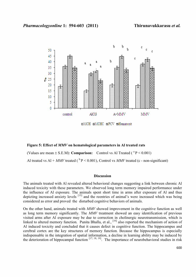
Hematological parameters
The haemoglobin and RBC were significantly (p ≤ 0.001) decreased in Al treated rat’s as compared
with normal control (Fig. 3 and 4). Whereas, in combined treatment (MMV with Al), the haemoglobin
and RBC level were significantly (p ≤ 0.001) increased as compared with Al treated rats. The levels of
HBS and RBC were within the normal limit in MMV alone treated rats. The other hematological
parameters such as MCH, MCHC, PCV and hematocrit (Hct) were significantly (p ≤ 0.001) decreased
in Al induced rats (Figure 5). On the other hand, the MCH, MCHC, PCV and hematocrit (Hct) level
were significantly (P ≤ 0.001) increased in MMV along with Al treated rats. There was no any
significant change in the MCH, MCHC, PCV and hematocrit (Hct) level in MMV alone treated rats
and the values were within the normal limit.

Pharmacologyonline 1: 594-603 (2011) Thirunavukkarasu et al.
599
Figure 3: Effect of MMV on hemoglobin changes in Al treated rats
(Values are mean ± S.E.M); Comparison: Control vs Al treated ( a P < 0.001),
Al treated vs Al + MMV treated ( b P < 0.001), Control vs MMV treated (c - non-significant).
Figure 4: Effect of MMV on RBC changes in different group of rats
(Values are mean ± S.E.M); Comparison: Control vs Al treated ( a P < 0.001),
Al treated vs Al + MMV treated ( b P < 0.001), Control vs MMV treated (c - non-significant)
Pharmacologyonline 1: 594-603 (2011) Thirunavukkarasu et al.
600
Figure 5: Effect of MMV on hematological parameters in Al treated rats
(Values are mean ± S.E.M): Comparison: Control vs Al Treated ( a P < 0.001)
Al treated vs Al + MMV treated ( b P < 0.001), Control vs MMV treated (c - non-significant)
Discussion
The animals treated with Al revealed altered behavioral changes suggesting a link between chronic Al
induced toxicity with these parameters. We observed long term memory impaired performance under
the influence of Al exposure. The animals spent short time in arms after exposure of Al and thus
depicting increased anxiety levels [15]
and the reentries of animal’s were increased which was being
considered as error and proved the disturbed cognitive behaviors of animals.
On the other hand, animals treated with MMV showed improvement in the cognitive function as well
as long term memory significantly. The MMV treatment showed an easy identification of previous
visited arms after Al exposure may be due to correction in cholinergic neurotransmission, which is
linked to altered memory function. Punita Bhalla, et al., [16] also reported the mechanism of action of
Al induced toxicity and concluded that it causes defect in cognitive function. The hippocampus and
cerebral cortex are the key structures of memory function. Because the hippocampus is especially
indispensable in the integration of spatial information, a decline in learning ability may be induced by
the deterioration of hippocampal function [17, 18, 19]
. The importance of neurobehavioral studies in risk

Pharmacologyonline 1: 594-603 (2011) Thirunavukkarasu et al.
601
assessment lies in the fact that behavior can be regarded as the net output of the sensory, motor and
cognitive functions occurring in the nervous system and can serve as potentially sensitive end points of
chemically induced neurotoxicity [20]
.
Al is known to concentrate in the water-lipid interface of membrane and interact with the phosphates
of the external hemilayer, thus diminishes the membrane external surface area. Additionally, Al can
disrupt the bilayer structure, causing a redistribution of membrane lipids, leading to shape change [21]
.
The Al results in significant hemorheological and hematological changes in rats such as a decrement in
RBC deformability at low shear stress levels, aggregation process and an increment in whole blood
viscosity at both native and standard Hct. The MCV, Hb and Hct of rats exposed to Al2 (SO4)3 has
been also found to be decreased. There is evidence that anemia is associated with Al accumulation in
the plasma and /or bone tissue which is known to cause chronic renal insufficiency [22]
. The present
results indicated that Al treatment resulted in a significantly decreased haemoglobin (Hb), total
erythrocytic count (TEC) and packed cell volume (PCV) along with increased while total leukocyte
count (TLC). Vittori et al. [23]
have reported morphological changes in the erythrocyte after Al toxicity
and finally the cell loses their typical biconcave shape. They also suggested that aluminium may
disturb erythropoiesis through combined effects on mature erythrocytes and cellular metabolism in late
erythroid progenitors. Also, the inhibition in erythropoiesis and iron metabolism due to aluminium
treatment probably hinders haemoglobin synthesis and erythroid cell maturation [24, 25]. On the other
hand, significant improvement in these parameters after MMV treatment it self suggests the protective
role of this drug against Al induced toxicity.
The increasing use in preparation and storage of food in Al vessels, cans, and foils may increase the
Al content, particularly in the food that are salty, acidic, or alkaline. There has been little concern
about toxic consequences of Al ingestion because the bioavailability was considered to be poor [26]
and
the gastrointestinal tract normally represented a barrier to Al absorption under normal circumstances
but this barriercan be breached [27]
. It has been clearly shown that individuals ingesting large amounts
of Al compound do absorb significant amounts resulting elevate plasma levels [26, 27]. In spite of this,
a persistent intoxication would cause compensating mechanisms to be triggered, leading to restoring
the hematocrit and hemoglobin concentration with a concomitant persistence of microcytosis and a
decrease MCH [28]
. The effects of Al on erythroid progenitors and on mature erythrocytes and the toxic
effects on erythropoiesis may be responsible for the decrease of hemoglobin and hematocrit levels [29].
It is already known that Al disorganizes the erythrocyte membrane by altering its mechanical
properties, suggesting a reduction of the mean lifespan of circulating erythrocytes, which could play a
major role in the anemia [30]
. Thus, the present findings revealed that the MMV can very well protect
the cells against the toxic stress caused by Al and it may be due to the presence of active molecules
such as saponin and flavanoids [Thirunavukkarasu, et al., 2010] which are well known antioxidants..
Acknowledgement
Authors are thankful to the Indian Council for Medical Research, New Delhi, for providing the
financial assistance and to the SASTRA University for PhD registration of Mr. S.V.Thirunavukkarasu.
References
1. Abbasali KM, T. Zhila and N. Farshad, 2005. Developmental Toxicity of Aluminium from High Doses
of AlCl3 in Mice. The J Appl Res 5: 575-579.

Pharmacologyonline 1: 594-603 (2011) Thirunavukkarasu et al.
602
2. Miu, A.C. and O. Benga, 2006. Aluminum and Alzheimer’s disease: a new look. Journal of
Alzheimer’s Dis 10:179–201.
3. Edwardson, J.A., J.M. Candy, P.G. Ince., F.K. McArthur., C.M. Morris, A.E. Oakley., G.A. Taylor,
and E. Bjertness. 1992. Aluminium accumulation, beta-amyloid deposition and neurofibrillary changes
in the central nervous system. Ciba Foundation Symposium, 169: 165–179.
4. Jeffery, E.H., Abreo K., Burgess E., Cannata J.B. and J.L. Greger. 1996. Systemic aluminium toxicity:
effects on bone, hematopoietic tissue, and kidney. Journal of.Toxicol Environ.Health, 48, 649.
5. Yokel, R.A, and P.J. Mcnamara, 2001. Aluminium Toxicokinetics: An Updated Mini Review. Pharmacol
and.Toxicol 88: 159.
6. Strong, M.J., Garruto, R.M., Joshi, J.G., Mundy, W.R. and T.J. Shafer, 1996. Can the mechanisms of
aluminium neurotoxicity be integrated into a unified scheme? Journal of Toxicol Environ Health., 48
(6): 599–613.
7. Ochmanski, W. and W. Barabasz. 2000. Aluminium-occurrence and toxicity for organisms of rat
fetuses and sucklings. Brain Res Bull., 55:229-234.
8. Julka, D. and K.D. Gillm. 1996. Altered calcium homeostasis: a possible mechanism of aluminium
induced neurotoxicity. Biochem Biophys Acta., 135: 47–54.
9. Kumar, V. and K.D. Gill. 2009. Aluminium neurotoxicity: neurobehavioural and oxidative aspects.
Arch Toxicol 83,965–978.
10. Thirunavukkarasu, S.V ., Venkataraman, S., Lokesh Upadhyay. 2010. In vitro antioxidant and
antibacterial activity of Polyherbal Manasamitra vatakam (MMV) drug. J Pharm Res 3(8): 2042 -2047.
11. Thirunavukkarasu, S.V., Venketaraman, S., Lokesh Upadhyay. 2010. Ameliorating effect an ayurvedic
herbo mineral preparation in AlCl3 induced cognitive dysfunction in rats. Biomedicine 30 (1): 40-47.
12. OECD. 2001. OECD guidline for testing of chemicals : guidline 423. Acute toxicity – Acute toxic
class Method, OECD, parris, France, http://iccvam.nieh.gov/suppDocs/ FedDocs/OECD.
13. Paxinos, G. and C. Watson, 1982. The Rat Brain in Stereotaxic Coordinates. Academic Press, New
York.
14. Kulkarni SK, (1999). Handbook of Experimental Pharmacology, 3rd edn. Vallabh. Prakashan, Delhi.
15. Elliott JM, Heal DJ, Marsden CA. Experimental Approaches to Anxiety and Depression. Chichester
New York: John Wiley, 1992.
16. Punita Bhalla, M.L. Garg, and D.K. Dhawan, 2010. Protective role of lithium during aluminium
induced neurotoxicity. Neuro Intersci 56: 256- 262.
17. Hu, H., Y.J. Yang, X.P. Li, and G.H. Chen. 2005. Effect of aluminum chloride on motor activity and
species-typical behaviors in mice. Zhonghua Lao Dong Wei Sheng Zhi Ye Bing Za Zhi., 23 (2): 132–
135.
18. Levesque L., C.A. Mizzen, D.R. McLachlan and P.E.Fraser . 2000. Ligand specific effects on
aluminium incorporation and toxicity in neurons and astrocytes. Brain Res 877: 191 – 202.
19. Evans, H.L. 1995. Markers of neurotoxicity: from behavior to autoantibodies against brain proteins.
Clinic Chem 41 (12 Pt. 2): 1874–1881.
20. Davies, P. 1990. Neurotransmitter-related enzyme in dementia of Alzheimer's type. Brain Res. 171:
319-327.
21. Connor, D.J., Harrell, L.E, and R.S. Jope, 1989. Reversal of an aluminum-induced behavioral deficit
by administration of deferoxamine. Behav Neurosci 103 (4): 779 – 783.
22. Fulton, B. and E.H. Jeffery, 1994. Heme oxygenase induction: a possible factor in aluminium-
associated anemia. Biol.Trace Element Res 40, 9.
23. Vittori, D., G. Garbossa, C. Lafourcade, G. Perez, A. Nesse, 2002. Human erythroid cells are affected
by aluminium. Alteration of membrane band 3 protein. Biochim Biophys Acta 1558, 142–150.

Pharmacologyonline 1: 594-603 (2011) Thirunavukkarasu et al.
603
24. Chmielnicka, J., M. Nasiadek., and E. Lewandowskażyndul. 1993. Effect of aluminium on some
stages of heme biosynthesis in rats. Roczn. PZH 44, 103.
25. Suwalsky, M., B.Un.gerer, F. Villena, B. Norris, H. Cardenas, and P. Zatta, 2001. Effects of AlCl on
toad skin, human erythrocytes, and model cell membranes. Brain Res Bull 55: 203-210.
26. Kowall, N.W., W.W. Pendlebury, J.B.Kesler, D.B. Perl, and M.F. Beal, , 1989. Aluminiuminduced
neurofibrillary degeneration affects a subset of neurons in rabbit cerebral cortex, basal forebrain and
upper brainstem. Neurosci 29: 329–377.
27. Mahdi, A.A., R. Chander, N.K. Kapoor, and S. Ahmad S. 1992. Role of free radical in Plasmodium
berghei infected mastomys natalensis brain. Ind J Exp Biol 30: 1193–1196.
28. Helliwell, B. and J.M.C. Gutteridge, 1990. The measurement and mechanism of lipid peroxidation in
biological systems. Trends in Biochem Sci 15: 129–135.
29. Shiga, T, Maeda, N. and K. Kon., 1990. Erythrocyte rheology. Crit Rev Oncol Hematology 10(1): 9–48.
30. Struys-Ponsar C, Florence A, Gauthier R, Crichton R, van den Bosch and P. de Aguilar, 1994. Ultra
structural changes in brain parenchyma during normal aging and in animal models of aging. J Neural
Transmision 44:111–132.