Embed Size (px)
Citation preview

Pain Management Research Institute/Orofacial/Phantom Tooth Pain 1
Phantom Tooth Pain Dr Russell Vickers PhD
Phantom tooth pain is a form of neuropathic pain of the oral cavity and further information can be found under that section. The information on this page is aimed at dental practitioners. The information is in two parts and is based on the following publications:
Vickers ER, Cousins M. Neuropathic orofacial pain. Part 1: Prevalence and pathophysiology. Australian Endodontic Journal 2000; 26: 19-26.
Vickers ER, Cousins M. Neuropathic orofacial pain. Part 2: Diagnostic procedures, treatment guidelines and case reports. Australian Endodontic Journal 2000; 26: 53-63.
Glossary of terms
after discharge continued firing of dorsal horn neurons due to repetitive peripheral stimulation
algesic pain producing
algogen pain producing substance
algogenic pain producing
allodynia pain from stimulus that does not normally cause pain
analgesic pain relieving
atypical facial pain a term no longer used but was characterised by vague signs and symptoms with significant psychological factors
atypical odontalgia severe, throbbing pain without major pathology, phantom tooth pain
central sensitisation a phenomenon occurring in the dorsal horn and other central structures causing allodynia and secondary hyperalgesia in uninjured tissue surrounding a site of injury
complex regional pain syndrome (CRPS) type I a syndrome that usually develops after an initiating noxious event, is not limited to the distribution of a single peripheral nerve, and is apparently disproportionate to the inciting event
CRPS type II burning pain, allodynia and hyperpathia usually in the hand or foot, after partial injury to a nerve or one of its major branches
hyperalgesia increased response to a painful stimuli neuronal plasticity nerve sprouting and neuroma formation causing neuropathic pain
neuropathic pain pain initiated or caused by a primary lesion or dysfunction in the nervous system
neuropraxis crush injury to a nerve

Pain Management Research Institute/Orofacial/Phantom Tooth Pain 2
neurotmesis sectioning or cutting of a nerve
nociception activation of peripheral nociceptor which is recognised centrally as pain
peripheral sensitisation a phenomenon where inflammatory mediators sensitise high threshold nociceptors
pharmacodynamics effect of drug on body
pharmacokinetics metabolic action of body on drug
physiological pain pain that serves a protective function (as a warning for tissue damage), is transient and well localised
sympathetic hyperfunction characterised by changes in skin temperature, blood flow, resting sweat output and presence of oedema
sympathetically maintained pain defined as pain that is maintained by sympathetic efferent innervation or circulating catecholamines
windup progressive increase in response of dorsal horn neurons due to repetitive peripheral stimulation
PART I – PREVALENCE AND PATHOPHYSIOLOGY Summary
Neuropathic pain is defined as “pain initiated or caused by a primary lesion or dysfunction in the nervous system”. Neuropathic orofacial pain has previously been known as “atypical odontalgia” (AO) and "phantom tooth pain". The patient afflicted with neuropathic oral / orofacial pain may present to the endodontist with a persistent, severe pain yet there are no clearly identifiable clinical or radiographic abnormalities. Accordingly, multiple endodontic procedures may be instigated to remove the likely anatomical source of the pain, yet the pain persists. There have been few studies and limited patient numbers investigating the condition. Two retrospective studies revealed the incidence of persistent pain following endodontic treatment to be 3-6% and 5% of patients; one author with wide experience in assessing the condition estimated its prevalence at 125,000 individuals in the USA alone. Dental treatment appears closely related to the onset of pain, 46% and 74% of patients, respectively, in two studies. In one other study, 50% of neuropathic orofacial pain patients reported persistent pain specifically following endodontic treatment. Patients predisposed to the condition may include those suffering from recurrent cluster or migraine headaches.
Neuropathic pain states include postherpetic neuralgia (shingles) and phantom limb / stump pain. The aberrant developmental neurobiology leading to this pain state is complex. Neuropathic pain serves no protective function, in contrast to physiological pain that warns of noxious stimuli likely to result in tissue damage. The relevant clinical features of neuropathic pain include: (i) precipitating factors such as trauma or disease (infection), and often a delay in onset after initial injury (days - months), (ii) typical complaints such as dysaesthesias (abnormal unpleasant sensations), pain that may include burning, and paroxysmal, lancinating or sharp qualities, and pain in an area of sensory

Pain Management Research Institute/Orofacial/Phantom Tooth Pain 3
deficit, (iii) on physical examination there may be hyperalgesia, allodynia and sympathetic hyperfunction, and (iv) the pathophysiology includes deafferentation, nerve sprouting, neuroma formation and sympathetic efferent activity.
Introduction
“The pain in my jaw is like a bad, nagging toothache or migraine pain, which can last up to two or three days. Sometimes I feel like taking my life because of the pain.” - a patient diagnosed with neuropathic orofacial pain.
Neuropathic pain is defined as “pain initiated or caused by a primary lesion or dysfunction in the nervous system” by the International Association for the Study of Pain (IASP) (1). Neuropathic oral / orofacial pain is a chronic pain condition relevant to the endodontist due to the likelihood of that practitioner being the first to recognise the problem. The condition has previously been known as “atypical odontalgia” (AO) and has been defined as a "severe throbbing pain in the tooth without major pathology" (1). Documentation of the condition in the dental literature is scarce, and similarly, there is a paucity of information regarding diagnostic and treatment / management procedures. Neuropathic orofacial pain was first described as a painful and unusual condition that occurs in the dentoalveolar structures and oral mucosa (2), with patients reporting pain that was moderate to severe in intensity, and with a pattern of referral that may cross the anatomical midline (of the mandible and maxilla) and possibly involve the face. Pain can occur in single or multiple sites (mucosal sites and single / multiple teeth) (Fig. 1). It has also been described as "phantom tooth pain" due to insufficient data in providing a physiological explanation, and its similarities compared with phantom limb pain (3, 4). The lack of clinical and radiographic evidence to establish a diagnosis pertaining to organic pathology gave credence to early investigators that there was a psychogenic basis to the condition (2).
There have been relatively few studies investigating the condition, and consequently, a limited number of patients have been analysed, with the largest cohorts of subjects reported by Rees and Harris 1979 (n=44) (2), Marbach 1978 (n=25) (5), Brooke 1980 (n=22) (6), Pöllman 1993 (n=44) (7), and Vickers et al. 1998 (n=50) (8). However, most of this published data is epidemiological and there is little data available for clinicians with respect to diagnostic tests and treatment / management strategies. The aetiology of neuropathic orofacial pain is not well understood, nor studied, but the limited data available show endodontic procedures to possibly account for up to 50% of the cases (9). The association between neuropathic pain and endodontic procedures as a possible causal factor is discussed in further detail in a later section.
The patient afflicted with neuropathic oral / orofacial pain may present to the endodontist with a persistent, severe pain yet there are no clearly identifiable clinical or radiographic abnormalities. Accordingly, multiple endodontic procedures may be instigated to remove the likely anatomical source of the pain. However, the resolution of neuropathic pain does not rely on surgical intervention but on appropriate pain management strategies. Indeed, where multiple surgical interventions are carried out, the likely outcome is a worsening of the condition. In addition to the challenge for the endodontist regarding

Pain Management Research Institute/Orofacial/Phantom Tooth Pain 4
diagnosis and management, the lack of pain relief from previous procedures may cause some patients to display openly negative emotions such as frustration, anger, mistrust and hostility toward his / her endodontist, based on patient psychological factors such as their expectations and assurances of cure (pain relief) from surgical intervention. As a consequence of the chronicity of the complaint and the high levels of pain associated with the condition, there is the possibility of psychological morbidity, and unfortunately, the real potential of suicide in some patients. Neuropathic pain has only recently been recognised by dental and medical practitioners as a pain state that deserves further understanding. Because of its recent awareness, its epidemiology and pathophysiology are undergoing extensive research in order that preventive measures and effective clinical treatments become possible. While much of this research is directed toward neuropathic pain states associated with medical conditions, there is now a growing awareness in the dental field for the need to investigate orofacial presentations of neuropathic pain. The purpose of this paper is to present our current knowledge of neuropathic pain in two parts: (1) its epidemiology and pathophysiology, and (2) its clinical appearance, appropriate diagnostic tests and management.
Historical perspectives
The first description of phantom tooth pain was by John Hunter over 200 years ago:
“There is one disease of the jaws which seems in reality to have no connection to the teeth, but of which the teeth are generally suspected to be the cause. This deserves to be taken notice of in this place, because operators have frequently been deceived by it, and even sound teeth have sometimes been extracted through an unfortunate mistake.
This pain is seated in some part of the jaws. As simple pain demonstrates noting, a tooth is often suspected, and is perhaps drawn out; but still the pain continues, with this difference, however, that it now seems to be the root of the next tooth; it is then supposed by either the patient or the operator, that the wrong tooth was extracted; wherefore, that in which the pain now seems to be is drawn, but with as little benefit. I have known cases of this kind, where all the teeth of the affected side of the jaw have been drawn out, and the pain continued in the jaw; in others, it has had a different effect, the sensation of pain has become more diffused, and has at last, attacked the corresponding side of the tongue. In the first case, I have known it recommended to cut down upon the jaw, and even to perforate and cauterize it, but all without effect. I have seen cases of some years standing, where the hemlock has succeeded when the bark has had no effect; but sometimes all attempts prove unsuccessful.”
Nomenclature
A problem for dental practitioners in diagnosing neuropathic pain has been the use of various terms ascribed by different investigators over the years. Originally, the condition was described as AO in response to the 'atypical' nature of the condition; this term was subsequently listed in the Taxonomy of Chronic Pain Syndromes by the IASP (1). Other terms have included idiopathic odontalgia (11), neurovascular odontalgia (12), and phantom tooth pain (5). More recently, neuropathic pain (“oral neuropathic pain” /

Pain Management Research Institute/Orofacial/Phantom Tooth Pain 5
“neuropathic orofacial pain”) has been the term used, and is based on the criteria of the condition’s underlying pathophysiology (3, 4, 9, 13). It is now accepted that the type of pain state be used, and is further qualified by the location of the pain or any associated medical condition; for example, a patient with pain as a result of diabetic neuropathy. However, it is important to point out that ‘neuropathic orofacial pain’ is an all embracing term and that several subsets may exist according to the location and aetiology of the neuropathic pain. For example, AO or phantom tooth pain implies pain in the mucosa at the site of a tooth extraction, often with a history of frequent previous restorative and endodontic procedures having been carried out on the tooth in question preceding extraction. Other locations of pain may be the mucosa and gingivae as a result of periodontal therapy – but it is difficult to say whether the pain has arisen in the periodontal tissues from scaling procedures, pulpal stimulation or both. More recently, there is now preliminary evidence to suggest that burning mouth / tongue syndrome (glossodynia, glossopyrosis) may be another form of neuropathic pain (14).
Prevalence
Neuropathic pain is observed in several different medical conditions including postherpetic neuralgia, phantom limb / stump pain, post spinal cord injury pain and complex regional pain syndromes. The incidence of neuropathic pain varies according to the underlying causal factor: (i) postherpetic neuralgia occurs in up to 75% of elderly patients with zoster infection but only 10-15% of the younger age group who suffer the condition, (ii) between 2-97% of patients may develop postamputation pain in the limb (phantom limb pain), (iii) 60% of patients with traumatic spinal cord injury have pain one year after the event, and (iv) complex regional pain syndrome type II (previously termed causalgia) occurs in 2-5% of patients after peripheral nerve injury. Other causal factors in the peripheral nervous system include malignant invasion of nerves and direct trauma to a nerve such as crush injury (neuropraxis), section or cutting (neurotmesis), or stretch / avulsion. Central nervous system (CNS) trauma, tumour and ischaemia can also be responsible for the development of neuropathic pain. The incidence of neuropathic pain following surgery is estimated to be 1-2% but probably varies according to the type of operation e.g. increased incidence in post thoracotomy (15). Postoperative neuropathic pain is more prevalent following amputation when there is concurrent pain, and when there has been severe postoperative pain. In addition, new research is focussing on the role of genetic factors in pain (16). Postherpetic neuralgia is described as the most common form of “neuropathic orofacial pain”, with Lynch and Elgeneidy (9) identifying 17 patients (0.3%) out of 5,000 patients referred to a medical pain management clinic; male / female ratio 2/15, and an age range of 26-58 years. The incidence of postherpetic neuralgia (all anatomical locations) is 125 per 100,000 of the population (17).
No formal prospective studies have been carried out to assess the extent of neuropathic orofacial pain. The only available data are several retrospective studies investigating potential causal factors (Table 1). Marbach et al. examined the possible role of endodontic procedures in triggering the condition, and while the study was limited in being able to draw definitive conclusions (it only investigated chronic pain in patients from one endodontist), it at least provides a window regarding the possible extent of the condition (10). In this study, 732 questionnaires were mailed out to patients who had

Pain Management Research Institute/Orofacial/Phantom Tooth Pain 6
completed endodontic treatment from one endodontist, with 463 (63%) usable responses. Marbach et al. found 3% of female patients met all four criteria of “phantom tooth pain”, including no obvious physical or radiographic abnormalities, and 6% of the group manifested three out four criteria. An interesting aspect of this study, and one that limited Marbach’s analysis of the problem, was the reluctance of four other endodontists to participate in the study and who remarked “Perhaps our greatest anxiety, however, is over the fact that a Pandora’s box would be opened up.” A later publication by Marbach (3) extrapolated these earlier findings and suggested that neuropathic orofacial pain (from endodontic procedures) may be present in some 125,000 individuals in the USA alone. In another study that reviewed 118 patients who had completed surgical endodontics, six patients (5%) had persistent pain following surgery, three patients had pain before the surgery and there was no postoperative pain reduction, and three patients developed pain following surgery (18). Nicolodi and Sicuteri (19), examined the prevalence of neuropathic pain in a group of patients suffering from “idiopathic headache” who had tooth extractions. They found that no one in the control group (n = 280, no history of recurrent headache) had persistent post tooth extraction pain. In the test group (n = 301, female / male = 171/130, mean age of 48 years) pain in the edentate site was reported by 20% (10/50) of cluster headache sufferers, and 14% of migraine sufferers. They also found that the more teeth extracted, the greater the pain, and the incidence of neuropathic pain was directly proportional to the number of teeth extracted. “Toothache” was the cause for tooth extraction in 67% of the headache group. Furthermore, in a cohort of chronic idiopathic orofacial pain patients, “dental therapy” was associated with the onset of pain in 28/50 cases (20).
A potential issue in determining the extent of the problem, has been the difficulty faced by various practitioners and investigators in diagnosing pain conditions, or in obtaining a correct diagnosis. For example, one study reported that there was no clearly identifiable cause in 38% of patients in a chronic orofacial pain group (21), and another study of several case reports described patients of having a diagnosis of atypical facial pain (or “atypical facial neuralgia”), yet these patients clearly fulfilled the description of neuropathic orofacial pain (22). It is plausible that other patients are misdiagnosed, thus underestimating the prevalence of neuropathic pain. Aghabeigi et al. (23) examined another group of “chronic idiopathic orofacial pain patients” whose features were suggestive of a diagnosis of neuropathic pain, and 15% of patients had evidence of post traumatic stress disorder (PTSD) that coincided with the onset of pain. However, no study has examined if the converse is true, that is, if there is a higher prevalence of neuropathic orofacial pain among PTSD patients compared with the general population. Psychological factors may well play a part in the development of neuropathic pain through unrealistic expectations of treatment; 65% of chronic pain patients (back, neck and limb) still had strong beliefs in the possibility of medical cure (including further surgery) for their pain despite an average of 9.7 years of pain, and of these patients, 60% had evidence of overinvestigation / treatment (24). Thus, in some cases of neuropathic pain, psychological variables may be indirectly responsible for the development of the condition through unnecessary additional treatment that is possibly the direct causal factor. The prevalence of neuropathic orofacial pain based on the figures suggested by Marbach may be an extraordinary claim (125,000 possible subjects in the USA), but it should be noted that the majority of people with chronic pain (for example, back pain) do
Pain Management Research Institute/Orofacial/Phantom Tooth Pain 7
not in fact seek extensive investigation and treatment - there may well be a ‘silent majority’ of orofacial pain patients as alluded to by the endodontists in the Marbach study (3). The reason that patients seek treatment for a chronic pain complaint is complex and multifactorial. It may involve ‘pain relief’, by reducing peripheral nociception and sensory aspects of neuropathic pain, altering the perception of pain information in central areas, and it may involve the management of ‘suffering’ (or psychological distress).
Further prospective studies evaluating the prevalence of neuropathic orofacial pain are warranted. In particular, specific studies concerning the association or development of neuropathic pain from endodontic procedures, as indicated by Lynch et al. (9), may prove valuable in a greater understanding of causal factors in medical variants of neuropathic pain.
Neurobiology of Pain and Pathophysiology of Neuropathic Pain
The purpose of pain is to provide an early warning of potential tissue damage leading to the initiation of appropriate withdrawal reflexes. In addition, pain initiates complex neurohumoral responses that help to maintain homeostasis in acute injury and pain. The response to tissue damage is similar irrespective of the injury such as a surgical incision, traumatic injury (e.g. fracture), soft tissue damage, disease processes (e.g. infection, cancer), and burns. While the degree of trauma and pain may be expected to correlate with the degree of the injury, there is a wide individual variation in the response to an injury, with evidence indicating that both physiological and psychological factors are involved. While most pain from injury proceeds through an orderly course, there is the potential for the development of vicious cycles of pain. Depending on the severity of trauma, peripheral sensitisation and areas of the CNS may be activated, leading to persistent or pathophysiological pain. The following summary of pain mechanisms is based on the extensive review by Siddall and Cousins (25).
Mechanisms in ‘physiological’ pain and neuropathic pain
There are several features of ‘physiological’ pain occurring after a brief noxious stimulus:
1. Pain from A delta and C fibres can be differentiated from A beta fibres. Low intensity, non-noxious stimuli activate low threshold receptors (A beta fibres), and high intensity noxious stimuli activate high threshold receptors (A delta and C fibres) that may lead to the sensation of pain.
2. Pain serves as a protective function. 3. Pain acts as a warning for potential tissue damage. 4. Pain is transient. 5. Pain is well localised. 6. The stimulus-response pattern is the same as other sensory modalities (e.g. touch).
Peripheral sensitisation
An acute injury leads to the Lewis triple response of oedema (wheal), vasodilation (flare) and pain. There is an antidromic response (towards the periphery) along collaterals of

Pain Management Research Institute/Orofacial/Phantom Tooth Pain 8
primary afferent fibres which results in the release of chemical mediators to cause an increased response to the noxious stimulus. Nociception primarily involves activation of C fibre afferents by thermal, chemical and mechanical stimuli. The triple response of Lewis describes the sequence of events at the site of injury, and involves the peripheral nervous system (PNS). During inflammation there is a release of intracellular contents from damaged cells and inflammatory cells including macrophages, lymphocytes and mast cells. Nociceptive stimulation also causes the release of peptides and hormones such as substance P, neurokinin A and calcitonin gene-related peptide (CGRP) from primary afferent terminals. These peptides alter the excitability of nociceptors (and sympathetic fibres), and induce vasodilation and extravasation of plasma proteins, and cause inflammatory cells to release chemical mediators. This produces a ‘sensitising soup’ of inflammatory mediators, including reactive oxygen species (ROS), histamine, cytokines, potassium, serotonin, bradykinin, nitric oxide (NO), leukotrienes, and prostaglandins and enzymes from the cyclooxygenase and lipoxygenase pathways. Following sensitisation, painful stimuli are perceived from low intensity, mechanical stimuli that are not normally painful, a condition termed allodynia. Furthermore, hyperalgesia may develop in adjacent tissues, hyperalgesia being defined as an increased response to painful stimuli (Fig. 2). Noxious skin stimuli sufficiently intense to produce tissue injury usually generate prolonged poststimulus sensory disturbances including allodynia and hyperalgesia. Hyperexcitability, in part, arises from changes in activity of the spinal cord. Long term consequences of pain thus result from both peripheral and central changes. These changes result in disturbance to the area of injury (primary hyperalgesia) but also to undamaged adjacent areas (secondary hyperalgesia and referred pain). Several factors have been demonstrated to be involved in the development of this form of pain. First, brief stimulation of peripheral, unmyelinated, afferent fibres can produce substantial and prolonged changes in the cutaneous receptive fields of dorsal horn neurons. The response of dorsal horn neurons can be influenced by the ‘history’ of previous noxious stimuli. Second, repetitive peripheral stimulation results in a progressive increase in response of dorsal horn neurons, termed “windup”, and continued firing, termed “after discharge”. Studies have also shown that after discharge is four times longer in a stimulated, sectioned peripheral nerve compared with an intact nerve. Third, in the presence of an intact nervous system, noxious C afferent fibre input usually does not produce prolonged excitability of the flexion reflex (in rat studies). In contrast, noxious muscle stimulation does produce prolonged changes in the reflex. Consequently, muscle trauma and spasm that can produce local ischaemia and inflammation may cause changes at the spinal level, leading to the development of a vicious cycle of enhanced muscle spasm and progressive increases in pain and the injury response. Fourth, the mechanism of long lasting changes in excitability of spinal neurons is related partially to N-methyl-D-aspartate (NMDA) receptors, with second messengers such as calcium, cyclic AMP and diacylglycerol (DAG) triggering prolonged changes. In addition, third messengers such as the protooncogene C-fos permit genetic encoding of an altered pattern of enhanced responsiveness of dorsal horn neurons.
NMDA activation can cause facilitation, windup, central sensitisation, changes in peripheral fields and induction of oncogenes. At the level of the CNS, neuropathic pain can be seen through a well studied marker, C-fos expression. C-fos is a protooncogene protein that can be measured by immunoreactivity. It targets genes, leading to genetic

Pain Management Research Institute/Orofacial/Phantom Tooth Pain 9
changes and subsequent long term responses e.g. to pain. It is induced in spinal cord neurons by noxious stimuli. Innocuous peripheral stimuli can also induce C-fos expression, but to a very limited extent; for example, capsaicin administration in an acute inflammation model showed C-fos expression to be present for less than 24 hours. In animal studies of pain, inflammation and drug pharmacodynamics (i.e. the effect of the drug on the body) using rat spinal cord, C-fos expression is induced or modulated in various laminae. With peripheral stimulation, the superficial laminae (I-II) of the spinal cord exhibit C-fos expression. However, in the case of allodynia in the rat model, C-fos expression in the deeper laminae is elicited, indicating the involvement of A-( afferents. C-fos expression thus provides important information on possible central mechanisms of neuropathic pain and the potential benefits of preemptive drug treatment. For example, intravenous morphine and morphine administered to the peripheral injury site has been shown to significantly decrease C-fos expression from intraplantar carrageenin in the rat. Other drugs demonstrating an effect on C-fos include local anaesthetics (lignocaine and bupivacaine), remifentanil, dexamethasone and nonsteroidal antiinflammatory drugs (NSAIDS).
Neuronal plasticity
Pathophysiological pain such as neuropathic pain can be explained, in part, by nervous system plasticity (Fig. 3). Involved in neuroplasticity are peptides such as nerve growth factor (NGF), a prime initiator of nerve ‘sprouting’ that regulates neuronal growth. However, in neuropathic pain NGF may play a role in altering responsiveness to sensory input. Neuroplasticity may occur at the peripheral and / or spinal cord level. The adjacent diagram was completed by a patient who underwent a left jaw joint operation and over the course of several years the pain spread to involve the entire left side of the body.
Damaged nerve fibres may also produce neuromas. Peripheral abnormalities can result from sensitisation of nociceptors or axon damage, permitting ectopic locations for spontaneous neuronal discharge (peripheral neuromas). These changes may also occur along the course of axons and produce microneuromas. Neuromas are also capable of spontaneous discharge, greatly enhanced and prolonged discharges, and show minimal accommodation. Properties of neuromas include sensitivity to mechanical stimuli, spontaneous firing and sensitivity to noradrenaline. The sensitivity of neuromas appears to be related to the time since nerve injury, being most intense within the first two weeks of neuroma formation, but then maintained at a lesser, continued level. Clinically, this situation is recognised by the production of intense, electric, shock like pain at the site of the neuroma, and radiating pain on percussion of the neuroma (Tinel’s sign). Damage to axon sheaths may also result in demyelination, resulting in nerve hyperexcitability. This causes paroxysms of pain and afferent activity may initiate reflex motor activity causing

Pain Management Research Institute/Orofacial/Phantom Tooth Pain 10
muscle spasm. This may account for the development of a temporomandibular disorder (TMD) that is secondary to the primary neuropathic oral pain (Fig. 4). Evidence has shown that peripheral neuroma pain may not necessarily be blocked by local anaesthetic at the site, suggesting that central mechanisms are involved. In the clinical situation of patients with neuropathic oral pain, Vickers et al. (8) have shown that there is a wide range of responses to topical anaesthetic application, with patients demonstrating either complete, partial or no pain reduction to somatosensory blockade. Evidence from animal studies suggests that there is a genetic predisposition in the development of spontaneous activity in neuromas. In the clinical situation, patients who develop neuromas frequently have similar problems after attempts to remove them.
In summary, the latter stages of injury involve invasion of the damaged area by phagocytes and fibroblasts. Damaged nerve endings and capillaries sprout and infiltrate into the area where sensory, sympathetic and motor fibres are already present. The C fibres are involved in detecting or ‘tasting’ the altered chemical environment, a phase that coincides with the beginning of scar formation, and the patient’s probable awareness of a persistent pain problem.
Sympathetically maintained pain
Another development in neuropathic pain may be abnormal (ongoing) sympathetic activity, termed sympathetically maintained pain (SMP). SMP is defined as pain that is maintained by sympathetic efferent innervation or by circulating catecholamines. Pathophysiology of SMP probably involves coupling between sympathetic and somatosensory pathways. This has been postulated to occur at peripheral nociceptors, at the dorsal root ganglion with subsequent sprouting of noradrenergic perivascular axons, and at central spinal cord sites. Both direct and indirect methods of excitation of peripheral nociceptors by noradrenaline have been proposed. Following nerve damage or chronic inflammation, a subset of C-polymodal nociceptors has been shown to develop sensitivity to sympathetic stimulation, and thus, may be directly stimulated by noradrenaline. Alternatively, noradrenaline may act indirectly via release of prostaglandins that, in turn, sensitise the nociceptor. Inflammation may also induce nociceptor sensitivity to noradrenaline. In considering endodontic and other dental procedures, the practitioner should consider the possibility, or indeed the likelihood, of preexisting ginigival / periodontal / periapical inflammation and the potential of the interaction of noradrenaline in dental anaesthetic cartridges.
SMP may also involve central mechanisms that may explain the development of hyperalgesia and more generalised pain. Pain transmission pathways may undergo plasticity through changes in receptor sensitivity, genetic changes and neuroanatomical reorganisation, leading to central sensitisation and windup. C-polymodal nociceptors can activate and sensitise wide dynamic range (WDR) neurons that then respond to activity in large diameter A-mechanoreceptors that are activated by light touch. The pain threshold is reduced, and previous subthreshold stimuli are then perceived as painful (i.e. hyperalgesia). This explains the frequently observed response in neuropathic oral pain patients where they cannot tolerate wearing a denture following extraction, particularly where the denture saddle rests upon an area of allodynia or hyperalgesia.
Pain Management Research Institute/Orofacial/Phantom Tooth Pain 11
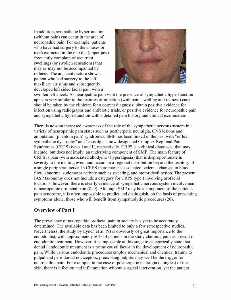
In addition, sympathetic hyperfunction (without pain) can occur in the area of neuropathic pain. For example, patients who have had surgery to the sinuses or teeth extracted in the maxilla (upper jaw) frequently complain of recurrent swellings (or swollen sensations) that may or may not be accompanied by redness. The adjacent picture shows a patient who had surgery to the left maxillary air sinus and subsequently developed left sided facial pain with a swollen left cheek. As neuropathic pain with the presence of sympathetic hyperfunction appears very similar to the features of infection (with pain, swelling and redness) care should be taken by the clinician for a correct diagnosis: obtain positive evidence for infection using radiographs and antibiotic trials, or positive evidence for neuropathic pain and sympathetic hyperfunction with a detailed pain history and clinical examination.
There is now an increased awareness of the role of the sympathetic nervous system in a variety of neuropathic pain states such as postherpetic neuralgia, CNS lesions and amputation (phantom pain) syndromes. SMP has been linked in the past with "reflex sympathetic dystrophy" and "causalgia", now designated Complex Regional Pain Syndromes (CRPS) types I and II, respectively. CRPS is a clinical diagnosis, that may include, but does not imply, an underlying component of SMP. The main feature of CRPS is pain (with associated allodynia / hyperalgesia) that is disproportionate in severity to the inciting event and occurs in a regional distribution beyond the territory of a single peripheral nerve. In CRPS there may be associated oedema, changes in blood flow, abnormal sudomotor activity such as sweating, and motor dysfunction. The present IASP taxonomy does not include a category for CRPS type I involving orofacial locations; however, there is clearly evidence of sympathetic nervous system involvement in neuropathic orofacial pain (8, 9). Although SMP may be a component of the patient's pain syndrome, it is often impossible to predict and distinguish, on the basis of presenting symptoms alone, those who will benefit from sympatholytic procedures (26).
Overview of Part I
The prevalence of neuropathic orofacial pain in society has yet to be accurately determined. The available data has been limited to only a few retrospective studies. Nevertheless, the study by Lynch et al. (9) is obviously of great importance to the endodontist, with approximately 50% of patients in the study claiming pain as a result of endodontic treatment. However, it is impossible at this stage to categorically state that dental / endodontic treatment is a prime causal factor in the development of neuropathic pain. While various endodontic procedures employ mechanical and chemical trauma to pulpal and periodontal nociceptors, preexisting pulpitis may well be the trigger for neuropathic pain. For example, in the case of postherpetic neuralgia (shingles) of the skin, there is infection and inflammation without surgical intervention, yet the patient

Pain Management Research Institute/Orofacial/Phantom Tooth Pain 12
may develop a neuropathic pain. Formal, prospective epidemiological studies are warranted to establish the exact prevalence of this condition associated with general dental treatment (restorations, scaling, extractions) and specifically, with endodontics.
The pathophysiology of neuropathic pain is complex. A great deal of research is currently underway to understand the mechanisms involved in this pain state. Our current knowledge is based on animal research models, and investigations of well described human conditions such as postherpetic neuralgia and phantom limb pain. Published data of neuropathic pain states focuses predominantly on anatomical locations arising from spinal cord dorsal root; there is scarce data pertaining to the trigeminal sensory system. While the sensory nervous system displays identical physiological phenomena (nerve conduction, etc.) throughout the body, differences in the degree of sensory innervation, such as the highly innervated dental pulp and periodontal ligament, may play an important role in the acquisition of neuropathic orofacial pain. A summary of the relevant clinical features of neuropathic pain includes:
• Precipitating factors such as trauma or disease (infection), and often a delay in onset after initial injury (days - months).
• Typical complaints such as dysaesthesias (abnormal unpleasant sensation), pain that may include burning, and paroxysmal, lancinating or sharp qualities, and pain in an area of sensory deficit.
• On physical examination there is hyperalgesia, allodynia and sympathetic hyperfunction (swelling, redness, sweating). The pathophysiology includes deafferentation, nerve sprouting, neuroma formation and sympathetic efferent activity.
References for Part 1
1. Merskey H., Bogduk N. Classification of chronic pain: descriptions of chronic pain syndromes and definitions of pain terms. Seattle, IASP Press, 2nd Ed., 1994, pp. 53-6.
2. Rees R.T., Harris M. Atypical odontalgia. Br J Oral Surg 1979; 16: 212-8. 3. Marbach J.J. Is phantom tooth pain a deafferentation (neuropathic) syndrome ? Part I:
evidence derived from pathophysiology and treatment. Oral Surg Oral Med Oral Pathol 1993; 75: 95-105.
4. Marbach J.J. Is phantom tooth pain a deafferentation (neuropathic) syndrome ? Part II: psychosocial considerations. Oral Surg Oral Med Oral Pathol 1993; 75: 225-32.
5. Marbach J.J. Phantom tooth pain. J Endodont 1978; 12: 362-72. 6. Brooke R.I. Atypical odontalgia. Oral Surg Oral Med Oral Pathol 1980; 49: 196-9. 7. Pöllmann L. Determining factors of the phantom tooth. N Y State Dent J 1993; Dec:
42-5. 8. Vickers E.R., Cousins M.J., Walker S., Chisholm K. Analysis of 50 patients with
atypical odontalgia: a preliminary report on pharmacological procedures for diagnosis and treatment. Oral Surg Oral Med Oral Pathol Oral Radiol Endod 1998; 85: 24-32.
9. Lynch M.E., Elgeneidy A.K. The role of sympathetic activity in neuropathic orofacial pain. J Orofac Pain 1996; 10: 297-305.
Pain Management Research Institute/Orofacial/Phantom Tooth Pain 13
10. Marbach J.J., Hulbrock J., Hohn C., Segal A.G. Incidence of phantom tooth pain: an atypical facial neuralgia. Oral Surg Oral Med Oral Pathol 1982; 53: 190-3.
11. Harris M. Psychogenic aspects of facial pain. Br Dent J 1974; 136: 199-202. 12. Mahan P.E., Alling C.C. Facial pain. Philadelphia, Lea & Febiger, 1991, pp. 304-5. 13. Epstein J.B., Marcoe J.H. Topical application of capsaicin for treatment of oral
neuropathic pain and trigeminal neuralgia. Oral Surg Oral Med Oral Pathol 1994; 77: 135-40.
14. Lauritano D., Spadari F., Formaglio F., Zambellini Artini M., Salvato A. Etiopathogenic, clinical-diagnostic and therapeutic aspects of the burning mouth syndrome. Research and treatment protocols in a patient group. Minerva Stomatologica 1998; 47: 239-51.
15. Brooker C., Cousins M.J., Molloy A.R. Neuropathic pain: a GP’s guide. Modern Medicine 1999; 42: 58-67.
16. Mogil J.S., Grisel J.E. Transgenic studies of pain. Pain 1998; 77: 107-28. 17. Loeser J.D. Herpes zoster and postherpetic neuralgia. Pain 1986; 25: 149-64. 18. Campbell R.L., Parks K.W., Dodds R.N. Chronic facial pain associated with
endodontic therapy. Oral Surg Oral Med Oral Pathol 1990; 69: 287-90. 19. Nicolodi M., Sicuteri F. Phantom tooth diagnosis and an anamnestic focus on
headache. N Y State Dent J 1993; Dec: 35-7. 20. Allerbring M., Haegerstam G. Characteristics of patients with chronic idiopathic
orofacial pain. Acta Odontol Scand 1993; 51: 53-8. 21. Yontchev E., Carlsson G.E., Hedegård B. Clinical findings in patients with orofacial
discomfort complaints. Int J Oral Maxillofac Surg 1987; 16: 36-44. 22. Fishbain D.A., Trescott J., Cutler B., Abdel-Moty E., Steele Rosomoff R., Rosomoff
H.L. Do some chronic pain patients with atypical facial pain overvalue and obsess about their pain ? Psychosomatics 1993; 34: 355-9.
23. Aghabeigi B., Feinmann C., Harris M. Prevalence of post-traumatic stress disorder in patients with chronic idiopathic facial pain. Br J Oral Maxillofac Surg 1992; 30: 360-4.
24. Pither C.E., Nicholas M.K. The identification of iatrogenic factors in the development of chronic pain syndromes: abnormal treatment behaviour?, In: Bond M.R., Charlton J.E., Woolf C.J., ed. Proc VIth World Congress on Pain. Elsevier, New York, 1991, 429-34.
25. Siddall P.J., Cousins M.J. Introduction to pain mechanisms: implications for neural blockade, In: Cousins M.J., Bridenbaugh P.O., ed. Neural blockade in clinical anesthesia and management of pain. Lippincott-Raven, New York, 3rd Ed., 1998, pp. 675-714.
26. Walker S.M., Cousins M.J. Complex regional pain syndromes: including "reflex sympathetic dystrophy" and "causalgia". Anaesth Intensive Care 1997; 25: 113-25.
PART 2 – DIAGNOSTIC PROCEDURES AND TREATMENT GUIDELINES
Summary of Part 2
Neuropathic orofacial pain can be difficult to diagnose because of the lack of clinical and radiographic abnormalities. Further difficulties arise if the patient exhibits significant

Pain Management Research Institute/Orofacial/Phantom Tooth Pain 14
distress and is a poor historian regarding previous diagnostic tests and treatments, such as somatosensory local anaesthetic blockade. Valuable information can be obtained by utilising the McGill Pain Questionnaire that allows the patient to choose words that describe the qualities of their pain in a number of important dimensions (sensory and affective). Basal pain intensity should be measured with the visual analogue scale, a simple instrument that can evaluate the efficacy of subsequent treatments. The endodontist can employ sequential analgesic blockade with topical anaesthetics and perineural administration of plain local anaesthetic to ascertain sites of neuropathology in the peripheral nervous system. These can be performed in the dental chair and in a patient blinded manner. Other, more specific, tests necessitate referral to a specialist anaesthetist at a multidisciplinary pain clinic. These tests include placebo controlled lignocaine infusions for assessing neuropathic pain, and placebo controlled phentolamine infusions for sympathetically maintained pain.
The treatment / management of neuropathic pain is multidisciplinary. Medication rationalisation utilises first-line antineuropathic drugs including tricyclic antidepressants such as amitriptyline and nortriptyline, and possibly an anticonvulsant such as carbamazepine, sodium valproate, or gabapentin if there are sharp, shooting qualities to the pain. Mexiletine, an antiarrhythmic and lignocaine analogue, may be considered following a positive patient response to a lignocaine infusion. All drugs need to be titrated to achieve maximum therapeutic effect and minimum side effects. Topical applications of capsaicin to the gingivae and oral mucosa are a simple and effective treatment in two out of three patients suffering from neuropathic orofacial pain. Temporomandibular disorder is present in two thirds of patients and should be assessed and treated with physiotherapy and where appropriate, occlusal splint therapy. Attention to the patient’s psychological status is crucial and requires the skill of a clinical psychologist and / or psychiatrist with pain clinic experience. Psychological variables include distress, depression, expectations of treatment, motivation to improve, background environmental factors, and advice for the patient concerning self responsibility to maintain taught pain management techniques. Unnecessary dental treatment to ‘remove the pain’ with dental extractions is contraindicated and aggravates neuropathic orofacial pain.
Introduction
The factors contributing to chronic pain fall into three broad categories: nociceptive, neuropathic and psychological / environmental. It is imperative to diagnose what category (or categories) of pain the patient presents with, as the treatment varies. Nociceptive factors result from stimulation of nociceptors by tissue damaging (noxious) stimuli where the nervous system is intact, such as early dental infection / pulpitis. Neuropathic pain results from damage, disease, or complete section (‘deafferentation’) of the peripheral nervous system (PNS) or CNS in the absence of a peripheral noxious stimulus. Psychological and / or environmental factors may predominate when no detectable noxious stimulus or damage to the nervous system is present and pain behaviour is presumed to arise primarily from psychological, social, and / or environmental factors. A patient may present with factors in one, two or all three areas contributing to pain and considerable overlap may exist within an individual, and
Pain Management Research Institute/Orofacial/Phantom Tooth Pain 15
multiple factors may play a role in perpetuating pain behaviour (1). In addition, there is the scope for associated factors such as myofascial pain to act as a contributing factor to persistent or recurrent pain; in the case of neuropathic orofacial pain there is the likelihood that two out of three patients will develop a secondary temporomandibular disorder (2). Iatrogenic causes may play an important role in the further development of neuropathic orofacial pain (Fig. 1). This may be seen by the endodontist or oral surgeon who provides treatment / surgery in a tooth or site of pain where there is no obvious clinical or radiographic abnormality, thereby aggravating any resident neuropathic pain problem.
Neuropathic pain is often reported by patients to be severe, and can be accompanied by significant levels of distress. Moreover, the impact of the pain is not relegated solely to the afflicted patient. The scope of changes attributed to chronic pain includes lost work productivity, reduced levels of social activities, and behavioural changes in the patient, spouse and family. It is now well recognised that early intervention can play a major role in more successful outcomes for patients. The basis of early intervention with its appropriate treatment(s), of course, relies on a correct and early diagnosis utilising validated methods and tests. In addition, the prognosis of neuropathic orofacial pain is dependent on curtailing the traditional Cartesian approach to painful sites where the concept is that the removal or amputation of the suspect site (tooth) will lead to the resolution of the pain. This approach is incorrect treatment for neuropathic pain; indeed, only further iatrogenic problems will be created. The development of a TMD secondary to the neuropathic pain is a prime example, in part caused by the unnecessary extraction of teeth, and will be further discussed. The purpose of Part 2 is to provide comprehensive guidelines, utilising our current state of knowledge, to enable the endodontist in addition to routine clinical and radiographic examinations, to understand, diagnose and manage where appropriate, chronic neuropathic orofacial pain. Furthermore, there are relatively simple indicators on instruments such as the McGill Pain Questionnaire (MPQ) that can provide valuable information for the endodontist as to when the need for referral to specialist multidisciplinary pain clinics is required.
Diagnostic procedures
Pain history
In addition to a routine medical history and clinical examination, the endodontist should obtain specific information in relation to a ‘pain history’. Pain maps are useful to delineate the origin of the pain and subsequent areas of radiation. The following information should be sought in a pain history based on the National Health and Medical Research Council guidelines for pain management (3):
• Circumstances associated with pain onset • Primary site of pain • Radiation of pain • Character of pain • Intensity of pain (at rest, at present, during last week, highest level) • Factors altering pain (what makes it worse, what makes it better)
Pain Management Research Institute/Orofacial/Phantom Tooth Pain 16
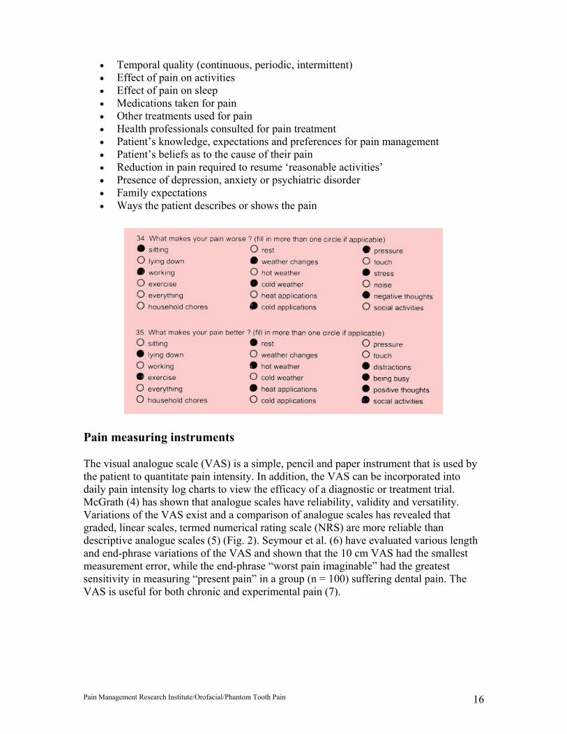
• Temporal quality (continuous, periodic, intermittent) • Effect of pain on activities • Effect of pain on sleep • Medications taken for pain • Other treatments used for pain • Health professionals consulted for pain treatment • Patient’s knowledge, expectations and preferences for pain management • Patient’s beliefs as to the cause of their pain • Reduction in pain required to resume ‘reasonable activities’ • Presence of depression, anxiety or psychiatric disorder • Family expectations • Ways the patient describes or shows the pain
Pain measuring instruments
The visual analogue scale (VAS) is a simple, pencil and paper instrument that is used by the patient to quantitate pain intensity. In addition, the VAS can be incorporated into daily pain intensity log charts to view the efficacy of a diagnostic or treatment trial. McGrath (4) has shown that analogue scales have reliability, validity and versatility. Variations of the VAS exist and a comparison of analogue scales has revealed that graded, linear scales, termed numerical rating scale (NRS) are more reliable than descriptive analogue scales (5) (Fig. 2). Seymour et al. (6) have evaluated various length and end-phrase variations of the VAS and shown that the 10 cm VAS had the smallest measurement error, while the end-phrase “worst pain imaginable” had the greatest sensitivity in measuring “present pain” in a group (n = 100) suffering dental pain. The VAS is useful for both chronic and experimental pain (7).

Pain Management Research Institute/Orofacial/Phantom Tooth Pain 17
The description of a pain condition (i.e. the patient’s choice of words applicable to the qualities of the pain condition) can provide simple but valuable information for diagnosis, selection of appropriate drugs, and levels of psychological distress. The McGill Pain Questionnaire (MPQ) is another pencil and paper pain measuring instrument that allows the patient to select appropriate pain word descriptors. It can measure pain intensity through various indices including sensory and affective components of pain. The MPQ has been used for evaluating acute (postoperative) orofacial pain from third molar teeth removal (8). In addition, the MPQ has been used to assess acute “toothache” patients and was able to differentiate different stages of pulpitis (irreversible versus reversible), with a correct prediction rate of 73% in subjects (9). Similar findings have been reported in using the MPQ to differentiate pulpitis from pericoronitis (10). Only one study (11), however, has used the MPQ for analysing chronic orofacial pain conditions; two chronic orofacial pain conditions were examined, atypical facial pain and trigeminal neuralgia, and the authors correctly predicted the diagnosis in 90% of patients. The MPQ has a standard form that was designed by Melzack (12) and the form consists of 78 words categorised into 20 groups, representing the four major dimensions of pain quality; sensory, affective, evaluative and miscellaneous pain descriptors (Fig. 3). These groups of words are scored and ranked to furnish four indices of pain quality; Pain Rating Index of Sensory descriptors [PRI(S)], Pain Rating Index of Affective descriptors [PRI(A)], Pain Rating Index of Evaluative descriptors [PRI(E)] and the Pain Rating Index of Miscellaneous descriptors [PRI(M)]. The four pain rating scores can then be added to give a fifth index, the total Pain Rating Index [PRI(T)]. The figure above lists the McGill pain word descriptors chosen by patients with neuropathic orofacial pain [based on data from (2)].
Local anaesthetic techniques

Pain Management Research Institute/Orofacial/Phantom Tooth Pain 18
A positive response (pain reduction) to somatosensory blockade of pain sites by various routes of administering local anaesthetic agents is suggestive of neuropathic pain (and nociceptive pain). In the orofacial region, the area can be tested by various techniques to determine possible sources of the location of pain. Ideally, several techniques should be employed to which the patient is blinded, including the use of placebo injections (placebo responses up to 60% in neuropathic pain). First, the administration of a topical anaesthetic to mucosal pain sites is advantageous by being noninvasive, with a study revealing that 37/38 patients with neuropathic pain responded with a reduction in pain when a topical anaesthetic cream was applied for five minutes (2). The topical anaesthetic of choice is EMLA cream 5% (AstraZeneca, Sydney, Australia) which is a eutectic mixture of lignocaine and prilocaine bases (Fig. 4). The eutectic mixture has a low melting point of 170C and is an oil at mouth temperature, thus facilitating absorption. The agent has been shown to have rapid oral mucosal absorption, to penetrate deep into the oral tissues, and to be safe from toxicity from extended application times of up to 30 minutes (13, 14). It is superior for sensory blockade compared with lignocaine 5% and 10% ointments (Xylocaine 5% and Xylocaine 10% Special Adhesive) and a benzocaine / amethocaine mixture (NUM) (13). The technique involves obtaining a VAS for basal pain intensity, followed by the removal of excess saliva from the test site using fresh cotton gauze. Isolation and placement of a placebo such as Vaseline on a cotton bud / roll for five minutes follows, and another VAS is obtained. The placebo is then wiped off and a liberal quantity (1 g) of EMLA applied for five minutes and a final VAS measured. The VAS responses can then be assessed to determine comparative pain reduction with EMLA.
Further local anaesthetic blockade can then be used to confirm other possible locations of peripheral neuropathy. This should be carried with sequential proximal anatomical blocks. For example, in testing the mandibular canine region, the analgesic sequence would be EMLA, mental nerve block, mandibular block using the ‘conventional’ technique blocking the mandibular nerve at the mandibular foramen, then a high mandibular block using the ‘closed mouth’ or Akinosi technique (15). This gives the endodontist potentially valuable information as to the location of any neuroma formation, and thus the likely success of utilising topical antineuropathic agents such as capsaicin. Obviously, in the situation of a patient who is a nonresponder to all blocks then there is the likelihood of CNS changes (centralisation of pain), thus greatly limiting the local therapeutic potential of capsaicin. Hypothetically, the probable selection of plain lignocaine (1-2%) as the test agent is preferable to lignocaine with vasoconstrictor (adrenaline / noradrenaline combinations) because of the potential of noradrenaline to actually increase pain (because of low pH) before the onset of lignocaine.
Three types of patient blinded, controlled local anaesthetic techniques are available. The first technique involves using normal saline as a control agent followed by a local anaesthetic agent such as plain lignocaine, obtaining VAS measures at baseline (prior to saline injection), five minutes after the saline block, and five minutes after the local anaesthetic block. The second technique is more robust and utilises two local anaesthetics with different half lives. The first block utilises plain lignocaine, then at a later stage, plain bupivacaine. Bupivacaine has a longer half life than lignocaine and a positive response would show pain reduction with both agents, but a longer period of pain

Pain Management Research Institute/Orofacial/Phantom Tooth Pain 19
reduction using bupivacaine. A third, superior technique, if time permitted, would employ triple patient blinded blocks: the first block using normal saline, the second block using plain lignocaine, and the third block injecting plain bupivacaine.
The intravenous lignocaine test requires referral to a specialist pain clinic with the services of an anaesthetist / pain specialist. This test has been reported to be useful in discriminating neuropathic pain from other forms of chronic pain (16). The test involves an infusion of lignocaine in increments up to a maximum of 2 mg/kg. It is placebo controlled and only one study has tested its application for neuropathic orofacial pain. In a test group of eight patients, 50% had a positive response with an average pain reduction of 68(10%. Interestingly, all the negative responders had a history of endodontic therapy in the area of pain but no further details were reported by the investigators (16).
Sympathetic nervous system procedures
A thorough clinical history may alert the endodontist to Sympathetically Maintained Pain (SMP). Clinically, the patient may give a history of episodes of oedema involving the face, and occasionally with a hot or burning quality (Fig. 5). An acknowledgement to simply asking the patient whether or not he /she experiences episodes of swelling in the face warrants referral for more definitive tests such as the phentolamine infusion.
A diagnosis of SMP is made when the pain is relieved by sympathetic blockade. However, there is no single test with adequate sensitivity and selectivity to distinguish SMP from sympathetically independent pain (SIP). The sympathetic chain receives efferent preganglionic fibres from the ventral roots of T1 (first thoracic nerve root) to L2 (second lumbar nerve root) and forms the stellate ganglion, thoracic paravertebral chain, coeliac plexus, lumbar paravertebral chain and superior hypogastric plexus. The sympathetic supply in the orofacial region originates from the stellate ganglion located in the neck. Studies using thermography (17, 18) and stellate ganglion block (19) have implicated sympathetically maintained pain (SMP) as occupying a role in neuropathic orofacial pain. Stellate ganglion block has been used as a diagnostic test for SMP in head, neck and upper limb (20). However, limitations of stellate block include false positive responses (spread of local anaesthetic to somatic fibres), false negative responses (incorrect placement of local anaesthetic), and an inability to conduct blinded, placebo controlled injections (21). More recently, intravenous administration of phentolamine (adrenoceptor antagonist) has been utilised as a diagnostic test for SMP and a positive response (pain relief) may be predictive of the success of a subsequent series of sympathetic blocks (22). In addition, the phentolamine test has the advantages of being less invasive and less painful, and blinded, placebo controlled infusions can be administered. The test is required to be performed by a specialist anaesthetist and patients are monitored with an electrocardiograph, pulse oximeter, and noninvasive blood pressure monitors. The test involves the initial administration of 500 mL of intravenous normal saline (0.9%) to limit potential hypotension from phentolamine. VAS scores are recorded at baseline and at five minute intervals. The trial involves a placebo infusion of saline over 10 minutes followed by phentolamine 15 mg over 10 minutes. If no analgesic response is reported by the patient and cardiovascular side effects are minimal (tachycardia and hypotension), further 5 mg bolus doses of phentolamine can be

Pain Management Research Institute/Orofacial/Phantom Tooth Pain 20
administered until a response is observed (pain relief or side effects). Only one study has employed the saline / phentolamine test to evaluate the sympathetic contribution to neuropathic orofacial pain with results showing that there was a significant response to the phentolamine challenge - 75% of patients (9/12) claimed a reduction (wide variation) in pain intensity ratings (2).
The involvement of SMP can be assessed through measuring sympathetic hyperfunction through changes in skin temperature (thermography and thermometry), blood flow (cutaneous Doppler), resting sweat output and the presence of oedema. Graff-Radford et al. (18) have suggested electronic thermography to be useful for SMP, although it is not a readily available test. However, the technique is promising, noninvasive and is selective in thermal accuracy (100%) with results showing the affected (painful) side of the face to be hotter than the nonpainful side (range 0.4-3.10C, mean=1.1(0.8, n=10).
Treatment / Pain Management Guidelines
Medication rationalisation
The selection of appropriate drugs for the treatment of neuropathic pain must take into account a number of principles: (i) adequate dose to achieve a therapeutic effect, (ii) titration of dose to minimise side effects, (iii) consideration of background medical conditions that may influence the pharmocokinetic profile of the drug, and (iv) drug interactions. Drugs appropriate for acute pain management may be, and often are, contraindicated for chronic pain management. Polypharmacy may reduce the overall amount of drug needed, but the dose must be calculated for each drug given. Blood concentrations of drugs may need to be monitored to assess if therapeutic concentrations are being reached. In medication rationalisation, the implementation of therapeutic drugs is crucial to effective pain management, yet an equally important task is to make recommendations for discontinuing other current, but inappropriate drugs, that are often used by patients with chronic pain. Management of neuropathic orofacial pain usually entails introducing the oral systemic first-line classes of drugs of a tricyclic antidepressant and / or an anticonvulsant.
Tricyclic antidepressants
Tricyclic antidepressants (TCAs) have a valuable role in the management of chronic pain, and the institution of a low dose TCA such as amitriptyline or nortriptyline as a first-line drug is frequently employed. Amitriptyline has both catecholaminergic and serotonergic activities. The dose is slowly increased from 25 mg nocte for the first week, to 50 mg nocte for the second week, and then to 75 mg nocte for the third and ensuing weeks, to build up patient tolerance to side effects such as sedation. For the elderly, it is prudent to commence with a dose of 10 mg nocte for the first week. It is necessary to maintain the tricyclic for at least 12-18 months before considering the discontinuation of the drug (23). TCAs provide background analgesia by preventing the reuptake of serotonin (and / or noradrenaline) at central sites. Serotonin and noradrenaline have analgesic or algogenic properties that are site specific; increased concentrations of serotonin and noradrenaline at central receptor sites provide analgesia, while increased concentrations at peripheral
Pain Management Research Institute/Orofacial/Phantom Tooth Pain 21
sites cause pain. Common side effects of TCAs include anticholinergic effects such as xerostomia, weight gain, and sedation. More serious side effects include postural hypotension, arrhythmias, and there is an overdose mortality rate of 11-15 times compared with anticonvulsants. The frequent side effect of sedation, however, may be beneficial as it assists the patient’s sleep pattern, which is often disturbed in chronic pain patients. TCAs are more acceptable for anxiolysis and night sedation than diazepam. The newer tetracyclic antidepressants are less toxic and have fewer side effects than the tricyclics, but do not appear to possess the analgesic properties of tricyclics.
Anticonvulsant drugs
Anticonvulsant drugs are appropriate where the pain has sharp, shooting, paroxysmal qualities. They have a membrane stabilising effect, causing a general reduction in the excitability of neurons. Anticonvulsants to be considered include carbamazepine (tegretol) and sodium valproate (epilim). Carbamazepine can increase brain concentrations of 5-hydroxytryptamine (serotonin) and sodium valproate can increase concentrations of gamma-aminobutyric acid (GABA). The starting dose for carbamazepine is usually 100 mg tds (and 100 mg nocte for elderly patients) and is gradually increased until a therapeutic effect (or side effects) is achieved. The effective dose may need to be as high as 400 mg tds, but at this dose side effects of sedation, ataxia and gastrointestinal (GIT) upset are common. Regular blood counts are necessary due to the possibility of thrombocytopenia and bone marrow depression. Sodium valproate appears to less toxic than carbamazepine, and is generally better tolerated. GIT disturbance, sedation and occasionally alopecia (hair loss) are side effects. In addition, disturbances of hepatic function may occur with sodium valproate and routine measurement of liver enzymes is necessary. Phenytoin (dilantin) is a second- or third-line anticonvulsant as it has a narrow therapeutic range and a potential side effect is marked gingival hyperplasia. Gabapentin (neurontin) is a recently introduced anticonvulsant that has an improved therapeutic effect to side effect ratio. However, its relatively high cost (~ $190 per monthly script) currently restrict its more widespread use as the drug is not currently listed with the Australian Pharmaceutical Benefit Scheme for pain management, in spite of published data establishing its efficacy for managing neuropathic pain (24-26).
Other oral systemic antineuropathic drugs
Clonazepam is a benzodiazepine primarily used for treating epilepsy, and accordingly its anticonvulsant properties render it useful for sharp, lancinating pain. Mexiletine, a cardiac antiarrhythmic and lignocaine analogue, is useful and should be considered if the patient has had a positive response to a lignocaine infusion. Drugs including long acting opioids, such as MS Contin and Kapanol which contain morphine as the active agent, should be used with caution, and as a last resort, as there is the possibility that over a period of time (after 2 years) opioids may increase the pain (seen as a resistance or tolerance) by increasing central concentrations of protein kinase C, and thus increasing central sensitisation (27). Patients with a history of chemical dependency should be excluded and there should be one prescriber and one dispenser. The use of short acting opioid analgesics, such as codeine and pethidine, often used for acute pain, are contraindicated in the long term management of neuropathic orofacial pain. NSAIDS,
Pain Management Research Institute/Orofacial/Phantom Tooth Pain 22
such as ibuprofen, have been shown to be effective for diabetic neuropathy but adverse effects of GIT ulceration preclude their long term use. Several other drugs and routes of administration such as intravenous ketamine and topical aspirin are effective for neuropathic pain, but may be limited in their application for intraoral use due to side effects (e.g. the ‘aspirin burn’ when aspirin is left to dissolve in the buccal sulcus). A systematic review of drug trials for peripheral neuropathic pain has been recently published (28).
Topical agents
Capsaicin (8-methyl-N-vanillyl-6-noneamide, cayenne pepper) is derived from capsicum fruit and its prime role for medical applications has been for the treatment of dermal lesions of postherpetic neuralgia (29). The substance has both algesic and analgesic properties, and depending on the concentration used, capsaicin can selectively activate, deactivate and even destroy small diameter afferents but leaves larger diameter afferents and the CNS intact. It can deplete several algogenic peptides including substance P, CGRP, somatostatin and vasoactive intestinal peptide (VIP). Capsaicin is a topical, first-line, antineuropathic agent with established efficacy in treating neuropathic oral pain. Vickers et al. (2) assessed the topical application of capsaicin in 30 neuropathic orofacial pain patients. Patients were instructed to apply the agent (0.025% capsaicin) for three minutes, twice daily to the area of intraoral pain. To limit any stinging / burning sensation from applying capsaicin, patients prerinsed their mouths with a topical anaesthetic mouthwash for three minutes. Results of the capsaicin trial showed a significant pain reduction in the test group. Nineteen patients reported a reduction in pain that ranged from 10-100% (average=58%). At long term followup (mean=13 months), there was no significant change in pain compared with results from the four week capsaicin trial; the positive responders had maintained a mean pain reduction of 50%. Other topical preparations specific for dermal use in conditions such as postherpetic neuralgia include powdered aspirin in chloroform or ethyl ether, and indomethacin, which inhibits prostaglandin synthesis (30).
Sympathetic blockade
There is limited data on the management of the SMP component of neuropathic orofacial pain. The recommended technique is a series of stellate blocks conducted by an anaesthetist. The concept is to repetitively break the pain cycle administering one block per week for 4-6 consecutive weeks using a long acting local anaesthetic such as bupivacaine. In an uncontrolled study (19), five subjects were given repeat stellate blocks (variable protocols) with all five subjects reporting pain reduction at follow up (10 months – 3 years). A possible alternative treatment is clonidine, an (2-adrenergic agonist that reduces sympathetic activity in the brainstem. Peripherally, clonidine acts at prejunctional (2 receptors to reduce noradrenaline release. Transdermal applications of clonidine reduce pain and hyperalgesia at the local site of application. Only one, uncontrolled study has assessed its efficacy for neuropathic orofacial pain. Clonidine was applied, 0.2 mg/g in a cream base, four times a day to the site of pain; results showed that 6/12 patients had a positive response but with a wide variability in reducing pain intensity ranging from 10-90% (31).

Pain Management Research Institute/Orofacial/Phantom Tooth Pain 23
Temporomandibular disorder
Approximately two thirds of patients with neuropathic orofacial pain subsequently develop a TMD (2). The adjacent diagram shows how a patient has placed a cross X indicating the phantom tooth pain site, and the secondary spread of pain to the masseter and trapezius muscles (TMD). Treatment of TMD, without attending to the causal neuropathic pain factor, however, does little in reducing the overall pain problem. Pain arising from TMD is predominantly due to bruxism. This can result in myofascial pain from masticatory muscle spasm (jaw clenching) and sensitisation of C-fibres in the periodontal, periapical and pulpal tissues of the oral cavity. Accordingly, there is a range of options available to help manage the condition including drugs, physiotherapy, and occlusal splints and other dental appliances. Drugs such as NSAIDS are appropriate for acute muscle spasm but should be used with caution; ideally NSAIDS should only be considered as a ‘rescue’ medication in the chronic pain patient. Physiotherapy encompasses a broad range of treatments that is initiated by the physiotherapist, and subsequently maintained by the patient. Initial treatment can include ultrasound, short wave diathermy, and laser to the affected musculature. Further professional instruction should be given to the patient regarding jaw (and neck) extension exercises and these should be rigorously pursued on a home basis by the patient. Other simple home physiotherapy measures include the application of heat packs on a frequent basis. Specific dental treatment can use occlusal splints to break the muscle spasm and to prevent tooth attrition. It should be noted, however, that while there is extensive documentation on splint design and clinical usage, well designed controlled studies indicate a significant placebo response from splints (32). On the other hand, where there is clear evidence of occlusal problems as a result of the extraction of teeth carried out in an attempt to ‘cure the pain’, then an occlusal splint as a prelude to occlusal rehabilitation with a permanent prosthetic appliance is warranted.
Psychological factors
For any patient with a chronic pain condition, the potential role of psychological factors in magnifying, minimising, or maintaining pain should not be underestimated. For example, Waddell et al. (33) were able to demonstrate that the amount of treatment that patients with back pain received bore more relationship to the distress and illness behaviour they exhibited, than to the physical disease. It is imperative that any chronic pain patient undergoes assessment by a clinical psychologist and / or psychiatrist with expertise in pain medicine. Pain is a subjective experience and is determined by psychological and environmental factors, in addition to the underlying pathology. There can be a wide individual variation by how a patient demonstrates his / her pain to their practitioner(s). Pain may often lead to anxiety and tension, which in turn, may increase the pain. Rudy (34) reported that pain intensity ratings are doubled when ‘stress’ is

Pain Management Research Institute/Orofacial/Phantom Tooth Pain 24
present, while others (35, 36) have previously shown a strong and positive correlation between pain severity and impairment of activities.
The chronic pain patient may be unable to cope with their situation which can lead to loss of self confidence, avoidance of others, and feelings of hopelessness and helplessness. They may blame the dentist / specialist for failing to solve the problem and there can be outright hostility and anger, and perhaps litigation, directed towards practitioners who they perceive have made the condition worse through inappropriate treatment. This can lead to an inability to communicate effectively and constructively with practitioners. The clinical psychologist (and psychiatrist to identify organic disorders) serves a key role to enable the patient to reach a better understanding of important pain management issues such as achieving a realistic outcome, self responsibility for long term management, and acceptance of the clinician’s advice. Operant conditioning may exist between patient and doctor, such as the continual advice on pain relief or cure for intractable chronic pain, and secondary emotional gain may be present between patient and caregiver, the continuance of pain to obtain support, sympathy and control (37).
It is important to obtain positive evidence of psychological factors playing a causal role as well; diagnosis by exclusion is clearly to be avoided. If a patient appears to have no clear pathological basis to his / her pain, referral for psychological assessment must be handled carefully lest the patient perceives it as being told 'the pain is in your head', which is likely to result in rejection of advice. If psychological assessment is sought, it should only be performed by a psychiatrist or clinical psychologist who has expertise in the field of pain, and the reason for the referral should be fully explained to the patient. For example, it should be explained that such pain problems are complex, and are associated with suffering and distress. In order to get a complete understanding of this pain, it is thus highly advisable to have input from appropriately experienced clinicians to assess the psychological aspects of pain, and the problems it causes. Most importantly, the medical / dental practitioner and the patient must have a common understanding prior to psychological / psychiatric assessments that the patient 'is not crazy' or 'imagining the pain'. For the referring clinician, it should be noted there is scope for previously undetected pathophysiology to exist in conditions that can be difficult to diagnose such as neuropathic orofacial pain.
Psychological strategies employed in pain management include cognitive behavioural therapy (modifying attitudes, beliefs and expectations), relaxation and biofeedback, and hypnosis. Pain management programmes are recommended for those who are not coping with their pain with the aims to (i) increase self perceived control over pain, (ii) increase self perceived independence, (iii) increase levels of physical and social activity, (iv)
Pain Management Research Institute/Orofacial/Phantom Tooth Pain 25
reduce levels of emotional distress, and (v) rationalise (discontinue or reduce) the use of inappropriate medication.
Overview of Part 2
Neuropathic orofacial pain is a condition that can be difficult to diagnose and treat. However, there are simple, yet effective, diagnostic procedures such as the MPQ and the utilisation of sequential analgesic blockade that can be discriminating in achieving a diagnosis. Once a diagnosis has been established, and indeed beforehand, a patient may expect substantial or unequivocal pain relief despite many years of suffering and failed treatments. Unfortunately, inordinate pressures can be placed on endodontists by chronic orofacial pain patients (and even their dentists) who suggest ablative treatment (extraction / further RCT) and perhaps, ongoing analgesic prescriptions. It is recognised that chronic orofacial pain conditions have similar pain intensity ratings, as indicated by the PRI(T) of the MPQ, compared with back pain, cancer pain and phantom limb pain (37). However, this important point is probably overlooked by the general public because chronic orofacial pain does not raise the emotive responses inherent in cancer pain, nor the government / business concerns of lost work productivity and related economic considerations associated with back pain.
For the endodontist, the use of capsaicin serves a dual purpose, as both a diagnostic agent and therapeutic agent. In addition, the endodontist can provide considerable advice to the patient, by giving a satisfactory understanding of the pathophysiology of neuropathic pain. In many cases of chronic neuropathic orofacial pain , the ‘bewildered’ general dental practitioner will refer the patient for an endodontic consultation. Accordingly, important management guidelines can be given by the endodontist to general dentists, particularly advice curtailing the unfortunate, but frequent desire, for dentists to carry out widespread pulp removal or the extraction of multiple teeth, in an attempt to ‘cure the pain’. This simple strategy may well save many patients from having a great deal of unnecessary treatment, expense and suffering. Initial treatment and long term management must utilise a broad range of measures individually tailored to the patient:
• rationalisation of drug regimens to reduce and eliminate, where possible, opioids, sedatives and other drugs of dependence; initiation of long term low dose tricyclic antidepressants, adjunct medications such as anticonvulsants (carbamazepine, sodium valproate) and membrane stabilisers (mexiletine);
• institution of specialist psychological / psychiatric treatment focusing on the development of effective coping strategies (e.g. relaxation techniques, cognitive and behavioral strategies), problem solving skills, and dealing with unresolved issues in the patient’s life);
• appropriate advice to the patient concerning dental treatment; • initiation of physical therapy to jaw, neck and other musculature for myofascial
pain from a secondary TMD; and • fabrication of dental splints and occlusal rehabilitation where appropriate, if an
associated TMD is present.
Pain Management Research Institute/Orofacial/Phantom Tooth Pain 26
Conceptually, chronic pain involves nociception and / or neuropathy, pain, suffering and pain behaviour. Early referral (within 6 months) of a chronic pain patient to a multidisciplinary pain clinic is recommended where possible, for comprehensive pain assessments by a medical pain specialist, clinical psychologist, physiotherapist and others (dentist, social worker, etc.). The medical management of neuropathic pain includes TCAs, with adjuvant medication such as anticonvulsants and membrane stabilisers. It is also imperative to assess the role of psychological and environmental factors. Pain can be magnified or minimised by gender effects, socioeconomic variables, ethnocultural differences, pending litigation / compensation, secondary gain and drug abuse. The practitioner should be aware of the following psychological variables in pain management: (i) unrealistic expectations, (ii) the motivation to improve, (iii) maintaining taught psychological coping skills and physical exercises, and (iv) accepting appropriate advice on the rationalisation of drugs. In summary, it is vital that the patient be given an accurate diagnosis to initiate appropriate treatment, and to quell the patient’s desire for a pain cure based on a ‘hard wired, simple telephone cable sysytem’. It is essential for the endodontist to gain and apply the knowledge of neuroplasticity of the nervous system associated with neuropathic pain. This will allow them to adopt a crucial role for the patient suffering from neuropathic orofacial pain, in order to achieve a successful outcome.
References for Part 2
1. Presley R.W., Cousins M.J. Current concepts in chronic pain management. Current Therapeutics 1992; June: 51-60.
2. Vickers E.R., Cousins M.J., Walker S., Chisholm K. Analysis of 50 patients with atypical odontalgia: a preliminary report on pharmacological procedures for diagnosis and treatment. Oral Surg Oral Med Oral Pathol Oral Radiol Endod 1998; 85: 24-32.
3. National Health and Medical Research Council. Acute pain management: scientific evidence. Ausinfo, Canberra, 1998, pp. 11-12.
4. McGrath P.A. The measurement of human pain. Endod Dent Traumatol 1986; 2: 124-9.
5. Sriwatanakul K., Kelvie W., Lasagna L., Calimlim J.F., Weis O.F., Mehta G. Studies with different types of analogue scales for measurement of pain. Clin Pharmacol Ther 1983; 34: 234-9.
6. Seymour R.A., Simpson J.M., Charlton J.E., Phillips M.E. An evaluation of length and end-phrase of visual analogue scales in dental pain. Pain 1985; 21: 177-85.
7. Price D.D., McGrath P.A., Rafii A., Buckingham B. The validation of visual analogue scales as ratio scale measures for chronic and experimental pain. Pain 1983; 17: 45-56.
8. Van Buren J., Kleinknecht R.A. An evaluation of the McGill Pain Questionnaire for use in dental pain assessment. Pain 1979; 6: 23-33.
9. Grushka M., Sessle B.J. Applicability of the McGill Pain Questionnaire to the differentiation of 'toothache' pain. Pain 1984; 19: 49-57.
10. Seymour R.A., Charlton J.E., Phillips M.E. An evaluation of dental pain using visual analogue scales and the McGill Pain Questionnaire. J Oral Maxillofac Surg 1983; 41: 643-8.

Pain Management Research Institute/Orofacial/Phantom Tooth Pain 27
11. Melzack R., Terrence C., Fromm G., Ansel R. Trigeminal neuralgia and atypical facial pain: use of the McGill Pain Questionnaire for discrimination and diagnosis. Pain 1986; 27: 297-302.
12. Melzack R. The McGill Pain Questionnaire: major properties and scoring methods. Pain 1975; 1: 277-99.
13. Vickers E.R., Punnia-Moorthy A. A comparison of the efficacy of three topical anaesthetic agents. Aust Dent J 1992; 37: 266-70.
14. Vickers E.R., Marzbani N., Gerzina T.M., McLean C., Punnia-Moorthy A., Mather L.E. Pharmacokinetics of EMLA cream 5% application to oral mucosa. Anesth Prog 1997; 44: 32-7.
15. Donkor P., Wong J., Punnia-Moorthy A. An evaluation of the closed mouth mandibular block technique. Int J Oral Maxillofac Surg 1990; 19: 216-9.
16. Saxen M.A., Adams W.R., Splonik K.S., Campbell R.L. Diagnostic lidocaine infusion in patients with chronic orofacial pain. Anesth Prog 1994; 41: 116-26. 17. Gratt B.M., Sickles E.A., Graff-Radford S.B., Solberg W.K. Electronic thermography in the diagnosis of atypical odontalgia: a pilot study. Oral Surg Oral Med Oral Pathol 1989; 68: 472-81.
17. Graff-Radford S.B., Ketelaer M.C., Gratt B.M., Solberg W.K. Thermographic assessment of neuropathic facial pain. J Orofac Pain 1995; 9: 138-45.
18. Lynch M.E., Elgeneidy A.K. The role of sympathetic activity in neuropathic orofacial pain. J Orofac Pain 1996; 10: 297-305.
19. Bonica J.J. Causalgia and other reflex sympathetic dystrophies. In: Bonica J.J., ed. The management of pain. Pennsylvania, Lea & Febiger, 1990, pp. 220-43.
20. Walker S.M., Cousins M.J. Complex regional pain syndromes: including "reflex sympathetic dystrophy" and "causalgia". Anaesth Intensive Care 1997; 25: 113-25.
21. Raja S.N., Treede R., Davis K.D., Campbell J.N. Systemic alpha-adrenergic blockade with phentolamine: a diagnostic test for sympathetically maintained pain. Anesthesiol 1991; 74: 691-8.
22. Diamond A.W., Coniam S.W. The management of chronic pain. Oxford, Oxford University Press, 1991, pp. 101-159.
23. Rosenberg J.M., Harrell C., Ristic H., Werner R.A., de Rosayro A.M. The effect of gabapentin on neuropathic pain. Clin J Pain 1997; 13: 251-5.
24. Gillin S., Sorkin L.S. Gabapentin reverses the allodynia produced by the administration of anti-GD2 ganglioside, an immunotherapeutic drug. Anesth Analg 1998; 86: 111-6.
25. Attal N., Brasseur L., Parker F., Chauvin M., Bouhassira D. Effects of gabapentin on the different components of peripheral and central neuropathic pain syndromes: a pilot study. Eur Neurol 1998; 40: 191-200.
26. Brooker C., Cousins M.J., Molloy A.R. Neuropathic pain: a GP’s guide. Modern Medicine 1999; 42: 58-67.
27. Kingery W.S. A critical review of controlled clinical trials for peripheral neuropathic pain and complex regional pain syndromes. Pain 1997; 73: 123-39.
28. Stanberry L.R., Bourne L., Bravo F.J., Bernstein D.I. Capsaicin-sensitive peptidergic neurons are involved in the zosteriform spread of herpes simplex virus infection. J Med Virol 1992; 38: 142-6.
29. Rowbotham M.C. Topical analgesic agents. In: Fields H.L., Liebeskind J.C., ed. Pharmacological approaches to the treatment of chronic pain: new concepts and

Pain Management Research Institute/Orofacial/Phantom Tooth Pain 28
critical issues. progress in pain research and management. Vol.1, Seattle, IASP Press, 1994, pp. 211-28.
30. Epstein J.B., Grushka M., Le N. Topical clonidine for orofacial pain: a pilot study. J Orofac Pain 1997; 11: 346-52.
31. Dao T.T.T., Lavigne G.J., Charbonneau A., Feine J.S., Lund J.P. The efficacy of oral splints in the treatment of myofascial pain of the jaw muscles: a controlled clinical trial. Pain 1994; 56: 85-94.
32. Waddell G., Bircher M., Finlayson D., Main C.J. Symptoms and signs: physical disease or illness behaviour? Br Med J 1984; 289: 739-41.
33. Rudy T.E. Psychophysiological assessment in chronic orofacial pain. Anesth Prog 1990; 37: 82-7.
34. Fordyce W.E., Lansky D., Calsyn D.A., Shelton J.L., Stolov W.C., Rock D.L. Pain measurement and pain behavior. Pain 1984; 18: 53-69.
35. Blumer D., Heilbronn M. The pain-prone disorder: a clinical and psychological profile. Psychosomatics 1981; 22: 395-402.
36. Mahan P.E., Alling C.C. Facial pain. Philadelphia, Lea & Febiger, 1991, pp. 304-5. 37. Vickers E.R., Cousins M.J., Woodhouse A. Pain description and severity of chronic
orofacial pain conditions. Aust Dent J 1998; 43: 403-9.
TMD Patient case study 1
History, presentation and investigations
A 43 year old female, married with three children, described a 10 year pain history. She lived with her family, worked as a travel consultant, and was a social drinker (1 alcoholic drink per day). She claimed ther was constant pain in tooth 16 (maxillary right first molar) in the period from 1988-91. The tooth was sensitive to pressure, and hot and cold stimuli. Her general dentist treated the tooth with root canal therapy (RCT) in 1991, and a crown was subsequently placed. The pain persisted and there was an increase in sensitivity to percussion and cold stimuli. Her general dental practitioner carried out a further RCT but there was no change in her symptomatology. She was then referred to an endodontist. The endodontist was unable to negotiate intracanal ledges and thus periapical surgery was performed in December, 1997. Healing was uneventful, however the pain continued. The adjacent tooth (15) then became sensitive to cold stimuli. An endodontic colleague who believed that the area of pain (15-16) was not associated with 16 sought a further opinion. An opinion from a prosthodontist followed and an occlusal splint was issued. The splint therapy was only “partially beneficial” (patient’s words) and she discontinued wearing the appliance. Periapical surgery was again performed on 16 in June 1998 (Fig. 7). No pathology was detected at the time of surgery, healing was again uneventful, but the pain persisted. In August 1998, 15 was still sensitive to cold stimuli, the pulp was extirpated and a Ledermix dressing placed. There was still no change in symptomatology and the patient was referred by the endodontist to the authors’ institution for multidisciplinary pain assessment.
Other investigations performed during the course of endodontic treatment included CT scan (no pathology detected), general medical assessments and an opinion from an ear, nose and throat surgeon. The patient’s previous dental history included the removal of
Pain Management Research Institute/Orofacial/Phantom Tooth Pain 29
supernumerary teeth at 7 years old, and the surgical removal of impacted third molar teeth and other supernumerary teeth at 14 years of age. Her medical history was unremarkable apart from an allergy to penicillin based drugs.
Pain characteristics and psychological factors
The pain was situated in the region of the 16 tooth and the adjacent buccal gingivae and mucosa. The pain had also spread extraorally to affect the facial tissues around the right temporomandibular joint (TMJ). The temporal quality of pain as reported by the patient was “constant”, but there were occasional short periods without pain. Pain intensity as measured by the VAS showed the pain to have a “usual” level of 5/10, that varied from 0-9 during the day, often no pain on awakening, but 7-9/10 by the afternoon (“0 = no pain, 10 = worst pain imaginable”). On the verbal descriptor scale, the pain was “medium to severe”. Modalities that worsened the pain included local pressure on the gingivae, tension, tiredness, eating, and dental procedures. Modalities that ameliorated the pain included sleep, analgesics and “keeping my mind off the pain”. MPQ word descriptors listed by the patient included “pounding, flashing, gnawing, hot, aching and tender” sensory qualities, and “exhausting, troublesome, penetrating and nagging” affective / miscellaneous qualities. Clinical examination showed a TMD was present and had developed subsequent to the neuropathic oral pain. The TMD signs and symptoms included TMJ pain, headaches, blocked ear, bruxism, sinusitis like symptoms, occasional facial numbness, and episodes of swelling and heat in the face and in the buccal vestibule of the lip.
There was no obvious clinical depression as determined by psychological assessment and completion of several psychological questionnaires including the MOS-56 Item Health Survey, the Beck Depression Inventory, and the Pain Self-Efficacy Questionnaire. The patient had indicated at her first pain centre consultation, that even with medical treatment, she realistically expected the pain “will not change”. Despite her lack of confidence in the prognosis of the condition, she was interested to find out what was the diagnosis of the condition and the cause of her pain.
Diagnosis and management
The medication at the time of her referral included prescription and ‘over the counter’ drugs: two Panadol tabs (paracetamol 500 mg tabs) prn, two Nurofen tabs (ibuprofen 200 mg tabs) prn, and Rohypnol ½ tab (flunitrazepam 2 mg tabs) prn. The patient was diagnosed with neuropathic orofacial pain with a secondary TMD. A six week trial of Zostrix (0.025% capsaicin) applied topically to the site of intraoral pain was commenced, concurrent with the introduction of a TCA. The TCA was amitriptyline; this was increased from 25 mg nocte to 75 mg nocte over three weeks and then maintained at that dosage. At a review at two months following the combined treatment, the patient claimed a 90% reduction in pain. She currently uses the capsaicin on an occasional basis when there is ‘breakthrough’ pain. This reduces the pain back to basal levels in 2-3 days. Her medical doctor, who currently manages the patient (amitriptyline prescriptions), reported slightly raised liver enzymes (a rare side effect of amitriptyline) that is being monitored.

Pain Management Research Institute/Orofacial/Phantom Tooth Pain 30
Patient case study 2
History, presentation and investigations
A 23 year old healthy male, a final year medical student, described a 4 year history of chronic pain situated in the left jaw and face. The original cause of the pain was thought to be chronic infection of the 38 (mandibular left third molar). An oral surgeon extracted the tooth but pain and infection persisted and another dentist extracted the adjacent second molar several months later. There were no further episodes of infection but the pain was still present and then spread to the maxillary left molars
There had been no diagnosis that could explain the chronic pain complaint from the investigations carried out over the 4 year history. During the course of the pain, he had seen three oral surgeons and one dentist who conducted clinical examinations, and periapical and orthopantomogram (OPG) radiographs (Fig. 9). Two ENT surgeons, two neurologists, an allergy specialist and four general medical practitioners also investigated him. In addition to routine clinical assessments by the medical specialists, investigations included blood tests, and CT and MRI scans of the brain.
Pain characteristics and psychological factors
The pain was centred in the left mandible 70% of the time, and 30% of the time it was centred in the left maxilla (Fig. 10). The pain intensity varied from 1-5/10 with “severe” pain episodes rated 8/10. There were periods of being pain free and of severe pain, both periods lasting days or weeks. Pain qualities ticked on the McGill Pain Questionnaire were: “throbbing, sharp, tugging, tingling, aching, taut, tiring, punishing, intense, spreading, tight, cold and dreadful”.
Psychologically, the patient believed that underlying, “perhaps sinister”, pathology was initially responsible for the pain. Later, unremarkable findings from investigations then led him to conclude it was non life threatening pathology but the pain had a “marked effect”. He felt “frustrated, despondent and anger towards unsympathetic doctors, no suicidal thoughts but periods of mild depression”. He questioned himself “can your mind create this pain from psychological and social factors?; I am doubtful of my normal senses - am I hallucinating regarding this pain?” He intentionally hid his condition from medical colleagues as he believed there was a stigma associated with chronic pain - a belief he attributed to the attitudes of his medical teachers (consultants and registrars) that these patients were “constantly complaining, narcotic dependent, and had personalities that were warped by the pain problem”. Study and university results were affected and he believed this would result in restricted employment opportunities. He relied on his
Pain Management Research Institute/Orofacial/Phantom Tooth Pain 31
parents for ongoing support and his social life was greatly affected with an 80% decline in pursuing hobbies and social activities.
Diagnosis and management
The patient self referred himself to a specialist oral surgical practice in a regional city visited on a part time basis by one of the authors (ERV). He had previously self prescribed Tofranil (imipramine 10 mg tabs) and discontinued it after 1-2 days due to side effects (“it was awful and I felt like a zombie even on 10mg”). The patient was diagnosed with neuropathic orofacial pain with a secondary TMD but he declined referral for multidisciplinary pain assessment at the authors’ institution. There was marked spasm in the left masseter muscle. He undertook a six week trial of Zostrix (0.025% capsaicin) but it was not beneficial. The patient currently self medicates and uses ‘rescue doses’ once / twice a month of Imigram (sumatriptan 50 mg tabs) and 4-6 tabs of Panadeine Forte (paracetamol 500 mg, codeine 30 mg per tab); his current use of medication for chronic pain is not very logical. In addition, he uses distraction strategies (work, study, television) and relaxation techniques to manage the pain.