Embed Size (px)
Citation preview
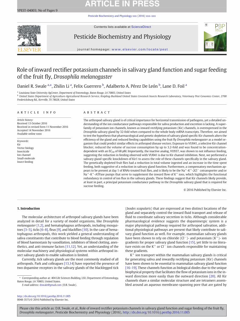
Pesticide Biochemistry and Physiology xxx (2016) xxx–xxx
YPEST-04003; No of Pages 9
Contents lists available at ScienceDirect
Pesticide Biochemistry and Physiology
j ourna l homepage: www.e lsev ie r .com/ locate /pest
Role of inward rectifier potassium channels in salivary gland function and sugar feedingof the fruit fly, Drosophila melanogaster
Daniel R. Swale a,⁎, Zhilin Li a, Felix Guerrero b, Adalberto A. Pérez De León b, Lane D. Foil a
a Louisiana State University AgCenter, Department of Entomology, Baton Rouge, LA 70803, United Statesb United States Department of Agriculture-Agricultural Research Service, Knipling-Bushland United States Livestock Insects Research Laboratory, Veterinary Pest Genomics Center, 2700Fredericksburg Rd., Kerrville, TX 78028, United States
⁎ Corresponding author at: 404 Life Sciences Building, LBaton Rouge, LA 70803, United States.
E-mail address: [email protected] (D.R. Swale).
http://dx.doi.org/10.1016/j.pestbp.2016.11.0050048-3575/© 2016 Published by Elsevier Inc.
Please cite this article as: D.R. Swale, et al., RoDrosophila melanogaster, Pesticide Biochemis
a b s t r a c t
a r t i c l e i n f oArticle history:Received 13 October 2016Received in revised form 11 November 2016Accepted 14 November 2016Available online xxxx
The arthropod salivary gland is of critical importance for horizontal transmission of pathogens, yet a detailed un-derstanding of the ion conductance pathways responsible for saliva production and excretion is lacking. A super-family of potassium ion channels, known as inward rectifying potassium (Kir) channels, is overexpressed in theDrosophila salivary gland by 32-fold when compared to the whole body mRNA transcripts. Therefore, we aimedto test the hypothesis that pharmacological and genetic depletion of salivary gland specific Kir channels alters theefficiency of the gland and reduced feeding capabilities using the fruit fly Drosophila melanogaster as a model or-ganism that could predict similar effects in arthropod disease vectors. Exposure to VU041, a selective Kir channelblocker, reduced the volume of sucrose consumption by up to 3.2-fold and was found to be concentration-dependent with an EC50 of 68 μM. Importantly, the inactive analog, VU937, was shown to not influence feeding,suggesting the reduction in feeding observed with VU041 is due to Kir channel inhibition. Next, we performed asalivary gland specific knockdown of Kir1 to assess the role of these channels specifically in the salivary gland.The genetically depleted fruit flies had a reduction in total volume ingested and an increase in the time spentfeeding, both suggestive of a reduction in salivary gland function. Furthermore, a compensatory mechanism ap-pears to be present at day 1 of RNAi-treated fruit flies, and is likely to be the Na+-K+-2Cl− cotransporter and/orNa+-K+-ATPase pumps that serve to supplement the inward flow of K+ ions, which highlights the functionalredundancy in control of ion flux in the salivary glands. These findings suggest that Kir channels likely provide,at least in part, a principal potassium conductance pathway in the Drosophila salivary gland that is required forsucrose feeding.
© 2016 Published by Elsevier Inc.
Keywords:KirVector biologyInsecticideIon channelSmall-moleculeInsect feeding
1. Introduction
The molecular architecture of arthropod salivary glands have beenanalyzed in detail for a variety of model organisms, like Drosophilamelanogaster [1,2], and hematophagous arthropods, including mosqui-toes [3–5], ticks [6–8], fleas [9], and blackflies [10]. In the case of hema-tophagous arthropods, this work yielded a general understanding ofsaliva constituents that contribute to blood feeding through regulationof blood haemostasis by vasodilation, inhibitors of blood clotting, anes-thetics, and anti-immune factors [11,12]. Yet, an understanding of themolecular machinery and physiological systems within arthropod in-sect salivary glands to enable salivation is limited.
Currently, tick salivary glands are the most commonly studied of allarthropods. Park and colleagues [13,14] have reported the presence oftwo dopamine receptors in the salivary glands of the blacklegged tick
SU, Department of Entomology,
le of inward rectifier potassiutry and Physiology (2016), h
(Ixodes scapularis) that are expressed at two distinct locations of thegland and separately control the inward fluid transport and release offluid to coordinate salivary secretion in ticks. Although considerablepharmacological evidence suggests the dopaminergic system is amajor physiological pathway required for arthropod salivation, addi-tional physiological pathways are present that likely contribute to sali-vary gland function as well. For example, mammalian salivary glandshave been shown to rely on chloride (Cl−)- and potassium (K+)- iongradients for proper salivary gland function [15], yet little to no litera-ture exists on the K+ or Cl− ion channels responsible for maintainingthese gradients.
K+ ion transport within the mammalian salivary glands is criticalfor generating saliva and inwardly rectifying potassium (Kir) channelshave been shown to be essential to mammalian salivary gland function[16–19]. These channels function as biological diodes due to the uniquebiophysical property that facilitates the flowof potassium ions in the in-ward direction more easily than the outward direction [20]. All Kirchannels share a similar molecular structure and are tetramers assem-bled around an aqueous membrane-spanning pore that are gated by
mchannels in salivary gland function and sugar feeding of the fruit fly,ttp://dx.doi.org/10.1016/j.pestbp.2016.11.005
2 D.R. Swale et al. / Pesticide Biochemistry and Physiology xxx (2016) xxx–xxx
polyvalent cations that occlude the pore at cell potentials more positivethan the K+ equilibrium potential (Ek) [21,22]. The numbers of genesencoding Kir channel constructs vary depending on species withhumans possessing 16 Kir channel encoding genes [20], Aedes aegyptimosquito possessing 5 [23], and D. melanogaster with 3 [24]. The Dro-sophila Kir genes are termed Ir, Irk2, and Irk3 and encode Kir1, Kir2,and Kir3, respectively [24]. Tissue expression patterns of DrosophilaKir channels are highly variable and are described in Zhuo and Hong-Sheng [24] with the expression patterns in excretory systems summa-rized in Table 1.
Several recent lines of genetic andpharmacological evidence suggestKir channels play important physiological roles in exocrine systems ofdipteran insects as well. In D. melanogaster, embryonic depletion ofKir1 and Kir2 mRNA in Malpighian tubules significantly reducestransepithelial secretion of fluid and K+ transport [25]. In Aedes aegyptiand Anopheles gambiaemosquitoes, researchers have shown that phar-macological inhibition of AeKir1 with structurally distinct small-mole-cules (i.e. VU573, VU041) disrupts the secretion of fluid and K+ inisolated Malpighian tubules, as well diuretic capacity, and K+ homeo-stasis in adult females [26–29]. Furthermore, the Ir gene encoding Dro-sophila Kir1 is enriched in exocrine tissues and gene expression isincreased by 37-fold in the salivary glands of larval and adult life stages(Table 1) [24,30]. TheDrosophila salivary glandmainly consists of secre-tory cells that synthesize and secrete proteins required for feeding [31],and the high expression of Ir in the salivary gland may suggest a role inpromoting secretion of salivary constituents.
Considering 1) the overexpression of Ir in Drosophila salivary, 2) thecritical role Kir channels serve inMalpighian tubules and 3) that salivarygland cells are also an exocrine tissue that rely on ionic gradients andwater transport to generate saliva, we hypothesized that Kir channelsare essential to proper salivary gland function and are critical in thehighly intricate physiological processes of arthropod feeding. Therefore,the goals of the present study were to use pharmacological inhibitionand salivary gland-specific genetic depletion of Kir channels in themodel organism D.melanogaster to determine the physiological impor-tance of Kir channels in fly salivary gland function asmeasured throughsucrose feeding efficiency.
2. Methods
2.1. Drosophila stocks and rearing conditions
Four strains of D.melanogasterwere used in this study. ThewildtypeOregon-R (OR) strainwas provided byDr. Jeffrey Bloomquist at the Uni-versity of Florida and was originally donated by Doug Knipple, CornellUniversity, Ithaca NY, USA. All GAL4-UAS fly strains were purchasedfrom Bloomington Drosophila Stock Center (Bloomington, IN, USA).The GAL4-UAS strain 6870 expresses the promoter in the larval andadult salivary glands, the strain 42644 expresses dsRNA for RNAi ofKir1 (Ir) under UAS control, and the strain 41554 expresses hairpinRNA (hpRNA) under the control of UAS for RNAi of GFP and was used
Table 1Anatomical expression of Kir channels expressed in excretory systems of Drosophila [24].
mRNA Signals
Localizations Ir Irk2 Irk3
Larval Adult Enrichment Larval Adult Larval Adult
Salivary gland 725 7480 37.2 67 235 3 8Crop – 2871 1.4 N/A 1722 – 6Midgut 782 506 2.5 36 117 7 4Hindgut 302 83 0.4 3856 4564 4 48M. tubules 844 1099 5.5 N/A 805 2898 4932
Note: Data was originally obtained from http://flyatlas.org and values shown were ex-tracted from Luan and Li [24]. Values indicate intensities of RNA signal. N/A: no informa-tive data. Enrichment is defined as the mRNA expression of the tissue/whole bodymRNA expression.
Please cite this article as: D.R. Swale, et al., Role of inward rectifier potassiuDrosophila melanogaster, Pesticide Biochemistry and Physiology (2016), h
as a negative knockdown control. The genotypes of each strain are asfollows: 6870, w[1118]; P{w[+mC] = Sgs3-GAL4.PD}TP1; 42644, y[1] sc[*] v [1]; P{y[+t7.7] v[+t1.8] = TRiP·HMS02480}attP2; 41554, y[1] sc[*] v [1]; P{y[+t7.7] v[+t1.8] = VALIUM20-EGFP.shRNA.2}attP2.
All fly strains have been maintained in culture at the Louisiana StateUniversity since April 2015. All fly strains were reared on standard me-dium in Drosophila tubes at 25 °C, 12·hour-12·hour photoperiod and55% relative humidity. For dissection, flies were anaesthetized by chill-ing on ice and decapitated before dissecting out salivary glands inSchneider's medium (Invitrogen, Paisley, Scotland, UK).
2.1.1. ChemicalsThe Kir channel inhibitor VU041 and the inactive analog VU937
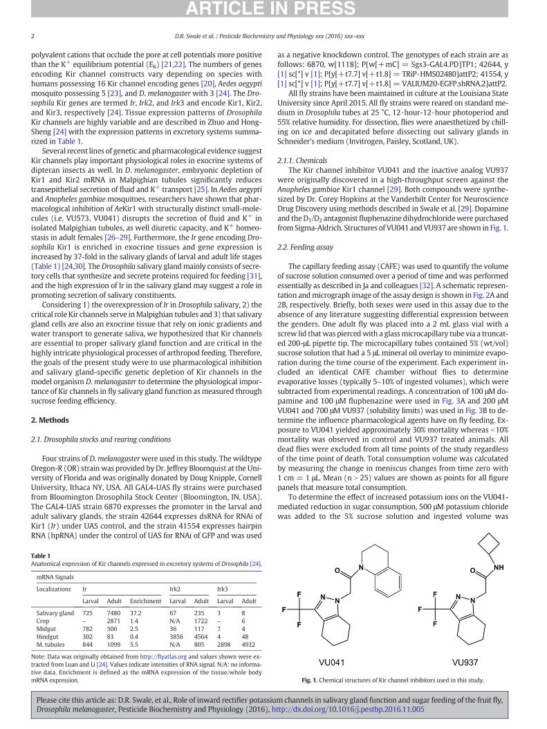
were originally discovered in a high-throughput screen against theAnopheles gambiae Kir1 channel [29]. Both compounds were synthe-sized by Dr. Corey Hopkins at the Vanderbilt Center for NeuroscienceDrug Discovery usingmethods described in Swale et al. [29]. Dopamineand theD1/D2 antagonistfluphenazine dihydrochloridewere purchasedfromSigma-Aldrich. Structures of VU041 andVU937 are shown in Fig. 1.
2.2. Feeding assay
The capillary feeding assay (CAFE) was used to quantify the volumeof sucrose solution consumed over a period of time and was performedessentially as described in Ja and colleagues [32]. A schematic represen-tation andmicrograph image of the assay design is shown in Fig. 2A and2B, respectively. Briefly, both sexes were used in this assay due to theabsence of any literature suggesting differential expression betweenthe genders. One adult fly was placed into a 2 mL glass vial with ascrew lid thatwas piercedwith a glassmicrocapillary tube via a truncat-ed 200-μL pipette tip. The microcapillary tubes contained 5% (wt/vol)sucrose solution that had a 5 μL mineral oil overlay to minimize evapo-ration during the time course of the experiment. Each experiment in-cluded an identical CAFE chamber without flies to determineevaporative losses (typically 5–10% of ingested volumes), which weresubtracted from experimental readings. A concentration of 100 μM do-pamine and 100 μM fluphenazine were used in Fig. 3A and 200 μMVU041 and 700 μM VU937 (solubility limits) was used in Fig. 3B to de-termine the influence pharmacological agents have on fly feeding. Ex-posure to VU041 yielded approximately 30% mortality whereas b10%mortality was observed in control and VU937 treated animals. Alldead flies were excluded from all time points of the study regardlessof the time point of death. Total consumption volume was calculatedby measuring the change in meniscus changes from time zero with1 cm = 1 μL. Mean (n N 25) values are shown as points for all figurepanels that measure total consumption.
To determine the effect of increased potassium ions on the VU041-mediated reduction in sugar consumption, 500 μM potassium chloridewas added to the 5% sucrose solution and ingested volume was
Fig. 1. Chemical structures of Kir channel inhibitors used in this study.
m channels in salivary gland function and sugar feeding of the fruit fly,ttp://dx.doi.org/10.1016/j.pestbp.2016.11.005
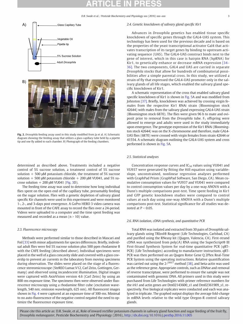
Fig. 2. Drosophila feeding assay used in this study modified from Ja et al. A) Schematicdiagram showing the feeding assay that utilizes a glass capillary tube held by a pipettetip and one fly added to each chamber. B) Photograph of the feeding chambers.
3D.R. Swale et al. / Pesticide Biochemistry and Physiology xxx (2016) xxx–xxx
determined as described above. Treatments included a negativecontrol of 5% sucrose solution, a treatment control of 5% sucrosesolution + 500 μM potassium chloride, the treatment of 5% sucrosesolution + 500 μM potassium chloride + 200 μM VU041, and 5% su-crose solution + 200 μM VU041 (Fig. 3D).
The feeding time assay was used to determine how long individualflies spent on the open end of the capillary tube, presumably feedingon the sugar solution. Flies with a genetic depletion of salivary glandspecific Kir channels were used in this experiment and were monitored1-, 2-, and 3-days post emergence. A GoPro HERO 3 video camera wasmounted in front of the CAFE assay to record the flies over a 24-period.Videos were uploaded to a computer and the time spent feeding wasmeasured and recorded as a mean (n N 10) value.
2.3. Fluorescence microscopy
Methods were performed similar to those described in Mascari andFoil [33]withminor adjustments for species differences. Briefly, individ-ual adult flies were fed 5% sucrose solution plus 500 ppm rhodamine Bwith the CAFE feeding (described above). Individual specimens wereplaced in thewell of a glass concavity slide and coveredwith a glass cov-erslip to prevent air currents in the laboratory from moving specimensduring observation. The slides were placed on the stage of a fluores-cence stereomicroscope (SteREO Lumar.V12, Carl Zeiss, Gottingen, Ger-many) and observed using incandescent illumination. Digital imageswere captured with AxioVision version 4.6 (Carl Zeiss) by using an800-ms exposure time. The specimens then were observed under fluo-rescence microscopy using a rhodamine filter cube (excitation wave-length, 540 nm; emission wavelength, 625 nm). All fluorescent imagesshown in Fig. 4 were captured at an exposure time of 300-ms. Minimalto no auto-fluorescence of the negative control negated the need to op-timize the fluorescence exposure time.
Please cite this article as: D.R. Swale, et al., Role of inward rectifier potassiuDrosophila melanogaster, Pesticide Biochemistry and Physiology (2016), h
2.4. Genetic knockdown of salivary gland specific Kir1
Advances in Drosophila genetics has enabled tissue specificknockdown of specific genes through the GAL4-UAS system. Thistechnology has been used for the previous decade and is based onthe properties of the yeast transcriptional activator Gal4 that acti-vates transcription of its target genes by binding to upstream acti-vating sequence (UAS). The GAL4-UAS construct binds next to thegene of interest, which in this case is hairpin RNA (hpRNA) forKir1, to genetically enhance or decrease mRNA expression [34–36]. The two components, GAL4 and UAS are carried in separateDrosophila stocks that allow for hundreds of combinatorial possi-bilities after a simple parental cross. In this study, we utilized astrain of fly that expressed the GAL4-UAS promoter only in the sal-ivary glands of all life stages, which enabled the salivary gland spe-cific knockdown of Kir1.
A schematic representation of the cross that enabled salivary glandspecific knockdown of Kir1 is shown in Fig. 5A and was modified fromJohnston [37]. Briefly, knockdown was achieved by crossing virgin fe-males from the respective Kir1 RNAi strain (Bloomington stock42644) with males from the salivary gland expressing GAL4-UAS strain(Bloomington stock 6870). The flies were given 96 h to mate and ovi-posit prior to removal from the Drosophila tube. F1 offspring wereallowed to emerge and adults were used in the study immediatelyupon emergence. The genotype expression of the Kir1 RNAi (Blooming-ton stock 42644) was on the X-chromosome and therefore, male GAL4-UAS flies (6870) were crossed with virgin females from strain 42644 or41554. A schematic diagram outlining the GAL4-UAS system and crossperformed is shown in Fig. 5A.
2.5. Statistical analyses
Concentration response curves and IC50 values using VU041 andVU937 were generated by fitting the Hill equation using variable-slope, unconstrained, nonlinear regression analyses performedwith GraphPad Prism (GraphPad Software, San Diego, CA). Mean cu-mulative consumption values for VU937 and VU041 were comparedto control consumption values per day by a one-way ANOVA with aDunn's multiple comparisons post-test. Time spent feeding in Kir1and GFP genetic knockdown studies were compared to controlvalues at each day using one-way ANOVA with a Dunn's multiplecomparisons post-test. Statistical significance for all studies was de-noted at P b 0.05.
2.6. RNA isolation, cDNA synthesis, and quantitative PCR
Total RNAwas isolated and extracted from30pairs ofDrosophila sal-ivary glands using TRIzol® Reagent (Life Technologies, Carlsbad, CA)and purified using the RNeasy kit (Qiagen, Valencia, CA). First-strandcDNA was synthesized from poly(A) RNA using the SuperScript® IIIFirst-Strand Synthesis System for real-time quantitative PCR (qRT-PCR) (Life Technologies) according to manufacturer instructions. qRT-PCR was then performed on an Qiagen Rotor Gene Q 2Plex Real-TimePCR System using the operating instructions. Relative quantificationwas carried out using the 2-DDCT method [38], and beta-actin was usedas the reference gene. Appropriate controls, such as DNAse and removalof reverse transcriptase, were performed to ensure the sample was notcontaminated with genomic DNA. All primers used in this study werepurchased from Life Technologies with primer reference numbers forthe irk1 and actin genes are Dm02143600_s1 and Dm02361909_s1, re-spectively. Five biological replicates were conducted and each was ana-lyzed in triplicate. The graphed output displays the average fold-changein mRNA levels relative to the wild type Oregon-R control salivaryglands.
mchannels in salivary gland function and sugar feeding of the fruit fly,ttp://dx.doi.org/10.1016/j.pestbp.2016.11.005
4 D.R. Swale et al. / Pesticide Biochemistry and Physiology xxx (2016) xxx–xxx
3. Results
3.1. Effect of pharmacological inhibition of Kir channels to sugar feeding
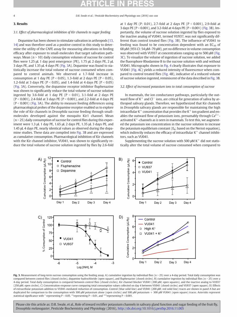
Dopamine has been shown to stimulate salivation in arthropods [13,14] and was therefore used as a positive control in this study to deter-mine the utility of the CAFE assay for measuring alterations in feedingefficacy after exposure to small-molecules that target salivation path-ways. Mean (n N 10) daily consumption volumes of sucrose for controlflies were 1.25 μL 1 day post emergence (PE), 1.75 μL 2 days PE, 2 μL3 days PE, and 1.35 μL 4 days PE (Fig. 3A). Dopamine was found to sta-tistically increase the total volume of sucrose consumed when com-pared to control animals. We observed a 1.7-fold increase inconsumption at 1 day PE (P b 0.05), 1.3-fold at 2 days PE (P b 0.05),1.2-fold at 3 days PE (P b 0.05), and 1.4-fold at 4 days PE (P b 0.05)(Fig. 3A). Conversely, the dopamine receptor inhibitor fluphenazinewas shown to significantly reduce the total volume of sucrose solutioningested by 3.6-fold at 1 day PE (P b 0.01), 3.1-fold at 2 days PE(P b 0.001), 2.4-fold at 3 days PE (P b 0.001), and 2.2-fold at 4 days PE(P b 0.001) (Fig. 3A). The ability to measure feeding differences usingpharmacological probes of the dopamine receptor enabled us to explorethe role of Kir channels in Drosophila sucrose feeding through small-molecules developed against the mosquito Kir1 channel. Mean(n N 25) daily consumption of sucrose for control flies during this exper-iment were 1.3 μL 1 day PE, 1.65 μL 2 days PE, 1.35 μL 3 days PE, and1.45 μL 4 days PE, nearly identical values as observed during the dopa-mine studies. These data are compiled into Fig. 3B and are expressedas cumulative consumption. Pharmacological inhibition of Kir channelswith the Kir channel inhibitor, VU041, was shown to significantly re-duce the total volume of sucrose solution ingested by flies by 2.6-fold
Fig. 3.Measurement of long-term sucrose consumption using the feeding assay. A) cumulativcompared between control flies (closed circles), dopamine hydrochloride (open square), and4-day period. Total daily consumption is compared between control flies (closed circles), K(250 μM; open circles). C) Concentration-response curve comparing total consumption valuesof extracellular potassium addition to VU041-mediated reduction of consumption. Control (duplicated for comparison to the consumption with 300 μM potassium alone (open circlestatistical significance with * representing P b 0.05. **representing P b 0.01, and ***representin
Please cite this article as: D.R. Swale, et al., Role of inward rectifier potassiuDrosophila melanogaster, Pesticide Biochemistry and Physiology (2016), h
at 1 day PE (P: 0.01), 2.7-fold at 2 days PE (P b 0.001), 2.9-fold at3 days PE (P b 0.001), and 3.2-fold at 4 days PE (P b 0.001) (Fig. 3B). Im-portantly, the volume of sucrose solution ingested by flies exposed tothe inactive analog of VU041, termed VU937, was not significantly dif-ferent than control treated flies (Fig. 3B). The influence of VU041 tofeeding was found to be concentration dependent with an EC50 of68 μM(95% CI: 54 μM–79 μM), yet no difference in volume consumptionwas observed with VU937 at concentrations ranging up to 500 μM (Fig.3C). To visualize the volume of ingestion of sucrose solution, we addedthe fluorophore Rhodamine B to the sucrose solution with and withoutVU041. Micrographs shown in Fig. 4 clearly illustrates that exposure toVU041 (Fig. 4C) yields a reduced intensity of fluorescence when com-pared to control treated flies (Fig. 4B), indicative of a reduced volumeof sucrose solution ingested, reminiscent of the data described in Fig. 3B.
3.2. Effect of increased potassium ions to total consumption of sucrose
In mammals, the ion conductance pathways, particularly the out-ward flow of K+ and Cl− ions, are critical for generation of saliva by ar-thropod salivary glands. Therefore, we hypothesized that Kir channelsin Drosophila salivary glands are responsible for maintaining the highintracellular K+ concentration that provides theK+ iongradient and en-ables the outward flow of potassium ions, presumably through Ca2+-activated K+-channels as is seen inmammals. To test this, we augment-ed the potassium ion concentration in the sucrose solution to increasethe potassium equilibrium constant (Ek; based on the Nernst equation),which indirectly reduces the efficacy of intracellular K+ channel inhibi-tors, such as VU041.
Supplementing the sucrose solution with 500 μM K+ did not statis-tically alter the total volume of sucrose consumed when compared to
e ingestion by individual flies (n N 25) over a 4-day period. Total daily consumption wasfluphenazine (closed circles). B) cumulative ingestion by individual flies (n N 25) over air channel blocker VU041 (200 μM; open squares), and the inactive analog to VU937collected on day 4 between VU041 (closed circles) and VU937 (open square). D) Effectsblue solid line) and VU041 (200 μM; red solid line) traces are shown in panel A but ares) and 500 μM potassium + 300 μM VU041 (open square) traces. Asterisks representg P b 0.001.
m channels in salivary gland function and sugar feeding of the fruit fly,ttp://dx.doi.org/10.1016/j.pestbp.2016.11.005
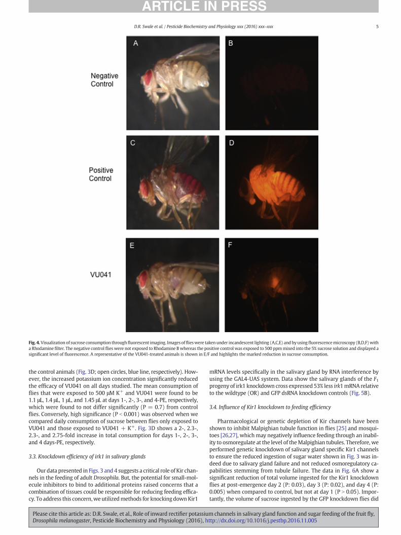
Fig. 4.Visualization of sucrose consumption throughfluorescent imaging. Images offlieswere takenunder incandescent lighting (A,C,E) and byusing fluorescencemicroscopy (B,D,F)witha Rhodamine filter. The negative control flies were not exposed to Rhodamine B whereas the positive control was exposed to 500 ppmmixed into the 5% sucrose solution and displayed asignificant level of fluorescence. A representative of the VU041-treated animals is shown in E/F and highlights the marked reduction in sucrose consumption.
5D.R. Swale et al. / Pesticide Biochemistry and Physiology xxx (2016) xxx–xxx
the control animals (Fig. 3D; open circles, blue line, respectively). How-ever, the increased potassium ion concentration significantly reducedthe efficacy of VU041 on all days studied. The mean consumption offlies that were exposed to 500 μM K+ and VU041 were found to be1.1 μL, 1.4 μL, 1 μL, and 1.45 μL at days 1-, 2-, 3-, and 4-PE, respectively,which were found to not differ significantly (P = 0.7) from controlflies. Conversely, high significance (P b 0.001) was observed when wecompared daily consumption of sucrose between flies only exposed toVU041 and those exposed to VU041 + K+. Fig. 3D shows a 2-, 2.3-,2.3-, and 2.75-fold increase in total consumption for days 1-, 2-, 3-,and 4 days-PE, respectively.
3.3. Knockdown efficiency of irk1 in salivary glands
Ourdata presented in Figs. 3 and 4 suggests a critical role of Kir chan-nels in the feeding of adult Drosophila. But, the potential for small-mol-ecule inhibitors to bind to additional proteins raised concerns that acombination of tissues could be responsible for reducing feeding effica-cy. To address this concern,we utilizedmethods for knockingdownKir1
Please cite this article as: D.R. Swale, et al., Role of inward rectifier potassiuDrosophila melanogaster, Pesticide Biochemistry and Physiology (2016), h
mRNA levels specifically in the salivary gland by RNA interference byusing the GAL4-UAS system. Data show the salivary glands of the F1progeny of irk1 knockdown cross expressed 53% less irk1mRNA relativeto the wildtype (OR) and GFP dsRNA knockdown controls (Fig. 5B).
3.4. Influence of Kir1 knockdown to feeding efficiency
Pharmacological or genetic depletion of Kir channels have beenshown to inhibit Malpighian tubule function in flies [25] and mosqui-toes [26,27], whichmay negatively influence feeding through an inabil-ity to osmoregulate at the level of theMalpighian tubules. Therefore, weperformed genetic knockdown of salivary gland specific Kir1 channelsto ensure the reduced ingestion of sugar water shown in Fig. 3 was in-deed due to salivary gland failure and not reduced osmoregulatory ca-pabilities stemming from tubule failure. The data in Fig. 6A show asignificant reduction of total volume ingested for the Kir1 knockdownflies at post-emergence day 2 (P: 0.03), day 3 (P: 0.02), and day 4 (P:0.005) when compared to control, but not at day 1 (P N 0.05). Impor-tantly, the volume of sucrose ingested by the GFP knockdown flies did
mchannels in salivary gland function and sugar feeding of the fruit fly,ttp://dx.doi.org/10.1016/j.pestbp.2016.11.005

Fig. 5. Salivary gland specific RNAi-medated knockdown of irk1. A) Schematic diagram ofthe GAL4/UAS system for directed gene knockdown. B) Quantitative RT-PCR analysisshowing Dmirk1 RNAi-based knockdown efficiency in the salivary glands of flies. Barsrepresent fold-difference of irk1 mRNA levels relative to beta-actin control group. Thestrains of flies used are as follows with Bloomington stock numbers in parentheses: WT(Oregon-R), GFP hpRNA (41554), irk1 hpRNA (42644), Salivary gland directed GAL4(1824).
Fig. 6. Long-term sucrose consumption in salivary gland specific irk1 knockdown animals.A) Cumulative ingestion by individual flies (n N 50) over a 4-day period. Total dailyconsumption is compared between control flies (black circles), GFP knockdown (opensquares), and salivary gland specific irk1 knockdown (red circles). Asterisks representstatistical significance at P b 0.05. B) Total time spent on capillary tube feeding onsucrose solution for 1-, 2-, 3-days post emergence. Bars represent mean (n:7–15) timespent feeding per treatment and error bars represent SEM. Bars not labeled by the sameletter represent statistical significance at P b 0.05. (For interpretation of the references tocolor in this figure legend, the reader is referred to the web version of this article.)
6 D.R. Swale et al. / Pesticide Biochemistry and Physiology xxx (2016) xxx–xxx
not differ from control. Although statistical significance was observed, asmaller than expected volume difference (c.a. 1 μL) between control andKir1 knockdownflieswas observed thatmay be due to the absence of anexternal stimuli preventing continuous feeding within the CAFE assay.Therefore, we assessed the timeeach individualfly rested on thebottomof the capillary tube, presumably feeding, at post emergence days 1, 2,and 3. Similar to the total consumption values, no significant differencein time spent feeding was observed for day 1, but a significant increase(P b 0.001) increase in time spent feeding was observed for the Kir1knockdown flies over control flies for days 2 and 3 with a 2.3- and1.9-fold increase, respectively (Fig. 6B).
4. Discussion
Despite the critical role the arthropod salivary gland serves in hori-zontal transmission of pathogens, an understanding of the machineryrequired for proper gland function is limited. Although rather limitedin scope, pharmacological studies against the isolated tick salivarygland have implicated several components involved in the process ofsalivary secretion: dopaminergic pathway [13], Na+-K+-ATPase [39],GABA [40], and the muscarinic acetylcholine receptor [41]. Althoughthese pathways are clearly important for saliva production and excre-tion, the complex nature of the gland suggests other pathways are likelycritical for proper function of the salivary gland. Indeed, the results ofthe present study provide compelling data that a superfamily of potas-sium ion channels, known as inward rectifier potassium channels, isan essential conductance pathway in the salivary gland that mediatesproper feeding in the model organism 4.1 Drosophila melanogaster.
Recent work on insect Kir channels have yielded insightful data sug-gesting these channels serve a critical role inMalpighian tubule function
Please cite this article as: D.R. Swale, et al., Role of inward rectifier potassiuDrosophila melanogaster, Pesticide Biochemistry and Physiology (2016), h
and fluid secretion [23,26,42]. The Malpighian tubules and salivaryglands are physiologically related tissues as both are a polarized epithe-lial tissue [43,44], require water and ion transport for function, and areconsidered, at least in part, to be an exocrine tissue. Furthermore, theKir1 channel has been shown to constitute the primary inward K+ con-ductance in the mosquito Malpighian tubule and the analogous genethat encodes Kir1 in Drosophila is highly upregulated in the salivaryglands of larval and adult flies [24]. Therefore, we hypothesized Kirchannels also serve a critical role in salivary gland function and aimedto elucidate the role of these channels through pharmacological and ge-netic manipulations of the Kir1 channel measured through feedingefficiency.
The recent identification of selective and potent small-molecules de-signed to target insect Kir channels [29,45–47] have enabled re-searchers to begin to characterize the physiological role of thesechannels in various tissue systems. In this study, we used the recentlydiscovered insect Kir channel modulator (VU041) and its inactive ana-log (VU937) [29] to characterize the influence these molecules have inthe feeding cascade. We found that exposure to VU041 during feedingsignificantly reduced the volume of sucrose ingested, whereas VU937had no influence to feeding efficiency, suggesting the observed pheno-type is through Kir inhibition. However, due to the capability thatsmall-molecules can inhibit unintended target sites and the fact Kirchannels are highly expressed in the Malpighian tubules (Table 1)[29], it was impossible to ensure the observed effect to feeding was di-rectly due to salivary gland failure. Therefore, we performed salivarygland specific RNAi-mediated knockdown of the Kir1 encoding gene.Results from this genetic depletion of Kir1 show a significantly less
mchannels in salivary gland function and sugar feeding of the fruit fly,ttp://dx.doi.org/10.1016/j.pestbp.2016.11.005

7D.R. Swale et al. / Pesticide Biochemistry and Physiology xxx (2016) xxx–xxx
efficient salivary gland (Fig. 6) and, when combined with the VU041-mediated reduction in sucrose consumption, strongly suggests the Dro-sophila salivary gland relies on the inward conductance of K+ ionsthrough Kir channels.
The data presented in this study raises the question as to what thephysiological role Kir channels have in salivary gland function at the cel-lular level. Consideration of knowledge and hypotheses based on therole of Kir channels in mammalian salivary gland function and salivaproduction can be applied to expand our understanding of salivarygland physiology in arthropods. First, it has been shown that electrolytesecretion in the mammalian salivary glands is based on the secondaryactive transport of anions, principally Cl− (and/or HCO3
−) ions [15]. Inthis model, K+ channels in the basolateral membrane of acinar cellsmaintain the membrane potential of the apical cell membrane to bemore negative than the Nernst potential for anions, thereby providinga driving force for the sustained electrogenic anion efflux across the api-cal membrane. The secondmodel for a role of Kir channels in the mam-malian salivary gland was described through cell-attached patch andwhole-cell patch-clamp studies. Here, researchers demonstrated thepresence of four primary K+ channels, two of which are the outwardmediated Ca2+-activated K+ channel and a Kir channel [18,19,48]. Theinwardly rectifying property of the Kir channel was hypothesized toperform fast uptake of accumulated K+ ions, in concert with Na+-K+-ATPase, into acinar cells with the K+ influx depending on the relationbetween the membrane potential and the concentration gradient ofK+ across the basolateral membrane. This buffering action likely pro-vides an ion gradient enabling the outward flow of K+ ions throughCa2+-activated K+ channels. Such K+ buffering action of Kir channelshas been proposed in brain astrocytes [49–51], in retinal Müller cells[52] and also in retinal pigmented epithelial cells [53].
To begin elucidating the role of Kir channels in the insect salivarygland based on themammalian hypotheses, we augmented the potassi-um ion concentration in the sucrose solution to increase the potassiumequilibrium constant (Ek; based on the Nernst equation), whichultimately reduces the efficacy of intracellular K+ channel inhibitors,such as Kir channel blockers [29,54]. The loss of VU041 potency(Fig. 3) supports the notion that Kir channels function as a means toprovide a pathway for rapid influx of K+ ions after depolarizationevents, a phenomenon oftentimes referred to as K+-spatial buffering.Therefore, we hypothesize that Kir channels in Drosophila salivaryglands are responsible, at least in part, formaintaining the high intracel-lular K+ concentration through a buffering-like action, which providesthe K+ ion gradient to enable the outward flow of potassium ions, pre-sumably through Ca2+-activated K+-channels as is seen in mammals.However, further studies rooted in cellular electrophysiology andmem-brane physiology are needed to provide additional support for thishypothesis.
Although the data of this study suggest Kir channels are likely theprimary mechanism for K+ spatial buffering within insect salivarygland cells, it is also evident that it is not the only transport pathway fa-cilitating inward flow of K+ ions. Genetic depletion of Kir1 channelsyielded a reduction of feeding at days 2, 3, and 4, but not on day 1(Fig. 6A) and similarly, the time spent feeding was not statisticallydifferent to controls at day 1 when compared to subsequent days(Fig. 6B). These data suggest the presence of a compensatory mecha-nism that accounts for the reduced expression of Kir1 in the geneticallydepleted animals, but one that is lost after day 1. Compensatory mech-anisms are commonly observed in animals with genetic depletions ofKir channels andmost oftentimes arise through upregulation of a differ-ent Kir gene. For instance,Wu and colleagues showed individual knock-down of any of the three Kir channel genes in Drosophila Malpighiantubules had no effect on the organ function, yet simultaneous knock-down of irk1 and irk2 had significant effects on transpeithelial K+ trans-port [55], suggesting Kir1 and Kir2 play redundant roles in Malpighiantubule function. Due to the expression of Kir1 and Kir2 mRNA in theDrosophila salivary gland [24], albeit at dramatic differences in mRNA
Please cite this article as: D.R. Swale, et al., Role of inward rectifier potassiuDrosophila melanogaster, Pesticide Biochemistry and Physiology (2016), h
expression level, it is plausible that the Ir2 gene is upregulated after ge-netic depletion of Kir1,whichmay account for the absence of an effect tofeeding at day 1. Furthermore, the Malpighian tubules partially rely onthe Na+-K+-2Cl− cotransporter and Na+-K+-ATPase pump to establisha high intracellular K+ ion gradient [56,57]. The compensatory systemsof the Malpighian tubules and the expression of the same conductancepathways in the salivary glands highlights the possibility that the Dro-sophila salivary gland is capable of utilizing these same pathways for es-tablishing the intracellular K+ ion concentration as well as providingredundancy into the system for salivary gland K+ excretion. However,additional studies are required to validate this notion.
This study provided the first insight into the role of K+ ion channelsin arthropod salivary gland physiology. Such knowledge helped under-stand the machinery arthropods evolved in the salivary gland to facili-tate food acquisition. A significant amount of research remains to beperformed to elucidate all the mechanisms of K+ ion transport that isrequired for proper gland function, but we provide clear evidence thatinward rectifying potassium channels expressed in the insect salivarygland are critical for its proper function as evidenced by alterations infeeding ability after chemical- or genetic- depletion. This study servesas a proof-of-concept that VU041 could serve as a lead compound forthe development of new/novel vector control agents aimed atdisrupting blood feeding and pathogen transport. Therefore, futurestudies will aim to expound on these data to characterize the functionalrelationship between Kir channels, Na+-K+-2Cl− cotransporter andNa+-K+-ATPase pumps as well as identify the role of Kir channels inthe salivary glands of arthropod disease vectors as a means for the de-velopment of new/novel vector control agents aimed at disruptingblood feeding and pathogen transport.
Author Contributions
Conceived, designed, andperformedexperiments:DRS, ZL. Analyzedthe data: DRS, ZL. Participated in writing of the manuscript: DRS, ZL,APDL, FG, LDF.
Acknowledgements
We thank the USDA-ARS (Kerrville, TX) and LSU-AgCenter for thefunding support for this research through Cooperative Agreement#58-3094-5-016. USDA is an equal opportunity employer.
References
[1] P.K. Choubey, J.K. Roy, Rab11, a vesicular trafficking protein, affectsendoreplication through Ras-mediated pathway in Drosophila melanogaster,Cell Tissue Res. (2016) (published online EpubSep) 27 http://dx.doi.org/10.1007/s00441-016-2500-0.
[2] R. Farkas, L. Pecenova, L. Mentelova, M. Beno, D. Benova-Liszekova, S. Mahmoodova,V. Tejnecky, O. Raska, P. Juda, S. Svidenska,M. Hornacek, B.A. Chase, I. Raska, Massiveexcretion of calcium oxalate from late prepupal salivary glands of Drosophilamelanogaster demonstrates active nephridial-like anion transport, Develop. GrowthDiffer. 58 (2016) 562–574 (published online EpubAug) http://dx.doi.org/10.1111/dgd.12300.
[3] J.M. Ribeiro, R. Charlab, V.M. Pham, M. Garfield, J.G. Valenzuela, An insight into thesalivary transcriptome and proteome of the adult female mosquito Culex pipiensquinquefasciatus, Insect Biochem. Mol. Biol. 34 (2004) 543–563 (published onlineEpubJun) http://dx.doi.org/10.1016/j.ibmb.2004.02.008.
[4] B. Arca, F. Lombardo, J.G. Valenzuela, I.M. Francischetti, O. Marinotti, M. Coluzzi, J.M.Ribeiro, An updated catalogue of salivary gland transcripts in the adult female mos-quito, Anopheles gambiae, J. Exp. Biol. 208 (2005) 3971–3986 (published onlineEpubOct) http://dx.doi.org/10.1242/jeb.01849.
[5] B. Arca, F. Lombardo, I.M. Francischetti, V.M. Pham, M. Mestres-Simon, J.F. Andersen,J.M. Ribeiro, An insight into the sialome of the adult female mosquito Aedesalbopictus, Insect Biochem. Mol. Biol. 37 (2007) 107–127 (published onlineEpubFeb) http://dx.doi.org/10.1016/j.ibmb.2006.10.007.
[6] J.M. Ribeiro, T.M. Endris, R. Endris, Saliva of the soft tick, ornithodoros moubata, con-tains anti-platelet and apyrase activities, Comp. Biochem. Physiol. A Comp. Physiol.100 (1991) 109–112.
[7] I.K. Santos, J.G. Valenzuela, J.M. Ribeiro, M. de Castro, J.N. Costa, A.M. Costa, E.R. daSilva, O.B. Neto, C. Rocha, S. Daffre, B.R. Ferreira, J.S. da Silva, M.P. Szabo, G.H.Bechara, Gene discovery in Boophilus microplus, the cattle tick: the transcriptomes
mchannels in salivary gland function and sugar feeding of the fruit fly,ttp://dx.doi.org/10.1016/j.pestbp.2016.11.005
8 D.R. Swale et al. / Pesticide Biochemistry and Physiology xxx (2016) xxx–xxx
of ovaries, salivary glands, and hemocytes, Ann. N. Y. Acad. Sci. 1026 (2004)242–246 (published online EpubOct) http://dx.doi.org/10.1196/annals.1307.037.
[8] I.M. Francischetti, V. My Pham, B.J. Mans, J.F. Andersen, T.N. Mather, R.S. Lane, J.M.Ribeiro, The transcriptome of the salivary glands of the female western black-leggedtick Ixodes pacificus (Acari: Ixodidae), Insect Biochem. Mol. Biol. 35 (2005)1142–1161 (published online EpubOct) http://dx.doi.org/10.1016/j.ibmb.2005.05.007.
[9] J.F. Andersen, B.J. Hinnebusch, D.A. Lucas, T.P. Conrads, T.D. Veenstra, V.M. Pham, J.M.Ribeiro, An insight into the sialome of the oriental rat flea, Xenopsylla cheopis (Rots),BMC Genomics 8 (2007) 102, http://dx.doi.org/10.1186/1471-21648-102.
[10] J.F. Andersen, V.M. Pham, Z. Meng, D.E. Champagne, J.M. Ribeiro, Insight into thesialome of the black fly, Simulium vittatum, J. Proteome Res. 8 (2009) 1474–1488(published online EpubMar) http://dx.doi.org/10.1021/pr8008429.
[11] J.V. Bishop, J.S. Mejia, A.A. Perez de Leon, W.J. Tabachnick, R.G. Titus, Salivary glandextracts of Culicoides sonorensis inhibit murine lymphocyte proliferation and noproduction by macrophages, Am.J.Trop. Med. Hyg. 75 (2006) 532–536 (publishedonline EpubSep).
[12] D.K. Brake, A.A. Perez de Leon, Immunoregulation of bovine macrophages by factorsin the salivary glands of Rhipicephalus microplus, Parasit. Vectors 5 (2012) 38, http://dx.doi.org/10.1186/1756-3305-5-38.
[13] D. Kim, L. Simo, Y. Park, Orchestration of salivary secretion mediated by two differentdopamine receptors in the blacklegged tick Ixodes scapularis, J. Exp. Biol. 217 (2014)3656–3663 (published online EpubOct 15) http://dx.doi.org/10.1242/jeb.109462.
[14] L. Simo, J. Koci, D. Kim, Y. Park, Invertebrate specific D1-like dopamine receptor in con-trol of salivary glands in the black-legged tick Ixodes scapularis, J. Comp. Neurol. 522(2014) 2038–2052 (published online EpubJun 15) http://dx.doi.org/10.1002/cne.23515.
[15] V.L.E. Di Cook, R. ML, Y. JA, in: A.D.L.R. Johnson, J. Christensen, J. ED, W. JH (Eds.),Physiology of the Gastrointestinal Tract, Raven Press, New York 1994,pp. 1061–1117.
[16] O.H. Petersen, I. Findlay, K. Suzuki, M.J. Dunne, Messenger-mediated control of po-tassium channels in secretory cells, J. Exp. Biol. 124 (1986) 33–52 (published onlineEpubSep).
[17] T. Nakamoto, V.G. Romanenko, A. Takahashi, T. Begenisich, J.E. Melvin, Apical maxi-K (KCa1.1) channels mediate K+ secretion by the mouse submandibular exocrinegland, Am. J. Phys. Cell Physiol. 294 (2008) C810–C819 published online EpubMar10.1152/ajpcell.00511.2007.
[18] E.A. Wegman, T. Ishikawa, J.A. Young, D.I. Cook, Cation channels in basolateral mem-branes of sheep parotid secretory cells, Am. J. Phys. 263 (1992) G786–G794 (pub-lished online EpubNov).
[19] T. Ishikawa, D.I. Cook, Effects of K+ channel blockers on inwardly and outwardlyrectifying whole-cell K+ currents in sheep parotid secretory cells, J. Membr. Biol.133 (1993) 29–41 (published online EpubApr).
[20] H. Hibino, A. Inanobe, K. Furutani, S. Murakami, I. Findlay, Y. Kurachi, Inwardly rec-tifying potassium channels: their structure, function, and physiological roles, Physi-ol. Rev. 90 (2010) 291–366 (published online EpubJan) http://dx.doi.org/10.1152/physrev.00021.2009.
[21] Z. Lu, R. MacKinnon, Electrostatic tuning of Mg2+ affinity in an inward-rectifier K+
channel, Nature 371 (1994) 243–246 (published online EpubSep 15) http://dx.doi.org/10.1038/371243a0.
[22] X. Tao, J.L. Avalos, J. Chen, R. MacKinnon, Crystal structure of the eukaryoticstrong inward-rectifier K+ channel Kir2.2 at 3.1 A resolution, Science 326 (2009)1668–1674 (published online EpubDec 18) http://dx.doi.org/10.1126/science.1180310.
[23] P.M. Piermarini, M.F. Rouhier, M. Schepel, C. Kosse, K.W. Beyenbach, Cloning andfunctional characterization of inward-rectifying potassium (Kir) channels fromMal-pighian tubules of the mosquito Aedes aegypti, Insect Biochem. Mol. Biol. 43 (2013)75–90 (published online EpubJan) 10.1016/j.ibmb.2012.09.009.
[24] Z. Luan, H.S. Li, Inwardly rectifying potassium channels in Drosophila, Shengli xue bao: Acta Physiol. Sinica 64 (2012) 515–519 (published online EpubOct 25).
[25] Y. Wu, M. Baum, C.L. Huang, A.R. Rodan, Two inwardly rectifying potassium chan-nels, Irk1 and Irk2, play redundant roles in Drosophila renal tubule function, Am.J. Phys. Regul. Integr. Comp. Phys. 309 (2015) R747–R756 (published onlineEpubOct) 10.1152/ajpregu.00148.2015.
[26] R. Raphemot, M.F. Rouhier, C.R. Hopkins, R.D. Gogliotti, K.M. Lovell, R.M. Hine, D.Ghosalkar, A. Longo, K.W. Beyenbach, J.S. Denton, P.M. Piermarini, Eliciting renalfailure in mosquitoes with a small-molecule inhibitor of inward-rectifying potassi-um channels, PLoS One 8 (2013), e64905, http://dx.doi.org/10.1371/journal.pone.0064905.
[27] R. Raphemot, M.F. Rouhier, D.R. Swale, E. Days, C.D. Weaver, K.M. Lovell, L.C. Konkel,D.W. Engers, S.F. Bollinger, C. Hopkins, P.M. Piermarini, J.S. Denton, Discovery andcharacterization of a potent and selective inhibitor of Aedes aegypti inward rectifierpotassium channels, PLoS One 9 (2014), e110772, http://dx.doi.org/10.1371/journal.pone.0110772.
[28] M.F. Rouhier, P.M. Piermarini, Identification of life-stage and tissue-specific splicevariants of an inward rectifying potassium (Kir) channel in the yellow fevermosqui-to Aedes aegypti, Insect Biochem. Mol. Biol. 48 (2014) 91–99 (published onlineEpubMay) http://dx.doi.org/10.1016/j.ibmb.2014.03.003.
[29] E.D.W.D.R. Swale, S.R. Bollinger, A.D. Gross, A. Inocente, E. Days, F. Kanga, R.M.Johnson, L. Yang, J.R. Bloomquist, C. Hopkins, P.M. Piermarini, J.S. Denton, An insec-ticide resitance-breaking mosquitocide targeting inward rectifier potassium chan-nels in vectors of Zika virus and Malaria, Sci. Rep. (2016) (Accepted).
[30] V.R. Chintapalli, J. Wang, J.A. Dow, Using FlyAtlas to identify better Drosophilamelanogaster models of human disease, Nat. Genet. 39 (2007) 715–720 (publishedonline EpubJun) 10.1038/ng2049.
Please cite this article as: D.R. Swale, et al., Role of inward rectifier potassiuDrosophila melanogaster, Pesticide Biochemistry and Physiology (2016), h
[31] D.J. Andrew, K.D. Henderson, P. Seshaiah, Salivary gland development in Drosophilamelanogaster, Mech. Dev. 92 (2000) 5–17 (published online EpubMar 15).
[32] W.W. Ja, G.B. Carvalho, E.M. Mak, N.N. de la Rosa, A.Y. Fang, J.C. Liong, T. Brummel, S.Benzer, Prandiology of Drosophila and the CAFE assay, Proc. Natl. Acad. Sci. U. S. A.104 (2007) 8253–8256 (published online EpubMay 15) http://dx.doi.org/10.1073/pnas.0702726104.
[33] T.M. Mascari, L.D. Foil, Laboratory evaluation of the efficacy of fluorescent bio-markers for sugar-feeding sand flies (Diptera: Psychodidae), J. Med. Entomol. 47(2010) 664–669 (published online EpubJul).
[34] D. Busson, A.M. Pret, GAL4/UAS targeted gene expression for studying DrosophilaHedgehog signaling, Methods Mol. Biol. 397 (2007) 161–201, http://dx.doi.org/10.1007/978-1-59745-516-9_13.
[35] J.A. Fischer, E. Giniger, T. Maniatis, M. Ptashne, GAL4 activates transcription in Dro-sophila, Nature 332 (1988) 853–856 (published online EpubApr 28) http://dx.doi.org/10.1038/332853a0.
[36] J.B. Duffy, GAL4 system in Drosophila: a fly geneticist's Swiss army knife, Genesis 34(2002) 1–15 (published online EpubSep-Oct) http://dx.doi.org/10.1002/gene.10150.
[37] D.S. Johnston, The art and design of genetic screens: Drosophila melanogaster, Nat. Rev.Genet. 3 (2002) 176–188 (published online EpubMar) http://dx.doi.org/10.1038/nrg751.
[38] K.J. Livak, T.D. Schmittgen, Analysis of relative gene expression data using real-timequantitative PCR and the 2(-Delta Delta C(T)) method, Methods 25 (2001) 402–408(published online EpubDec) 10.1006/meth.2001.1262.
[39] D. Kim, J. Urban, D.L. Boyle, Y. Park, Multiple functions of Na/K-ATPase in dopamine-induced salivation of the blacklegged tick, Ixodes scapularis, Sci. Rep. 6 (2016)21047, http://dx.doi.org/10.1038/srep21047.
[40] P.J. Lindsay, W.R. Kaufman, Potentiation of salivary fluid secretion in ixodid ticks: anew receptor system for gamma-aminobutyric acid, Can. J. Physiol. Pharmacol. 64(1986) 1119–1126 (published online EpubAug).
[41] G.R. Needham, J.R. Sauer, Control of fluid secretion by isolated salivary glands of thelone star tick, J. Insect Physiol. 21 (1975) 1893–1898 (published online EpubDec).
[42] K.W. Beyenbach, H. Skaer, J.A. Dow, The developmental, molecular, and transport bi-ology of Malpighian tubules, Annu. Rev. Entomol. 55 (2010) 351–374, http://dx.doi.org/10.1146/annurev-ento-112408-085512.
[43] T. Pannabecker, Physiology of the Malpighian tubules, Annu. Rev. Entomol. 40(1995) 493–510.
[44] S.D. Tran, J. Wang, B.C. Bandyopadhyay, R.S. Redman, A. Dutra, E. Pak, W.D. Swaim,J.A. Gerstenhaber, J.M. Bryant, C. Zheng, C.M. Goldsmith, M.R. Kok, R.B. Wellner,B.J. Baum, Primary culture of polarized human salivary epithelial cells for use in de-veloping an artificial salivary gland, Tissue Eng. 11 (2005) 172–181 (published on-line EpubJan-Feb) http://dx.doi.org/10.1089/ten.2005.11.172.
[45] R. Raphemot, T.Y. Estevez-Lao, M.F. Rouhier, P.M. Piermarini, J.S. Denton, J.F. Hillyer,Molecular and functional characterization of Anopheles gambiae inward rectifier po-tassium (Kir1) channels: a novel role in egg production, Insect Biochem. Mol. Biol.51 (2014) 10–19 (published online EpubAug) http://dx.doi.org/10.1016/j.ibmb.2014.05.002.
[46] R. Raphemot, D.F. Lonergan, T.T. Nguyen, T. Utley, L.M. Lewis, R. Kadakia, C.D.Weaver, R. Gogliotti, C. Hopkins, C.W. Lindsley, J.S. Denton, Discovery, characteriza-tion, and structure-activity relationships of an inhibitor of inward rectifier potassi-um (Kir) channels with preference for Kir2.3, Kir3.x, and Kir7.1, Front. Pharmacol.2 (2011) 75, http://dx.doi.org/10.3389/fphar.2011.00075.
[47] M.F. Rouhier, R. Raphemot, J.S. Denton, P.M. Piermarini, Pharmacological validationof an inward-rectifier potassium (Kir) channel as an insecticide target in the yellowfever mosquito Aedes aegypti, PLoS One 9 (2014), e100700, http://dx.doi.org/10.1371/journal.pone.0100700.
[48] T. Ishikawa, E.A. Wegman, D.I. Cook, An inwardly rectifying potassium channel inthe basolateral membrane of sheep parotid secretory cells, J. Membr. Biol. 131(1993) 193–202 (published online EpubFeb).
[49] K. Higashi, A. Fujita, A. Inanobe, M. Tanemoto, K. Doi, T. Kubo, Y. Kurachi, An inward-ly rectifying K(+) channel, Kir4.1, expressed in astrocytes surrounds synapses andblood vessels in brain, Am. J. Phys. Cell Physiol. 281 (2001) C922–C931 (publishedonline EpubSep).
[50] P. Kofuji, E.A. Newman, Potassium buffering in the central nervous system, Neurosci-ence 129 (2004) 1045–1056, http://dx.doi.org/10.1016/j.neuroscience.2004.06.008.
[51] B. Djukic, K.B. Casper, B.D. Philpot, L.S. Chin, K.D. McCarthy, Conditional knock-out ofKir4.1 leads to glial membrane depolarization, inhibition of potassium and gluta-mate uptake, and enhanced short-term synaptic potentiation, J. Neurosci. 27(2007) 11354–11365 (published online EpubOct 17) http://dx.doi.org/10.1523/JNEUROSCI.0723-07.2007.
[52] M. Ishii, Y. Horio, Y. Tada, H. Hibino, A. Inanobe, M. Ito, M. Yamada, T. Gotow, Y.Uchiyama, Y. Kurachi, Expression and clustered distribution of an inwardly rectify-ing potassium channel, KAB-2/Kir4.1, on mammalian retinal Muller cell membrane:their regulation by insulin and laminin signals, J. Neurosci. 17 (1997) 7725–7735(published online EpubOct 15).
[53] S. Kusaka, Y. Horio, A. Fujita, K. Matsushita, A. Inanobe, T. Gotow, Y. Uchiyama, Y.Tano, Y. Kurachi, Expression and polarized distribution of an inwardly rectifyingK+ channel, Kir4.1, in rat retinal pigment epithelium, J. Physiol. 520 (2) (1999)373–381 (published online EpubOct 15).
[54] D.R. Swale, J.H. Sheehan, S. Banerjee, A.S. Husni, T.T. Nguyen, J. Meiler, J.S. Denton,Computational and functional analyses of a small-molecule binding site in ROMK,Biophys. J. 108 (2015) 1094–1103 (published online EpubMar 10) http://dx.doi.org/10.1016/j.bpj.2015.01.022.
[55] Y. Wu, M. Baum, C.L. Huang, A.R. Rodan, Two Inwardly Rectifying Potassium Chan-nels, Irk1 and Irk2, Play Redundant Roles in Drosophila Renal Tubule Function,Am. J. Phys. Regul. Integr. Comp. Phys. (2015) (published online EpubJul 29, ajpregu00148 02015) http://dx.doi.org/10.1152/ajpregu.00148.2015.
mchannels in salivary gland function and sugar feeding of the fruit fly,ttp://dx.doi.org/10.1016/j.pestbp.2016.11.005

9D.R. Swale et al. / Pesticide Biochemistry and Physiology xxx (2016) xxx–xxx
[56] Y. Wu, J.N. Schellinger, C.L. Huang, A.R. Rodan, Hypotonicity stimulates potassium fluxthrough theWNK-SPAK/OSR1 kinase cascade and the Ncc69 sodium-potassium-2-chlo-ride cotransporter in theDrosophila renal tubule, J. Biol. Chem. 289 (2014)26131–26142(published online EpubSep 19) http://dx.doi.org/10.1074/jbc.M114.577767.
Please cite this article as: D.R. Swale, et al., Role of inward rectifier potassiuDrosophila melanogaster, Pesticide Biochemistry and Physiology (2016), h
[57] A.R. Rodan, M. Baum, C.L. Huang, The Drosophila NKCC Ncc69 is required fornormal renal tubule function, Am. J. Phys. Cell Physiol. 303 (2012)C883–C894 (published online EpubOct 15) http://dx.doi.org/10.1152/ajpcell.00201.2012.
mchannels in salivary gland function and sugar feeding of the fruit fly,ttp://dx.doi.org/10.1016/j.pestbp.2016.11.005