Embed Size (px)
Citation preview
Research Article
Pervasive supply of therapeutic lysosomal enzymesin the CNS of normal and Krabbe-affectednon-human primates by intracerebral lentiviralgene therapyVasco Meneghini1,†, Annalisa Lattanzi1,‡, Luigi Tiradani1, Gabriele Bravo1, Francesco Morena2,
Francesca Sanvito3, Andrea Calabria1, John Bringas4, Jeanne M Fisher-Perkins5, Jason P Dufour5,
Kate C Baker5, Claudio Doglioni3, Eugenio Montini1, Bruce A Bunnell5, Krystof Bankievicz4,
Sabata Martino2, Luigi Naldini1 & Angela Gritti1,*
Abstract
Metachromatic leukodystrophy (MLD) and globoid cell leukodys-trophy (GLD or Krabbe disease) are severe neurodegenerative lyso-somal storage diseases (LSD) caused by arylsulfatase A (ARSA) andgalactosylceramidase (GALC) deficiency, respectively. Our previousstudies established lentiviral gene therapy (GT) as a rapid andeffective intervention to provide pervasive supply of therapeuticlysosomal enzymes in CNS tissues of MLD and GLD mice. Here, weinvestigated whether this strategy is similarly effective in juvenilenon-human primates (NHP). To provide proof of principle for toler-ability and biological efficacy of the strategy, we established acomprehensive study in normal NHP delivering a clinically rele-vant lentiviral vector encoding for the human ARSA transgene.Then, we injected a lentiviral vector coding for the human GALCtransgene in Krabbe-affected rhesus macaques, evaluating for thefirst time the therapeutic potential of lentiviral GT in this uniqueLSD model. We showed favorable safety profile and consistentpattern of LV transduction and enzyme biodistribution in the twomodels, supporting the robustness of the proposed GT platform.We documented moderate inflammation at the injection sites,mild immune response to vector particles in few treated animals,no indication of immune response against transgenic products,and no molecular evidence of insertional genotoxicity. Efficientgene transfer in neurons, astrocytes, and oligodendrocytes close tothe injection sites resulted in robust production and extensivespreading of transgenic enzymes in the whole CNS and in CSF,leading to supraphysiological ARSA activity in normal NHP and
close to physiological GALC activity in the Krabbe NHP, in whichbiological efficacy was associated with preliminary indication oftherapeutic benefit. These results support the rationale for theclinical translation of intracerebral lentiviral GT to address CNSpathology in MLD, GLD, and other neurodegenerative LSD.
Keywords brain; gene therapy; lentiviral vectors; leukodystrophy; non-human
primates
Subject Categories Genetics, Gene Therapy & Genetic Disease; Neuroscience
DOI 10.15252/emmm.201505850 | Received 11 September 2015 | Revised 22
February 2016 | Accepted 1 March 2016
Introduction
Genetic deficiency in the lysosomal enzymes arylsulfatase A (ARSA)
and galactosylceramidase (GALC) causes metachromatic leukodys-
trophy (MLD) and globoid cell leukodystrophy (GLD or Krabbe
disease), respectively. Clinical manifestations are neurological with
prominent white matter signs in the central and peripheral nervous
system (CNS, PNS), reactive astrocytic gliosis, and neuroinflamma-
tion (Vellodi, 2005; Kohlschutter, 2013). Treatment options are
limited, may provide benefit only to some patients, and do not halt
all disease manifestations. Allogeneic hematopoietic cell transplan-
tation (HCT) can delay onset or mitigate progression of some CNS
manifestations of GLD and MLD when performed in the pre-symptomatic
stage (Escolar et al, 2005; Duffner et al, 2009; Krageloh-Mann et al,
2013; Martin et al, 2013). Nevertheless, HCT-treated patients still
1 SR-TIGET, Division of Regenerative Medicine, Stem Cells and Gene Therapy, IRCCS San Raffaele Scientific Institute, Milan, Italy2 Department of Chemistry, Biology and Biotechnologies, Biochemistry and Molecular Biology Unit, University of Perugia, Perugia, Italy3 Anatomy and Histopathology Department, San Raffaele Scientific Institute, Milano, Italy4 University of California San Francisco (UCSF), San Francisco, CA, USA5 Division of Regenerative Medicine, Tulane National Primate Research Center, Covington, LA, USA
*Corresponding author. Tel: +39 02 2643 4623; Fax: +39 02 2643 4668; E-mail: [email protected]†Present address: Institute Imagine, Paris, France‡Present address: Genethon, Evry Cedex, France
ª 2016 The Authors. Published under the terms of the CC BY 4.0 license EMBO Molecular Medicine 1
Published online: March 29, 2016

experience severe neurological defects. As a result, MLD and GLD
remain devastating diseases with poor prognosis, especially in the
early onset forms.
Transplantation of autologous hematopoietic stem cells (HSC)
engineered using lentiviral vectors (LV) to express supraphysiologi-
cal ARSA levels benefits both CNS and PNS manifestations in MLD
patients if performed prior to or very soon after appearance of major
symptoms (Biffi et al, 2013). This remarkable advantage is associ-
ated with stable reconstitution of ARSA activity to normal levels in
the cerebrospinal fluid (Biffi et al, 2013). However, the expected
delay in enzymatic reconstitution of CNS tissues by HSC-derived
myeloid cells might hamper timely therapeutic advantage of this
treatment in symptomatic (even at the early stages) or late-onset
patients. Indeed, recent studies showing the synergic effect of intrac-
erebral gene therapy and HCT in the severe and rapidly progressing
GLD mouse model clearly show that high enzymatic activity is
necessary but not sufficient to counteract the disease progression if
not provided in the early asymptomatic stage and with the appropri-
ate timing to all affected tissues (Ricca et al, 2015). In fact, storage,
neuroinflammation, CNS, and PNS damage are present well before
the onset of symptoms in GLD mice (Meisingset et al, 2013) as well
as in GLD and MLD patients (Igisu & Suzuki, 1984; Siddiqi et al,
2006; Biffi et al, 2013; Krageloh-Mann et al, 2013). So, there is a
strong rationale for the development of alternative or complemen-
tary strategies able to achieve rapid and efficient reconstitution of
the functional enzyme in CNS tissues, in order to counteract disease
progression or stabilize the pathology. Intracerebral delivery of
vectors coding for the functional proteins might fit these
requirements.
Intracerebral gene delivery of adeno-associated vectors (AAV)
has reached clinical testing for several LSD (Worgall et al, 2008;
Leone et al, 2012; Tardieu et al, 2014) and a phase I/II clinical trial
evaluating intracerebral AAV-mediated delivery of the ARSA
enzyme in late infantile MLD patients has started recently (Clini-
calTrials.gov Identifier: NCT01801709). Clinical findings indicate a
favorable safety profile of the AAV-mediated approach, while effi-
cacy is overall unclear. Limitations to AVV-mediated GT are related
to pre-existing immunity to the viruses (High & Aubourg, 2011;
Mingozzi & High, 2013) and size of the transgene that can be deliv-
ered (Grieger & Samulski, 2012). LV accommodate larger gene
inserts than AAV, display low immunogenicity upon intracerebral
(Lattanzi et al, 2014) and systemic delivery (Cantore et al, 2015),
and transduce mammalian neuronal and glial cells with high effi-
ciency (Abordo-Adesida et al, 2005; Wong et al, 2006; Lattanzi
et al, 2010). The results of the first clinical trial using a LV-based
gene therapy vector (ProSavin) to treat chronic Parkinson’s disease
have been recently published, showing a favorable safety profile
and indication of efficacy in all treated patients (Palfi et al, 2014).
Our previous studies on MLD and GLD murine models estab-
lished the LV-mediated GT platform as a rapid, effective, and safe
therapeutic intervention that exploits endogenous neural cells as a
long-lasting source of therapeutic enzyme following a single vector
injection in white matter tracts (Lattanzi et al, 2010, 2014). Also,
they documented lack of adverse effects and absence of insertional
genotoxicity in the adult and early postnatal CNS (Lattanzi et al,
2014). Indeed, the continuous improvement in LV design has
strongly reduced the risk of insertional mutagenesis related to the
use of integrating LV (Montini et al, 2009; Biffi et al, 2011).
While these results highlight a major rationale for LV applica-
tion in intracerebral gene therapy platforms for LSD, scaling up of
the strategy in large animals is required in the perspective of clin-
ical development. Here, we investigated feasibility, safety, and
efficacy of intracerebral LV gene therapy to deliver high levels of
therapeutic lysosomal enzymes in non-human primates (NHP), in
a physiological background and, for the first time ever, in a
spontaneous Krabbe NHP model that recapitulates the human
pathology.
Results
Rationale of the study
We have previously demonstrated comparable patterns of vector
biodistribution as well as widespread enzyme expression and activ-
ity in CNS tissues upon a single intracerebral injection of LV.ARSA
and LV.GALC in white matter (WM) tracts of MLD and GLD mice,
respectively, and of WT littermates (Lattanzi et al, 2010, 2014).
Here, we sought to assess whether a similar favorable outcome
could be achieved upon two injections of therapeutic LV in the
brain of non-human primates (NHP), to ultimately provide a ratio-
nale for safe and effective clinical development and potential broad
application of this platform to address CNS pathology in MLD and
GLD. We envisaged targeting the thalamic region in addition to WM
tracts (Colle et al, 2010; Rosenberg et al, 2014; Zerah et al, 2015),
thus exploiting cortical–spinal tracts and thalamic-cortical connec-
tions to enhance vector dispersion and distribution of transgene
products from transduced cells. Also, we chose to treat juvenile
animals, to mimic the potential treatment of infantile/early juvenile
patients. To obtain strong evidence of safety and biological efficacy,
we established a comprehensive study in normal NHP, delivering a
clinically relevant LV.hARSA manufactured according to the same
process used for the current hematopoietic stem cell gene therapy
trial of MLD (Biffi et al, 2013). The use of significant sample size
(n = 6) and the possibility to set up the protocol (i.e. dose, volume
and site of injection, enhanced delivery system) in a control group
(LV.GFP-injected NHP; n = 2) allowed us to evaluate in detail
behavior, neuroinflammation, immune response to vector and
transgenes, and potential genotoxicity in addition to vector and
transgene biodistribution in this experimental setting. Then, in
order to collect crucial information regarding the efficacy of this
strategy in a severe LSD setting, we took advantage of the unique
NHP model of Krabbe disease, which has never been used so far to
test therapeutic approaches. The limited availability of Krabbe NHP
(n = 1) and non-affected controls (n = 1), and some technical
issues related to their use reduced the possibility to evaluate dif-
ferent experimental conditions and imposed a slightly modified
(and perhaps suboptimal) protocol in terms of LV.hGALC dose and
delivery strategy as compared to that described for LV.hARSA injec-
tions. Due to these differences in the procedures, results related to
LV.ARSA and LV.GALC treatments are presented separately, but
with a focus on common patterns and final outcome, with the aim
of supporting reliability and robustness of the proposed LV-
mediated gene therapy platform and providing for the first time
ever preliminary evidence of its therapeutic efficacy in the exclusive
NHP model of LSD.
EMBO Molecular Medicine ª 2016 The Authors
EMBO Molecular Medicine Effective lentiviral GT in NHP brains Vasco Meneghini et al
2
Published online: March 29, 2016

Feasibility, biological efficacy, and safety profile of LVintracerebral injections delivering the hARSA enzyme in juvenilenormal NHP
Safe and consistent delivery of LV.hARSA in normal NHP
We used normal juvenile NHP (Macaca fascicularis; age 2–3
months; n = 9; Appendix Table S1), which are less restricted to
HIV as compared to other macaques and thus provide a suitable
model to asses LV gene transfer before human testing. Importantly,
we used a clinically relevant LV.hARSA batch manufactured accord-
ing to the same process used for the ex vivo HSC gene therapy trial
of MLD (Biffi et al, 2013) (Appendix Table S2) and validated for effi-
cacy and safety in MLD mice (As2�/�) (Appendix Fig S1). We used
convection enhanced delivery CED (Lonser et al, 2015) with the aim
of obtaining reproducible delivery of vector suspension while
enhancing LV particle dispersion. Since no published data were
available regarding CED-mediated LV intracerebral injection in juve-
nile NHP, we optimized surgical procedures by using two NHP (P1
and P2; pilot group), which were injected unilaterally in the EC and
thalamus (Fig 1A) with different doses/volumes of LV.GFP
(Table 1) and different CED parameters (Appendix Table S3). Based
on the results collected from these pilot animals, we decided to
inject 5 × 10⁷ TU/injection site (80 ll) of LV.hARSA (Table 1), a
combination that allowed limiting the total time of surgery
(Appendix Table S3) without affecting the volume of injected
suspension, as assessed by 3D rendering of injected brains gener-
ated from post-surgery MRI scan (Fig 1B and Appendix Table S4).
According to data showing increased GFP expression in the caudal
brain regions and spinal cord upon targeting posterior WM tracts in
mice (A. Gritti, unpublished data), we randomly assigned the 7 NHP
to two study groups (study group 1, n = 3; study group 2, n = 4)
based on the site of the second injection (thalamus and posterior
EC, respectively) (Table 1 and Fig 1A and B). One NHP assigned to
study group 2 died before surgery due to complication of anesthesia
(Appendix Table S1), and thus, we treated 3 NHP/group. The vital
signs (blood pressure, heart rate, O2 saturation, body temperature)
monitored during surgery were within the normal range. The
volume of injected vector suspension estimated from 3D brain
rendering confirmed the consistency of the injection procedure
(Fig 1B and Appendix Table S4). Behavior, motricity (Appendix Fig
S2A), as well as body weight (Appendix Fig S2B) and clinical chem-
istry parameters (Appendix Fig S2C) of treated NHP remained
within the normal range during the 3-month follow-up. These
results suggested that CED-mediated LV intracerebral injection in
juvenile NHP was well tolerated with no serious adverse events. At
the end of experiment, animals were perfused with saline. Brain,
cervical spinal cord (Fig 1C and D) sciatic nerves, liver, spleen, and
gonads were collected and divided into blocks that were either
frozen or fixed in PFA. All brain slices and organs presented no
macroscopic lesions.
Efficient LV-mediated transduction of neurons, astrocytes,
and oligodendrocytes
Indirect immunofluroscence (IF) assay followed by confocal analy-
sis showed relevant numbers of GFP+ cells within and around the
injection sites of LV.GFP-treated NHP (Fig 2A). We estimated the
percentage of transduced cells by GFP labelling and nuclear counter-
staining in serial tissue slices encompassing the injection area. A
total of 445 nuclei and 173 GFP+ cells (38%) were counted in four
slices derived from two tissue blocks. This percentage was compara-
ble to that scored after LV injection in the murine CNS (Lattanzi
et al, 2010). Quantitative analysis of the transduced cell types
showed that 50.5 � 5.7% of the GFP+ cells were neurons (NeuN),
22.3 � 5.7% were astrocytes (GFAP), and 24.4 � 10.6% were
oligodendrocytes (CNPase) (mean � SEM; n = 2, 2–4 slices/brain)
(Fig 2A). A similar pattern of cell transduction was observed in
LV.hARSA-injected NHP, in which numerous neurons and oligoden-
drocytes overexpressing the ARSA protein were detected around the
injection sites (Fig 2B and C and Appendix Fig S3). Physiological
ARSA expression was barely detectable in IF analysis (Fig 2D,
Appendix Fig S3). These data indicate that LVs proficiently trans-
duced neurons, astrocytes and oligodendrocytes upon
intraparenchymal injection in the juvenile NHP brain, resulting in
robust transgene expression.
Spread of LV particles along white matter tracts and extensive hARSA
distribution in CNS tissues
We measured integrated LV genome, transgene expression (GFP
and ARSA; mRNA and protein) in tissue blocks cut from brain,
spinal cord, PNS, and organ specimens of LV-injected NHP.
The VCN ranged between 0.1 and 2.25 for diploid genome with
no differences related to treatment (Table 2), with maximum values
in close correspondence to the injection sites (Fig 3, Appendix Figs
S4 and S5). By evaluating the number of tissue blocks containing
detectable integrated LV genome (VCN > 0.001) in all the slices, we
estimated a global vector diffusion of 24–48 mm along the
anterior–posterior axis and 20–30 mm along the medial–lateral and
dorsal–ventral axes, respectively (Table 2). The significant increase
in the volume of transduced hemisphere observed in NHP of the
study group 2 (Table 2) suggested more efficient dispersion of LV
particles along fibers in the EC system as compared to the thalamus.
Vector diffusion in the contralateral hemisphere was minimal, and
we did not detect LV genome in spinal cord tissues of all injected
NHP. Importantly, VCN values below the threshold of detection
were measured in sciatic nerves and in liver, spleen, and gonads of
all the injected animals. These data suggested robust LV transduc-
tion around sites of injection, moderate diffusion of LV particles,
preferentially along WM tracts, and the absence of off-target LV
integration.
Western blot (WB) and immunohistochemistry (IHC) analysis on
tissues of pilot NHP showed robust and widespread GFP expression
along the rostrocaudal axis, in both cell bodies and processes, reach-
ing the cervical spinal cord (Fig EV1). Tissue blocks with detectable
GFP expression corresponded to � 50% (P1) and � 70% (P2) of
the injected hemisphere volume and to � 24% (P1) and � 45%
(P2) of the contralateral hemisphere volume, indicating that two
unilateral injections are sufficient to ensure protein transport far
from the injected areas in the juvenile NHP brain.
In line with VCN distribution, the expression of transgenic
hARSA mRNA was high close to the injection sites and decreased in
distant regions of the injected hemisphere in LV.hARSA-injected
NHP (Appendix Fig S5). Accordingly, total ARSA mRNA expression
reached > 100-fold the physiological levels close to the injection
sites (assessed by comparing the region-matched contralateral tissue
blocks) (Fig 3). IHC analysis confirmed an overall 50% increase of
ARSA expression in the injected hemisphere of LV.hARSA-injected
ª 2016 The Authors EMBO Molecular Medicine
Vasco Meneghini et al Effective lentiviral GT in NHP brains EMBO Molecular Medicine
3
Published online: March 29, 2016

A
B
C D
Figure 1. LV.GFP- and LV.hARSA-injected NHP: treatment groups and tissue collection.
A Coordinates for injection in normal NHP of the pilot group, study group 1 and study group 2, were calculated on the bases of pre-surgery MRI scans.B 3D rendering of injected brains generated based on post-surgery MRI scans showing the injection sites and the brain volume containing the vector suspension in the
different treatments groups. EC, external capsule; Thal, thalamus; CR, corona radiata; ant, anterior; post, posterior.C The brain was cut in 6-mm-thick slices using an adjustable brain matrix, obtaining 9–10 slices/brain.D Each brain slice was divided along the midline and each part subdivided into blocks. The cervical spinal cord was cut in four blocks.
EMBO Molecular Medicine ª 2016 The Authors
EMBO Molecular Medicine Effective lentiviral GT in NHP brains Vasco Meneghini et al
4
Published online: March 29, 2016
NHP as compared to physiological expression (detected in pair-
matched blocks of LV.GFP-injected NHP) (Fig 4). The overexpres-
sion was less evident in the contralateral hemisphere, due to the
difficulty of distinguishing the likely moderate increase of
immunoreactive signal consequent to hARSA transport and uptake
over the physiological ARSA expression.
Supraphysiological levels of ARSA activity in the whole CNS of
LV.hARSA-injected NHP
Analysis performed in a relevant and consistent number of blocks in
all brain slices of 2 representative NHP (S1.2 and S2.2) and in
selected blocks/brain slices of other NHP showed up to twofold
increase of ARSA activity as compared to physiological levels
(measured in matched blocks of UT and LV.GFP-injected NHP)
(Fig 5A), not only close to the injection sites, as anticipated by IHC
data, but also in distant regions of the contralateral hemisphere
(Fig 5B and C), with a significant enhancement in NHP receiving the
second injection in the posterior EC as compared to those injected in
the thalamus (Fig 5D). Supraphysiological (> 20% over the basal
levels) ARSA activity was present in � 80% of blocks analyzed in
the injected hemisphere of NHP in both study groups and in � 20
and � 50% of blocks analyzed in the contralateral hemisphere of
NHP in study group 1 and study group 2, respectively. Importantly, a
higher percentage of blocks with ARSA activity > 30 and > 50% over
the physiological levels was present in the contralateral hemispheres
of NHP in study group 2 when compared to study group 1
(Fig EV2A).
The transgenic hARSA protein reconstituted in the brain of
injected NHP showed biochemical features indistinguishable from
those of the native enzyme (Fig EV2B; chromatographic analysis)
and displayed efficient catalytic activity toward the natural substrate
(sulfatide) (Morena et al, 2014) (Fig EV2C). A trend for increased
ARSA activity was also observed in the spinal cord of LV.hARSA-
injected NHP (Fig 5E). No detectable increase of ARSA above
normal levels was observed in the sciatic nerve of both groups
(Fig 5F). The twofold increase of ARSA activity measured in the
cerebrospinal fluid (CSF) of LV.hARSA-injected NHP (Fig 5G) indi-
cated enzyme release and transport in the liquor and pointed to
long-term persistence of functional enzyme expression from
transduced cells due to the treatment.
Favorable safety profile of intracerebral LV.hARSA gene therapy
Histopathological evaluation was made on all available brain slices
and on organ specimens, and a severity score was attributed
according to size and number of observed lesions, which included
perivascular mononuclear infiltration, gliosis, and mononuclear
infiltration in the adjacent neuropil (Appendix Table S5). Histopatho-
logical lesions were observed exclusively in close proximity of the
injection sites of five out of eight animals, including one animal with
severe, two with moderate, and two with mild inflammatory lesions
(Appendix Table S5). Infiltrates were mainly composed by CD3+
and CD20+ cells (T and B lymphocytes), and CD11c+ macrophages/
dendritic cells (Fig EV3A and B). The presence of infiltrating
macrophages was confirmed by mRNA expression of chemokine
(C-C motif) ligand 2 (CCL2), which correlated with histology and
IHC data (Fig EV3C). Few cells with neuronal appearance displaying
homogenous eosinophilic cytoplasm (interpreted as intracytoplasmic
protein accumulation) and lateral displacement of nucleus were
found in gray matter regions close to injection sites
(Appendix Table S5 and Fig EV3D). All remaining brain specimens,
including matched blocks of the contralateral hemisphere
(Fig EV3B), and all examined organs (sciatic nerves, liver, spleen,
and gonads) presented no detectable histologic lesions.
We found a low titer of antibodies against LV particles in 2 out of
8 animals (Fig EV3E and F). Importantly, we never found p24 anti-
gen in sera collected at the end of experiment, indicating the
absence of replication-competent LV particles in the circulation.
Low titer of antibodies against GFP was present in one of the pilot
NHP (Fig EV3G), while no antibodies against hARSA were found in
LV.hARSA-injected NHP.
In order to assess a potential genotoxic effect of LV-mediated
gene transfer at the molecular level, we characterized the genomic
integration profile of the LV constructs used in this study in brain
tissues of 4 treated NHP (P1, P2, S1.3, and S2.2). We amplified the
vector–genome junctions by linear amplification-mediated (LAM)-
PCR (Paruzynski et al, 2010). The LAM-PCR products sequenced by
454 pyrosequencing were mapped on the Macaca Fascicularis
genome using a dedicated bioinformatics pipeline. Overall, we
retrieved > 4,000 unique integration sites. The proportion of
sequencing reads representing each integration site within each data
sets (surrogate readout for the relative abundance of vector-marked
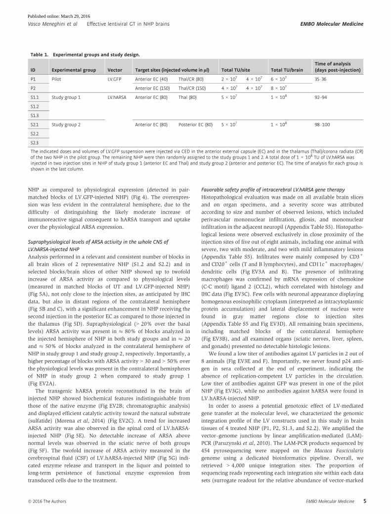
Table 1. Experimental groups and study design.
ID Experimental group Vector Target sites (injected volume in ll) Total TU/site Total TU/brainTime of analysis(days post-injection)
P1 Pilot LV.GFP Anterior EC (40) Thal/CR (80) 2 × 10⁷ 4 × 10⁷ 6 × 10⁷ 35–36
P2 Anterior EC (150) Thal/CR (150) 4 × 10⁷ 4 × 10⁷ 8 × 10⁷
S1.1 Study group 1 LV.hARSA Anterior EC (80) Thal (80) 5 × 10⁷ 1 × 10⁸ 92–94
S1.2
S1.3
S2.1 Study group 2 Anterior EC (80) Posterior EC (80) 5 × 10⁷ 1 × 10⁸ 98–100
S2.2
S2.3
The indicated doses and volumes of LV.GFP suspension were injected via CED in the anterior external capsule (EC) and in the thalamus (Thal)/corona radiata (CR)of the two NHP in the pilot group. The remaining NHP were then randomly assigned to the study groups 1 and 2. A total dose of 1 × 10⁸ TU of LV.hARSA wasinjected in two injection sites in NHP of study group 1 (anterior EC and Thal) and study group 2 (anterior and posterior EC). The time of analysis for each group isshown in the last column.
ª 2016 The Authors EMBO Molecular Medicine
Vasco Meneghini et al Effective lentiviral GT in NHP brains EMBO Molecular Medicine
5
Published online: March 29, 2016
A
B
C D
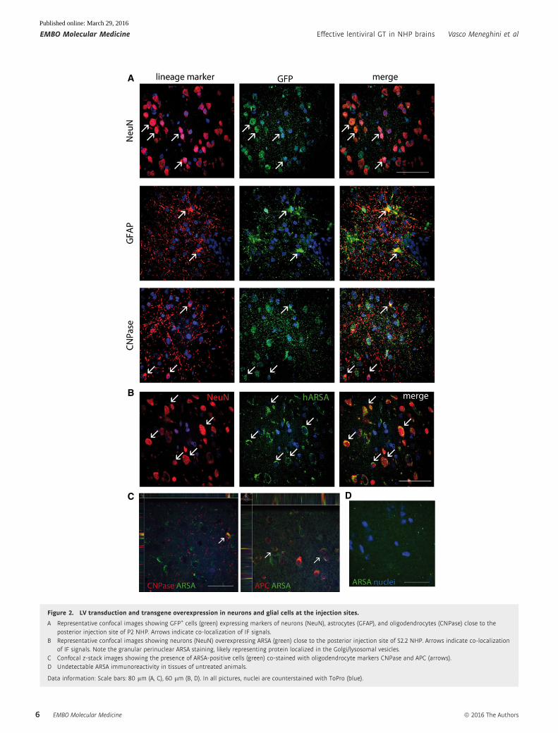
Figure 2. LV transduction and transgene overexpression in neurons and glial cells at the injection sites.
A Representative confocal images showing GFP+ cells (green) expressing markers of neurons (NeuN), astrocytes (GFAP), and oligodendrocytes (CNPase) close to theposterior injection site of P2 NHP. Arrows indicate co-localization of IF signals.
B Representative confocal images showing neurons (NeuN) overexpressing ARSA (green) close to the posterior injection site of S2.2 NHP. Arrows indicate co-localizationof IF signals. Note the granular perinuclear ARSA staining, likely representing protein localized in the Golgi/lysosomal vesicles.
C Confocal z-stack images showing the presence of ARSA-positive cells (green) co-stained with oligodendrocyte markers CNPase and APC (arrows).D Undetectable ARSA immunoreactivity in tissues of untreated animals.
Data information: Scale bars: 80 lm (A, C), 60 lm (B, D). In all pictures, nuclei are counterstained with ToPro (blue).
EMBO Molecular Medicine ª 2016 The Authors
EMBO Molecular Medicine Effective lentiviral GT in NHP brains Vasco Meneghini et al
6
Published online: March 29, 2016

cell clones in brain tissues of LV-injected NHP) showed a pattern of
polyclonal marking in all the four tissue samples analyzed without
evidence for expanded clones within the transduced brain
(Fig EV3H). In agreement with previous reports (Bartholomae et al,
2011; Lattanzi et al, 2014), we found that about 60% of LV integra-
tions mapped within genes (Fig EV3I). Of importance, Gene Ontol-
ogy analysis showed preferential targeting of genes related to
neural/neuronal function, without evidence for in vivo selection of
insertions occurring at cancer genes (Appendix Table S6). Overall,
these results indicated a favorable safety profile of intracerebral LV
gene therapy in juvenile NHP.
Therapeutic potential of LV-mediated intracerebral gene therapyin the Krabbe NHP model
Intracerebral LV.hGALC delivery is well tolerated in the Krabbe NHP
While there are no large animal models of MLD, a mutation causing
globoid cell leukodystrophy (GLD) in the rhesus monkey has been
previously described (Luzi et al, 1997). The rhesus macaque model
of Krabbe disease has a dinucleotide deletion that abolishes galacto-
sylceramidase (GALC) activity and shows clinical signs (muscle
tremors of head and limbs, ataxia, hypertonia, and incoordination)
and immunopathologic alterations (central and peripheral demyeli-
nation, the presence of multinucleated globoid cells, and psychosine
accumulation) resembling those found in humans (Baskin et al,
1998; Weimer et al, 2005; Borda et al, 2008). While these animals
represent a unique LSD model to provide proof of principle of safety
and efficacy of gene therapy platforms, their limited availability
hampers the use of a large sample size to test different therapeutic
approaches. We had the opportunity to investigate for the first time
LV-mediated intracerebral gene delivery in one Krabbe-affected
juvenile NHP and in one normal animal from the same colony. We
administered a LV encoding for the hGALC cDNA tagged with Myc
peptide (Appendix Table S2), which we tested for efficacy in
GALC-deficient cells in vitro and upon intracerebral injection in
GALC-deficient Twitcher mice (Appendix Fig S6). With the aim of
matching the age of LV.hARSA-injected NHP and because of the
rapid disease progression in Krabbe-affected animals (average life
span 100 days), a young affected animal (JT02; 53 days) was
chosen for the study. The normal NHP (JV02) was 89 days old at
the time of treatment (Appendix Table S7). We applied a protocol
similar to that described for LV.hARSA injection in normal NHP.
Since a controlled CED-mediated system was not available, we
used a standard Hamilton-driven injection, splitting the total
injected volume in multiple deposits. We performed two unilateral
injections targeting the internal capsule (three deposits; 5 × 10⁶
TU/20 ll/deposit) and the thalamus (2 × 10⁷ TU/40 ll). The total
injected LV.hGALC dose in JT02 and JV02 was 0.35 × 10⁸ TU/
brain as compared to 1 × 10⁸ TU/brain in LV.hARSA-injected
NHP.
Prior to treatment (at 7, 14, and 28 postnatal days), both animals
underwent neurobehavioral assessment using a Neurobehavioral
Assessment Scale (NBAS) (Brazelton, 1973) standardized for use
with juvenile rhesus macaques (Champoux et al, 1994, 1997, 2002)
that examines central aspects of juvenile’s neurobehavioral perfor-
mance including orientation, state control, motor maturity, and
reflexes. With the exception of the 7-day testing period, scores for
study animals fell within historical ranges for genetic status and
age. In addition, JT02 scored lower than the control animals for
most measures and most time points, including motor maturity,
activity, neuromotor items, and muscle tone items (Fig EV4A and
Appendix Table S8).
The surgical procedure was well tolerated with no serious adverse
events. All physiological parameters were normal during surgery
and in the post-surgery follow-up. Body weight of treated animals
was comparable to historical ranges from animals of similar genetic
status and age (Fig EV4B). These results closely resembled those
obtained in age-matched LV-injected normal NHP.
Three months after treatment, animals were perfused with saline.
Tissues (CNS and organ specimens) were collected to assess
Table 2. Diffusion of LV particles in the NHP brains and estimate of transduced brain volume.
NHP Experimental group VCN at inj. sites 1st/2nd
Diffusion of LVparticles (mm)
% of hemisphere volumecontaining vector
% of total brain volumecontaining vectorAP ML DV Injected Contralateral
P1 Pilot group 1.17/1.62 30 30 24 13 5 9
P2 0.58/0.48 48 20 20 16 1 8.5
Mean � SEM 0.96 � 0.27 14.5 � 1.5 3 � 2 8.7 � 0.25
S1.1 Study group 1 0.67/1.15 24 20 20 12 2 7
S1.2 2.25/0.18 36 20 20 8 2 5
S1.3 0.25/1.46 24 30 30 13 0 6.5
Mean � SEM 0.99 � 0.32 11.0 � 1.5 1.3 � 0.7 6.2 � 0.6
S2.1 Study group 2 0.37/0.49 42 20 30 20 1 10.5
S2.2 0.75/1.52 30 20 30 21 1 11
S2.3 0.13/0.13 24 30 20 15 3 9
Mean � SEM 0.56 � 0.21 18.7 � 1.8* 1.7 � 0.7 10.2 � 0.6*
The table shows the vector copy number (VCN) at injection sites, the diffusion of LV particles along the anterior–posterior (AP), medial–lateral (ML), and dorsal–ventral (DV) axes, the percentage of hemisphere volume containing integrated LV in the injected and contralateral hemispheres and in the total brain of NHP inthe pilot group and in study groups 1 and 2. Data are presented as mean values � SEM. Data are analyzed by Kruskal–Wallis test followed by Dunn’s multiplecomparison tests. *P < 0.05 versus study group 1.
ª 2016 The Authors EMBO Molecular Medicine
Vasco Meneghini et al Effective lentiviral GT in NHP brains EMBO Molecular Medicine
7
Published online: March 29, 2016

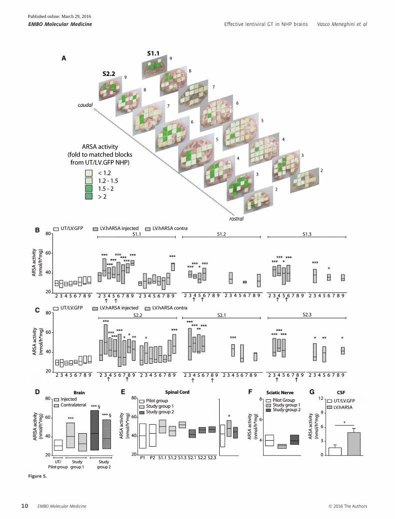
Figure 3. Integrated LV genome and transgene overexpression in LV.hARSA-injected NHP.Vector copy number (VCN) cartography shows integrated LV genome (assessed by qPCR) along the rostrocaudal axis (slices 1–10) in LV.hARSA-injected S1.1 and S2.2 NHP in aside-by-side comparison with ARSA mRNA expression (assessed by qPCR analyses using probe and primers annealing to sequences conserved in both the human andMacacafascicularis and expressed as fold increase to region-matched blocks of the contralateral hemisphere). Grading of colors for VCN ranged from white (VCN < 0.001;corresponding to CT > 37) to dark orange (VCN = 1–3). The highest VCN is found in close correspondence to the injection sites, as confirmed by comparison with post-surgeryMR images (yellow and orange circles indicate viral suspension close to the injection sites). Grading of colors for ARSA expression ranges from white (< 1.5-fold thephysiological level) to dark purple (100-fold the physiological level).
EMBO Molecular Medicine ª 2016 The Authors
EMBO Molecular Medicine Effective lentiviral GT in NHP brains Vasco Meneghini et al
8
Published online: March 29, 2016
pathology, vector, and transgene biodistribution, as well as specific
GALC activity (Appendix Fig S7).
We documented moderate astrogliosis and microgliosis in corre-
spondence to the injection sites in JV02 (Fig EV5A and B). This
local reaction was not easily assessable by immunofluorescence (IF)
in JT02, since Krabbe-affected monkeys show diffused astrogliosis
and microgliosis in several brain regions (Borda et al, 2008).
Increased GFAP and Iba-1 expression assessed by WB analysis
(Fig EV5B) and upregulation of chemokine CCL2 mRNA expression
levels in regions close to the injection sites of JT02 as compared to
untreated Krabbe NHP (Fig EV5C) suggested the occurrence of local
inflammation and macrophage recruitment as a consequence to the
surgical procedure. Tumor necrosis factor-a (TNF-a) and inter-
leukin-1b (IL-1b) mRNA expression levels were increased in JT02
tissues when compared to physiological levels but were not signifi-
cantly altered as a consequence of LV injection, as assessed by
comparison with tissues from untreated Krabbe-affected animals
(Fig EV5D and E).
Local LV.hGALC-mediated transduction establishes widespread
distribution of a functional GALC enzyme in Krabbe-affected CNS
tissues and CSF
We found integrated LV genome in proximity to the injection sites
(slices 9–13; VCN range: 0.00013–0.061 for diploid genome)
(Fig 6A). The lower VCN values as compared to those measured in
LV.GFP- and LV.hARSA-injected NHP (Table 2) likely reflect the
lower injected LV dose that, coupled to the limited particle
dispersion resulting from non-CED-mediated delivery, ultimately
determined the volume of injected hemisphere containing integrated
LV, which was � 3% as compared to 11–18% in LV.hARSA-injected
NHP (Table 2). GALC mRNA expression levels ranged between
twofold and eightfold the physiological levels (assessed by compar-
ing region-matched contralateral tissue blocks) (Fig 6B), in close
correlation with VCN values. Integrated LV genome and hGALC
mRNA were undetectable in spinal cord, sciatic nerves, liver,
spleen, and gonads of both JT02 and JV02, confirming the absence
of off-target LV integration upon injection in normal and
Krabbe-affected brains.
Importantly, by IF staining followed by confocal analysis, we
showed relevant numbers of GALC-expressing neurons and glial
cells within and around the injection sites of LV.hGALC-injected
NHP (Fig 6C), as well as in matched blocks of the contralateral
hemisphere up to � 2 cm rostral and caudal from the injection site
(Fig 6D and E).
Taken together, results obtained in LV.GFP-, LV.hARSA-, and
LV.hGALC-injected NHP showed a reproducible and transgene-inde-
pendent pattern of tissue transduction and vector biodistribution
upon intracerebral LV delivery. Specifically, a small pool of
LV.hGALC-transduced cells efficiently produced and released the
hGALC protein, which was available for cross-correction of
surrounding GALC-deficient cells in the context of a disease-affected
brain.
In order to evaluate the biological efficacy of the treatment, we
measured the specific GALC activity (Martino et al, 2009) in a
relevant number of blocks in brain slices and in spinal cord
tissues of JT02 and JV02, as well as in tissues of age-matched
untreated Krabbe-affected and normal NHP. Tissues from untreated
Krabbe-affected NHP (n = 4; Appendix Table S7) showed unde-
tectable GALC activity. In contrast, � 70% of physiological GALC
activity was measured in the injected and contralateral brain
hemispheres (Fig 7A and B), and � 40% of physiological GALC
activity was measured in spinal cord tissues (Fig 7C) of JT02. GALC
A
B
C
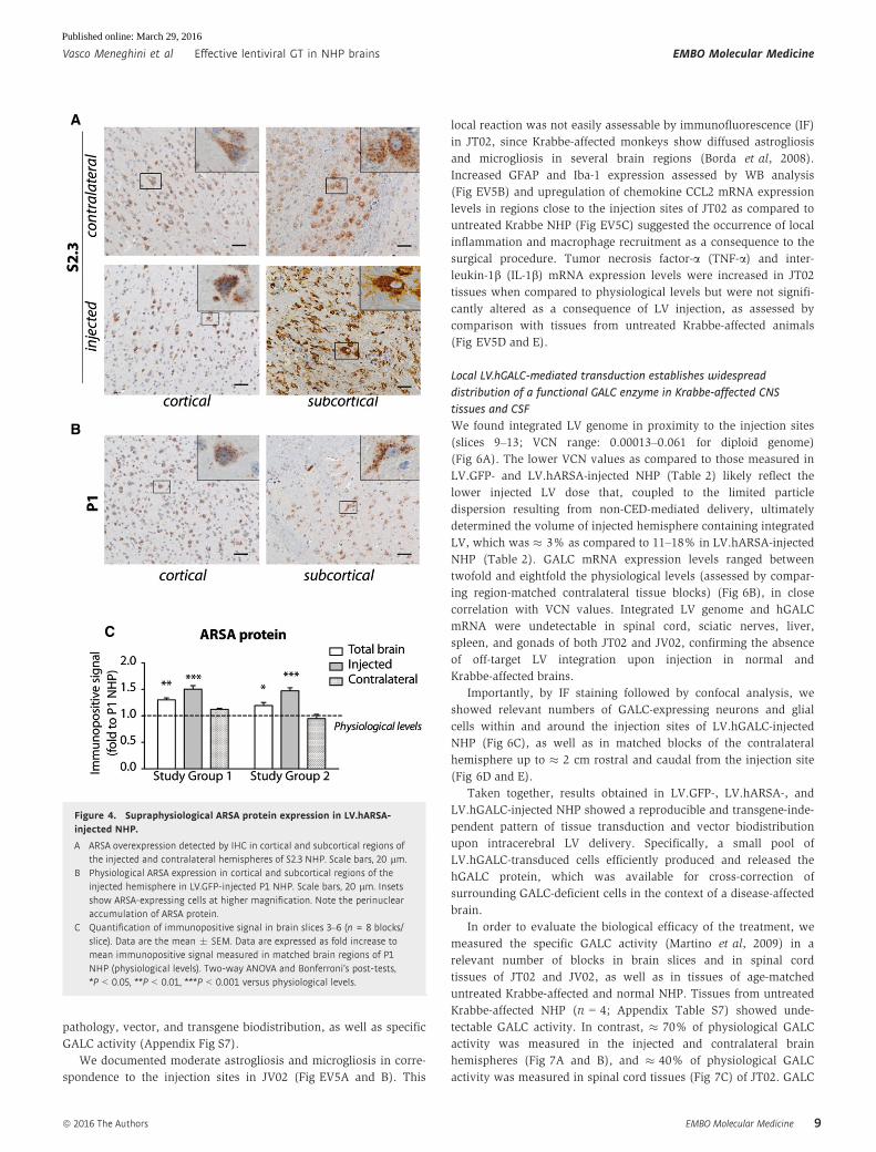
Figure 4. Supraphysiological ARSA protein expression in LV.hARSA-injected NHP.
A ARSA overexpression detected by IHC in cortical and subcortical regions ofthe injected and contralateral hemispheres of S2.3 NHP. Scale bars, 20 lm.
B Physiological ARSA expression in cortical and subcortical regions of theinjected hemisphere in LV.GFP-injected P1 NHP. Scale bars, 20 lm. Insetsshow ARSA-expressing cells at higher magnification. Note the perinuclearaccumulation of ARSA protein.
C Quantification of immunopositive signal in brain slices 3–6 (n = 8 blocks/slice). Data are the mean � SEM. Data are expressed as fold increase tomean immunopositive signal measured in matched brain regions of P1NHP (physiological levels). Two-way ANOVA and Bonferroni’s post-tests,*P < 0.05, **P < 0.01, ***P < 0.001 versus physiological levels.
ª 2016 The Authors EMBO Molecular Medicine
Vasco Meneghini et al Effective lentiviral GT in NHP brains EMBO Molecular Medicine
9
Published online: March 29, 2016
A
B
C
D E F G
Figure 5.
EMBO Molecular Medicine ª 2016 The Authors
EMBO Molecular Medicine Effective lentiviral GT in NHP brains Vasco Meneghini et al
10
Published online: March 29, 2016

overexpression (+30–40% over physiological level) was observed
in brain tissues of JV02 (Fig 7A and B). The chromatographic profile
of GALC (Lattanzi et al, 2010) obtained from selected brain slices
and spinal cord of JT02 and untreated non-affected NHP demon-
strated the comparable native conformation of transgenic and WT
GALC (Fig 7D). Importantly, GALC activity in the CSF of JT02
reached � 10% of that measured in JV02 (Fig 7E), which likely
reflected supraphysiological levels of GALC activity, as also detected
in JV02 CNS tissues (Fig 7A and B). These data suggested that LV-
mediated gene therapy supplies therapeutically relevant levels of
functional hGALC enzyme in CNS tissues and CSF of Krabbe NHP.
Preliminary indication of safety and therapeutic efficacy of
intracerebral LV.hGALC gene therapy
Taking advantage of the unique Krabbe’s behavioral manifestations,
we sought to obtain descriptive information on response to treat-
ment as a preliminary evaluation of safety and therapeutic efficacy
of the LV gene therapy strategy. To this end, JT02 and JV02 were
evaluated monthly (beginning at 2 months of age) using a modified
Bayley’s scale for infant development that included problem-
solving, motor ability, and temperament tests (Champoux et al,
1994). Results were compared with historical data relative to age-
and genotype-matched juveniles. JT02 had one assessment prior to
surgery (2 months) while JV02 had 2 pre-surgery assessments
(2 and 3 months). The control juvenile JV02 was uncooperative,
irritable, and hostile during testing and often would make no
attempt to complete items presented, resulting in unusually low
scores on the cognitive subtest and high scores on the behavior/
social orientation subtest (Appendix Table S9), while motor scores
fell within the historical range (Appendix Table S9 and Fig 7F).
Interestingly, JT02 showed unprecedentedly high motor perfor-
mance scores in the post-surgery assessment when compared to
untreated Krabbe-affected juveniles. This score measures muscle
tone and strength of muscle contractions (i.e. resistance to the
experimenter gently flexing the limbs) as well as righting reflexes
(i.e. reorienting the body when shifted out of normal position by the
experimenter). JT02’s scores showed the age-dependent increase
seen in normal juveniles, and never seen in Krabbe-affected juve-
niles, which show instead an age-dependent decrease. Indeed, while
JT02’s motor scores fell below normal animals and within range for
affected juveniles at 2–4 months, JT02’s score was higher than the
score of any prior Krabbe-affected juvenile at 5 months (3 months
after gene therapy), at a level that fell within normal range and with
a mean score almost identical to that of normal age-matched juve-
niles (Fig 7F).
Overall, these data provide the first preliminary evidence of
safety and therapeutic benefit of LV.hGALC gene therapy in Krabbe
NHP.
Discussion
Here, we show that lentiviral vector-mediated intracerebral gene
therapy in juvenile NHP displays a favorable safety profile and
provides stable therapeutically relevant enzyme activity in the whole
brain, spinal cord, and liquor of treated animals after injections in
only two CNS regions. A consistent pattern of vector and enzyme
biodistribution was observed in LV.hGALC- and LV.hARSA-injected
NHP even in the presence of differences in treatment protocols
(i.e. diseased versus normal animals, different transgenes, vector
Figure 5. Supraphysiological ARSA activity in LV.hARSA-injected NHP.
A Schematic representation of ARSA activity in the brain of S1.1 and S2.2 NHP along the rostrocaudal axis (slices 2–9). Data are expressed as fold increase to ARSAactivity detected in matched blocks of UT and LV.GFP-injected NHP (physiological levels). Grading of colors ranges from light yellow (< 1.2-fold) to dark green(≥ twofold).
B, C Enzymatic activity measured in single brain slices of NHP of study group 1 (B) and study group 2 (C) (filled gray bars, injected hemisphere; striped gray bars,contralateral hemisphere) and in UT and LV.GFP-injected NHP (white bars; physiological levels). Arrows on x-axis indicate the injection sites; n = 2–12 tissue blocks/slice. Number of tissue blocks analyzed per animal: 51 (UT/LV.GFP), 98–122 (S1.1 and S2.2), and 12–22 (S1.2, S1.3, S2.2, S2.3). Two-way ANOVA followed byBonferroni’s multiple comparison tests, *P < 0.05; **P < 0.01; ***P < 0.001 versus matched slices of UT/LV.GFP.
D Summary of ARSA activity in the injected and contralateral hemispheres of NHP in study group 1 and study group 2. Number of tissue blocks analyzed were asfollows: 51 (UT/LV.GFP), 105 (study group 1, injected hemisphere) and 52 (study group 1, contralateral hemisphere); 103 (study group 2, injected hemisphere) and 72(study group 2, contralateral hemisphere). One-way ANOVA and Tukey’s multiple comparison tests, ***P < 0.001 versus UT/LV.GFP, §P < 0.05 versus study group 1.
E ARSA activity in the spinal cord of individual LV.GFP- (n = 4 blocks/animal, 2–3 replicates) and LV.hARSA-injected NHP (n = 2–4 blocks/animal; n = 10 and n = 11total blocks for study group 1 and study group 2, respectively). Graph on the right represents the summary of ARSA activity in each treatment group. One-wayANOVA and Dunnet’s multiple comparison test. *P < 0.05 versus pilot group.
F ARSA activity in the sciatic nerve of LV.GFP- (n = 2) and LV.hARSA-injected NHP (n = 3). In (B–F), data are expressed as floating bars (line at mean).G ARSA activity in the cerebrospinal fluid (CSF) of UT/LV.GFP (n = 3) and LV.hARSA-injected NHP (n = 5). Data are expressed as mean � SEM. Student’s t-test,
*P = 0.035.
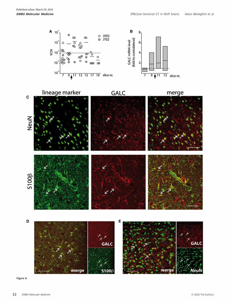
Figure 6. Efficient transduction of Krabbe CNS tissue by LV.hGALC.
A Vector copy number (VCN) indicating the distribution of integrated LV genome along the rostrocaudal axis (slices 7–19) in LV.hGALC-injected NHP (JT02 and JV02)assessed by qPCR. Each dot represents the VCN measured in one block within the slice. Lower threshold (dotted line): VCN < 0.001, corresponding to CT > 37.
B GALC mRNA expression along the rostrocaudal axis of JT02 (slices 7–13) is expressed as fold increase to region-matched blocks of the contralateral hemisphere(dotted line, y = 1). Data are expressed as floating bars (min to max, line at mean; n = 4–8 blocks/slice). Arrows on x-axis in (A) and (B) indicate the injection sites.
C Confocal images showing GALC+ cells expressing markers of neurons (NeuN) and astrocytes (S100b) close to the injection site (slice 10) in JT02. Arrows indicateco-localization of IF signals. Scale bar: 70 lm (upper panel) and 40 lm (lower panel).
D, E Confocal images showing GALC-expressing astrocytes (S100b; D) and neurons (NeuN; E) in the contralateral hemispheres, anterior (slice 8) and posterior (slice 12)to the injection site. Arrows indicate co-localization of IF signals. Scale bars: 40 lm.
◀
▸
ª 2016 The Authors EMBO Molecular Medicine
Vasco Meneghini et al Effective lentiviral GT in NHP brains EMBO Molecular Medicine
11
Published online: March 29, 2016
A B
C
D E
Figure 6.
EMBO Molecular Medicine ª 2016 The Authors
EMBO Molecular Medicine Effective lentiviral GT in NHP brains Vasco Meneghini et al
12
Published online: March 29, 2016

dose, injected volume, and delivery system), suggesting the robust-
ness of the proposed LV-mediated gene therapy platform and
providing the first preliminary evidence of therapeutic efficacy in
the exclusive NHP model.
By comparing results from the two experimental systems, it
appears that vector dose, injection in posterior white matter
regions, and CED-mediated delivery (Lonser et al, 2015), which
allowed homogeneous and reproducible vector distribution, can be
considered as factors that positively affect the extent of transduced
volume and the enhancement of LV particle dispersion. The trans-
duced brain volume in this study was overall lower when compared
to that obtained following intracerebral AAV delivery (Colle et al,
2010; Rosenberg et al, 2014; Zerah et al, 2015). This can be
explained by the multiple injection protocols (six injection sites
with 6–12 deposits) used in those studies as compared to the two-
injection protocol in this study. Also, it is in line with a more
limited diffusion capacity of LV as compared to AAV, likely due to
the larger size and, possibly, surface features of the lipid-bound LV
particle. By delivering low vector doses (0.35–1 × 10⁸ TU/brain) in
few intraparenchymal sites, we limited the invasiveness of the
procedure while achieving efficient gene transfer in the targeted
CNS regions, in which we documented transgene overexpression in
neurons and glial cells. As previously shown in the murine CNS
(Lattanzi et al, 2010, 2014), LV proficiently transduced NHP oligo-
dendrocytes. This specific feature is important in the perspective of
clinical development. Indeed, oligodendrocytes are the more abun-
dant cell types in white matter areas and the most affected cell
population in MLD and GLD but are hardly transduced by other
vector types, including AAV (Colle et al, 2010; Piguet et al, 2012;
Rosenberg et al, 2014). Also, while enzyme-deficient oligodendro-
cytes are efficiently cross-corrected in vitro or in the mouse brain
(Lattanzi et al, 2010; Piguet et al, 2012; Santambrogio et al, 2012),
this mechanism appears less efficient in large brains (Colle et al,
2010).
The transduction of a small pool of endogenous cells established
a widespread expression of transgenic products. The presence of
close to normal (GALC) or supranormal (ARSA) enzymatic activity,
even in the most caudal brain regions and in spinal cord at
3 months post-gene therapy (but likely even at earlier time points,
based on levels of transgene expression observed 1 month post-GT
in pilot animals) confirmed long-distance enzyme distribution by
possible axonal transport (Ciron et al, 2009; Colle et al, 2010;
Rosenberg et al, 2014) and cross-correction, the property of lysoso-
mal enzymes to be secreted and recaptured by surrounding cells.
Moreover, the presence of twofold normal ARSA activity and
> 10% of normal GALC activity in the liquor of treated NHP (while
no GALC protein could be isolated from the CSF of Krabbe-affected
NHP before treatment) suggested sustained production and effi-
cient bioavailability of the enzymes. Of note, the 100% increase of
ARSA levels observed in the physiological background at 3 months
post-treatment are comparable to the physiological ARSA levels
detected in the liquor of MLD patients 12–24 months post-HSC
gene therapy (Biffi et al, 2013). These results provide a rationale to
propose this approach to stabilize CNS damage and prevent further
deterioration in the late-onset forms of MLD and GLD but also to
counteract the rapid progression of CNS pathology in early symp-
tomatic infantile/early juvenile patients. While a long-term follow-
up would be needed to assess persistency of enzymatic levels in
our model, our results point against the occurrence of vector
silencing or immune response against the vector or transgenes. Of
note, antibodies against envelope and capsid proteins were occa-
sionally and transiently seen in NHP and PD patients 3 and
6 months after administration of a lentiviral vector-based thera-
peutics delivering dopaminergic-related genes (ProSavin) without
affecting transgene expression (Jarraya et al, 2009; Palfi et al,
2014).
Studies of healthy individuals carrying pseudodeficient variants
of the ARSA gene suggest that 10% of normal ARSA activity is suffi-
cient to prevent the onset of MLD symptoms (Gieselmann, 2006).
Similarly, while all individuals with Krabbe disease have very low
GALC enzyme activity (0–5% of physiological levels), there is a
relatively broad range of GALC activities in the healthy population.
Indeed, the presence of multiple polymorphisms in both GALC
alleles might lower enzyme activity to 8–20% of normal without
resulting in clinical disease (Wenger, 1993, 2000). In this view, the
extent of ARSA and GALC expression and activity observed in the
brain of LV.hARSA- and in the Krabbe-affected LV.hGALC-treated
NHP are compatible with foreseeable therapeutic activity in
patients, which remains to be evaluated in rigorous clinical trials in
which selective inclusion/exclusion criteria as well as biochemical
Figure 7. Gene therapy rescues GALC activity in CNS tissues of Krabbe-affected NHP.
A Specific GALC enzymatic activity in single brain slices of WT untreated (UT; white bars, physiological levels), WT LV.hGALC-treated (JV02), and Krabbe-affectedLV.hGALC-treated (JV02) animals (filled gray bars, injected hemisphere; striped gray bars, contralateral hemisphere). Two-way ANOVA followed by Bonferroni’smultiple comparison tests; *P < 0.05, **P < 0.01 versus matched slices of WT UT.
B, C Summary of GALC activity in brain (B) and spinal cord (C; n = 3 blocks/animal) samples from JT02 and JV02, as well as from WT and Krabbe UT controls.D Chromatographic profile of GALC in selected brain slices and spinal cord (SC) tissue of untreated (UT) WT and LV.hGALC-treated Krabbe NHP (JT02). Extracts were
run through a Sephadex S-300 gel filtration column. Fractions (0.2 ml) were assayed for enzyme activity using MUGAL substrate in the presence (○) or absence of11 lM AgNO3 (●).
E GALC activity in the CSF of JT02 (pre- and post-treatment) and JV02 (post-treatment). Dots represent technical replicates.F Beginning at 2 months (m) of age, animals were evaluated monthly for motor performance using a modified Bayley’s scale for infant development. For the study
animals (JV02 and JT02), scores are reported as pre-surgery and post-surgery. Affected animals score significantly lower than normal animals at any age considered.JT02’s motor scores increase over time post-LV.GALC injection, being close to the mean observed in normal juveniles at 5 months. Historical data for normal andaffected animals are presented as means plus one standard deviation. Historical data are analyzed by two-way ANOVA and Bonferroni’s posttests, P < 0.05 at6 months, P < 0.001 at all other ages in affected versus normal animals; n = 5–20 animals/group, except for normal animals at 6 months (n = 1). See alsoAppendix Table S9.
Data information: Data in (A-C) are represented as floating bars (min to max, line at mean; n = 2–6 blocks/slice). Number of blocks analyzed/animal: 16 (WT UT), 24(JT02), and 22 (JV02). GALC activity in Krabbe UT samples is < 0.001 nmol/h*mg; n = 3 blocks. One-way ANOVA and Bonferroni’s multiple comparison test (B) or Dunnet’smultiple comparison test (C); *P < 0.05, **P < 0.001 versus WT UT.
▸
ª 2016 The Authors EMBO Molecular Medicine
Vasco Meneghini et al Effective lentiviral GT in NHP brains EMBO Molecular Medicine
13
Published online: March 29, 2016

end points will be key to correctly estimate the clinical impact of
this approach and to highlight potential therapeutic hurdles that are
dependent on the biochemical and kinetic properties of the individ-
ual enzymes.
The local neuroinflammation in the vicinity of the injection
areas, which we found in some treated animals, was previously
reported in AAV- (Colle et al, 2010; Salegio et al, 2012; Rosenberg
et al, 2014) and LV-injected NHP (Kordower et al, 2000; Jarraya
et al, 2009), being mainly associated with tissue damage (conse-
quent to the surgical procedure), persistence of injected soluble
material, and/or immune response. Our data suggest that macro-
phage and mononuclear cell infiltrates observed to a different extent
in LV.GFP-, LV.hARSA-, and, indirectly, in LV.hGALC-injected NHP
at 3 months post-treatment are not correlated to an immune
A
C
E
F
D
B
Figure 7.
EMBO Molecular Medicine ª 2016 The Authors
EMBO Molecular Medicine Effective lentiviral GT in NHP brains Vasco Meneghini et al
14
Published online: March 29, 2016

response against transgenes or LV capsid proteins. In contrast to a
recent study (Zerah et al, 2015), recruitment of B and T cells at sites
of neuroinflammation was not associated with anti-hARSA antibod-
ies (undetectable in LV.hARSA-injected NHP, as expected consider-
ing the 96% identity between human and Macaca fascicularis ARSA
proteins) or anti-GFP antibodies (detectable at low titer in only one
of the two LV.GFP-injected NHP, both showing similarly mild
inflammation). Also, only one out of five NHP showing
mononuclear cell infiltration developed detectable antibodies
against the VSV envelope and LV capsid protein, and similar titers
of antibodies were detected in one NHP in which cell infiltration
was absent. The mechanism by which LV injections may have
caused these focal lesions remains unclear. Importantly, there was
no clinical impact of these histopathological findings, as docu-
mented by normal general safety, hematological, and behavioral
parameters.
The use of self-inactivating long-term repeats in new generation
LV reduces the potential for gene activation at the insertion sites
(Montini et al, 2006). LV-delivered ARSA and GALC genes do not
impart a proliferative advantage to neural cells (Neri et al, 2011).
This, in addition to the targeting of postmitotic cells (Colin et al,
2009; Lattanzi et al, 2014) and the preferential integration into the
body rather than promoters of active genes, minimizes the risk of
oncogenesis as compared to earlier generation retroviral vectors.
Instead, promoter insertion and enhancer-mediated activation of
oncogenes are the preferred mechanisms of insertion-driven oncoge-
nesis (Cesana et al, 2014). Our earlier studies have shown that LV-
transduced cells undergo proliferation and apoptosis associated with
physiological neonatal brain development. In contrast, we never
observed hyperproliferation of transduced cells or formation of
tumor masses in mice analyzed at PND40 and at 6 months post-
neonatal injection (Lattanzi et al, 2014). Intragenic LV insertions
may interfere with normal cell function, triggering the formation of
aberrant transcripts that result in gain or loss of function mutations
(Cesana et al, 2012; Moiani et al, 2012). However, this mechanism
has not been associated with cell transformation in the follow-up of
HSC gene therapy trials, in which no adverse genotoxic events have
been shown to date (Cartier et al, 2009; Aiuti et al, 2013; Biffi et al,
2013). It is also possible that mutant proteins produced by aberrant
transcripts acquire detrimental functions and cause cell death or
functional impairments. The impact of this phenomenon in non-
proliferating tissues, however, would be essentially limited to the
cell(s) in which the damaging insertion has occurred, and it is diffi-
cult to predict/assess in this experimental setting. The polyclonal
pattern of LV integration, the preferential targeting of neural-specific
genes without preferential insertions in genes involved in tumor
formation or neurological disorders that we report here for the first
time in NHP are in line with previous data obtained in rodents
(Bartholomae et al, 2011; Lattanzi et al, 2014) and further point to a
low genotoxic risk associated with the proposed GT platform. The
CNS-restricted distribution of LV particles (Jarraya et al, 2009;
Lattanzi et al, 2014) and, consequently, the absence of off-target LV
integration in the periphery represent an additional favorable safety
trait of this GT platform.
According to previous studies performed in mice (brain volume
� 0.5 cm3) (Lattanzi et al, 2010, 2014), we estimated that
� 1 × 10⁶ TU/cm3 could be safe and potentially effective in achiev-
ing enzyme correction in the brain of juvenile NHP (brain volume
� 60–65 cm3). Based on the respective brain volumes of NHP and
human infantile/adult patients (� 500–1,000 cm3), the equivalent
vector dose to be injected in patients would be � 5 × 10⁸–1 × 10⁹
TU. This roughly corresponds to � 0.5–1 ml of high-titer clinical
grade LV batches (Biffi et al, 2013), which could be easily divided
into multiple intraparenchymal vector deposits in order to minimiz-
ing the vector dose at each injection site while maintaining robust
cell transduction, transgene production, and expected biological
efficacy.
We have previously demonstrated similar transduction efficiency
and biodistribution of ARSA and GALC enzymes upon intracerebral
LV gene delivery in mice. Here, we provide proof of concept that
this equivalence holds true in larger brains. Considering the ethical
and practical limitations of working with NHP, eight normal
monkeys represent a considerable sample size to study safety and
biodistribution in a physiological background. The rhesus monkey
model of Krabbe disease represents the first reported observation of
a lysosomal storage disease in any non-human primate species
(Wenger, 2000). By treating one Krabbe-affected juvenile animal,
we could assess for the first time ever vector and transgene biodis-
tribution as well as rescue of enzymatic activity in CNS tissues of a
large animal that shares high degree of pathological and clinical
similarity to the human disease. This analysis provided formal proof
of biological efficacy of the strategy, as it was predicted from the
results obtained in LV.hARSA-injected normal NHP. Neurobehav-
ioral assessment using scales standardized for use with juvenile
rhesus macaques showed progressive improvement of motor perfor-
mance (muscle tone and strength of muscle contractions) in the
gene therapy-treated Krabbe NHP, which reached those of age-
matched normal animals at 3 months post-gene therapy. Notably,
motor scores of Krabbe-affected macaques generally fell below
normal animals at any age considered and tend to decline with age,
resembling the relentless motor deterioration observed in individu-
als with the infantile form of Krabbe disease (Wenger, 1993). So,
our results provide indication (although very preliminary) of thera-
peutic benefit of the lentiviral gene therapy strategy in this model.
Given the variability in disease progression that characterize
Krabbe-affected macaques (Baskin et al, 1998; Weimer et al, 2005;
Borda et al, 2008), further studies including more animals might
better address potential immune response against the GALC protein
(a minor issue if considering patients with residual protein and
enzymatic activity) and definitely prove whether metabolic rescue
translates into clinical–pathological amelioration and overall
therapeutic benefit.
In conclusion, our study advances intracerebral lentiviral gene
therapy for clinical development. Lentiviral vectors may cooperate
with other gene/cell therapy strategies to address some of the unre-
solved challenges that have limited so far the development of
effective treatment options for MLD and GLD patients.
Materials and Methods
Study design
Non-human primate studies were limited in sample size for feasibil-
ity and ethical reasons. For experiments in normal NHP, we chose to
use n = 2–3 animals/treatment group (random assignment) in order
ª 2016 The Authors EMBO Molecular Medicine
Vasco Meneghini et al Effective lentiviral GT in NHP brains EMBO Molecular Medicine
15
Published online: March 29, 2016

to reliably detect potential differences related to treatment. In
contrast, only one Krabbe-affected monkey and one normal monkey
of comparable age were available for treatment with LV.hGALC.
However, the use of a similar experimental protocol in the two stud-
ies (age at injection, injection sites, tissue collection and analysis,
and readouts) and the availability of historical data/samples from
the Krabbe NHP colony allowed us to go beyond a qualitative assess-
ment of data. The sample size of mouse studies was chosen accord-
ing to earlier studies of intracerebral LV gene therapy (Lattanzi et al,
2010, 2014). Mice were randomly assigned to experimental groups.
Untreated mice (mutant and WT) served as controls for the treat-
ment groups. No animal (mice and NHP) administered the intended
dose and surviving the procedures was excluded from the analysis.
Whenever possible results are shown in dot-plot graphs or in
low–high bar graphs (line at mean) in order to show intragroup
variability. Investigators involved in the histopathological analysis
were blinded. Investigators performing animal handling, sampling,
euthanasia, and raw data analysis were not blinded.
Non-human primates
Normal NHP
Ten pregnant females (Macaca fascicularis) were purchased by
BIOPrim, Baziege, France, and housed at MIRCen, CEA/INSERM,
Fontenay aux roses, France. Nine babies (two females and seven
males) were delivered and were used for this study when they
reached 2–3 months of age (juveniles). Details on the animals used
can be found in Appendix Table S1.
Krabbe-affected NHP
One 53-day-old Krabbe-affected rhesus monkey (Macaca mulatta)
and one age-matched unaffected normal animal from the colony
housed at TNPRC were used. Tissues from four Krabbe-affected
monkeys (available in Prof. Bunnell’s lab) were used as controls for
molecular and enzymatic analyses. Details on the animals used can
be found in Appendix Table S7.
Plasmids and vector production
Low-endotoxin plasmid containing the full-length coding sequence
of the human (h)ARSA and VSV-pseudotyped lentiviral vector (LV)
batches was prepared by MolMed S.p.A. (http://www.molmed.
com) and manufactured according to the process approved for clini-
cal use (Biffi et al, 2013) and thus subject to stringent quality
assessment, although the procedures were not performed under
GMP in order to contain costs.
The plasmid containing the full-length coding sequence of the
human (h)GALC gene was kindly provided by Dr. Shen, Baylor
Univ. Medical Center, Dallas, USA. The human gene coding for
GALC was tagged with the Myc epitope. Our in vitro and in vivo
data on murine models indicate that Myc epitope does not interfere
with the lysosomal targeting and enzymatic activity.
Laboratory grade/scale VSV-pseudotyped third-generation LV
encoding for green fluorescent protein (LV.GFP) or hGALC-myc
(LV.hGALC) under the control of the human phosphoglycerate
kinase (PGK) promoter was produced by transient four-plasmid
co-transfection into 293T cells and purified by ultracentrifugation,
according to established protocols (Amendola et al, 2005).
Reagents, cloning procedures, and sequence information are avail-
able upon request. Infection titer was estimated on 293T cells by
limiting dilution (LV.GFP) or by qPCR (LV.hGALC). Vector particle
was measured by HIV-1 gag p24 antigen immunocapture (NEN Life
Science Products, Zaventem, Belgium). Vector infectivity was calcu-
lated as the ratio between titer and particles. Details on LV batches
used can be found in Appendix Table S2. Eight vials (1 ml/vial) of
LV.hARSA, LV.GFP (100 ll/vial), and LV.hGALC (100 ll/vial) were
randomly selected, stored frozen, and delivered to MirCen
(LV.hARSA and LV.GFP) and to TNPRC (LV.hGALC) 2 weeks before
the scheduled day of treatment.
Surgical procedures
LV injection using convection enhanced delivery (CED)
After underskin epinephrine–xylocaine local administration, animals
were positioned in a MRI-compatible stereotaxic apparatus. Unilat-
eral (left hemisphere) craniotomy was performed to position a CED
cannula to infuse LV.GFP or LV.hARSA (2 × 10⁷ – 5 × 10⁷ TU/injec-
tion site) into two targeted regions of the brain parenchyma, namely
anterior external capsule/corona radiata and thalamus (pilot group
and study group 1); anterior external capsule/corona radiata and
posterior external capsule (study group 2). Surgical coordinates
were determined from preoperative MRI using the known distance
from the scan plane containing the ear bars (anteroposterior axis)
and the mediolateral distance from midline on the coronal image
containing the target. The infusion system consisted of a fused silica
reflux-resistant cannula connected to a loading line (containing
vector suspension) and an infusion line (containing sterile saline). A
1-ml syringe mounted on an MRI-compatible infusion pump
(Harvard Apparatus Inc, Holliston, Massachusetts, USA) regulated
the flow of fluid through the delivery cannula. Approximately
40–150 ll of vector suspension was infused through the cannula via
infusion pump to the target site with a dosing scheme consisting of
ascending infusion rates. Immediately after the infusion procedure,
the wound site was closed in anatomical layers, and the animal was
transferred to the MRI room for a post-infusion scan. After that, the
animal was returned to the operating room for extubation and
recovery procedures.
LV injection in Krabbe-affected (JT02) and normal (JV02) NHP
Animals were anesthetized with ketamine hydrochloride 10 mg/kg
IM, intubated, and maintained on isoflurane and oxygen. A midline
skin incision was made over the dorsal aspect of the skull. All
muscle and subcutaneous tissue were reflected with a periosteal
elevator and the skin and underlying tissue is retracted to ensure
constant exposure of the skull. The animals were positioned in a
MRI-compatible stereotaxic frame (1430M, Kopf Instruments). The
stereotaxic instrument was mounted on the stereotaxic head frame
and rostral/caudal and medial/lateral coordinates (achieved with a
Micromanipulator; model 1760-61 Kopf instruments) were located
just above the skull. The stereotaxic instrument was removed to
allow for drilling an access hole into the skull. A 4-mm drill bit was
used to drill through the outer cortex and medullary cavity of the
skull. A constant sterile water lavage of the drilling site was
performed to minimize airborne bone dust and to keep surrounding
tissues cool. A 1.5-mm drill bit was used to drill through the inner
cortex and clean the margins of the defect to ensure that no damage
EMBO Molecular Medicine ª 2016 The Authors
EMBO Molecular Medicine Effective lentiviral GT in NHP brains Vasco Meneghini et al
16
Published online: March 29, 2016

occurs to underlying structures. Once adequate exposure to specific
anatomical locations of the brain is obtained, the stereotaxic
instrument was placed back on the stereotaxic head frame. The
predetermined rostral/caudal and medial/lateral coordinates of the
brain were obtained by adjusting the stereotaxic instrument. Injec-
tions were performed with a 50-ll Hamilton syringe with a 2-inch
22-gauge needle. Injection rate was approximately 1 ll/min. Coordi-
nates for JV02: injections were performed in the right hemisphere,
in a rostrocaudal plane at the level of the external auditory meatus.
One injection was performed in the thalamus (15 mm ventral to the
surface of the brain and 3 mm lateral to midline; 2 × 10⁷ TU/40 ll),and three injections were performed in the internal capsule
(15.5 mm ventral, 8.5 mm lateral; 13 mm ventral, 8 mm lateral;
10 mm ventral, 6.5 mm lateral; 5 × 10⁶ TU/20 ll/site). Coordinatesfor JT02: injections were performed in the right hemisphere, in a
rostrocaudal plane 4 mm cranial to the external auditory meatus.
One injection was performed in the thalamus (19 mm ventral and
3 mm lateral; 2 × 10⁷ TU/40 ll), and three injections were
performed in the internal capsule (23 mm ventral, 7 mm lateral;
19 mm ventral, 7 mm lateral; 19 mm ventral, 9 mm lateral; 5 × 10⁶
TU/20 ll/site). Total TU injected/brain for JT02 and JV02 was
3.5 × 10⁷.
Quantification of VCN
VCN in murine samples was quantified as previously described
(Lattanzi et al, 2010). Genomic DNA was extracted from NHP brain
tissues, spinal cord, sciatic nerve, liver, spleen, and gonads follow-
ing manufacturer’s instructions (#200600, DNA Extraction Kit,
Agilent Technologies, Inc. Santa Clara, CA 95051, USA). Samples
were quantified by NanoDrop ND-1000 Spectrophotometer (Euro-
clone, Pero, Italy). Vector copies per genome were quantified by
TaqMan analysis using 50 ng of template DNA. DNA extracted from
untreated NHP was used as negative control. LV backbone was
amplified by using forward primer (50-TACTGACGCTCTCGCACC-30
– final concentration: 10 lM), reverse primer (50-TCTCGACGCAGGACTCG-30 – final concentration: 10 lM), and FAM/MGB probe
(50-FAM-ATCTCTCTCCTTCTAGCCTC-MGB-30 – 5 lM final concen-
tration). As there was an internal reference gene for normalization,
we amplified a fragment of the Macaca fascicularis TAF7 RNA poly-
merase II gene (TATA box-binding protein-associated factor) by
using a Custom TaqMan Gene Expression Assays containing both
probe and primers (Rh02916247_s1, reporter: FAM, quencher:
MGB, Life Technologies). Other details can be found in
Appendix Supplementary Methods.
LV diffusion along the three axes was calculated considering
consecutive blocks with detectable VCN along the axis. The volume
of each brain block was estimated by dividing the average hemi-
sphere volume of NHP in the study (32.5 cm3; estimated through
MRI analysis and consistent with published volumes for the Macaca
fascicularis and Macaca mulatta brain) by the number of blocks per
hemisphere. P1 NHP: 0.25 cm3; P2 NHP: 0.37 cm3; Study groups:
0.32 cm3. JT02 and JV02: 0.25 cm3.
Quantification of ARSA and GALC mRNA
Blocks with detectable VCN as well as surrounding blocks in the
same or different slices (injected hemisphere) and matched brain
blocks of the contralateral hemisphere were selected for mRNA
extraction. Total RNA was isolated and purified from brain tissues
using RNeasy Lipid Tissue Kit (Qiagen, Hilden, Germany), following
the manufacturer’s instructions. The quantity of RNA was
determined by 260/280 nm optical density reading on NanoDrop
ND-1000 Spectrophotometer (NanoDrop, Pero, Italy). Reverse tran-
scription was carried out using 2 lg of total RNA and the Quantitect
Reverse Transcriptase kit (Qiagen, Hilden, Germany).
We evaluated the exogenous hARSA and hGALC mRNA
expression by quantitative PCR, using primers (forward, 50-GGCTGTTGGGCACTGACAAT-30, final concentration: 10 lM; reverse, 50-ACGTCCCGCGCAGAATC-30, final concentration: 10 lM) and probe
(50-FAM-TTTCCTTGGCTGCTCGCCTGTGT-MGB-30, final concentra-
tion: 5 lM) annealing the vector construct WPRE sequence. mRNA
samples extracted from brain of UT NHP were used as negative
controls.
In parallel, we assessed total ARSA and GALC mRNA expression
using a Custom TaqMan Gene Expression Assays, containing both
probe and primers (ARSA: Rh02828799_m1 reporter: FAM,
quencher: MGB, Life Technologies; GALC: Rh02827142_m1
reporter: FAM, quencher: MGB, Life Technologies). As an internal
reference for normalization, we amplified a fragment of the Macaca
fascicularis TAF7 RNA polymerase II gene, using a Custom
TaqMan� Gene Expression Assays, containing both probe and
primers (Rh02916247_s1, reporter: FAM, quencher: MGB, Life Tech-
nologies). Reactions were carried out in a total volume of 12.5 ll, ina ViiATM 7 Real-Time PCR System (Life Technologies-Applied Biosys-
tems, Carlsbad, CA, USA).
Expression levels of transgenic hARSA and hGALC mRNA were
calculated as fold to TAF7 expression levels. Samples with WPRE
probe CT > 37 (corresponding to a fold value < 0.0001) were
considered as having undetectable levels of exogenous hARSA or
hGALC expression.
Total ARSA and GALC mRNA expression levels in tissue blocks
of the injected hemisphere were expressed as fold to mRNA expres-
sion levels detected in pair-matched blocks of the contralateral
hemisphere. Only samples with TAF7 probe CT within 22–26 were
considered in the analyses. Data were obtained by n = 2 experi-
ments performed in duplicate. Within the same experiments,
samples with a CT variability > 2 for TAF7 probe CT, WPRE probe
CT, total GALC probe CT, or total ARSA probe CT within the dupli-
cate were not considered in the analyses. Samples with interexperi-
ment variability > 50% were analyzed at least in n = 3 experiments.
Immunofluorescence
Selected post-fixed samples from brain were recovered from paraf-
fin using xylene and then rehydrated with different dilutions of
ethanol in water. Unmasking was performed in citrate/Tris–EDTA
buffer in a bain-marie for 30 min. Slides were incubated with
blocking solution [0.5% milk + 10% normal goat serum
(NGS) + 1% BSA + 0.3% Triton X-100 in PBS] for 2 h at RT and
then incubated overnight at 4°C with primary antibody diluted in
blocking solution. After washing in PBS (3 × 10 min each), anti-
body staining was revealed using species-specific fluorophore-
conjugated secondary antibodies diluted in 10% NGS + 1%
BSA + 0.1% Triton X-100 in PBS. Tissue sections were counter-
stained with DAPI (40,6-diamidino-2-phenylindole; 10236276001,
ª 2016 The Authors EMBO Molecular Medicine
Vasco Meneghini et al Effective lentiviral GT in NHP brains EMBO Molecular Medicine
17
Published online: March 29, 2016

Roche, Basel, Switzerland) or ToPro-3 (T3605, Life Technologies,
Invitrogen, Carlsbad, CA, USA), washed in PBS and mounted on
glass slides using Fluorsave (#345789, Calbiochem, Billerica, MA,
USA).
Cell counts
GFP staining was evaluated in coronal brain sections selected close to
the anterior or posterior injection sites (two blocks, two slices/block,
and five fields/slice). GFP staining was performed in combination with
lineage-specific markers (NeuN, neurons; GFAP, astroglia; APC and
CNPase, oligodendroglia) and nuclear counterstaining. Triple-labeled
sections were scanned with Perkin Elmer UltraVIEW ERS spinning
disk confocal microscope at 40× magnification; GFP+ cells showing
defined nuclei were counted and co-localization with neuronal or glia
markers was evaluated. Results were expressed as follows: (i) percent-
age of GFP+ cells on total number of nuclei (extent of transduction);
(ii) percentage of double-positive GFP+NeuN+, GFP+GFAP+, or
GFP+CNPase+ cells over the total number of GFP+ cells.
Immunohistochemistry
Post-fixed tissues samples were stained with: (i) anti-CD3, anti-
CD20, and anti-CD11c to assess the nature of infiltrating cells.
Detection of signal was performed with UltraVision Quanto Detec-
tion System HRP DAB (Thermo ScientificTM); (ii) anti-ARSA antibody
recognizing an epitope conserved in both human and Macaca fascic-
ularis by using Ventana autostainer. Samples included cortical and
subcortical white and gray matter regions of the left and right
hemispheres of slices 3–6. All specimens were examined in blind.
ARSA-positive signal was quantified, and a score was assigned
based on the following factors: pattern of distribution of stained
cells, approximate intracellular granulation size and number (low
versus high quantity of granules), and the level of the staining in
the surrounding extracellular matrix (ECM). The sum of the partial
scores gave the final grade for each specimen. For each NHP, the
mean score was calculated and data were expressed as fold increase
to the score assigned to P2 NHP (control).
Enzymatic activity and DAE chromatography
ARSA and GALC activity assay as well as DAE chromatography were
performed according to previously described protocols (Martino
et al, 2005, 2009; Morena et al, 2014), which have been validated in
our previous studies (Lattanzi et al, 2010, 2014; Neri et al, 2011).
Detection of antibodies in the serum (ELISA assay)
Pre- and post-surgery sera collected from LV-injected NHP were
tested to detect the presence of antibodies against transgenes (GFP
and hARSA) and LV particles through enzyme-linked immunosor-
bent assay (ELISA). Details can be found in Appendix Supplementary
Methods.
p24 enzyme-linked immunosorbent assay
The presence of vector particle in the sera of LV-injected NHP was
determined by immunosorbent assay by using the Alliance HIV-1
p24 antigen ELISA kit (Perkin Elmer), following manufacturer’s
instructions. Details can be found in Appendix Supplementary
Methods.
LAM-PCR and 454 pyrosequencing
The procedures and primer sequences for linear amplification-
mediated (LAM)-PCR for LV integration site retrieval have been
previously described (Schmidt et al, 2007). Briefly, we used 1 lgof DNA extracted from brain tissues as template for 100-cycle
linear PCR pre-amplification of genome–vector junctions, followed
by magnetic capture of the biotinylated target DNA, hexanu-
cleotide priming, restriction digest using Tsp509I and HpyCHIV4,
and ligation of a restriction site–complementary linker cassette.
The first exponential biotinylated PCR product was captured via
magnetic beads and re-amplified by a nested second exponential
PCR. We separated LAM-PCR amplicons on high-resolution
Spreadex gels (Elchrom Scientific) to evaluate PCR efficiency and
the bands pattern for each sample. We adapted the LAM-PCR
samples for 454-pyrosequencing by fusion primer PCR to add the
Roche 454 GS-FLX adaptors (Paruzynski et al, 2010): Adaptor A,
plus a six nucleotides barcode, was added at the LTR end of the
LAM-PCR amplicon; adaptor B was added at the linker cassette
side. Each sample was amplified with fusion primers carrying each
a different barcode sequence. Fusion primer PCR products were
assembled in equimolar ratio into libraries, avoiding repetition of
identical barcodes, and sequenced at GATC biotech. Sequence
reads were mapped to reference genome of Macaca Fascicularis,
with a web-available version of BLAST. All the hits with an iden-
tity score lower than 95% were discarded, as well as those with a
starting alignment position beyond the third base. Each integration
site (IS) was annotated using an own developed annotation tool
(Calabria et al, 2014). For each IS, the output of the tool contains
the following information: chromosome position, feature’s name
(in which the IS is contained), starting and ending position,
distance of the IS from the feature’s transcription starting site, and
the IS relative position with respect to the feature. Raw sequence
reads are available at the NCBI sequence reads archives (SRA) at
the following link: http://www.ncbi.nlm.nih.gov/bioproject/
PRJNA311501.
The raw data were converted to a list of GeneID, using DAVID
online software (http://david.abcc.ncifcrf.gov/home.jsp) and then
filtered using the same software. Gene Ontology was performed
using the functional annotation tool. Results were filtered with the
following parameters: P-value < 0.05; fold change > 2; Benjamini
value < 0.05; false discovery rate < 0.05.
Statistics
Data were analyzed with GraphPad Prism version 5.0a for Macin-
tosh and expressed as the mean � standard error mean (SEM) if
not otherwise stated. Nonparametric (Kruskal–Wallis) or parametric
tests (Student’s t-test, and one-way or two-way analysis of vari-
ance) followed by ad hoc post-tests were used according to data
sets. The number of samples analyzed and the statistical test used
are indicated in the legends to each figure. The summary of
statistics for the main figures and tables is provided in
Appendix Table S10.
EMBO Molecular Medicine ª 2016 The Authors
EMBO Molecular Medicine Effective lentiviral GT in NHP brains Vasco Meneghini et al
18
Published online: March 29, 2016
Study approval
Mouse studies were conducted under an approved protocol of the
“Institutional Committee for the Good Animal Experimentation” of
the San Raffaele Scientific Institute and are reported to the Ministry
of Health, as required by Italian law.
All the procedures and protocols involving NHP were performed
under approval of: (i) MIRCen ethical committee, in accordance
with the animal welfare guidelines and recommendations provided
by the directive 2010/63/UE (decree 2013-118) (normal NHP); (ii)
Institutional Animal Care and Use Committee (IACUC) of the
TNPRC conformed to the requirements of the Animal Welfare Act
(Normal and Krabbe-affected NHP).
For details on mouse studies, in vitro studies, surgical and post-
surgical monitoring, magnetic resonance imaging, tissue collection
and processing, quantification of VCN, Western blot analyses,
histopathology, ELISA assay, p24 enzyme-linked immunosorbent
assay, and primary and secondary antibodies, see the Appendix Sup-
plementary Methods.
Expanded View for this article is available online.
AcknowledgementsWe thank Lucia Sergi Sergi for the help with LV.GFP and LV.hGALC production;
Tiziana Plati for the help with p24 assay; Fabrizio Benedicenti for the help with
LAM-PCR; Andrea Annoni, Alessio Cantore, and Silvia Gregori for the help with
immune response studies and critical discussion of data; MolMed S.p.A for
LV.hARSA production; Alessandra Biffi for critical discussion of data; Eleonora
Calzoni for analysis of ARSA activity using sulfatide; the MirCen team: Lev Stim-
mer and Helene Touin for histopathology, Laurent Watroba for coordinating
surgery procedures, Julien Flament for MRI analysis and 3D brain reconstruc-
tion, Francois Lachapelle for coordinating the MiRCen staff dedicated to the
project, all the vets and technicians providing professional support during the
duration of the project; and the TNPRC team: Peter J. Didier for necroscopy and
histopathology and Cyndi Trygg for technical support and logistics. This work
was supported by: Fondazione Telethon, grant number TGT11B02 to AG; NIH
grant OD 11104 to the TNRPC; TNPRC Pilot Research project to BAB and AG.
Author contributionsVM designed and performed experiments, analyzed the data, and wrote the
manuscript. AL produced LV.hGALC and performed mouse studies. GB
performed molecular analyses, Western blot, immune response studies, and
cytokine expression profile. LT performed immunofluorescence analysis on
LV.GFP-injected NHP and molecular analysis on LV.hGALC-injected NHP. KB
contributed to the experimental plan for the studies performed at MirCen
and performed surgery using CED-mediated delivery. JB coordinated logistics
and helped with CED-mediated delivery and data collection. FM performed
ARSA and GALC activity assays. SM supervised biochemical assays, analyzed
the data, and critically revised the manuscript. AC performed integration site
analyses and analyzed the data. FS and CD performed immunohistochem-
istry on NHP tissues. EM supervised integration site studies and analyzed
the data. JPD performed surgery at TNPRC. JMFP performed immunofluores-
cence on tissues from NHP injected at TNPRC. KCB supervised the behav-
ioral analysis at TNPRC; BAB coordinated the studies performed at TNPRC.
LN contributed to the experimental plan and critically discussed data. AG
designed the experimental plan, supervised the project and facilitated
collaboration between the different parties, analyzed the data, and wrote
the manuscript.
Conflict of interestLN is an inventor on pending and issued patents on lentiviral vector technol-
ogy filed by the Salk Institute, Cell Genesys, Telethon Foundation and/or San
Raffaele Scientific Institute. The other authors declare that they have no
conflict of interest.
The paper explained
ProblemMetachromatic leukodystrophy (MLD) and globoid cell leukodys-trophy (GLD or Krabbe disease) are recessive lysosomal storagediseases (LSD) caused by arylsulfatase A (ARSA) and galacto-sylceramidase (GALC) deficiency, respectively, leading to severedemyelination, neurodegeneration and premature death, andcurrently lacking any approved treatment. A major goal in thedesign of novel treatments is to achieve stable and widespreaddistribution of the therapeutic enzymes across the blood–brainbarrier in CNS tissues. This rationale was recently validated in aclinical trial of hematopoietic stem cell (HSC)-based gene therapy(GT) for MLD (Biffi et al, 2013). However, the enzymatic reconstitu-tion of CNS tissues mediated by HSC GT takes a few months toestablish and might not benefit the fast-developing disease in themajority of symptomatic patients. So, there is a strong rationale forthe development of alternative/complementary strategies able toprovide rapid and efficient supply of the functional enzyme in CNStissues, in order to counteract disease progression and/or stabilizethe pathology.
ResultsAfter many years of pre-clinical studies in MLD and GLD murinemodels, we tested here the safety and efficacy of LV-mediated GT in aconsiderable number of normal juvenile non-human primates (NHP)and in the unique NHP model of Krabbe disease, the only describedLSD model in any non-human primate species. Our data providestrong evidence of biological efficacy and preliminary indication oftherapeutic benefit. We proved that a small pool of neural cells trans-duced at high efficiency by low LV doses stably express and releasetransgenic enzymes that are functionally and biochemically indistin-guishable from the native enzymes, at levels that can be foreseen astherapeutic if achieved in human patients. We showed for the firsttime undetectable genotoxicity related to integration of LV genome inneural cells in the context of intracerebral GT in juvenile NHP. Theseresults, coupled to the absence of off-target LV integration in visceralorgans, and to negligible immune response against LV and transge-nes, represent additional favorable safety traits of this GT platform.
ImpactThe results of the comprehensive study performed here in normaljuvenile NHP using a clinically relevant vector should greatly facilitatethe clinical development of this strategy. Importantly, by treating oneKrabbe-affected juvenile animal, we assessed for the first time vectorand transgene biodistribution as well as rescue of enzymatic activityin CNS tissues of a large animal that share high degree of pathologi-cal and clinical similarity to the human disease. These results suggestthat the LV GT platform might be used to counteract the rapidprogression of CNS pathology in early symptomatic infantile/earlyjuvenile patients, but also to stabilize CNS damage and preventfurther deterioration in the late-onset forms of the diseases. Thebroad distribution of enzymes throughout the nervous system andthe cross-correction of endogenous non-transduced cells(enzyme-deficient cells, in the case of Krabbe-affected NHP) extendthe potential impact of this strategy in the field of LSD and, likely,other neurodegenerative diseases.
ª 2016 The Authors EMBO Molecular Medicine
Vasco Meneghini et al Effective lentiviral GT in NHP brains EMBO Molecular Medicine
19
Published online: March 29, 2016
References
Abordo-Adesida E, Follenzi A, Barcia C, Sciascia S, Castro MG, Naldini L,
Lowenstein PR (2005) Stability of lentiviral vector-mediated transgene
expression in the brain in the presence of systemic antivector immune
responses. Hum Gene Ther 16: 741 – 751
Aiuti A, Biasco L, Scaramuzza S, Ferrua F, Cicalese MP, Baricordi C, Dionisio F,
Calabria A, Giannelli S, Castiello MC et al (2013) Lentiviral hematopoietic
stem cell gene therapy in patients with Wiskott-Aldrich syndrome. Science
341: 1233151
Amendola M, Venneri MA, Biffi A, Vigna E, Naldini L (2005) Coordinate dual-
gene transgenesis by lentiviral vectors carrying synthetic bidirectional
promoters. Nat Biotechnol 23: 108 – 116
Annoni A, Brown BD, Cantore A, Sergi LS, Naldini L, Roncarolo MG (2009) In
vivo delivery of a microRNA-regulated transgene induces antigen-specific
regulatory T cells and promotes immunologic tolerance. Blood 114:
5152 – 5161
Bartholomae CC, Arens A, Balaggan KS, Yanez-Munoz RJ, Montini E, Howe SJ,
Paruzynski A, Korn B, Appelt JU, Macneil A et al (2011) Lentiviral vector
integration profiles differ in rodent postmitotic tissues. Mol Ther 19:
703 – 710
Baskin GB, Ratterree M, Davison BB, Falkenstein KP, Clarke MR, England
JD, Vanier MT, Luzi P, Rafi MA, Wenger DA (1998) Genetic
galactocerebrosidase deficiency (globoid cell leukodystrophy, Krabbe
disease) in rhesus monkeys (Macaca mulatta). Lab Anim Sci 48:
476 – 482
Biffi A, Bartolomae CC, Cesana D, Cartier N, Aubourg P, Ranzani M, Cesani
M, Benedicenti F, Plati T, Rubagotti E et al (2011) Lentiviral vector
common integration sites in preclinical models and a clinical trial
reflect a benign integration bias and not oncogenic selection. Blood
117: 5332 – 5339
Biffi A, Montini E, Lorioli L, Cesani M, Fumagalli F, Plati T, Baldoli C,
Martino S, Calabria A, Canale S et al (2013) Lentiviral hematopoietic
stem cell gene therapy benefits metachromatic leukodystrophy. Science
341: 1233158
Borda JT, Alvarez X, Mohan M, Ratterree MS, Phillippi-Falkenstein K, Lackner
AA, Bunnell BA (2008) Clinical and immunopathologic alterations in rhesus
macaques affected with globoid cell leukodystrophy. Am J Pathol 172:
98 – 111
Brazelton TB (1973) Assessment of the infant at risk. Clin Obstet Gynecol 16:
361 – 375
Calabria A, Leo S, Benedicenti F, Cesana D, Spinozzi G, Orsini M, Merella S,
Stupka E, Zanetti G, Montini E (2014) VISPA: a computational pipeline for
the identification and analysis of genomic vector integration sites. Genome
Med 6: 67
Cantore A, Ranzani M, Bartholomae CC, Volpin M, Valle PD, Sanvito F, Sergi
LS, Gallina P, Benedicenti F, Bellinger D et al (2015) Liver-directed
lentiviral gene therapy in a dog model of hemophilia B. Sci Transl Med 7:
277ra228
Cartier N, Hacein-Bey-Abina S, Bartholomae CC, Veres G, Schmidt M,
Kutschera I, Vidaud M, Abel U, Dal-Cortivo L, Caccavelli L et al (2009)
Hematopoietic stem cell gene therapy with a lentiviral vector in X-linked
adrenoleukodystrophy. Science 326: 818 – 823
Cesana D, Sgualdino J, Rudilosso L, Merella S, Naldini L, Montini E (2012)
Whole transcriptome characterization of aberrant splicing events induced
by lentiviral vector integrations. J Clin Invest 122: 1667 – 1676
Cesana D, Ranzani M, Volpin M, Bartholomae C, Duros C, Artus A, Merella S,
Benedicenti F, Sergi Sergi L, Sanvito F et al (2014) Uncovering and
dissecting the genotoxicity of self-inactivating lentiviral vectors in vivo.
Mol Ther 22: 774 – 785
Champoux M, Suomi SJ, Schneider ML (1994) Temperament differences
between captive Indian and Chinese-Indian hybrid rhesus macaque
neonates. Lab Anim Sci 44: 351 – 357
Champoux M, Higley JD, Suomi SJ (1997) Behavioral and physiological
characteristics of Indian and Chinese-Indian hybrid rhesus macaque
infants. Dev Psychobiol 31: 49 – 63
Champoux M, Hibbeln JR, Shannon C, Majchrzak S, Suomi SJ, Salem N Jr,
Higley JD (2002) Fatty acid formula supplementation and neuromotor
development in rhesus monkey neonates. Pediatr Res 51: 273 – 281
Ciron C, Cressant A, Roux F, Raoul S, Cherel Y, Hantraye P, Deglon N,
Schwartz B, Barkats M, Heard JM et al (2009) Human alpha-iduronidase
gene transfer mediated by adeno-associated virus types 1, 2, and 5 in the
brain of nonhuman primates: vector diffusion and biodistribution. Hum
Gene Ther 20: 350 – 360
Colin A, Faideau M, Dufour N, Auregan G, Hassig R, Andrieu T, Brouillet E,
Hantraye P, Bonvento G, Deglon N (2009) Engineered lentiviral vector
targeting astrocytes in vivo. Glia 57: 667 – 679
Colle MA, Piguet F, Bertrand L, Raoul S, Bieche I, Dubreil L, Sloothaak D,
Bouquet C, Moullier P, Aubourg P et al (2010) Efficient intracerebral
delivery of AAV5 vector encoding human ARSA in non-human primate.
Hum Mol Genet 19: 147 – 158
Duffner PK, Caviness VS Jr, Erbe RW, Patterson MC, Schultz KR, Wenger
DA, Whitley C (2009) The long-term outcomes of presymptomatic
infants transplanted for Krabbe disease: report of the workshop held on
July 11 and 12, 2008, Holiday Valley, New York. Genet Med 11:
450 – 454
Escolar ML, Poe MD, Provenzale JM, Richards KC, Allison J, Wood S, Wenger
DA, Pietryga D, Wall D, Champagne M et al (2005) Transplantation of
umbilical-cord blood in babies with infantile Krabbe’s disease. N Engl J
Med 352: 2069 – 2081
Gieselmann V (2006) Cellular pathophysiology of lysosomal storage diseases.
In Fabry Disease: Perspectives from 5 Years of FOS, Mehta A, Beck M,
Sunder-Plassmann G (eds), chapter 4. Oxford: Oxford PharmaGenesis
Grieger JC, Samulski RJ (2012) Adeno-associated virus vectorology,
manufacturing, and clinical applications. Methods Enzymol 507: 229 – 254
High KA, Aubourg P (2011) rAAV human trial experience. Methods Mol Biol
807: 429 – 457
Igisu H, Suzuki K (1984) Progressive accumulation of toxic metabolite in a
genetic leukodystrophy. Science 224: 753 – 755
Jarraya B, Boulet S, Ralph GS, Jan C, Bonvento G, Azzouz M, Miskin JE, Shin
M, Delzescaux T, Drouot X et al (2009) Dopamine gene therapy for
Parkinson’s disease in a nonhuman primate without associated dyskinesia.
Sci Transl Med 1: 2ra4
Kohlschutter A (2013) Lysosomal leukodystrophies: Krabbe disease and
metachromatic leukodystrophy. Handb Clin Neurol 113: 1611 – 1618
Kordower JH, Emborg ME, Bloch J, Ma SY, Chu Y, Leventhal L, McBride J, Chen
EY, Palfi S, Roitberg BZ et al (2000) Neurodegeneration prevented by
lentiviral vector delivery of GDNF in primate models of Parkinson’s
disease. Science 290: 767 – 773
Krageloh-Mann I, Groeschel S, Kehrer C, Opherk K, Nagele T, Handgretinger R,
Muller I (2013) Juvenile metachromatic leukodystrophy 10 years post
transplant compared with a non-transplanted cohort. Bone Marrow
Transplant 48: 369 – 375
Lattanzi A, Neri M, Maderna C, di Girolamo I, Martino S, Orlacchio A,
Amendola M, Naldini L, Gritti A (2010) Widespread enzymatic correction
of CNS tissues by a single intracerebral injection of therapeutic
EMBO Molecular Medicine ª 2016 The Authors
EMBO Molecular Medicine Effective lentiviral GT in NHP brains Vasco Meneghini et al
20
Published online: March 29, 2016

lentiviral vector in leukodystrophy mouse models. Hum Mol Genet 19:
2208 – 2227
Lattanzi A, Salvagno C, Maderna C, Benedicenti F, Morena F, Kulik W, Naldini
L, Montini E, Martino S, Gritti A (2014) Therapeutic benefit of lentiviral-
mediated neonatal intracerebral gene therapy in a mouse model of
globoid cell leukodystrophy. Hum Mol Genet 23: 3250 – 3268
Leone P, Shera D, McPhee SW, Francis JS, Kolodny EH, Bilaniuk LT, Wang DJ,
Assadi M, Goldfarb O, Goldman HW et al (2012) Long-term follow-up after
gene therapy for canavan disease. Sci Transl Med 4: 165ra163
Lonser RR, Sarntinoranont M, Morrison PF, Oldfield EH (2015) Convection-
enhanced delivery to the central nervous system. J Neurosurg 122:
697 – 706
Luzi P, Rafi MA, Victoria T, Baskin GB, Wenger DA (1997) Characterization of
the rhesus monkey galactocerebrosidase (GALC) cDNA and gene and
identification of the mutation causing globoid cell leukodystrophy (Krabbe
disease) in this primate. Genomics 42: 319 – 324
Martin HR, Poe MD, Provenzale JM, Kurtzberg J, Mendizabal A, Escolar ML
(2013) Neurodevelopmental outcomes of umbilical cord blood
transplantation in metachromatic leukodystrophy. Biol Blood Marrow
Transplant 19: 616 – 624
Martino S, Consiglio A, Cavalieri C, Tiribuzi R, Costanzi E, Severini GM,
Emiliani C, Bordignon C, Orlacchio A (2005) Expression and purification of
a human, soluble Arylsulfatase A for Metachromatic Leukodystrophy
enzyme replacement therapy. J Biotechnol 117: 243 – 251
Martino S, Tiribuzi R, Tortori A, Conti D, Visigalli I, Lattanzi A, Biffi A, Gritti A,
Orlacchio A (2009) Specific determination of beta-galactocerebrosidase
activity via competitive inhibition of beta-galactosidase. Clin Chem 55:
541 – 548
Meisingset TW, Ricca A, Neri M, Sonnewald U, Gritti A (2013) Region- and
age-dependent alterations of glial-neuronal metabolic interactions
correlate with CNS pathology in a mouse model of globoid cell
leukodystrophy. J Cereb Blood Flow Metab 33: 1127 – 1137
Mingozzi F, High KA (2013) Immune responses to AAV vectors: overcoming
barriers to successful gene therapy. Blood 122: 23 – 36
Moiani A, Paleari Y, Sartori D, Mezzadra R, Miccio A, Cattoglio C, Cocchiarella
F, Lidonnici MR, Ferrari G, Mavilio F (2012) Lentiviral vector integration in
the human genome induces alternative splicing and generates aberrant
transcripts. J Clin Invest 122: 1653 – 1666
Montini E, Cesana D, Schmidt M, Sanvito F, Ponzoni M, Bartholomae C,
Sergi Sergi L, Benedicenti F, Ambrosi A, Di Serio C et al (2006)
Hematopoietic stem cell gene transfer in a tumor-prone mouse model
uncovers low genotoxicity of lentiviral vector integration. Nat Biotechnol
24: 687 – 696
Montini E, Cesana D, Schmidt M, Sanvito F, Bartholomae CC, Ranzani M,
Benedicenti F, Sergi LS, Ambrosi A, Ponzoni M et al (2009) The genotoxic
potential of retroviral vectors is strongly modulated by vector design and
integration site selection in a mouse model of HSC gene therapy. J Clin
Invest 119: 964 – 975
Morena F, di Girolamo I, Emiliani C, Gritti A, Biffi A, Martino S (2014) A new
analytical bench assay for the determination of arylsulfatase a activity
toward galactosyl-3-sulfate ceramide: implication for metachromatic
leukodystrophy diagnosis. Anal Chem 86: 473 – 481
Neri M, Ricca A, di Girolamo I, Alcala’-Franco B, Cavazzin C, Orlacchio A,
Martino S, Naldini L, Gritti A (2011) Neural stem cell gene therapy
ameliorates pathology and function in a mouse model of globoid cell
leukodystrophy. Stem Cells 29: 1559 – 1571
Palfi S, Gurruchaga JM, Ralph GS, Lepetit H, Lavisse S, Buttery PC, Watts C,
Miskin J, Kelleher M, Deeley S et al (2014) Long-term safety and
tolerability of ProSavin, a lentiviral vector-based gene therapy for
Parkinson’s disease: a dose escalation, open-label, phase 1/2 trial. Lancet
383: 1138 – 1146
Paruzynski A, Arens A, Gabriel R, Bartholomae CC, Scholz S, Wang W, Wolf S,
Glimm H, Schmidt M, von Kalle C (2010) Genome-wide high-throughput
integrome analyses by nrLAM-PCR and next-generation sequencing. Nat
Protoc 5: 1379 – 1395
Piguet F, Sondhi D, Piraud M, Fouquet F, Hackett NR, Ahouansou O, Vanier
MT, Bieche I, Aubourg P, Crystal RG et al (2012) Correction of brain
oligodendrocytes by AAVrh. 10 intracerebral gene therapy in
metachromatic leukodystrophy mice. Hum Gene Ther 23: 903 – 914
Ricca A, Rufo N, Ungari S, Morena F, Martino S, Kulik W, Alberizzi V, Bolino A,
Bianchi F, Del Carro U et al (2015) Combined gene/cell therapies provide
long-term and pervasive rescue of multiple pathological symptoms in a
murine model of globoid cell leukodystrophy. Hum Mol Genet 24:
3372 – 3389
Rosenberg JB, Sondhi D, Rubin DG, Monette S, Chen A, Cram S, De BP,
Kaminsky SM, Sevin C, Aubourg P et al (2014) Comparative efficacy and
safety of multiple routes of direct CNS administration of adeno-associated
virus gene transfer vector serotype rh.10 expressing the human
arylsulfatase A cDNA to nonhuman primates. Hum Gene Ther Clin Dev 25:
164 – 177
Salegio EA, Samaranch L, Jenkins RW, Clarke CJ, Lamarre C, Beyer J, Kells AP,
Bringas J, Sebastian WS, Richardson RM et al (2012) Safety study of
adeno-associated virus serotype 2-mediated human acid
sphingomyelinase expression in the nonhuman primate brain. Hum Gene
Ther 23: 891 – 902
Santambrogio S, Ricca A, Maderna C, Ieraci A, Aureli M, Sonnino S,
Kulik W, Aimar P, Bonfanti L, Martino S et al (2012) The
galactocerebrosidase enzyme contributes to maintain a functional
neurogenic niche during early post-natal CNS development. Hum Mol
Genet 21: 4732 – 4750
Schmidt M, Schwarzwaelder K, Bartholomae C, Zaoui K, Ball C, Pilz I,
Braun S, Glimm H, von Kalle C (2007) High-resolution insertion-site
analysis by linear amplification-mediated PCR (LAM-PCR). Nat Methods
4: 1051 – 1057
Siddiqi ZA, Sanders DB, Massey JM (2006) Peripheral neuropathy in Krabbe
disease: electrodiagnostic findings. Neurology 67: 263 – 267
Tardieu M, Zerah M, Husson B, de Bournonville S, Deiva K, Adamsbaum C,
Vincent F, Hocquemiller M, Broissand C, Furlan V et al (2014) Intracerebral
administration of AAV rh.10 carrying human SGSH and SUMF1 cDNAs in
children with MPSIIIA disease: results of a phase I/II trial. Hum Gene Ther
25: 506 – 516
Vellodi A (2005) Lysosomal storage disorders. Br J Haematol 128: 413 – 431
Weimer MB, Gutierrez A, Baskin GB, Borda JT, Veazey RS, Myers L, Phillippi-
Falkenstein KM, Bunnell BA, Ratterree MS, England JD (2005) Serial
electrophysiologic studies in rhesus monkeys with Krabbe disease. Muscle
Nerve 32: 185 – 190
Wenger DA (1993) Krabbe disease. In GeneReviews(R), Pagon RA, Adam
MP, Ardinger HH, Wallace SE, Amemiya A, Bean LJH, Bird TD, Dolan CR,
Fong CT, Smith RJH, Stephens K (eds). Seattle, WA: University of
Washington
Wenger DA (2000) Murine, canine and non-human primate models of Krabbe
disease. Mol Med today 6: 449 – 451
Wong LF, Goodhead L, Prat C, Mitrophanous KA, Kingsman SM, Mazarakis
ND (2006) Lentivirus-mediated gene transfer to the central nervous
system: therapeutic and research applications. Hum Gene Ther
17: 1 – 9
ª 2016 The Authors EMBO Molecular Medicine
Vasco Meneghini et al Effective lentiviral GT in NHP brains EMBO Molecular Medicine
21
Published online: March 29, 2016

Worgall S, Sondhi D, Hackett NR, Kosofsky B, Kekatpure MV, Neyzi N, Dyke JP,
Ballon D, Heier L, Greenwald BM et al (2008) Treatment of late infantile
neuronal ceroid lipofuscinosis by CNS administration of a serotype 2 adeno-
associated virus expressing CLN2 cDNA. Hum Gene Ther 19: 463 – 474
Zerah M, Piguet F, Colle MA, Raoul S, Deschamps JY, Deniaud J, Gautier B,
Toulgoat F, Bieche I, Laurendeau I et al (2015) Intracerebral gene therapy
using AAVrh. 10-hARSA recombinant vector to treat patients with early-
onset forms of metachromatic leukodystrophy: preclinical feasibility and
safety assessments in nonhuman primates. Hum Gene Ther Clin Dev 26:
113 – 124
License: This is an open access article under the
terms of the Creative Commons Attribution 4.0
License, which permits use, distribution and reproduc-
tion in any medium, provided the original work is
properly cited.
EMBO Molecular Medicine ª 2016 The Authors
EMBO Molecular Medicine Effective lentiviral GT in NHP brains Vasco Meneghini et al
22
Published online: March 29, 2016