Embed Size (px)
Citation preview

RESEARCH Open Access
Perturbations of the ileal mycobiota bynecrotic enteritis in broiler chickensQing Yang1, Jing Liu1, Kelsy J. Robinson1,2, Melanie A. Whitmore1, Sydney N. Stewart1,3 and Guolong Zhang1*
Abstract
Background: Intestinal microbiota is critical for maintaining animal health and homeostasis. However, involvementof the fungal community, also known as the mycobiota, in animal health and disease is poorly understood. Thisstudy was aimed to examine the association between the intestinal mycobiota and the severity of necrotic enteritis(NE), an economically significant poultry disease.
Methods: A total of 90 day-of-hatch Cobb broilers were infected with Eimeria maxima on d 10, followed by an oralchallenge with C. perfringens on d 14 to induce NE, while another 10 broilers were served as mock-infectedcontrols. On d 17, the lesions in the jejunum were scored, and the ileal digesta were subjected to DNA isolationand real-time PCR quantification of total bacterial and fungi populations. Internal transcribed spacer 2 (ITS2)amplicon sequencing was also performed to profile the ileal mycobiota composition. Changes in the ilealmycobiota in response to NE were investigated. Spearman correlation analysis was further conducted to identifythe correlations between relative abundances of individual ileal fungi and the severity of NE.
Results: While the total bacterial population in the ileum was increased by 2- to 3-fold in NE chickens, the totalfungal population was progressively declined in more exacerbated NE, with the most severely infected chickensshowing a nearly 50-fold reduction relative to mock-infected controls. Richness of the ileal mycobiota also tendedto reduce in chickens with NE (P = 0.06). Compositionally, among 30 most abundant fungal amplicon sequencevariants (ASVs), 11 were diminished and 7 were enriched (P < 0.05), while 12 remained largely unchanged in NE-afflicted chickens (P > 0.05). Multiple Wallemia and Aspergillus species were markedly diminished in NE (P < 0.05) andalso showed a significant negative correlation with NE severity (P < 0.05).
Conclusions: Dysbiosis of the ileal mycobiota is induced evidently by NE and the extent of the dysbiosis ispositively correlated with disease severity. These findings suggest a possible role of the intestinal mycobiota in NEpathogenesis and highlight the mycobiota as a new potential target for NE mitigation in poultry.
Keywords: Antimicrobial resistance, C. perfringens, Dysbiosis, ITS sequencing, Microbiome, Mycobiota, Necroticenteritis, Poultry
© The Author(s). 2021 Open Access This article is licensed under a Creative Commons Attribution 4.0 International License,which permits use, sharing, adaptation, distribution and reproduction in any medium or format, as long as you giveappropriate credit to the original author(s) and the source, provide a link to the Creative Commons licence, and indicate ifchanges were made. The images or other third party material in this article are included in the article's Creative Commonslicence, unless indicated otherwise in a credit line to the material. If material is not included in the article's Creative Commonslicence and your intended use is not permitted by statutory regulation or exceeds the permitted use, you will need to obtainpermission directly from the copyright holder. To view a copy of this licence, visit http://creativecommons.org/licenses/by/4.0/.The Creative Commons Public Domain Dedication waiver (http://creativecommons.org/publicdomain/zero/1.0/) applies to thedata made available in this article, unless otherwise stated in a credit line to the data.
* Correspondence: [email protected] of Animal and Food Sciences, Oklahoma State University,Stillwater, OK, USAFull list of author information is available at the end of the article
Yang et al. Journal of Animal Science and Biotechnology (2021) 12:107 https://doi.org/10.1186/s40104-021-00628-5

IntroductionThe gastrointestinal (GI) tract of humans and animals ispopulated with a diverse group of microbes known asthe microbiota that include bacteria, fungi, archaea, pro-tists, and viruses, with bacteria being the most predom-inant [1, 2]. The bacterial microbiota is well known tobe critically involved in host physiology and immunedevelopment [1, 2]; however, the role of the fungal com-munity, known as the mycobiota, that plays in healthand diseases is less studied and understood. Recent stud-ies have suggested that a healthy intestinal mycobiotaappears to be important for maintaining host homeosta-sis, modulating host immune responses, and competitiveexclusion of pathogens [3–5]. For example, colonizationof C. albicans protects mice against infections of virulentfungi and bacteria by stimulating the expansion of Th17cells, activating neutrophils, and thus enhancing hostdefense against extracellular pathogens in mice [6, 7].Alterations in the intestinal mycobiota are also linked toexaggerated inflammation in diseases such as human in-flammatory bowel disease (IBD) [8, 9], allergic airwaydiseases [10, 11], and colorectal cancer [12]. The inter-play between the intestinal mycobiota and microbiota iscritical for intestinal homeostasis [4]. Studies haveshown disease-specific bacteria-fungi networks [8, 13],highlighting the significance of the fungal community inhost health and underscoring a need for further investi-gation of the mycobiome.Little is known about the intestinal mycobiota in
poultry. We recently revealed the mycobiota in theupper GI tract to be more diverse than the mycobiota inthe lower GI tract of chickens [14]. Unlike the intestinalbacterial microbiota, which appears to become stabilizedbetween d 21–28, the cecal mycobiota remains unstablebeyond d 28 [14]. A study of the turkey ileal mycobiotarevealed a similar kinetic trend [15]. Furthermore, alter-nations in the ileal mycobiome are significantly corre-lated with the bacterial changes in response to a low-dose antibiotic and probiotics [15]. However, the in-volvement of the intestinal mycobiota in poultry diseaseshas not been studied to date. The intestinal mycobiota-microbiota interplay in the context of a disease remainsunknown.Necrotic enteritis (NE), caused by pathogenic Gram-
positive bacterium C. perfringens, is one of the most com-mon and economically devastating enteric diseases inpoultry [16]. NE-induced disruption of the intestinalmicrobiota is well-documented [17]. Although thechanges in the microbiota diversity vary among studies,the intestinal microbiota in NE chickens is generally char-acterized by an overgrowth of C. perfringens and Escheri-chia/Shigella, with a reduction of lactic acid bacteria (e.g.Lactobacillus and Weissella) and short-chain fatty acid(SCFA) producers (e.g. Lachnospiraceae species) [17].
However, the impact of NE on the intestinal mycobiota ofchickens is currently unknown. The purposes of this studywere to investigate the ileal mycobiota changes in re-sponse to NE in broiler chickens and further reveal apossible correlation between the mycobiota and diseaseseverity, laying a foundation for potential development ofthe mycobiome-based approaches to mitigating NE inpoultry.
Materials and methodsChickens and co-infection model of NENon-vaccinated day-of-hatch male Cobb broiler chickswere obtained from Cobb-Vantress (Siloam Springs,AR), tagged individually with wing bands, and assignedrandomly to floor pens with fresh wood shavings. Chickswere provided ad libitum with tap water and anantibiotic-free corn-soybean starter diet (crude protein21.5%) that meets or exceeds the nutrient requirementsof the National Research Council (NRC) recommenda-tions (1994). The lighting schedule was set as 23 L:1D inthe first week and 18 L:6D afterwards. The roomtemperature was maintained at 32 °C in the first weekand reduced to 30 °C and 27 °C in the second and thirdweek, respectively. All animal procedures were approvedby the Institutional Animal Care and Use Committee atOklahoma State University under the protocol numberAG-16-10.On d 10, a total of 100 chickens were individually
weighed after overnight fasting and transferred to 17battery cages with 5–6 animals per cage for experimentalinduction of NE as previously described [18, 19]. Upontransfer, 90 chickens in 15 cages were immediately chal-lenged with 5 × 10³ sporulated oocysts of E. maximastrain M6 [20] in 1 mL saline via oral gavage, while theremaining 10 chickens in two cages were gavaged with1 mL saline only and served as mock-infected controls.On d 14, after overnight fasting, 90 chickens that re-ceived E. maxima were orally inoculated again with ap-proximately 4 × 10⁸ colony-forming unit (CFU) of netB-and tpeL-positive C. perfringens strain Brenda B [21] in2 mL of overnight culture, which was prepared by se-quential passage in cooked meat medium and fluidthioglycollate medium as described. Ten chickens in themock-infected group were administrated with 2mL fluidthioglycollate medium only.All animals were monitored twice daily till d 17 for be-
havior and clinical signs. Mortalities were recorded dailyand chickens reluctant to move were euthanized to alle-viate undue pain. All surviving birds were weighed indi-vidually and euthanized through CO2 asphyxiation on d17. Gross lesions of NE in the small intestine were eval-uated in a blind manner using a 0–6 scoring system asproposed [18]. Briefly, the lesion scoring criteria were asfollows: score 0 = no gross lesions, score 1 = thin or
Yang et al. Journal of Animal Science and Biotechnology (2021) 12:107 Page 2 of 16

friable intestinal walls, score 2 = focal necrosis or ulcer-ation (1–5 foci), score 3 = focal necrosis or ulceration(6–15 foci), score 4 = focal necrosis or ulceration (> 16foci), score 5 = patches of 2- to 3-cm long necrosis, andscore 6 = extensive necrosis typical of field cases. Con-tents from the proximal ileum (2–3 cm distal to Meckel’sdiverticulum) were collected and stored at − 80 °C formicrobial DNA extraction. Weight loss of infectedchickens between d 10 and d 17 was calculated, relativeto mock-infected controls.
Microbial DNA extractionMicrobial genomic DNA of the ileal contents was ex-tracted using the ZR Fecal DNA MicroPrep Kit (ZymoResearch, Irvine, CA) following the manufacturer’sprotocol. The resulting DNA concentration and puritywere quantified using Nanodrop 1000 Spectrophotom-eter (Thermo Fisher Scientific, Wilmington, DE) andused subsequently for microbial quantification and fun-gal ITS2 amplicon sequencing.
Quantification of total fungal and bacterial populationsTotal populations of the fungi and bacteria in the ilealdigesta were measured using Femto Fungal and BacterialDNA Quantification Kits (ZYMO Research, Irvine, CA),respectively. Sample dilution and quantitative PCR(qPCR) were performed according to the manufacturer’sdirections. Known quantities of purified genomic DNAof Saccharomyces cerevisiae and Escherichia coli wereused to establish standard curves for fungi and bacteria,respectively. Total genome copies of the fungi or bac-teria were estimated using the following formula as rec-ommended by the manufacturer: genome copynumber = DNA (g) / (g-to-bp constant × genome size),where the g-to-bp constant is 1.096 × 10− 21 g/bp and theaverage genome size of bacteria and fungi is 3.87Mb[22] and 40Mb [23], respectively. Results were expressedas fungal or bacterial genome copy number/g digesta,and the ratio of total genome copies of fungi to that ofbacteria was further calculated for individual animals.
Fungal ITS2 sequencing and bioinformaticsMicrobial DNA of the ileal contents was subjected toITS2 sequencing for the mycobiota profiling. The ITS2 re-gion was amplified by PCR using the primers ITS3-2024F(5′-GCATCGATGAAGAACGCAGC-3′) and ITS4-2409R (5′-TCCTCCGCTTATTGATATGC-3′) [24]. TheITS2 amplicon library was constructed using the NEB-Next® Ultra™ DNA Library Prep Kit (New England Biolabs,Ipswich, MA) and subsequently sequenced on an IlluminaHiSeq platform by Novogene (Beijing, China). PE250paired-end reads were then processed using QIIME 2v.2019.10 [25]. After demultiplexing and removal ofadapters, sequence reads were denoised using Deblur [26]
to generate amplicon sequence variants (ASVs). Taxo-nomic classification of fungal ASVs was implementedusing Naive Bayes classifiers against the UNITE referencedatabase (v.8.2). The taxonomies of top 30 and NE-correlated fungal ASVs were confirmed by BLAST againstthe NCBI nucleotide database. The ASVs present in lessthan 5% of chickens were excluded from the analysis. Themycobiome sequencing data was normalized using thecumulative-sum scaling method in the R ‘metagenomeSeq’package to correct uneven sampling depths [27].Alpha and beta diversities of the fungal community
were computed with R ‘phyloseq’ package (v.1.30.0) [28].The number of ASVs, Pielou’s evenness index, andShannon index were used to indicate the richness, even-ness, and overall alpha diversity, respectively, whereasbeta diversity was determined using weighted and un-weighted UniFrac distances [29]. The mycobiota com-position was indicated as relative abundances offungal taxa at phylum, family, genus, and ASV levels.Fungal ASVs present in at least 20% of chickens weresubjected to the linear discriminate analysis (LDA) effectsize (LEfSe) analysis [30] to identify differentiallyenriched fungi between healthy and mildly-infectedchickens as well as between mild and severe NE, withthe cutoff at P < 0.05 and LDA score > 2.0. Spearmanrank correlation analysis was performed between relativeabundances of the fungal taxa existing in > 20% of chick-ens and NE severity indicated by lesion scores andweight loss. Spearman correlation coefficient was com-puted using the corr.test function in R ‘psych’ package(v.1.9.12.31) and displayed in Heatmap using the ‘pheat-map’ package (v.1.0.12) in R. Fold changes in relativeabundance of significant NE-correlated taxa were calcu-lated relative to that of mock-infected healthy chickens.Furthermore, ileal fungi-bacteria correlation was per-formed based on the Spearman correlation. NE severity-correlated fungal and bacterial taxa common in > 20% ofchickens were included in the correlation analysis. Thecorrelation matrix was plotted with R ‘corrplot’ package(v.0.84). Additionally, the ‘ggplot2’ package (v.3.3.0) [31]was used to make graphs in R.
Bacterial 16S rRNA gene sequencing and bioinformaticsMicrobial DNA of the chicken ileal contents was alsosubjected to 16S rRNA gene sequencing for profiling themicrobiota as previously described [32, 33]. The V4 re-gion of the bacterial 16S rRNA gene was amplified byPCR using the primers 515F (GTGCCAGCMGCCGCGGTAA) and 806R (GGACTACHVGGGTWTCTAAT) and sequenced on Illumina MiSeq, processed withQIIME2, denoised with Deblur, and classified using theGreengenes database. The composition of the ilealmicrobiota was indicated by relative abundance of bac-terial taxa at phylum, order, family, genus, and ASV
Yang et al. Journal of Animal Science and Biotechnology (2021) 12:107 Page 3 of 16

levels. Those bacteria that were commonly present in >20% of the samples were further calculated for their as-sociation with the lesion score using Spearman correl-ation analysis as described above for the mycobiome.
Statistical analysisStatistical analysis and visualization were achieved inGraphPad Prism 8 (GraphPad Software, La Jolla, CA)and RStudio (v.1.2.1578) (RStudio, Boston, MA). Statis-tical significance was measured using parametric or non-parametric methods, depending on the normality of dataas determined by the Shapiro-Wilk test. Weight loss,total fungi or bacteria, and the fungal/bacterial ratiowere subjected to one-way analysis of variance(ANOVA) and Tukey’s post-hoc test, while alpha diver-sity and fungal relative abundance among groups werecompared using Kruskal-Wallis and pairwise Wilcoxonrank-sum tests. The significance of beta diversity was
measured by permutational multivariate analysis of vari-ance (PERMANOVA) with 999 permutations using theR ‘vegan’ package (v.2.5.6). In Spearman correlation, thefalse discovery rate (FDR) was controlled using theBenjamini-Hochberg procedure. P < 0.05 or FDR < 0.05was considered statistically significant.
ResultsGrowth impairment and ileal fungal load reduction by NEAs expected, sequential infections of chickens with E.maxima and C. perfringens induced clinical symptomsof NE including lethargy, anorexia, and diarrhea.Among 90 chickens infected, 33 died or were eutha-nized due to NE illness by d 17. All surviving chick-ens were scored for the severity of intestinal lesionsusing a 0–6 scale of a scoring scheme [18]. While theintestines of all 10 mock-infected chickens were ap-parently healthy and received a score of 0, all infected
Fig. 1 Weight loss and populations of the ileal fungi and bacteria in chickens with different severities of necrotic enteritis (NE). Chickens weresequentially infected with E. maxima and C. perfringens to induce NE and separated into five groups based on their intestinal lesion scores. NE-induced weight loss (%) A was calculated relative to mock-infected controls. The total fungal genome copy numbers B and total bacterialgenome copy numbers C per gram of the ileal contents were quantified using qPCR. The percentage (%) of total fungal genome copies relativeto total bacterial genome copies D was also calculated. Results were expressed as means ± SEM and individual dots represented the results ofindividual animals. Statistical significance (P < 0.05), denoted by different superscripts, was determined using one-way ANOVA and Tukey’spost-hoc test
Yang et al. Journal of Animal Science and Biotechnology (2021) 12:107 Page 4 of 16

chickens had intestinal abnormalities, with lesions oc-curring primarily in the jejunum and proximal ileum.Among all infected chickens that were survived, 15were scored 1, and 17 were given a score of 2. Fivechickens were scored 5, while 13 birds were scored 6.Two infected chickens with a score of 3 and anothertwo with a score of 4 were not included in the ana-lyses because of the small sample size.As a result, chickens were categorized into five groups
(score-0, − 1, − 2, − 5, and − 6) based on their intestinallesion scores. Although all groups of chickens had asimilar body weight on d 10 prior to infection, weightloss of infected chickens was gradually increased as thelesions became more severe, with animals scored 1, 2,and 5 showing an approximately 30% weight loss ascompared with mock-infected controls, while chickens
scored 6 having a 52% reduction in weight gain betweend 10–17 (Fig. 1A).To evaluate the influence of NE on the intestinal mi-
crobial load, total fungal and bacterial populations in theileal contents were quantified using qPCR. While mock-infected control (score-0) chickens had approximately2 × 106 of fungal genome copies/g digesta, infectedchickens showed a significant progressive decrease (P <0.05) with exacerbation of NE (Fig. 1B). Chickens with ascore of 1 or 2 harbored approximately 1 × 10⁶ of fungalgenome copies/g digesta; however, the fungal genomeswere decreased to 3.0 × 10⁵/g digesta in chickens scored5, which further declined to 4.3 × 10⁴/g digesta in se-verely infected chickens with a lesion score of 6 (Fig.1B). By contrast, the genome copies of total bacteriawere increased only by 2- to 3-fold in NE chickens,
P = 0.574C
P = 0.062A
P = 0.613B
P = 0.001R2 = 0.152
DP = 0.045R2 = 0.126
E
Fig. 2 Alpha and beta diversities of the ileal mycobiota in healthy and NE chickens. Chickens were sequentially infected with E. maxima and C.perfringens to induce NE and separated into five groups based on their intestinal lesion scores. The number of amplicon sequence variants (ASVs)A, Peilou’s evenness index B, and Shannon index C were shown in box and whisker plots. Each box indicated median, 25th and 75th percentiles,while whiskers extended to 1.5 interquartile range. Significance was evaluated using Kruskal-Wallis test, while pairwise comparisons wereimplemented using Mann–Whitney U test. Different superscripts denoted significance (P < 0.05) in pairwise comparisons. Principal coordinatesanalysis (PCoA) plots were based on unweighted D and weighted UniFrac distances E. Each dot represented an individual ileal digesta sample.The x and y axes indicated the percentage of variation explained by two principal coordinates. Significance was determined using PERMANOVA
Yang et al. Journal of Animal Science and Biotechnology (2021) 12:107 Page 5 of 16

showing a plateau with chickens scored 2 (Fig. 1C). As aresult, total fungal genome copies accounted for ap-proximately 0.1% of total bacterial genome copies in theileum of healthy broiler chickens, but was gradually de-clined in more severe NE (Fig. 1D). Total fungal popula-tion was decreased by approximately 5-fold to represent0.02% of total bacteria in mildly infected chickens with ascore of 1 or 2 and further reduced to only account forapproximately 0.004% and 0.0006% of the bacterialpopulation in more severe NE chickens with a lesionscore of 5 and 6, respectively.
Alternations in the diversity of the ileal mycobiota in NETo investigate potential alternations of the intestinalmycobiome in NE, microbial DNA was isolated from theileal digesta of both infected and mock-infected broilersand subjected to ITS2 sequencing. A total of 3,978,808raw reads were obtained from 65 ileal digesta samplesand analyzed using QIIME 2. After Deblur denoising,1,512,578 sequence reads were left, with an average of23,270 ± 1015 (SD) reads per sample, from which a totalof 642 ASVs were generated. Samples with lesion scoresof 3 and 4 were excluded from downstream analyses dueto the small sample size. A total of 503 fungal ASVspresent in > 5% of chickens with NE scores of 0, 1, 2, 5,and 6 were subjected to downstream analyses. Com-pared with healthy mock-infected (score-0) chickens
harboring a median of 162 fungal ASVs, the fungal ASVswere gradually declined in NE chickens (P = 0.062)(Fig. 2A), indicating reduced richness of the fungal com-munity in NE. Although Pielou’s evenness index wassimilar between healthy and all groups of NE chickens(Fig. 2B), the overall alpha diversity of ileal mycobiotameasured by Shannon index was not changed obviouslyin NE chickens in comparison with healthy controls(Fig. 2C).Comparisons of beta diversities of the ileal mycobiome
between healthy and NE chickens revealed significantdifferences as indicated by unweighted UniFrac (P =0.001, R2 = 0.152) (Fig. 2D) and weighted UniFrac (P =0.045, R2 = 0.126) (Fig. 2E). Pairwise comparisons alsorevealed significant differences between score-6 chickensand other groups in unweighted UniFrac as well as sig-nificantly different weighted UniFrac distances betweenscore-0 and score-6 chickens as well as between score-0and score-2 chickens (P < 0.05) (Table S1). These resultssuggested that severe NE induced a pronounced shift ofthe ileal mycobiota.
Shifts in the mycobiota composition in the ileum of NEchickensTo further examine the ileal mycobiota compositionalchanges in response to NE, all fungi present in > 5% ofthe chickens were classified at phylum, family, genus,
Fig. 3 The compositions of the ileal mycobiota among chickens with different severities of NE. Chickens were sequentially infected with E.maxima and C. perfringens to induce NE and separated into five groups based on their intestinal lesion scores. Relative abundances of all phyla(A), top 10 families (B), top 20 genera (C), and top 20 amplicon sequence variants (ASVs) (D) of the ileal mycobiota were displayed
Yang et al. Journal of Animal Science and Biotechnology (2021) 12:107 Page 6 of 16
and ASV levels and compared among groups. The ilealmycobiota consisted of three phyla, 57 families, 90 gen-era, and 503 ASVs. The identities of top 30 fungal ASVs,averaging 92.3% of the total fungal population in allsamples, were further confirmed by BLAST search of theNCBI nucleotide database (Table S2). Three phyla in-cluded Ascomycota, Basidiomycota, and Mucoromycota(Fig. 3A), with the predominant phylum Ascomycotacomprising 73.5%–83.3% of the total fungal population,while the second most abundant phylum Basidiomycotaconstituting 15.8%–25.8% of the mycobiota (Table S3).However, none of the phyla showed a significant differ-ence among different groups (Table S3). Wallemia wasthe only genus in the Wallemiaceae family and also thesecond most abundant genus in the ileum of chickens(Fig. 3B and C). Wallemia showed a gradual declinefrom 24.5% in healthy chickens to 10.1% in score-6chickens (P = 0.02 and FDR = 0.08) (Table S3). By
contrast, several other genera such as Pichia, Candida,Trechispora, Pseudotremella, and Malassezia wereenriched in NE chickens in comparison with healthycontrols (Fig. 3B, C, and Table S3).At the ASV level, a differential response of the ileal
mycobiome to NE was observed. The most abundantASV, an unidentified Fusarium species (F1) comprising53.1%–62.2% of the total fungal population showed noobvious changes among healthy and infected chickens(Fig. 3D and Table S3). However, the second most abun-dant ASV, Aspergillus vitricola (F2), accounted for 10.7%of the mycobiota in healthy broilers and was graduallyreduced to 3.5% in score-6 chickens (Fig. 3D and TableS3). Wallemia mellicola (F3, F7, F8, F9, and F11) and W.tropicalis (F5) also showed a progressive decline whenNE was aggravated (Table S3). Conversely, Pichiakudriavzevii (F4) and Trichosporon asahii (F12) weremore abundant in chickens with NE (Table S3).
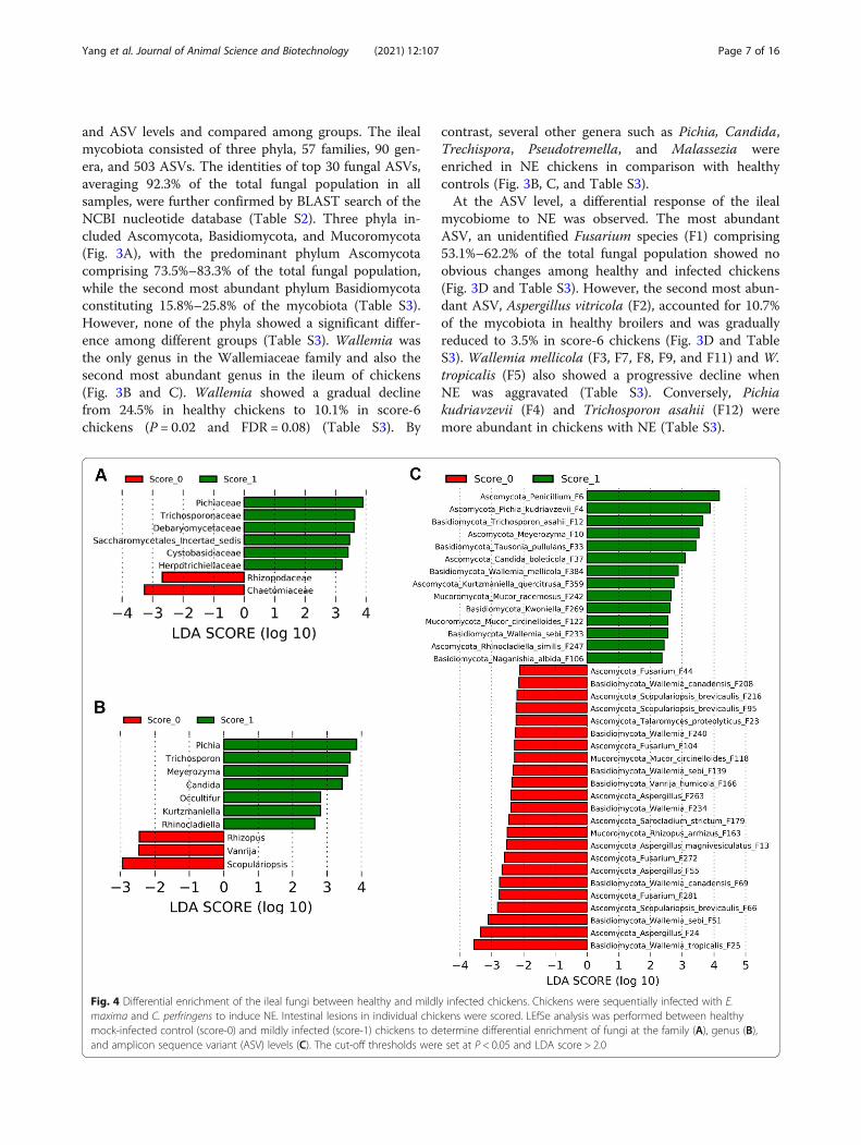
Fig. 4 Differential enrichment of the ileal fungi between healthy and mildly infected chickens. Chickens were sequentially infected with E.maxima and C. perfringens to induce NE. Intestinal lesions in individual chickens were scored. LEfSe analysis was performed between healthymock-infected control (score-0) and mildly infected (score-1) chickens to determine differential enrichment of fungi at the family (A), genus (B),and amplicon sequence variant (ASV) levels (C). The cut-off thresholds were set at P < 0.05 and LDA score > 2.0
Yang et al. Journal of Animal Science and Biotechnology (2021) 12:107 Page 7 of 16

Differential enrichment of the mycobiota in response toNETo identify discriminative fungi between healthy and NEchickens, LEfSe was first performed between score-0 andscore-1 chickens at the family, genus, and ASV levelsamong those common taxa that were present in > 20%of chickens using thresholds of P < 0.05 and LDA score >2. In comparison with healthy chickens, families such asChaetomiaceae and Rhizopodaceae were reduced, whilePichiaceae, Trichosporonaceae, Debaryomycetaceae, Sac-charomycetales incertae sedis, Cystobasidiaceae, andHerpotrichiellaceae were enriched in score-1 broilers
(Fig. 4A). At the genus level, mild NE diminished Scopu-lariopsis, Vanrija, and Rhizopus, but enriched Pichia,Trichosporon, Meyerozyma, Candida, Occultifur, Kurtz-maniella, and Rhinocladiella (Fig. 4B). A total of 37ASVs showed differential enrichment between healthyand mild NE chickens. Multiple members of Wallemia(F25, F51, F69, F139, F208, F234, and F240), Aspergillus(F13, F24, F55, and F263), and Fusarium (F44, F104, andF281) as well as Scopulariopsis brevicaulis (F95 andF216) were reduced in mild NE, while a diverse array offungi such as a Penicillium species (F6). P. kudriavzevii(F4), a Meyerozyma species (F10), and T. asahii (F12) as
Fig. 5 Differential enrichment of the ileal fungal taxa between chickens with mild and severe NE. Chickens were sequentially infected with E.maxima and C. perfringens to induce NE. Intestinal lesions in individual chickens were scored. LEfSe analysis was performed between chickenswith mild (score-1) and severe NE (score-6) to determine differential enrichment of fungi at the family (A), genus (B), and amplicon sequencevariant (ASV) levels (C). The cut-off thresholds were set at P < 0.05 and LDA score > 2.0
Yang et al. Journal of Animal Science and Biotechnology (2021) 12:107 Page 8 of 16

A
0.4 0.2 0 -0.2 -0.4R-value
B
Lesion Score Weight Loss
Family
Genus
Lesion Score Weight Loss
0.4 0.2 0 -0.2 -0.4R-value
4 3 2 1 0 -1Log2 (fold)
Family
Score 0 1 2 5 6
Genus
Score 0 1 2 5 6 4 3 2 1 0 -1
Log2 (fold)
4 2 0 -2 -4 -6Log2 (fold)
ASV
Score 0 1 2 5 6
0.4 0.2 0 -0.2 -0.4R-value
ASV
Lesion Score Weight Loss
Fig. 6 (See legend on next page.)
Yang et al. Journal of Animal Science and Biotechnology (2021) 12:107 Page 9 of 16

well as two relatively rare Wallemia members (F233 andF384) were increased in mildly infected NE chickens(Fig. 4C). Fungi that were enriched in mild NE mainlybelonged to Saccharomycetes, Tremellomycetes, andChaetothyriales (data not shown).To further determine differentially abundant fungi in
the ileum between chickens with mild and severe NE,LEfSe was performed between score-1 and score-6chickens. At the family level, Wallemiaceae, Herpotri-chiellaceae, Bulleraceae, and Saccharomycetaceae wereenriched in chickens with mild NE, while ChaetomiaceaeTrichocomaceae, Hypocreaceae, Hydnodontaceae, Tri-chosporonaceae, Malasseziaceae, Filobasidiaceae, and anunidentified family within Onygenales were more abun-dant in severely infected chickens (Fig. 5A). At the genuslevel, score-1 chickens showed an enrichment of Walle-mia, Pseudotremella, Rhinocladiella, Kurtzmaniella, andKazachstania, but genera such as Talaromyces, Trechis-pora, Trichosporon, Malassezia, Trichoderma, Naga-nishia, and Xeromyces were increased in score-6chickens (Fig. 5B). At the ASV level, a total of 46 ASVswere differentially enriched between mild and severeNE. For example, Penicillium F6 and multiple Wallemiamembers such as W. mellicola (F3, F7, F8, F9, F49, F79,and F382), W. tropicalis (F5 and F71), W. canadensis(F133 and F160), and W. sebi (F481) were abundantlypresent in score-1 chickens, while enrichments of Can-dida glabrata, T. asahii, Aspergillus sydowii, and Asper-gillus flavus were observed in score-6 chickens (Fig. 5C).
Correlation between the ileal fungal abundance and NEseverityTo identify the ileal fungi that are strongly correlatedwith NE severity, Spearman’s rank correlation was per-formed separately between the fungal taxa present in >20% of the chickens and two NE severity parameters (in-testinal lesion score and weight loss). Among 33 familiesand 47 genera that were common in > 20% of samples,11 families and 12 genera showed a significant correl-ation (FDR < 0.05) with at least one indicator of NE se-verity (Fig. 6A). While Wallemiaceae/Wallemia wasnegatively correlated with disease severity (FDR < 0.05),Trichosporonaceae/Trichosporon, Pichiaceae/Pichia, Filo-basidiaceae/Naganishia, an unidentified member ofOnygenales, and Bulleraceae/Pseudotremella showed asignificant positive correlation (FDR < 0.05) with both
NE severity indicators. Out of 198 fungal ASVs presentin > 20% of chickens, 39 ASVs showed a significant cor-relation with NE severity, with 14 showing a positivecorrelation and 25 showing a negative correlation(FDR < 0.05) (Fig. 6A). For example, 17 Wallemia spe-cies such as W. mellicola (F3, F7, F8, F9, and F11), W.tropicalis (F25), W. sebi (F34 and F51) and six Aspergil-lus members (e.g., A. vitricola F52 and unclassified As-pergillus) exhibited a strong negative correlation with atleast one indicator of NE severity (Fig. 6A). As revealedin a heatmap, multiple Wallemia members were dimin-ished, while a diverse group of fungi were enriched inexacerbated NE (Fig. 6B).A closer examination of the 30 most abundant fungal
ASVs further revealed different patterns of the fungalchange in response to NE. While W. mellicola (F3, F7,F8, F9, and F11) and W. tropicalis (F5) were graduallydiminished in more severe NE; W. tropicalis F25 showedan abrupt decrease even in chickens with mild NE(Fig. 7A), suggesting both inter-species and intra-speciesvariations of Wallemia in response to NE. Similarly, dif-ferent sensitivities to NE were observed among Aspergil-lus species. While A. vitricola (F2) displayed aprogressive decline, A. magnivesiculatus (F13) wasabruptly abolished in all NE chickens (Fig. 7A). On theother hand, A. sydowii (F20) appeared to be enrichedonly in chickens with severe NE (Fig. 7B). An unspeci-fied Trechispora (F18), C. glabrata (F14), and Talaro-myces proteolyticus (F23) were also sharply increased insevere NE chickens (P < 0.05), but P. kudriavzevii (F4)and T. asahii (F12) showed a progressive increase withNE severity (Fig. 7B). Notably, nearly 50% of top 30 fun-gal ASVs showed no obvious shift change in response toNE including the most abundant Fusarium ASV (F1)(Fig. 7C). Perhaps as confirmation, another Fusarium (F.fujikuroi F15) was unaltered in NE either (Fig. 7C).
Correlation between the ileal mycobiota and microbiotain the context of NEIn addition to the mycobiota, the bacterial microbiota ofthe ileum was also investigated by 16S rRNA gene se-quencing. Spearman correlation analysis was conductedbetween the ileal fungi and bacteria to explore their po-tential interactions in the context of NE. Among 12 fun-gal and 29 bacterial genera that were commonly presentin > 20% of the chickens and also showed a significant
(See figure on previous page.)Fig. 6 Correlation between the ileal mycobiota composition and NE severity. Chickens were sequentially infected with E. maxima and C.perfringens to induce NE. Intestinal lesions in individual chickens were scored. Spearman correlation analysis was performed between the ilealmycobiota profile and the intestinal lesion score and weight loss. A Spearman correlation at family, genus, and amplicon sequence variant (ASV)levels. B Heatmap displaying log2 transformations of fold changes in fungal abundance in NE chickens at the family, genus, and ASV levels,relative to mock-infected controls. Significance of Spearman correlations was subjected to FDR correction using the Benjamini-Hochbergprocedure. *FDR < 0.05, **FDR < 0.01
Yang et al. Journal of Animal Science and Biotechnology (2021) 12:107 Page 10 of 16

correlation with NE severity (FDR < 0.05), Wallemiashowed a negative correlation with Clostridium, but apositive correlation with Cyanobacteria, Subdoligranu-lum, Corynebacterium, Blautia, Staphylococcus, Cuneati-bacter, Aerococcus, Lactonifactor, Oscillospiraceae,Erysipelatoclostridium, and an unidentified genus inLachnospiraceae (Fig. 8A). Positive correlations also
occurred between Clostridium and fungal genera includ-ing Pichia, Candida, Trichosporon, Pseudotremella, andNaganishia, while a majority of the fungal-bacterial in-teractions were negative (Fig. 8A).At the ASV level, among 39 fungal and 64 bacterial
ASVs showing a significant correlation with the severityof NE (FDR < 0.05), positive correlations were common
P = 0.004P = 0.003P = 0.001
P = 0.05
P = 0.24P = 0.59P = 0.15
P = 0.008P = 0.01
P = 0.07
P = 0.01
P = 0.33
P = 0.02P = 0.06P = 0.02
P = 0.001P = 0.05P = 0.002 P = 0.009P = 1.44E-04P = 3.44E-04
P = 0.41P = 0.20P = 0.40P = 0.75
P = 0.59P = 0.74
A
B
C
P = 0.007
P = 0.81
Fig. 7 Relative abundances of top 30 ileal fungal amplicon sequence variants (ASVs) among chickens with different severities of NE. Ileal fungiwere diminished (A), increased (B), or unaltered (C) in NE. Each box indicated median, 25th and 75th percentiles, while whiskers of the box plotsextended to 1.5 interquartile range. Significance was determined using Kruskal-Wallis test and was indicated on the top of each plot. Groups notsharing common superscripts were significantly different at P < 0.05 as measured by pair-wise Mann–Whitney U test
Yang et al. Journal of Animal Science and Biotechnology (2021) 12:107 Page 11 of 16

among NE-diminished fungi such as Wallemia speciesand NE-reduced bacteria (majorly lactic acid bacteriaand SCFA-producing bacteria, such as group B
Lactobacillus, L. reuteri, Subdoligranulum variabile,Weissella species, and Blautia species) (Fig. 8B). On thecontrary, negative interactions mainly occurred between
Fig. 8 The ileal fungal-bacterial correlations in chickens with NE. Spearman correlation was performed to evaluate potential interactions betweenthe ileal fungi and bacteria at the genus (A) and amplicon sequence variant (ASV) levels (B). Only fungal and bacterial taxa that were present in> 20% of chickens and also correlated significantly with NE severity were included in the analysis. Significant positive and negative correlations(FDR < 0.05) were denoted by red and blue squares, respectively, while the strength of a correlation was indicated by the extent of the color.White squares indicated nonsignificant correlations
Yang et al. Journal of Animal Science and Biotechnology (2021) 12:107 Page 12 of 16

NE-enriched fungi (e.g., P. kudriavzevii F4, T. asahiiF12, C. glabrata F14, and Naganishia friedmannii F97)and bacterial species that were decreased in NE (Fig.8B).
DiscussionThe bacterial microbiota and fungal mycobiota residingin the GI tract contribute to the health or diseases of thehost [1–5]. Disturbance in the intestinal microbiota hasbeen linked to chicken NE [17], while the involvementof the mycobiome in NE is yet to be investigated. In thecurrent study, we unraveled an altered ileal fungal com-munity in NE-afflicted chickens using ITS2 amplicon se-quencing and further identified a number of fungi thatare strongly correlated with the severity of NE. Ourstudy also revealed the mycobiota-microbiota correla-tions in NE, suggesting that the mycobiome, in additionto the microbiome, might be potentially involved in NEand targeted to mitigate NE, although direct experimen-tal evidence is needed.We found that chicken intestinal mycobiota is domi-
nated by Ascomycota followed by Basidiomycota. Thetwo fungal phyla make up 97%–99% of the ileal myco-biota in broilers, in agreement with a recent report [14].Similarly, Ascomycota and Basidiomycota are two majorfungal phyla found in the human GI tract [8, 9]. Asco-mycota is also the most abundant fungal phylum in theintestine of piglets [34]. In the present study, Fusariumis the most predominant in the chicken ileum, regardlessof the health status. Fusarium, Wallemia, and Aspergil-lus collectively comprise 84% of the ileal fungal commu-nity, while Pichia, Candida, Penicillium, Mucor, andTrichosporon also show a high prevalence in the chickenileum. Similar to our results, Candida, Trichosporon,and Rhodotorula are major fungi isolated from chickenfeces [35], which are also among predominant fungi inthe GI tract of healthy turkeys [36]. As for the source ofthe intestinal fungi, Fusarium, Aspergillus, Penicillium,and Mucor were found to be prevalent in corn, soybeanmeal, and finished poultry feed [37, 38], suggesting thatthe intestinal mycobiota is mainly originated from thefeed.In this study, we found that richness, but not evenness
or Shannon index, of the ileal mycobiota tends to de-crease in NE, which is similar to earlier studies with IBDand ulcerative colitis patients [3, 8], although other stud-ies described no obvious changes in fungal richness be-tween Crohn’s disease patients and healthy cohorts [13,39]. One major finding of our research is that the totalileal fungal population is drastically reduced in severeNE. The fungal population approximately constitutes ap-proximately 0.1% of the total bacteria in the ileum ofhealthy chickens, which is consistent with the previousreport in humans and mice [40], but is gradually
diminished in NE, with an approximately 50-fold reduc-tion in severely infected, score-6 chickens. On the otherhand, the total bacterial load is increased by 2- to 3-foldin NE chickens. As a result, a progressive decline in thefungal-bacterial ratio occurs in exacerbated NE. Interest-ingly, the fungal load and the fungal-bacterial ratio areincreased in patients with Crohn’s disease [39]. More re-search is warranted to further investigate whethercolonization and proliferation of C. perfringens in thesmall intestine in NE lead to diminished mycobiota.In this study, we observed an obvious difference in the
sensitivity to NE among the ileal fungal taxa. Among the30 most abundant fungi, approximately 40% remain un-changed, while another 40% are declined and theremaining 20% are enriched in NE. Among those thatare altered in NE, some show a gradual increase or de-crease, while others are changed abruptly. For example,C. glabrata (F14), A. sydowii (F20), T. proteolyticus (F23)and a Trechispora species (F18) are enriched only in theileum of severally infected chickens.Among those fungi that are dramatically reduced in
NE are a number of Wallemia taxa that are com-monly found in the air, house dust, soils, and plants[11, 41]. Wallemia is also a commensal in the intes-tine of humans and mice [11]. In this study, we re-vealed that several Wallemia species such as W.mellicola, W. tropicalis, W. sebi, and W. canadensisare negatively correlated with NE severity. Consist-ently, Wallemia is reported to produce UCA1064-Aand 1064-B with beneficial antitumor, antifungal, andantimicrobial activities [42, 43]. However, W. sebi andW. mellicola have been found to be associated withskin infections and allergic airway diseases [10, 11].Further studies are needed to understand the involve-ment of Wallemia in NE.Apart from Wallemia, Aspergillus is also apparently
reduced in NE. A. vitricola, A. magnivesiculatus, andseveral unidentified Aspergillus species are negativelycorrelated with NE severity. Aspergillus is ubiquitous infeed and can cause aspergillosis in avian species orhumans [44]. Although it can be a source of dietary my-cotoxins with a detrimental effect on poultry health andperformance [37], Aspergillus also produces beneficialmetabolites such as lovastatin, terreulactones, 11-αhydroprogesterone, quadrone, and terpeptin [45]. Thepotential of Aspergillus for disease resistance in poultrywarrants further investigation.Along with a striking decrease in Wallemia and Asper-
gillus, other fungi such as P. kudriavzevii (teleomorph ofCandida krusei), C. glabrata, M. restricta, T. asahii, andN. friedmannii are enriched in the ileum of chickenswith NE, especially in severe NE. P. kudriavzevii isolatedfrom chicken feces exhibits probiotic properties in vitro[46]. With a capacity to bind to aflatoxin B1, dietary
Yang et al. Journal of Animal Science and Biotechnology (2021) 12:107 Page 13 of 16

supplementation of P. kudriavzevii mitigates the adverseeffect of aflatoxin B1 on the growth performance ofbroilers [47]. Candida species are a part of commensalmycobiota in the GI tract but may cause candidiasis inpoultry and humans [36, 41, 44]. Candida infectionsmost frequently occur in the upper GI tract of chickensand may result in growth retardation or even mortality[44]. We revealed a positive correlation between C. glab-rata and NE severity, which is in agreement with reportsthat Candida such as C. albicans, C. glabrata, and C.tropicalis are associated with IBD [8, 13, 39]. The role ofP. kudriavzevii and Candida overgrowth in NE is notclear, and further studies are warranted.Malassezia is a commensal fungus that colonizes not
only the skin but also in the GI tract of humans andanimals [48]. M. restricta has been found to be asso-ciated with IBD and several other intestinal inflamma-tory disorders in humans [48]. M. restricta, withpotent pro-inflammatory properties, is enriched inCrohn’s disease patients, and oral administration ofM. restricta exacerbates colitis in mice [9]. T. asahiiis another commensal fungus that can cause oppor-tunistic infections [14, 49]. In agreement with ex-panded intestinal Trichosporon in IBD patients [8], wefound that T. asahii is positively correlated with NEseverity in chickens. In addition, N. friedmannii(formerly Cryptococcus friedmannii) is reported tocause onychomycosis in humans [50]. The overgrowthof opportunistic fungi in NE-infected chickens mightincrease susceptibility of chickens to mycosis andprobably threat public health.Fungi and bacteria co-colonize the GI tract and
interact with each other directly or indirectly throughphysical contact, microbial metabolites, and modifica-tion of immune status [4, 51]. The cross-talk betweenthe mycobiota and the microbiota is critical for main-taining intestinal homeostasis [4, 51] and has beendemonstrated in turkeys [15] and human IBD [8, 13].We revealed a dramatic shift of the ileal microbiotain NE (unpublished). The current study has furtherrevealed a strong positive or negative correlation be-tween a number of fungal and bacterial taxa in NE.For example, most Wallemia species are correlatednegatively with C. perfringens colonization in theileum but correlated positively with a number ofSCFA-producing bacteria, consistent with an earlierreport on a positive association between Wallemiaand SCFA-producing Oscillospiraceae [52]. On theother hand, P. kudriavzevii (F4), T. asahii (F12), C.glabrata (F14), and M. restricta (F60) are positivelycorrelated with C. perfringens with a negative correl-ation with SCFA-producing bacteria and often lacticacid bacteria. Such an antagonism between C. glab-rata and lactic acid bacteria (e.g., Lactobacillus and
Weissella) was also reported earlier [53, 54]. It will beimportant to study the role of the fungi-bacteriainterplay in the development of NE and whether suchinteractions can be explored for control and preven-tion of NE.Eimeria infection is an important predisposing factor
for NE in poultry with the ability to cause damage to theintestinal epithelium, providing niches or nutrients to fa-cilitate the colonization and proliferation of C. perfrin-gens [18]. Eimeria in conjunction with C. perfringenschallenge is thus the most commonly used approach toexperimentally induce NE, while the same dose ofEimeria or C. perfringens alone causes no or only mildintestinal lesions [18]. Consistently, co-infection withEimeria and C. perfringens causes more pronouncedmicrobiota changes than inoculation separately withEimeria or C. perfringens [55, 56]. However, the impactof Eimeria or C. perfringens on the intestinal mycobiotais currently unknown and warrants further investigation.
ConclusionThis study revealed for the first time dysbiosis of thechicken ileal mycobiota induced by NE. The total fungalpopulation is drastically reduced in NE and alterationsin the mycobiota are more pronounced in exacerbatedNE. Furthermore, we reported positive and negative cor-relations between a number of fungi and bacteria. Thesefindings suggest a possible role of the intestinal myco-biota in NE pathogenesis and highlight the mycobiota asa new potential target for NE management in poultry.
AbbreviationsANOVA: Analysis of variance; ASV: Amplicon sequence variant; BW: Bodyweight; CFU: Colony-forming unit; FDR: False discovery rate;GI: Gastrointestinal; IBD: Inflammatory bowel disease; ITS2: Internaltranscribed spacer 2; LDA: Linear discriminate analysis; LEfSe: Lineardiscriminant analysis effect size; NE: Necrotic enteritis; NRC: National ResearchCouncil; OTU: Operational taxonomic unit; PCoA: Principal coordinatesanalysis; PERMANOVA: Permutational multivariate analysis of variance;qPCR: Quantitative PCR; SCFA: Short-chain fatty acid
Supplementary InformationThe online version contains supplementary material available at https://doi.org/10.1186/s40104-021-00628-5.
Additional file 1: Table S1. Pairwise comparison of beta diversity ofthe ileal mycobiota between healthy and NE chickens. Table S2.Taxonomy of top 30 fungal amplicon sequence variants (ASVs) in thechicken ileum identified through a BLAST search of the NCBI nucleotidedatabase. Table S3. Relative abundance (%) of top fungal taxa in thechicken ileum.
AcknowledgmentsThe authors would like to thank Dr. John R. Barta at the University of Guelph,Canada for kindly providing E. maxima strain M6. The authors are alsograteful to Dr. Lisa Bielke at Ohio State University for providing the C.perfringens strain Brenda B.
Yang et al. Journal of Animal Science and Biotechnology (2021) 12:107 Page 14 of 16

Authors’ contributionsThe contributions of the authors were as follows: QY, MW, SS, JL, and KRconducted the animal trial and processed all samples. QY, JL, and KRperformed data analysis. QY wrote the manuscript. GZ conceived the animaltrial and revised the manuscript. All authors read and approved the finalmanuscript.
FundingThis work was supported by the USDA National Institute of Food andAgriculture (grant no. 2018–68003-27462 and 2018–67011-28041), the RalphF. and Leila W. Boulware Endowment Fund, and Oklahoma AgriculturalExperiment Station Project H-3112.
Availability of data and materialsThe raw sequencing reads of this study have been deposited in the NCBISequence Read Archive (SRA) database under BioProject PRJNA725022.
Declarations
Ethics approval and consent to participateAll animal procedures were approved by the Institutional Animal Care andUse Committee of Oklahoma State University under protocol number AG-16-10.
Consent for publicationNot applicable.
Competing interestsThe authors declare that they have no competing interests.
Author details1Department of Animal and Food Sciences, Oklahoma State University,Stillwater, OK, USA. 2Present address: Poultry Production and Product SafetyResearch Unit, USDA–Agricultural Research Service, Fayetteville, AR, USA.3Present address: Safety and Security Division, Institute for Public Research,CNA, Arlington, VA, USA.
Received: 14 May 2021 Accepted: 5 August 2021
References1. Peixoto RS, Harkins DM, Nelson KE. Advances in microbiome research for
animal health. Annu Rev Anim Biosci. 2021;9(1):289–311. https://doi.org/10.1146/annurev-animal-091020-075907.
2. Fan Y, Pedersen O. Gut microbiota in human metabolic health and disease.Nat Rev Microbiol. 2021;19(1):55–71. https://doi.org/10.1038/s41579-020-0433-9.
3. Li XV, Leonardi I, Iliev ID. Gut mycobiota in immunity and inflammatorydisease. Immunity. 2019;50(6):1365–79. https://doi.org/10.1016/j.immuni.2019.05.023.
4. Santus W, Devlin JR, Behnsen J. Crossing kingdoms: how the mycobiotaand fungal-bacterial interactions impact host health and disease. InfectImmun. 2021;89(4):e00648–20.
5. Wu X, Xia Y, He F, Zhu C, Ren W. Intestinal mycobiota in health anddiseases: from a disrupted equilibrium to clinical opportunities. Microbiome.2021;9(1):60. https://doi.org/10.1186/s40168-021-01024-x.
6. Tso GHW, Reales-Calderon JA, Tan ASM, Sem X, Le GTT, Tan TG, et al.Experimental evolution of a fungal pathogen into a gut symbiont. Science.2018;362(6414):589–95. https://doi.org/10.1126/science.aat0537.
7. Shao TY, Ang WXG, Jiang TT, Huang FS, Andersen H, Kinder JM, et al.Commensal Candida albicans positively calibrates systemic Th17immunological responses. Cell Host Microbe. 2019;25(3):404–17.e6.
8. Sokol H, Leducq V, Aschard H, Pham HP, Jegou S, Landman C, et al. Fungalmicrobiota dysbiosis in IBD. Gut. 2017;66(6):1039–48. https://doi.org/10.1136/gutjnl-2015-310746.
9. Limon JJ, Tang J, Li D, Wolf AJ, Michelsen KS, Funari V, et al. Malassezia isassociated with Crohn‘s disease and exacerbates colitis in mouse models.Cell Host Microbe. 2019;25(3):377–88.e6.
10. Skalski JH, Limon JJ, Sharma P, Gargus MD, Nguyen C, Tang J, et al.Expansion of commensal fungus Wallemia mellicola in the gastrointestinal
mycobiota enhances the severity of allergic airway disease in mice. PLoSPathog. 2018;14(9):e1007260. https://doi.org/10.1371/journal.ppat.1007260.
11. Zajc J, Gunde-Cimerman N. The genus Wallemia-from contamination offood to health threat. Microorganisms. 2018;6(2):46. https://doi.org/10.3390/microorganisms6020046.
12. Coker OO, Nakatsu G, Dai RZ, Wu WKK, Wong SH, Ng SC, et al. Entericfungal microbiota dysbiosis and ecological alterations in colorectal cancer.Gut. 2019;68(4):654–62. https://doi.org/10.1136/gutjnl-2018-317178.
13. Hoarau G, Mukherjee PK, Gower-Rousseau C, Hager C, Chandra J, RetuertoMA, et al. Bacteriome and mycobiome interactions underscore microbialdysbiosis in familial Crohn‘s disease. mBio. 2016;7(5):e01250–16.
14. Robinson K, Xiao Y, Johnson TJ, Chen B, Yang Q, Lyu W, et al. Chickenintestinal mycobiome: initial characterization and its response to bacitracinmethylene disalicylate. Appl Environ Microbiol. 2020;86(13):e00304–20.
15. Ward TL, Weber BP, Mendoza KM, Danzeisen JL, Llop K, Lang K, et al.Antibiotics and host-tailored probiotics similarly modulate effects on thedeveloping avian microbiome, mycobiome, and host gene expression.mBio. 2019;10(5):e02171–19.
16. Wade B, Keyburn A. The true cost of necrotic enteritis. World Poult. 2015;31(7):16–7.
17. Antonissen G, Eeckhaut V, Van Driessche K, Onrust L, Haesebrouck F,Ducatelle R, et al. Microbial shifts associated with necrotic enteritis.Avian Pathol. 2016;45(3):308–12. https://doi.org/10.1080/03079457.2016.1152625.
18. Shojadoost B, Vince AR, Prescott JF. The successful experimental inductionof necrotic enteritis in chickens by Clostridium perfringens: a critical review.Vet Res. 2012;43(1):74. https://doi.org/10.1186/1297-9716-43-74.
19. Cooper KK, Songer JG. Virulence of Clostridium perfringens in anexperimental model of poultry necrotic enteritis. Vet Microbiol. 2010;142(3–4):323–8. https://doi.org/10.1016/j.vetmic.2009.09.065.
20. Al-Badri R, Barta JR. The kinetics of oocyst shedding and sporulation in twoimmunologically distinct strains of Eimeria maxima, GS and M6. ParasitolRes. 2012;111(5):1947–52. https://doi.org/10.1007/s00436-012-3041-4.
21. Latorre JD, Adhikari B, Park SH, Teague KD, Graham LE, Mahaffey BD, et al.Evaluation of the epithelial barrier function and ileal microbiome in anestablished necrotic enteritis challenge model in broiler chickens. Front VetSci. 2018;5:199. https://doi.org/10.3389/fvets.2018.00199.
22. diCenzo GC, Finan TM. The divided bacterial genome: structure, function,and evolution. Microbiol Mol Biol Rev. 2017;81(3):e00019–7.
23. De Fine Licht HH, Hajek AE, Eilenberg J, Jensen AB. Utilizing genomics tostudy entomopathogenicity in the fungal phylum entomophthoromycota: areview of current genetic resources. Adv Genet. 2016;94:41–65. https://doi.org/10.1016/bs.adgen.2016.01.003.
24. White TJ, Bruns T, Lee S, Taylor J. Amplification and direct sequencing offungal ribosomal RNA genes for phylogenetics. In: Innis MA, Gelfand DH,Sninsky JJ, White TJ, editors. PCR protocols: a guide to methods andapplications. New York: Academic Press; 1990. p. 315–22.
25. Bolyen E, Rideout JR, Dillon MR, Bokulich NA, Abnet CC, Al-Ghalith GA, et al.Reproducible, interactive, scalable and extensible microbiome data scienceusing QIIME 2. Nat Biotechnol. 2019;37(8):852–7. https://doi.org/10.1038/s41587-019-0209-9.
26. Amir A, McDonald D, Navas-Molina JA, Kopylova E, Morton JT, Zech Xu Z,et al. Deblur rapidly resolves single-nucleotide community sequencepatterns. mSystems. 2017;2(2):e00191–16.
27. Paulson JN, Stine OC, Bravo HC, Pop M. Differential abundance analysis formicrobial marker-gene surveys. Nat Methods. 2013;10(12):1200–2. https://doi.org/10.1038/nmeth.2658.
28. McMurdie PJ, Holmes S. phyloseq: an R package for reproducible interactiveanalysis and graphics of microbiome census data. PLoS One. 2013;8(4):e61217.
29. Lozupone C, Knight R. UniFrac: a new phylogenetic method for comparingmicrobial communities. Appl Environ Microbiol. 2005;71(12):8228–35.https://doi.org/10.1128/AEM.71.12.8228-8235.2005.
30. Segata N, Izard J, Waldron L, Gevers D, Miropolsky L, Garrett WS, et al.Metagenomic biomarker discovery and explanation. Genome Biol. 2011;12(6):R60. https://doi.org/10.1186/gb-2011-12-6-r60.
31. Wickham H. ggplot2: elegant graphics for data analysis. 2nd ed. New York:Springer; 2016. https://doi.org/10.1007/978-3-319-24277-4.
32. Wang X, Tsai T, Deng F, Wei X, Chai J, Knapp J, et al. Longitudinalinvestigation of the swine gut microbiome from birth to market revealsstage and growth performance associated bacteria. Microbiome. 2019;7(1):109. https://doi.org/10.1186/s40168-019-0721-7.
Yang et al. Journal of Animal Science and Biotechnology (2021) 12:107 Page 15 of 16

33. Liu J, Stewart SN, Robinson K, Yang Q, Lyu W, Whitmore MA, et al. Linkagebetween the intestinal microbiota and residual feed intake in broilerchickens. J Anim Sci Biotechnol. 2021;12(1):22. https://doi.org/10.1186/s40104-020-00542-2.
34. Hu J, Nie Y, Chen J, Zhang Y, Wang Z, Fan Q, et al. Gradual changes ofgut microbiota in weaned miniature piglets. Front Microbiol. 2016;7:1727.
35. Subramanya SH, Sharan NK, Baral BP, Hamal D, Nayak N, Prakash PY, et al.Diversity, in-vitro virulence traits and antifungal susceptibility pattern ofgastrointestinal yeast flora of healthy poultry, Gallus gallus domesticus. BMCMicrobiol. 2017;17(1):113. https://doi.org/10.1186/s12866-017-1024-4.
36. Sokol I, Gawel A, Bobrek K. The prevalence of yeast and characteristics ofthe isolates from the digestive tract of clinically healthy turkeys. Avian Dis.2018;62(3):286–90. https://doi.org/10.1637/11780-121117-Reg.1.
37. Ghaemmaghami SS, Modirsaneii M, Khosravi AR, Razzaghi-Abyaneh M.Study on mycoflora of poultry feed ingredients and finished feed in Iran.Iran J Microbiol. 2016;8(1):47–54.
38. Oliveira GR, Ribeiro JM, Fraga ME, Cavaglieri LR, Direito GM, Keller KM, et al.Mycobiota in poultry feeds and natural occurrence of aflatoxins, fumonisinsand zearalenone in the Rio de Janeiro state, Brazil. Mycopathologia. 2006;162(5):355–62. https://doi.org/10.1007/s11046-006-0070-5.
39. Liguori G, Lamas B, Richard ML, Brandi G, da Costa G, Hoffmann TW, et al.Fungal dysbiosis in mucosa-associated microbiota of Crohn‘s diseasepatients. J Crohns Colitis. 2016;10(3):296–305. https://doi.org/10.1093/ecco-jcc/jjv209.
40. Li J, Chen D, Yu B, He J, Zheng P, Mao X, et al. Fungi in gastrointestinaltracts of human and mice: from community to functions. Microb Ecol. 2018;75(4):821–9. https://doi.org/10.1007/s00248-017-1105-9.
41. Limon JJ, Skalski JH, Underhill DM. Commensal fungi in health anddisease. Cell Host Microbe. 2017;22(2):156–65. https://doi.org/10.1016/j.chom.2017.07.002.
42. Jančič S, Frisvad JC, Kocev D, Gostinčar C, Džeroski S, Gunde-Cimerman N.Production of secondary metabolites in extreme environments: food- andairborne Wallemia spp. produce toxic metabolites at hypersaline conditions.PLoS One. 2016;11(12):e0169116.
43. Takahashi I, Maruta R, Ando K, Yoshida M, Iwasaki T, Kanazawa J, et al.UCA1064-B, a new antitumor antibiotic isolated from Wallemia sebi:production, isolation and structural determination. J Antibiot (Tokyo). 1993;46(8):1312–4. https://doi.org/10.7164/antibiotics.46.1312.
44. Dykstra MJ, Charlton BR, Chin RP, Barnes HJ. Fungal infections. In: SwayneDE, Glisson JR, McDougald LR, Nolan LK, Suarez DL, Nair VL, editors. Diseasesof poultry. 13th ed. Oxford: Wiley-Blackwell; 2013. p. 1075–96. https://doi.org/10.1002/9781119421481.ch25.
45. Ashtekar N, Anand G, Prakash PY, Rajeshkumar KC. Aspergillus terreus:taxonomy, biology, and bioactive secondary metabolites with potentialapplications. In: Singh J, Gehlot P, editors. New and future developments inmicrobial biotechnology and bioengineering. Amsterdam: Elsevier; 2021. p.215–23. https://doi.org/10.1016/B978-0-12-821005-5.00015-6.
46. García-Hernández Y, Rodríguez Z, Brandão LR, Rosa CA, Nicoli JR, ElíasIglesias A, et al. Identification and in vitro screening of avian yeasts for useas probiotic. Res Vet Sci. 2012;93(2):798–802. https://doi.org/10.1016/j.rvsc.2011.09.005.
47. Magnoli AP, Rodriguez MC, Poloni VL, Rojo MC, Combina M, ChiacchieraSM, et al. Novel yeast isolated from broilers‘ feedstuff, gut and faeces asaflatoxin B1 adsorbents. J Appl Microbiol. 2016;121(6):1766–76. https://doi.org/10.1111/jam.13297.
48. Spatz M, Richard ML. Overview of the potential role of Malassezia in guthealth and disease. Front Cell Infect Microbiol. 2020;10:201. https://doi.org/10.3389/fcimb.2020.00201.
49. Duarte-Oliveira C, Rodrigues F, Gonçalves SM, Goldman GH, Carvalho A,Cunha C. The cell biology of the Trichosporon-host interaction. Front CellInfect Microbiol. 2017;7:118.
50. Ekhtiari M, Farahyar S, Falahati M, Razmjou E, Ashrafi-Khozani M, Ghasemi Z,et al. The first report of onychomycosis caused by Cryptococcus friedmannii(Naganishia friedmannii) a basidiomycetous yeast. Med Mycol Case Rep.2017;15:25–7. https://doi.org/10.1016/j.mmcr.2017.01.002.
51. Kruger W, Vielreicher S, Kapitan M, Jacobsen ID, Niemiec MJ. Fungal-bacterial interactions in health and disease. Pathogens. 2019;8(2):70. https://doi.org/10.3390/pathogens8020070.
52. Lin M, Feng L, Cheng Z, Wang K. Effect of ethanol or lactic acid on shortchain fatty acid production and microbial community in short-term
sequentially transfers by ruminal fermented with wheat straw in vitro.Process Biochem. 2020;102:369–75.
53. Boris S, Barbés C. Role played by lactobacilli in controlling the population ofvaginal pathogens. Microbes Infect. 2000;2(5):543–6. https://doi.org/10.1016/S1286-4579(00)00313-0.
54. Fan D, Coughlin LA, Neubauer MM, Kim J, Kim MS, Zhan X, et al. Activationof HIF-1α and LL-37 by commensal bacteria inhibits Candida albicanscolonization. Nat Med. 2015;21(7):808–14. https://doi.org/10.1038/nm.3871.
55. Stanley D, Wu SB, Rodgers N, Swick RA, Moore RJ. Differential responses ofcecal microbiota to fishmeal, Eimeria and Clostridium perfringens in anecrotic enteritis challenge model in chickens. PLoS One. 2014;9(8):e104739.https://doi.org/10.1371/journal.pone.0104739.
56. Yang WY, Lee Y, Lu H, Chou CH, Wang C. Analysis of gut microbiota andthe effect of lauric acid against necrotic enteritis in Clostridium perfringensand Eimeria side-by-side challenge model. PLoS One. 2019;14(5):e0205784.https://doi.org/10.1371/journal.pone.0205784.
Yang et al. Journal of Animal Science and Biotechnology (2021) 12:107 Page 16 of 16